Note : Les descriptions sont présentées dans la langue officielle dans laquelle elles ont été soumises.
CA 02849634 2014-03-21
WO 2012/038698
PCT/GB2011/001376
1
IMPROVED FEMORAL IMPLANT REVISION TOOL
The present invention relates to a surgical implement suitable for use in the
revision
of orthopaedic prosthetic implants. More particularly but not exclusively, it
relates to
an ultrasonically-vibratable surgical implement for removing a stem of a hip
joint
prosthesis embedded within a femur.
Orthopaedic joint replacements, such as hip joint prostheses, generally have
service
lives of fifteen to twenty years. However, with human lifespans increasing,
this
means that many patients with such implants will experience problems requiring
revision of the prosthesis, i.e. its removal and replacement. Prostheses may
eventually
fracture as a result of simple metal fatigue or overstressing. Other problems
include
wear and damage to the "ball and cup" elements that articulate the portion of
the
prosthesis implanted in the femur to the portion of the prosthesis mounted to
the
pelvis. If resulting polymer and metal fragments enter body tissues, they may
cause
an immune reaction, as the body attempts to absorb them. Absorption is not
possible,
but the immune reaction may meanwhile locally destroy existing bone material.
Another problem arises if the implant in the femur starts to come loose, which
may
CONFIRMATION COPY
CA 02849634 2014-03-21
WO 2012/038698
PCT/GB2011/001376
2
cause severe pain and inflammation, as well as locally weakening the
surrounding
bone.
It is therefore important to be able to remove a orthopaedic implant from a
femur or
other bone while causing minimal damage to surrounding bone material, thus
facilitating implantation of the new implant and aiding subsequent healing. A
femoral
implant typically has an elongate stem which is inserted into the cavity or
channel of
the shaft of the femur, the ball of the articulation being mounted to the
proximal end
of the stem. The stem may be secured within the shaft of the femur using a
polyacrylate cement (a "cemented" implant) or it may be provided with a
textured
surface to encourage ingrowth of cancellous bone to anchor the stem in place
(an
"uncemented" implant).
It is known to use ultrasonically-vibratable tools to soften the cement
holding
cemented implants in place, allowing relatively rapid and straightforward
subsequent
extraction of the implant. However, to remove uncemented implants, a surgeon
must
directly cut away the cancellous bone that has formed between an inner surface
of the
channel of the femur and the stem of the implant, before the implant can be
extracted.
This is currently a lengthy and difficult procedure.
Typically, a proximal section of the wall of the femur is cut open and
temporarily
hinged back to provide lateral access to an upper part of the stem of the
implant. A
wire saw is passed behind the implant and is moved down the stem, gradually
sawing
away the cancellous bone encircling the stem. However, the approach of cutting
through the bone to access the stem cannot be used all the way down the stem,
or the
CA 02849634 2014-03-21
WO 2012/038698
PCT/GB2011/001376
3
femur could be permanently weakened. The cancellous bone around a distal
portion
of the stem must therefore be dealt with by alternative methods. In principle,
an
osteotome blade might be inserted down between the stem and the bone of the
shaft,
to chisel away the cancellous bone. However, the required direction of
approach of
the blade is usually hampered by the proximal portion of the implant. A
surgeon may
therefore first need to saw through the stem of the implant and remove the
proximal
portion, before chiselling out a remaining stub of the distal portion of the
stem, or
cutting around the stub with a trephine. The implant typically comprises a
hard cobalt
steel, so sawing through it is slow, wears away saw blades rapidly, and
creates a great
deal of metal swarf, which must be kept away from soft tissues of the body.
Such a prolonged procedure may be harmful to the patient, who must be kept
under
general anaesthetic throughout. Many patients requiring such a procedure will
be in
imperfect health, making prolonged anaesthesia risky. Additionally, during
such
lengthy and labour intensive procedures, there is a risk of fatigue and
impaired
performance on the part of the surgeon.
It is hence desirable that an alternative approach be devised for cutting away
cancellous bone that has formed between an elongate stem of a femoral implant
(or
other long bone implant) and an inner surface of the shaft of the femur (or
other long
bone). Such an approach should ideally be quicker, less labour-intensive, more
precise and more convenient than existing approaches.
CA 02849634 2014-03-21
WO 2012/038698
PCT/GB2011/001376
4
It is hence an object of the present invention to provide a surgical tool,
suitable for
cutting cancellous bone around an orthopaedic implant in situ, which obviates
the
above disadvantages and provides some or all of the above benefits.
According to a first aspect of the present invention, there is provided a
cutting
element for an ultrasonically-vibratable surgical tool, comprising elongate
waveguide
means mountable adjacent its proximal end to a source of ultrasonic vibrations
and
having elongate blade means extending from adjacent its distal end, wherein a
longitudinal axis of the waveguide means and a longitudinal axis of the blade
means
intersect at an acute non-zero angle.
Preferably, the blade means comprises a cutting edge at its distal tip.
The cutting element may comprise an osteotome element.
The cutting element may be adapted to cut cancellous bone, optionally
cancellous
bone retaining orthopaedic implant means within a lumen of a bone.
In a preferred embodiment, the blade means has an arcuate cross-sectional
profile.
The blade means may have a constant cross-sectional profile along at least a
majority
of its length.
The blade means advantageously comprises a portion of an elongate hollow
cylinder.
CA 02849634 2014-03-21
WO 2012/038698
PCT/GB2011/001376
A distal portion of the blade means may have a cross-sectional profile greater
in
diameter than a remainder thereof.
A distal portion of the blade means may be provided with at least one
longitudinal
groove means extending along a concave face thereof.
In a preferred embodiment, the blade means is provided on a face thereof
adjacent its
distal tip with spacing means adapted to contact a substrate surface to guide
the blade
means.
The spacing means thus prevents direct contact between said face of the blade
means
and said substrate surface.
The spacing means may be provided on a concave face of a blade means having an
arcuate cross-sectional profile.
The spacing means may comprise a layer of thermoplastics material, optionally
a
fluoropolymer, a polyether ether ketone or a high density polyalkene.
The spacing means may alternatively or additionally comprise a plurality of
rib means
upstanding from said face of the blade means.
Said rib means may extend substantially longitudinally of the blade means.
CA 02849634 2014-03-21
WO 2012/038698
PCT/GB2011/001376
6
Preferably, a thickness of the blade means is significantly lower than a
thickness of
the waveguide means.
Advantageously, a profile of the waveguide means and a profile of the blade
means
blend smoothly adjacent their junction.
The cutting element may comprise a curved junction region including a distal
end of
the waveguide means and a proximal end of the blade means.
The waveguide means may comprise an elongate solid cylindrical body.
Preferably, said angle between the respective longitudinal axes of the
waveguide
means and the blade means is between 100 and 45 .
Advantageously, said angle is between 25 and 35 , optionally approximately 30
.
Preferably, the cutting element has an overall length of approximately (2n +
1) X/2,
where n is a positive integer and X is a wavelength of an ultrasonic vibration
within
the material of the cutting element.
Optionally, the cutting element may have an overall length of approximately
3X/2.
Advantageously, the waveguide means has a length of approximately (2n + 1)
X/4.
Optionally, the waveguide means may have a length of approximately 3X/4.
CA 02849634 2014-03-21
WO 2012/038698
PCT/GB2011/001376
7
The blade means may have a length of approximately (2m + 1) k/4, where m is a
positive integer.
Optionally, the blade means may have a length of approximately 3X/4.
The blade means and the waveguide means may each have a length of (2n + 1)
X/4.
Optionally, the blade means and the waveguide means may each have a length of
approximately 3X/4.
Preferably, the cutting element is so configured that when vibrated by said
source of
ultrasonic vibrations, a first antinode of the ultrasonic vibrations is
located adjacent a
proximal end of the waveguide means, a second antinode of the ultrasonic
vibrations
is located adjacent a distal tip of the blade means and a node of the
ultrasonic
vibrations is located adjacent a junction of the waveguide means and the blade
means.
Advantageously, the cutting element is so configured that said ultrasonic
vibrations
undergo a gain in amplitude across the junction of the waveguide means and the
blade
means.
The source of ultrasonic vibrations preferably comprises a source of
longitudinal-
mode ultrasonic vibrations.
8
The cutting element is advantageously so mountable to the source of ultrasonic
vibrations that said longitudinal mode ultrasonic vibrations are directed
substantially
parallelly to the waveguide means.
According to a second aspect of the present invention, there is provided a
surgical tool
comprising a cutting element as described in the first aspect above,
operatively
connected to a source of ultrasonic vibrations.
Preferably, said source of ultrasonic vibrations comprises a source of
longitudinal-
mode ultrasonic vibrations.
According to a third aspect of the present invention, there is provided a
method of
separating a stem of an orthopaedic implant from surrounding bone, comprising
the
steps of providing a surgical tool having a cutting element as described in
the first
aspect above, applying blade means thereof to a region between an inner
surface of a
cavity in a bone and a stem of an orthopaedic implant embedded in said cavity,
and
causing the cutting element to vibrate at an ultrasonic frequency so as to
sever
bonding material extending within said region.
Said bonding material may comprise cancellous bone.
Said bonding material may comprise a polymeric cement composition.
According to a further aspect of the present invention there is provided a
cutting
element for an ultrasonically-vibratable surgical tool, comprising elongate
CA 2849634 2017-12-06
8a
waveguide means mountable adjacent its proximal end to a source of ultrasonic
vibrations and having elongate blade means extending from adjacent its distal
end,
wherein a longitudinal axis of the waveguide means and a longitudinal axis of
the
blade means intersect at an acute non-zero angle, and wherein the blade means
is
provided on a face thereof adjacent its distal tip with spacing means adapted
to
contact a substrate surface to prevent direct contact between said face of the
blade
means and the substrate surface.
Embodiments of the present invention will now be more particularly described
by
way of example and with reference to the accompanying drawings, in which;
CA 2849634 2017-12-06
CA 02849634 2014-03-21
WO 2012/038698
PCT/GB2011/001376
9
Figure 1 is a frontal elevation of an upper portion of a femur with a first
implant in
place;
Figure 2 is a longitudinal cross-sectional view of the femur of Figure 1,
showing the
location of a stem of the first implant within the femur;
Figure 3 is a frontal elevation of the femur of Figure 1, part-way through a
conventional revision procedure;
Figure 4A is a perspective view of a first cutting tool embodying the present
invention:
Figure 4B is a schematic representation of a vibrational amplitude along the
tool of
Figure 4A, in use;
Figure 4C is a cross-sectional perspective view of the tool of Figure 4A;
Figure 5 is a transverse cross-section of a blade of the tool of Figure 4A;
Figure 6 is a scrap longitudinal cross-sectional elevation of a distal portion
of the tool
of Figure 4A;
Figure 7 is a schematic transverse cross-section of the tool of Figure 4A in
use;
Figure 8A is a scrap longitudinal cross-sectional elevation of a distal
portion of a
second cutting tool embodying the present invention;
Figure 8B is a scrap perspective view of the distal portion of the tool of
Figure 8A;
Figure 8C is a scrap plan view from above of the distal portion of the tool of
Figure
8A;
Figure 9A is a scrap perspective view of a distal portion of a third cutting
tool
embodying the present invention;
Figure 9B is a sCrap plan view from above of the distal portion of the tool of
Figure
9A;
CA 02849634 2014-03-21
WO 2012/038698
PCT/GB2011/001376
Figure 10 is a perspective view of a fourth cutting tool embodying the present
invention;
Figure 11 is a perspective view of a fifth cutting tool embodying the present
invention; and
Figure 12 is a perspective view of the tool of Figure 11 in operative
alignment with a
second femoral implant.
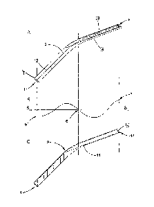
Referring now to the Figures, and to Figures 1 and 2 in particular, a human
femur 100
is shown. In a previous surgical procedure, a first orthopaedic implant 200
has been
implanted into the femur 100, such that a rounded head 201 of the first
implant 200
replaces a head of the femur 100, and an elongate stem 202 of the first
implant 200 is
embedded in an elongate central cavity 102 of the femur 100.
A suitable surgical cement 203, normally a polyacrylate composition, may have
been
used to secure the first implant 200 in place. Alternatively, the first
implant 200 may
have been secured in place by the natural growth of cancellous bone 103,
particularly
between the stem 202 of the first implant 200 and the walls 104 of the shaft
of the
femur 100. In some cases, most or all of a surface of the stem 202 of the
first implant
200 has a roughened surface, to which the cancellous bone 103 may "key". Other
implants 200 have smaller roughened zones located on a proximal portion 204 of
the
stem 202 (although cancellous bone 103 will still adhere to non-roughened
zones of
the stem 202, albeit initially less strongly).
If any implant 200 becomes damaged or worn out, or begins to come loose, a
revision
procedure will be necessary to remove the existing implant 200 and replace it
with
CA 02849634 2014-03-21
WO 2012/038698
PCT/GB2011/001376
11
another. It is hence necessary to separate the implant 200 from the femur,
while
leaving the femur 100 in a sufficiently sound condition to hold a replacement
implant
200 securely.
Where the implant 200 is cemented, known ultrasonically-vibratable tools may
be
used to soften the cement 203 sufficiently for the implant 200 to be
extracted, and
further known ultrasonically-vibratable tools may be used to remove the
remains of
the cement 203, before the new implant 200 is cemented within the femur 100.
This
is usually a relatively rapid procedure, minimising patient trauma and time
spent
under anaesthetic.
Currently, cutting away cancellous bone 103 is more difficult. Chiselling away
cancellous bone 103 adjacent a proximal end of the stem 202 may be possible to
a
limited extent, but since the implant 200 is in situ, there is very little
freedom of
motion for conventional osteotomes because of surrounding tissues. It is
therefore
necessary, as shown schematically in Figure 3, to cut open a "window" in the
walls
104 of the femur 100, temporarily folding back a flap 105 of bone, so that the
cancellous bone 103 may be approached laterally, freeing the proximal portion
204 of
the stem 202. (NB the "window" may extend to a proximal rim of the walls 104
of
the femur 100, when chiselling longitudinally from the open proximal end of
the
femur 100 is completely impractical). To sever cancellous bone 103 not
directly
accessible through this "window", a wire saw is passed behind the stem 202 and
is
used to saw down through this cancellous material.
CA 02849634 2014-03-21
WO 2012/038698
PCT/GB2011/001376
12
Even though the flap 105 will be replaced after the procedure and will
eventually heal
and merge with the walls 104 of the femur 100, this weakens the femur 100, and
this
approach should not be used all the way down to a distal tip 205 of the stem
202. The
window does not allow useful access to conventional osteotomes. It is usually
necessary for the stem 202 to be sawn through, the proximal portion 204 to be
removed, and then for the distal tip 205 to be chiselled out separately. The
implant
200 generally comprises a hard cobalt steel, so sawing it through is a slow,
labour
intensive procedure, wearing away saw blades and creating metal fragments that
must
be prevented from entering body tissues.
Revision of an implant 200 can thus be a lengthy procedure, causing high
levels of
patient trauma and involving long periods under anaesthesia. It may also lead
to
manual fatigue on the part of the surgeon.
A first cutting tool or osteotome 1 embodying the present invention is shown
in
Figures 4A and 4C. The first tool 1 comprises an elongate cylindrical
waveguide 2
having an elongate cutting blade 3 extending from a distal end of the
waveguide 2. A
proximal end 4 of the waveguide 2 is in practice fitted with a threaded
connector or
the like (here omitted for clarity), by which the tool 1 is connected to a
source of
ultrasonic vibrations, such as a longitudinal mode ultrasound generator of
known
form.
The cutting blade 3 has a substantially constant hollow semicircular profile
(see also
Figure 5), and its distal tip 8 is provided with a bevel 15 leading to a
relatively sharp
distal cutting edge 14.
CA 02849634 2014-03-21
WO 2012/038698
PCT/GB2011/001376
13
The blade 3 extends from the waveguide 2 at an angle: a longitudinal axis 12
of the
waveguide 2 and a longitudinal axis 13 of the blade 3 intersect at an angle of
300 in
this example, although this angle may vary while still producing an effective
tool 1.
The waveguide 2 and blade 3 blend smoothly into each other across a curved
joining
zone 9. A concave face of the joining zone 9 has a smooth curved profile.
However,
a concave surface of the blade 3 continues as a groove extending straight
across the
joining zone 9 until it meets the waveguide 2 (see Figure 4C). Thus, the
thickness of
the tool 1 tapers across the joining zone 9, the wall 11 of the blade 3 being
substantially thinner than the cylindrical waveguide 2.
Ideally, the tool 1 may be formed from a single cylindrical stock piece of
metal,
which is first bent smoothly through a desired angle at the joining zone 9.
The blade
3 is then machined out in a single straight pass. This creates the hollow
semicircular
profile of the blade 3 and the gradual taper across the joining zone 9, the
groove
becoming shallower and ending as the waveguide 2 curves away beneath it.
Mt,- tool 1 shown is devised to be used with longitudinal-mode ultrasonic
vibrations
of a known frequency, and hence a known wavelength in a given material (to a
first
approximation at least).
As shown in Figure 4B, the waveguide 2 in this case has a length of
approximately
three-quarters of the wavelength of the ultrasonic vibrations therein. The
source of
ultrasonic vibrations is connected to the waveguide 2 at its proximal end 4,
such that
CA 02849634 2014-03-21
WO 2012/038698
PCT/GB2011/001376
14
there is a first anti-node 5 in the vibrations at this point. The length of
the waveguide
2 produces a nodal point 6 in the vibrations, located within the joining zone
9. The
blade 3 also has a length of three-quarters of the wavelength of the
ultrasonic
vibrations therein. Thus, there will be a second anti-node 7 at the distal tip
8 of the
blade 3.
It is found that with the profile of the joining zone 9 shown, there is a
remarkably
good transmission of energy "around the bend" in the tool 1, from the
waveguide 2 to
the blade 3. Additionally, the reduction in the cross-sectional area of the
tool 1, from
the cylindrical waveguide 2 to the thin semi cylindrical walls 11 of the blade
3,
produces a gain in the amplitude of the vibrations. Across an abrupt step in
the
diameter of the tool, the gain is a function of the ratio of the cross
sectional areas each
side of the step. Across a more gradual change in cross-sectional area, as in
this tool
1, it is found that a similar gain can be achieved.
As a result, this tool 1 may be energised with longitudinal-mode ultrasonic
vibrations
to produce a reciprocal motion of the distal cutting edge 14, directed
parallel to the
longitudinal axis 13 of the blade, with an amplitude of at least 60
micrometres.
This motion, applied to cancellous bone 103, is easily sufficient to chisel it
away
without requiring a user to do more than apply the distal tip 8 to the
cancellous bone
and activate the ultrasonic vibrations. (It should also cut through bone
cement 203
with ease).
CA 02849634 2014-03-21
WO 2012/038698
PCT/GB2011/001376
Figures 5 and 6 show preferred features of the blade 3, Since the blade 3 is
likely to
contact the stem 202 of an implant 200 or surrounding tissues in use, it is
preferred
that each lateral rim 10 of the blade 3 should be rounded smoothly, rather
than being
left with rougher or sharply-angled edges. This should reduce the risk of
damage to
the blade 3, especially since some implants 200 have roughened surfaces.
Figure 6 shows how the bevel 15 is preferably formed on an inner, concave face
of
the blade 3, defining a sharper distal cutting edge 14. This need not
necessarily be as
sharp as that of a conventional hand-impelled osteotome, but it should be
significantly
sharper than any other edge or rim on the tool 1, such that it is safe when
not
energised, and the only significant cutting element when the tool 1 is
energised.
Figure 7 shows, in schematic form, the first tool 1 in use (some relative
sizes and
proportions have been adjusted for clarity, rather than strict accuracy). In
this
example, a stem 202 of an implant 200 is held within the wall 104 of a femur
100 by
cancellous bone 103 (the thickness of which is exaggerated in this Figure).
The tool 1
is aligned such that the longitudinal axis of the blade 3 is directed
substantially in
parallel to the longitudinal axis of the stem 202. To guide the blade 3, a
concave face
of the blade 3 may be contacted with the stem 202 and the blade 3 may then be
run
down the stem 202. This should keep the blade 3, and the rims 10 of blade 3 in
particular, away from the walls 104 of the femur 100. The osseous bone of the
walls
104 would be more resistant to cutting than the cancellous bone 103, but any
unnecessary damage to the osseous bone should be avoided. The user is then
able to
press the blade 3 smoothly down the stem 202, the distal cutting edge 14
cutting
through the cancellous bone 103 as it goes. Very little force should be needed
once
CA 02849634 2014-03-21
WO 2012/038698
PCT/GB2011/001376
16
the tool 1 is ultrasonically vibrated. Only a limited number of passes would
be
required to isolate the stem 202 from the surrounding cancellous bone 103,
particularly when the curvature of the blade 3 is matched closely to that of
the stem
202 (unlike in Figure 7, where the differences in radius of curvature are
exaggerated
for effect).
The curve of the joining zone 9 of the tool 1 allows the blade 3 to be
presented at the
correct angle to be used as described above, while the waveguide 2 and a
remainder of
the tool 1 mounted thereto are conveniently canted away from surrounding
tissues. A
manually-impelled osteotome with such a geometry would be difficult to impel
longitudinally of the femur 100, but the discovery that ultrasonic vibrations
may be
transmitted reliably and controllably around a curve in mid-tool (with a gain
in
amplitude into the bargain) allows the ultrasonically-vibratable tool 1 of the
present
invention to cut with minimal force and maximal convenience.
The tool 1 may conveniently be used with longitudinal-mode ultrasound
generators
operating at between 20 kHz and 60 kHz, which are already used in a range of
surgical tools. Since the optimal length of the tool 1 depends on the
wavelength of
the vibrations produced in the tool 1, it would be possible to produce tools
of a range
of desired dimensions, each achieving resonance at the exact frequency that
puts a
node in the joining zone 9 and an anti-node at the distal tip 8.
Figures 8A to 9B, show two variant forms of tool embodying the present
invention. It
may be beneficial to profile the blade to allow easier passage of cut debris
away from
the cutting edge. In a second cutting tool 21 (Figures 8A and 8C), a majority
of the
CA 02849634 2014-03-21
WO 2012/038698
PCT/GB2011/001376
17
blade 23 has a constant profile, but adjacent a distal end it comprises a
coaxially-
extending section 25 of greater diameter, joined to the main blade 23 by a
flaring
section 24.
Ultrasonically-vibrated tools can cause significant local heating in use. In a
third
cutting tool 31 (Figures 9A, 9B), a series of parallel longitudinal grooves 38
are
formed along a distal section of the blade 33, extending across the bevel 15
to the
cutting edge 14. These allow cooling water to be delivered down the blade 33
to the
cutting edge 14.
While in each of the tools 1, 21, 31 illustrated, the bevel 15 is shown in the
inner,
concave surface of the blade 3, 23, 33, it would also be possible to bevel the
outer,
convex surface if desired.
Although running the concave surface of the blade 3 down the stem 202 of an
implant
200 guides the blade 3 with the required accuracy, it has been found in trials
that
prolonged contact between the blade 3 and the stem 202, particularly over
large
contact areas, may lead to fatigue problems in the metal of the blade 3.
Figures 10
and 11 show two-improvements to the blade 3 that help to obviate this problem.
Figure 10 shows a fourth tool 41 embodying the present invention, which is
similar in
most respects to the first tool 1. There is an elongate cylindrical waveguide
2,
connectable to a source of ultrasonic vibrations, with an elongate blade 43
extending
from its distal end. At a distal end of the blade 43, a bevel 15 leads to a
distal cutting
edge 14.
CA 02849634 2014-03-21
WO 2012/038698
PCT/GB2011/001376
18
As in the case of the blades 3, 23, 33 above, this blade 43 has towards its
distal tip a
thin-walled part-circular cross-section. The lateral rims 10 of the hollow
part-
cylindrical profile thus formed are again rounded-off.
In order to obviate metal fatigue resulting from contact between the
ultrasonically-
vibrated, concave face of the titanium blade 43 and the cobalt steel stem 202,
the
concave face of the blade 43 is provided with a lining or insert 47 of poly
(tetrafluoroethylene), polyether ether ketone, high density polyethylene (i.e.
PTFE,
PEEK or HDPE), or other thermoplastics material having a degree of resilience,
mechanical integrity and low coefficient of friction. This lining 47 extends
proximally from immediately adjacent the bevel 15 along a major portion of the
blade
43, and it conforms to the profile of the concave face of the blade 43, having
a
substantially constant thickness. (Instead of a lining 47, a coating of PTFE
or the like
could also be applied to this concave surface).
In use, when this blade 43 is brought up to a stem 202, only the insert 47
will contact
the stem 202. As the blade 43 is passed down the stem 202, there will be
minimal
vibrating metal-Metal contact and hence minimal risk of metal fatigue.
Figure 11 shows a fifth tool 51 embodying the present invention, which employs
a
different approach. The fifth tool 51 again has most of the features of the
first tool 1,
including an elongate blade 53 extending from a distal end of an elongate
cylindrical
waveguide 2 to a terminal bevel 15 and a distal cutting edge 14. This blade 53
again
CA 02849634 2014-03-21
WO 2012/038698
PCT/GB2011/001376
19
has a thin-walled part-circular cross-section towards its distal tip, with
rounded off
lateral rims 10 to the hollow, part-cylindrical profile thus formed.
The blade 53 of the fifth tool 51 is also provided with a set of
longitudinally-
extending upstanding ribs or fins 59, spaced around its concave face. The fins
59
extend proximally from the bevel 15 (a distal end of each fin 59 may, as
shown,
continue the bevelled profile) along a major portion of the blade 51. Each fin
is
radially upstanding to a constant height above the concave face of the blade
51.
Thus, when this blade 53 is brought into contact with a stem 202 of an implant
200,
and as the blade 53 is passed down to stem 202, the blade 53 and stem 202 will
only
be in contact along an upper surface of the fins 59.
Figure 12 shows the fifth tool 51 in operative alignment with the stem 202 of
a second
femoral implant 210, to demonstrate this point (the second femoral implant 210
has
minor differences of detail, compared to the first 200, but its stem 202 is
substantially
identical). The stem 202 is almost cradled within the part-cylindrical profile
of the
blade 53, contacting the blade 53 only along the upper surface of each fin 59.
The
blade 53 can thus pass freely along the stem 202 as it cuts through the
cancellous bone
103 surrounding the stem 202.
Because the contact area between the fins 59 and the stem 202 is so small, any
metal
fatigue in the blade 53 will be localised within the fins 59. Even if there is
localised
damage to a fin 59, this would have little effect on the performance of the
tool 51 as a
whole.
CA 02849634 2014-03-21
WO 2012/038698
PCT/GB2011/001376
As can be seen from Figures 11 and 12, the upstanding longitudinal fins 59
along the
inner, concave face of the blade 53 could be considered to define channels 58
between
them. These could be used to pass cooling water down the blade 53 to its
distal
cutting edge 14 (as for the grooves 38 of the third tool 31), and/or could
provide
convenient passage away from the cutting edge 14 for fragmentary debris
created as
the cutting edge 14 passes through cancellous bone 103.
In the particular example of the fifth tool 51 shown, the fins 59 are formed
integrally
with the blade 53 as the tool 51 is machined into shape. In a variant (not
shown) the
fins 59 are instead formed as part of a pre-formed insert or lining, e.g. of
PTFE, PEEK
or the like, mounted to the inner, concave surface of the blade 53.
It is also possible to form circumferentially-extending upstanding fins on the
inner,
concave face of the blade 51, which would also produce the same stand-off
function
to obviate metal fatigue in the operative portions of the blade 53. In this
variant, the
fins would not define longitudinal channels for passage of debris and/or
cooling
water, and might be slightly less convenient for longitudinal motion, but the
tool
should still be superior to a tool 1 with a plain concave surface leading to
extensive
vibrating metal-metal contact. Either should be far superior to existing tools
and
methods described above.