Note : Les descriptions sont présentées dans la langue officielle dans laquelle elles ont été soumises.
BIOSCAFFOLDS FOR FORMATION OF MOTOR ENDPLATES
AND OTHER SPECIALIZED TISSUE STRUCTURES
George J. Christ, Justin M. Saul, John B. Scott,
Benjamin T. Corona, Benjamin Harrison, and Catherine Ward
Related Applications
This application claims the benefit of U.S. provisional application serial
number
61/541,652, filed September 30, 2011.
Field
The present disclosure concerns biomaterial constructs useful to support the
delivery
of agents of interest.
Background
Muscle deficiencies due to a host of congenital or acquired conditions,
including, but
not limited to, surgery, inflammation, traumatic injury, and disease, can lead
to the
irrecoverable loss of muscle function. For those who suffer from such defects,
there are
currently few clinical tieatinentN available.
Cell-based approaches have been studied to regenerate or re-create muscle
tissues
such as skeletal muscle de novo with the use of materials (e.g., polymers or
natural
scaffolds) to support the attachment, growth, and/or proliferation of cells
that have some of
the characteristics of native skeletal muscle. However, and particularly in
cases where the
magnitude of the injury or disease significantly exceeds the regenerative
capacity of the
remaining viable tissue (e.g., volumetric muscle loss resulting from traumatic
injury), better
therapeutic solutions are needed to create functional muscle tissue.
Summary of the Invention
Provided herein are methods useful to promote the formation of functional
clusters
on a tissue, for example, motor endplates (MEPs) or a component thereof on
skeletal muscle
tissue. In some embodiments, promotion of a cluster of acetylcholine receptors
on the
muscle tissue promotes the muscle phenotype, such as inclusion of
multinucleated myotubes,
expression of mature muscle markers (e.g., myogenin, MHC, titin, etc.) prior
to and after
- 1 -
CA 2849987 2017-10-23
CA 02849987 2014-03-25
WO 2013/049563
PCT/US2012/057904
implantation of thc tissue engineered construct, so that when re-innervation
does occur in
vivo, the tissue engineered implant can still function, and preferably
function more optimally.
Also provided herein are scaffolds useful for growing cells thereon. In some
embodiments, the scaffold includes one or more agents of interest at separate
and discrete
locations in or on the scaffold. In some embodiments, the scaffold includes
beads (e.g.,
microbeads or nanobeads) therein or thereon, wherein said beads comprise an
agent of
interest. In some embodiments, the agent of interest is incorporated onto the
beads though
covalent coupling.
In some embodiments, the scaffold is a skeletal muscle cell scaffold or a
cardiac
muscle cell scaffold. In some embodiments, the agent of interest is agrin.
In some embodiments, the scaffold includes fibrin, collagen, agarose,
cellulose,
alginate, agarose, keratin, hydroxymethyl cellulose, or
poly(hydroxyethylmethacrylate).
Also provided are methods of making a skeletal muscle implant, including
providing
a scaffold as described herein and seeding cells (e.g., muscle cells such as
myoblast cells or
satellite cells) onto said scaffold. In some embodiments, the methods also
include isolating
muscle cells from a donor tissue.
Further provided are methods of making a scaffold including beads therein or
thereon,
including one or more of the steps of providing a material (e.g., a hydro gel)
including beads,
which beads include an agent of interest; applying the material onto a
template (e.g., a fiber
template); polymerizing the material around the template to form a polymerized
material; and
then, selectively dissolving the template to create hollow spaces (e.g.,
hollow tubes) within
the polymerized material.
Still further provided are methods of culturing organized skeletal muscle
tissue from
precursor muscle cells (e.g., myoblast cells or satellite cells), including
cyclically stretching
and relaxing the muscle cells on a scaffold in vitro for a time sufficient to
produce the
organized skeletal muscle tissue; and further including agrin at separate and
discrete locations
in or on said scaffold, the agrin provided in an amount effective to promote
the formation of
aggregated acetylcholine receptors in the organized skeletal muscle adjacent
to one or more
of said separate and discrete locations.
In some embodiments, the separate and discrete locations are provided in a
ratio of
between 1:10 and 10:1, or at an approximately 1:1, 1:2, 1:3, 1:4, 1:5, 5:1,
4:1, 3:1, or 2:1
ratio, with respect to formed myotubes in the organized skeletal muscle
tissue.
Also provided are methods of culturing organized skeletal muscle tissue from
precursor muscle cells including cyclically stretching and relaxing said
muscle cells seeded
- 2 -
onto a fibrin or fibrinogen scaffold in vitro for a time sufficient to produce
said organized skeletal
muscle tissue, wherein the scaffold includes a plurality of channels aligned
along a first axis.
Further provided are multi-layered skeletal muscle tissue produced by the
methods as
taught herein. In some embodiments, the tissue includes elongated multi-
nucleated muscle fibers.
In some embodiments, the tissue includes or expresses acetylcholine (ACh)
receptors. In some
embodiments, the tissue includes aggregated ACh receptors. In some
embodiments, the tissue
includes aggregated ACh receptors forming a pretzel shape characteristic of
motor end plates. In
some embodiments, the tissue includes aggregated ACh receptors which are at a
ratio of 10:1 and
1:10, or between 5:1 and 1:5, or between 1:2 and 2:1, with respect to said
elongated multi-
nucleated muscle fibers. In some embodiments, the tissue is suturable.
Still further provided are methods of treating a skeletal muscle injury or
defect in a
subject in need thereof including grafting an engineered tissue as described
herein into the subject
in a treatment-effective configuration.
Also provided is an engineered tissue as taught herein for use in repairing a
skeletal
muscle injury or defect.
The invention also provides a scaffold comprising one or more agents of
interest at
separate and discrete locations in or on the scaffold.
The invention also provides a scaffold for growing cells thereon comprising
one or more
agents of interest at separate and discrete locations in or on the scaffold,
wherein the agents of
interest are growth or development factors.
The invention also provides a method of making a skeletal muscle implant
comprising:
providing the scaffold as defined herein; and
seeding muscle cells onto said scaffold.
The invention also provides a method of making the scaffold as defined herein,
comprising:
providing a hydrogel comprising one or more agents of interest at separate and
discrete locations on or in the hydrogel;
applying said hydrogel onto a fiber template;
polymerizing said hydrogel around said fiber template to form a polymerized
hydrogel; and then,
selectively dissolving said fiber template to create hollow spaces within said
polymerized hydrogel, to thereby make the scaffold.
The invention also provides a method of making the scaffold as defined herein,
comprising:
- 3 -
CA 2849987 2019-12-03
providing a hydrogel comprising one or more agents of interest at separate and
discrete locations on or in the hydrogel;
applying said hydrogel onto a fiber template;
polymerizing said hydrogel around said fiber template to form a polymerized
hydrogel; and then,
selectively dissolving said fiber template to create hollow spaces within said
polymerized hydrogel, to thereby make the scaffold;
wherein the agents of interest are growth or development factors.
The invention also provides a method of culturing organized skeletal muscle
tissue from
precursor muscle cells, comprising cyclically stretching and relaxing said
muscle cells on a
scaffold in vitro for a time sufficient to produce said organized skeletal
muscle tissue, and further
comprising:
including agrin at separate and discrete locations in or on said scaffold,
said agrin
provided in an amount effective to promote the formation of aggregated
acetylcholine receptors
in said organized skeletal muscle adjacent to one or more of said separate and
discrete locations.
The invention also provides a method of culturing organized skeletal muscle
tissue from
precursor muscle cells comprising: cyclically stretching and relaxing said
muscle cells seeded
onto a scaffold in vitro for a time sufficient to produce said organized
skeletal muscle tissue,
wherein said scaffold comprises fibrinogen, and
wherein said scaffold comprises a plurality of channels aligned along a first
axis.
The invention also provides a multi-layered skeletal muscle tissue produced by
the
process of the invention.
The invention also provides a multi-layered skeletal muscle tissue produced by
the
method of the invention.
The invention also provides a use of the tissue as defined herein for grafting
said tissue in
a treatment-effective configuration for treating a skeletal muscle injury or
defect in a subject in
need thereof.
The invention also provides a tissue of the invention for use in repairing a
skeletal muscle
injury or defect.
The invention also provides the tissue of the invention for use in repairing a
skeletal
muscle injury or defect in a subject.
- 3a -
CA 2849987 2019-12-03
Brief Description of the Drawin2s
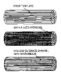
FIG. 1A-1B provides a schematic example of the formation of a fibrin hydrogel
scaffold
(A), which can be (B) seeded with cells and/or containing polystyrene beads
capable of providing
acetylcholine receptor agonist stimulation through agonist presentation
(triangles).
FIG 2 reports the effects of agrin-presenting beads on C2C12 cells cultured on
fibrin
hydrogels. Microparticle bead delivery vehicles without added agrin produce no
response from
treated cells (A). Physically adsorbing agrin to the bead surface allows for
spatially targeted
induction of a clustering response in membrane-bound acetylcholine receptors
after 1 day of
treatment at areas of contact between cells and bead delivery vehicles (B).
Though agrin-
adsorbed beads are ineffective at generating a clustering response after 3
days of treatment (data
not shown), linking agrin to the microparticle surface using EDAC/sulfo-NHS
covalent
crosslinking chemistry allows for induction of clustering behavior at 5 days
of treatment (C) or
beyond. Dotted lines show approximate cell-seeded area in the plane of focus,
and arrows
indicate foci of acetylcholine receptor clustering. All areas of clustering
occur where beads are
present, indicating the importance of the agrin-presenting beads in promoting
clustering behavior.
Results also suggest that adsorption of agrin to bead surface is _________
- 3b -
CA 2849987 2019-12-03
CA 02849987 2014-03-25
WO 2013/049563
PCT/US2012/057904
superior in short-term applications, while covalent crosslinking between agrin
and the bead
enables cell signaling in the long term.
FIG. 3A-3B shows scanning electron microscope (SEM) images of scaffolds
fabricated from pMMA templates and fibrin for nerve applications with a 105
[JI,M template.
FIG. 4 shows receptor clustering in myotube-like cell membranes in response to
5
days of treatment with agrin-delivering microparticles. Confocal imaging
eliminates out-of-
plane signal, producing an image of a very thin "slice" of tissue. Progressive
slices can be
used to show the evolution of signals across all three dimensions of a
structure. Micrographs
of fluorescent labeling using a-bungarotoxin beginning beneath microparticles
(A),
advancing vertically (B), and ending near the center of the microparticle
vertical thickness
(C) show that cells respond to agrin delivery via formation of AChR clusters
(staining) at
areas of microparticle contact. Dotted outlines depict locations of
microparticles with agrin
covalently linked to bead surface. As seen across the three sequential sub-
images, clustering
behavior is independent of bead orientation at area of contact, meaning a bead
may equally
signal a cell it is on top of, beside, or contacting at any oblique angle.
Similarly, one agrin-
delivering particle can signal multiple adjacent cells, indicating that
production of a response
in one cell does not meaningfully deplete the agrin-coupled bead's ability to
impart its signal.
FIG. 5 illustrates the efficacy of myoblast-like cell seeding of patterned
fibrin
scaffolds. Scaffolds were fabricated via polymerization of fibrinogen in the
presence of
thrombin and calcium around a sacrificial pMMA template. The template was
dissolved,
leaving a porous network of hollow cylindrical channels within a macroscopic
fibrin
biomaterial. C2C12 cells were seeded statically by adding in suspension on top
of the
scaffold (from the left of the image), cultured for 1 day in growth medium,
and differentiated
toward a myotube-like phenotype for 10 further days. Low-magnification imaging
of a
sagittal scaffold section shows cells readily colonized the entire thickness
of the scaffold (left
to right of image) by migration during the growth and early differentiation
phases as
visualized by DAPI nuclear stain. Higher-magnification views near the top,
middle, and
bottom of the scaffold thickness reveal that myotube-like cells have fused
within the scaffold
and are expressing acetylcholine receptors (data not shown).
- 4 -
Detailed Description of Preferred Embodiments
Provided herein and further described below are compositions and methods
useful for
producing functional muscle tissue in vitro for implantation in vivo.
As used herein in the description of the invention and the appended claims,
the
singular forms "a," "an" and "the" are intended to include the plural forms as
well, unless the
context clearly indicates otherwise. Furthermore, the terms "about" and
"approximately" as
used herein when referring to a measurable value such as an amount of a
compound, dose,
time, temperature, and the like, is meant to encompass variations of 20%, 10%,
5%, 1%,
0.5%, or even 0.1% of the specified amount. Also, as used herein, "and/or" or
"I" refers to
and encompasses any and all possible combinations of one or more of the
associated listed
items, as well as the lack of combinations when interpreted in the alternative
("or").
"Implant" refers to a product configured to repair, augment or replace (at
least a
portion of) a natural tissue of a subject (e.g., for veterinary or medical
(human) applications).
The term "implantable" means the device can be inserted, embedded, grafted or
otherwise
chronically attached or placed on or in a patient. Implants include a support
with or without
having cells seeded thereon and/or subjected to bioconditioning according to
some
embodiments as described herein.
"Subjects" are generally human subjects and include, but are not limited to,
"patients." The subjects may be male or female and may be of any race or
ethnicity,
including, but not limited to, Caucasian, African-American, African, Asian,
Hispanic,
Indian, etc. The subjects may be of any age, including prenatal, newborn,
neonate, infant,
child, adolescent, adult and geriatric subjects.
Subjects may also include animal subjects, particularly vertebrate subjects,
e.g.,
mammalian subject such as canines, felines, bovines, caprines, equines,
ovines, porcines,
rodents (e.g., rats and mice), lagomorphs, non-human primates, etc., or fish
or avian
subjects, for, e.g., veterinary medicine and/or research or laboratory
purposes.
"Treat" refers to any type of treatment that imparts a benefit to a subject,
e.g., a
patient afflicted with or at risk for developing a disease (e.g., a
musculoskeletal disease),
injury, or other impairment or defect. Treating includes actions taken and
actions refrained
from being taken for the purpose of improving the condition of the patient
(e.g., the relief of
one or more symptoms), delay in the onset or progression of the disease, etc.
Treatment
includes that of any disease or injury associated with the loss or dysfunction
of skeletal
- 5 -
CA 2849987 2017-10-23
muscle, such as the treatment of volumetric muscle loss or congenital defects
in the limbs or face,
for which current treatments do not fully repair the defects. Other examples
include, but are not
limited to, the loss or denervation of skeletal muscle due to disease
conditions such amyotrophic
lateral sclerosis (ALS), post-polio syndrome, muscular dystrophy, etc.
In some embodiments, treating includes reconstructing skeletal muscle tissue
(e.g., where
such tissue has been damaged or lost by, e.g., injury or disease) by
implanting an scaffold (e.g.,
an anisotrophic scaffold, with or without muscle cells) into a subject in need
thereof. Scaffolds
may be implanted, e.g., at or adjacent to the site of injury, and/or at
another site in the body of a
subject that would impart a benefit to the subject, as would be appreciated by
one of skill in the
art.
Muscle cells used to carry out the present invention may be isolated according
to methods
known in the art, and are preferably mammalian muscle cells, including, but
not limited to,
human, other primate such as monkey, baboon, pig, sheep, goat, horse, dog,
rodent such as
mouse, rat, etc. In general, such cells are skeletal muscle cells. Muscle
cells of other species,
including birds, fish, reptiles, and amphibians, as well as arthropods and/or
invertebrate skeletal
muscle may also be used, if so desired. In some embodiments, the cells are
precursor cells, or
cells that are capable of differentiating into mature, multinucleated muscle
cells, under
appropriate culture conditions and stimuli as described herein. Muscle
precursor cells are known.
See, e.g., U.S. Patent No. 6,592,623.
In some embodiments, skeletal muscle progenitor cells isolated from muscle
tissue are
used. In some embodiments, stem cells are used, and may be optionally
differentiated toward the
skeletal muscle phenotype before and/or after seeding onto the scaffold.
"Skeletal muscle cells"
include, but are not limited to, myoblasts, satellite cells and myotubes.
''Myoblasts" are a type of muscle precursor cell that can fuse with each other
and give rise
to myotubes. Myoblasts are thought to arise from satellite cells and are
normally closely
associated with myofibers during the course of their life cycle in the
vertebrate organism
(Zammit PS et al. The skeletal muscle satellite cell: the stem cell that came
in from the cold.
Journal of Histochemistry & Cytochemistry. 2006; 54(11): 1177-1191). If the
myofiber is injured,
the satellite cells become activated and give rise to myoblasts, which are
capable of further
dividing and repairing damaged muscle and/or forming new fibers. Typically,
after muscle
injuries myofibers become necrotic and are removed by macrophages (Hurme et
al. (1991)
Healing of skeletal muscle injury: an ultrastructural and immunohistochemical
study, Med. Sci
Sports Exerc. 23, 801-810). This induces proliferation and fusion of myoblasts
as described
above to form multinucleated and elongated myotubes, which self-assemble to
form a more
organized structure, namely muscle fibers (Campion (1984) The muscle satellite
- 6 -
CA 2849987 2019-01-09
CA 02849987 2014-03-25
WO 2013/049563
PCT/US2012/057904
cell: a review, mt. Rev. Cytol. 87, 225-251). Myoblasts may be harvested from
an appropriate
donor and isolated by standard techniques.
"Myotubes" are elongated, multinucleateci cells, normally formed by the fusion
of
myoblasts. Myotubes can develop into mature muscle fibers, which typically
have
peripherally-located nuclei and myofibrils in their cytoplasm (e.g., as found
in mammals).
"Isolated" as used herein signifies that the cells are placed into conditions
other than
their natural environment. Tissue or cells are "harvested" when initially
isolated from a
subject, e.g., a primary explant. In one embodiment, cells may be isolated
from a donor (e.g.,
living or cadaveric) or obtained from other cell sources. In one embodiment,
cells may be
obtained from the muscle.
Cells may be syngeneic (i.e., genetically identical or closely related, so as
to minimize
tissue transplant rejection), allogeneic (i.e., from a non-genetically
identical member of the
same species) or xenogeneic (i.e., from a member of a different species) with
respect to the
subject. Syngeneic cells include those that are autogeneic or autologous
(i.e., from the patient
to be treated) and isogeneic (i.e., a genetically identical but different
subject, e.g., from an
identical twin). Cells may be obtained from, e.g., a donor (either living or
caddvetic) or
derived from an established cell strain or cell line. For example, cells may
be harvested from
a donor (e.g., a potential recipient of a bioscaffold graft) using standard
biopsy techniques
known in the art.
Any suitable culture media can be used to grow cells in the present invention,
including medias comprising serum and other undefined constituents, defined
medias, or
combinations thereof, such as RPMI, DMEM, etc.
The "primary culture" is the first culture to become established after seeding
disaggregated cells or primary explants into a culture vessel. "Expanding" or
"expansion" as
used herein refers to an increase in number of viable cells. Expanding may be
accomplished
by, e.g., "growing" the cells through one or more cell cycles, wherein at
least a portion of the
cells divide to produce additional cells. "Growing" as used herein includes
the culture of cells
such that the cells remain viable, and may or may not include expansion and/or
differentiation of the cells.
"Passaged in vitro" or "passaged" refers to the transfer or subculture of a
cell culture
to a second culture vessel, usually implying mechanical or enzymatic
disaggregation,
reseeding, and often division into two or more daughter cultures, depending
upon the rate of
proliferation. If the population is selected for a particular genotype or
phenotype, the culture
- 7 -
CA 02849987 2014-03-25
WO 2013/049563
PCT/US2012/057904
becomes a "cell strain" upon subculture, i.e., the culture is homogeneous and
possesses
desirable characteristics (e.g., the ability to express a certain protein or
marker).
"Express" or "expression" of a protein or other biological marker means that a
gene
encoding the same of a precursor thereof is transcribed, and preferably,
translated. Typically,
according to the present invention, expression of a coding region of a gene
will result in
production of the encoded polypeptide, such that the cell is "positive" for
that protein or other
biological marker. Expression of certain proteins or other markers may be
indicative of a
certain phenotype, as known in the art.
"Scaffolds" on which cells may be seeded and grown to produce cultured tissue
.. include any suitable support. See, e.g., U.S. Patent Nos. 6,998,418;
6,485,723; 6,206,931;
6,051,750; and 5,573,784. Preferably, the scaffold is configured to support
the attachment,
proliferation and/or differentiation of cells thereon.
The scaffold may be formed from any suitable material, including, but not
limited to,
synthetic or natural polymers, other biopolymers, and combinations thereof. In
some
.. embodiments, scaffolds include collagen supports or decellularized tissue
supports (e.g.,
obtained from smooth muscle or skeletal muscle, such as a decellularized
mammalian (e.g.,
porcine) bladder such as bladder acellular matrix (BAM)). In some embodiments,
scaffolds
include a polymeric matrix (e.g., collagen, a hydrogel, etc.). In some
embodiments, scaffolds
include fibrin or fibrinogen.
The scaffold may be of any suitable configuration, but in some embodiments
comprises, consists of, or consists essentially of a generally flat planar
portion, such as a
sheet. In other embodiments, the scaffold comprises, consists of, or consists
essentially of a
generally tubular configuration. The scaffold may be of any suitable
thickness, but in some
embodiments are at least 20, 30, 50 or 100 lam thick, up to 600, 800, or 1000
pm thick, or
more. In some embodiments, scaffolds are 0.5 mm to 20 mm, or 1 mm to 15 mm, or
3 mm to
10 mm thick.
In some embodiments, scaffolds have sufficient mechanical integrity for
skeletal
muscle applications. In some embodiments, scaffolds have a tensile strength of
from 10 kPa
to 1000 kPa, or 50 kPa to 500 kPa, or 100 kPa to 300 kPa. In some embodiments,
scaffolds
.. have a tensile strength of at least 10, 50, 100 or 300 kPa.
In some embodiments, scaffolds have a Young's modulus of from 10 kPa to 5000
kPa,
or from 100 kPa to 2500 kPa, or from 500 kPa to 1000 kPa. In some embodiments,
scaffolds
(e.g., fibrin scaffolds) have a Young's modulus of from 100 kPa to 350kPa. The
Young's
modulus for scaffolds fabricated by the techniques described herein can be
tailored to mimic
- 8 -
the native modulus of muscle cells and tissue, with scaffolds of higher and
lower moduli
readily fabricated, allowing for the use of materials that have
physiologically-relevant
mechanical properties and/or that can withstand the rigors of various in vitro
and in vivo
environments.
In some embodiments, scaffolds have mechanical integrity sufficient to
withstand the
mechanical stimulation (e.g., cyclic loading) in a bioreactor to produce the
desired skeletal
muscle tissues. For example, in some embodiments scaffolds are able to
withstand the cell
seeding and preferred bioreactor pre-conditioning protocols described for at
least 5, 10, 15,
17 or 20 days or more, in PCT application no. PCT/US2011/047600, filed August
12, 2011,
to Christ et al.
In some embodiments, the scaffold is conditioned in a bioreactor to support
the
growth and maturation of cells. In other embodiments, the scaffold is directly
implanted in
vivo.
In some embodiments, scaffolds as provided herein are useful for the localized
delivery of agents of interest. For example, in some embodiments, the scaffold
may include
components that are actively presented to cells to promote functional
formation and/or
maintain phenotypic characteristics of cells in vitro and/or following
implantation.
For example, agents may be used that promote the formation of motor endplates
(MEPs) on skeletal muscle constructs, which can improve the maintenance of
phenotypic
characteristics of skeletal muscle cells. Without wishing to be bound by
theory, it is thought
that the promotion of MEP formation in skeletal muscle constructs will promote
the
maintenance of an innervated muscle phenotype prior to and/or after
implantation of the
tissue engineered construct, so that when re-innervation by the native tissues
does occur, the
tissue-engineered implant will retain at least a portion of this
functionality. For example, it is
expected that this will increase the rate and magnitude of functional muscle
recovery seen
with the implanted constructs. In some embodiments, the rate of recovery is
from 2, 3, or 4
to 6, 8 or 10 weeks. In some embodiments, the magnitude of recovery is at
least 30, 40, 50,
60, 70, 80 or 90 % of the original force generation. In some embodiments, the
magnitude of
recovery is from 30 to 90 %, or 40 to 80%, or 50 to 75% the original force
generation.
In addition to the use of agrin to promote MEPs in skeletal muscle constructs,
other
agents may be used to promote desired phenotypes in other cell types,
including, but not
limited to, smooth muscle cells, endothelial cells, nerve cells, Schwann
cells, bone cells, etc.
By incorporating chemical cues through incorporation into scaffolds (e.g.,
muscle scaffold,
blood vessel scaffolds, etc.) or by the direct presentation (e.g., by
injection) of the agent,
- 9 -
CA 2849987 2017-10-23
CA 02849987 2014-03-25
WO 2013/049563
PCT/US2012/057904
more functional phenotypes can be obtained or maintained for regenerative
medicine/tissue
engineering applications. For example, an angiogenic compound such as VEGF can
be
seeded on or carried by the scaffold to facilitate the formation of vascular
cells or vasculature
in the tissue.
In some embodiments, scaffolds provide localized delivery of agents of
interest by
incorporating on or into beads. "Beads" as uses herein refers to discrete
particles of any
geometrical shape (spheres, rectangles, cones, etc.), typically on the micro
(10-6) or nano (10-
9) scale (microbeads or nanobeads). In some embodiments, beads include or are
made of
materials such as polystyrene or other polymeric material such as
poly(ethylene), poly(lactic
acid), poly(glycolic acid), or poly(lactic-co-glycol acid); or gold, silver,
iron oxide, etc. The
particles may include one or more agents of interest (e.g., growth or
development factors,
such as agrin, acetylcholine, etc.) useful to promote phenotypes of interest
in the engineered
tissue. In some embodiments, beads have an average diameter of less than 0.1
um. In other
embodiments, beads have an average diameter of more than 1 mm. In some
embodiments,
beads have an average diameter of 0.1 inn to 1 mm, or 1 inn to 0.5 mm, or 10
gm to 100 am,
For example, in some embodiments, the local delivery of agrin to skeletal
muscle
tissue may promote the formation of acetylcholine receptor clustering or other
components
involved in the formation of motor endplates and/or neuromuscular junctions.
Other agents
that may be used include, but are not limited to, heparin-binding growth-
associated molecule
(HB-GAM), muscarine, acetylcholine receptor antibodies, carbachol, and
nicotine. Biglycan
or other agents that stabilize dystrophin-associated protein complexes may
also be used. See
U.S. Patent Application Publication No. 2011/0183910 to Fallon et al.
Any suitable method to couple the agent of interest to the beads may be used.
For
example, the agent may be adsorbed or covalently coupled to the beads through
various
chemistries including EDAC/NHS (for carboxyl-amine coupling) or inaleimide
chemistry
(for coupling to sulfhydryl groups). The beads can be placed on or within the
scaffold to
promote the formation of discrete functional clusters on the tissue, In some
embodiments,
beads are provided on or in the scaffold at a density of from 103, 104, or 105
per cm3, to 108,
109, 1010 or 2 x 10¹⁰ per cm'.
In some embodiments, beads comprising an agent may be directly injected at a
site of
interest. For example, beads comprising agrin (e.g., provided in a carrier
such as a hydrogel)
may be injected into a site of denervation or muscle motor loss to maintain
phenotype in vivo.
In some embodiments, the scaffold contains aligned or substantially aligned
hollow
conduit channels (e.g., along an axis of the support), or other porous
architecture that
- 10 -
CA 02849987 2014-03-25
WO 2013/049563
PCT/US2012/057904
supports the infiltration of cells (e.g., myoblasts, myotubes, etc.) and/or
sub-cellular
components. These scaffolds may be fabricated by a templating approach or
other methods to
achieve the preferred architecture.
In some embodiments, the scaffold is a three-dimensional fibrin or fibrinogen
scaffold. Fibrinogen is a soluble plasma glycoprotein that can be converted by
thrombin into
fibrin, such as normally occurs during blood coagulation. These scaffolds
include those made
by forming the scaffold around a dissolvable core to mold the inner channels.
For example,
the fibrin or fibrinogen scaffold may be formed around poly(methyl-
methaerylate) (pMMA)
beads, after which the beads are dissolved with acetone to form an
interconnected
microporous network. See, e.g., Linnes et al., Biomaterials 28 (2007) 5298-
5306. In other
embodiments, scaffolds may be formed around polymer rods. See, e.g., Flynn et
al,,
Biomaterials (2003) 4265-4272; Stokols et al., Tissue Engineering 12(10)
(2006) 2777-2787.
In some embodiments, the channels may be formed from a modification of the
production of fibrinogen-based porous scaffolds with polymer (e.g., 100 kDa
pMMA, or
poly(tetrafluoro ethylene)) fibers are extruded on a piston extrusion system.
Fiber diameter
may be varied based on the extrusion temperature and rate of uptake on
collection godets.
The fibers may then be packed into a desired geometry.
In some embodiments, any hydrogel material and fiber combination can be used,
so
long as an appropriate solvent can be used to selectively remove/dissolved the
fibers. Other
polymeric materials that may be used as the sacrificial template fibers
include, but are not
limited to, polystyrene, poly(ethylene glycol), poly(tetrafluoroethylene)
poly(vinyl alcohol),
poly(lactic acid), poly(glycolic acid), poly(lactic-co-glycolic acid),
poly(caprolactone), and
alginate,
Other hydrogel materials that may be used include, but are not limited to,
collagen,
agarose, cellulose (and modifications thereof), alginate, agarose, keratin,
hydroxymethyl
cellulose, and poly(hydroxyethylmethacrylate). It is preferable in some
embodiments that the
template and hydrogel materials have different solubilities in order to
promote selective
(sacrificial) removal of the template fibers. In some embodiments, beads are
included in the
hydrogel phase.
A schematic representation of a fabrication process for supports according to
some
embodiments is given in Figure 1. First, a fiber template is formed (A). The
fiber may be a
polymer, e.g., pMMA. The fibers may be formed by different processes and
formed to
desired diameters (e.g., by melt extrusion). A hydrogel material (e.g.,
fibrinogen) is then
backfilled into the template (e.g., by centrifugation or positive or negative
pressure). The
- 11 -
hydrogel material may then be polymerized (e.g., fibrinogen may be polymerized
to fibrin
with thrombin and calcium). The polymer mold or template may then be
selectively
dissolved by a solvent (e.g., pMMA may be dissolved by acetone), creating, for
example,
hollow tubes which may serve as aligned conduits. Locally-stimulating beads
(circles) that
may include an agent such as agrin (triangles) may be incorporated into the
construct (B).
These may be included in the hydrogel during the backfill process or may be
incorporated
after the support is formed, e.g., through absorption, covalent crosslinking,
etc.
In some embodiments, hollow channels in the scaffold have a diameter or
average
diameter of from 0.1, 1, 5, or 10 um to 1, 5 or 10 millimeters, or 100, 500,
1000 or 5000
millimeters. Other geometries may also be used, as desired. For example,
scaffolds may be
provided with substantially spherical pores. See, e.g., U.S. Patent No.
7,972,628 to Ratner et
al.
In some embodiments, cells are seeded onto (e.g., onto the surface) or into
(e.g.,
within the volume of) a scaffold. Cell seeding may be accomplished statically,
e.g. by adding
a suspension of cells in appropriate culture medium to one or more faces of
the scaffold,
after which cells settle onto and/or infiltrate the scaffold via gravity
and/or native cellular
motility. Cell seeding may also be accomplished dynamically using a
bioreactor, which may
act to guide or push/pull cells into the scaffold, for example by agitation or
fluid flow of the
cell suspension around and/or through the bulk of a scaffold and/or its porous
architecture.
In some embodiments, scaffolds with beads and/or cells are subjected to
bioreactor
pre-conditioning to facilitate maturation of myoblasts to form myotubes and
other markers
of mature skeletal muscle cells. See, e.g., patent application publication no.
2006/0239981 to
Yoo et al.; and PCT patent application serial no. PCT/US2011/047600, filed
August 12,
2011, to Christ et al. Any suitable bioreactor device can be used, such as
those described in
the aforementioned patent applications.
In some embodiments, a "mold" is provided which is configured to fit within
the
bioreactor and also designed to confine a cell suspension on top of and/or
within one or more
of the scaffolds and/or scaffolds seeded with cells. The mold may be made of a
light-weight
material (with a total weight, e.g., of 1-5 grams) and preferably does not
significantly
damage the underlying cellular structures when placed onto the scaffold and/or
scaffold
seeded with cells.
Multiple cell seeding protocols (i.e., more than just this one additional one,
to include
additional increases in bioreactor preconditioning) may also be performed. As
an example, if
- 12 -
CA 2849987 2017-10-23
CA 02849987 2014-03-25
WO 2013/049563
PCT/US2012/057904
each additional cell seeding is carried out during a time of between 3 and 4
days, the number
of cell seedings according to some embodiments may be 2, 3, 4, 5, 6, 7, or 8
or more.
The length of stretching of the scaffold may be to a dimension at least 5%
greater in
length than the static position, and in some embodiments preferably not
greater that 15%, and
the relaxing may comprise retracting the scaffold to a dimension not greater
in length than the
static position. In some embodiments the "static position" may be intermediate
between the
stretched and relaxed position, and in such cases the relaxing may comprise
retracting the
scaffold to a dimension at least 5% lesser in length than the static position.
The first time period, during which the stretching and relaxing occurs, may be
of any
suitable length, for example from 2 or 3 minutes up to 10, 20 or 30 minutes in
duration or
more. The step of cyclically stretching and relaxing is typically carried out
at least two or
three times during the first time period (e.g., from 2, 3 or 4 times, up to 10
or 20 times).
The second time period during which the scaffold is maintained in a static
position,
may be of any suitable duration. In some embodiments the second time period is
shorter than
the first time period, and may be from 1 or 2 minutes in duration up to 10 or
20 minutes in
duration. In other embodiments the second time period is longer than the first
time period,
and may be from 10 or 20 minutes in duration up to 40, 60 or 90 minutes in
duration, or
more. In some embodiments, such as where the first time period contains
comparatively long
intervals between stretching and relaxing, the need for a second time period
may be obviated
altogether.
In some embodiments, the scaffold is cyclically stretched and relaxed during a
first
''active' time period to a dimension of 10 % greater and lesser in length than
the static
dimension at a rate of 3 cycles per minute for a total of five minutes,
followed by a 25 minute
'rest" second time period, continuously for 1 to 3 weeks of in vitro culture.
In some
embodiments, this protocol may result in an increase in the number of
multinucleated cells,
thicker myotube width, better cellular alignment, etc., in the construct. In
some embodiments,
this protocol may result in an increase in the number of multinucleated cells,
thicker myotube
width, better cellular alignment, etc., in the construct (by, e.g., 10, 20,
50, 80 or 100%).
In some embodiments, the construct (scaffold plus cells) is characterized by
containing one or more neuromuscular junction features, particularly post-
synaptic features
such as the expression of acetylcholine (ACh) receptors, and in some
embodiments the ACh
receptors are aggregated. In some embodiments, aggregated ACh receptors may
include those
which have or approximate the characteristic pretzel shape of a motor endplate
in innervated
mature fibers in vivo.
- 13 -
CA 02849987 2014-03-25
WO 2013/049563
PCT/US2012/057904
In native tissue, the neuromuscular junction (NMJ), or nerve-muscle synapse,
include
the pre- and post-synaptic specializations of the motor neuron and muscle,
respectively, the
intervening synaptic basal lamina, and the specialized Schwann cell cap
(Salpeter, et al
(1987) The Vertebrate Neuromuscular Junction. New York, Alan R. Liss.). The
presynaptic
apparatus is typically marked by ordered arrays of synaptic vesicles, a subset
of which are
poised to fuse with the plasma membrane at the active zones, and release
acetylcholine that is
recognized by acetylcholine receptors (AChRs) on the muscle, and ultimately
results in
electrical activation and contraction of the muscle (Heuser, et al (1981) J.
Cell Biol. 88: 564).
Immediately across the 50 urn synaptic cleft from these zones are the crests
of the
postjunctional folds. These crests bristle with Acetylcholine
receptors'(AChRs), which can
reach densities of >10,000 molecules/um2 (Huh et al. (2002) Mol. Neurobiol.
25: 79). The
localized and tightly regulated secretion of acetylcholine into the narrow
synaptic cleft,
coupled with the high AChR density in the postsynaptic membrane, ensures rapid
and reliable
synaptic transmission between neuron and muscle, Perturbations of these
specializations,
such as the decrease in the number of functional AChRs seen in myasthenia
gravis, can lead
to debilitating and often fatal clinical outcomes (Oosterhuis, et al (1992)
Neurology &
Neurosurgery 5: 638).
In some embodiments, beads comprising an agent of interest is provided on the
scaffold such that a proper stoichiometry, or ratio of beads:cells (i.e.,
number of beads per
cell) is achieved. In some embodiments, the ratio of beads to cells is 1:100
to 100:1, or 1:50
to 50:1, or 1:20 to 20:1, or 1:15 to 15:1, 1:10 to 10:1, or 1:8 to 8:1, or1:5
to 5:1, or 1:4 to 4:1,
or 1:3 to 3:1, or 1:2 to 2:1, or approximately 1:1, 1:2, 1:3, 1:4, 1:5, 5:1,
4:1, 3:1, or 2:1.
In the case of skeletal muscle, in some embodiments it may be optimal to
provide
beads comprising an agent that promotes the formation of neuromuscular
junction features,
such as agrin, in an approximately 1:1, 1:2, 1:3, 3:1, or 2:1 ratio of beads
to cells to promote
the formation of approximately one point of innervation per cell. This may be
preferable in
some embodiments to reduce colliding waves of excitation that may occur when
there are
multiple sites of innervations per cell (e.g., fused cells formed after
bioreactor conditioning).
The density of the beads on or in the scaffold may be altered, as desired, to
obtain the
stoichiometry of choice.
In some embodiments, myotubes have a length of about 300 to 1500 um and a
diameter of about 20 to 30 um. These sizes may be used in some embodiments to
calculate
the concentration/distribution of beads in or on the scaffold in order to
obtain the desired
stoichiometry.
- 14 -
CA 02849987 2014-03-25
WO 2013/049563
PCT/US2012/057904
Skeletal muscle tissue produced as described herein may be used in vitro to
examine
the pharmacological or toxicological properties of compounds of interest
(e.g., by adding the
compound of interest to a culture medium in which the tissue is immersed, and
examining the
histological or mechanical properties of the tissue as compared to a control
tissue).
Skeletal muscle tissue constructs produced by the methods of the present
invention
are in some embodiments "suturable" in that they have sufficient structural
integrity to be
surgically sutured or otherwise fastened at either end when implanted, and
thereafter are
capable of developing tension upon contraction.
Skeletal muscle tissue constructs produced as described herein may be used for
the
reconstruction of damaged tissue in a patient, e.g., a patient with a
traumatic injury of an arm
or leg. Such tissue may be formed on a support (which is also implanted) or
removed from
the support prior to implantation into the subject. The skeletal muscle tissue
may be
implanted to "build'' soft tissue (e.g., at the interface between an amputated
limb and a
prosthetic device) or to reconstruct (partially or totally) a damaged muscle
(e.g., a muscle of
the face, hand, foot, arm, leg, back or trunk). The skeletal muscle tissue in
some
embodiments has, in some embodiments, a size or volume of at least 1, 2, or 3
or more cubic
centimeters (not counting the volume of the support if present), and/or a
length of 1 cm to 50
cm, to provide sufficient tissue mass for implantation in a patient (e.g., in
association with an
existing muscle of the patient) and reconstruction of a skeletal muscle
involved in, for
example, movement of fingers.
For allogenic transplant into a patient, tissue constructs as described herein
may be
matched or tissue-typed in accordance with known techniques, and/or the
subject may be
administered immune suppressive agents to combat tissue transplant rejection,
also in
accordance with known techniques.
The present invention is explained in greater detail in the following non-
limiting
Examples.
- 15 -
CA 02849987 2014-03-25
WO 2013/049563
PCT/US2012/057904
EXAMPLES
Example 1. Expression of acetylcholine receptors. During the characterization
of
tissue engineered muscle repair construct (TEMR) morphology following
bioreactor
preconditioning, it was discovered that multinucleated cells of TEMR
constructs expressed
acetylcholine (ACh) receptors and, in rare cases, exhibited aggregation of
these receptors.
Interestingly, the aggregation of ACh receptors in one construct is beginning
to exhibit the
characteristic pretzel shape of a motor endplate in mature fibers. These
findings are indicative
of mature TEMR constructs that produce clinically relevant force, as
innervation of
implanted TEMR constructs is thought to promote functional restoration of
traumatically
injured muscle tissue, and it is now known that these cells 1) express ACh
receptors and 2)
have the capacity to develop a motor endplate.
Example 2. Agrin-stimulated acetylcholine receptor clustering. For proof-of-
concept studies on the ability to achieve clustering of acetylcholine
receptors via stimulation
.. by agrin, a C2C12 mouse myoblast cell line was used. A 2-dimensional
version of the
scaffolds described above was also used to allow histological evaluation.
C2C12 cells were seeded on 2-dimensional fibrin hydrogels for 4-10 days.
Separately,
agrin was coated onto or covalently immobilized on the surface of polystyrene
beads. At 3-6
days of C2C12 cell culture, the agrin-coated beads were placed onto the cells
for 24 hours or
more. Cells were then stained for alpha-bungarotoxin, a marker of
acetylcholine receptor
clustering.
Agrin-coated beads on C2C12 cells cultured on fibrin hydrogels resulted in
areas of
clustering (FIG. 2). All areas of clustering occured where beads were present,
indicating that
the agrin-coated beads promoted clustering behavior. Physical adsorption of
agrin onto beads
was shown to be superior for short-term signaling of cells (e.g., 1 or 2
days), while covalent
immobilization enabled agrin delivery over longer time periods (e.g., 5 or
more days). A
higher magnification image confirmed that acetylcholine receptor clustering
occurs only
where beads are located (not shown).
FIG. 3A-3B show scanning electron microscope (SEM) images of scaffolds
fabricated from pMMA templates and fibrin for nerve applications with a
1051.tm template.
Clustering behavior was verified at areas of contact between cells and beads
covalently linked to agrin using confocal microscopy (FIG. 4). Moreover, the
unique ability
of confocal microscopy to isolate signals in a vertical direction demonstrated
independence of
- 16 -
bead positioning relative to the cell for the purposes of clustering induced,
as well as the ability
of an agrin-delivering bead to signal multiple adjacent cells.
Example 3. Incorporation of agrin-coated beads into scaffolds. Agrin-coated
beads
are incorporated into 2-D hydrogels and the 3-D scaffolds described above
(fabricated using a
poly(tetrafluoroethylene) tubular or rectangular mold), myoblasts are seeded
onto the formed 3-
D scaffolds, and studies similar to those described above in Example 2 are
conducted to
determine the efficacy of cell seeding in three dimensions as well as the
level of acetylcholine
receptor clustering achieved when beads are part of the scaffold material.
A rectangular, 3-D, patterned fibrin scaffold was seeded by static addition of
a cell
suspension and subsequent infiltration. As shown in FIG. 5, C2C12 cells
substantially infiltrated
the full thickness of the porous architecture via native cell motility,
demonstrating the suitability
of both the fibrin biomaterial and the sacrificial patterning process to
myoblast-like cell seeding.
Furthermore, microscopy revealed that the seeded myotube-like cells fused
within the
scaffold and expressed acetylcholine receptors (data not shown).
The foregoing is illustrative of the present invention, and is not to be
construed as
limiting thereof The invention is defined by the following claims, with
equivalents of the claims
to be included therein.
***
In some aspects, embodiments of the present invention as described herein
include the
following items:
1. A scaffold for growing cells therein comprising one or more agents of
interest at separate
and discrete locations in the scaffold, wherein the agents of interest are
growth or development
factors,
wherein the agents of interest are provided as particles or on beads, and
wherein the separate and discrete locations are provided in the scaffold at a
ratio of
approximately 1:1, 1:2, 1:3, 3:1, or 2:1 relative to the cells.
- 17 -
Date Recu/Date Received 2021-10-13
1.1. A scaffold for growing cells therein, said scaffold comprising one
or more agents of
interest, wherein the agents of interest are growth or development factors and
are
incorporated on beads in or on the scaffold.
2. The scaffold of item 1, wherein said one or more agents of interest are
provided on beads.
2.1. The scaffold of item 1.1, wherein the beads are provided in or on
the scaffold at a ratio of
approximately 1:1, 1:2, 1:3, 3:1, or 2:1 relative to cells to be seeded on the
scaffold.
3. The scaffold of item 2, wherein said beads are microbeads.
4. The scaffold of item 2 or 3, wherein said agents of interest are
incorporated onto said
beads though covalent coupling.
5. The scaffold of any one of items 2 to 4, wherein said beads are provided
in or on said
scaffold at a density of 103 to 2 x 10' beads per cubic centimeter of scaffold
volume.
6. The scaffold of any one of items 2 to 5, wherein said beads comprises
polystyrene,
poly(ethylene), poly(lactic acid), poly(glycolic acid), or poly(lactic-co-
glycol acid).
7. The scaffold of any one of items 2 to 5, wherein said beads comprise
gold, silver, or iron
oxide.
8. The scaffold of any one of items 1 to 7, wherein said one or more agents
of interest
comprises agrin.
9. The scaffold of any one of items 1 to 8, wherein said scaffold comprises
fibrin, collagen,
agarose, cellulose, alginate, agarose, keratin, hydroxymethyl cellulose, or
poly(hydroxyethylmethacrylate).
10. The scaffold of any one of items 1 to 9, wherein the scaffold comprises
a polymeric
matrix.
- 18 -
Date Recu/Date Received 2021-10-13
11. The scaffold of any one of items 1 to 9, wherein the scaffold comprises
a decellularized
tissue support.
12. The scaffold of any one of items 1 to 9, wherein the scaffold comprises
a decellularized
smooth muscle tissue or a decellularized skeletal muscle tissue.
13. The scaffold of any one of items 1 to 12, wherein said scaffold is a
skeletal muscle cell
scaffold comprising skeletal muscle cells or precursors thereof, or a cardiac
muscle cell scaffold
comprising cardiac muscle cells or precursors thereof.
14. A method of making a skeletal muscle implant comprising:
providing the scaffold as defined in any one of items 1 to 12; and
seeding muscle cells onto said scaffold.
15. The method of item 14, wherein said muscle cells are myoblast cells or
satellite cells.
16. The method of item 14 or 15, further comprising the step of
isolating muscle cells from a
donor tissue in vitro.
17. A method of making the scaffold as defined in item 10, comprising:
providing a hydrogel comprising the one or more agents of interest at separate
and
discrete locations in the hydrogel;
applying said hydrogel onto a fiber template;
polymerizing said hydrogel around said fiber template to form a polymerized
hydrogel;
and then,
selectively dissolving said fiber template to create hollow spaces within said
polymerized
hydrogel, to thereby make the scaffold.
17.1 A method of making the scaffold as defined in item 10, comprising:
providing a hydrogel comprising the one or more agents of interest
incorporated on beads
in the hydrogel;
applying said hydrogel onto a fiber template;
- 19 -
Date Recu/Date Received 2021-10-13
polymerizing said hydrogel around said fiber template to form a polymerized
hydrogel;
and then,
selectively dissolving said fiber template to create hollow spaces within said
polymerized
hydrogel, to thereby make the scaffold.
18.
The method of item 17, wherein said hydrogel comprises fibrin, collagen,
agarose,
cellulose, alginate, agarose, keratin, hydroxymethyl
cellulose, or
poly(hydroxyethylmethacrylate).
19.
The method of item 17 or 18, wherein said fiber template comprises
polystyrene,
poly(ethylene glycol), poly(tetrafluoroethylene), poly(vinyl alcohol),
poly(lactic acid),
poly(glycolic acid), poly(lactic-co-glycolic acid), poly(caprolactone), or
alginate.
20. The method of any one of items 17 to 19, wherein said one or more
agents of interest are
provided on beads.
21. The method of item 20, wherein said agent of interest is incorporated
onto said beads
though covalent coupling.
22.
The method of item 20 or item 21, wherein said beads are provided in said
hydrogel at a
concentration of 103 to 2 x 10¹⁰ beads per cubic centimeter of scaffold
volume.
23. A multi-layered skeletal muscle tissue produced by the method of any
one of items 17 to
22, wherein said tissue comprises elongated multi-nucleated muscle fibers.
23.1. A multi-layered skeletal muscle tissue produced by the method of any one
of items 17 to
19, wherein said tissue comprises elongated multi-nucleated muscle fibers and
said beads.
24. The tissue of item 23, wherein said tissue expresses acetylcholine
(ACh) receptors.
25. The tissue of item 23, wherein said tissue comprises aggregated ACh
receptors.
- 20 -
Date Recu/Date Received 2021-10-13
26. The tissue of item 22, wherein said tissue comprises aggregated ACh
receptors forming a
pretzel shape characteristic of motor end plates.
27. The tissue of item 23, wherein said tissue comprises aggregated ACh
receptors which are
at a ratio of between 5:1 and 1:5 with respect to said elongated multi-
nucleated muscle fibers.
28. The tissue of any one of items 23 to 27, wherein said tissue is
suturable.
29. Use of the tissue as defined in any one of items 23 to 28 for grafting
said tissue in a
treatment-effective configuration for treating a skeletal muscle injury or
defect in a subject in
need thereof.
30. The tissue of any one of items 23 to 28 for use in repairing a skeletal
muscle injury or
defect in a subject.
- 21 -
Date Recu/Date Received 2021-10-13