Note : Les descriptions sont présentées dans la langue officielle dans laquelle elles ont été soumises.
CA 02851985 2014-04-11
WO 2013/064740 PCT/F12012/051053
1
A skeletal method and arrangement utilizing
electromagnetic waves
The field of the invention
The invention relates to skeletal quantitative ultrasound (QUS), included with
photo-acoustic (PA) excitation and/or detection of ultrasonic signals in bone.
The state of the art
Essentially, the excitation and/or detection is proposed to be done by means
of a beam of electromagnetic wave or impulse waveform, produced e.g. by a
laser or pulsed laser source, which is mediated via electromagnetic
waveguide (e.g. an optical fibre, collimator, lenses, masks and/or an
arrangement of mirrors) and targeted onto the human tissue. An input of the
electromagnetic wave into the human tissue is followed by electromagnetic-
mechanical conversion (e.g. photo-acoustic conversion) which generates
heat and mechanical vibration into the tissue. Correspondingly, at an output
of electromagnetic waves, mechanical vibrations of the tissue are detected
(e.g. by means of optical interferometry, optical coherence tomography or
laser Doppler vibrometry). The objective is thereby to generate and/or detect
mechanical waves (e.g. ultrasonic waves) in a bone, bones or the skeleton.
The potential applications relate to assessment and therapy of a bone, bones
or the skeleton. Bone assessment can include screening or diagnosing of
bone disease, such as osteoporosis, and monitoring of fracture healing.
Therapy can include, e.g., facilitation of fracture healing by mechanical
vibration.
In particular, the invention relates closely to earlier patent US 7601120 B2
(Petro Moilanen et al) on noninvasive assessment of bones, which proposes
simultaneous in vivo QUS measurement of two or more modes of Lamb
CA 02851985 2014-04-11
WO 2013/064740 PCT/F12012/051053
2
waves in human long bones such as the radius and tibia. Such measurement
is based on so-called axial transmission technique, which refers to excitation
and detection at a given source receiver distance (or a number of distances)
along the long axis of a bone. In particular, one of the said ultrasonic modes
can be associated to the first arriving signal (FAS) and the other one to
fundamental flexural (i.e. antisymmetric) Lamb mode (AO).
The speed of sound of the FAS can be interpreted according to those of the
fundamental symmetric Lamb mode (SO) and lateral compression wave
(Nicholson et al 2002; Bossy et al 2002). The lateral compression wave is a
compression wave which propagates along the outer (periosteal) boundary
of the bone, at a velocity closely consistent with that of a bulk compression
wave. In particular, the FAS appears as a transient mode of which apparent
propagation velocity can be assessed from the time of flight together with
the known source-receiver distance. A number of source-receiver distances
and measurement of propagation at two opposite directions with symmetrical
arrangement of sources and receivers is needed in order to correct for the
delays in the time of flight due to passing through the soft coating tissue.
Such correction is possible by traditional ultrasonic means, an array of
piezoceramic contact ultrasound transducers, which has shown on in vivo
studies to provide good assessment of bone mineral density and cortical
thickness in particular when the centre frequency of excitation is tuned
sufficiently low (preferably 100-400 kHz) (Kilappa et al 2011). Moreover, this
approach has provided excellent prediction of bone fractures, comparable or
better than DM (Moilanen et al, subm). By today, none of the information
above can be considered novel.
A number of Lamb modes can also be excited and detected individually in
bone. One of the most particular interest is the AO Lamb mode, of which
velocity is strongly associated to thickness of the cortical bone, and thus
essentially enables estimation of cortical wall thickness inversely from the
CA 02851985 2014-04-11
WO 2013/064740 PCT/F12012/051053
3
measured ultrasound velocity (Moilanen et al UMB 2007). However, it has
shown truly challenging with the traditional ultrasound technique based on
piezoceramic contact ultrasound transducers to excite and detect this AO
mode through the soft tissue coating (Moilanen et al 2008). This is explained
due to the fact that the soft coating tissue provides a propagation path to
interferences, which often are relatively strong, while at the same time the
AO mode is known to have weak displacement amplitude apart from bone,
within the surrounding soft tissue. Moreover, as the propagation velocities of
the interference modes are close to that of the AO mode, it is truly
challenging to extract the AO from the response signals recorded on top of
the soft coating. Therefore, specific attention is required for tuning the
excitation and detection to potentially enable the in vivo measurement of the
AO mode. In addition to AO Lamb mode, a number of other Lamb modes may
prove diagnostically useful.
Bone strength (or fragility) is determined by a number of properties, such as
elastic stiffness, bone mineral density, porosity and cortical thickness. In
particular, it has been shown that microscopic porosity of cortical bone is
the
major determinant which describes variations between individuals in elastic
stiffness or bone mineral density (Granke at al 2011). The porosity on the
other hand is known to be one important factor, in addition to cortical
thickness, which describes the fragility of cortical bone (Yeni et al 1997,
Zebaze et al 2010). The bone fragility is thus determined by multiple factors,
which could most completely be assessed by ultrasound. To this end,
however, multimodal ultrasound is clearly needed in order to provide
complete characterization of bone fragility. Also these prior art examples
indicates that multimodal axial transmission combining e.g. the measurement
of the FAS (first arriving signal) and AO modes is clearly needed in order to
provide more complete characterization of bone fragility.
CA 02851985 2014-04-11
WO 2013/064740 PCT/F12012/051053
4
Short description of the invention
The object of the invention is to accomplish an improved osteoporosis
assessment technology which gives essentially accurate measurement results
for the needs of osteoporosis analysis. This is achieved by a skeletal method
utilizing electromagnetic waves to be utilized at least in one of skeletal
actuation, skeletal detection and skeletal therapy. In the method is
performed at least one of first and second method steps, where in the first
method step is generated by means of electromagnetic waves at least one
mechanical wave at at least one generation location into the skeleton
through soft tissue, and in the second method step is detected by means of
electromagnetic waves skeletal vibrations due to at least one mechanical
wave, is recorded the detected at least one mechanical wave at at least one
recording location to form mechanical wave information, and distance of said
at least one recording location from said at least one generation location is
known, and further in the second method step is determined skeletal
properties based on at least one recorded signal.
The focus of the invention is also a skeletal arrangement utilizing
electromagnetic waves to be utilized at least in one of skeletal actuation,
skeletal detection and skeletal therapy. The arrangement comprises at least
one first and second means and, where the first means are for generating by
means of electromagnetic waves at least one mechanical wave at at least
one generation location into the skeleton through soft tissue, and the second
means are for detecting by means of electromagnetic waves skeletal
vibrations due to at least one mechanical wave, means for recording the
detected at least one mechanical wave at at least one recording location to
form mechanical wave information, and distance of said at least one
recording location from said at least one generation location is known, and
means for determining skeletal properties based on at least one recorded
signal.
CA 02851985 2014-04-11
WO 2013/064740
PCT/F12012/051053
The invention is based on utilization of at least one of first and second
method steps, where in the first method step is generated at least one
mechanical wave into the skeleton through soft tissue, and in the second
method step is detected skeletal vibrations due to at least one mechanical
5 wave, is recorded the detected at least one mechanical wave to form
mechanical wave information, and in the second method step is determined
skeletal properties based on at least one recorded signal.
The benefit of the invention is that quality of measurement results of bone
properties is improved to be utilized for example in osteoporosis analysis.
Also measurement sensors with essentially small dimensions can be used,
which enables use of low-frequency for example ultrasound with small and
controllable form of mechanical interference to improve controlled excitation
of elastic wave modes, and enables miniaturization of for example
multielement sensors, and gives both ergonomic benefit in the use of the
sensors and economical benefit in the production of the sensors. The
invention also enables electromagnetic excitation to be used for therapy
purposes to generate mechanical vibration in bone.
Short description of figures
Figure 1 presents electromagnetic wave excitation and detection
according to the present invention.
Figure 2 presents a delayed excitation and detection embodiment
according to the present invention.
Figures 3A-3D
present means for performing positioning movements of
electromagnetic wave sensors.
CA 02851985 2014-04-11
WO 2013/064740 PCT/F12012/051053
6
Detailed description of the invention
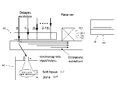
In figure 1 is presented electromagnetic wave excitation and detection
according to the present invention. Reference number 100 refers to first
means 100 for generating by means of electromagnetic waves at least one
mechanical wave at at least one generation location into the skeleton 107
through soft tissue 105. Reference sign 120 in figure 1 refers to
electromagnetic input function performed by the first means 100. Reference
sign 122 in figure 1 refers to electromagnetic output function. In figure 2 is
presented as an example a phase delayed excitation and detection
embodiment according to the present invention. The arrangement comprises
second means 103 for detecting the electromagnetic output. In said
detection is detected by means of electromagnetic waves skeletal vibrations
due to at least one mechanical wave. In a preferred phase delayed
embodiment light beam is guided through an optical fiber where after it is
absorbed to the skeleton and generates for example an ultrasound wave.
Time-delayed excitation is attained by employing a time delay (&) between
trigger signals of for example laser diodes.
Referring to figure 2 the arrangement comprises means 104 for recording
the detected at least one mechanical wave at least one recording location to
form mechanical wave information. Distance of said at least one recording
location from said at least one generation location is known. The
arrangement comprises means 108 for determining skeletal properties based
on at least one recorded signal. Said means 104, 108 are arranged for
example in a computer processor utilizing calculative programs, when
needed. The computer processor 104, 108 is presented schematically in the
figure 2. Wired or wireless data transmission is used between the computer
processor 104, 108 and the first 100 and second 103 means to perform data
transmissions between them. Said means 103, 104, 108 can be utilized also
CA 02851985 2014-04-11
WO 2013/064740 PCT/F12012/051053
7
in other embodiments of the invention than the delayed excitation and
detection embodiment of figure 2.
In a method according to the invention is performed at least one of first and
second method steps, where in the first method step is generated by means
of electromagnetic waves at least one mechanical wave at at least one
generation location into the skeleton 107 through soft tissue 105. In the
second method step is detected by means of electromagnetic waves skeletal
vibrations due to at least one mechanical wave, is recorded the detected at
least one mechanical wave at at least one recording location to form
mechanical wave information, and distance of said at least one recording
location from said at least one generation location is known, and further in
the second method step is determined skeletal properties based on at least
one recorded signal. When whether the first or the second method step is
performed, for example means of mechanical and/or piezomechanical effects
can be utilized together with the first or the second method step and the
first
100 or second means 103, 104, 108 utilized in said first or second method
step. A therapy embodiment according to the invention can be realized by
using the first means 100 according to the first method step.
One preferred arrangement according to the invention comprises means 100
for performing multimodal axial transmission in generation of at least one
mechanical wave by means of electromagnetic waves. The arrangement can
also comprise means 100 for tuning at least one of centre frequency and
pattern of the mechanical wave to facilitate an in vivo excitation of at least
one Lamb mode into the bone. Means 100 preferably comprise
electromagnetic sources, i.e. electromagnetic wave sensors 100, and at least
one processor, which in the preferred embodiment of figure 2 can be
arranged for tuning at least one of centre frequency and pattern of the
mechanical wave by performing phased delay excitation of an array of the
electromagnetic sources 100 to facilitate the in vivo excitation of at least
one
CA 02851985 2014-04-11
WO 2013/064740 PCT/F12012/051053
8
Lamb mode into the bone. Means 100 can be further arranged for optimizing
magnitude of the phase delay by utilizing a feedback based on at least one
of maximization of an amplitude of detected signal and minimization of
bandwidth of detected signal, and means 108 for determining the phase
velocity of the Lamb mode excited on the basis of the magnitude of the
phase delay together with an average distance between the sources in the
array of electromagnetic sources 100.
The second means 103 for detecting (figure 2) by means of electromagnetic
waves skeletal vibrations comprise at least one of a optical interferometer
103, optical coherence tomography device 103 and laser Doppler vibrometer
103, and correspondingly the detection of skeletal vibrations is based on at
least one of optical interferometry, optical coherence tomography and laser
Doppler vibrometry. The optical interferometer 103, optical coherence
tomography device 103 and laser Doppler vibrometer 103 can also be named
as electromagnetic wave sensors 103. The preferred detection of skeletal
vibrations in a bone by the second means 103 is based on the detection of at
least one of first arriving signal (FAS) and Lamb ultrasound modes. One
preferred arrangement comprises means 108 for identifying at least one
Lamb mode in the mechanical wave information, and for determining speed
of the at least one mechanical wave on the basis of the identified at least
one Lamb mode to evaluate at least one property of the skeleton. Also one
preferred arrangement of the invention can comprise means 108 for
mapping bone material properties of the skeleton on the basis of the formed
mechanical wave information.
In figures 3A-3D is presented means 106 for performing positioning
movements of electromagnetic wave sensors 100, 103 by performing at least
one of the following movements: tuning movement of perpendicular
positioning of the electromagnetic wave sensors, movement of adaptive axial
positioning of the electromagnetic wave sensors, movement of tangential
CA 02851985 2014-04-11
WO 2013/064740 PCT/F12012/051053
9
positioning of the electromagnetic wave sensors, movement of azimuthal
positioning of the electromagnetic wave sensors and axial scanning
movement of the electromagnetic wave sensors. Figures 3A-3D are explained
in details later on in this description.
In following description is described in details one of the preferred modes of
the present invention. Photo-acoustic (=PA, later on in this description)
means, i.e. electromagnetic wave sensors, essentially enable flexible tuning
of the excitation and detection which, by a number of ways, can facilitate the
in vivo excitation and detection of Lamb waves in human bones. The idea is
to generate a mode that is strong and easy to recognize at the receiver. This
mode should also be sensitive to at least one clinically relevant property of
bone (e.g. cortical bone thickness, elastic stiffness or bone mineral
density).
Tuning of excitation and/or detection by PA can be comprised of the
following aspects: A. Tuning of optical wavelength (wavelength of the
electromagnetic beam) so as to provide maximal light absorption in the bone
conditioned on minimizing the absorption in the covering soft tissue. The
ultrasonic source (i.e. source of mechanical waves) is thereby generated into
the bone or as close to the bone as possible. B. Tuning of illuminated
surface area so as to provide maximum allowable light intensity on the skin.
C. Tuning of the shape of an illuminated surface so as to produce the
strongest possible targeted mode at the receiver. Optimal shape can be, e.g.,
a sphere, line or crest. D. Tuning of the mechanical (e.g. ultrasonic) centre
frequency of excitation, so as to provide (a) optimal excitability and (2)
sufficient (or optimal) sensitivity to at least one clinically useful property
of
bone. E. Tuning of the magnitude of phase delay in the case of phase
delayed excitation, so as to facilitate selective excitation of one particular
mode.
CA 02851985 2014-04-11
WO 2013/064740 PCT/F12012/051053
While an array of contact ultrasound transducers already enable accurate
assessment of the first arriving signal (FAS) velocity, the following points,
related to excitation, could enhance the FAS measurement. Consider an
array of contact ultrasound sources and two contact ultrasound receivers,
5 one at each end of the source array.
1. Replacement of contact US sources by PA means (optical fibre or
lasers diodes) enables to increase the number of sources due to a
smaller element diameter. The accuracy of ultrasound velocity
10 assessment can thus be increased.
2. Position of the photo-acoustic source or an array of photo-acoustic
sources can easily be scanned, so as to further increase the accuracy
of velocity determination.
3. PA means can enable rapid tuning of the acoustic centre frequency of
excitation, so as to enable assessment of dispersion of the FAS
velocity, successively from rapidly iterated measurements by scanning
the centre frequency. Such dispersion assessment is supposed to
provide a way for FAS based cortical thickness estimation, as an
alternative to the AO Lamb mode.
Excitation and detection of the AO mode can largely be affected by
appropriate tuning of the source 100 and receiver 103. The following
approaches of tuning the excitation can thus be considered.
1. Excitation of interference modes into the soft tissue coating should be
minimized and excitation of a Lamb AO mode in bone should be
maximized.
Ways to minimize the energy excited into the soft tissue coating and
maximize that excited into the AO mode in bone.
2. Proper choice of the optical wavelength to minimize optical absorption
in the soft tissue. The lower the optical absorption the weaker the PA
CA 02851985 2014-04-11
WO 2013/064740 PCT/F12012/051053
11
source is. When the PA source is weak in the soft tissue, energy
excited into interference modes in the soft tissue is also weak.
3. Proper choice of the optical wavelength to minimize optical scattering,
so as to enable sharp beam towards the bone.
4. Proper choice of optical wavelength to maximize the optical absorption
in bone, so as to produce a strong PA source in the bone.
5. Proper tuning of the acoustic excitation frequency, so as to facilitate
the excitation of AO through the soft tissue coating. The AO is most
efficiently excited at very low ultrasonic frequencies, preferably at 20-
120 kHz, however, piezo elements of such frequencies have
inappropriately large diameter for the purpose. PA means enable point
sources at such frequencies.
6. Proper adjustment of the shape and size of area irradiated by the
laser (preferably a narrow line), so as to maximize the amount of PA
energy within the safety limits for the human tissue, but to minimize
the surface area to facilitate the excitation of the AO Lamb mode. The
excitation power is a function of the beam intensity and the surface
area irradiated.
7. Phase delayed excitation by an array of sources can be used to further
facilitate the excitation of AO.
8. Blocking of disturbing the direct propagation path through the soft
tissue coating has shown by initial modeling and in vitro experiments
to reduce the intensities of direct interference modes in the soft
coating, so as to largely facilitate the detection of the AO mode at the
detector on top of the soft coating. To this end, the detector can also
be a traditional contact ultrasound transducer.
CA 02851985 2014-04-11
WO 2013/064740 PCT/F12012/051053
12
Ways to facilitate the detection of AO mode
1. Tuning the optimal sensitivity of the detector to low ultrasonic
frequencies (<120 kHz). This is most optimally done by PA means,
such as a laser interferometer.
2. Implementation of a point or narrow line detector, also enabled by PA
means.
3. Using optical clearing techniques of the soft tissue coating to let the
detector beam penetrate close to the bone surface. (This technique
has shown to be challenging and potentially impossible to implement).
PA (Photo-acoustics) measurements require clamping of the forearm or lower
leg and guiding the source(s) and receiver(s) into an appropriate position
with respect to the bone to be measured. It is a task to design such an
apparatus suitable for clinical measurements.
1. Important features are convenient position adjustments and
appropriate feedback based on the ultrasound signal being measured
during the fine adjustments of the position. The main requirements
are reasonably rapid and reproducible positioning.
Alternatively, the PA source could be packed together with contact US
receivers inside a hand-held probe. Such a design could be implemented by a
laser diode or an array of laser diodes, combined potentially with miniature
translation stage to provide means for scanning of the source position. Such
a setup could provide a potential embodiment for the hybrid device.
2. According to the experience from present US devices, a hand-held
probe together with instant response from the measured signal enable
intuitive positioning.
Alternatively, the PA source could be packed together with one or two PA
receivers inside a hand-held probe, wherein the source is implemented by a
laser diode or an array of laser diodes and the receiver by, e.g., a pair of
CA 02851985 2014-04-11
WO 2013/064740 PCT/F12012/051053
13
interferometric detectors. Such a design could provide a potential
embodiment for the full PA device, suitable for clinical use.
The novel and inventive characters of the invention can be considered to
arise at least from the following few facts:
1. Combination of known photo-acoustic (PA) methods with known
methods of skeletal quantitative ultrasound (QUS), in a way which is
not obvious. Careful choice of several parameters (such as optical
wavelength, beam intensity and dimensions of illuminated area at the
skin, tuning the PA source for optimal acoustic wavelength, and
potentially hampering the propagation of interference modes) is
required to be done simultaneously.
2. PA means enable flexible tuning of the excitation (and detection).
A. Point or point-like (including thin line) sources are enabled also
at very low ultrasonic centre frequencies (f = 20-120 kHz),
which are not possible by piezo ceramic elements of which
physical diameter is large when tuned to such frequencies.
Additionally, PA means also enable implementation of point-like
detectors. The point-like source and receivers are known to be
optimal for facilitating the excitation and detection of the AO
Lamb mode in particular (useful also for other modes),
including that excitability of the AO mode typically increases
with decreasing frequency.
B. Instantaneous tuning of the centre frequency of ultrasonic
excitation by certain PA sources (laser diodes), so as to enable
dispersion assessment of transient ultrasound modes (such as
the FAS). Such tuning of the centre frequency is not possible by
piezo elements (for short transients). Dispersion of the FAS is
sensitive to cortical thickness whereas a FAS measurement at a
CA 02851985 2014-04-11
WO 2013/064740
PCT/F12012/051053
14
fixed frequency is mostly sensitive to elastic stiffness and bone
mineral density.
C. Phase-
delayed excitation to further facilitate the excitation of
ultrasonic modes. Advantages of PA arise from the possibility to
point-like sensor elements, which enable inclusion of several
sensor elements inside a short clinical array probe.
3. Device design which may be of critical importance for the success
with
clinical applications of the method proposed.
The arrangement development according to the one preferred embodiment
will specifically aim at enabling clinically relevant in vivo measurements of
the thickness-sensitive SGW mode (= consistent with Lamb AO). To this end
the specific objectives of the project are:
- To introduce a PA technique for wide-band (low-frequency) and
flexible signal generation in bone.
- To use PA to selectively excite the AO mode as a means to reduce
mode distortion caused by the overlying soft tissue.
- To use PA to remotely image bone surface vibrations from above the
overlying soft tissue.
- To optimize the technique for accurate and fast scanning of the
measured distance long enough for clinical use.
- To optimize by modelling the measuring setup for in vivo
measurements on bone.
- To optimize signal processing for enabling clinical in vivo
measurements.
- To design and construct a portable instrument.
CA 02851985 2014-04-11
WO 2013/064740 PCT/F12012/051053
These objectives will enable clinically relevant multimode (FAS + SGW) in
vivo characterization of osteoporosis, which will be relatively inexpensive
and
which will provide a more complete assessment of bone than has been
possible thus far.
5
Different options of implementation of the PAQUS (photo-acoustic
skeletal quantitative ultrasound) devices will be investigated.
1. Replacement of the source and receiver of the ultrasonic axial
10 transmission scanner (Fig. 1) by non-contact (photo-acoustic; PA) means.
A. Phase 1: Hybrid device - PA source combined with contact
ultrasound detection.
B. Phase 2: Full PA implementation - excitation and detection by
15 PA means.
Enhancement of excitation by using a (PA) phased delay array probe. Direct
assessment of cortical thickness from the specular reflection (pulse-echo
measurement), as implemented by PA means.
The two clinically useful properties of elastic guided waves (Lamb waves) are
thickness-sensitivity and sensitivity to material properties. The latter
depends
on penetration depth and characteristic vibration profile of each specific
mode.
The slow guided wave (SGW or Wave2) is consistent with properties of the
AO Lamb mode. The fast first arriving signal (FAS or Wave1) is an apparent
mode observable in the measured signal and its velocity can be interpreted.
Ranges of optimal thickness-sensitivity of the FAS and SGW can be
interpreted according to the appropriate models.
CA 02851985 2014-04-11
WO 2013/064740 PCT/F12012/051053
16
Influence of the soft overlying tissue is particularly challenging for
excitation
and detection of the SGW (associated to AO) in particular, due to rapid
leakage of the acoustic energy into the surrounding tissue (which causes
rapid attenuation with distance) and characteristic displacement profile
according to which this mode has detectable displacement amplitude in bone
but the amplitude drops rapidly in soft coating, apart from the bone, and is
thus hardly detectable on top of the coating (Viktorov 1967; Yapura and
Kinra, 1995). Moreover, interferences due to other stronger modes in the
coating hamper identification of the weak AO mode (Moilanen et al., 2008).
The choice of long wavelength (low frequency) can, to some extent, reduce
this soft tissue impact. For a particularly long wavelength the AO mode can
have a measurable displacement even on top of the (thin) soft coating. To
this end, frequencies as low as, e.g, 50 kHz can be considered optimal.
Photo-acoustics enables excitation and detection of such low frequencies
while the same would be challenging with piezo-elements due to large
physical dimensions of such transducers. For excitation of the AO mode,
sharp (i.e. mediated onto a small surface area) and strong impulse,
perpendicular to the elastic waveguide, is indeed known to be optimal.
Energy of an optical signal is mediated into the energy of an acoustic signal
(i.e. ultrasound) via photo-acoustic transformation. While this process occurs
due to optical absorption, efficiency of the photo-acoustic transformation is
mainly determined by absorption coefficient, characteristic to each material
and optical wavelength. In addition, penetration depth of the optical beam
plays a role.
For cortical bone these optical parameters are dependent on wavelength.
Cortical bone has highest optical absorption at excitation wavelengths longer
than 1400 nm, where the effective penetration depth into cortical bone is
CA 02851985 2014-04-11
WO 2013/064740 PCT/F12012/051053
17
about 1 mm. Laser excitation at these wavelengths is thus optimal to
generate strongest possible photo-acoustic waves in bone.
Further considerations are needed to mediate the signal through the soft
tissue coating. In general, the soft tissue affects optical absorption and
scattering, and limits thus efficiently the amount of light energy arriving to
bone. For example according to related absorption spectra, absorption is
minimal (and thus optimal) at 600-1100 nm (result for the skin). There is
thus no direct match between the optimal values for the bone and soft tissue
and efficient photo-acoustic excitation is always a tradeoff between
absorption in the soft coating and bone. Therefore, care is needed to the
choice of optimal excitation wavelength.
In above three exemplary cases, excitation at 532 nm will produce the
strongest but smallest PA source which is only located in subsurface of the
soft tissue. Features of a traditional contact ultrasound transducer at the
soft
tissue surface are thus mimicked, with the advantage of tuneable surface
area independent of the excitation frequency which is not possible with piezo
elements. For a piezo element its dimensions are always functions of the
centre frequency. In particular, at low ultrasonic frequencies the physical
size
of a traditional piezo element limits its suitability for the present
application.
The wavelength of 532 nm is optimal for the excitation of FAS in particular,
while measurement of this wave mode has been designed and optimized for
the contact transducers previously (Kilappa et al 2011). Secondly, this
wavelength might due to its small surface size also enable excitation of the
SGW (associated to AO) through a thin soft coating.
Excitation at 1064 nm wavelength will generate the weakest and biggest PA
sources in both soft tissue and bone. Penetration into the bone could enable
excitation of the SGW (associated to AO), while the large size of the source
is
unoptimal for the purpose.
CA 02851985 2014-04-11
WO 2013/064740 PCT/F12012/051053
18
Excitation at 1680 nm wavelength will cause a strong and sharp PA source in
the soft tissue and bone, optimal for excitation of the SGW associated to AO.
Strong absorption in the soft tissue (stronger than that in bone), on the
other hand, may cause adverse interferences between the PA sources in the
soft tissue and bone.
Excitation at 1250 nm can be considered the most optimal wavelength for
producing a strong SGW associated to AO. At this wavelength there is an
absorption peak in bone and the absorption in soft tissue has decreased to
the level comparable with that of bone. A preliminary experimental result
supports the assumption that at a low ultrasonic frequency range the
amplitude spectra excited at 1250 nm wavelength is stronger than that
excited at 1680 nm.
The optical beam can be either focussed onto the skin surface or the area of
optical exposure can be adjusted by masking an unfocussed beam. Direct
focussing of the beam generates a sharp and strong point (or line) source,
which is optimal for excitation of the SGW (associated to AO) in particular.
Intensity of such focussed beam, however, is hard to control accurately and
locally the intensity may easily exceed the limits of safety. Masking of
unfocussed beam is thus focussed a more controlled and safe option, even
though masking cannot generate such optimal point source than focussing.
Sources generated by masking were line sources with the short dimension
(width) along the propagation direction. Values of 1-5 mm were considered
for the width and 5-15 mm for the length of the line source. Advantage of a
larger beam area is mediation of greater amount of energy safely into the
tissue, resulting in a stronger response.
In hybrid version of the photo-acoustic axial transmission scanner, the
source is implemented by non-contact means whereas the receivers are
traditional contact ultrasound transducers. A pair of receivers is used in
order
CA 02851985 2014-04-11
WO 2013/064740 PCT/F12012/051053
19
to enable bidirectional measurement for the accurate correction of soft tissue
effects.
When exciting and detecting ultrasonic signals in bone in vivo by using the
PAQUS hybrid setup, for example the FAS mode can be clearly identified in
the recorded signals.
Excitation of individual Lamb modes (e.g. AO or SO mode) can be facilitated
by phased delay excitation. It has been thus employed a potentially
noncontacting IDT(interdigital transducer)-like excitation to allow efficient
generation of a Lamb mode (e.g. the SO or SO mode). The idea is to
generate a mode that is strong and easy to recognize at the receiver. It
should also be sensitive to at least one clinically relevant property of bone
(e.g. cortical bone thickness, elastic stiffness or bone mineral density). To
do
so we illuminate four spots (e.g. spheres, lines or crests) on the skin that
lie
on the shortest line of sight between the transmission and reception area.
The size of these spots is chosen to provide maximum allowable light
intensity on the skin. Their shape is chosen to so as to produce the strongest
possible targeted mode at the receiver. The inter-spot distance is chosen to
match the time of flight requirement (spatial phase matching) for a targeted
wave mode (e.g. AO along the radius bone at 50 kHz). The centre frequency
of the targeted mode is selected such that it maximizes the amplitude, by
minimizing using feedback the absolute bandwidth, of the received signal.
The optical spectrum of the illuminating laser is chosen such that it provides
an optimal light absorption profile in the bone conditioned on minimizing the
absorption in the covering soft tissue. The temporal profile of each
illuminating pulse and the pulsing pattern onto each illuminated spot is
chosen such as to produce a sonic pattern that generates a strong mode into
the bone. The illumination of the laser spots (temporally and spatially)
should
fulfil the phase matching requirement like in an IDT transducer (which
CA 02851985 2014-04-11
WO 2013/064740 PCT/F12012/051053
depends on sound speed in the bone and on the distance between the
spots).
PA wave will be coupled into human limb by ultrasonic coupling liquid,
5 reflecting at different tissue boundaries. The echoes propagate back into
the
PA sensor and are received by a piezo-detector. As cortical bone has much
higher acoustic impedance than other soft tissues, the echoes at bone ¨ soft
tissue boundaries are much stronger than those reflected from softtissue-soft
tissue boundaries, which are easy to be distinguished. Measuring the time
10 difference of two echoes from bone-soft tissue boundaries, the bone
thickness can be estimated if the acoustic speed in the bone is known.
Finally figures 3A-3D are explained more in detail. Proper positioning of an
ultrasonic probe into the bone is critical for a successful ultrasound
15 measurement. In particular, the transverse and circumferential
directions
with respect to the long axis of bone are important. With a hand-held array
probe the proper positioning can be found intuitively by manual movements
of the probe, using the properties of a measured response signal as a
feedback. The proper anatomical position is typically found within the range
20 of 30 degrees.
In a PAQUS setup, when an external laser unit (or units) 210, i.e. source of
electromagnetic radiation 210 is used through an optical fibre 216, the
degrees of freedom of moving the laser beam(s) are preferably minimized.
Especially, it is challenging to arrange rotation of the laser beam.
Therefore,
it is preferred, that the degrees of freedom required for proper positioning
are arranged by moving the human limb into a proper position, while the
ultrasonic source(s) and detector(s) 103 remain fixed.
To arrange the rotation of a human limb, a possible embodiment includes
two circles 212, 214 of which the outer one 212 is fixed and the inner one
CA 02851985 2014-04-11
WO 2013/064740 PCT/F12012/051053
21
214 has a freedom to rotate. Ultrasonic transducers (PA and conventional
ones) are fixed into (or with respect of) the outer circle. In the hybrid
setup
the transducers include a PA source mediated, e.g., from an external pulse
laser unit, and two conventional contact US receivers. Force sensors are
included with the US receivers to monitor the contact pressure. The receivers
remain fixed, while means are arranged to scan the axial position of the PA
source.
The purpose is to position the mass centre of the cross-section of a bone
(e.g. radius) into the centre point of the circle, and then rotate the bone
into
an appropriate angle. The arm is fixed by specific clamps 218 which have
been mounted via linear units into the inner circle 214. Reference sign 200
refers to support part 200 to a base structure, and reference sign 226 refers
to a crank 226 to move the inner circle 214 in relation to the outer circle.
Reference sign 204 refers to an electromagnetic waves collimator 204.
In the following, the human forearm is used as an example of the human
limb, and radius as an example of a bone to be measured.
Means 224 to move the ultrasonic sensors
1. Means can be provided to move the ultrasonic sensors 103 away while
clamping the forearm, and to return the sensors back to the
measurement position.
2. Means can be provided to fine tune the perpendicular position (x) of
the ultrasonic sensors 103.
Adaptive axial positioning (z-direction)
1. The forearm is fixed into the elbow and wrist clamps.
2. Means are provided to measure the positions of these two clamps.
CA 02851985 2014-04-11
WO 2013/064740 PCT/F12012/051053
22
3. The distance of the two clamps represents the bone length and is
determined from the measured positions.
4. The axial measurement position is determined in a relation to the
bone length.
5. Means 124 are provided to move (by a motor) or to guide the
movement (by signs indicating "forward", "backward" and "hold") of
the forearm into the correct axial position.
6. Axial positions of the two clamps are fixed.
Tangential (x and y) and azimuthal positioning
7. The inner circle is rotated to adjust the azimuthal angle.
8. The tangential positions of the two clamps will be adjusted by four
independent linear units.
9. Positioning is tuned successively by position measurements and using
the measured signals as a feedback. Positioning can be manual or
automated.
Axial scanning
10. Means 224 are provided to successively move the source beam within
a limited range (e.g. 30 mm) between the two receivers, the said
range being symmetric with respect the two sensors 103.
11. Response signals are recorded at the sensors 103 at each position of
the source.
Alternative configurations
12. In the hybrid setup, the source 210 can be replaced by a laser diode
or an array of laser diodes. In this case, an alternative configuration is
CA 02851985 2014-04-11
WO 2013/064740 PCT/F12012/051053
23
possible included that the forearm clamp 218 system is fixed into the
table and the sources and receivers into the inner rotating ring.
Second alternative design includes a hand held array probe in which
case the forearm clamp and positioning mechanism is not necessary.
13. The receivers, ie. sensors 103 can be replaced by PA receivers (e.g.
interferometers) and the source by conventional ultrasonic transducer
or an array of conventional ultrasound transducers.
14. The receivers can be replaced by PA receivers (e.g.
interferometers),
so as to enable a full PA device.
15. The sources and receivers can also act in an imaging mode, or specific
imaging sensors can be included, so as to enable (geometrical)
imaging of the limb based on ultrasonic or PA pulse-echo method
Imaging can provide additional diagnostic information, such as the
profile or map of cortical thickness. Moreover, imaging can be used to
determine the orientation of the bone and position of the mass centre
of the cross section of the bone, according to which one can automate
the positioning of the bone in the mechanism described.
The computer processor 104, 108 is presented schematically in the figures A-
3D. Wired or wireless data transmission is used between the computer
processor 104, 108 and the positioning means 106 described in figures 3A-
3D to perform data transmissions needed between them.
Although the invention has been presented in reference to the attached
figures and specification, the invention is by no means limited to those, as
the invention is subject to variations within the scope allowed for by the
claims.