Note : Les descriptions sont présentées dans la langue officielle dans laquelle elles ont été soumises.
CA 02856571 2015-01-22
TIBIAL BASEPLATE WITH ASYMMETRIC PLACEMENT
OF FIXATION STRUCTURES
FIELD OF THE DISCLOSURE
[0001] The present disclosure relates to orthopaedic prostheses and, more
particularly,
to tibial baseplate components in a knee prosthesis.
BACKGROUND OF THE DISCLOSURE
[0002] Orthopaedic prostheses are commonly utilized to repair and/or
replace
damaged bone and tissue in the human body. For a damaged knee, a knee
prosthesis may be
implanted using a proximal tibial baseplate component, a tibial bearing
component, and a distal
femoral component. The tibial baseplate component is affixed to a proximal end
of the
patient's tibia, which is typically resected to accept the baseplate
component. The femoral
component is implanted on a distal end of the patient's femur, which is also
typically resected
to accept the femoral component. The tibial bearing component is placed
between the tibial
baseplate component and the femoral component, and may be fixed or slidably
coupled to the
tibial baseplate component.
[0003] The tibial baseplate component provides support for the tibial
bearing
component. Forces generated by use of the knee prosthesis are transferred
through the tibial
bearing component to the tibial baseplate component, and ultimately to the
tibia. In order to
ensure long term performance of the knee prosthesis, stable and firm
sccurement of the tibial
baseplate component to the proximal end of the patient's tibia is desired.
SUMMARY
[0004] This application is related to U.S. Non-Provisional Patent
Application
Serial No. 13/593,339, filed August 23, 2012 and to U.S. Non-Provisional
Patent
Application Serial No. 14/278,805, filed May 15, 2014.
1
CA 02856571 2014-05-21
WO 2013/077919
PCT/US2012/052132
[0005] The present disclosure provides an orthopaedic knee prosthesis
including a
tibial baseplate component having a distal, bone-contacting surface with one
or more fixation
structures extending distally therefrom, the fixation structures being
asymmetrically arranged
within the outer periphery of the baseplate.
[0006] For designs utilizing a plurality of fixation pegs that extend
distally from the
bone-contacting surface of the tibial baseplate, fixation pegs are
asymmetrically arranged in
opposite anterior/lateral and posterior/medial regions of the tibial
baseplate, thereby
maximizing distance between the fixation pegs, avoiding overlap with the
intramedullary canal,
avoiding areas of low bone density, and avoiding cortical impingement by
positioning the
fixation pegs in regions of cancellous bone.
[0007] For designs utilizing a single keel that extends distally from the
bone-contacting
surface of the tibial baseplate, the keel is medialized with respect to the
outer periphery of the
tibial baseplate, where the degree of medialization increases as prosthesis
sizes grow
progressively.
[0008] According to an embodiment thereof, the present disclosure provides
a tibial
prosthesis system comprising: a first tibial baseplate comprising: a first
proximal surface; a
first distal surface opposite the first proximal surface, the first distal
surface sized and shaped to
substantially cover a proximal resected surface of a tibia; a first medial
face; a first lateral face
opposite the first medial face; a first total width measured from the first
medial face to the first
lateral face; and a first keel extending distally from the first distal
surface, the first keel spaced
from the first medial face by a first medial distance and spaced apart from
the first lateral face
by a first lateral distance; and a second tibial baseplate comprising: a
second proximal surface;
a second distal surface opposite the second proximal surface, the second
distal surface sized
and shaped to substantially cover a proximal resected surface of a tibia; a
second medial face; a
second lateral face opposite the second medial face; a second total width
measured between the
second medial face and the second lateral face, the second total width
differing from the first
total width whereby the first and second tibial baseplates comprise unique
nominal sizes; and a
second keel extending distally from the second distal surface, the second keel
spaced apart
from the second medial face by a second medial distance and spaced apart from
the second
2
CA 02856571 2014-05-21
WO 2013/077919
PCT/US2012/052132
lateral face by a second lateral distance, a first ratio of the first medial
distance to the first total
width differing from a second ratio of the second medial distance to the
second total width.
[0009] According to another embodiment thereof, the present disclosure
provides a
tibial baseplate configured for implantation upon a patient's proximal tibia,
the tibial baseplate
comprising: a medial compartment; a lateral compartment opposite the medial
compartment; a
proximal surface; a distal surface opposite the proximal surface, the distal
surface sized and
shaped to substantially cover the patient's proximal tibia; an outer periphery
cooperatively
defined by an anterior face, a medial face, a lateral face, and at least one
posterior face; a first,
anterior-posterior axis located between the medial face and the lateral face
and intersecting the
anterior face, the first axis extending centrally between the medial and
lateral compartments
throughout its length; a plurality of fixation pegs extending distally from
the distal surface,
each of the plurality of fixation pegs being positioned inward of the outer
periphery for
implantation into the patient's proximal tibia, the plurality of fixation pegs
comprising: a
medial fixation peg located at the medial compartment; and a lateral fixation
peg located at the
lateral compartment, the lateral fixation peg being positioned more anteriorly
than each other
fixation peg among the plurality of fixation pegs.
[0010] According to yet another embodiment thereof, the present disclosure
provides a
tibial baseplate configured for implantation upon a patient's proximal tibia,
the tibial baseplate
comprising: a medial compartment; a lateral compartment opposite the medial
compartment; a
proximal surface; a distal surface opposite the proximal surface, the distal
surface sized and
shaped to substantially cover the patient's proximal tibia; an outer periphery
cooperatively
defined by an anterior face, a medial face, a lateral face, and at least one
posterior face; at most
one medial fixation peg associated with the medial compartment, the medial
fixation peg
extending distally from the distal surface and positioned for implantation
into the patient's
proximal tibia; and at most one lateral fixation peg associated with the
lateral compartment, the
lateral fixation peg extending distally from the distal surface and positioned
for implantation
into the patient's proximal tibia, the lateral fixation peg being located
closer to the anterior face
than the medial fixation peg.
[0011] According to still another embodiment thereof, the present
disclosure provides
a tibial baseplate configured for implantation upon a patient's proximal
tibia, the tibial
baseplate comprising: a medial compartment; a lateral compartment opposite the
medial
compartment; a proximal surface; a distal surface opposite the proximal
surface, the distal
3
CA 02856571 2014-05-21
WO 2013/077919
PCT/US2012/052132
surface sized and shaped to substantially cover the patient's proximal tibia;
an outer periphery
cooperatively defined by an anterior face, a medial face, a lateral face, and
at least one posterior
face; a first, anterior-posterior axis located between the medial face and the
lateral face and
intersecting the anterior face, the first axis extending centrally between the
medial and lateral
compartments throughout its length; a first fixation peg extending distally
from the distal
surface, the first fixation peg being inset from the outer periphery for
implantation into the
patient's proximal tibia, the first fixation peg being medially spaced from
the first axis by a first
distance; and a second fixation peg extending distally from the distal
surface, the second
fixation peg being inset from the outer periphery for implantation into the
patient's proximal
tibia, the second fixation peg being laterally spaced from the first axis by a
second distance, the
second distance less than the first distance.
BRIEF DESCRIPTION OF THE DRAWINGS
[0012] The above-mentioned and other features and advantages of this
disclosure, and
the manner of attaining them, will become more apparent and the invention
itself will be better
understood by reference to the following description of embodiments of the
invention taken in
conjunction with the accompanying drawings, wherein:
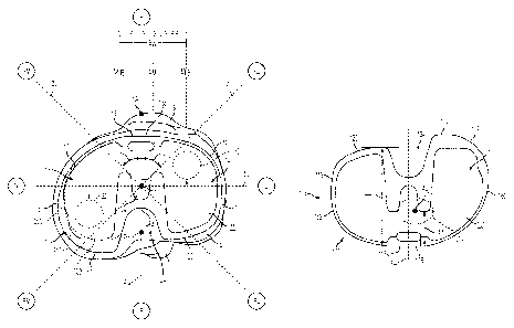
[0013] FIG. 1 is a proximal plan view of a tibial baseplate made in
accordance with the
present disclosure, the baseplate having a lateral fixation peg and a medial
fixation peg, the
baseplate shown implanted upon the resected proximal surface of a patient's
tibia, the baseplate
shown having an asymmetric outer periphery in solid lines and a symmetric
outer periphery in
phantom;
[0014] FIG. 2A is a first, distal plan view of the baseplate of FIG. 1,
showing
medial/lateral positioning of the fixation pegs and the overall medial bias
thereof;
[0015] FIG. 2B is a second, distal plan view of the baseplate of FIG. 1
similar to FIG.
2A, showing anterior/posterior positioning of the fixation pegs;
[0016] FIG. 2C is a third, distal plan view of the baseplate of FIG. 1
similar to FIGS.
2A and 2B, also showing anterior/posterior positioning of the fixation pegs;
[0017] FIG. 3 is a lateral elevational view of the baseplate of FIG. 1;
[0018] FIG. 4 is a distal plan view of an alternative baseplate;
4
CA 02856571 2014-05-21
WO 2013/077919
PCT/US2012/052132
[0019] FIG. 5 is a distal plan view of another alternative baseplate;
[0020] FIG. 6 is a graph illustrating the anterior/posterior positioning of
the fixation
pegs of FIGS. 1-3 across a range of prosthesis sizes;
[0021] FIG. 7 is a posterior perspective view of the baseplate of FIG. 1,
shown with a
tibial bearing component mounted thereon;
[0022] FIG. 8 is a proximal plan view of another tibial baseplate made in
accordance
with the present disclosure, the baseplate having a single fixation keel for
fixation to the
patient's tibia;
[0023] FIG. 9A is an anterior elevational view of the baseplate of FIG. 8;
[0024] FIG. 9B is another anterior elevational view of the baseplate of
FIG. 8;
[0025] FIG. 10 is a graph illustrating the medialization of the fixation
keel of FIGS. 8
and 9 across a range of prosthesis sizes;
[0026] FIG. 11 is another graph illustrating the medialization of the
fixation keel of
FIGS. 8 and 9 across a range of prosthesis sizes;
[0027] FIG. 12 is a graph illustrating the anterior/posterior positioning
of the fixation
keel of FIGS. 8 and 9 across a range of prosthesis sizes; and
[0028] FIG. 13 is a distal plan view of another baseplate similar to the
baseplate shown
in FIGS. 2A-2C, but having another lateral fixation peg and another medial
fixation peg; and
[0029] Fig. 14 is a perspective view of a posterior-stabilized femoral
component in
accordance with the present disclosure.
[0030] Corresponding reference characters indicate corresponding parts
throughout the
several views. The exemplifications set out herein illustrate exemplary
embodiments of the
invention and such exemplifications are not to be construed as limiting the
scope of the
invention in any manner.
CA 02856571 2015-01-22
DETAILED DESCRIPTION
[0031] The present disclosure provides a tibial baseplate component for a
knee
prosthesis including asymmetrically arranged distal fixation structures which
promote secure
and stable long term fixation of the tibial baseplate to a patient's proximal
tibia.
[0032] In order to prepare the tibia and femur for receipt of a knee joint
prosthesis of
the present disclosure, any suitable methods or apparatuses for preparation of
the knee joint
may be used. The surgical procedure may involve, for example, forming an
incision in the
patient's skin near the knee joint, resecting the distal end of the patient's
femur (not shown), and
resecting the proximal end of the patient's tibia T (FIG. 1). Resecting the
proximal end of the
patient's tibia T (FIG. 1), in particular, may involve guiding a saw blade
through an appropriate
cutting guide slot to form a substantially planar resected surface S of tibia
T, as shown in FIG.
1.
[0033] Exemplary surgical procedures and associated surgical instruments
are
disclosed in Zimmer's "LPS-Flex Fixed Bearing Knee, Surgical Technique"
bearing copyright
dates of 2004, 2007 and 2008, "NexGen Complete Knee Solution, Surgical
Technique for the
CR-Flex Fixed Bearing Knee" bearing a copyright date of 2003, "NexGen
Complete Knee
Solution Extramedullary/1ntramedullary Tibial Resector, Surgical Technique"
bearing
copyright dates of 2000, 2008 and 2009, "NexGen Trabecular MetalTM Monoblock
Tibial
Components, Surgical Technique Addendum," bearing copyright dates of 2005 and
2007,
"NexGen Trabecular MetalTM Tibial Tray, Surgical Technique," bearing
copyright dates of
2007 and 2009, and "Trabecular Metal TM Monoblock Tibial Components," bearing
a copyright
date of 2007 (collectively, the "Zimmer Surgical Techniques").
[0034] As used herein, "proximal" refers to a direction generally toward
the torso of a
patient, and "distal" refers to the opposite direction of proximal (i.e., away
from the torso of a
patient). "Anterior" refers to a direction generally toward the front of a
patient or knee, and
"posterior" refers to the opposite direction of anterior (i.e., toward the
back of the patient or
knee). "Lateral" refers to a direction generally away from the middle of the
patient and the
sagittal plane, and "medial" refers to the opposite direction of lateral
(i.e., toward the middle of
the patient and the sagittal plane). When referring to one of the patient's
knees, "lateral" refers
6
CA 02856571 2014-05-21
WO 2013/077919
PCT/US2012/052132
to the direction generally away from the other knee, and "medial" refers to
the direction
generally toward the other knee.
[0035] These anatomical regions are labeled in certain drawings for
clarity. In FIG. 1,
for example, the anterior region of tibia T is labeled "A," the posterior
region of tibia T is
labeled "P," the lateral region of tibia T is labeled "L," and the medial
region of tibia T is
labeled "M." Therebetween and moving in a clock-wise direction, the
anterior/lateral region of
tibia T is labeled "AL," the posterior/lateral region of tibia T is labeled
"PL," the
posterior/medial region of tibia T is labeled "PM," and the anterior/medial
region of tibia T is
labeled "AM." The AL, PL, PM, and AM regions can be described as dividing
tibia T into four
corners or quadrants. These labels are referenced throughout the following
paragraphs.
[0036] The embodiments shown and described herein illustrate components for
a right
knee prosthesis. Right and left knee prosthesis configurations are generally
mirror images of
one another about a sagittal plane. Thus, it will be appreciated that the
aspects of the prosthesis
described herein for a right knee configuration are equally applicable to a
left knee
configuration.
1. Tibial Baseplate
[0037] Referring now to FIG. 1, tibial baseplate 10 is shown disposed atop
a proximal
resected surface S of a patient's tibia T. The upper or proximal surface 11 of
baseplate 10 is
shown in FIG. 1. This proximal surface 11 of baseplate 10 is configured to
receive a tibial
bearing component 53 (FIG. 7) in a fixed or a sliding relationship, for
example. To arrange
baseplate 10 and the tibial bearing component 53 in a fixed relationship, the
tibial bearing
component 53 may be adhered to, mechanically fastened to, molded directly onto
(as discussed
further below), or otherwise fixedly coupled to baseplate 10. The illustrative
baseplate 10
includes a raised rim 13 around proximal surface 11 to receive, surround, and
hold the tibial
bearing component 53 therein, but it is contemplated that other structures may
be provided on
baseplate 10 to receive and hold the tibial bearing component 53 on baseplate
10. In turn, tibial
bearing component 53 is configured to interact with the patient's distal femur
or a prosthetic
femoral component, such as femoral component 70 shown in Fig. 14 and described
below.
[0038] Baseplate 10 may be partially or entirely constructed of a highly
porous
biomaterial. A highly porous biomaterial is useful as a bone substitute and as
cell and tissue
receptive material. A highly porous biomaterial may have a porosity as low as
55%, 65%, or
7
CA 02856571 2015-01-22
75% or as high as 80%, 85%, or 90%. An example of such a material is produced
using
Trabccular MetalTM Technology generally available from Zimmer, Inc., of
Warsaw, Indiana.
Trabecular Metalrm is a trademark of Zimmer, Inc. Such a material may be
formed from a
reticulated vitreous carbon foam substrate which is infiltrated and coated
with a biocompatible
metal, such as tantalum, by a chemical vapor deposition ("CVD") process in the
manner
disclosed in detail in U.S. Patent No. 5,282,861 to Kaplan. In addition to
tantalum, other
metals such as niobium, or alloys of tantalum and niobium with one another or
with
other metals may also be used.
[0039] Generally, the porous tantalum structure includes a large plurality
of ligaments
defining open spaces therebetween, with each ligament generally including a
carbon core
covered by a thin film of metal such as tantalum, for example. The open spaces
between the
ligaments form a matrix of continuous channels having no dead ends, such that
growth of
cancellous bone through the porous tantalum structure is uninhibited. The
porous tantalum
may include up to 75%, 85%, or more void space therein. Thus, porous tantalum
is a
lightweight, strong porous structure which is substantially uniform and
consistent in
composition, and closely resembles the structure of natural cancellous bone,
thereby providing
a matrix into which cancellous bone may grow to provide fixation of baseplate
10 to the
patient's bone.
[0040] The porous tantalum structure may be made in a variety of densities
in order to
selectively tailor the structure for particular applications. In particular,
as discussed in the
above-incorporated U.S. Patent No. 5,282,861, the porous tantalum may be
fabricated to
virtually any desired porosity and pore size, and can thus be matched with the
surrounding
natural bone in order to provide an improved matrix for bone ingrowth and
mineralization.
[0041] Bearing component 53 may be molded directly onto baseplate 10,
specifically
proximal surface 11 of baseplate 10. If baseplate 10 is constructed of a
highly porous
biomaterial, as discussed above, the material that is used to construct
bearing component 53
(e.g., polyethylene) may interdigitate into the pores of baseplate 10 during
the molding process.
The pores may be located at and beneath proximal surface 11 of baseplate 10,
so the resulting
molded bearing component 53 may also be located at and beneath proximal
surface 11 of
baseplate 10. The resulting structure may be a monoblock component having a
strong,
8
CA 02856571 2014-05-21
WO 2013/077919
PCT/US2012/052132
wear-resistant connection between baseplate 10 and bearing component 53,
especially along
proximal surface 11 of baseplate 10.
[0042] Baseplate 10 includes outer periphery 12, which may be visible in a
top plan
view (FIG. 1) or a bottom plan view (FIGS. 2A-2C) with baseplate 10 positioned
in a generally
transverse anatomical plane. As shown in FIG. 1, outer periphery 12 is
cooperatively defined
by anterior face 18, posterior/lateral face 20, posterior/medial face 22, PCL
cutout area 24,
lateral face 62, and medial face 60. Each of these surfaces is described
further below.
[0043] Baseplate 10 also includes lateral compartment 14, medial
compartment 16, and
interior compartment 17 therebetween. Lateral compartment 14 and medial
compartment 16
are separated by an anterior-posterior home axis AH, which is discussed
further below.
Because FIG. 1 is a proximal view of the patient's right tibia T, lateral
compartment 14 of
baseplate 10 is located on the right side of FIG. 1 and medial compartment 16
of baseplate 10 is
located on the left side of FIG. 1.
[0044] With bearing component 53 in place against baseplate 10 (FIG. 7) to
articulate
with adjacent femoral component 70, for example, lateral compartment 14 of
baseplate 10 will
be positioned generally beneath lateral condyle 74 of femoral component 70 to
support and
articulate with lateral condyle 74, and medial compartment 16 of baseplate 10
will be
positioned generally beneath medial condyle 72 of femoral component 70 to
support medial
condyle 72. Tibial bearing component 53 (Fig. 7) may be disposed between
medial and lateral
condyles 72, 74 of femoral component 70 and medial and lateral compartments
16, 14 to
provide a low-friction articular interface, as described below. In the
illustrative embodiment,
femoral component 70 includes cam 76 adapted to articulate with a spine of a
tibial bearing
component, e.g., spine 58 of tibial bearing component 53 (Fig. 7). However, it
is contemplated
that femoral component 70 may omit spine 76 to provide an uninterrupted space
between
medial and lateral condyles 72, 74 in some prosthesis designs.
[0045] Anterior face 18 of the illustrative baseplate 10 is disposed
anteriorly on
periphery 12 of baseplate 10 (i.e., in the A region of tibia T). Anterior face
18 is generally
centrally located between lateral and medial compartments 14, 16. More
specifically, as
shown in FIG. 1, anterior face 18 includes a linear or flat portion 18a that
is generally centrally
located between lateral and medial compartments 14, 16. In this illustrated
embodiment, flat
portion 18a of anterior face 18 defines the anterior-most extent of baseplate
10.
9
CA 02856571 2014-05-21
WO 2013/077919
PCT/US2012/052132
[0046] Posterior/lateral face 20 of the illustrative baseplate 10 is
disposed generally
opposite anterior face 18 in the posterior region of lateral compartment 14
(i.e., near the PL
region of tibia T). Posterior/medial face 22 of the illustrative baseplate 10
is disposed generally
opposite anterior face 18 in the posterior region of medial compartment 16
(i.e., near the PM
region of tibia T). The PCL cutout area 24 is disposed between
posterior/lateral face 20 and
posterior/medial face 22 (i.e., near the P region of tibia T). From both
posterior/lateral face 20
and posterior/medial face 22, the PCL cutout area 24 extends generally
anteriorly until
reaching apex 24a.
[0047] Lateral face 62 of the illustrative baseplate 10 is disposed
laterally of lateral
compartment 14 on periphery 12 of baseplate 10 (i.e., near the L region of
tibia T). Medial face
60 of the illustrative baseplate 10 is located medially of medial compartment
16 on periphery
12 of baseplate 10 (i.e., near the M region of tibia T).
2. Home Axis and Other Reference Axes of Tibial Baseplate
[0048] In the context of patient anatomy, such as tibia T described herein,
"home axis"
AH of tibia T extends anteriorly from a posterior point Pp on tibia T to an
anterior point PA on
tibia T. The posterior point Pp and the anterior point PA of tibia T are
discussed further below.
[0049] The posterior point Pp is generally disposed in the area where the
patient's
posterior cruciate ligament (PCL) attaches to tibia T. More specifically, the
posterior point Pp
is generally disposed at the geometric center of the attachment between the
patient's PCL and
tibia T. The patient's PCL typically attaches to tibia T in two ligament
"bundles," the first
bundle having a more anterolateral attachment location and the second bundle
having a more
posteromedial attachment location. In FIG. 1, the posterior point Pp is shown
at the geometric
center of the first bundle. It is also within the scope of the present
disclosure that the posterior
point Pp may be located at the geometric center of the second bundle or at the
geometric center
of the first and second bundles, together.
[0050] The anterior point PA is disposed on the patient's anterior tibial
tubercle B. In
FIG. 1, the anterior point PA is medially spaced from the tubercle midpoint BM
(at marking 1/2)
by an amount equal to 1/6 of the overall medial/lateral tubercle width Bw
(which spans
between markings 0 and 1). Stated another way, the anterior point PA is
laterally spaced from
the tubercle medial end BmE (at marking 0) by an amount equal to 1/3 of the
overall
CA 02856571 2015-01-22
medial/lateral tubercle width Bw (which spans between markings 0 and 1), such
that the
anterior point PA lies on the "medial third" of the anterior tibial tubercle B
(at marking 1/3).
[0051] In the context of a prosthesis, such as tibial baseplate 10
described herein,
"home axis" AH of baseplate 10 refers to an anterior-posterior extending axis
of baseplate 10
that aligns with home axis AH of tibia T upon implantation of baseplate 10
onto resected
surface S of tibia T in a proper rotational and spatial orientation (as shown
in FIG. 1).
According to an exemplary embodiment of the present disclosure, and as shown
in FIG. 1,
home axis AH of baseplate 10 is centrally located between the inner-most
portion of lateral
compartment 14 and the inner-most portion of medial compartment 16 of
baseplate 10
throughout its length. In other words, home axis AH of baseplate 10 is
equidistant from the
inner-most portion of lateral compartment 14 and the inner-most portion of
medial
compartment 16 of baseplate 10 to divide the interior compartment 17
therebetween into
substantially equal halves.
[0052] In the illustrative embodiment of FIG. 1, home axis AH of baseplate
10 bisects
anterior face 18 of baseplate 10 (which is located anteriorly on periphery 12
of baseplate 10)
and is generally perpendicular to flat portion 18a of anterior surface 18.
Also, home axis AH of
baseplate 10 bisects PCL cutout area 24 of baseplate 10 (which is located
posteriorly on
periphery 12 of baseplate 10) and is generally perpendicular to apex 24a of
PCL cutout area 24.
It is contemplated that home axis AH of baseplate 10 may be oriented to other
features of
baseplate 10, it being understood that proper alignment and orientation of
baseplate 10 upon
resected surface S of tibia T will position home axis AH of baseplate 10
coincident with home
axis AH of tibia T.
[0053] The home axes AH of tibia T and baseplate 10 are further described
in U.S.
Patent Application Publication No. 2012/0022659, filed July 22, 2011, entitled
"ASYMMETRIC TIBIAL COMPONENTS FOR A KNEE PROSTHESIS,".
[0054] A pair of reference axes 26, 28 is presented in FIG. 1. A first
reference axis 26
extends diagonally across baseplate 10 from the back-left PM region of tibia T
to the front-right
AL region of tibia T, intersecting home axis AH to define a first angle a with
home axis AH, as
shown in FIG. 1. A second reference axis 28 extends diagonally across
baseplate 10 and
perpendicularly to the first axis 26 from the back-right PL region of tibia T
to the front-left AM
11
CA 02856571 2015-01-22
region of tibia T, intersecting home axis AH to define a second angle 13 with
home axis An, as
shown in FIG. 1. The first and second angles a and 13 are each approximately
45 degrees such
that, when combined, the first and second angles a and 13 together total
approximately 90
degrees.
[0055] The first and second reference axes 26, 28 illustratively intersect
one another
and home axis AH at a common point X within periphery 12 of baseplate 10.
According to an
exemplary embodiment of the present disclosure, point X is generally centered
within
periphery 12 of baseplate 10 to maximize the aggregated extent of each
reference axis 26, 28
that is located within periphery 12 of baseplate 10 while maintaining the
desired first and
second angles a and13, as discussed above. Point X is illustratively
positioned along home axis
AH between flat portion 18a of anterior face 18 and apex 24a of PCL cutout
area 24.
[0056] Illustratively, a medial-lateral axis 50 also extends through point
X in a
direction perpendicular to home axis AH. Together, the medial-lateral axis 50
(e.g., the x-axis)
and the anterior-posterior home axis AH (e.g., the y-axis) cooperate to define
a component
coordinate system (e.g., an x-y coordinate system) useful for quantifying and
identifying
certain features of baseplate 10.
3. Shape of Outer Periphery of Tibial Baseplate
[0057] According to an exemplary embodiment of the present disclosure, and
as shown
in FIG. 1, baseplate 10 has an asymmetric outer periphery 12. The asymmetric
outer periphery
12 may be designed to closely match the corresponding periphery of resected
surface S of tibia
T. In the illustrated embodiment of FIG. 1, for example, medial compartment 16
is larger than
lateral compartment 14. Medial compartment 16 is wider than lateral
compartment 14, so
medial face 60 is spaced further apart from the anterior-posterior home axis
AH than lateral face
62. Medial compartment 16 is also deeper than lateral compartment 14, so
posterior/medial
face 22 is spaced further apart posteriorly from the medial-lateral axis 50
than posterior/lateral
face 20. For at least these reasons, the outer periphery 12 of baseplate 10 is
asymmetric.
[0058] The asymmetric shape of baseplate 10 is further described in U.S.
Patent
Application Publication No. 2012/0022659, filed July 22, 2011, entitled
"ASYMMETRIC
TIBIAL COMPONENTS FOR A KNEE PROSTHESIS,".
12
CA 02856571 2014-05-21
WO 2013/077919
PCT/US2012/052132
[0059] It is also within the scope of the present disclosure that baseplate
10 may have a
symmetric outer periphery 212, as shown in phantom in FIG. 1. In this
embodiment, lateral
compartment 14 and medial compartment 16 are the same shape and size. Lateral
compartment 14 and medial compartment 16 are the same width, so lateral face
62 and the
modified medial face 260 (shown in phantom) are equidistant from the anterior-
posterior home
axis AH. In this manner, an anterior-posterior axis of symmetry through outer
periphery 212 of
symmetric baseplate 10 may overlay "home axis" AH and may serve as a reference
for lateral
compartment 14, medial compartment 16, lateral face 62, medial face 260,
lateral and fixation
pegs 30, 32 (described below) and other components of baseplate 10. Thus, in
addition to
being centered within interior compartment 17 between lateral compartment 14
and medial
compartment 16 of the symmetric embodiment of baseplate 10, the anterior-
posterior home
axis AH would also be centered between lateral face 62 and the modified medial
face 260
(shown in phantom). Lateral compartment 14 and medial compartment 16 also
define a
common anterior/posterior depth, so posterior/lateral face 20 and the modified
posterior/medial face 222 (shown in phantom) are equidistant from the medial-
lateral axis 50.
Generally, a symmetric outer periphery 212 allows the same baseplate 10 to be
implanted onto
either a patient's right tibia or left tibia.
4. Fixation Pegs
[0060] Referring next to FIGS. 2A-2C and 3, the underside or distal surface
34 of
baseplate 10 is shown. Distal surface 34 is the surface which contacts
resected surface S of
tibia T (FIG. 1) after implantation of baseplate 10. As shown in FIG. 3,
distal surface 34 is
located opposite proximal surface 11. Baseplate 10 includes a plurality of
fixation structures,
illustratively lateral fixation peg 30 and medial fixation peg 32, that extend
distally from distal
surface 34 and into tibia T (FIG. 1).
[0061] Each fixation peg 30, 32 is inset from outer periphery 12 of
baseplate 10. Each
fixation peg 30, 32 may have a minimum inset distance 39 (FIG. 2A) that
exceeds 0 mm, such
as 1 mm, 3 mm, 5 mm, or more, for example. For purposes of the present
disclosure, and as
shown in FIG. 2A, the minimum inset distance 39 is the smallest distance
measured between
outer periphery 12 of baseplate 10 and the outer perimeter of each fixation
peg 30, 32.
[0062] According to an exemplary embodiment of the present disclosure,
fixation pegs
30, 32 of baseplate 10 are constructed of a highly porous biomaterial, such as
the
13
CA 02856571 2015-01-22
above-described porous tantalum material. Distal surface 34 of baseplate 10
may also be
constructed of a highly porous biomaterial. With distal surface 34 of
baseplatc 10 resting
against resected surface S of tibia T and fixation pegs 30, 32 of baseplate 10
extending distally
into tibia T, the highly porous biomaterial may provide a matrix into which
cancellous bone
may grow to provide fixation of baseplate 10 to tibia T.
[0063] As shown in FIG. 3, the illustrative fixation pegs 30, 32 are
hexagonal in
cross-section near distal surface 34 of baseplate 10. As fixation pegs 30, 32
continue extending
distally away from distal surface 34 of baseplate 10, fixation pegs 30, 32
transition to a circular
cross-section. The hexagonal to circular transition of fixation pegs 30, 32 is
also evident in
FIGS. 2A-2C. In FIG. 1, by contrast, each fixation peg 30, 32 is represented
by a phantom
circle to schematically show the general location of each fixation peg 30, 32,
not necessarily
the size or shape of each fixation peg 30, 32. Exemplary fixation pegs 30, 32
are shown at
pages 16-19 of the "Zimmer Tibial Bascplate, Pocket Guide United States
Version,".
[0064] According to an exemplary embodiment of the present disclosure, and
as
discussed further below, lateral and medial fixation pegs 30, 32 are
asymmetrically arranged on
distal surface 34 of baseplate 10. In one exemplary embodiment, fixation pegs
30, 32 are
asymmetrically arranged about the anterior-posterior home axis AH, such that
the
anterior-posterior home axis AH is not an axis of symmetry of fixation pegs
30, 32. In another
embodiment, fixation pegs 30, 32 are asymmetrically arranged about the medial-
lateral axis 50,
such that the medial-lateral axis 50 is not an axis of symmetry of fixation
pegs 30, 32. In yet
another embodiment, fixation pegs 30, 32 are asymmetrically arranged about
both the
anterior-posterior home axis AH and the medial-lateral axis 50, such that
neither the
anterior-posterior home axis AH nor the medial-lateral axis 50 is an axis of
symmetry of
fixation pegs 30, 32.
5. Anterior/Lateral (AL) and Posterior/Medial (PM) Positioning of
Fixation Pegs
100651 Returning now to FIG. 1, lateral fixation peg 30 in lateral
compartment 14 of
baseplate 10 is positioned anteriorly relative to the medial-lateral axis 50
and anteriorly of
medial fixation peg 32. Thus, lateral fixation peg 30 is more generally
positioned in the AL
region of tibia T while being substantially distanced from the PL region of
tibia T. The AL bias
14
CA 02856571 2014-05-21
WO 2013/077919
PCT/US2012/052132
of lateral fixation peg 30 is evident in FIG. 1, because from the center point
X, the first axis 26
extends toward the AL region and approaches or even intersects lateral
fixation peg 30, while
the second axis 28 extends toward the PL region extends further away from
lateral fixation peg
30.
[0066] In the medial compartment 16 of baseplate 10, medial fixation peg 32
is
positioned posteriorly relative to the medial-lateral axis 50 and posteriorly
of lateral fixation
peg 30. Thus, medial fixation peg 32 is more generally positioned in the PM
region of tibia T
while being substantially distanced from the AM region of tibia T. The PM bias
of medial
fixation peg 32 is evident in FIG. 1, because from the center point X, the
first axis 26 extends
toward the PM region and approaches or even intersects medial fixation peg 32,
while the
second axis 28 extends toward the AM region and travels away from medial
fixation peg 32. In
this exemplary embodiment, both fixation pegs 30, 32 are generally positioned
along the same
first reference axis 26 which spans the PM and AL regions.
[0067] An alternative baseplate 10' is shown in FIG. 4 for contrast. Outer
periphery 12'
of the alternative baseplate 10' of FIG. 4 is generally the same as outer
periphery 12 of
baseplate 10 (shown in solid lines in FIG. 1) ¨ both are asymmetric in shape.
However, unlike
fixation pegs 30, 32 of FIGS. 2A-2C, which are located on opposite sides of
the medial-lateral
axis 50, fixation pegs 30', 32' of FIG. 4 are aligned along and intersect with
medial-lateral axis
50'. With respect to baseplate 10', both the anterior-posterior home axis AH'
and the
medial-lateral axis 50' are axes of symmetry for fixation pegs 30', 32', such
that fixation pegs
30', 32' may be said to be symmetrically oriented with respect to the
component coordinate
system.
[0068] Another alternative baseplate 10" is shown in FIG. 5 for contrast.
Outer
periphery 12" of the alternative baseplate 10" of FIG. 5 is generally the same
as outer periphery
212 of baseplate 10 (shown in phantom in FIG. 1) ¨ both are symmetric in
shape. Lateral
compartment 14" of the alternative baseplate 10" is generally the same size
and shape as medial
compartment 16" of the alternative baseplate 10". Therefore, the anterior-
posterior home axis
AH" is an axis of symmetry for outer periphery 12" of baseplate 10". Like
fixation pegs 30', 32'
of FIG. 4, fixation pegs 30", 32" of FIG. 5 are aligned along and intersect
with medial-lateral
axis 50". With respect to baseplate 10", both the anterior-posterior home axis
AH" and the
medial-lateral axis 50" are axes of symmetry for fixation pegs 30", 32", such
that that fixation
CA 02856571 2014-05-21
WO 2013/077919
PCT/US2012/052132
pegs 30", 32" may be said to be symmetrically oriented with respect to the
component
coordinate system.
[0069] Returning again to FIG. 1, the asymmetric positioning of lateral and
medial
fixation pegs 30, 32 near opposite AL and PM corners or quadrants,
respectively, allows
fixation pegs 30, 32 to be widely spaced apart across distal surface 34 of
baseplate 10.
Advantageously, this wide spacing facilitates avoidance of the anatomic
intramedullary canal
of tibia T upon implantation (which may be located near the intersection point
X), particularly
where baseplate 10 is used for a small-stature patient. By avoiding placement
of fixation pegs
30, 32 within the intramedullary canal of tibia T, the associated areas of low
bone density are
avoided and, instead, fixation pegs 30, 32 may be implanted into areas of
higher bone density,
thereby promoting firm and stable long-term fixation of tibial baseplate 10 to
tibia T. If
fixation pegs 30, 32 are constructed of a highly porous biomaterial, as
discussed above, this
firm and stable long-term fixation may be achieved by cancellous bone growth
into the porous
fixation pegs 30, 32. Also advantageously, the wide spacing between fixation
pegs 30, 32
encourages bone ingrowth therebetween. By contrast, if fixation pegs 30, 32
are too close
together, there may not be enough space for bone to grow therebetween.
[0070] Also, the asymmetric arrangement of lateral and medial fixation pegs
30, 32 on
opposite sides of the medial-lateral axis 50 may enhance the torsional
stability of baseplate 10
when implanted upon tibia T (FIG. 1). During normal use, a significant portion
of the forces
generated on baseplate 10 are directed anteriorly or posteriorly. Activities
which primarily
generate such anteriorly-directed or posteriorly-directed forces include
walking, running,
squatting, and climbing stairs, for example. As shown in FIG. 3, such
anteriorly-directed and
posteriorly-directed forces give rise to anterior torsional moments MA and
posterior torsional
moments Mp, respectively, which urge rotation of baseplate 10 anteriorly and
posteriorly about
the medial-lateral axis 50. Having lateral and medial fixation pegs 30, 32
positioned on
opposite sides of the medial-lateral axis 50 (i.e., the axis of rotation), as
illustrated in FIG. 3 and
discussed in detail above, presents greater resistance to such rotation.
[0071] Furthermore, positioning lateral and medial fixation pegs 30, 32 in
the AL and
PM regions of tibia T, rather than the PL and AM regions of tibia T, may avoid
impingement of
pegs 30, 32 on adjacent cortical bone upon implantation of baseplate 10.
Advantageously, the
AL and PM regions of tibia T (where fixation pegs 30, 32 are located) are
typically populated
with substantial areas of cancellous bone, thereby promoting firm and stable
long-term fixation
16
CA 02856571 2014-05-21
WO 2013/077919
PCT/US2012/052132
of tibial baseplate 10 to tibia T and promoting bone ingrowth. By contrast,
the PL and AM
regions of tibia T (where fixation pegs 30, 32 are not located) are typically
populated with
substantial areas of cortical bone. By avoiding the PL and AM regions of tibia
T, the potenital
for impingement of fixation pegs 30, 32 upon cortical bone is minimized.
6. Lateral/Medial Positioning of Fixation Pegs
[0072] Because lateral fixation peg 30 extends from lateral compartment 14
and medial
fixation peg 32 extends from medial compartment 16, as discussed above,
lateral fixation peg
30 can be said to be positioned "more laterally" on distal surface 34 of
baseplate 10 than medial
fixation peg 32. Similarly, medial fixation peg 32 is positioned "more
medially" on distal
surface 34 of baseplate 10 than lateral fixation peg 30. Thus, as shown in
FIG. 2A, fixation
pegs 30, 32 are spaced apart by a medial-lateral separation distance 36. For
purposes of the
present disclosure, the medial-lateral separation distance 36 is measured on
center between
fixation pegs 30, 32 along a direction perpendicular to home axis AH and
parallel to
medial-lateral axis 50 (FIG. 2B). In an exemplary embodiment, the medial-
lateral separation
distance 36 is between 20 mm and 55 mm, with smaller separation distances 36
corresponding
to smaller nominal prosthesis sizes, and larger separation distances 36
corresponding to larger
nominal prosthesis sizes.
[0073] According to an exemplary embodiment of the present disclosure,
lateral
fixation peg 30 and/or medial fixation peg 32 are medially biased in their
respective
compartments 14, 16. In lateral compartment 14, the illustrative lateral
fixation peg 30 is
medially biased toward home axis AH. In medial compartment 16, the
illustrative medial
fixation peg 32 is medially biased away from home axis AH. The medial bias of
fixation pegs
30, 32, is evident in FIG. 2A, for example, where central peg axis 38 (which
is centered along
the medial-lateral separation distance 36 between fixation pegs 30, 32) is
medially biased
toward medial compartment 16 and away from home axis AH. Because central peg
axis 38 is
centered along medial-lateral separation distance 36, central peg axis 38
divides medial-lateral
separation distance 36 into equal halves ¨ one half being located between
lateral fixation peg
30 and central peg axis 38 and the other half being located between medial
fixation peg 32 and
central peg axis 38.
[0074] If fixation pegs 30, 32 were equally spaced apart from home axis AH,
central
peg axis 38 would coincide with home axis AH. However, in FIG. 2A, pegs 30, 32
are not
17
CA 02856571 2014-05-21
WO 2013/077919
PCT/US2012/052132
equally spaced apart from home axis AH. Instead, lateral fixation peg 30 is
located closer to
home axis AH than medial fixation peg 32. As a result, central peg axis 38
between fixation
pegs 30, 32 is medially spaced or offset toward medial compartment 16 and away
from home
axis AH by offset distance 40. Therefore, fixation pegs 30, 32 may be said to
be
asymmetrically, medially biased relative to home axis AH. In an exemplary
embodiment,
offset distance 40 is between 3 mm and 6 mm. Smaller prosthesis sizes may have
smaller
values for offset distance 40, while larger prosthesis sizes may have larger
values for offset
distance 40.
7. Anterior/Posterior Positioning of Fixation Pegs
[0075] As discussed above, lateral fixation peg 30 is positioned relatively
more
anteriorly on distal surface 34 of baseplate 10 than medial fixation peg 32.
Stated differently,
medial fixation peg 32 is positioned relatively more posteriorly on distal
surface 34 of
baseplate 10 than lateral fixation peg 30. Thus, as shown in FIG. 2B, pegs 30,
32 are spaced
apart by an anterior-posterior separation distance 42. For purposes of the
present disclosure,
the anterior-posterior separation distance 42 is measured on center between
fixation pegs 30,
32 along a direction parallel to home axis AH. In an exemplary embodiment, the
anterior-posterior separation distance 42 is between 5 mm and 11 mm, with
smaller separation
distances 42 corresponding to smaller prosthesis sizes, and larger separation
distances 42
corresponding to larger prosthesis sizes.
[0076] The alternative baseplates 10', 10" of FIGS. 4 and 5 are provided
for contrast.
Because lateral and medial fixation pegs 30', 32' of the alternative baseplate
10' of FIG. 4, for
example, are aligned in an anterior-posterior direction, lateral and medial
fixation pegs 30', 32'
lack an anterior-posterior separation distance analogous to the anterior-
posterior separation
distance 42 of FIG. 2B. Or stated differently, lateral and medial fixation
pegs 30', 32' have an
anterior-posterior separation distance equal to 0 mm. Similarly, lateral and
medial fixation
pegs 30", 32" of the alternative baseplate 10" of FIG. 5 are aligned in an
anterior-posterior
direction and, therefore, have an anterior-posterior separation distance equal
to 0 mm.
[0077] Turning now to FIG. 2C, another way of quantifying the
anterior/posterior
asymmetry of fixation pegs 30, 32 is by contrasting their different positions
relative to a
common reference marker. In FIG. 2C, for example, the common reference marker
is flat
portion 18a of anterior face 18 of baseplate 10, with measurements being taken
posteriorly
18
CA 02856571 2015-01-22
therefrom in a direction parallel to home axis AH. Lateral fixation peg 30 is
spaced posteriorly
from anterior face 18 by a relatively smaller lateral peg distance 46, while
medial fixation peg
32 is spaced posteriorly from anterior face 18 by a relatively larger medial
peg distance 48.
The lateral anterior/posterior depth 44 of lateral compartment 14 of baseplate
10 is also shown
being measured from anterior face 18 to posterior/lateral face 20 of baseplate
10, and this
lateral anterior/posterior depth 44 exceeds both peg distances 46, 48.
Similarly, medial
anterior/posterior depth 45 of medial compartment 16 of baseplate 10 is also
shown being
measured from anterior face 18 to posterior/medial face 22 of baseplate 10,
and medial
anterior/posterior depth 45 exceeds both peg distances 46, 48, as well as
lateral
anterior/posterior depth 44. If baseplate 10 had a symmetric outer periphery
212 (shown in
phantom in FIG. 1) instead of the asymmetric outer periphery 12 of FIG. 2C,
lateral depth 44
and medial depth 45 would be the same.
100781 The alternative baseplates 10', 10" of FIGS. 4 and 5 are provided
for contrast.
Because lateral and medial fixation pegs 30', 32' of the alternative baseplate
10' of FIG. 4, for
example, are aligned in an anterior-posterior direction, the lateral peg
distance 46' from anterior
face 18' to lateral fixation peg 30' is the same as the medial peg distance
48' from anterior face
18' to medial fixation peg 32'. The same is also true for lateral peg distance
46" and medial peg
distance 48" of the alternative baseplate 10" of FIG. 5. Because the
alternative baseplate 10' of
FIG. 4 has an asymmetric outer periphery 12', medial depth 45' differs from
lateral depth 44'.
Because the alternative baseplate 10" of FIG. 5 has a symmetric outer
periphery 12", on the
other hand, medial depth 45" is the same as lateral depth 44".
8. Asymmetric Positioning of Fixation Pegs for Set of Prostheses
[0079] Baseplatc 10 may be provided in a kit or set of different prosthesis
sizes. In one
embodiment, nine baseplates 10 are provided in the set, with baseplates 10
growing
progressively in lateral anterior/posterior depth 44 and/or other dimensions,
for example. The
progressive growth of periphery 12 of baseplates 10 across the set or family
of baseplate sizes
is described in detail in U.S. Patent Application Publication No. 2012/0022660
filed July 22,
2011 and entitled ASYMMETRIC TIBIAL COMPONENTS FOR A KNEE PROSTHESIS
(Attorney Docket: Z1M0815-02).
19
CA 02856571 2014-05-21
WO 2013/077919
PCT/US2012/052132
[0080] Referring next to FIG. 6, exemplary peg distances 46, 48 are
graphically
presented for a set of prostheses of different sizes. More specifically,
exemplary peg distances
46, 48 are graphically presented for a set of prostheses having different
lateral depths 44. The
vertical axis of FIG. 6 shows peg distances 46, 48 (in millimeters), while the
horizontal axis of
FIG. 6 shows various lateral depths 44 (also in millimeters) and the
corresponding nominal size
indicator (1-9). The data points located farther to the left represent smaller
lateral depths 44
(and therefore smaller nominal prosthesis sizes), and data points located
farther to the right
represent larger lateral depths 44 (and therefore larger nominal prosthesis
sizes). In accordance
with FIG. 2C, peg distances 46, 48 and lateral depth 44 are measured
posteriorly from flat
portion 18a of anterior face 18.
[0081] For each given prosthesis size (i.e., each discrete value of lateral
depth 44), a
pair of points are presented for lateral and medial peg distances 46, 48,
respectively, with a
space between the pair of points. This space indicates that peg distances 46,
48 are different for
each of the nine given prosthesis sizes. Medial peg distances 48 consistently
exceed the
corresponding lateral peg distances 46 for each of the nine given prosthesis
sizes. For example,
each medial peg distance 48 may exceed the corresponding lateral peg distance
46 by 7 mm to
11 mm. In this manner, each of the given prostheses has anterior/posterior
asymmetry of
fixation pegs 30, 32 with respect to anterior face 18.
[0082] FIG. 6 also demonstrates that, as the prosthesis size increases,
medial peg
distances 48 may increase at a faster rate than lateral peg distances 46. In
the illustrated
embodiment of FIG. 6, medial peg distances 48 increase at a rate (i.e., slope)
of approximately
0.9, while lateral peg distances 46 increase at a rate of approximately 0.6.
As a result, the
difference between medial peg distance 48 and its corresponding lateral peg
distance 46
increases as the prosthesis size increases, causing fixation pegs 30, 32 to
become more and
more spaced apart as the prosthesis size increases.
[0083] With respect to the alternative baseplate 10' of FIG. 4, by
contrast, where the
lateral peg distance 46' is the same as the medial peg distance 48', the peg
distances 46', 48'
would overlap graphically in FIG. 6. Thus, for any given prosthesis size, a
single point
corresponding to both lateral peg distance 46' and medial peg distance 48'
would be presented
in FIG. 6, without a space therebetween. Also, because fixation pegs 30', 32'
of baseplate 10'
are aligned along medial-lateral axis 50', and not forward of or behind medial-
lateral axis 50'
like fixation pegs 30, 32 of baseplate 10, the overlapping peg distances 46',
48' of the
CA 02856571 2014-05-21
WO 2013/077919
PCT/US2012/052132
alternative baseplate 10' would fall somewhere between the spaced-apart peg
distances 46, 48
of FIG. 6. The same result would occur with the overlapping peg distances 46",
48" of the
alternative baseplate 10" of FIG. 5.
[0084] According to an exemplary embodiment of the present disclosure, the
above-described distances, including inset distance 39, medial-lateral
separation distance 36,
offset distance 40, anterior-posterior separation distance 42, lateral peg
distance 46, and medial
peg distance 48, are measured along distal surface 34 of baseplate 10. As a
result, the distances
are measured near the intersection of each peg 30, 32 with distal surface 34
(e.g., near the
proximal end of each peg 30, 32). In embodiments where pegs 30, 32 are
perpendicular to
distal surface 34, the distances could also be measured away from distal
surface 34 (e.g., near
the distal end of each peg 30, 32) without impacting the measurements. In
embodiments where
pegs 30, 32 are canted relative to distal surface 34, however, the
measurements could vary if
taken away from distal surface 34 (e.g., near the distal end of each canted
peg 30, 32).
Therefore, for consistency, the measurements are taken along distal surface 34
of baseplate 10.
9. Force Testing of Asymmetric Fixation Pegs
[0085] A first prosthesis was manufactured, as shown in FIG. 7, by mounting
bearing
component 53 onto baseplate 10, with baseplate 10 having an asymmetric outer
periphery 12
and asymmetrically arranged lateral and medial fixation pegs 30, 32 (FIGS. 1-
3). A second
prosthesis (not shown) was manufactured by mounting a similar bearing
component 53 onto an
alternative baseplate 10', with the alternative baseplate 10' having an
asymmetric outer
periphery 12' but aligned lateral and medial fixation pegs 30', 32' (FIG. 4).
A third prosthesis
(not shown) was manufactured by mounting a similar bearing component 53 onto
another
alternative baseplate 10", with the other alternative baseplate 10" having a
symmetric outer
periphery 12" and aligned lateral and medial fixation pegs 30", 32" (FIG. 5).
[0086] The illustrative bearing component 53 has lateral articular surface
54, medial
articular surface 56, and spine 58 located therebetween. When bearing
component 53 is
assembled onto baseplate 10, as shown in FIG. 7, lateral articular surface 54
of bearing
component 53 aligns with lateral compartment 14 of baseplate 10, medial
articular surface 56
of bearing component 53 aligns with medial compartment 16 of baseplate 10, and
spine 58
aligns with interior compartment 17 (FIG. 1) of baseplate 10. For the first
and second
prostheses, bearing component 53 had a thickness T of 20 mm. For the third
prosthesis,
21
CA 02856571 2015-01-22
bearing component 53 had a thickness T of 17 mm. Bearing component 53 and its
associated articular surfaces 54, 56 are described in detail in :
U.S. Non-Provisional Patent Application Serial No. 13/459,037, filed April 27,
2012;
U.S. Non-Provisional Patent Application Serial No. 14/490,153, filed September
18, 2014;
U.S. Non-Provisional Patent Application Serial No. 13/459,041, filed on April
27, 2012;
U.S. Non-Provisional Patent Application Serial No. 13/459,048 filed on April
27, 2012;
U.S. Non-Provisional Patent Application Serial No. 14/181,033 filed February
14, 2014;
U.S. Non-Provisional Patent Application Serial No. 13/459,056 filed April 27,
2012; and
U.S. Non-Provisional Patent Application Serial No. 14/284,028 filed May 21,
2014,
all entitled "TIBIAL BEARING COMPONENT FOR A KNEE PROSTHESIS
WITH IMPROVED ARTICULAR CHARACTERISTICS".
[0087] As shown in FIG. 7, a lateral compressive force FcL was applied onto
lateral
articular surface 54 of each bearing component 53, and a medial compressive
force Fcm was
applied onto medial articular surface 56 of each bearing component 53. The
compressive
forces FcL, Foil measured 202 N.
[0088] Simultaneously with application of the compressive forces FcL, Fcm,
an
anterior-facing force FAp was applied to the distal/posterior base of spine
58, as shown in FIG.
7. The anterior-facing force FAp measured 725 N for the first and second
prostheses and was
scaled up to 791 N for the third prosthesis to account for the thinner bearing
component 53.
[0089] Forces FcL, FCM, and FAp were designed in magnitude and area of
application to
replicate forces exerted on tibial bearing component 53 by a prosthetic
femoral component,
e.g., femoral component 70, during a kneeling motion. An exemplary femoral
component
22
CA 02856571 2015-01-22
which articulates with tibial bearing component 53 is described in U.S. Patent
Application
Serial No. 13/459,061 filed April 27, 2012 (Attorney Docket No. ZIM0915-07),
and are further
described in U.S. Patent Application Serial No. 13/459,064 filed April 27,
2012 (Attorney Docket
No. ZIM0915-08), and are further described in U.S. Patent Application Serial
No. 13/459,060
filed April 27, 2012 (Attorney Docket No. ZIM0915-09), and are further
described in U.S. Patent
Application Serial No. 13/161,624 filed June 16, 2014 all entitled "FEMORAL
COMPONENT
FOR A KNEE PROSTHESIS WITH IMPROVED ARTICULAR CHARACTERISTICS".
[0090] Finite element analysis was performed on the first, second, and
third prostheses
to evaluate and compare stresses experienced at the interface of baseplates
10, 10', 10" and a
simulated tibial bone that was well fixed to each respective baseplate. Peak
stresses
experienced in the above-described loading scenario were substantially reduced
for the first
baseplate 10 having asymmetrically arranged fixation pegs 30, 32 as compared
to the second
baseplate 10' having aligned fixation pegs 30', 32' and the third bascplatc
10" having aligned
fixation pegs 30", 32". More particularly, a 51% reduction in peak stress was
observed in the
first baseplate 10 as compared to the second baseplate 10', and a 46%
reduction in peak stress
was observed in the first baseplate 10 as compared to the third baseplate 10".
10. Additional Fixation Pegs
100911 In addition to lateral fixation peg 30 described above, lateral
compartment 14 of
tibial baseplate 100 may further include at least one additional lateral
fixation peg 330. As
shown in FIG. 13, the additional lateral fixation peg 330 is substantially
centered within the PL
23
CA 02856571 2014-05-21
WO 2013/077919
PCT/US2012/052132
quadrant. The illustrative lateral fixation peg 330 is positioned
anteriorly/posteriorly between
lateral fixation peg 30 and medial fixation peg 32, such that lateral fixation
peg 30 is the
anterior-most fixation peg on tibial baseplate 100 and medial fixation peg 32
is the
posterior-most fixation peg on tibial baseplate 100. As a result of lateral
fixation peg 30 being
medially biased toward home axis AH, as described above, the illustrative
lateral fixation peg
330 is located laterally outward of lateral fixation peg 30 and is the lateral-
most fixation peg on
tibial baseplate 100.
[0092] In addition to medial fixation peg 32 described above, medial
compartment 16
of tibial baseplate 100 may further include at least one additional medial
fixation peg 332. As
shown in FIG. 13, the additional medial fixation peg 332 is substantially
centered within the
AM quadrant. The illustrative medial fixation peg 332 is positioned
anteriorly/posteriorly
between lateral fixation peg 30 and medial fixation peg 32, such that lateral
fixation peg 30 is
the anterior-most fixation peg on tibial baseplate 100 and medial fixation peg
32 is the
posterior-most fixation peg on tibial baseplate 100. As a result of medial
fixation peg 32 being
medially biased away from home axis AH, as described above, the illustrative
medial fixation
peg 332 is located laterally inward of medial fixation peg 32.
11. Fixation Keel
[0093] Turning to FIGS. 8 and 9, tibial baseplate 100 is provided that is
substantially
similar to baseplate 10 of FIGS. 1-3, except that baseplate 100 includes a
single fixation
structure, illustratively keel 130, that extends distally from distal surface
134 and into tibia T
(FIG. 1). Keel 130 may be monolithically or integrally formed as part of
tibial baseplate 100,
or keel 130 may be separately attachable to distal surface 134 of tibial
baseplate 100.
Structures of baseplate 100 that correspond to structures of baseplate 10 have
corresponding
reference numerals, with the number 100 being added to the reference numerals
of baseplate 10
to arrive at the corresponding reference numerals of baseplate 100, except as
otherwise noted.
[0094] The illustrative keel 130 of FIG. 9A has a cylindrical core 131
defining
longitudinal axis AK (i.e., the axis of the cylinder defined by cylindrical
core 131) and having
two or more fins 133 extending radially outwardly therefrom, the fins being
arranged
symmetrically relative to the cylindrical core 131. More particularly, fins
133 extend along
substantially all of the longitudinal extent PDK (FIG. 9B) of keel 130, as
best shown in FIGS.
9A and 9B , such that fins 133 terminate at or near the distal tip of keel
133. In an exemplary
24
CA 02856571 2015-01-22
embodiment, longitudinal extent PDK of tibial keel cylindrical core 131 may
range from 27 mm
to 48 mm, with smaller nominal sizes of baseplate 100 having relatively lesser
extents PDK and
larger nominal sizes of baseplate 100 having relatively greater extents PDK.
[0095] Keel fins 133 also define keel fin angle 7 with respect to
longitudinal axis AK of
cylindrical core 131 of keel 130. In an exemplary embodiment, keel angle 7 is
equal to between
22 degrees and 27 degrees. Keel fin angle 7 and longitudinal extent
longitudinal extent PDK of
cylindrical core 131 cooperate to define a medial/lateral keel extent MLK
(Fig. 9B) of between
38 mm and 54 mm, with smaller nominal sizes of baseplate 100 having relatively
lesser extents
MLK and larger nominal sizes of baseplate 100 having relatively greater
extents MLK.
Advantageously, this medial/lateral extent MLK defined by fins 133 of keel 130
present high
resistance to rotation of tibial baseplate 100 in vivo, and enhance the
overall strength of
baseplate 100.
[0096] In an exemplary embodiment, keel 130 defines a substantially
cylindrical outer
profile as illustrated in Fig. 9A. Where such cylindrical outer profile is
employed, an
exemplary embodiment of core 131 of keel 130 may maintain an outer diameter
between 14
mm and 16 mm, with such diameter remaining constant across the longitudinal
extent.
However, it is contemplated that core 131 of keel 130 may have a conical,
tapered outer profile,
particularly for small-stature baseplate sizes. The taper angle may be formed,
for example, by
tapering core 131 of keel 130 from a circular outer diameter of 17.1 mm at the
proximal
terminus of keel 130 (i.e., at the junction between keel 130 and distal
surface 134 of tibial
baseplate 100) to a circular diameter of 13.4 mm at the distal terminus of
keel 130. An
exemplary conical keel used in conjunction with a small-stature baseplate size
is disclosed in
U.S. Provisional Patent Application Serial No. 61/592,574 filed January 30,
2012 (Attorney
Docket No. ZIM0919) and in U.S. Provisional Patent Application Serial No.
61/621,374 filed
April 6, 2012 (Attorney Docket No. ZIM0919-01), both entitled ASYMMETRIC
TIBIAL
COMPONENTS FOR A KNEE PROSTHESIS.
[0097] Prior art tibial baseplates include constant-diameter keels in this
diameter range,
such as the Zimmer NexGen Stemmed Tibial Plates and Natural Knee II Modular
Cemented
Tibial Plates. The NexGen Stemmed Tibial Plates and Natural Knee II Modular
Cemented
Tibial Plates are shown at pages 14 and 28, respectively, of the "Zimmer
Tibial Baseplate,
Pocket Guide United States Version,".
CA 02856571 2015-01-22
[0098] In FIG. 8, keel 130 is represented by a phantom oval to show the
general
location of keel 130, not necessarily the size or shape of keel 130. Rather
than being
cylindrical in shape, it is also within the scope of the present disclosure
that core 131 of keel
130 may be conical in shape, with an outer diameter that tapers distally.
[0099] As discussed above, fixation pegs 30, 32 of baseplate 10 (FIGS. 1-3)
may be
designed to interact with cancellous bone surrounding the intramedullary canal
of the patient's
tibia T. To enhance this interaction with the cancellous bone, fixation pegs
30, 32 may be
constructed of a highly porous biomaterial that accepts bone ingrowth. Keel
130 of baseplate
100 (FIGS. 8 and 9), by contrast, may be designed to fit into the
intramedullary canal of the
patient's tibia T. Like fixation pegs 30, 32, keel 130 may also be constructed
of a highly porous
biomaterial that accepts bone ingrowth. Alternatively, rather than achieving
fixation via bone
ingrowth, keel 130 may be constructed of a solid metal that achieves fixation
via a tight
interference fit with the patient's surrounding bone.
[00100] Although keel 130 may be the only fixation structure on baseplate
100, it is also
within the scope of the present disclosure to combine keel 130 with additional
fixation
structures. In one embodiment, keel 130 may be combined with the above-
described fixation
pegs 30, 32 (FIGS. 1-3). On another embodiment, keel 130 may be combined with
sharp
spikes (not shown). Such spikes may be located in the same general areas
discussed above with
respect to fixation pegs 30, 32. However, unlike the blunt-tipped and porous
fixation pegs 30,
32, the spikes may be sharp-tipped to pierce the patient's bone and may be
solid in construction.
The spikes may also have external ribs or barbs to enhance fixation with the
patient's bone.
[00101] Keel 130 may also include a tapered bore (not shown) extending
proximally
into the distal tip of keel 130, designed to mate with a corresponding locking-
taper surface of a
tibial stem extension.
12. Lateral/Medial Positioning of Fixation Keel
[00102] As shown in FIG. 9A, keel 130 is asymmetrically disposed on distal
surface 134
of baseplate 100 with respect to home axis AH. More particularly, the
longitudinal keel axis AK
of keel 130 is biased medially with respect to the vertical plane that
contains home axis AH, i.e.,
26
CA 02856571 2014-05-21
WO 2013/077919
PCT/US2012/052132
keel axis AK is offset toward medial compartment 116 and away from lateral
compartment 114
by offset distance 163. Throughout the following paragraphs, home axis AH and
the vertical
plane that contains home axis AH are used interchangeably.
[00103] According to an exemplary embodiment of the present disclosure,
offset
distance 163 is measured along distal surface 134 of baseplate 100. As a
result, offset distance
163 is measured medially from the intersection of home axis AH and distal
surface 134 to the
intersection of keel axis AK and distal surface 134 (e.g., near the proximal
end of keel 130). In
embodiments where keel axis AK is perpendicular to distal surface 134, offset
distance 163
could also be measured away from distal surface 134 (e.g., near the distal end
of keel 130)
without impacting the measurement. In embodiments where keel axis AK is canted
relative to
distal surface 134, however, the measurement could vary if taken away from
distal surface 134
(e.g., near the distal end of the canted keel 130). Therefore, for
consistency, the measurement
is taken along distal surface 134 of baseplate 100.
[00104] In embodiments where baseplate 100 has a symmetric outer periphery
112, an
anterior-posterior axis of symmetry through outer periphery 112 may be used as
a "home axis"
AH for referencing medial face 160, lateral face 162, keel 130, and other
components of
baseplate 100. This home axis AH would be substantially centered between
medial face 160
and lateral face 162. With keel axis AK being medially offset from the central
home axis AH,
keel axis AK would be positioned closer to medial face 160 than lateral face
162. Thus, medial
distance 164 between keel axis AK and the medial-most portion of medial face
160 would be
less than lateral distance 166 between keel axis AK and the lateral-most
portion of lateral face
162.
[00105] In embodiments where baseplate 100 has an asymmetric outer
periphery 112, as
shown in FIGS. 8 and 9, home axis AH would not constitute an axis of symmetry
and would be
positioned closer to lateral face 162 than medial face 160. Depending on the
degree to which
keel axis AK is medially offset from home axis AH, keel axis AK may still be
positioned closer
to medial face 160 than lateral face 162. Thus, medial distance 164 between
keel axis AK and
the medial-most portion of medial face 160 may be less than lateral distance
166 between keel
axis AK and the lateral-most portion of lateral face 162.
[00106] The degree of medialization of keel 130 may be expressed as a ratio
or a
percentage and may be calculated by dividing the offset distance 163 between
keel axis AK and
27
CA 02856571 2014-05-21
WO 2013/077919
PCT/US2012/052132
home axis AH by the total medial/lateral width of distal surface 134 (i.e.,
medial distance 164
plus lateral distance 166). For baseplate 100 having the dimensions set forth
in Table 1 below,
for example, the degree of medialization would be approximately 6% (calculated
as 5 mm / 88
mm x 100%).
Table 1: Sample Dimensions of a Large-Size Baseplate 100
ens Width
Offset Distance 163 between Keel Axis AK and Home Axis AR 5 6
Medial Distance 164 41 47
Lateral Distance 166 47 53
Total Width (Medial Distance 164 + Lateral Distance 166) 88 N/A
[00107] Advantageously, the medial bias of keel 130 (i.e., the relatively
short medial
distance 164 and the relatively long lateral distance 166) more closely aligns
keel 130 with the
intramedullary canal of the patient's tibia T (FIG. 1). Thus, upon
implantation of baseplate 100
onto the patient's tibia T, keel 130 may be centered or nearly centered within
the intramedullary
canal. In this manner, keel 130 may avoid impinging onto hard, cortical bone
around the
intramedullary canal, thereby promoting firm and stable long-term fixation of
tibial baseplate
100 to tibia T. The medial bias of keel 130 may also be important if it
becomes necessary to
attach a distal stem extension (not shown) to keel 130, such as during a
revision surgical
procedure. In this manner, tibial baseplate 100 may achieve an optimum
metaphyseal fit on
tibia T in the region of keel 130 and diaphyseal fit on tibia T in the region
of the distal stem
extension.
13. Lateral/Medial Positioning of Fixation Keel for Set of Prostheses
[00108] Baseplate 100 may be provided in a kit or set of different
prosthesis sizes. In
one embodiment, nine nominal sizes of baseplate 100 are provided in the set,
with baseplates
100 growing progressively in size.
[0100] According to an exemplary embodiment of the present disclosure, the
degree of
medialization of keel 130 increases as the prostheses in the set grow in size.
Thus, rather than
maintaining a fixed relationship between medial distance 164 and lateral
distance 166 as the
prostheses grow in size, medial distance 164 makes up a smaller and smaller
portion of the total
width as the prostheses grow in size, and lateral distance 166 makes up a
larger and larger
28
CA 02856571 2014-05-21
WO 2013/077919
PCT/US2012/052132
portion of the total width as the prostheses grow in size. Stated differently,
the rate at which
keel 130 moves toward medial face 160 exceeds that rate at which the
prostheses grow in size.
[0101] The dimensions of another sample baseplate 100 are provided in Table
2 below.
Baseplate 100 of Table 2, which has a total width of 58 mm, is smaller than
baseplate 100 of
Table 1 above, which has a total width of 88 mm.
Table 2: Sample Dimensions of a Small-Size Baseplate 100
Offset Distance 163 between Keel Axis AK and Home Axis AH 1 2
Medial Distance 164 29 50
Lateral Distance 166 29 50
Total Width (Medial Distance 164 + Lateral Distance 166) 58 N/A
[0102] As baseplates 100 of the present set grow in size from Table 2 to
Table 1 (i.e.,
from a small nominal size having a 58 mm total width to a large nominal size
having an 88 mm
total width), the degree of medialization of keel 130 increases relative to
home axis AH (from
2% to 6%). Also, as keel 130 moves medially from the small size of Table 2 to
the large size of
Table 1, medial distance 164 makes up a smaller portion of the total width
(from 50% to 47%),
and lateral distance 166 makes up a larger portion of the total width (from
50% to 53%).
[0103] Advantageously, increasing the degree of medialization of keel 130
as baseplate
100 grows in size may better track the position of the intramedullary canal as
the patient's tibia
T (FIG. 1) grows in size. Therefore, keel 130 may be positioned inside the
intramedullary
canal rather than in hard, cortical bone around the intramedullary canal.
[0104] The increasing medialization of keel 130 is presented graphically in
FIG. 10,
where exemplary offset distances 163 between keel axis AK and home axis AH are
shown for a
set of prostheses of different sizes. More specifically, exemplary offset
distances 163 between
keel axis AK and home axis AH are shown for a set of prostheses having
different medial/lateral
widths (i.e., medial distance 164 plus lateral distance 166). The data points
located farther to
the left represent smaller medial/lateral widths (and therefore smaller
prosthesis sizes), and
data points located farther to the right represent larger medial/lateral
widths (and therefore
larger prosthesis sizes). Although adjacent nominal prosthesis sizes may share
the same offset
distance 163 between keel axis AK and home axis AH (compare, for example, the
corresponding offset distances 163 of the size 5 and size 6 implants, shown as
the fifth- and
29
CA 02856571 2014-05-21
WO 2013/077919
PCT/US2012/052132
sixth-from left data points respectively), the overall trend in FIG. 10 is
that offset distance 163
increases as total medial/lateral width increases.
[0105] In a smaller bone, the metaphyseal region of tibia T is more closely
aligned with
the diaphyseal region of tibia T. Therefore, keel 130 may achieve an optimum
metaphyseal
and diaphyseal fit with a relatively small offset distance 163 (e.g., 1 mm, 2
mm). In a larger
bone, by contrast, the metaphyseal region of tibia T is more offset from the
diaphyseal region
of tibia T. Therefore, keel 130 may require a relatively large offset distance
163 (e.g., 4 mm, 5
mm) to achieve an optimum metaphyseal and diaphyseal fit. FIG. 10 presents
exemplary offset
distances 163, but for any given size, offset distance 163 may vary by +/- 0.5
mm, +/- 1.0 mm,
+/- 1.5 mm, or +/- 2.0 mm, for example.
[0106] As discussed above, the degree of medialization of keel 130 may be
expressed
as a percentage by dividing the offset distance 163 between keel axis AK and
home axis AH by
the total medial/lateral width. In FIG. 11, the offset distances 163 from FIG.
10 are shown as
percentages of the total medial/lateral width. The overall trend in FIG. 11 is
that the degree of
medialization of keel 130 increases as medial/lateral width increases. With
respect to a
relatively small nominal size 3 implant, for example, the medial offset of
keel 130 from home
axis AH is 3% of the total medial/lateral implant width. With respect to a
relatively large
nominal size 7 implant, the medial offset of keel 130 from home axis AH is 5%
of the total
medial/lateral implant width.
14. Anterior/Posterior Positioning of Fixation Keel for Set of
Prostheses
[0107] As shown in FIG. 8, the anterior/posterior keel distance 147 may be
measured
posteriorly from flat portion 118a of anterior face 118 to keel axis AK, for
example. The lateral
depth 144 of lateral compartment 114 is also shown being measured posteriorly
from flat
portion 118a of anterior face 118 to posterior/lateral face 120 of baseplate
100 in FIG. 8, and
this lateral depth 144 exceeds keel distance 147.
[0108] According to an exemplary embodiment of the present disclosure, keel
distance
147 is measured along distal surface 134 of baseplate 100. As a result, keel
distance 147 is
measured posteriorly from the intersection of flat portion 118a of anterior
face 118 and distal
surface 134 to the intersection of keel axis AK and distal surface 134 (e.g.,
near the proximal
end of keel 130). In embodiments where keel axis AK is perpendicular to distal
surface 134,
keel distance 147 could also be measured away from distal surface 134 (e.g.,
near the distal end
CA 02856571 2014-05-21
WO 2013/077919
PCT/US2012/052132
of keel 130) without impacting the measurement. In embodiments where keel axis
AK is
canted relative to distal surface 134, however, the measurement could vary if
taken away from
distal surface 134 (e.g., near the distal end of the canted keel 130).
Therefore, for consistency,
the measurement is taken along distal surface 134 of baseplate 100.
[0109] Across a set of different tibial baseplates 100 haying varying
nominal sizes, the
anterior/posterior positioning of keel 130 may vary. In FIG. 12, for example,
exemplary
anterior/posterior keel distances 147 are shown for a set of prostheses of
different sizes. The
overall trend in FIG. 12 is that keel distance 147 increases as lateral depth
144 increases.
Moving keel 130 further and further from anterior face 118 as baseplate 100
increases in size
may avoid anterior cortical bone impingement by keel 130, especially if keel
130 also increases
in size (e.g., diameter, length) along with baseplate 100. FIG. 12 depicts
exemplary keel
distances 147, but for any given size, keel distance 147 may vary by +/- 0.5
mm, +/- 1.0 mm,
+/- 1.5 mm, or +/- 2.0 mm, for example.
15. Proximal Keel Expansion
[0110] As shown in FIG. 9A, the illustrative keel 130 includes a blind
proximal bore
180 therein that is sized to receive a fixation structure, such as a set screw
(not shown), from
proximal surface 111 of baseplate 100. The fixation structure may be used to
attach a tibial
bearing component onto proximal surface 111 of baseplate 100, for example.
[0111] The illustrative bore 180 of FIG. 9A is centered along home axis AH.
However,
because keel axis AK is offset from home axis AH, bore 180 becomes offset in
keel 130. To
ensure that the walls of keel 130 surrounding bore 180 are adequately thick
along the axial
extent of bore 180 (e.g., 1.5 mm), keel 130 may expand radially outwardly
around bore 180 to
form bulge 182.
[0112] As keel 130 becomes more and more offset from home axis AH and bore
180,
bulge 182 may become larger and larger in size. For example, for medium
nominal prosthesis
sizes (e.g., sizes 5 and 6) haying medium offset distances 163 between keel
axis AK and home
axis AH (e.g., 3 mm), bulge 182 may increase the diameter of keel 130 by 0.5
mm. For large
nominal prosthesis sizes (e.g., sizes 7-9) haying large offset distances 163
between keel axis
AK and home axis AH (e.g., 4 mm, 5 mm), bulge 182 may increase the diameter of
keel 130 by
31
CA 02856571 2015-01-22
1.4 mm. For small nominal prosthesis sizes (e.g., sizes 1-4) having small
offset distances 163
between keel axis AK and home axis AH (e.g., 1 mm, 2 mm), bulge 182 may be
excluded.
32