Note : Les descriptions sont présentées dans la langue officielle dans laquelle elles ont été soumises.
CA 02856837 2014-07-11
CARDIAC ACTIVITY VISUALIZATION WITH FREQUENCY DISCRIMINATION
FIELD OF THE INVENTION
The present invention relates generally to electro-
anatomical mapping, and particularly to methods and systems
for visualizing electro-cardio signals.
BACKGROUND OF THE INVENTION
Various techniques are known in the art for spatially
mapping cardiac signals in a heart cavity. For example, U.S.
Patent Application Publication 2011/0190625, whose
disclosure is incorporated herein by reference, describes a
non-contact cardiac mapping method that includes: (i)
inserting a catheter into a heart cavity having an
endocardium surface, the catheter including multiple,
spatially distributed electrodes; (ii) measuring signals at
the catheter electrodes in response to electrical activity
in the heart cavity with the catheter spaced from the
endocardium surface; and (iii) determining physiological
information at multiple locations of the endocardium surface
based on the measured signals and positions of the
electrodes with respect to the endocardium surface. Related
systems and computer programs are also disclosed.
U.S. Patent Application Publication 2009/0306641, whose
disclosure is incorporated herein by reference, describes a
method for providing an electro-anatomical representation of
a patient's heart which includes measuring signals at one or
more electrodes at multiple positions in the patient's heart
cavity over a time period including multiple heart beat
cycles, at least some of the signals being in response to
electrical activity in the patient's heart cavity. An
algorithm is applied to one or more specific signals of the
measured signals to determine a triggering event in the
specific signal. The signals measured at the one or more
electrodes are synchronized by the computer with one another
1
CA 02856837 2014-07-11
according to a heartbeat cycle based on the triggering
event. The electro-anatomical representation of the
patient's heart is generated by the computer based on the
synchronized measured signals and positions of the catheter
electrodes.
SUMMARY OF THE INVENTION
An embodiment of the present invention described herein
provides a method including measuring electrical activity at
multiple points on a surface of a heart of a patient. User
input indicative of a spectral slice selected from a
frequency band is received. Respective levels of the
electrical activity within the selected spectral slice are
calculated. The calculated levels are displayed on a map of
the heart.
In some embodiments, measuring the electrical activity
includes contacting the surface of the heart with a catheter
at the multiple points and measuring electro-cardiac signals
at the respective multiple points using the catheter. In
other embodiments, calculating the levels of the electrical
activity within the selected spectral slice includes
computing a frequency spectrum of the electro-cardiac
signals at the respective multiple points. In yet other
embodiments, displaying the levels includes measuring
respective positions of the catheter while the catheter
touches the points, and displaying the levels at the
respective positions on the map of the heart.
In some embodiments, receiving the user input includes
receiving the selected spectral slice from a slide bar input
device. In other embodiments, displaying the calculated
levels includes assigning the levels respective colors, and
coloring the map of the heart in accordance with the colors.
In yet other embodiments, displaying the levels includes
receiving a digitized three-dimensional image of the heart,
2
CA 02856837 2014-07-11
correlating the calculated levels to the multiple points on
the image, and displaying the map of the correlated
calculated levels and the image on a user display.
There is additionally provided herein, in accordance
with an embodiment of the present invention, an apparatus
including an interface and a processor. The interface is
configured to receive user input indicative of a spectral
slice selected from a frequency band. The processor is
configured to measure electrical activity at multiple points
on a surface of a heart of a patient, to calculate
respective levels of the electrical activity within the
selected spectral slice, and to display the calculated
levels on a map of the heart.
The present invention will be more fully understood
from the following detailed description of the embodiments
thereof, taken together with the drawings in which:
BRIEF DESCRIPTION OF THE DRAWINGS
Fig. 1 is a schematic, pictorial illustration of an
electro-anatomical mapping system, in accordance with an
embodiment of the present invention;
Fig. 2 is a block diagram that schematically
illustrates an electro-cardio signal
frequency
discrimination system, in accordance with an embodiment of
the present invention;
Figs. 3A and 3B are diagrams illustrating a
visualization of electro-cardiac activity on an image of the
heart, in accordance with an embodiment of the present
invention; and
Fig. 4 is a flow chart that schematically illustrates a
method for visualizing electro-cardiac activity with
frequency discrimination, in accordance with an embodiment
of the present invention.
3
CA 02856837 2014-07-11
DETAILED DESCRIPTION OF EMBODIMENTS
OVERVIEW
Patient electro-cardiac signals are sometimes monitored
during therapeutic and cardiac procedures. Electro-cardiac
signals can be sampled locally using catheters which are
navigated into the heart cavity. Electro-anatomical mapping
systems use the local electro-cardiac signals in conjunction
with heart map images, to identify local regions in the
heart where various pathologies may be present. For example,
regions with the high frequency of the local electro-cardiac
signals are indicative with heart tissue associated with
fibrillation and other heart dysfunctions.
Embodiments of the present invention described herein
include improved methods and systems for visualizing
electro-cardiac activity. In the disclosed embodiments, a
frequency discrimination system visualizes the level of
electro-cardiac activity for a particular spectral slice
that is selected by an operator. In this manner, regions of
associated with a particular frequency slice of the electro-
cardiac signal is spatially mapped onto an image of the
heart.
In an embodiment, the operator selects a desired
frequency of the electro-cardiac signal. In response, the
system is configured to display the level of electrical
activity across the heart surface, restricted to that
frequency. Different amplitudes of electrical activity at
the desired frequency may be represented by different
colors, for example. The operator may select the desired
spectral slice using a suitable control in real-time. As the
operator modifies the selected spectral slice, the
visualization changes accordingly. The operator may observe
the change of coloring for different spectral slices, and
4
CA 02856837 2014-07-11
use this form of visualization technique to identify various
heart pathologies.
For example, areas of the heart that are associated
with fibrillation and/or fractionated electrograms may
exhibit a predominance of high-frequency activity, in
comparison with areas of normal electrical activity. Direct
visualization of the frequency distribution of electrical
activity over the area of a heart chamber may therefore be
useful in identifying and planning treatment of arrhythmias.
SYSTEM DESCRIPTION
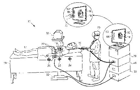
Fig. 1 is a schematic, pictorial illustration of an
electro-anatomical mapping system 10, in accordance with an
embodiment of the present invention. A catheter 15 is
percutaneously inserted into a living body 17 of a patient
lying on a gurney 19. Catheter 15 is connected to an
electro-anatomical mapping and navigation unit (EMNS) 20 in
system 10. Catheter 15 is navigated into a heart 18 of the
patient. An example of a catheter navigation and tracking
system (EMNS 20) is the CARTO system (Biosense Webster,
Diamond Bar, CA).
In an embodiment, one or more electro-cardiac signal
(ECS) probe sensors 22 are attached to the surface of
patient body 17 near heart 18 in order to receive electro-
cardiac signals. Probe sensors 22 are connected to EMNS 20.
The signals acquired by sensors 22 may be used, for example,
for gating the visualization to a particular phase of the
electrocardiogram (ECG) cycle.
One or more magnetic field generators 26 create a
magnetic field through the body of the patient, which induce
signals in position sensors within the distal tip of
catheter 15 (not shown in the diagram). The induced signals
are used by EMNS 20 to track the position of catheter 15 in
heart 18.
5
CA 02856837 2014-07-11
The local electro-cardiac signals are sampled when the
distal tip of catheter 15 locally contacts the heart tissue.
The position of the catheter distal tip during tracking is
displayed to an operator 70 on an output display 60 on a
monitor 50, and recorded along with the local electro-
cardiac signals. The known position of the distal tip of
catheter 15 during sampling of the electro-cardiac signals
enables EMNS 20 to record the electrical activity at the
positions of multiple points on the surface of the heart
cavity in patient 17.
In some embodiments, although not necessarily, an
imaging system (IS) 30 is used to obtain the image of the
heart. Imaging system 30 comprises an imaging source 32,
which may use magnetic resonance imaging (MRI), X-ray
computed tomography (CT), fluoroscopy or any suitable
imaging technique to obtain the heart image. The image of
the heart is then digitized and stored in IS 30.
An electro-cardio signal frequency discrimination
system (ESFDS) 40 receives the digitized heart image in IS
30 and the local electro-cardiac signals obtained from EMNS
20. (In alternative embodiments, IS 30 is omitted, and both
position information and local electrical activity levels
are received from EMNS 20.) ESFDS 40 correlates the heart
image data and the local electro-cardiac signals data at
multiple points on the surface of the heart cavity.
In some embodiments, ESFDS 40 performs a frequency
transformation so as to obtain the frequency spectrum of the
electro-cardiac data at each point on the heart surface.
Thus, the ESFDS forms a three-dimensional (3D) spatial map
of the heart with the local frequency spectrum electro-
cardiac signal at the multiple points.
ESFDS 40 is configured to receive a user input from
operator 70, indicating a particular spectral slice whose
amplitude is to be visualized. Operator terminal 50
6
CA 02856837 2014-07-11
comprises display 60 and a user input device, such as a
touch slide bar 65. The operator can choose the desired
spectral slice (frequency range) of the electro-cardiac
signal by moving his finger on frequency slide bar 65 as
shown in the inset of Fig. 1. ESFDS 40 calculates the
respective levels of the electrical activity within the
selected spectral slice, e.g., the voltage levels of the
electro-cardiac signals. The calculated levels of the
selected spectral slice can be viewed on the map of heart 18
and viewed on display 60 by operator 70. Alternatively to
slide bar 65, ESFDS 40 may use any other suitable control to
receive the spectral slice selection from operator 70.
The exemplary system 10 shown in Figure 1 is for
visual clarity and not by way of limitation of the
embodiments of the present invention. In some embodiments,
system 10 may comprise both imaging system 30 and EMNS 20
which can be operated during the same diagnostic session. In
other embodiments, system 10 may comprise only ESFDS 40 and
EMNS 20 to provide electro-anatomical mapping. Further
alternatively, ESFDS 40 may use imaging data that has been
previously acquired and uploaded to the ESFDS. In yet other
embodiments, system 10 may be used in conjunction with other
therapeutic procedures, for example, where catheter 15 is
also configured to perform cardiac tissue ablation.
Fig. 2 is a block diagram that schematically
illustrates electro-cardio signal frequency discrimination
system 40, in accordance with an embodiment of the present
invention. Local ECS data from electro-anatomical mapping
and navigation unit (EMNS) 20, and heart image map data from
imaging system (IS) 30 are sent to ESFDS 40 via an ESFDS
interface 100. ESFDS interface 100 also receives the
frequency slice selected by operator 70 from user input
device 65 on operator terminal 50 (e.g., touch slide bar
65).
7
CA 02856837 2014-07-11
ESFDS 40 further comprises a processor 110 and a memory
120. Processor 110 receives the local ECS data and the heart
image map, calculates the frequency spectrum of the local
ECS data, and correlates the processed data to the heart
image map. The correlated map of the heart image with the
processed local ECS data is stored in memory 120. Processor
110 also outputs the calculated levels (e.g., voltage
amplitude) of the electro-cardiac signal activity at the
frequency slice set by input device 65. The respective
levels are spatially mapped onto an image of the heart
previously acquired by IS 30 at multiple points along the
surface of the heart and outputted to display 60.
In some embodiments, ESFDS 40 may be a separate unit.
In other embodiments, ESFDS 40 may be integrated within EMNS
20, IS 30, or in any other suitable configuration to perform
the functions described herein. Some elements of ESFDS 40
may be implemented in hardware, e.g., in one or more
Application-Specific Integrated Circuits (ASICs) or Field-
Programmable Gate Arrays (FPGAs). Additionally or
alternatively, some ESFDS elements can be implemented using
software, or using a combination of hardware and software
elements. In some embodiments, processor 110 comprises a
general-purpose computer, which is programmed in software to
carry out the functions described herein. The software may
be downloaded to the computer in electronic form, over a
network, for example, or it may, alternatively or
additionally, be provided and/or stored on non-transitory
tangible media, such as magnetic, optical, or electronic
memory.
ELECTRO-CARDIAC FREQUENCY DISCRIMINATION
The frequency spectrum of local electro-cardio signals
sampled by catheter 15 at multiple points along the surface
of the heart gives an indication of local heart dysfunction.
For example, areas of the heart that exhibit high frequency
8
CA 02856837 2014-07-11
electro-cardiac activity may indicate fibrillation or
fractionated electro-cardiograms in comparison to other
areas of normal electrical activity.
Figs. 3A and 3B are diagrams illustrating electro-
cardiac signal mapping onto an image of the heart, in
accordance with an embodiment of the present invention. In
Fig. 3A, operator 70 decides to view the voltage amplitude
of the electro-cardiac signal in a spectral slice having a
low frequency denoted fa). To do so, operator 70 moves touch
slide bar 65 on display 60 to select low frequency fLO
spectral slice within the frequency band assessable by slide
bar 65. In response, ESFDS 40 displays an image of the heart
and a region 150 of heart 18 with electrical activity at
frequency fLO. Since lower frequency electro-cardio signals
are associated with normal heart function, the electro-
cardiac signals with a low frequency fLO component are
present over most of the surface of the heart cavity as
shown in Fig. 3A.
In order to assess localized heart dysfunction, in Fig.
3B operator 70 selects a spectral slice on slide bar 65 with
a high frequency, fm, a frequency that is known to be
associated with arrhythmias as described previously. In this
case, operator 70 can view a localized damaged region 160 of
heart 18 on display 60.
Slide bar 65 is typically configured to allow operator
70 to choose a spectral slice within a range of frequencies
obtained in the frequency transformation of the electro-
cardiac signal data. The lower and upper edges of the slide
bar are thus configured to be the lowest and highest
frequencies, respectively, from the transformation. In an
embodiment, the lowest and highest frequencies that are
displayed are 0.01 Hz and 300 Hz, respectively. The
9
CA 02856837 2014-07-11
frequency range of 10-25 Hz is typically the band of
interest used for identifying heart dysfunction in
accordance with the embodiments described herein.
In some embodiments, ESFDS 40 assigns respective colors
to the respective levels of the electro-cardiac signal in
the spectral slice (e.g., voltage amplitude). These colors
are then overlaid on the 3D map of the heart on display 60
and viewed by operator 70. In this form of visualization,
regions of high activity at the selected frequency will be
marked with a certain color, while regions of low activity
at the selected frequency will be marked with a different
color. Alternatively, ESFDS 40 may use any other suitable
form of visualization.
Operator 70 may raster his finger on the slide bar to
change the selected frequency quickly in order to view and
observe any changes in position of localized damaged region
160. Changes in the distribution of electrical activity
across the heart surface from one frequency to another can
be a useful diagnostic input.
In some embodiments, system 10 may also comprise ESFDS
40 along with an additional unit to perform therapeutic
procedures, such as ablation therapy. Catheter 15 can be
navigated to region 160 not only to sample the local
electro-cardiac signal but also to ablate the damaged tissue
identified by ESFDS 40. This method restores the heart to
normal function immediately after diagnosis of heart
dysfunction by ESFDS 40 during the same medical procedure.
In some embodiments, the electro-cardio signal voltage
waveforms obtained by EMNS 20 at various points on the
surface of the heart are converted to a frequency spectrum
by processor 110, for example, by the use of Fast Fourier
Transform (FFT) computations. In other embodiments, the
calculated levels (e.g., voltage amplitude) of the frequency
spectrum of the electro-cardiac signals at multiple points
CA 02856837 2014-07-11
along the surface of the heart cavity pre-correlated to the
acquired image of the heart is uploaded and stored in memory
120. The pre-correlated data may be acquired prior to the
diagnostic procedure.
Fig. 4 is a flow chart that schematically illustrates a
method for visualizing cardiac electrical activity, in
accordance with an embodiment of the present invention. In a
storing step 200, electro-cardio signal frequency
discrimination system (ESFDS) 40 stores electro-cardiac
frequency information along with a spatial image mapping at
multiple locations on a surface of heart 18 in memory 120.
In a receiving step 210, ESFDS interface 100 receives a
spectral slice selection from input device 65. The selection
defines the spectral slice of the electro-cardiac signal
frequency band that operator 70 wants to view spatially
mapped onto the image of heart 18 on display 60. In a
displaying step 220, ESFDS 40 displays the amplitude of the
selected input frequency along the spatial image of the
heart based on the mapping obtained from step 200.
Although the embodiments described herein mainly
address using frequency discrimination in cardiac diagnostic
procedures, the methods and systems described herein can
also be used in other applications, such as in electro-
encephalography (EEG).
It will thus be appreciated that the embodiments
described above are cited by way of example, and that the
present invention is not limited to what has been
particularly shown and described hereinabove. Rather, the
scope of the present invention includes both combinations
and sub-combinations of the various features described
hereinabove, as well as variations and modifications thereof
which would occur to persons skilled in the art upon reading
the foregoing description and which are not disclosed in the
prior art. Documents incorporated by reference in the
11
CA 02856837 2014-07-11
present patent application are to be considered an integral
part of the application except that to the extent any terms
are defined in these incorporated documents in a manner that
conflicts with the definitions made explicitly or implicitly
in the present specification, only the definitions in the
present specification should be considered.
12