Note : Les descriptions sont présentées dans la langue officielle dans laquelle elles ont été soumises.
CA 02860645 2016-01-29
APPARATUS AND METHODS FOR TISSUE CLOSURE
BACKGROUND
Field
This relates to tissue closure devices, including surgical suturing devices as
well as
such devices that can be used for intra-abdominal suturing and suturing of
puncture wounds
generated by surgical trocars and other puncturing devices.
Related Art
Minimally invasive methods for conducting surgery on internal organs, tissues,
ligaments and bones usc extremely small instruments such as catheters,
laparoscopes, and the
like. The instruments are introduced using very small incisions, for example
on the order of
five to 18 mm in diameter, into which a trocar or other introducing device is
placed. The
trocars may have a diameter, for example, between 3 mm and 30 mm, with the
smaller
trocars leaving the opening substantially unchanged. The larger trocars may
enlarge the
opening. The trocars provide a reliable and fixed opening for introducing and
removing
various surgical instruments, viewing devices and other instruments used
during the surgical
procedure.
While the incisions and the trocar opening are quite small by traditional
surgical
standards, they still require closure after completing the surgical procedure.
Surgical closure
reduces the possibility of post-surgical infection, post-surgical herniation
(for example in
abdominal surgeries), subsequent bleeding or other effects. Closure can be
accomplished
either through manual suturing or suturing instruments used to complete the
closure. In either
case, suturing is made difficult by the small opening size, for example not
only for
manipulating the suture but also for visualizing the procedure. Closure is
also made more
difficult by the need to suture the subcutaneous tissue, for example fascial
layers, separate
from closure of the overlying skin, and doing so through a very small opening
in the skin.
Conventional closure techniques such as those for closing openings in the
abdominal
wall pass sutures through the abdominal wall tissue a distance from the
original trocar
incision. One or more sutures are then tied off to close the subcutaneous
layer followed by
suitable closure of the skin layer. It has been noted that the distance of the
suture location
from the original incision opening is important in order to secure a suitable
amount of
abdominal wall tissue for forming a reliable closure. If the distance is too
small, the closure
1
may not be enough to reliably close the opening without later complications.
See, for example, US
Patent Publication 20060030868.
Tissue closure devices, for example laparoscopic port closure devices, may be
introduced into
the opening after removal of the trocar device to make easier the suturing of
the trocar opening. Various
methods and structures may help in closing the opening, but may require a
significant number of steps
for completing the closure. Some devices may require a significant amount of
manual care in suturing
the opening and tying off the suture, as well as close visualization for
accomplishing the closure.
Additionally, some devices have a significant number of components or special
devices in order to
accomplish the closure, or they may not provide consistent and reliable
results even under normal
operating circumstances.
SUMMARY
Apparatus and methods are provided that are easy to use for closing a tissue
opening, for
example a trocar opening used in a minimally invasive surgical procedure. One
or more of the examples
of the apparatus and methods described are easy to use and provide a reduced
number of steps to
produce a consistent and reliable closure. The apparatus and methods may be
more simple than
conventional techniques, and can be possibly done without scope visualization
or without the aid of
insufflation of the abdominal cavity (e.g. pneumoperitoneum) under appropriate
circumstances. One or
more of these features can be provided with the apparatus and methods
described herein.
In accordance with one aspect, there is provided a device for use in suturing
tissue, the device
comprising:
a proximal portion for being positioned on a first side of tissue and a distal
portion for being
positioned on a second side of tissue,
wherein at least one of the proximal and distal portions of the device
includes a first structure
defining a first mating component and a second mating component for the first
mating component,
wherein the second mating component includes first and second elements,
wherein at least one of the first and second elements is configured for
receiving a suture,
wherein the second element includes a cartridge and a flexible target
supported by the cartridge,
and wherein the cartridge is configured to be supported by the first element,
and
wherein the first element and the second element have complementary keying
surfaces.
2
CA 2860645 2018-12-17
In accordance with another aspect, there is provided a device for use in
suturing tissue, the
device comprising:
a proximal portion for being positioned on a first side of tissue and a distal
portion for being
positioned on a second side of tissue,
wherein at least one of the proximal and distal portions of the device
includes a first structure
defining a first mating component and a second mating component for the first
mating component,
wherein the second mating component includes first and second elements,
wherein at least one of the first and second elements is configured for
receiving a suture,
wherein the second element includes a cartridge and a flexible target
supported by the cartridge,
and wherein the cartridge is configured to be supported by the first element,
and
wherein the first element includes a first side and a second side and wherein
a cross-section
between the first and second sides defines a dimension that changes between
the first and second
sides.
In accordance with another aspect, there is provided a device for use in
suturing tissue, the
device comprising:
a proximal portion for being positioned on a first side of tissue and a distal
portion for being
positioned on a second side of tissue,
wherein at least one of the proximal and distal portions of the device
includes a first structure
defining a first mating component and a second mating component for the first
mating component,
wherein the second mating component includes first and second elements,
wherein at least one of the first and second elements is configured for
receiving a suture,
wherein the second element includes a cartridge and a flexible target
supported by the cartridge,
and wherein the cartridge is configured to be supported by the first element,
and
wherein the cartridge is formed from a material more rigid than a material of
the flexible target.
2a
CA 2860645 2018-12-17
In accordance with another aspect, there is provided a device for use in
suturing tissue, the
device comprising:
a proximal portion for being positioned on a first side of tissue and a distal
portion for being
positioned on a second side of tissue,
wherein at least one of the proximal and distal portions of the device
includes a first structure
defining a first mating component and a second mating component for the first
mating component,
wherein the second mating component includes first and second elements,
wherein at least one of the first and second elements is configured for
receiving a suture,
wherein the second element includes a cartridge and a flexible target
supported by the cartridge,
and wherein the cartridge is configured to be supported by the first element,
and
wherein the first structure on the first mating component is a movable element
having a cavity
and the second mating component is configured to be insertable into the
cavity.
In one example of apparatus and methods for closing a tissue opening, for
example an
abdominal trocar opening used in surgery, a closure device is used for closing
the tissue opening. A
passageway in the closure device, which in one example may be a trans-lateral
passageway, is used
to guide a needle or other suture carrier along the passageway and through a
tissue layer to be closed.
A surface or barrier formed on the closure device distal of an opening to the
passageway helps to protect
an operator's finger while a suture is being introduced to the passageway in
the body. The surface or
barrier may be a shield, disc, plate guard or other blocking element that can
reduce the possibility of
needle stick when an operator's finger or fingers are placed under the surface
and the introducer is
introduced into the opening from the proximal side of the surface. Where the
closure device has
a single suture introducing opening, the surface can surround the opening with
sufficient
coverage over the operator's fingers without fully encircling the closure
device. Where the closure
device has two or more suture-introducing openings, respective surfaces can
surround the
2b
CA 2860645 2018-12-17
CA 02860645 2014-07-02
WO 2013/103682
PCT/US2013/020094
openings, or a single surface can surround all openings and extend completely
around the
body. In an example where the closure device has an even number of suture-
introducing
openings, the openings may be arranged in pairs, for example on diametrically
opposite sides
of the body. For such openings, the surface can conveniently extend completely
around the
body.
In one example of a closure device having a needle-introducing opening, the
body of
the device may include finger grasping areas for accommodating an operator's
fingers. The
grasping areas may include surfaces complementary to finger curvature, and may
include
surface variations for helping the operator to grasp the body of the closure
device. The
surface variations may be ridges, grooves, knurling, dimples, surface texture
variations or
other surface variations to help the operator reliably grasp the body of the
closure device.
In another example of apparatus and methods for closing a tissue opening, for
example a trocar opening, a closure device with a passageway for receiving a
suture-carrying
needle or other closure device includes a surface or other construction
adjacent the opening
for shielding an operator's fingers from needle sticks. The closure device may
also include a
slot or other longitudinally extending opening extending from a surface of the
body to the
passageway. The slot allows the suture to be disengaged from the body of the
closure device
after the needle or other closure device has carried the suture along the
passageway and
through a tissue layer. The suture can be disengaged from the passageway while
the closure
device is still positioned in the trocar opening or during or after the
closure device is removed
from the trocar opening. In a closure device having a shield for an operator's
fingers, the slot
may also extend into the shield, thereby permitting separation of the suture
from the
passageway over the entire length of the passageway. In one example, the slot
extends
longitudinally of the closure device body, and may also extend partially about
a perimeter of
the body. Where the closure device includes a plurality of suture-introducing
passageways, a
respective number of slots, each corresponding to a passageway, permit
separation of the
suture from the closure device completely from above the shield to below the
shield.
In a further example of a closure device for closing a tissue opening, for
example a
trocar opening, the closure device includes a longitudinally extending body
having a
proximal portion and a distal portion. The distal portion of the closure
device body includes
one or more projections extending at least partly laterally of the body and
having a suture-
receiving target material. The target material is resiliently flexible and
initially has an
unperforated proximal surface. The target surface is also configured to be
capable of being
3
CA 02860645 2014-07-02
WO 2013/103682
PCT/US2013/020094
punctured by a needle or other device suitable for carrying a suture multiple
times in a given
patient in a surgical setting. In one example, the target surface material is
selected so as to
reliably hold a length of suture using friction between the outer wall of the
suture material
and target surface material, for example a size 0 suture, under normal
operating conditions. A
suitable target surface material includes silicone rubber, but may include but
be not limited to
other commonly available biocompatible thermoset or thermoplastic materials
(e.g.
Polyurethane, polyethylene, C-flex, and the like) that may consist of a single
or compounded
materials, suitable to provide the desired friction force to hold and retain
the commonly used
surgical suture materials. The target material may consist of a single layer
material or may be
of several layers, each layer having different characteristics or properties
designed to achieve
the friction necessary to hold the suture in place but at the same time
flexible enough to allow
any needle or other suture carrying device to penetrate. With the described
configuration,
specially-configured target materials are not required.
In an additional example of a closure device for closing a tissue opening, for
example
a trocar opening, the device includes a longitudinally extending body. The
body includes a
proximal portion and a distal portion, the proximal portion being used for
manipulating the
closure device and the distal portion for extending within the trocar opening
and for making
easier the placement and retrieval of one or more sutures. The body further
includes one or
more indicators or markings for visibly indicating proper location or
positioning of the
closure device for optimal operation. For example, a visible marking can be
used to indicate
maximum tissue depth, or maximum depth of the closure device into the tissue.
In another
example, or in addition, a visible marking can be used as visual warning to
the operator that
the patient's soft tissue bed may be too insubstantial in terms of thickness
and, if the indicator
is at or near the skin layer upper surface, that the needle will exit at a
point above the skin
level instead of below it as desired. In another example, or in addition to
one or more of the
other markers, a visible marking can be used to indicate desired closure
device positioning
within a trocar opening relative to a tissue layer to begin or transition the
closure device to
another configuration. For example, the tissue layer may be the peritoneum,
and the visible
marking may be used to indicate that a target element for a suture is
positioned for
deployment. In another example, the visible marking may be used as a visual
warning to the
operator that the patient's soft tissue bed may be too insubstantial in terms
of thickness and, if
the indicator is at or near the skin layer upper surface, that the needle will
exit at a point
above the skin level instead of below it as desired. In a further example, a
visible marking
4
CA 02860645 2014-07-02
WO 2013/103682
PCT/US2013/020094
may be used to indicate when the closure device has been positioned within the
tissue
opening, for example relative to a peritoneum layer, to permit the easiest
retrieval of a suture
that may be held at that time on a part of the closure device.
In a further example of a closure device for closing a tissue opening, for
example a
trocar opening, the device includes a longitudinally extending body having a
proximal
portion and a distal portion. The distal portion includes a suture holding
portion extending a
first distance laterally from a first axial position on the body. The body
further includes a
substantially straight, suture-receiving passageway extending at an angle to a
central axis of a
body. The passageway includes an entrance opening and an exit opening on the
body distal of
the entrance opening and it is substantially aligned with the suture holding
portion. The exit
opening on the body is positioned proximally at a second axial position
relative to the first
axial position and spaced therefrom a second distance. The angle of the
passageway, the
second distance and the first distance are chosen so that a ratio of the
second distance to the
first distance is no more than preferably approximately 2.5:1. For example, if
the suture
holding portion is spaced from the body approximately 1 cm, the distance from
the first axial
position on the body to the exit opening is no more than approximately 2.5 cm.
The ratio can
be less than 2.5:1, but in one example, the first distance between the first
axial position on the
body and the suture holding portion is at least 1 cm. Additionally, if the
first distance between
the first axial position on the body and the suture holding portion is at
least 1 cm, the ratio
can be less than 2.5:1, for example by adjusting the second distance or by
adjusting the angle
of the passageway, or both.
Examples of accessories and their use with closure devices for tissue
openings,
needles or suture introducers can include one or more features for making
their use easier
with closure devices. One feature may include a combination of needle length
and handle
configured such that when the needle is fully introduced into the closure
device, the handle or
other structure on the needle contacts a structure on the closure device
substantially
preventing further introduction of the needle into the closure device, and
therefore
substantially preventing further ingress of the needle into a tissue layer.
Such a configuration
reduces the possibility of unintended needle stick of a tissue layer, organ or
other nearby
surface. Additionally, such a configuration in combination with an
appropriately-designed
closure device for known patient anatomies allows an operator to more
confidently follow the
closure steps in less time and with fewer redundant steps. Alternatively, a
visual marking or
CA 02860645 2014-07-02
WO 2013/103682
PCT/US2013/020094
position indicia provided in the needle structure may also be used as a means
to indicate the
depth of the needle during suture introduction.
In another feature for an accessory and its use, such as that for a needle or
suture
introducer, the needle may include a distal point and a suture-retention
structure proximal of
the needle point. The retention structure may include a groove or undercut
having an entrance
opening where the entrance opening is sized larger than the suture and allows
loading of the
suture into the groove or undercut. The groove or undercut is sized such that
the narrowest
opening includes a maximum spacing slightly less than the suture diameter to
be used with
the needle. The maximum spacing is selected so as to reliably hold the suture
within the
groove when the suture is in a relaxed configuration, and for example under
the weight of
gravity. However, for a greater force such as might be applied manually, the
suture can be
removed from thc groove and out the opening. Such a greater force would be
greater than the
force of gravity on the suture hanging from the groove. In the present
applications for a
needle in use with a closure device, the groove opens distally and has a
groove bottom
proximal of the opening.
A further feature for an accessory and its use, such as that for a needle or
suture
introducer having a groove for releasably retaining a suturc, the groove is
formcd in a side of
the needle proximal of a distal needle tip. The needle includes an indicator
on a side surface
of the needle at a proximal portion of the needle, and the indicator is
positioned
perimetrically with respect to the needle at about the same position as the
groove is located.
The indicator can be a raised, longitudinally-extending the ridge or land, an
arrow, grooves,
unique finger holdings or other configurations indicating the groove position.
The operator
can use the indicator to establish in which radial direction the groove
entrance opening is
facing when the needle tip is not easily visualized, such as when it has
passed through or
beyond a tissue layer. This feature is also beneficial to the operator or
surgical team when
manually loading the suture into the needle groove in an operating room or
suite where the
lighting has been dimmed or is absent.
Accessories for use with closure devices for tissue openings can incorporate
any one
or more of the features described herein with respect to a needle or suture
introducer. The
needle may also include a handle having finger grip surfaces or other manual
assist
configurations for helping the operator manipulate the needle.
The apparatus described herein, as well as other apparatus, can be used in
accordance
with one or more methods for closing tissue openings. In the context of using
one or more
6
CA 02860645 2014-07-02
WO 2013/103682
PCT/US2013/020094
methods with a closure device described herein, such as may be used for
closure of
abdominal trocar openings, the closure device can be inserted into a trocar
opening after the
trocar has been removed. In one example, the closure device can be inserted
until the skin
surface comes adjacent a proximal indicator, for example a circumferential
line about the
closure body. Additionally, where scope-based visualization is available,
clearance of a distal
indicator interior to the peritoneum indicates that the target wings or other
target elements can
be deployed laterally of the closure body. Alternatively, where scope-based
visualization is
not used or available, the skin surface adjacent the proximal indicator will
indicate that the
target wings or other target elements can be deployed laterally for most
patients. Sutures can
then be introduced and closure effected as desired.
The apparatus described herein, as well as other apparatus, can be used in
accordance
with another example of a method for closing tissue openings. Regardless of
whether or not
the foregoing method of positioning a closure device is used, sutures can be
introduced into
tissue layers through a closure device using a suture-carrying introducer, for
example a
needle. The needle can be loaded with a suture manually or automatically by
moving a suture
portion along the shaft of a needle toward a groove opening at a distal
portion of the needle.
The suture portion is moved along the shaft proximal to distal or distal to
proximal until the
suture portion enters the groove opening, for example radially inward from the
shaft outer
surface, after which the suture portion is moved proximally of the needle tip.
The operator
may appreciate a tactile sensation when the suture enters the groove opening
and also observe
it visually depending upon the lighting in the operating room. A slight force
is applied to the
suture portion to overcome a restriction in the groove opening having a
dimension slightly
less than an outer dimension of the suture, after which the suture passes the
restriction and
reaches the bottom of the groove, for example at the proximal-most portion of
the groove.
The retention of the suture portion in the groove can be tested if desired by
directing the
needle point downward with the suture also extending downward by gravity. The
spacing of
the restriction in the groove opening is such that the suture remains within
the groove as the
force necessary to move the suture portion past the groove restriction is
greater than the force
of gravity on the suture.
The suture can then be introduced, as well as other sutures, into the tissue
and closure
effected as desired.
In additional examples, apparatus such as that described herein as well as
other
apparatus can be used in accordance with a further example of a method for
closing tissue
7
CA 02860645 2014-07-02
WO 2013/103682
PCT/US2013/020094
openings. Whether or not any other methods herein of positioning a closure
device or loading
a suture-carrying introducer are used, a closure device inserted into a trocar
opening can be
loaded with one or more sutures without requiring the operator to intervene to
manually or
with additional tools detach a suture from an introducer and attach the suture
to the closure
device. In one example, the closure device is inserted into the trocar opening
with a relatively
high friction target material on a distal portion of the closure device, for
example below the
peritoneum. The target material is high friction relative to the suture
material to be used with
the closure device. Additionally in one example, the target material is un-
perforated prior to
initial use and presents a uniform, un-breached surface facing proximally for
receiving the
suture. The target material is also resiliently flexible, and when a suture
introducer, for
example a suture-carrying needle, breaches the proximal-facing surface and
embeds the
suture in the target material, the friction generated in the target material
grasps and holds the
suture even as the introducer is withdrawn. One exemplary introducer may have
a reduced-
opening groove for loosely holding a suture portion that can be removed by
withdrawing the
introducer from the target material. Other sutures may be introduced and
closure effected as
desired.
In another example of a method for closing a tissue opening, a closure device
may be
preloaded with one or more sutures and introduced into a tissue opening, for
example a trocar
opening. When the closure device is suitably inserted, one or more respective
wings or
suture-holding elements may be deployed substantially laterally of a closure
device body. In
one example, a suture is releasably positioned and held at least 1 cm
laterally away from the
closure device body. In another example, a suture may be held in a resiliently
flexible
material, for example embedded in silicone rubber or other suitable bio-
compatible material
that provides the desired friction properties in conjunction with the suture.
The tissue layer
overlying the suture is then pierced with a retriever element, the suture
retrieved and then
pulled through the tissue layers to a position outside the patient. Similar
steps can be followed
for retrieving any additional sutures, and then closure completed as desired.
Other closure
device configurations are also possible for use in this procedure, for example
one in which
one or more sutures are held by physical restrictions or interference fits
between the suture
and the suture holder (for example on a wing or other suture-holding element).
Additionally,
the closure device can include a guide channel or other retriever guide
defining the path for
the retriever necessary to substantially guarantee contact between the distal
tip of the retriever
and the target suture, for example even without visualization. Such a guide
may be a channel,
8
CA 02860645 2014-07-02
WO 2013/103682
PCT/US2013/020094
such as one extending along a line intersecting the target suture position, or
a trans-lateral
channel extending from one side of the closure device body to the other side
and then
intersecting the target suture position. A plurality of such guides may be
included, for
example for respective suture positions.
In a further example of a method for closing a tissue opening, a closure
device,
including any of those described herein, may be introduced into a tissue
opening, for example
after removal of a trocar. If the closure device has been properly introduced
into the opening,
for example as indicated by a proximal indicator being adjacent a skin surface
or as indicated
by visualization of a distal indicator being inferior to the tissue layer to
be closed (for
example a peritoneum and abdominal fascia), one or more wings or other suture
target
elements can be deployed laterally of the closure device body. The target or
target elements
may be deployed preferably at least 1 cm from the closure device body
(measured
perpendicular to the body). They are deployed by manipulating an actuator rod
relative to the
closure device body, such as through manipulator rings or other grasping
elements. In one
example, the target elements may be locked in an insertion position (un-
deployed) until
unlocked. Once deployed, the target elements can also be locked in the
deployed
configuration, for example through re-engagement of a locking element. When
the closure
device is positioned and configured as desired, the operator can grasp the
body of the closure
device at a body position distal of suture openings. While grasping the body,
a suture
carrying introducer is inserted into an opening and guided through tissue to
be closed and into
a respective target element. In one example, the guide is a trans-lateral
passageway with an
entrance opening above the operator's grasp on the closure device body and an
exit along a
line substantially intersecting the suture target. A closure device may be
used that has a guard
or shield between the operator's gasp on the closure device body and the
suture openings.
In the method described in the foregoing paragraph, the suture may be
introduced
using an introducer with a stop or other element on the introducer that
engages a
corresponding surface on the closure device that prevents further ingress of
the introducer
through the tissue and through the target. The introducer may include grasping
surfaces
and/or alignment indicators for assisting the operator in properly positioning
the suture in the
target element. The introducer can be removed leaving the suture in the target
element, for
example embedded in a relatively high friction, resiliently flexible material,
for example
silicone rubber, and other sutures introduced in a similar manner, if desired.
When the desired
number of sutures have been embedded in their respective targets, a lock or
latch is released
9
CA 02860645 2014-07-02
WO 2013/103682
PCT/US2013/020094
and the suture targets returned to their insertion configuration using the
manipulator rings or
other grasping elements. The closure device can then be withdrawn from the
tissue opening
carrying with it the suture or sutures previously passed through the desired
tissue layer. The
tissue layers may then be closed by securing the sutures, and the overlying
skin layer may
also be closed. In all methods and devices described herein, it is understood
that the devices
and steps taken to achieve the desired result can be repeated for subsequent
closures on the
same patient as required.
These and other examples are set forth more fully below in conjunction with
drawings, a brief description of which follows.
BRIEF DESCRIPTION OF THE DRAWINGS
FIG. 1 is an upper left front isometric view of a tissue closure device
according to onc
example and a suture introducer according to one example inserted into a guide
of the closure
device.
FIG. 2 is a left side elevation view of the assembly shown in FIG. 1 including
a partial
schematic of a tissue layer into an opening in which the closure device may be
inserted.
FIG. 3 is a right side elevation view of the assembly of FIG. 1.
FIG. 4 is a lower right rear isometric view of the assembly of FIG. 1.
FIG. 5 is a bottom plan view of the assembly of FIG. 1.
FIG. 6 is a front elevation view of the closure device of FIG. 1.
FIG. 7 is a left side elevation view of the closure device of FIG. 1.
FIG. 8 is a bottom plan view of the closure device of FIG. 1.
FIG. 9 is a top plan view of the closure device of FIG. 1.
FIG. 10 is a longitudinal cross-section view of the closure device taken along
10-10 of
FIG. 9.
FIG. 11 is a longitudinal cross-sectional view of the closure device taken
along line
11-11 of FIG. 9.
FIG. 12 is a left side elevation view of the closure device of FIG. 1 in an
insertion
configuration.
FIG. 13 is an upper isometric view of a slide unit having finger rings for
changing the
configuration of the closure device of FIG. 1.
FIG. 14 is a top plan view of the slide unit of FIG. 13.
FIG. 15 is a front elevation view of the body of the closure device of FIG. 1.
FIG. 16 is a side elevation view of the body of the closure device of FIG. 1.
CA 02860645 2014-07-02
WO 2013/103682
PCT/US2013/020094
FIG. 17 is a top plan view of the body of the closure device of FIG. 1.
FIG. 18 is a bottom plan view of the closure device of FIG. 1.
FIGS. 19-23 are transverse cross-sectional views of the closure device body
taken
along the respective lines in FIG. 16.
FIG. 24 is an upper isometric view of a suture target wing for the closure
device of
FIG. 1.
FIG. 25 is a bottom plan view of the suture target wing of FIG. 24.
FIG. 26 is a side elevation view of the suture target wing of FIG. 24.
FIG. 27 is a rear elevation view of a suture target wing of FIG. 24.
FIG. 27A is a longitudinal cross section of the target wing of FIG. 27 taken
along line
27A-27A.
FIG. 28 is a lower front isometric view of the suture target wing of FIG. 24.
FIG. 29 is a front isometric view of a needle assembly from the assembly of
FIG. 1.
FIG. 30 is a front plan view of the needle assembly of FIG. 29.
FIG. 31 is a detailed view of the tip of the needle assembly of FIG. 29 taken
at 31-31.
FIG. 32 is an isometric view of another example of a needle assembly that can
be
used with any of the closure devices described herein.
FIG. 33 is a side elevation view of the needle assembly of FIG. 32.
FIGS. 33A-33C our detailed views of a distal portion of the needle assembly of
FIGS.
32-33.
FIG. 34 is a side elevation view of the needle assembly of FIG. 32.
FIG. 35 is another example of a tissue closure device for use with any of the
needle
assemblies and for any of the procedures described herein.
FIG. 36 is an upper isometric view of a further example of a tissue closure
device for
use with any of the needle assemblies and for any of the procedures described
herein.
FIG. 37 is a side elevation view of the tissue closure device of FIG. 36.
FIG. 37A is a transverse cross-section of the tissue closure device of FIG. 36
taken
along line 37A-37A of FIG. 37.
FIG. 37B is a transverse cross-section of the tissue closure device of FIG. 36
taken
along line 37B-37B of FIG. 37.
FIG. 37C is a transverse cross-section of the tissue closure device of FIG. 36
taken
along line 37C-37C of FIG. 37.
FIG. 38 is a top plan view of the tissue closure device of FIG. 36.
11
CA 02860645 2014-07-02
WO 2013/103682
PCT/US2013/020094
FIG. 39 is another side elevation view of the tissue closure device of FIG.
36.
FIG. 40 is a side isometric and exploded view of a suture-receiving wing for
use with
a tissue closure device, including any of the tissue closure devices described
herein.
FIG. 41 is a bottom plan view of the components of FIG. 40.
FIG. 42 is a bottom plan and exploded view of a suture-receiving wing
according to
another example.
FIG. 43 is a side elevation view of assembly of FIG. 42.
FIG. 44 is a right-side elevation of the assembly of FIG. 42.
FIG. 45 is a left upper side isometric view of another example of a suture-
receiving
wing assembly.
FIG. 46 is an upper right isometric view of the assembly of FIG. 45.
FIG. 47 is a lower right isometric view of the assembly of FIG. 45.
FIG. 48 is an upper right isometric and exploded view of another example of a
suture-
receiving wing assembly.
FIG. 49 is a bottom plan view of the assembly of FIG. 48.
FIG. 50 is a rear elevation view of the assembly of FIG. 48.
FIG. 51 is a side elevation view of wing assembly in accordance with another
example described herein.
FIG. 52 is a top plan and exploded view of an example of the wing assembly of
FIG.
51.
FIG. 53 is a side elevation and exploded view of the assembly of FIGS. 52.
FIG. 54 is an isometric and exploded side view of another example of the wing
assembly of FIG. 51.
FIG. 55 is a top plan and exploded view of another example of a wing assembly
of
FIG. 51.
FIG. 56 is a side elevation, partial cutaway and exploded view of the assembly
of
FIG. 55.
FIG. 57 is a top plan and exploded view of another example of a wing assembly
of
FIG. 51.
FIG. 58 is a side elevation and exploded view of the assembly of FIG. 57.
DETAILED DESCRIPTION
This specification taken in conjunction with the drawings sets forth examples
of
apparatus and methods incorporating one or more aspects of the present
inventions in such a
12
CA 02860645 2014-07-02
WO 2013/103682
PCT/US2013/020094
manner that any person skilled in the art can make and use the inventions. The
examples
provide the best modes contemplated for carrying out the inventions, although
it should be
understood that various modifications can be accomplished within the
parameters of the
present inventions.
Examples of closure devices and of methods of making and using the closure
devices
are described. Depending on what feature or features are incorporated in a
given structure or
a given method, benefits can be achieved in the structure or the method. For
example, closure
devices using a finger shield or guard may be easier and safer to use. Closure
devices having
finger gasping surfaces with selected configurations may also help in holding
and using the
closure device. Closure devices having visual indicator marks also make such
closure devices
easier and more reliable to use. Moreover, closure devices having
predetermined suture
delivery configurations may also provide more rcliable and consistent bites of
appropriatc
tissue layers desirable for suturing these layers than conventional
techniques.
Improvements are also provided to components with which the closure devices
may
be used. For example, needles or other suture introducers may make it easier
to use the
closure device where the needle has improved suture holding and release
characteristics.
They may also be easicr to use with needle insertion indicators or stops for
rcducing the
possibility of excessive needle insertion. Indicators on the needles may also
be used with
suture holding grooves in the needle tip to indicate the relative orientation
of the suture
holding groove when such grooves are not directly visible to the operator.
These and other benefits will become more apparent with consideration of the
description of the examples herein. However, it should be understood that not
all of the
benefits or features discussed with respect to a particular example must be
incorporated into a
closure device, component or method in order to achieve one or more benefits
contemplated
by these examples. Additionally, it should be understood that features of the
examples can be
incorporated into a closure device, component or method to achieve some
measure of a given
benefit even though the benefit may not be optimal compared to other possible
configurations. For example, one or more benefits may not be optimized for a
given
configuration in order to achieve cost reductions, efficiencies or for other
reasons known to
the person settling on a particular product configuration or method.
Examples of a number of closure device configurations and of methods of making
and
using the closure devices are described herein, and some have particular
benefits in being
used together. However, even though these apparatus and methods are considered
together at
13
CA 02860645 2014-07-02
WO 2013/103682
PCT/US2013/020094
this point, there is no requirement that they be combined, used together, or
that one
component or method be used with any other component or method, or
combination.
Additionally, it will be understood that a given component or method could be
combined
with other structures or methods not expressly discussed herein while still
achieving desirable
results.
Closure devices for trocar openings are used as examples of a closure device
that can
incorporate one or more of the features and derive some of the benefits
described herein, and
in particular closure devices for abdominal tissue openings. Closure of trocar
openings in
abdominal walls present particular issues for acceptable results, and closure
devices for
abdominal openings will be considered in more detail. However, closure devices
other than
for abdominal wound closures can benefit from one or more of the present
inventions.
It should be understood that terminology used for orientation, such as front,
rear, side,
left and right, upper and lower, and the like, are used herein merely for ease
of understanding
and reference, and are not used as exclusive terms for the structures being
described and
illustrated.
In accordance with one example of apparatus that can be used for closing a
tissue
opening, for example a trocar opening in the abdominal wall, and where the
apparatus reflects
one or more methods that can be used for tissue closure, a closure device and
needle
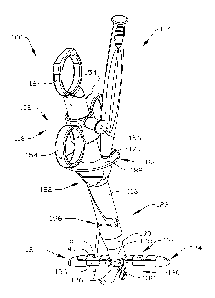
assembly 100 (FIGS. 1-5) includes a closure assembly or closure device 102 and
a needle
assembly 104. The closure device 102 can be used with the needle assembly 104
as discussed
herein, or with other suture introducers or needles, and the needle assembly
104 as discussed
herein can be used with other closure devices. However, for purposes of some
of the
examples, the closure device 102 and the needle assembly 104 will be
considered as being
used together. Additionally, the present discussion for the application of the
closure assembly
will be in the context of closure of an abdominal opening, but it should be
understood that
other tissue closures can be carried out with one or more of the components of
the assembly.
In the context of a trocar opening 106 (FIG. 2) in an abdominal wall 108, the
opening
106 extends through a skin and superficial layer 110 that may include muscle,
depending on
the location in the abdomen at which the opening is made. The skin and
superficial layer 110
will be referred to as the skin layer 110 for simplicity. Underlying the skin
layer 110 is a
fascial layer 112 having a thin peritoneum 114 (not separately shown for
simplicity). The
peritoneum forms the lining of the abdominal cavity outside the internal
organs (not shown),
and it is through the skin layer 110, fascial layer 112 and peritoneum 114
that the trocar
14
CA 02860645 2014-07-02
WO 2013/103682
PCT/US2013/020094
opening and trocar permit access for an operator to the internal organs. Once
the surgery is
complete, the trocar opening is closed by closing the fascial layer 112 and
peritoneal layer
114, while taking care to avoid puncturing or injuring any underlying organs.
One way to
minimize puncturing underlying organs during the closure process is to retract
the tissue
layers away from the underlying organs and to limit or carefully control the
ingress of suture
introducers or retrievers beyond the tissue wall (peritoneal layer), for
example in the manner
described more fully below.
The closure device 102 in the present example includes a closure body 116
(FIGS. 1-
5). The body extends from a proximal portion 118 to a distal portion 120.
Generally, the
proximal portion 118 is used to control and manipulate the closure device, and
the distal
portion 120 forms a working structure to be inserted under the peritoneal
layer. The distal
portion 120 in thc present examples is used to present a target at a known and
predetermined
location where a suture can be reliably placed or retrieved, for example even
without
visualization, and in such a way that suture bites can be made at optimal
locations for
forming reliable closures. For example, the distal portion 120 can be used as
a target for
inserting one or more sutures through the fascial layer 112 and into the
target, and in another
example, the distal portion 120 can be used as a target for inserting a
retrieval tool through
the fascial layer 112 to the target for retrieving a pre-disposed suture
portion from the target
and withdrawing the suture through the fascial layer 112 and a tissue opening
106 to help in
closing the opening.
The closure device 102 also includes an intermediate or middle portion 122,
which
will be generally considered that portion of the closure body 116 residing
within the opening
106 during normal use. The middle portion 122 generally will extend between
the outer
surface of the skin layer 110 and the peritoneal layer 114. The middle portion
122 includes at
least one element that helps to reliably and repeatably place a suture
introducer or retriever at
the predetermined target site without the operator having to substantially
adjust or vary the
direction of movement of the introducer or the retriever. In the present
examples, as discussed
more fully below, the at least one element in the middle portion 122 that
helps to reliably and
repeatably place a suture introducer or retriever at the predetermined target
site is a channel
or passageway, for example a trans lateral passageway, through the body 116 of
the closure
device.
Considering the closure device 102 in more detail, the distal portion 120 in
the present
example includes a plurality of suture-receiving elements 124. The elements
124 may be
CA 02860645 2014-07-02
WO 2013/103682
PCT/US2013/020094
wings that form targets for a suture introducer such as the needle assembly
104. The wings
124 extend outwardly in substantially opposite directions from the closure
body 116 in the
deployed configuration shown in FIGS. 1-5. They are substantially 180 apart
and extend
substantially perpendicular to a central axis of the closure body. In other
examples, the
closure device could have a single wing or plural wings, whether arranged in
pairs or
otherwise. When an-anged in pairs, they can be arranged in two, four, six or
more pairs, as
desired.
The wings 124 (see also FIGS. 24-28) are pivotally mounted to respective
portions of
a mounting structure 126 at the distal end of the closure body 116. The wings
124 are
mounted at opposite sides of a channel or groove 128 (FIG. 15) disposed along
the central
axis of the closure body, so that the wings can pivot simultaneously between
the opened or
deployed configuration shown in FIGS. 1-5 and a closed or insertion
configuration shown in
FIG. 12. The wings 124 are linked to and operated through a pull rod 130
(FIGS. 1-3)
through respective link arms or expanders 132. Pull rod 130 extends upward
into and is
substantially centered on the central axis of the closure body 116 for
longitudinal movement
within the body. Upward movement of the pull rod 130 pulls the link arms or
expanders
upward to move the wings 124 from a collapsed or insertion configuration shown
in FIG. 12
to the expanded or deployed configuration shown in FIGS. 1-5. Downward
movement of the
pull rod 130 within the body 116 fold the link arms 132 down relative to the
body, thereby
pulling the wings 124 downward to the closed configuration. Alternatively,
pull rod 130 may
be directly linked or engaged to the wings 124, eliminating the link arms or
expanders 132.
Upward movement of the pull rod 130 actuates the wings 124 from a collapsed or
insertion
configuration shown in FIG. 12 to the expanded or deployed configuration shown
in FIGS. 1-
5. Downward movement of the pull rod 130 within the body 116 actuates the
wings 124 to a
closed configuration or geometry capable of atraumatic insertion into the
body.
Suitable precision machining, injection molding, casting or other such forming
of the
wings, link arms 132 and pull rod 130, and their positioning and mounting to
or within the
closure body 116 allows accurate positioning of the wings 124 when in the
deployed
configuration shown in FIGS. 1-5. Therefore, when they are in the deployed
configuration,
the wing positions are accurately and reliably known relative to other points
on the closure
device. Likewise, the position of any point on the wings 124 is also
accurately and reliably
known relative to any other point on the closure device. Therefore, with
suitable precision
16
CA 02860645 2014-07-02
WO 2013/103682
PCT/US2013/020094
machining or forming of components on the closure device, the precise location
of any point
on a wing 124 is known and can be used as a target for inserting or receiving
a suture.
Each wing 124 includes a predetermined target location 134 (FIG. 1). The
target
location can be used for reliably receiving a suture portion, for example
through an
introducer, or for reliably retrieving a suture portion previously placed at
the target location.
In the present examples, each wing 124 includes a target element 136 securely
positioned in
the target area 134 of the wing, and partly underneath a target approach
opening 138 formed
in a top surface 140 of the wing. In the present examples where the
introducers or retrievers
approach the target opening at an angle, the target approach opening 138 is
also formed with
a central axis at an angle, substantially parallel or conforming to the angle
of approach of the
introducer or retriever. The target approach opening 138 includes a wall 142
(FIG. 10) shown
as extending substantially parallel to the central axis 142A of the opening.
However, the
walls can be conical or other selected shape or cross-sectional configuration.
The walls are
also shown as circular cylindrical, but they can have other configurations as
well.
The size and shape of the target approach opening 138 can be determined based
on a
number of considerations. When used in conjunction with a suture introducer,
these
considerations may include the suture introducer diameter, the flcxibility of
the introducer,
the travel length of the introducer from an exit port on the closure body 116
to the opening
138, and the desired tolerance between the expected range of motion of the tip
of the
introducer and the minimum opening cross-sectional configuration.
The target element 136 in the present example is a structure having
substantially the
same shape as a cavity 143 in the wing 124 and sized sufficiently so that the
target element is
reliably retained in the cavity. Alternatively, rather than being pre-cut to
size and then
assembled, the target element can be insert molded directly into the cavity
143 and can fill
the opening 138. The target element 136 is formed from a material sufficiently
soft that the
suture introducer can traverse and embed the suture in the material. As shown
in FIGS. 1-5,
the material also is sufficiently soft so that the tip of the introducer can
pass through the
material. The target element is positioned so as to be substantially centered
under the target
approach opening 138.
The target element includes a proximally-facing surface 144 (FIG. 10) with
which the
suture introducer comes into contact. In the present example, the surface 144
is initially
unperforated, and lacks any openings, slits, slices or other breaks in the
surface to ease the
penetration of the introducer past the surface. For situations where multiple
closures are
17
CA 02860645 2014-07-02
WO 2013/103682
PCT/US2013/020094
required in the same surgical setting and patient, the surface 144 can be used
multiple times
as long as the target element 136 is able to maintain the embedded suture. The
target element
for the surface and a substantial portion of the material below the surface is
substantially
resiliently flexible. In one configuration, the target element is formed from
a material that has
a sufficiently high coefficient of friction relative to the suture material so
as to suitably retain
the embedded suture portion during normal operation. For example, it is
desirable to ensure
that the target element substantially retains the suture all the time while
the wings are
collapsed and the closure device is being withdrawn from the trocar opening
106. The suture
portion can then be cut from the closure device or more simply manually pulled
from the
target element 136 material to allow an operator to complete the closure of
the trocar
opening. In one example, the target element is formed from silicone rubber.
Other materials
(for example biocompatiblc thermoset and thermoplastic materials) may be used
to suitably
form the target having the desired characteristics. Additionally, the target
material may
consist of a single layer material or may be of several layers, each layer
having different
characteristics or properties designed to achieve the friction desired to hold
the suture in place
but at the same time flexible enough to allow any needle or other suture
carrying device to
penetrate.
The pull rod 130 (FIGS. 1-11) controlling the positions of the wings can take
a
number of configurations. In the present example, the pull rod is a
substantially straight,
longitudinally extending bar having a rectangular cross-section extending from
beyond the
distal end of the body through a similarly shaped channel in the middle
portion and into the
proximal portion of the body. The pull rod 130 is positioned on a central axis
of the body.
The pull rod is substantially rigid so as to reliably transmit a force to
enable movement of the
wings 124 with minimal to no bending. The proximal end of the pull rod 130 is
secured by a
pin 146 to an actuator mechanism so that when the actuator mechanism is moved,
the pull rod
130 and therefore the wings 124 are also moved. The pin 146 is positioned to
move axially
within a pair of oppositely formed slots 148 (FIGS. 11 and 16 and 20) in a
side wall of the
proximal portion of the closure body 116. The slots are formed so as to allow
easy translation
of the pin within the slots. Alternatively (not shown), the pull rod 130 may
be directly linked
or engaged with an actuator mechanism by means of a T-bar end configuration or
the like,
eliminating the need for a pin 146. Further design alternatives (as shown in
FIGS. 35 - 39)
include elimination of the actuator sleeve 150 and finger rings 154. In this
configuration, the
18
CA 02860645 2014-07-02
WO 2013/103682
PCT/US2013/020094
top ring 164 is configured to move relative to the main body of the closure
device and serves
as the actuator mechanism for moving the pull rod 130 upwards or downwards.
In the present example, the pin 146 is fixed to the pull rod and to an
actuator sleeve
150 on the closure body 116. The sleeve 150 has a substantially cylindrical
body 152 (FIGS.
10-14) configured to slide up and down along an outer surface of the proximal
portion of the
body. The pull rod 130 and the sleeve 150 are substantially axially fixed
relative to each
other.
The sleeve 150 is an actuator device that an operator can use to manipulate
the wings
124. Manipulator elements such as finger rings 154 on the sleeve 150 make it
easier for an
operator to move the actuator device up and down over the closure body 116.
Other
manipulator elements may be used, for example grip surfaces, curved trigger-
shaped surfaces
and the like. The finger rings 154 are oriented diametrically opposite each
other on the sleeve
body 152 for convenient manipulation by the operator.
The actuator sleeve 150 may include a biased locking or latching element
having a
cap or sleeve 156 (FIGS. 1-3, 5, and 12) secured on a boss or post 158 (FIGS.
13-14) on an
outside surface of the cylindrical body 152. The locking element 156 in the
present example
is biased inward and is movable over the post 158 substantially radially
relative to the body
116, and includes a locking pin (not shown) fixed to the cap. The locking pin
is insertable
into and removable from one of two (in the present example) locking openings
160 and 162
(FIGS. 12 and 16). Locking opening 160 locks the sleeve 150 and therefore the
wings 124 in
a deployed configuration, and the locking opening 162 locks the wings 124 in
an insertion
configuration such as is shown in FIG. 12. The spring bias in the locking pin
keeps the lock
in place until manually unlocked, and locks the sleeve 150 in place as soon as
the pin is
aligned with a given opening 160 or 162. Alternatively, the lock can be a set
screw or other
manual securement, an umbrella-style latch mechanism, living hinge or the
like. The pin 146
in the pull rod and the slots 148 help to keep the sleeve 150 from pivoting
about the body so
that the locking pin will no longer align with one of the openings 160 or 162.
A top ring 164 (FIGS. 1-7 and 10-12) is fixed to the top of the proximal
portion of the
body 116. The top ring can be used as a thumb ring by the operator and is
oriented so as to
extend substantially parallel to the finger rings 154 and substantially
orthogonal to the wings
124 when deployed. The operator can use the top ring as a reference or base as
to which the
actuator sleeve 150 is moved back and forth. A compression spring 166 (FIGS.
10-11) is
positioned in a bore 168 and biases the actuator sleeve 150 away from the top
ring 164 by
19
CA 02860645 2014-07-02
WO 2013/103682
PCT/US2013/020094
way of contact between the spring 166 and the pin 146. Therefore, the closure
device is
biased so that the wings are in their insertion configuration (FIG. 12) unless
the sleeve 150 is
fixed by the locking pin in the opening 160.
In the present example, the closure device includes an opening 170 (FIGS. 1, 9
and
17), which opens into a passageway 172. In the present examples, a second
passageway 174
includes a respective opening 176 (FIG. 17) substantially identical to but
diametrically
opposite from the opening 170. Only the opening 170 and the passageway 172
will be
described in detail, and it will be understood that the second passageway 174
and second
opening 176 are substantially identical to the first. Other openings and
passageways may be
included to correspond to additional target elements other than the
illustrated wings 124 such
as are shown in FIG. 1.
The opening 170 is formed in the proximal portion of thc closure body 116, and
the
passageway 172 extends from the opening 170 at an angle, for example trans-
laterally of the
body 116. The passageway terminates at an exit opening 178 (FIGS. 3-4) formed
in a side
wall of the closure body 116 in the middle portion 122 of the body. Generally,
the
passageway is substantially straight and includes a center axis that passes
through
substantially the center of the target access opening 138 in the corresponding
wing 124 (FIG.
1). The passageway can be configured in length and cross-sectional dimension
in conjunction
with a suture introducer such as the needle assembly 104 so that the tip of
the needle or suture
introducer assembly 104 substantially always passes through the opening 138,
even without
visualization. Alternatively, the passageway 172 can be slightly curved
between the entry
opening 170 and exit opening 178.
The opening 170 in the present example opens into a conical or funnel-shaped
lead-in
or approach to the rest of the passageway 172. The approach makes it easier to
introduce the
needle assembly 104 to the passageway. The remainder of the passageway to the
exit opening
178 has a substantially constant cross-sectional configuration and area until
reaching the
external surface of the closure body 116. In the present examples, the
passageway 172 is
substantially circular in cross-section and in one example is sized to
smoothly accommodate
the needle shaft of the needle assembly 104 and two times the cross-section of
a suture to
account for a double backed portion of suture, without wear on the suture or
binding in the
passageway.
As can be seen by comparing FIGS. 1-12, the passageway 172 extends from the
opening 170 on one side of the finger rings 154 to the wing 124 on the
opposite side of the
CA 02860645 2014-07-02
WO 2013/103682
PCT/US2013/020094
finger rings. The passageway 172 does not cross the rectangular bore in which
the pull rod
130 travels. Additionally, respective passageways do not intersect, thereby
ensuring that a
subsequent needle passage does not interfere with or damage a previously-
positioned suture.
The actual length of a passageway may be selected as a function of the
vertical height or axial
length over which it is desired to have the suture pass through the tissue
adjacent the exit
opening 178 to the corresponding target area 134. The overall length may also
be selected as
a function of the axial position of the opening 170. However, as described
more fully below,
it is desirable to establish a relationship between the axial height from the
exit opening 178 to
the top of the wing 124 and the lateral distance from the closure body 116 to
the center of the
target opening 138.
When the closure device 102 is used to pass a suture through the surrounding
tissue
and into a target area 134, it may include a suture escape slot such as
opening 180 extending
from the entrance opening 170 to the exit opening 178. The slot opening 180 is
a
substantially straight opening formed into the wall from the surface of the
closure body 116
to intersect the passageway 172 along with the entrance and exit openings 170
and 178,
respectively. The slot opening 180 is contained in a plane that also contains
the central axis of
the passageway 172. Consequently, a suture passing through the passageway 172
can be
relatively easily manually extricated from the passageway by the operator by
shifting the
suture outwardly through the slot opening 180 facilitating a more rapid
procedure. The width
of the slot opening may be slightly greater than the maximum cross-sectional
dimension of
the suture. However, the largest gap spacing of the slot opening is less than
the smallest
cross-sectional dimension of the needle or other introducer element extending
along the
passageway, so that the suture introducer does not move laterally
significantly as it traverses
the passageway. Other configurations can omit a suture escape slot.
The closure body 116 includes one or more surfaces such as gripping surfaces
182.
The gripping surfaces are positioned on a proximal portion of the closure
body, and in the
present examples below the actuator sleeve 150. The gripping surfaces may be
finger
grasping or gripping surfaces distal of the openings 170 and 176 but still
proximal of the
middle portion 122 of the closure body. The gripping surfaces may include
surface
configurations complementary to finger curvature, and they may include surface
variations or
textures for helping the operator to reliably grasp the body of the closure
device. In the
illustrated example, the surface variations include transversely extending
ridges 184 (FIGS. 7
and 11). In other configurations, the surface variations may include grooves,
knurling,
21
CA 02860645 2014-07-02
WO 2013/103682
PCT/US2013/020094
dimples, surface texture variations or other configurations to help the
operator reliably grasp
the body of the closure device. In the illustrated example, the gripping
surface includes
curved surfaces as well as surface variations. Additionally, the gripping
surfaces include
oppositely facing, orthogonal finger grips on the body.
The illustrated example includes, though closure devices can omit one or the
other or
both, a finger shield or guard 186 around the openings 170 and 176 for the
passageways and
proximal of the grasping areas. The shield or guard 186 helps to protect the
operator's fingers
or hand from accidental needle stick as a needle is introduced into one or the
other of the
openings. In the illustrated example, the guard or shield is partly distal of
the openings but
still proximal of the middle portion 122 of the body. This allows the
operator's finger or
fingers to be placed distal of the guard while still above the surface of the
skin, and while a
suture is being introduced to an opening in the closure device. In the present
example, the
guard or shield has a flat proximal surface 188 and an elliptical perimeter.
The guard or
shield is substantially planar and is thick enough to withstand impact and
bending during
normal use.
In the illustrated example, the openings 170 and 176 are formed in the
proximal
surface 188 of the guard or shield 186, on substantially diametrically
opposite sides of the
center axis of the body. The slots 180 also extend through the guard or shield
and outward to
the perimeter thereof. In the present example, the grasping surfaces 182 are
oriented to extend
in a direction substantially parallel to the major axis of the elliptical
guard or shield. Other
configurations and relative dimensions of the gripping surfaces and the guard
or shield may
also be used.
A substantial portion of the proximal area of the closure body 116 is
substantially
cylindrical, for example to permit the actuator sleeve 115 to slide along the
body surface.
Other configurations for the outer surface adjacent the actuator sleeve may
also be used while
still permitting the actuator sleeve to open and close the wings 124. The
finger grasping
surfaces are noncircular and non-cylindrical to make easier the grasping and
manipulating the
closure device. The remainder of the body of the closure device is
substantially cylindrical in
the perimeter surface, for example to easily accommodate the shape of the
trocar opening.
The middle portion 122 of the closure body 116 has a substantially straight
cylindrical
sidewall, except for the slots 180 and the exit openings, and it extends to a
first taper surface
190. The taper surface 190 extends distally to a reduced diameter body surface
192 between
the taper 190 and the mounting structure 126 for the wings. The taper surface
190 is
22
CA 02860645 2014-07-02
WO 2013/103682
PCT/US2013/020094
configured and placed axially on the closure body to permit a portion of the
fascial layer 112
ingress against the closure body and further underneath the path of the suture-
carrying needle.
The closure body 116 in the present example includes at least one indicator or
marker
for indicating a location of the closure body relative to surrounding tissue.
In the illustrated
example, a proximal indicator 194 is formed in at least part of the body
surface and extends at
least partly around a perimeter of the body at a given axial position on the
body. The
proximal indicator 194 can be used to provide the operator with an indication
of the
maximum depth to which the operator should insert the device into the trocar
hole relative to
the skin layer prior to commencing the closure procedure with the closure
device. The
proximal indicator in the present example is the most proximal indicator of a
plurality of
indicators. The proximal indicator helps to reduce the possibility that the
closure device or
needle or suture introducer component is introduced too great a distance
beyond the
abdominal wall. The axial position of the proximal indicator away from the
closed wings 124
is selected as a function of typical tissue thickness for openings for which
the closure devices
to be used. Once the maximum tissue depth is reached, visualization or other
indicators can
be used to confirm if desired that it is appropriate to deploy the wings 124.
A proximal
indicator can also be used for other purposes.
In the present examples, an intermediate indicator 196 is positioned
circumferentially
around the closure body 116 in the middle portion 122. The intermediate
indicator 196 is
positioned slightly below the exit openings, and otherwise substantially
encircles the body.
The intermediate indicator at the position below the exit openings can be used
to note the
location of the exit openings for example relative to the fascial layer 112.
For example, as can
be seen in FIG. 2, the intermediate indicator 196 occurs below the exit
openings but above
the start of the fascial layer 112. Consequently, introduction of a suture
through the fascial
layer will have the suture pass through the full thickness of the fascial
layer, for example as
illustrated in FIG. 2. It is desirable to have the indicator 196 slightly
below the exit openings
so that the operator can confirm that a suture introduced through the closure
device will
transit the entire thickness of the fascial layer. This indicator 196 also
acts as visual warning
to the operator that the patient's soft tissue bed may be too insubstantial in
terms of thickness
and, if the indicator is at or near the skin layer upper surface, that the
needle will exit at a
point above the skin level instead of below it as desired.
A distal marker or other indicator can be used to indicate a desired position
for the
closure device within the trocar opening relative to the peritoneal layer to
begin or transition
23
CA 02860645 2014-07-02
WO 2013/103682
PCT/US2013/020094
the closure device from an insertion configuration to a deployed or open
configuration. For
example, indicator 198, when visible beyond the peritoneal layer, indicates
that the wings 124
have sufficiently cleared the peritoneal layer and can be opened to safely
deploy the wings in
the typically insufflated abdominal cavity. The wings can then be deployed by
pulling up on
the finger rings 154, thereby sliding the actuator sleeve 150 proximally to
pull up on the pull
rod 130.
In a further example, a distal marker 200 (FIG. 16) may be positioned on the
body
proximal of the body distal end and of the wings 124. The distal marker 200
can be used
when the closure device is preloaded with one or more sutures before insertion
into the trocar
opening. When the wings 124 are preloaded and deployed, and the peritoneal
layer is
proximal of the distal indicator 200, there is sufficient clearance between
the peritoneal layer
and the target area 134 on thc wings for a retriever such as one with jaws or
a clasp to
actuate, secure a suture portion, and withdraw the suture portion through the
fascial layer and
outside the skin layer. Other markers or indicators can also be used as
desired.
One or more of the indicators may be formed by grooves or other surface
discontinuities visible or that can otherwise be sensed on the closure body.
The indicators can
be painted, pad printed, tcxturized, or may include some other detectable
material or indicator
for sensing the position of the indicator relative to the surrounding tissue.
In the example shown in FIG. 2, the proximal indicator 194 and the distal
indicator
198 are formed on the closure body 116 to indicate the maximum tissue depth
for insertion of
the closure device. The spacing between the two indicators is determined by
the maximum
expected tissue depth for a large population of expected patients, and the two
indicators
formed accordingly. Other populations such as morbidly obese or exceptionally
thin patients
may lead to adjustments on the locations of these indicators. The intermediate
indicator 198
is positioned axially on the external surface of the closure body slightly
below the exit
openings for the passageways 172 and 174. The exit openings in turn are
positioned on the
closure body an axial distance above the deployed level of the wings 124
sufficient to permit
traversal by the suture-carrying needle of the entire thickness of the fascial
layer 112.
One or more of the indicators can therefore be used for visual or other forms
of
confirmation for the operator that the positioning of the closure device will
achieve the
desired suture placement and closure configuration. Similarly, with the
closure device having
the spacing configuration such that the exit openings are a predetermined
distance from the
upper level of the deployed wings, and such that the target areas 134 on the
wings are spaced
24
CA 02860645 2014-07-02
WO 2013/103682
PCT/US2013/020094
a predetermined distance from the adjacent body surface, the operator knows
that proper
positioning of the closure device will produce the desired suture bite in the
fascial layer.
Therefore, with the intermediate indicator 196 located slightly below the exit
openings, and
the distal marker 198 located at the level of the upper wing surfaces, any
visual inspection
that reveals the peritoneal layer resting on the wings 124 and the
intermediate locator 196
above the fascial layer, gives the operator secondary confirmation that the
desired suture bite
in the fascial layer results.
In one configuration of a closure device 102, such as where the device is used
as a
target for sutures introduced after the closure device is fully deployed with
the wings 124
under the peritoneal layer 114, the closure device 102 can be given a design
to achieve a
desired suture bite of the fascial layer. For the average patient population,
a desired suture
bite is one where the suture traverses the peritoneal layer 114 (FIG. 2) about
1 cm from the
edge of the trocar opening 106. Therefore, the center of the target access
opening 138 in the
top of the wing 124 can be positioned a distance 202 of 1 cm measured
perpendicular to the
adjacent surface of the closure body. Therefore, when the closure device 102
is positioned in
the trocar opening as depicted in FIG. 2, the fascial layer 112 closes around
the distal portion,
including the taper 190 of the closure body. The target access opening 138 is
then positioned
at the peritoneal layer 1 cm from the adjacent edge of the trocar opening.
Likewise, visual
inspection will reveal that the distal indicator 198, which is 1 cm away from
the target access
opening 138, is properly positioned relative to the peritoneal layer, and
subsequent insertion
of sutures will produce the desired suture bite for the fascial layer.
Likewise in the configuration of the closure device described in the
immediately
preceding paragraph, for average patient populations, having the exit openings
178 for the
suture carriers sufficiently spaced from the tops of the deployed wings 124
will ensure that
the sutures will traverse the entire thickness of the fascial layer 112.
Therefore, as depicted in
FIG. 2, the exit openings 178 of the passageways for the suture carriers are
placed a distance
204 axially above the tops of the deployed wings 124 equal to about 2.5 cm.
Similarly as
depicted in FIG. 2, the suture-carrying needle of the assembly 104 enters the
fascial layer 112
well away from the trocar opening wall and traverses the entire thickness of
the fascial layer.
Likewise, where the fascial layer closes around the tapered portion 190 and
the distal portion
of the closure body above the wings 124, the suture exits the peritoneal layer
about 1 cm
outward of the trocar opening wall. Therefore, the distances 202 and 204 can
be considered to
form a right triangle where the two perpendicular sides have a ratio of 2.5:1.
A closure device
CA 02860645 2014-07-02
WO 2013/103682
PCT/US2013/020094
where the target access opening 138 is 1 cm away from the adjacent body wall
and the exit
openings are 2.5 cm above the deployed wings will produce the most desired or
optimal
suture bite. Therefore, one desired ratio between the perpendicular sides of
the triangle is
approximately 2.5:1 where the target access opening is on the wing 124 1 cm
away from the
body. Where the distance 202 is 1 cm, the ratio can be less than 2.5:1 but it
is still desirable
that the suture traverse the entire thickness of the fascial layer 112. This
result is achievable
by configuring the closure device with these dimensions and suitable
tolerances in the
passageways and by configuring the needle assembly 104 so that the needle
shaft and its tip
as a result do not bend significantly out of the linear path from the exit to
the target defined
by the passageway 174. The target access opening in other examples can be as
little as 0.5 cm
and may be as much as 2.0 cm away from the body (measure perpendicularly).
Also in other
examples, the exit openings can be as close as 1.0 cm above the top of the
wings or space as
much 4.0 cm away. While the closure device can be designed with other
configurations to
produce different suture bites, it is believed that the closure devices
described can reliably
and repeatably produce the desired suture bite and closure configuration.
Additionally, while
visualization of the indicators or markers is useful for confirming proper
positioning, precise
manufacture of the closure device and the needle assembly or other introducer
assembly can
reliably and consistently produce the intended result in the tissue beds
likely to be
encountered in a clinical setting.
A suture introducer can be used with the closer devices discussed herein as
well as
with other tissue closure devices. One example of a suture introducer includes
the needle
assembly 104 (FIGS. 29-31). The needle assembly 104 includes a proximal handle
portion
300 and a distal needle portion 302. The handle 300 includes a disc-shaped cap
304 having a
substantially flat end face and an outer diameter approximately the same as
the maximum
outer diameter of the rest of the handle. In the present example, the handle
300 includes a pair
of oppositely-disposed finger grasping surfaces 306, shaped to optimally
conform to curved
finger surfaces. The grasping surfaces or textures may include surface
variations 308 to help
in reliably gripping the handle and manipulating the needle assembly. In this
example, the
grasping surfaces 306 are positioned immediately distal of the cap 304.
The handle may also include relatively shallow concave surfaces 310 extending
on
opposite sides of the handle over a substantial length of the handle, and on
the same sides as
the grasping surfaces 306. The surfaces 310 also help the operator to properly
hold and
manipulate the needle assembly in a closure procedure.
26
CA 02860645 2014-07-02
WO 2013/103682
PCT/US2013/020094
The needle 302 includes a needle shaft 312 extending over a substantial length
of the
needle. The needle terminates at a needle tip 314, having a strength and
configuration suitable
for maintaining the point and strong enough to penetrate the resilient
material of the target
element 136. The needle 302 extends from the base 316 of the handle over a
predetermined
length to the tip 314. The base 316 is sized or otherwise configured to
contact the shield or
guard 186 so that further ingress of the needle is prevented. Therefore, the
predetermined
length of the needle 302 is selected such that, when the base 316 of the
handle contact the
guard 186 of the closure assembly, the needle tip 314 extends no more than a
predetermined
distance beyond the target element 136. In one example, the predetermined
distance beyond
the target element 136 may be less than or approximately the same as the
distance that the
pull rod 130 extends below the bottom surfaces of the deployed wings 124. Pre-
selecting the
amount of exposed needle tip extending beyond the wings 124 helps to reduce
the possibility
of unintended needle stick, for example of internal organs within the
insufflated abdominal
cavity. The handle or other parts of the needle assembly can, in addition or
instead, have
other surfaces for contacting the closure device to limit further ingress of
the needle
assembly.
The needle includes a distal end portion 318 having a suture retaining or
carrying
portion in the form of a groove 320. The groove is formed in one side surface
of the needle
shaft 312 starting at an axial position adjacent where the needle point 314
transitions to the
full needle shaft diameter. The groove includes an entrance portion 322 formed
in part by a
proximally-extending ramp surface 324 and a protrusion 326. The larger spacing
between the
ramp surface 324 and the protrusion 326 is about several times the diameter of
the suture to
be used with the needle 302. The smallest spacing between the protrusion 326
and the ramp
portion 324 is approximately the diameter of the suture.
The protrusion 326 extends distally from the needle shaft 312 and it is spaced
apart
from a long side 330 of the groove. The protrusion converges on the open side
and on the
groove side to a rounded tip 332 to minimize any damage to the suture when the
suture
contacts the protrusion. The minimum spacing for the groove between the long
side 330 and
the protrusion side 334 is slightly less than a diameter of the suture to be
used with the
needle, for example a size 0 suture. The minimum spacing for the groove is
selected so that
the suture, represented schematically at 336, requires a proximally-directed
force to pull the
suture past the restriction in the groove, at which point the suture would be
depicted in FIG.
31 as squeezed or elongated in the direction of less constriction until the
suture is past the
27
CA 02860645 2014-07-02
WO 2013/103682
PCT/US2013/020094
restriction. The protrusion 326 can be formed as a rigid section or as a
flexible section such
that it can be deflected during suture loading or during the release of the
suture. For example,
the operator or a machine can grasp the suture on opposite sides of the groove
and pull the
suture into the groove 320 in the proximal direction along the ramp 324 and
against the
resistive force created by the restrictive spacing between the walls 330 and
334. The
restrictive spacing is preferably such as to keep the suture from falling out
of the groove by
force of gravity if the needle and suture are pointed downward with the suture
hanging from
the groove 320. The restrictive spacing is selected so that a greater force is
required to
remove the suture from the groove, and the greater force is on the order of
the frictional force
developed between a suture portion and the material of the target element 136,
such as
silicone rubber, when a suture portion is embedded in the target material. The
restrictive
spacing of the groove can bc formed by cutting (for example, EDM, laser
cutting), grinding
or otherwise forming the groove to have a dimension smaller than the outer
dimension of the
suture, biasing the protrusion 332 toward the groove wall 330 or in other
ways.
The needle assembly also includes an indicator 338, in the present example on
the
handle, for indicating the relative orientation of the groove 320 to ease
loading of suture
especially in dark operating rooms as the groove feature is discrete and could
be difficult to
see clearly. The indicator can also be used to determine the orientation of
the groove and the
suture therein when the needle assembly and suture have been introduced into a
passageway
in the closure device or further into the tissue or target. In the illustrated
example, the
indicator 338 is a longitudinally extending ridge formed on the handle and
extending
substantially the full length of the handle. A gap may be formed between
sections of the
ridge. The indicator 338 may be positioned during use so that the groove 320
and the
supported suture in the target element 136 face away from the body 116 of the
closure device.
Another example of a suture needle, and one that can be used with any of the
examples of closure devices described herein, includes a more pronounced
offset opening. In
the present example, a needle combination or suture introducer 350 (FIGS. 32-
34) may be
substantially identical to that described previously, except for the
differences described in
connection with and illustrated in FIGS. 32-34. In the present example, the
handle 352
includes a pronounced and tactile indicator or marker 354 in the shape of an
arrow or
triangle, with an apex directed toward the needle tip 356. The marker 354 is
on the same side
or at the same angular or arcuate position on the needle shaft as is the
entrance portion 358 of
a suture-carrying groove 360 (FIG. 33B). Alternatively, the needle can have a
handle portion
28
CA 02860645 2014-07-02
WO 2013/103682
PCT/US2013/020094
configured in any of the other ways described herein, or other configurations
known to those
skilled in the art.
The needle shaft extends along a central axis 362 (FIG. 33A) to the suture-
carrying
groove 360 defined by a groove formed in the needle shaft between a protrusion
364 and the
remainder of the needle shaft. In the present example, the terminus or distal
end of the
protrusion 364 curves or bends slightly inward toward the central axis 362 to
further offset or
bias its outer surface inside of the outer-most perimeter surface 366 of the
needle tip base 368
and/or to reduce the size of the opening for the suture. The characteristics
and function of the
opening 358 and groove 360 are substantially the same for the suture and its
use as described
herein. In the present example, the protrusion 364 is recessed below the
envelope defined by
the outer perimeter of the needle tip base 368 (perimeter 366 shown in FIG.
33C) than is the
rounded tip 332 in the example described with respect to FIGS. 29-31. As can
be seen in the
cross-section of FIG. 33C, the protrusion 364 is positioned significantly
interior to the
perimeter 366 of the needle tip base 368. The protrusion 364 is positioned
approximately
halfway between the outer-most perimeter surface 366 of the needle tip base
368 and the
central axis 362 of the needle shaft. In this configuration, the possibility
of tissue or other
adjacent material engaging the free end of the protrusion 364 is even further
reduced while
minimizing trauma to said tissue or adjacent material. Furthermore, the
protrusion 364 may
be formed as a flexible section by reducing its dimension (e.g. thickness) in
order to provide
flexibility which allows it to deflect towards the centerline of the needle
when an external
load or compression force is applied on the outer perimeter of the protrusion
364, as in the
case of a tissue recoiling when the needle is being inserted through the
tissue. The deflection
further captures or secures the suture while being inserted into the tissue
and through the
target element 136 of the closure device.
Alternative to the opening being offset, the opening 358 may be considered
recessed
below the adjacent surfaces of the needle tip base. In this example, at least
the free end of the
protrusion external surface is positioned closer to the central axis of the
needle than is the
adjacent surface of the needle tip base at the same arcuate or angular
position about the
central axis of the needle as the protrusion (vertically downward when viewing
the cross-
section in the direction of FIG. 33C). From a side view, such as that shown in
FIGS. 33A and
33B, the top surface 370 of the protrusion at the opening and at its free end
is higher than the
lower surface 372 of the needle tip extending parallel to the needle axis
(FIGS. 33B and
33C). In the configuration shown in FIGS. 33A-33C, the center of gravity of
the needle tip
29
CA 02860645 2014-07-02
WO 2013/103682
PCT/US2013/020094
base 368 is significantly below (as viewed in FIGS. 33A-33C) the central axis
362.
Additionally, the perimeter surface profile of the needle tip base 368 extends
significantly
radially outward relative to the outer boundary of the protrusion 364, which
provides a
shadow or protection benefit to the protrusion 364, when the needle is viewed
proximally
from the distal end.
In the configuration of the needle end shown in FIGS. 32-34, the size of the
entrance
portion or opening 358 to the groove 360 is smaller, as measured in an axial
dimension, than
that in the example of FIGS. 29-31. The distal-most tip of the protrusion 364
is closer to the
ramp surface 374 forming part of the entrance portion to the groove.
The recessed protrusion can be formed in several ways. In one example, a
protrusion,
boss or other extension can be formed on the needle tip base 368 so as to move
the center of
mass of the needle tip basc away from the central axis 362. In another
example, the groove
360 can be other than coaxial with the central axis 362, for example so that
the distal end
portion 370 of the protrusion 364 is closer to the central axis 362 than is
the base of the
protrusion 364 where the protrusion 364 meets the needle shaft.
In another example, the recessed protrusion is produced by radially offsetting
the
needle tip from the needle tip base. In one example, the base of the needle
tip adjacent thc
opening is enlarged or upset relative to the needle shaft (for example that
portion of the
needle shaft supporting the protrusion.) The needle tip base adjacent the
opening 358 can be
bent at an angle to the central axis so that a central axis of the base 368
diverges away from
the needle shaft. The base is offset by providing an offset portion, for
example 376, by
bending the material between the needle shaft and the base 368. The base 368
can be bent
again so that the central axis of the base 368 is at an angle to the central
axis 362. The needle
tip 356 supported by the base 368 can then be ground or otherwise formed into
the shape
illustrated. In the example shown, the triangular portion (as viewed in FIG.
33A) of the
needle tip 356 is substantially symmetric about the central axis 362. In
another example, the
needle tip and base can be formed by removing (such as by grinding) less of
the material
adjacent the opening compared to the material on the side away from the
opening. The
structure and use of the needle is otherwise the same as the other examples
described herein.
In another example of a tissue closure device 400 (FIG. 35), which may have
any or
all of the features and configurations of the tissue closure devices described
herein, which
may be used with any of the suture introducer or needle assemblies described
herein, for
which may be used with any of the methods described herein, an indicator 196A
is provided,
CA 02860645 2014-07-02
WO 2013/103682
PCT/US2013/020094
and/or one or more suture-receiving wings 124 may be provided to improve the
resulting
tissue closure configuration in some patient populations, for example obese
patients. In the
present example, the tissue closure device is substantially identical to those
described herein
except that the outer diameter of the body 116/116A is approximately 14 mm
instead of
approximately 12 mm for the configuration described with respect to FIG. 1,
the height or
length H1 from the wings 124 to the bottom of the guard 186 is approximately 7
cm rather
than approximately 5 cm, and the height H2 of the intermediate indicator 196A
is
approximately 4 cm rather than approximately 2.5 cm above the deployed wings
124, and the
lateral distance W1 is approximately 1.5 cm rather than 1 cm. In this
configuration, the ratio
of H2:W1 is approximately 2.7 rather than the approximately 2.5 for the
configuration
described with respect to FIG. I. It is contemplated that it is possible that
the ratio could
range from approximately 2 or less to approximately 3 or more, while a ratio
between two
and three has been found to be suitable. The ratio can be increased, for
example, by
increasing Wl, for example by changing the angle of the slots through which
the needle
passes, or curving the slots so that the needle enters the target on the wings
124 further from
the body 116A. Other ways can also be used to increase the ratio. The ratio
can be decreased
by adjusting these parameters in the other direction.
In the present example of FIG. 35, the intermediate indicator 196A is also
placed
substantially at the exit opening for the needle, and otherwise substantially
encircles the body
116A. The intermediate indicator 196A has substantially the same structure and
function as
the intermediate indicator 196, but placed at a different axial position
relative to the wings
124. Consequently, when the wings 124 are brought up against the peritoneum,
the adjacent
surface of the peritoneum serves as the datum line or baseline for determining
where a needle
and suture are to be introduced there-above to provide the desired suture bite
through the
peritoneum. In the present example of FIG. 35, the dimensions and ratios are
believed to
provide the optimum suture bite for the majority of obese patients so that the
suture will pass
through the full thickness of the fascia layer, for example as illustrated in
the comparable
configuration of FIG. 2. When the wings 124 are retracted against the
peritoneum, this
configuration allows the user to index off the peritoneum and to more reliably
estimate where
the fascia would be so that the suture can be introduced immediately above the
fascia. By
indexing off the peritoneum, sutures can be introduced more reliably at the
desired locations
to optimize the integrity of the closure and to increase patient to patient
consistency of
closure. For obese patients, this is done with the closure device having the
configuration
31
CA 02860645 2014-07-02
WO 2013/103682
PCT/US2013/020094
described with respect to FIG. 35, and for patients other than obese patients
this can be done
with the closure device described with respect to FIGS. 1 and following.
The diameter of the body 116A is slightly larger in the present configuration,
for
example for obese patients, for example to accommodate a possibly larger
tissue opening left
by larger trocars. This configuration increases the possibility that the
desired level of
insufflation or pneumoperitoneum can be maintained or preserved as well as
possible.
In another example of apparatus that can be used for closing a tissue opening,
and
where the apparatus reflects one or more methods that can be used for tissue
closure, a
closure device 502 (FIGS. 36-39) can be used with any of the needle assemblies
described
herein, or with other suture introducers or needles. However, for purposes of
the present
examples, the closure device 502 will be considered as being used with one or
another of the
necdle assemblics described herein. The closure device 502 in the present
examplc includes a
closure body 516 extending from a proximal portion 518 to a distal portion
520, serving a
function similar to that described above with respect to the embodiments of
FIGS. 1 and
following.
In the present example, the distal portion 520 includes a plurality of suture-
receiving
elements in a pair of wings 524 configured and operating in a manner similar
to the wings
124, except that at least one of the wings includes a plurality of suture-
receiving elements. In
the example illustrated in FIGS. 36-39, each wing includes a pair of suture-
receiving
elements (not shown) accessible through respective target-approach openings
538A-538D.
Each opening in the pair is positioned on the wing so that the openings are
substantially
symmetric with respect to a centerline or center plane through the wing, and
include bores or
other channel configurations for reliably receiving needles or other suture-
carrying elements.
The wings 524 in the present example are arranged in pairs, substantially
opposite each other
relative to a central axis of the closure device. As with other configurations
of the closure
devices, additional wings can be included, in pairs or otherwise, with single
openings, pairs
of openings, or more than two openings in at least one of the wings, in some
of the wings, or
in all of the wings.
In the present example, the closure device 502 includes an opening 570 which
opens
into a passageway 572. In the present example, there is one passageway
corresponding to
respective openings 538, namely openings 570A, 570B, 570C, and 570D,
respectively. Each
of the openings have respective passageways 572. The openings, passageways and
openings
538 have the same or similar structures, functions and uses as the openings
170, passageways
32
CA 02860645 2014-07-02
WO 2013/103682
PCT/US2013/020094
172 and openings 138. Other openings 570 and passageways 572 may be included
to
correspond to respective target element other than the illustrated wings 524.
In the present example, the body 516, openings and passageways are configured
so
that an opening 570 above one side of one wing (for example 570D shown in FIG.
36) opens
into a passageway that would direct a needle or other suture carrying device
to the opposite
wing in the pair of wings, but on the same side of the opposite wing has side
on which the
opening originates. Therefore, as shown in FIG. 36, opening 570D is directed
to the target-
approach opening 538D. Likewise with the other openings and passageways.
Alternatively,
another closure device may be configured so that an opening 570 above one side
of one wing
opens into a passageway that would direct a needle, for example, to the
opposite wing in the
pair of wings and on the opposite side of the wing from which the opening 570
began. For
example, as the openings are labeled in FIG. 36, the latter-described
configuration would
have the opening 570D correspond to the opening 538C. Other combinations are
also
possible.
It is noted that any of the closure devices discussed herein, and the suture-
receiving
elements used with them, can be configured as single-wing closure devices. The
method of
closure may vary depending on the number of wings on the closure device, but
the
configurations of the wings described herein along with the components
incorporated or that
can be made part of a given wing can be incorporated into a number of closure
device
configurations. Additionally, the suture-receiving or target elements
described herein can be
incorporated into a number of closure devices, whether with or without wings
as described
herein.
The closure device in the example of FIGS. 36-39 may be suitable for closure
of
larger openings than that described with respect to FIG. 1 and following. For
larger openings,
the cross-section of the body 516 may have a larger diameter, or may have a
different shape,
for example oval, as desired. For example, and oval cross-section may
facilitate having
oppositely-configured pairs of openings and passageways such as those shown in
FIGS. 36-
39. Additionally, noncircular bodies may be useful for closure of noncircular
tissue openings,
for example tissue tears or longitudinal tissue openings.
The target element 136 (FIG. 1) may take a number of configurations, and it
may be
implemented in a number of ways. In the example shown in FIG. 1, the target
element may
have one configuration in which the target element in the wing 124 is a
monolithic structure,
for example molded in place. In other examples, the target element may be
inserted or
33
CA 02860645 2014-07-02
WO 2013/103682
PCT/US2013/020094
inserted and removed. Additionally, as described below, the target element can
be part of an
assembly, which assembly is placed in a wing of the closure device, such as
wing 124, having
a configuration appropriate for receiving a target assembly. In these
examples, the assemblies
will all have a target element having one or more of the characteristics of
the target elements
described herein, including the material characteristics, ability to hold a
suture in place, as
well as any or all of the other characteristics suitable for target elements
in applications of
closure devices such as those described herein. Target elements incorporated
into target
assemblies will have shapes, sizes and surface characteristics that may be
different from those
of the target element 136 described herein.
In the following examples of target assemblies (FIGS. 40-50), the target
assemblies
are formed as two-piece assemblies. They can also be one piece assemblies, for
example if
they arc co-molded in such a way that they appear to be one piece assemblies,
even though
there may be two or more different types of materials included in the
assembly. They can also
have more than two pieces. However, for purposes of the present examples, the
target
assemblies are two-piece structures.
In one example of a target assembly, for example target assembly 600 in FIGS.
40-41,
the target element 602 is positioned at least partly inside an outer component
604. The outer
component 604 typically includes at least one wall 606 defining an opening in
which the
target element 602 is positioned for allowing a needle or other element to
contact the target
element. The outer component 604 may be any number of elements, including a
cartridge, a
holder, a carrier, a housing, or similar structure. The outer component 604
supports the target
element and positions the target element within a suitable opening in the wing
124, such as
wing 124A. In the present examples, the outer component reliably secures the
target element
within it, for example so that the target element is not removable from the
outer component
without substantially compromising one or the other of the elements of the
assembly. While it
is possible that the target element can be removable from the outer component
604, for
example for replacement and reuse, the present examples will treat the target
assembly 600 as
being a one-time use component and disposable.
In several examples of target assemblies described herein, including target
assembly
600, the outer component 604, sometimes referred to herein as an insert,
includes one or more
retention features. A retention features used to help hold the target assembly
in place in a
wing during normal applications. A retention feature may take the form of
surface
configurations, for example, surface textures, surface shapes as well as
complementary
34
CA 02860645 2014-07-02
WO 2013/103682
PCT/US2013/020094
surface configurations, namely surface configurations that are complementary
to
corresponding surfaces in a wing. For example, the surface configurations may
be mating
surfaces between the insert and adjacent surfaces in the wing. Alternatively,
a retention
feature may be a mechanical element that is moved in such a way as to hold the
target
assembly in place, for example a slide mechanism, cover or the like.
In the example of the assemblies shown in FIGS. 40-41, the target assembly is
held in
place in the wing 124A through engagement of a detent on an outer or exposed
surface on the
insert 604 engaging a complementary surface on the wing. In the present
example, the insert
604 includes a vertically extending (as viewed in FIG. 40) detent 608
approximately centered
on a distal side surface of the insert 604. The length of the detent is
preferably sufficient to
reliably engage the complementary surface on the wing, and in the present
example extends
the height of the vertical face of the distal surface. In the present example,
the detent 608
extends from a medial groove 610, formed to accommodate adjacent structures
such as pull
rod 130 and link arms 132, to an upper transition surface 612 curving inward
to a top surface
of the insert. In the present example, the remainder of the surfaces of the
insert and the target
element 602 substantially conform to the adjacent surfaces of the wing I24A.
As shown in
FIG. 41, the plan view of the insert 604 is substantially square in outline,
and a lateral
receiving slot 614 in the wing has substantially straight walls laterally
except for the distal
wall 616. An approximate midpoint of the distal wall 616, a vertically
extending groove 618
is formed in the distal wall. The groove 618 receives and helps to hold the
detent 608, thereby
helping to retain the insert in the lateral slot.
In use, the target assembly 600 is pressed laterally into the lateral slot 614
in the wing,
from either side. It is pressed inward until such time as the detent 608
engages the groove
618. The dimensions and configurations of the detent 608 and groove 618 are
selected so as
to reliably hold the target assembly in place during ordinary operating
conditions.
Lateral movement of the target assembly into and out of the wing is desirable
because
there are relatively few forces applied to the wing or the target assembly in
that direction
during normal operation. Consequently, such a configuration is less likely to
result in the
target assembly moving out of position under normal conditions for a given
retention
configuration. The target assembly can also be inserted proximally from a
distal point on the
wing, from the tip end, or from other directions, but such configurations may
not be as
desirable.
CA 02860645 2014-07-02
WO 2013/103682
PCT/US2013/020094
The retention feature for the target assembly, for example the detent 608 and
groove
618, can be placed at any number of locations relative to the target assembly.
For example,
the detent 608 can be placed on any surface of the target assembly, including
the target
element 602 and/or insert 604, that will be adjacent or sufficiently close to
a corresponding
surface in the wing 124 to form a reliable retention function. Additionally,
the surface
configurations described herein to achieve the retention function can be
reversed relative to
each other and still achieve the same purpose. For example, the detent 608 can
be formed on
the distal wall 616 of the wing, and a complementary groove formed in the
insert 604, and
still accomplish a retention function. Other configurations are also possible.
Additionally, in
some examples, it may be desirable to configure the lateral slot and the
insert of the target
assembly to minimize counter forces between the target assembly and walls of
the slot once
the retention feature has been overcome, for example by pushing the insert or
otherwise
releasing the retention feature.
In the examples of target assemblies described herein, the insert may be
formed from
silicone rubber, resilient urethane or the like having a relatively rigid
configuration to
withstand pressure, for example that arising by pushing that would be used to
place the insert
in the lateral slot. Other biocompatiblc materials may also be used. The
target element would
be made from the same material as the other target elements described herein.
In these
configurations, the target assembly would be discarded after the procedure.
The remaining
structures of the tissue closure assembly can then be resterilized and reused,
as is known to
those skilled in the art. For example, the closure assembly could be
constructed from stainless
steel and be amenable to instrument cleaning methods known in the art followed
by either
autoclave sterilization or ethylene oxide sterilization modalities.
In another example of a retention feature (FIGS. 42-44), a target assembly 630
includes a vertically extending detent 632 positioned near a lateral side
surface of the target
assembly. The detent 632 and its complementary surface in the wing 124B are
otherwise
formed in substantially the same configuration as that described with respect
to FIGS. 40-41.
If the detent 632 is positioned at a trailing portion of the target assembly,
any counter forces
that might be developed through friction or other contact between the detent
632 and adjacent
surfaces is nonexistent until such time as the target assembly is almost fully
in place.
Conversely, the retention feature can be placed at a leading portion of the
target assembly,
possibly also generating more counter forces.
36
CA 02860645 2014-07-02
WO 2013/103682
PCT/US2013/020094
In any examples of a target assembly, features can be included for minimizing
the
possibility of inadvertent or undesirable positioning of the target assembly
in the wing. In
some examples, keying structures can be included to reduce or eliminate the
possibility of
undesirable positioning of a target assembly in a wing. In one example of such
structures
shown in FIGS. 42-44, a ledge or wall structure 634 is formed on a proximal
side surface of
the target assembly 630, and extends laterally along the wall. Additionally, a
complementary,
laterally extending wall or groove 636 is formed in the proximal side surface
of the slot 638
in the wing 124B. In the present example, the target assembly would not be
capable of
significant insertion into the slot 638 in the wing 124B in any orientation
other than the
proper one and one where the target assembly is rotated 180' about a proximal-
distal
extending axis, so that the ledge 634 still fits within the groove, but the
target assembly is
upside down. In other orientations, the ledge 634 would contact the distal
wall surface and
the detent 632 would contact the proximal wall surface, and the target
assembly would be
relatively difficult if not impossible to insert into the lateral slot. Other
keying arrangements
can be used besides or in addition to a ledge and groove, as described in the
present example.
Other keying or orientation configurations can be used either separately or in
combination with other configurations to reduce the possibility of inadvertent
or undesirable
positioning of the target assembly in the wing. In one example, for example as
shown in
FIGS. 42-44, the target assembly is shaped so as to prevent or substantially
inhibit or
otherwise affect the progress of properly positioning the target assembly in
its corresponding
wing. For example, where the target assembly 630 includes a trapezoidal shape
in top plan
view, and the slot profile has a shape that conforms, the target assembly may
still be
positioned improperly in the slot with sufficient lateral force, but an
improperly positioned
target assembly will affect the progress of positioning in such a way that the
user would
notice a problem. In the present example, the shape and mentions of the target
assembly 630
and the corresponding shape and dimensions of the lateral slot 638 in the wing
are such that
the target assembly fits snugly in the lateral slot when properly positioned.
In the present
example, the trapezoidal shape of the target assembly 630 is formed by angling
the proximal
and distal surfaces out of square so that they are converging. Alternatively,
one of the sides
can be angled and the other could be straight. It is also noted that this
particular example
shows an at least partly asymmetrical shape together with the ledge 634, the
day or similar
structures can be used separately if desired. They can also be used together
as shown in the
example of FIGS. 42-44, or in combination with other features.
37
CA 02860645 2014-07-02
WO 2013/103682
PCT/US2013/020094
Other orientation configurations can include a stop wall that might inhibit
further
forward progress if the target assembly was improperly oriented, notches or
walls or other
complementary surfaces that would stop the target assembly when properly
positioned and
fully seated, as well as other configurations.
Any of the examples of wing and target assembly configurations described
herein can
include configurations facilitating removal of the target assembly. In the
example shown in
FIGS. 42-43, a visual indicator as well as a surface contributing to simple
removal of the
target assembly is provided by a concave or arcuate surface 640. The concave
surface 640
serves as a depression surface for a thumb or finger to press against the
target assembly to
push it out or release it out of the lateral slot. Once the detent 632 is
released from the
corresponding groove in the wing, in the example shown in FIGS. 42-44, the
target assembly
can be removed and discarded at the end of a procedure. Additionally, if the
target assembly
also includes a shape such as the trapezoid shown in FIGS. 42, the target
assembly is easier to
remove once the detent is released because of the reduced drag between
adjacent surfaces.
Other ways to facilitate removal may include surface dimensions reducing
frictional
engagement, and the like.
In another example, retention features and keying features can be incorporated
into
similar structures. In the example shown in FIGS. 45-47, a target assembly 650
may include a
plurality of retention structures such as detents 652 for engaging
complementary structures in
the form of grooves 654 in the lateral slot 656 in the wings 124C. In this
example, the target
assembly 650 can be substantially square in top and bottom plan views, and
substantially
rectangular in a side elevation views, but the pair of detents 652 will not
retain adequately the
target assembly in place in the lateral slot unless the detents engage the
grooves 654. While it
is still possible in the present example for the target assembly to be
inserted upside down, and
the target assembly can be inserted laterally from either side of the wing,
the pair of detents
help to hold the target assembly in place when properly positioned, and may
also serve an
orienting function.
A number of retention and keying or orienting functions can be incorporated
into one
assembly. As shown in FIGS. 48-50, a target assembly 670 can include a
plurality of detent
features and a plurality of orienting or keying features. In the present
example, the target
assembly includes a plurality of detent structures 672. In the present
example, the detents
extend vertically, as viewed in FIG. 48, and are positioned at trailing end
portions of the
target assembly. The detents engage complementary grooves 674 formed in
proximal and
38
CA 02860645 2014-07-02
WO 2013/103682
PCT/US2013/020094
distal side walls of a lateral slot 676 in the wing 124D. Additionally, the
target assembly has
proximal and distal side walls converging toward each other so that, in plan
view, the target
assembly has a trapezoidal shaped. Likewise, the side profile of the slot 676
has a
complementary shape. The target assembly also has a varying thickness from the
leading-
edge surface 678 to the trailing edge surface 680, as illustrated in FIG. 50.
Additionally, the
profile of the lateral slot 676 has a complementary shape. Consequently, there
is only one
proper orientation of the target assembly in the corresponding wing 124D that
will not have
progress impeded because of the shape or other orientation or keying function.
Any or all of the features of the removable or separable target assembly
embodiments
depicted in or described with respect to FIGS. 40-50 can be incorporated into
a monolithic
target element formed from a single material. For example, the assembly 600
(FIGS. 40-41)
can be formed as a monolithic target element, for example with the material of
the target
element 602, and having a detent such as that described as the detent 608 in
FIGS. 40-41.
Other than being formed monolithic and from the material the same as or
similar to the
material of the target element 602, a monolithic target element can otherwise
be the same or
substantially the same as that described with respect to the assembly 600 of
FIGS. 40-41.
Alternatively or additionally, the assembly 630 (FIGS. 42-44) can be formed as
a
monolithic target element, for example with the material of the target
elements described
herein, and having one or more of a detent and/or a keying arrangement such as
those
described with respect to the assembly 630. Other than being formed monolithic
and from the
same or similar material to that of the target elements described herein, a
monolithic target
element can otherwise be the same or substantially the same as that described
with respect to
the assembly 630 of FIGS. 42-44.
Alternatively or additionally, the assembly 650 (FIGS. 45-47) can be formed as
a
monolithic target element, for example with the material of the target
elements described
herein, and having one or more of the detents and/or surface configurations as
those
described with respect to the assembly 650. Other than being formed monolithic
and from the
same or similar material to that of the target elements described herein, a
monolithic target
element can otherwise be the same or substantially the same as that described
with respect to
the assembly 650 of FIGS. 45-47.
Alternatively or additionally, the assembly 670 (FIGS. 48-50) can be formed as
a
monolithic target element, for example with the material of the target
elements described
herein, and having one or more of the detents and/or surface configurations as
those
39
CA 02860645 2014-07-02
WO 2013/103682
PCT/US2013/020094
described with respect to the assembly 670. Other than being formed monolithic
and from the
same or similar material to that of the target elements described herein, a
monolithic target
element can otherwise be the same or substantially the same as that described
with respect to
the assembly 670 of FIGS. 48-50.
In other examples, which may be incorporated into any of the closure
assemblies
described herein, all or part of the movable part forming the wing can be
separable into two
or more components that can be releasably joined (for example through
complimentary
attachment elements), any one or more of which components can be disposable
and
replaceable. In one example, the target assembly may form a substantial
portion of the wing,
such as wing 700 shown in FIGS. 51-58. It can be designed to be one mating
component 702
to mate with another mating component 704 forming the remaining portion of the
wing 700,
for example by coupling them together for use in the operation. In one
example, a disposable
or replaceable wing portion 706 (FIGS. 52-53) may be configured to include a
target element
or target assembly (configured in the manner of any of those described herein)
and be
coupled or attached to the remaining or reusable wing portion 708 of the wing
supported by
the closure body. Upon assembly, the two wing portions combine to provide a
continuous
structure that may be used according to the methods of use described herein.
Various methods of mating the two components or coupling the wing portion
holding
the insert with the remaining portion of the closure device assembly are
possible, and may
include the attachment elements described with respect to the FIGS. 52-58
herein. In the
example shown in FIGS. 52-53, a post 710 is provided on one or the other of
the mating
components, in this example on the reusable wing 708, and a key feature 712 is
provided that
projects orthogonally on the distal end of the post 710 on the closure
assembly. The
disposable wing portion 706 featuring the resilient rubber insert has a
corresponding mating
receptacle 714. This mating receptacle, at the proximal end of the replaceable
wing
component 706 features a keyway complementary to the post 710 and key 712 and
that
accepts the post and key from the opposite wing portion. When first joined,
the two wing
components are positioned at 90 degrees to each other, and when fully
inserted, the wing
portion holding the insert may be rotated to a lock position within a
complementary locking
slot in the disposable wing portion 706.
In another example, the coupling could be achieved as shown in FIGS. 55-56. In
this
example, one or the other of a replaceable wing 720 and a reusable wing 722
may include
two cantilever projections 724 (in this example on the reusable wing 722) and
the cantilever
CA 02860645 2014-07-02
WO 2013/103682
PCT/US2013/020094
projections may engage corresponding detents 726 projecting within a
cantilever-receiving
opening 728 in the proximal end of the replaceable wing portion 720 holding or
configured to
hold the resilient rubber insert. Insertion of the cantilever surfaces into
the mating surfaces of
the remaining wing portion of the closure assembly would provide locking or
coupling of the
two portions as the cantilever projections could deflect inwardly during
insertion and then
return at least partly to the non-deflected position upon full insertion (the
non-deflected
configuration shown in FIG. 56). Upon this full insertion, the detents 726
mate with
corresponding grooves 730 on the cantilever elements 724 of the reusable wing
component.
Another example of reusable and disposable wing elements may be joined by
sliding
components together, such as laterally. In one example shown in FIGS. 57-58,
one or the
other of a replaceable wing 740 and a reusable wing 742 may include a
continuous slot 744,
in the present example in the replaceable wing component holding thc rubber
insert. The
continuous slot 744 has a keyway that accepts a mating feature or post 746
with a key 748 on
the other portion of the wing.
In the examples described with respect to FIGS. 52-58, the attachment elements
are
post 710 and key feature 712, corresponding mating receptacle 714, two
cantilever
projections 724 and corresponding &tents 726 in thc cantilever-receiving
opening 728,
continuous slot 744, and post 746 with the key 748.
For all of these embodiments above, upon completion of a procedure, the
replaceable
wing portion holding the resilient insert could be unlocked, detached and
discarded and the
remaining wing portion and the overall closure device assembly to which it is
attached could
be cleaned and resterilized.
In these embodiments of FIGS. 51-58, it is also contemplated that the wing
portion
attached to the closure assembly would articulate when the proximal handle of
the device was
manipulated as described in the methods of use described in this
specification.
In these embodiments, the coupling mechanism features are reversible to the
extent
that the features that enable mating coupling could be on the wing portion
containing the
insert and on the remaining wing portion as shown in FIGS. 51-58 or the
features could be
reversed so that the same mating and locking would be possible except the
features described
above are on the opposite wing component.
It should be understood that the inventions include any and all means to
attach or
couple or lock the mating surfaces of the device either within or over each
other. For
example, the inventions would include embodiments where the wing portion
attached to the
41
CA 02860645 2014-07-02
WO 2013/103682
PCT/US2013/020094
closure device assembly could include surfaces or features over which the
target assembly
(i.e. wing portion that provides capture of the suture in the methods of use)
could be mated
and/or reversibly locked. For example, the target assemblies are described
herein as being
insertable and removable from a cavity or slot in a wing. It should also be
understood that a
target assembly could be configured to be assembled onto a wing component,
such as by
extending on or over a portion of a wing component. For example, alternative
to being
inserted in a cavity or slot in the wing, a wing component can include a
skeleton or other
supporting frame that would be one mating component that a target assembly as
a second
mating component can be mounted onto or attached to for support and movement
with the
wing. In another example, a target assembly as one mating component can be
clipped onto a
wing component as the other mating component, or embedded or otherwise joined
on the
wing component.
All removable or separable target assembly embodiments depicted in or
described
with respect to FIGS. 40-58 are amenable to kitting wherein the disposable
elements could be
provided to the user separate from the remaining structures of the tissue
closure assembly.
For example, in one embodiment two removable target assemblies could be
provided in a
sterile pack ready for loading into a resterilized, reusable trocar closure
assembly. A kit could
alternatively contain one assembly or more than two, as desired. In one
embodiment, it is
envisioned that one or more suture needles to be used with such a tissue
closure assembly
could also be included in the sterile kit with the disposable, target
assemblies. Alternatively,
the suture needle could be constructed of sturdier materials to withstand
reuse and
resterilization and thus potentially eliminated from such a kit as well.
One or more of the presently-described apparatus can be used for closing
tissue
openings using methods described herein, and other apparatus can be used with
one or more
of these methods as well. In one process, a tissue opening (for example, one
formed by a
trocar) can be closed by removing the trocar and introducing a closure device
into the
opening. In a first example, the closure device is not preloaded with any
sutures, and in a
second example the closure device is preloaded with one or more sutures.
In the first example, a closure device such as that of 102 illustrated in
FIGS. 1-12 is
placed in the insertion configuration, shown in FIG. 12. The actuator sleeve
is placed at a
distal position (FIG. 12) and the locking pin of the lock 156 inserted into
the distal opening
162 on the closure body (FIG. 16). The wings 124 extend axially of the body
and have their
respective flat faces facing each other. The closure device 102 is then
inserted into the trocar
42
CA 02860645 2014-07-02
WO 2013/103682
PCT/US2013/020094
opening 106 until the upper skin layer approaches the proximal indicator 194.
If the opening
is under visualization, the closure device can be inserted until the distal
indicator 198 passes
beyond the peritoneal layer 114. Once it is decided that the closure device is
inserted
sufficiently for deployment of the wings 124, the locking element 156 is
withdrawn from the
second opening 162 and the actuator sleeve moved proximally until the locking
pin can be
engaged with the proximal opening 160. As the actuator sleeve translates
proximally, the pull
rod 130 translates axially in the proximal direction within its rectangular
shaft in the body.
The pull rod 130 pulls the link arms 132 upward causing wings 124 to pivot
about retaining
pins through the mounting structure 126 at the distal end of the body. When
the locking
element 156 engages the proximal opening 160 the wings 124 are fully deployed
substantially laterally and perpendicular to the central axis of the body and
to the pull rod
130. The upper surfaces 140 of the wings arc substantially adjacent the
peritoneal layer and
the target access openings 138 are aligned with their respective passageways
in the body 116.
The needle assembly 104 has been previously loaded with an adequate length of
suture 336 by placing a bight of an end portion of the suture on the shaft 312
proximal of the
protrusion 332 (FIG. 31). While the suture contacts the side of the needle,
the suture is moved
distally along the converging surface toward the tip 332 and down into the
opening 320. Thc
suture 336 is then moved proximally along the ramp surface 324 and under the
protrusion
326 and moved against the groove restriction by application of an increased
force until the
suture 336 seats in the bottom of the groove. The holding of the suture can be
tested by
releasing the suture and letting it hang from the groove. The suture-carrying
needle can then
be introduced into the opening 170 in the closure device as represented in
FIGS. 1-5.
The operator gasps the closure device at the grasping surfaces 182 and lifts
the
closure device against the peritoneal layer and separates the tissue layers
further away from
the underlying organs or other tissues in the abdominal insufflated cavity.
With the operator's
fingers underneath the shield or guard 186, the needle assembly is inserted
into the opening
170 and advanced along the passageway 172 with the needle indicator 338 on the
handle
oriented as shown in FIGS. 1-5. As the needle tip exits the exit opening 178,
the needle tip
penetrates the tissue surrounding the trocar opening in the area above the
fascial layer 112.
As the needle shaft continues along the passageway and the needle tip
penetrates further into
the tissue, the needle tip approaches the target access opening 138, all the
while carrying the
loaded suture end portion.
43
CA 02860645 2014-07-02
WO 2013/103682
PCT/US2013/020094
The needle point punctures the proximal facing surface of the target element
136 and
continues through the target element until the base 316 of the needle handle
bottoms out
against the guard 186 or the conical opening 170, as predetermined by the
relative sizes and
positions of the guard and the handle. If operating under scope-based
visualization, suitable
embedding of the suture in the target element can be confirmed. The needle
assembly can
then be withdrawn from the target element, with the frictional engagement
between the
silicone rubber of the target element and the suture acting to hold the suture
embedded in the
target element as the needle is withdrawn. The needle assembly is fully
withdrawn from the
closure device and reloaded with the other end of the suture or another suture
(or another
needle assembly can be used). While the operator grasps the closure device
about the
grasping surfaces 182 and lifts the device, the suture-loaded needle assembly
104 is
introduced to the second opening 176 and along the second passageway 174 to
embed the
suture in the target element of the diametrically opposite wing 124 (see
phantom needle in
FIG. 2). The needle assembly is then withdrawn again. With closure devices
having more
wings and passageways, the process (procedural steps described above) can be
repeated for
each one.
The locking element 156 is then released and the spring bias moves the
actuating
sleeve distally, closing the wings 124 until the locking button 156 engages
the second
opening 162 and locks in place. The closure device holding the embedded suture
ends in each
wing can then be removed from the trocar opening, also drawing suture into the
tissue and
out through the trocar opening. The suture lengths residing in the passageways
can be
removed out of the slots 180 either before or after the closure device is
removed from the
trocar opening. The embedded sutures can also be manually removed from the
silicone rubber
target elements in the wings. The sutures can then be tied off as desired to
complete the
closure.
In the second example process, one or more sutures are first anchored on
respective
wings of a closure device and then subsequently introduced into a tissue
opening, for
example a trocar opening. With a pair of wings, a single length of suture can
have its ends
anchored in respective wings with the connecting loop remaining outside the
trocar opening.
The suture ends can be anchored on the wings for example by being embedded in
a relatively
high friction material such as silicone rubber, or by engaging one or more
structures on or
about the wings such as by interference fit or frictional engagement, by
wrapping, by a cleat
arrangement, by a miniaturized clamp or by various other active or passive
means. In one
44
CA 02860645 2014-07-02
WO 2013/103682
PCT/US2013/020094
configuration, a suture end portion is anchored by spaced apart anchor
elements on each side
of the wing and with the intermediate suture length extending over an open
area forming the
target area for a grasping or retrieving element. The grasping or retrieving
element will then
approach the target area, for example with jaws or other grasping mechanisms
open to engage
the suspended suture portion. The sutures can be left outside the body of the
closure device.
The closure device with the sutures mounted and in the insertion configuration
is then
inserted into the trocar opening until the indicators show that the closure
device is sufficiently
inserted. The wings are then deployed, and the closure device positioned
within the trocar
opening to form a gap between the wings and the peritoneal layer. While the
operator grasps
the closure device at the grasping surfaces 182, a retrieval tool is
introduced into the opening
170 and along the passageway 172. The retrieval tool is sufficiently sharp to
pierce the tissue
layers in order to reach the embedded suture on the associated wing. The
retrieval tool may
include jaws or other grasping elements for securely holding the suture.
Alternatively, the
retrieval tool can utilize a groove which enables the operator to let the
suture 'run' instead of
being grasped when withdrawn as commonly done in orthopedic applications
involving
sutures. This method would include loading the midsection of the suture,
providing an
adequate length of suture on the respective anchor elements before insertion
of the device. In
either embodiment, as the working portion of the retrieval tool exits the
peritoneal layer, the
working portion is activated, for example by opening jaws, and the tool is
advanced to
retrieve the midsection of the suture mounted in the anchor element. Once the
tool is fully
advanced in the passageway, such as up to a stop point, the jaws or other
working portion are
closed and the suture is either allowed to run or is grasped and withdrawn
until it exits the
tissue layers and the passageway, and then released once a suture end is
visible. The retrieval
tool is then inserted into the other passageway to retrieve the other suture
element in like
manner. These procedural steps can be repeated if multiple wings are used. It
is also noted
that other mechanical means can be used to retrieve the suture.
When each of the suture end portions have been retrieved and released, the
closure
device can be reset to the insertion configuration and removed from the trocar
opening.
Because the sutures are free of the closure device, suture escape slots are
neither used nor
necessary to remove suture elements from the passageways. Traction draws the
free ends of
the suture tight with the intermediate loop portion against the opening and
the suture ends are
then tied off to effect a full thickness closure of the fascial layer.
CA 02860645 2016-01-29
With a closure device where the suture is anchored on the wings before the
closure
device is inserted into the trocar opening, the closure device may be
configured to have a
longer body to provide the gap between the peritoneal layer and the target
wings, to thereby
accommodate the working portions of the retrieval tool. In this configuration,
the distal
marker 198 is moved proximally to the distal end of the taper 190. The length
of the body, for
example the taper, is increased a like amount so that the distal indicator 198
still substantially
aligns with the peritoneal layer. Additionally, because the trajectory of the
retrieval tool is
still determined by the central axis of the passageway 172, which has now
shifted axially
upward away from the target wings, the target access opening or the target
area for the
retrieval tool is shifted radially outward from the closure body beyond 1 cm
(assuming the
angle of inclination of the passageway is unchanged). The actual increase in
the distance 202
(FIG. 2) can be calculated as a function of the increase in body length of the
closure device.
The closure devices can be formed from a number of bio-compatible materials,
such
as polycarbonate, and the various metal parts may be formed from suitable
medical grade
materials including stainless steel. The needle handle may also be formed,
machined, cast or
molded from a suitable bio-compatible material. The needle shaft and tip may
be formed
from a hardened stainless steel or similar material. In some examples, though
other
dimensions may be used as well, a standard length closure device would have an
outside
diameter of approximately 12 mm and a 2.5 inch working length, with the
passageway exit
holes about 2.5 cm above the peritoneum and with the wings producing a lateral
bite of about
1 cm. An extra-length device for example may have a 3.5" working length and
the exit holes
would be about 3 cm from the peritoneum and with the wings provide a lateral
bite of about 1
cm. The needle on the needle assembly may have an approximately 10 cm minimum
working
length with a maximum 2 mm outside diameter.
The scope of the claims should not be limited by the preferred embodiments set
forth in
the examples, but should be given the broadest interpretation consistent with
the description as
a whole.
46