Note : Les descriptions sont présentées dans la langue officielle dans laquelle elles ont été soumises.
CA 02861360 2014-11-03
SYSTEMS AND METHODS FOR REDUCING THE PROLIFERATION OF
MICROORGANISMS
TECHNICAL FIELD
[0001] This disclosure relates to systems and methods for reducing the
likelihood of
infections caused by microorganisms. In particular, this disclosure relates to
systems,
articles, and methods for reducing the likelihood of nosocomial infections
using an effective
dose of electromagnetic radiation in a preferred range of predominant (center)
wavelengths.
BACKGROUND
[0002] Infection is a primary concern in healthcare settings. Nosocomial
infections are
infections that originate in a hospital or a healthcare service unit, often
the result of infectious
microorganisms entering the body through open wounds, skin lesions or
incisions, or mucous
membranes. Microorganisms including harmful bacteria can cause infections in
the body
when they traverse the protective layers of the skin. There can be increased
susceptibility to
infection where skin ulcerations exist or where dermal layers of the skin are
breached, e.g., a
catheter insertion site. When infections occur, they can cause significant
morbidity and
mortality which can increase both the cost of healthcare and the length of
hospitalization for
the patient.
[0003] Catheters are placed into the body for many reasons. It is well known
in the
medical arts that the skin or other entrance points should be thoroughly
cleansed prior to the
introduction of any catheter to help prevent infection. It is also common
practice to place a
sterile, adhesive flexible membrane over the catheter insertion site to
further protect against
microorganism infection at the catheter entry site. It can be difficult,
however, to maintain
sterility at catheter insertion sites over a length of time. Despite ongoing
infection prevention
and intervention measures, nosocomial infections originating from
catheterization procedures
remain a serious healthcare problem.
1
CA 02861360 2015-05-26
. [0004] Some infection prevention measures include replacing
catheter dressings and
disinfecting the insertion site with chemical disinfectants or sterilizing
agents. These
procedures can increase the chances of dislodging the underlying catheter,
however, and can
additionally cause harm to the skin and blood vessels. Furthermore, some
patients react
unfavorably to chemical disinfectants through allergic reactions or irritation
of the skin or
other tissue.
SUMMARY
[0004a] Certain exemplary embodiments provide a system for reducing the
likelihood of
infection in a living system, comprising: a solid-state light source
configured to produce an
effective dose of electromagnetic radiation in the violet portion of the
electromagnetic
spectrum so as to reduce the proliferation of microorganisms on a target
surface, wherein said
effective dose of electromagnetic radiation is delivered to said target
surface via an optical
fiber in optical communication with said light source, and wherein said
optical fiber is
configured to emit said effective dose of electromagnetic radiation from both
a distal end
portion and along a length of said optical fiber; and an electronic control
module configured
to allow a user to toggle emission of said effective dose between on and off
states.
[0004b] Other exemplary embodiments provide a system for reducing the
likelihood of
infection at or near a catheterization site, comprising: a solid-state light
source capable of
producing an effective dose of electromagnetic radiation sufficient to reduce
proliferation of a
population of infectious microorganisms, wherein said electromagnetic
radiation has a center
wavelength between about 385 nm and about 425 nm; and a catheter at least
partially
engaged with at least one optical fiber configured to transmit said effective
dose of
electromagnetic radiation from a proximal end of said waveguide to a distal
end of said
waveguide, wherein said waveguide is capable of projecting said effective dose
of said
electromagnetic radiation onto a target surface at or near said
catheterization site; wherein
said waveguide is configured to emit said effective dose of electromagnetic
radiation from
both a distal end portion and along a length of said optical fiber.
[0004c] Other exemplary embodiments provide use of a system comprising a light
source
and an elongate catheter to reduce the likelihood of infection in a living
system, the system
comprising: the light source capable of producing an effective dose of
electromagnetic
radiation sufficient to cause a reduction in proliferation of a microorganism
and having a
center wavelength between about 385 nm and about 425 nm; and the elongate
catheter,
comprising: a housing having a bore extending therethrough from a proximal
catheter end
2
CA 02861360 2015-05-26
to a distal catheter end, an elongate lumen in fluid communication with said
bore extending
from said proximal catheter end and configured for insertion into tissue of an
animal body at
a catheter insertion site; and one or more optical fibers at least partially
engaged with said
elongate catheter, wherein a distal end of said waveguide is configured to
receive said
electromagnetic radiation, and both a length of said waveguide and a proximal
end of said
waveguide are configured to project said effective dose of electromagnetic
radiation about
said catheter insertion site to cause necrosis in said infectious
microorganism.
[0005] In one exemplary aspect, a system for reducing the likelihood of
infection in a
living system is provided. The system includes a light source capable of
producing an
effective dose of electromagnetic radiation so as to reduce the proliferation
of
microorganisms on a target surface, where the electromagnetic radiation has a
center
wavelength between about 385 nm and about 425 nm. The system further includes
a
protective dressing configured to cover all, or a portion of the target
surface, where the
dressing includes a window that is substantially transparent to the
electromagnetic radiation.
[0006] In one embodiment, the microorganisms are one or more of: bacteria,
fungi, or
protist.
[0007] In one embodiment, the system further includes a support body capable
of
securing the light source proximate to the target surface in an orientation
suitable to project
the electromagnetic radiation through the dressing and onto the target
surface. In one
embodiment, the target surface is a selected portion of skin, tissue, bone,
muscle fiber, lumen,
or organ.
[0008] In one embodiment, the protective covering includes a clear acrylic
substrate and
an adhesive layer configured to adhere the protective covering to the target
surface.
[0009] In one embodiment, the dressing includes one or more layers of a solid,
liquid, or
gel material.
[0010] In one exemplary aspect, a system for reducing the likelihood of
infection caused
by a catheterization process is provided. The system includes a light source
capable of
producing an effective dose of electromagnetic radiation sufficient to reduce
proliferation of a
population of infectious microorganisms, where the electromagnetic radiation
has a center
wavelength between about 385 nm and about 425 nm. The system further includes
optical
components and support structures for projecting the electromagnetic radiation
onto, and
adjacent a catheter insertion site, where a catheter is inserted into a body
part of a living
system.
3
CA 02861360 2015-05-26
= [0011] In one embodiment, projecting the radiation onto, and adjacent the
incision site
includes utilizing one or more waveguides configured to carry the
electromagnetic radiation
from a distal end to a proximal end of the catheter. The distal end of the
waveguide is
configured to receive the output of the light source, and the proximal end is
configured to
project the electromagnetic radiation onto the incision site.
3a
CA 02861360 2014-11-03
=
[0012] In one embodiment, the waveguide is embedded in a catheter having a
central bore
for transporting fluids into and out of the living system.
[0013] In one embodiment, the system further includes a protective dressing
configured
to reversibly hold the projecting means proximate to the catheter insertion
site.
[0014] In one embodiment, the protective dressing is one or more of a solid,
liquid, or gel
dressing.
[0015] In one embodiment, the system further includes a computer control
module
configured to allow user input for controlling one or more of exposure time,
exposure
intensity, and time between repeated exposures of the electromagnetic
radiation.
[0016] In one exemplary aspect, a method for reducing the likelihood of
infection in a
living system is provided. The method includes providing a light source
capable of
producing an effective dose of electromagnetic radiation sufficient to cause a
reduction in
proliferation of a microorganism. The light source has a center wavelength
between about
385 nm and about 425 nm. The method further includes providing a dressing for
covering an
exposure area that is susceptible to infection through the presence of the
microorganisms.
The method further includes projecting the electromagnetic radiation through
the dressing,
and onto the exposure area in an effective dose sufficient to reduce the
proliferation of the
microorganisms.
[0017] In one embodiment, the light source includes a laser, diode, excitable
gas, or
filament.
[0018] In one embodiment, the exposure area is a catheter insertion site,
where a catheter
has been introduced into the living system. In one embodiment, the exposure
area includes
skin of the living system.
[0019] In one embodiment, the dressing is one or more of a solid, liquid or
gel dressing
that is substantially transparent to the electromagnetic radiation.
[0020] In one embodiment, projecting the electromagnetic radiation through the
dressing
includes projecting the output of the light source toward the dressing; or
carrying the output of the light source to an area proximate to the exposure
area through the
use of one or more waveguides, and directing an output end of the waveguide
onto the
dressing so as to irradiate the exposure area with the electromagnetic
radiation.
[0021] In one embodiment, the exposure area receives a plurality of effective
doses over a
selected period of time to further prevent colonization of the microorganisms.
[0022] In one embodiment, the effective dose is determined based on the type
of
microorganism(s) on or near the exposure area.
4
CA 02861360 2014-11-03
[0023] Certain advantages of the systems and methods described herein include:
a non-
invasive treatment method for reducing the likelihood of nosocomial and other
infections;
reduction of undesirable microorganism population in and around a catheter
insertion site
without the use of sterilizing agents and other chemicals, or ultra-violet
radiation, which has
been shown to cause skin cancer; a catheterization system that does not
require frequent
dressing changes; and the ability to protect against infection from different
types of
undesirable microorganism populations with a single system; among others.
[0024] Unless otherwise defined, all technical and scientific terms used
herein have the
same meaning as commonly understood by one of ordinary skill in the art.
Although
methods and materials similar or equivalent to those described herein can be
used in the
practice or testing of any described embodiment, suitable methods and
materials are
described below. In addition, the materials, methods, and examples are
illustrative only and
not intended to be limiting. In case of conflict with terms used in the art,
the present
specification, including definitions, will control.
[0025] The foregoing summary is illustrative only and is not intended to be in
any way
limiting. In addition to the illustrative aspects, embodiments, and features
described above,
further aspects, embodiments, and features will become apparent by reference
to the drawings
and the following detailed description.
DESCRIPTION OF DRAWINGS
[0026] The present embodiments are illustrated by way of the figures of the
accompanying drawings in which like references indicate similar elements, and
in which:
[0027] FIG. 1 shows a system for reducing the likelihood of infection,
according to one
embodiment;
[0028] FIG. 2 shows a system for reducing the likelihood of infection,
according to one
embodiment.
[0029] FIG. 3A shows a system for reducing the likelihood of infection,
according to one
embodiment;
[0030] FIG. 3B shows an alternative arrangement of the system shown in FIG.
3A,
according to one embodiment;
[0031] FIG. 4 shows a system for reducing the likelihood of infection at or
near a catheter
insertion site, according to one embodiment;
CA 02861360 2014-11-03
[0032] FIG. 5 shows a system for reducing the likelihood of infection at or
near a catheter
insertion site, according to one embodiment;
[0033] FIG. 5A shows a cross-sectional view of a terminal end of the catheter
543
described with respect to FIG. 5, according to one embodiment;
[0034] FIG. 6 shows a catheterization system, according to one embodiment;
[0035] FIG. 6A shows a cross-sectional view of a terminal end of the catheter
housing
601 described with respect to FIG. 6;
[0036] FIG. 7 shows a system for reducing the likelihood of microorganism
growth,
according to one embodiment;
[0037] FIG. 8 shows steps of a method for reducing the likelihood of
microorganism
growth on a target surface, according to one embodiment; and
[0038] FIG. 9 shows steps of a method for reducing the likelihood of
microorganism
growth on a target surface, according to one embodiment.
DETAILED DESCRIPTION OF ILLUSTRATIVE EMBODIMENTS
[0039] In general, systems, articles, and methods are disclosed for reducing
the likelihood
of infection resulting from undesirable microorganism growth in, or on living
systems, or on
equipment that comes into contact with living systems. The term
"microorganism," as used
herein, generally refers to microscopic organisms such as bacteria, fungi,
protists, and other
microorganisms capable of multiplying or colonizing to cause an infection in a
host. It will
be understood, however, that the systems and methods described herein for
reducing the
likelihood of infection from these organisms can also be applied for
controlling, preventing,
or reducing the likelihood of infections from viruses. "Microorganism growth,"
as used
herein, generally refers to an increase in a population of microorganisms.
[0040] Nosocomial infections (e.g., infections originating in a hospital) are
an increasing
primary care concern because of the risk of further illness to the patient.
The likelihood of
developing an infectious disease generally increases when a pathogen enters a
body through
mucous membranes, inhalation, or when skin is pierced, often times providing a
direct route
for pathogens to enter the blood stream. In many living systems, the skin is a
primary barrier
for preventing infection and disease by foreign substances.
[0041] Catheters are used in hospitals, ambulances, triage units, and even in
battlefields
as a way to rapidly introduce fluids, medicines, and other agents directly
into a patient's
bloodstream or other parts of the body. Catheters are often used in providing
intravenous
6
CA 02861360 2014-11-03
(IV) therapy to patients by accessing veins and arteries in the arms and legs,
for example. In
many cases, the benefits of this direct access into the body outweigh certain
health risks,
which include, among others, risk of infection. Even when proper sterilization
techniques are
performed prior to insertion of a catheter, there is a risk of microorganism
growth in the
insertion area which provides a direct route for infectious agents to enter
the body.
[0042] In one exemplary aspect, the likelihood of microorganism growth can be
reduced
on and around a catheter insertion site by irradiating the area with
electromagnetic radiation,
e.g., light, having a center wavelength between about 385 nanometers (nm) and
about 425
nm. As used herein, the term "center wavelength" generally refers to a peak
emission
wavelength of a given color of light. For example, some laser light has a
bandwidth that
includes wavelengths of light on low- or high-energy sides (i.e., red-shifted
or blue-shifted,
respectively) of the predominant color (center wavelength) of the light.
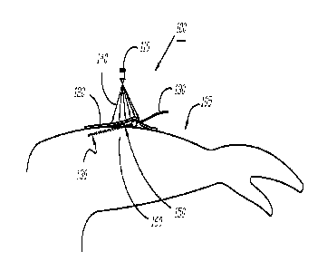
[0043] Referring now to FIG. 1, a system 100 for reducing the likelihood of
infection is
shown, according to one embodiment. In this embodiment, a patient's left arm
105 is shown;
inserted into the arm 105 is a catheter 130. The portion of the catheter 130
shown as a solid
line in FIG. 1 exists outside of the arm, while the dashed portion indicates
the portion of the
catheter within the body, e.g., under the skin 160. The catheter 130 is shown
inserted through
an insertion site 150 in the arm 105 which can be, e.g., an incision or a
break in the skin's
continuity from insertion of a needle, and a dressing 120 is shown covering
both the insertion
site 150 and a portion of the exterior-exposed catheter.
[0044] The dressing 120 can be any type of bandage, adhesive, or covering used
for
reducing the risk of infection in patients. Common dressings for this purpose
include, not by
way of limitation, absorbent acrylic, hydrocolloid, hydrogel, foam,
transparent films, and
composites, among others. Those skilled in the art will appreciate that
hospitals, health care
clinics, ambulance services, and other health care providers often use a
variety of dressings
for this and other purposes. In one preferred embodiment, the dressing 120 is
an absorbent
clear acrylic dressing sold under the TegadermTm brand, produced by 3M Skin
and Wound
Care Division, 3M Corporation, St. Paul, Minnesota, USA. Such a dressing
usually includes
a transparent or translucent sheet of acrylic with an adhesive ring disposed
about the
periphery of the sheet that adheres to the patient's skin to keep the dressing
in place. The
transparent or translucent sheet allows heath care providers to monitor a
catheter while
minimizing the disturbance that can otherwise be caused by frequent dressing
changes. In
another preferred embodiment, the dressing 120 includes a substantially
sterile transparent or
semi-transparent film configured to reduce or prevent the introduction of
microorganisms to
7
CA 02861360 2014-11-03
the insertion site 150. An integral adhesive can surround the film about its
periphery to
adhere the film to the patient's skin. One exemplary dressing of this type is
sold under the
SorbaviewTM brand by Centurion Medical Products Corporation, Williamston, MI,
United
States.
[0045] The catheter 130 can be any type of tube, lumen, or cannula. Such
catheters can
be used, e.g., for introducing substances to, or removing fluids or other
substances from a
body. Exemplary catheters include those used for intravenous therapy, and
those configured
to be inserted into a body cavity, duct, or vessel to allow drainage (e.g., in
the case of a
urinary catheter), to administer fluids, or provide access by surgical
instruments to internal
body parts e.g., in the practice of angioplasty or endoscopy. The catheter 130
can be a
temporary catheter, e.g., an "indwelling" catheter or a permanent catheter
generally referred
to as a "permcath" and may be flexible or rigid depending on the needs of the
patient and the
treatment plan of the caregiver.
[0046] In this and all other embodiments described herein, the likelihood of
developing
an infection as a result of catheterization can be reduced by irradiating the
insertion site 150
and the surrounding area with an effective dose of light having a center
wavelength between
about 385 nm and about 425 nm, e.g., light having a center wavelength of 385
nm, 390 nm,
395 nm, 400 nm, 401 nm, 402 nm, 403 nm, 404 nm, 405 nm, 406 nm, 407 nm, 408
nm, 409
nm, 410 nm, 415 nm, 420 nm, or 425 nm. In general, and without wishing to be
bound by
theory, it is believed that an effective dose of light in the wavelength range
of between about
385 nm and about 425 nm (hereinafter referred to as "violet" light) causes
either
microorganism death, or a disruption in microorganism reproduction, or both,
and thus can be
used as an antimicrobial agent, in the sense that it can reduce proliferation
of
microorganisms.
[0047] In a preferred embodiment, the light source 110 is capable of emitting
light with a
center wavelength of about 405 nm. Exemplary light sources 110 for this
purpose include,
not by way of limitation, lasers, diodes, excitable gases or filaments, and
other light sources.
In this and all other embodiments, the light source 110 can be configured to
deliver violet
light to a selected treatment area in an effective dose capable of reducing or
preventing the
proliferation of microorganisms. For example, the light source 110 can be
configured in
cooperation with lenses, windows, filters, wavegu ides, light pipes, or any
other optical
component so as to deliver violet light to a selected target area as
described.
[0048] In one embodiment, a light source 110 capable of producing or
transmitting light
radiation (indicated by reference numeral 140 in FIG. 1) in the aforedescribed
wavelength
8
CA 02861360 2014-11-03
range can be positioned a selected distance from the surface of the arm (i.e.,
the skin 160) so
as to irradiate a desired area around the catheter insertion site 150 in an
effective dose as
shown in FIG. 1.
[0049] In general, the skin 160 surrounding the insertion site 150 can be
exposed to an
effective dose of radiation, e.g., radiation having a center wavelength
between about 385 nm
and about 425 nm, for a selected length of time. The exposure time can be
controlled using a
timer, for example, through the use of an electronic, e.g., computer-
controlled control module
or other methods. In another embodiment, the exposure time can be controlled
manually,
e.g., through use of a switch, button, or other control that allows a
caregiver to irradiate the
target area with an effective dose of radiation as described for a selected
amount of time.
[0050] In general, the area of the skin exposed to the effective dose of
radiation 140 can
be chosen according to the type of catheter used, as well as the type of
dressing used (if any),
the presence of open sores, lesions, or other breaches of the skin (if any),
and other
considerations that can be determined by the user of the system 100 (e.g., a
healthcare
worker). For example, in the case of a single intravenous catheter, prepared
under relatively
sterile conditions and placed by an experienced medical provider, the
caregiver may decide to
irradiate a small area (e.g., 3-4 cm) around the insertion site with an
effective dose of
radiation as described to prevent the likelihood of infectious microorganism
growth in that
area. In another example having somewhat opposite circumstances, a patient may
be
delivered to a hospital after an automobile accident, where paramedics
emergently inserted an
intravenous catheter to reduce the likelihood of shock. In this case, where
thorough
sterilization techniques may have been secondary to stabilizing the victim,
the irradiation area
of the skin around the catheter insertion site 150 can be enlarged to
encompass a greater area,
e.g., 10-12 cm around the insertion site 150. Furthermore, in the latter case,
the exposure
time can be increased a desired amount to account for the increased risk of
infection under
the circumstances described.
[0051] In some embodiments, the light source 110 can be mounted in a preferred
configuration and orientation so as to provide an effective dose of radiation
as described to
the treatment area. For example, the light source 110 can be mounted a
selected distance
from the patient's skin so as to provide reproducible exposure of an effective
dose of
radiation as described to a desired area of the body, e.g., a catheter
insertion site. One
suitable approach for this purpose includes using molded plastic components
that attach to a
body part (e.g., attach to an arm using a strap), while simultaneously
providing a bridge or
other frame component configured to secure the light source 110 in a preferred
configuration
9
CA 02861360 2014-11-03
to irradiate selected area(s) of the patient's skin. In another embodiment,
the light source can
be attached directly to the dressing in a configuration that directs the
emitted light 140 toward
the target area. In one example of such an embodiment, an array of LED lights
capable of
providing an effective dose of violet light to cause reduction in
proliferation of
microorganisms can be integrated into one side of a dressing using glues,
adhesives, hook-
and-loop fastening systems, cloths, or other methods that will be apparent to
skilled artisans.
[0052] Referring now to FIG. 2, a system for reducing the likelihood of
infection is
shown, according to one embodiment. This system, similar to the system shown
and
described with respect to FIG. 1, includes a light source 210 capable of
producing an
effective dose of violet light to reduce the likelihood of microorganism
proliferation on a
target area of the skin 240. A catheter 230 is shown inserted into the skin
240, where the
dashed lined indicates the portion of the catheter under the skin layers.
[0053] The blow-up region shows a dressing 235 having a plurality of layers,
250, 251,
and 252 which can be the same or different materials. In one example, one of
the layers (e.g.,
layer 250) is an acrylic sheet that is transparent to violet light; one of the
layers (e.g., layer
251) includes a cotton or other absorbent material; and one of the layers,
(e.g., layer 252)
includes a gel layer. In this example, the light rays 214 output from the
light source 210 can
propagate through the layers to reach the skin layer 240 in an effective dose
to reduce or
prevent proliferation of microorganisms. It will be understood that the
dressing 235 can
include one or more layers of material as necessary to provide a desired
treatment for the
patient. For example, a burn victim may benefit from a dressing having a
silver-containing
gel layer in contact with their skin (e.g., layer 252) which covers the
inserted catheter 230.
[0054] Referring now to FIG. 3A, one embodiment of a system 300 for reducing
the
likelihood of infection is shown. Similar to the embodiment of FIG. 1, FIG. 3
shows a
catheter 330 inserted into the skin 380 of a patient's arm 305 through an
insertion site 350
(e.g., an incision in the skin produced by an IV needle). The insertion site
350 and
surrounding area is covered by a dressing 320 that is at least partially
transmissive with
respect to violet light. In a preferred embodiment, the dressing 320 is an
absorbent, clear
acrylic dressing sold under the Tegaderm brand (vide supra).
[0055] In this and other embodiments, violet light can be generated remote
from the
selected target area by a light source 310, which can be a laser, diode, or
other light source
capable of producing an effective dose of violet light at the treatment site.
The violet light
can be carried by a waveguide 360, e.g., a fiber optic cable, to a dispersion
optic 395 capable
of dispersing the light from the fiber optic onto a desired treatment area. It
will be
CA 02861360 2014-11-03
understood that the term "dispersion" as used herein, refers to increasing the
irradiance area
of the effective dose of radiation from a source to a target, e.g., from the
output end of a
waveguide to a larger area on a patient's skin; the term does not refer to the
spatial or
temporal separation of light into components of different wavelengths.
[0056] The dispersion optic 395 can be, in one example, a lens that causes
divergence of
light from a terminal end of the waveguide 360 to a desired size (e.g., area).
The lens can be
made of any suitable material to perform this function, e.g., glass, plastics,
etc., and various
types of lenses may be used. For example, traditional curved dielectrics made
of glass can
de-focus or de-collimate the output of the waveguide to achieve irradiance
over a desired area
on the patient's skin. In another example, so-called "flat" lenses may be
used, such as lenses
that incorporate photonic crystals. In such an embodiment, light from the
waveguide can be
injected into a flat slab having photonic crystals that produce a negative
index of refraction
and cause the light to be emitted over a broad area. The injected light can
spread over two-
dimensional space within the slab; when applied to the insertion site 350, the
slab can blanket
the area with violet light. In yet another example, the dispersion optic 395
can be a Fresnel
lens, which can be flexible to accommodate being placed on curved surfaces,
such as the
surface of a body part.
[0057] FIG. 3B shows an alternative arrangement of the system 300 shown in
FIG. 3A,
according to one embodiment. Here, the positions of the dispersion optic 395
and the
dressing 320 are reversed, i.e., the dispersion optic 395 is juxtaposed
between the catheter
330 and a portion of the patient's skin 350, and the dressing 320. Compared to
the
embodiment shown in FIG. 3A, the arrangement shown in FIG. 3B can reduce or
eliminate
loss associated with light propagating through the dressing 320 and can result
in increased
irradiance to the target area. In this embodiment, the fiber optic 360 can
extend through the
dressing 320; a terminal end of the fiber optic 360 can be coupled to the
dispersion optic 395
using methods known in the optics art fields.
[0058] In general the type of dressing 320 used in these and other embodiments
can be
chosen according to preference. In one exemplary embodiment, the dressing used
in the
embodiment of FIG. 3A includes a window capable of allowing violet light to
pass
therethrough, e.g., it is transmissive with respect to violet light; on the
other hand, the
dressing used in the embodiment shown in FIG. 3B can be an occlusive dressing
for
protecting the underlying skin, catheter, and dispersion optic.
[0059] Referring now to FIG. 4, a system 400 for reducing the likelihood of
infection at
or near a catheter insertion site is shown, according to one embodiment. Here,
similar to the
11
CA 02861360 2014-11-03
embodiments described above, a catheter 430 is shown inserted into a patient's
arm 460, e.g.,
subdermally, through an insertion site 450. A light source 445 capable of
producing an
effective dose of violet light 447 to cause reduction in proliferation of
microorganisms at the
target site is attached to a dressing 420. The dressing 420 is adhered to the
patient's arm 460
through the use of an adhesive ring 490 disposed about the periphery of the
dressing 420. In
this embodiment, the light source 445 of the system 400 includes an integral
power source so
that the unit is substantially self-contained, e.g., it does not require an
external power source
such as an external battery or require use of building-supplied alternating
current to produce
the effective dose of violet light. Various self-contained power sources can
be used that will
be apparent to those skilled in the art of light-emitting diodes, for example,
and includes,
without limitation, integrated batteries, capacitors, and the like.
[0060] Referring now to FIG. 5, a system 500 for reducing the likelihood of
infection
related to catheter insertion is shown, according to one embodiment. The
system 500
includes a light source 510 capable of producing light in the wavelength range
of between
about 385 nm and about 425 nm. Suitable light sources for this purpose
include, without
limitation, lasers, diodes, various types of lamps, excitable filaments, chemi-
and
electroluminescent materials, and fluorescent and phosphorescent materials,
among others.
The light output of the light source 510 can be directed into a waveguide 540
(e.g., a fiber
optic) via one or more light-coupling optics 570; the type and configuration
of the coupling
optics 570 can be chosen according to the intended use and other factors, and
the particular
configuration will be known by those skilled in the art of fiber optics and
light injection.
[0061] Referring to FIGS. 5 and 5A, in this embodiment, a portion of a
catheter 543
includes a hollow, flexible sleeve 545 that houses the waveguide 540 therein;
a void space
(generally indicated by reference numeral 546) allows fluid to be transported
within the
catheter 543 between the waveguide 540 and the inner surface of the sleeve
545, between
proximal 520 and distal 544 ends. In one embodiment, a distal end of the
catheter 544 can be
configured to receive the light output of the light source 510 in a distal end
of the waveguide
540. The distal end can also include a fluid port 546 for inserting fluid
into, or drawing fluid
out of the catheter 543.
[0062] Referring back to FIG. 5, the catheter can be inserted into a patient's
skin 560 at
an insertion site 550 as previously described. The waveguide 540 can extend
from the distal
end to a proximal end 520 where the catheter can be configured to allow light
to be emitted
from the waveguide 540 into surrounding tissue for the purpose of reducing
populations of
infectious microorganisms that may be present due to catheterization. In one
embodiment,
12
CA 02861360 2014-11-03
the catheter 543 can include two sections that can be reversibly coupled. In
such an
embodiment, a union 530 allows an exterior portion of the catheter 543 to be
joined to a
proximal portion of the catheter 521 that comes into close proximity to, and,
in some cases,
penetrates the patient's dermal layers as shown in FIG. 5. In such an
embodiment, the
proximal portion of the catheter 521 can be configured to emit the light from
the waveguide
540 along the length of the proximal portion 521, e.g., through use of a
partially lossy
waveguide, or by channeling portions of light from the central core waveguide
540 to the
surface of the catheter 543. In this manner, the proximal portion of the
catheter 521 can
irradiate the surface of the patient's skin 560, the dermal layers, and the
surrounding sub-
dermal layers (not illustrated in FIG. 5 for clarity) with violet light for
the purpose of
reducing populations of infectious microorganisms. It will be understood in
this and other
embodiments that catheters of the type described herein can be inserted into
biological
lumens, such as a patient's bladder or gastrointestinal tract, for the purpose
of reducing the
likelihood of infection.
[0063] In general, the systems and methods described herein can be used to
treat
infectious biofilms. As those skilled in the medical arts will appreciate,
biofilms composed
of gram-positive or gram-negative bacteria, yeasts, or other organisms can
originate from the
patient's skin, exposure to contaminated medical equipment or healthcare
workers, or other
sources, and can be difficult to treat. In one approach, an infectious biofilm
can be treated by
exposing the biofilm to an effective dose of violet light to cause reduction
in the proliferation
of the infectious microorganism.
[0064] Referring now to FIGS. 6 and 6A, a catheter system 600 is shown. The
catheter
system is configured to reduce the likelihood of infection by delivering an
effective dose of
violet light to the surface of skin 602 at, and around the insertion site 610
of the catheter
lumen 603, to reduce or inhibit the proliferation of infectious
microorganisms. In this
embodiment, the catheter system includes a housing 601, which can be flexible
or rigid
depending on the intended use that includes a central bore 608 for
transporting fluids along
the length of the housing 601. The catheter lumen 603 is a tube that extends
the central bore
608 out of the housing 601 and allows fluid to flow between the proximal end
of the housing
601 and the patient's blood stream, e.g., in situations where the system 600
is being used for
IV therapy. It will be understood that the catheter system 600 can be used for
other
treatments as well (vide supra).
[0065] The housing 601 includes one or more waveguides 604a-604d (e.g., fiber
optics)
arranged concentrically about the central bore 608. It will be understood that
the
13
CA 02861360 2014-11-03
configuration of waveguides and the central bore shown in FIGS. 6 and 6A is
one of many
possibilities, and other arrangements are equally contemplated. The waveguides
604a-604d
extend from the proximal end (e.g., nearest to the patient's skin, as shown)
to a distal end of
the housing 601. The distal end of the housing is configured allow distal ends
of the
waveguides 604a-604d to receive the output of a light source (not shown in
FIG. 6 ¨ 6A for
clarity) capable of producing an effective dose of violet light to reduce
proliferation of
microorganisms near the incision site 610. The distal end of the housing can
also be
configured to allow access to fluids in the central bore 608, so that fluids
can be drawn from,
or injected into, the patient.
[0066] The proximal end of the housing 601 houses the proximal terminal ends
of the
waveguides 604a-604d. In this embodiment, convex protuberances 606a-606d
extend from
the housing to produce a lensing effect capable of causing the light emitted
from the
waveguide to disperse across a wider area, although in some embodiments this
feature can be
optional. The configuration of the waveguides is such that the area
immediately surrounding
the catheter insertion site 610 can be flooded with violet light. In this
embodiment, the
application of intense violet light can be focused near the area where the
skin has been
breached for catheterization. As previously described, this can cause a
disruption in the
ability of infectious microorganisms to reproduce, and thus reduce the
likelihood of infection.
In some circumstances, the catheter insertion site may be the area where the
blood stream is
vulnerable to outside infectious agents.
[0067] Referring now to FIG. 7, a system for reducing the likelihood of
microorganism
proliferation is shown. A light source 710 provides output of an effective
dose of violet light
(indicated by reference numeral 714) which can be directed onto a surface 740.
The surface
740 can be the surface of living tissue, similar to the embodiments described
herein. In some
embodiments, however, the surface can be non-living, for example, and without
limitation, a
surface of a piece of medical equipment, table- and countertops, processing
areas, or other
surface. In one embodiment, the surface 740 is a portion of processed food.
Examples of
processed foods include, without limitation, meats, such as steaks and other
butchery cuts,
eggs, breads, pastas, fish, confectionery items such as cakes and cookies,
vegetables, and
other foodstuffs. In another embodiment, the surface 740 is a portion of
packaging used to
package foods, such as a packaging tray for the foods just described.
[0068] In general, the system 700 can be used to reduce the likelihood of
microorganism
proliferation in and on foodstuffs by irradiating the target, e.g., the
foodstuff or the packaging
containing the foodstuff, with an effective dose of violet light sufficient to
reduce or prevent
14
CA 02861360 2014-11-03
microorganism reproduction. The system 700 can be used in, e.g., food
processing facilities
where foods are processed prior to distribution. For example, the system 700
can be used as
part of a food processing system where foods are irradiated prior to packaging
so that the
proliferation of microorganisms on the food is reduced. Similarly, the system
700 can be
used in food stores to reduce the likelihood of microorganism growth on
foodstuffs, thereby
prolonging the so-called shelf-life of the food. In one example, foods can be
irradiated with
an effective dose of violet light according to a schedule, e.g., every two
days, to reduce
microorganism growth.
[0069] In general, methods for reducing the likelihood of microorganism growth
on a
target surface are provided. Referring now to FIG. 8, the steps of a method
800 are shown,
according to one embodiment. The method 800 can be used to reduce the
likelihood of
microorganism growth on a target surface, and, in some embodiments, within a
host matrix.
The method begins at step 801 by identifying a target. The target can be, in
multiple
embodiments, skin, tissue, muscle fiber, and other parts of living systems;
one or more
surfaces of medical equipment; one or more surfaces having the likelihood to
become
exposed to biological fluids, such as hospital beds, ambulance patient
treatment areas,
lavatory areas, etc.; catheters; food processing equipment; packaged food; and
other surfaces.
In a preferred embodiment, the target area is an area of living tissue
immediately adjacent to,
and surrounding a catheter insertion site, or other area where a body's
barrier to infectious
agents has been compromised.
[0070] Next, at step 802, the target area is irradiated with an effective dose
of violet light
so as to reduce the proliferation of microorganisms, e.g., infectious
microorganisms. As
described heretofore, "violet" light is generally considered to include light
having a center
wavelength of between about 385 nm and about 425 nm, e.g., light having a
center
wavelength of 385 nm, 390 nm, 395 nm, 400 nm, 401 nm, 402 nm, 403 nm, 404 nm,
405 nm,
406 nm, 407 nm, 408 nm, 409 nm, 410 nm, 415 nm, 420 nm, or 425 nm.
"Irradiated" and
"irradiating" as used herein, carries the common meaning and includes exposing
the target
surface with electromagnetic radiation from a light source. In general, the
intensity of the
violet light can be chosen according to user preference or circumstance to
produce an
effective dose sufficient to affect the proliferation of microorganisms. For
example, a high
intensity (high flux) can be used when an active microorganism population is
witnessed, e.g.,
an infection is present, so as to affect the greatest population of the
microorganisms as
possible. Alternatively, a lesser intensity (lower flux) can be used as a
preventive measure to
keep lesser populations of microbes from reproducing and causing infection in
a body.
CA 02861360 2014-11-03
[0071] The decision at step 803 can involve situations where irradiation is
scheduled.
"Scheduled irradiation" includes, e.g., irradiating a target surface on
regular or otherwise
timed or scheduled intervals. For example, in some circumstances, patients are
given a
catheter that may stay in the body for extended periods of time, e.g., 3-5
days. During this
time, the catheter insertion site can be exposed to infectious microorganisms,
which can
increase the risk of bodily infection. Accordingly, a caregiver can set a
timer, e.g., through
an electronic control module, that activates the light source and causes the
target surface to be
exposed for a selected amount of time, at selected intervals. For example, the
caregiver can
set the timer to expose a patient's catheter insertion site for five minutes,
every two hours.
The intensity of the exposure can similarly be set and controlled for every
exposure through
the control module. If the answer to the "scheduled exposure?" question in
step 803 is "yes,"
then the method returns to step 802 to expose the target surface; the loop
between step 802
and step 803 iterates until the decision at step 803 is "no." The method then
ends at step 804.
[0072] Referring now to FIG. 9, a method 900 for reducing the likelihood of
microorganism growth at or near catheter insertion sites is shown, according
to one
embodiment. This method 900 can be used, without limitation, in a variety of
settings,
including hospitals, veterinary clinics, triage units, emergency rooms,
primary care clinics,
ambulances, and in outside areas such as battlefields. This and other methods
described
herein can be practiced by, without limitation, physicians, veterinarians,
ambulance crews,
EMT's, firefighters, soldiers, or anyone placing a catheter within a living
system. Beginning
at step 901, the catheter insertion site is located, e.g., on the skin of a
patient's arm or hand;
while optional, in preferred embodiments, the site can be disinfected to
reduce the population
of microbes that may already be present, which is a practice those skilled in
the art will
recognize.
[0073] Next, at step 902, the catheter is inserted into the patient. In
general, but without
limitation, this step is often performed by inserting a needle through the
patient's skin and
into a blood vessel, such as in the case of IV therapy. A lumen (e.g., a
hollow, flexible tube)
is then advanced into the blood vessel along the path defined by the needle;
the needle is then
withdrawn, leaving the lumen within the blood vessel. The lumen is generally
connected to
other catheter structures and tubing to allow fluids to be drawn out of, or
inserted into, the
patient. In some embodiments, such as the embodiment of FIG. 6, a portion of
the catheter
body includes one or more waveguides configured to irradiate the insertion
site with violet
light from a violet light source.
16
CA 02861360 2014-11-03
=
[0074] Next, at step 903, the insertion site can be optionally covered to
protect the
catheter and the catheter insertion site. In some embodiments, the catheter
and catheter
insertion site can be covered with a dressing having a transparent window to
allow caregivers
to monitor the state of the catheter and catheter insertion site. In one
exemplary embodiment,
the dressing includes a transparent window having a flexible, clear acrylic
window, and
adhesive along its periphery allowing the covering to adhere to the patient's
skin. Preferably,
the material of the transparent window is transparent to violet light.
Coverings sold under the
Tegaderm brand (vide supra) are preferred.
[0075] Next, at step 904, the catheter insertion site and surrounding area is
exposed to an
effective dose of violet light to cause reduction in the proliferation of any
microorganisms
present. In general, the intensity of the violet light can be chosen according
to circumstances
as described with respect to the method of FIG. 8. In general, the insertion
site can be
exposed from a selected vantage point, e.g., from above, from the side, or in
a "blanket"
fashion, if, e.g., the light source is the type as described in FIGS. 3-3B.
[0076] Next, decision 905 asks whether the exposure is scheduled on a
repeating basis.
In some cases, a caregiver may decide to give the patient a single dose of
radiation; in other
cases, e.g., when the catheter is a "permcath" the caregiver may elect to
administer repeating
doses of radiation over a selected period of time. In the latter case, step
904 is repeated until
the number of selected exposures has been reached.
[0077] A number of illustrative embodiments have been described. Nevertheless,
it will
be understood that various modifications may be made without departing from
the spirit and
scope of the various embodiments presented herein. For example, the concepts
described
herein can be applied toward other scenarios where microorganism growth and
proliferation
can be problematic. For example, microorganism growth is known to cause
structural
damage to building components such as wood framework and stucco. To combat
this
problem, light sources capable of producing an effective dose against
microorganism
reproduction can be placed in areas where microbes live, or have the
capability of colonizing.
In one embodiment, high-intensity LEDs can be placed in the framework of
buildings as they
are being constructed; the LEDs can be activated on a selected schedule (e.g.,
once a day) to
reduce the likelihood of microorganism growth in areas that would otherwise be
accessible
only through demolition.
[0078] In one embodiment, the concepts, systems, and methods described herein
can be
applied to combating the problem of microorganism growth inside of fuel tanks,
e.g., airliner
fuel tanks. It is known that certain bacteria can degrade aluminum fuel tanks
which can be
17
CA 02861360 2014-11-03
costly to repair; likewise, it is known that certain bacteria can degrade
fuels such as aviation
fuel. Accordingly, light sources capable of producing an effective dose of
violet light to
interfere with microorganism reproduction can be installed in various types of
tanks, e.g., fuel
tanks. In such an approach, it can be advantageous for obvious reasons to use
a light source
that produces little heat, such as an LED, or utilize waveguides to carry
violet light from a
light source to the interior of the tank.
[0079] In one embodiment, the concepts, systems, and methods described herein
can be
used in the restaurant industry to reduce the likelihood of microorganism
growth on cooking
and eating surfaces.
[0080] In general, the effective dose of violet light to cause a reduction in
the
proliferation of a microorganism can be adjusted for different types of
microorganisms.
18