Note : Les descriptions sont présentées dans la langue officielle dans laquelle elles ont été soumises.
CA 02865317 2014-08-21
WO 2013/126597
PCT/US2013/027175
1
METHODS AND COMPOSITIONS FOR PREVENTING OR TREATING
OPHTHALMIC CONDITIONS
TECHNICAL FIELD
[0001] The present technology relates generally to compositions and methods of
preventing or treating ophthalmic diseases or conditions such as diabetic
macular
edema. In particular, the present technology relates to administering aromatic-
cationic peptides in effective amounts to prevent or treat ophthalmic diseases
or
conditions.
BACKGROUND
[0002] The following description is provided to assist the understanding of
the
reader. None of the information provided or references cited is admitted to be
prior
art to the present invention.
[0003] Diseases and degenerative conditions of the optic nerve and retina are
the
leading causes of blindness in the world. A significant degenerative condition
of the
retina is age-related macular degeneration (ARMD). ARMD is the most common
cause of blindness in people over 50 in the USA and its prevalence increases
with age.
ARMD is classified as either wet (neovascular) or dry (non-neovascular); the
dry
form of the disease is more common. Wet ARMD is considered an advanced stage
of
dry ARMD and is associated with angiogenesis. Macular degeneration occurs when
the central retina has become distorted and thinned usually associated with
age but
also characterized by intra-ocular inflammation and angiogenesis (wet ARMD
only)
and/or intra-ocular infection. Inappropriate angiogenesis or
neovascularization is
thought to result from retinal ischemia, as the subsequent generation of free
radicals,
resulting in oxidative tissue damage, local inflammation and production of
growth
factors (such as VEGF and FGF) and inflammatory mediators.
[0004] The retina is the part of the eye that is sensitive to light. The
macula is the
central portion of the retina, a small area rich in cones, the specialized
nerve endings
that detect color and upon which daytime vision depends and is the part of the
eye
responsible for detailed central vision. Retinopathy is a leading cause of
blindness in
type I diabetes, and is also common in type 11 diabetes. The degree of
retinopathy
CA 02865317 2014-08-21
WO 2013/126597
PCT/US2013/027175
2
depends on the duration of diabetes, and generally begins to occur ten or more
years
after onset of diabetes. Diabetic retinopathy may be classified as non-
proliferative,
where the retinopathy is characterized by increased capillary permeability,
edema and
exudates, or proliferative, where the retinopathy is characterized by
neovascularization extending from the retina to the vitreous, scarring,
deposit of
fibrous tissue and the potential for retinal detachment. Diabetic retinopathy
is
believed to be caused by the accumulation of glycosylated proteins due to high
blood
glucose that cause biochemical and hemodynamic abnormalities in the retina
that in
turn lead to chronic retinal hypoxia. Several other less common retinopathies
include
choroidal neovascular membrane (CNVM), cystoid macular edema (CME), epi-
retinal
membrane (ERM) and macular hole. Diseases of the macula, such as diabetic
macular edema, account for a major proportion of legal blindness.
[0005] Glaucoma is made up of a collection of eye diseases that cause vision
loss by
damage to the optic nerve. Elevated intraocular pressure (IOP) due to
inadequate
ocular drainage is the primary cause of glaucoma. Glaucoma often develops as
the
eye ages, or it can occur as the result of an eye injury, inflammation, tumor
or in
advanced cases of cataract or diabetes. It can also be caused by the increase
in IOP
caused by treatment with steroids. Drug therapies that are proven to be
effective in
glaucoma reduce IOP either by decreasing vitreous humor production or by
facilitating ocular draining. Such agents are often vasodilators and as such
act on the
sympathetic nervous system and include adrenergic antagonists.
SUMMARY
[0006] The present technology relates generally to the treatment or prevention
of
ophthalmic diseases or conditions in mammals through administration of
therapeutically effective amounts of aromatic-cationic peptides to subjects in
need
thereof.
[0007] In one aspect, the present disclosure provides a method for preventing,
treating, or ameliorating the symptoms of diabetic macular edema in a
mammalian
subject in need thereof, the method comprising: administering to the subject a
therapeutically effective amount of a peptide D-Arg-2'6'-Dmt-Lys-Phe-NH2 or a
CA 02865317 2014-08-21
WO 2013/126597
PCT/US2013/027175
3
pharmaceutically acceptable salt thereof, wherein the peptide is administered
ocularly,
orally, or parenterally.
[0008] In some embodiments, the therapeutically effective amount of the
peptide is
administered ocularly to one or more regions of the eye. In some embodiments,
the
one or more regions of the eye is selected from the group consisting of the
posterior
chamber, ora serrata, ciliary muscle, ciliary zonules, canal of Schlemm,
pupil, anterior
chamber, cornea, iris, lens cortex, lens nucleus, ciliary process,
conjunctiva, inferior
oblique muscle, inferior rectus muscle, medial rectus muscle, retinal arteries
and
veins, optic disc, dura mater, central retinal artery, central retinal vein,
optic nerve,
vorticose vein, bulbar sheath, macula, fovea, sclera, choroid, superior rectus
muscle,
and retina. In some embodiments, the region of the eye is the macula. In some
embodiments, the mammal is a human.
[0009] In some embodiments, the subject is at risk of having, suspected of
having,
or diagnosed as having one or more of macular degeneration, central retinal
thickness,
intraretinal water content, macular central fovea thickness, contrast
sensitivity, or loss
of visual acuity. In some embodiments, the peptide is administered in
combination
with at least one additional therapeutic agent. In some embodiments, the
additional
therapeutic agent is administered before peptide administration, after peptide
administration, simultaneously with peptide administration, or a combination
thereof
[0010] In one aspect, the present disclosure provides a composition for
preventing,
treating, or ameliorating the symptoms of diabetic macular edema in a
mammalian
subject in need thereof, comprising: a therapeutically effective amount of a
peptide D-
Arg-2'6'-Dmt-Lys-Phe-NH2 or a pharmaceutically acceptable salt thereof,
wherein the
composition is administered ocularly, orally, or parenterally.
[0011] In some embodiments, the composition is administered ocularly to one or
more regions of the eye. In some embodiments, the one or more regions of the
eye is
selected from the group consisting of the posterior chamber, ora serrata,
ciliary
muscle, ciliary zonules, canal of Schlemm, pupil, anterior chamber, cornea,
iris, lens
cortex, lens nucleus, ciliary process, conjunctiva, inferior oblique muscle,
inferior
rectus muscle, medial rectus muscle, retinal arteries and veins, optic disc,
dura mater,
CA 02865317 2014-08-21
WO 2013/126597
PCT/US2013/027175
4
central retinal artery, central retinal vein, optic nerve, vorticose vein,
bulbar sheath,
macula, fovea, sclera, choroid, superior rectus muscle, and retina.
[0012] In some embodiments, the region of the eye is the macula. In some
embodiments, the mammal is a human. In some embodiments, the subject is at
risk of
having, suspected of having, or diagnosed as having one or more of macular
edema,
central retinal thickness, intraretinal water content, macular central fovea
thickness,
contrast sensitivity, or loss of visual acuity. In some embodiments, the
composition is
administered in combination with at least one additional therapeutic agent. In
some
embodiments, the additional therapeutic agent is administered before, after,
or
simultaneous to the composition, or a combination thereof
[0013] In one aspect, the present disclosure provides a method for the
treatment,
prevention, or amelioration of microvascular damage caused by acute ocular
ischemia
in a mammalian subject in need thereof, the method comprising: administering
to the
subject a therapeutically effective amount of a peptide D-Arg-2'6'-Dmt-Lys-Phe-
NH2
or a pharmaceutically acceptable salt thereof, wherein the peptide is
administered
ocularly, orally, or parenterally.
[0014] In some embodiments, the method further comprises the step of
performing a
revascularization procedure on the subject. In some embodiments, the subject
is
administered the peptide prior to or following the formation of ocular
ischemia. In
some embodiments, the subject is administered the peptide before, during, or
after the
revascularization procedure, or continuously before, during, and after the
revascularization procedure.
[0015] In one aspect, the disclosure provides a method of treating or
preventing an
ophthalmic condition comprising administering to a subject in need thereof an
aromatic-cationic peptide. In some embodiments, the aromatic-cationic peptide
is a
peptide having:
at least one net positive charge;
a minimum of four amino acids;
a maximum of about twenty amino acids;
a relationship between the minimum number of net positive charges (pm) and
the total number of amino acid residues (r) wherein 3pm is the largest number
that is
CA 02865317 2014-08-21
WO 2013/126597
PCT/US2013/027175
less than or equal to r + 1; and a relationship between the minimum number of
aromatic groups (a) and the total number of net positive charges (pt) wherein
2a is the
largest number that is less than or equal to pt + 1, except that when a is 1,
pt may also
be 1. In particular embodiments, the mammalian subject is a human.
[0016] In one embodiment, 2pm is the largest number that is less than or equal
to
r+1, and may be equal to pt. The aromatic-cationic peptide may be a water-
soluble
peptide having a minimum of two or a minimum of three positive charges.
[0017] In one embodiment, the peptide comprises one or more non-naturally
occurring amino acids, for example, one or more D-amino acids. In some
embodiments, the C-terminal carboxyl group of the amino acid at the C-terminus
is
amidated. In certain embodiments, the peptide has a minimum of four amino
acids.
The peptide may have a maximum of about 6, a maximum of about 9, or a maximum
of about 12 amino acids.
[0018] In one embodiment, the peptide may have the formula Phe-D-Arg-Phe-Lys-
NH2 or 2',6'-Dmp-D-Arg-Phe-Lys-NH2. In a particular embodiment, the aromatic-
cationic peptide has the formula D-Arg-2',6'-Dmt-Lys-Phe-NH2.
[0019] In one embodiment, the peptide is defined by formula I:
R5 R10
R
R4 R6 11 0
R3 I.
R7 0 I. R12
H2C 0 H2C 0
R1 H H
\ __,,,,...,N .N==
z N N
H NH2
R2
0 (CH2)3 0 (CH2)n
I
1
NH
1 NH2
,C\
HN NH2
CA 02865317 2014-08-21
WO 2013/126597
PCT/US2013/027175
6
wherein R1 and R2 are each independently selected from
(i) hydrogen;
(ii) linear or branched C1-C6 alkyl;
1-(CH 26 where m = 1-3;
(iii)
4cl-12 __________ <
¨CH2¨C=CH2
(v)
R35 R45 R55 R65 R75 R85 R95 R105 R11 and K-12
are each independently selected from
(i) hydrogen;
(ii) linear or branched C1-C6 alkyl;
(iii) C1-C6 alkoxy;
(iv) amino;
(v) C1-C4 alkylamino;
(vi) C1-C4 dialkylamino;
(vii) nitro;
(viii) hydroxyl;
(ix) halogen, where "halogen" encompasses chloro, fluoro, bromo, and iodo;
and
n is an integer from 1 to 5.
[0020] In a particular embodiment, R15 R25 R35 R45 R55 R65 R75 R85 R95 R105
R115 and
R12 are all hydrogen; and n is 4. In another embodiment, R1, R25 R35 R45 R55
R65 R75
R85 R95 and R11 are all hydrogen; R8 and R12 are methyl; R1 is hydroxyl; and
n is 4.
CA 02865317 2014-08-21
WO 2013/126597
PCT/US2013/027175
7
[0021] In one embodiment, the peptide is defined by formula II:
OH R7
R8
R6
R-r-µ,4 R- R9
0 CH2 0 CH2
RI\
zNQD) NH2
R2
(CH2)3 O (CH2)n 0
NH
NH2
HN NH2
wherein R1 and R2 are each independently selected from
(i) hydrogen;
(ii) linear or branched C1-C6 alkyl;
1¨(CH 26 where m = 1-3;
(iii)
CH2 ______________ <
(iv) S
H2
C C = CH 2
(v)
R3 and R4 are each independently selected from
(i) hydrogen;
(ii) linear or branched Ci-C6 alkyl;
(iii) C1-C6 alkoxy;
(iv) amino;
(v) C1-C4 alkylamino;
(vi) C1-C4 dialkylamino;
(vii) nitro;
(viii) hydroxyl;
(ix) halogen, where "halogen" encompasses chloro, fluoro, bromo, and iodo;
R5, R6, R7, R8, and R9 are each independently selected from
CA 02865317 2014-08-21
WO 2013/126597
PCT/US2013/027175
8
(i) hydrogen;
(ii) linear or branched C1-C6 alkyl;
(iii) C1-C6 alkoxy;
(iv) amino;
(v) C1-C4 alkylamino;
(vi) C1-C4 dialkylamino;
(vii) nitro;
(viii) hydroxyl;
(ix) halogen, where "halogen" encompasses chloro, fluoro, bromo, and iodo;
and
n is an integer from 1 to 5.
[0022] The aromatic-cationic peptides may be administered in a variety of
ways. In
some embodiments, the peptides may be administered intraocularly, orally,
topically,
intranasally, ocularly, intraperitoneally, parenterally, intravenously,
subcutaneously,
or transdermally (e.g., by iontophoresis).
[0023] In one aspect, the present disclosure provides a pharmaceutical
composition
comprising a therapeutically effective amount of the peptide D-Arg-2'6'-Dmt-
Lys-
Phe-NH2 or Phe-D-Arg-Phe-Lys-NH2 formulated for topical, iontophoretic, or
intraocular administration.
[0024] In one aspect, the present disclosure provides an ophthalmic
formulation
comprising a therapeutically effective amount of the peptide D-Arg-2'6'-Dmt-
Lys-
Phe-NH2 or a pharmaceutically acceptable salt thereof, such as an acetate salt
and/or a
tri-fluoro-acetate salt or Phe-D-Arg-Phe-Lys-NH2 or a pharmaceutically
acceptable
salt thereof, such as an acetate salt and/or a tri-fluoro-acetate salt. In one
embodiment, the formulation is soluble in the cornea, aqueous humor, and lens
of the
eye. In one embodiment, the formulation further comprises a preservative. In
one
embodiment, the preservative is present in a concentration of less than 1%.
[0025] In one embodiment, the formulation further comprises an active agent
selected from the group consisting of: an antioxidant, a metal complexer, an
anti-
inflammatory drug, an antibiotic, and an antihistamine. In one embodiment, the
antioxidant is vitamin A, vitamin C, vitamin E, lycopene, selenium, a-lipoic
acid,
CA 02865317 2014-08-21
WO 2013/126597
PCT/US2013/027175
9
coenzyme Q, glutathione, or a carotenoid. In one embodiment, the formulation
further comprises an active agent selected from the group consisting of:
aceclidine,
acetazolamide, anecortave, apraclonidine, atropine, azapentacene, azelastine,
bacitracin, befunolol, betamethasone, betaxolol, bimatoprost, brimonidine,
brinzolamide, carbachol, carteolol, celecoxib, chloramphenicol,
chlortetracycline,
ciprofloxacin, cromoglycate, cromolyn, cyclopentolate, cyclosporin,
dapiprazole,
demecarium, dexamethasone, diclofenac, dichlorphenamide, dipivefrin,
dorzolamide,
echothiophate, emedastine, epinastine, epinephrine, erythromycin,
ethoxzolamide,
eucatropine, fludrocortisone, fluorometholone, flurbiprofen, fomivirsen,
framycetin,
ganciclovir, gatifloxacin, gentamycin, homatropine, hydrocortisone,
idoxuridine,
indomethacin, isoflurophate, ketorolac, ketotifen, latanoprost, levobetaxolol,
levobunolol, levocabastine, levofloxacin, lodoxamide, loteprednol, medrysone,
methazolamide, metipranolol, moxifloxacin, naphazoline, natamycin, nedocromil,
neomycin, norfloxacin, ofloxacin, olopatadine, oxymetazoline, pemirolast,
pegaptanib, phenylephrine, physostigmine, pilocarpine, pindolol, pirenoxine,
polymyxin B, prednisolone, proparacaine, ranibizumab, rimexolone, scopolamine,
sezolamide, squalamine, sulfacetamide, suprofen, tetracaine, tetracyclin,
tetrahydrozoline, tetryzoline, timolol, tobramycin, travoprost, triamcinulone,
trifluoromethazolamide, trifluridine, trimethoprim, tropicamide, unoprostone,
vidarbine, xylometazoline, pharmaceutically acceptable salts thereof, and
combinations thereof
BRIEF DESCRIPTION OF THE FIGURES
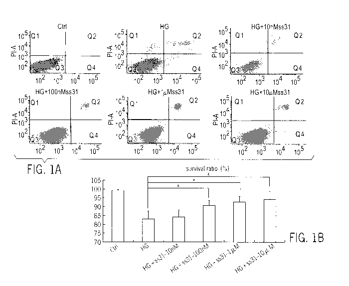
[0026] FIGs. lA and 1B show the effects of different concentrations of D-Arg-
2',6'-
Dmt-Lys-Phe-NH2(SS-31) (10 nM, 100 nM, 1 [iM and 10 [tM) used as co-treatment
with 30 mM glucose (HG). FIG. lA shows analysis for apoptosis, as assessed by
a
Flow cytometry after Annexin V/PI staining, which showed that the survival
ratios for
HRECs (Q3) was 99.3%, 83.2%, 84.3%, 90.7%, 92.8%, and 94.3%, respectively 24
hours after treatment. FIG. 1B is a graphic representation of the survival
ratio for
HRECs. Data for D-Arg-2',6'-Dmt-Lys-Phe-NH2 (SS-31) concentrations of 100nM,
1 M, and 10 M were significantly higher than that seen with high glucose
exposed
cells in the absence of co-treatment with D-Arg-2',6'-Dmt-Lys-Phe-NH2 (SS-31).
*
p<0.05 vs. 30mM high glucose treated group.
CA 02865317 2014-08-21
WO 2013/126597
PCT/US2013/027175
[0027] FIGs. 2A-2F is a series of micrographs showing co-treatment with D-Arg-
2',6'-Dmt-Lys-Phe-NH2 (SS-31) reduced intracellular reactive oxygen species
(ROS)
in HRECs exposed to 30 mM glucose for 24 h and 48 h. Intracellular ROS was
measured using dihydroethidium. 2A, 2D normal culture media; 2B, 2E 30 mM
glucose; and 2C, 2F 30 mM glucose + D-Arg-2',6'-Dmt-Lys-Phe-NH2 (SS-31) (100
nM) at 24 and 48 h, respectively.
[0028] FIGs. 3A and 3B show that D-Arg-2',6'-Dmt-Lys-Phe-NH2 (SS-31) prevents
the mitochondrial potential loss of HRECs treated with high-glucose. FIG. 3A.
The
Ak-lim of HRECs was measured by flow cytometry after JC-1 fluorescent probe
staining. High glucose (30 mM) treatment resulted in a rapid loss of
mitochondrial
membrane potential of the cultured HRECs at 24 and 48 hours. In contrast, flow
cytometric analysis showed that 30mM glucose co-treated with D-Arg-2',6'-Dmt-
Lys-
Phe-NH2 (SS-31) increased Ak-lim compared with the high glucose alone group.
FIG.
3B. Quantitative analysis of AkTim in high glucose HRECs co-treated with D-Arg-
2',6'-Dmt-Lys-Phe-NH2 (SS-31) for 24 and 48 hours, High glucose alone
adversely
affected Ak-lim. In contrast, D-Arg-2',6'-Dmt-Lys-Phe-NH2 (SS-31) restored Ak-
lim to
control levels. Values represent mean SD of six separate experiments
performed in
triplicate. *P< 0.05.
[0029] FIGs. 4A and 4D are confocal microscopic images showing that HRECs in
the normal glucose group and the D-Arg-2',6'-Dmt-Lys-Phe-NH2 (SS-31) co-
treated
group have more exact overlapping cytochrome c staining and HSP60 staining at
24
and 48 hours, indicating the co-localization of cytochrome c and mitochondria.
Twenty four and 48 hours after treatment, cytochrome c was obviously increased
in
the cytoplasm of HRECs treated with 30 mM glucose. FIGs. 4B and 4E show the
cytochrome c content in mitochondria and cytoplasm as determined by Western
blot.
FIGs. 4C and 4F show quantitative analysis of the percentage of cytochrome c
content
in mitochondria and cytoplasm of HRECs co-treated with high glucose and D-Arg-
2',6'-Dmt-Lys-Phe-NH2 (SS-31) for 24 and 48 h.
[0030] FIG. 5A and FIG. 5B show increased expression of caspase-3 in HRECs
treated with high glucose (HG) was reduced by D-Arg-2',6'-Dmt-Lys-Phe-NH2 (SS-
31) co-treatment as detected by western blot. Caspase-3 expression was
normalized to
the expression of 3-actin. FIGs. 5C-E show D-Arg-2',6'-Dmt-Lys-Phe-NH2 (SS-31)
CA 02865317 2014-08-21
WO 2013/126597
PCT/US2013/027175
11
increases the expression of Trx2 in the high glucose-treated HRECs. FIG. 5C
shows
the mRNA level of Trx2 in HRECs exposed to 30 mM glucose treated with D-Arg-
2',6'-Dmt-Lys-Phe-NH2 (SS-31) for 24 h and 48 h. FIG. 5D shows the level of
Trx2
protein expression measured by Western blot. FIG. 5E shows quantitative
analysis of
the protein level of Trx2 in HRECs 24 and 48 h after high glucose with or
without D-
Arg-2',6'-Dmt-Lys-Phe-NH2 (SS-31) co-treatment.
[0031] FIG. 6 is a photograph of the effects of D-Arg-2',6'-Dmt-Lys-Phe-NH2
(SS-
31) on the lenses of diabetic rats. Top row: lenses obtained from diabetic
rats; bottom
row: lenses obtained from diabetic rats treated with D-Arg-2',6'-Dmt-Lys-Phe-
NH2
(SS-31) or Phe-D-Arg-Phe-Lys-NH2 (SS-20).
[0032] FIG. 7 is a series of photographs showing the effects of D-Arg-2',6'-
Dmt-
Lys-Phe-NH2 (SS-31) and Phe-D-Arg-Phe-Lys-NH2 (SS-20) on the lenses of
diabetic
rats. Diabetes was induced by high fat diet and streptozotocin (HFD/STZ) (top
row)
or streptozotocin (STZ) alone (bottom row).
[0033] FIG. 8 is a series of micrographs showing the lens epithelium from
normal
rats, diabetic rats, and diabetic rats treated with D-Arg-2',6'-Dmt-Lys-Phe-
NH2 (SS-
31). Diabetes was induced by STZ.
[0034] FIG. 9 is a series of micrographs showing the lens epithelium from
normal
rats, diabetic rats, and diabetic rats treated with D-Arg-2',6'-Dmt-Lys-Phe-
NH2 (SS-
31). Diabetes was induced by HFD/STZ.
[0035] FIG. 10A and 10B are charts showing the integrity of the blood-retinal
barrier of normal rats (NRC), diabetic rats, and diabetic rats treated with
Phe-D-Arg-
Phe-Lys-NH2 (SS-20) or D-Arg-2',6'-Dmt-Lys-Phe-NH2 (SS-31), as analyzed by
Evans blue extravasation. (A) diabetes induced by STZ; (B) diabetes induced by
HFD/STZ.
[0036] FIG. 11 is a series of micrographs showing retinal microvessels of
normal
rats (NRC), diabetic rats (HFD/STZ), and diabetic rats treated with D-Arg-
2',6'-Dmt-
Lys-Phe-NH2 (SS-31).
CA 02865317 2014-08-21
WO 2013/126597
PCT/US2013/027175
12
[0037] FIG. 12 is a series of micrographs showing retinal microvessels of
normal
rats, diabetic rats (STZ), and diabetic rats treated with D-Arg-2',6'-Dmt-Lys-
Phe-NH2
(SS-31).
[0038] FIGS. 13A-13D is a series of micrographs showing the distribution of
the
tight junction protein claudin-5 in retinal microvessels in normal rats (A),
STZ rats
(B), STZ/Phe-D-Arg-Phe-Lys-NH2-treated rats (SS-20) (C), or STZ/D-Arg-2',6'-
Dmt-
Lys-Phe-NH2-treated rats (SS-31) (D).
[0039] FIG. 14 is a chart showing the lack of cytotoxicity of D-Arg-2',6'-Dmt-
Lys-
Phe-NH2 (SS-31) on trabecular meshwork cells from non-diseased individuals
(HTM)
and trabecular meshwork cells from glaucoma patients (GTM) administered D-Arg-
2 ',6'-Dmt-Lys-Phe-NH2 (SS-31).
[0040] FIG. 15 is a series of confocal micrographs showing co-treatment with D-
Arg-2',6'-Dmt-Lys-Phe-NH2 (SS-31) dose-dependently inhibited the decrease in
mitochondrial potential (Am) elicited by 200 [tM H202 in trabecular meshwork
cells
from glaucoma patients (GTM).
[0041] FIG. 16 is a series of charts showing co-treatment with D-Arg-2',6'-Dmt-
Lys-Phe-NH2 (SS-31) inhibited the decrease in mitochondrial membrane potential
(Am), as measured by TMRM and flow cytometry, in trabecular meshwork cells
from glaucoma patients (GTM) induced by 200 [LM H202.
[0042] FIG. 17 is a chart comparing mitochondrial membrane potential (Am) in
GTM and HTM cells.
[0043] FIG. 18 is a series of micrographs showing the morphology changes in
GTM
cells in response to D-Arg-2',6'-Dmt-Lys-Phe-NH2 (SS-31) treatment as viewed
using
inverted phase contrast microscopy.
[0044] FIG. 19 is a series of micrographs showing co-treatment with D-Arg-
2',6'-
Dmt-Lys-Phe-NH2 (SS-31) reduced the loss of mitochondrial membrane potential
in
GTM cells caused by 400 [tM H202 in a dose-dependent manner as viewed using
confocal microscopy.
CA 02865317 2014-08-21
WO 2013/126597
PCT/US2013/027175
13
[0045] FIG. 20 is a series of micrographs showing co-treatment with D-Arg-
2',6'-
Dmt-Lys-Phe-NH2 (SS-31) reduced the loss of mitochondrial membrane potential
(Am) in GTM cells caused by 400 04 H202 as viewed by TMRM and confocal
microscopy (200x magnification).
[0046] FIG. 21 is a series of micrographs showing the morphology changes in
GTM
cells in response to D-Arg-2',6'-Dmt-Lys-Phe-NH2 (SS-31) treatment as viewed
using
inverted phase contrast microscopy.
[0047] FIG. 22 is a chart showing that D-Arg-2',6'-Dmt-Lys-Phe-NH2 (SS-31) had
no effect on the viability of primary human retinal pigment epithelial (RPE)
cells (as
measured by the MTT assay).
[0048] FIG. 23A is a chart showing the effect of different concentrations of
tBHP
on the viability (as measured by an MTT assay) of RPE cells. FIG. 23B is a
chart
showing the effects of different concentrations of D-Arg-2',6'-Dmt-Lys-Phe-NH2
(SS-
31) on cell viability when exposed to increasing concentrations of tBHP.
[0049] FIG. 24A-24C is a series of micrographs illustrating the pathological
effects
in a choroidal neovascularization (CNV) mouse model. FIG. 24D is a graph
showing
CNV area in treated and control groups.
[0050] FIG. 25 is a series of micrographs illustrating different pathological
findings
in an oxygen-induced retinopathy (OIR) mouse model. Note areas of avascularity
and
new vascularization in a P17 OIR mouse as compared to a P17 normal mouse.
[0051] FIG. 26A-26D is a series of micrographs showing the effects of
administering D-Arg-2',6'-Dmt-Lys-Phe-NH2 (SS-31) in the OIR mouse model. FIG.
26E is a graph showing the neovascular area of the control and treated groups.
D-
Arg-2',6'-Dmt-Lys-Phe-NH2 (SS-31) reduced the avascular area.
[0052] FIG. 27A is a chart showing the effect of different doses of tBHP on
cell
viability of a 661W cone cell line derived from a mouse retinal tumor. FIG.
27B is a
chart showing the effect of 1 [iM D-Arg-2',6'-Dmt-Lys-Phe-NH2 (SS-31) in
reducing
tBHP-induced 661W cell death.
CA 02865317 2014-08-21
WO 2013/126597
PCT/US2013/027175
14
[0053] FIG. 28 is a series of micrographs showing the thickness of the retinal
outer
nuclear layer (ONL) in a mouse model of retina degeneration in control and D-
Arg-
2',6'-Dmt-Lys-Phe-NH2(SS-31)-treated mice.
[0054] FIG. 29 is a series of micrographs showing the cone cell density in
retinal
flat mounts stained with peanut agglutinin (PNA), which selectively stains
cone inner
and outer segments in control and D-Arg-2',6'-Dmt-Lys-Phe-NH2(SS-31)-treated
mice.
[0055] FIG. 30 is a series of micrographs showing staining for acolein, a
marker for
oxidative lipid damage in a mouse model of retina degeneration.
[0056] FIG. 31 is a series of graphs showing fluorescence intensity of
intracellular
ROS production in three groups of RPE cells using FACS analysis. FIG. 31A
shows
ROS production in control RPE cells; FIG. 31B shows ROS production in RPE
cells
treated with 500 ILIM tBHP for 3 h; FIG. 31C shows ROS production in RPE cells
treated with 500 ILIM tBHP for 3 h and 1 ILIM D-Arg-2',6'-Dmt-Lys-Phe-NH2 (SS-
31).
FIG. 31D is a bar graph showing ROS fluorescence.
[0057] FIG. 32 is a series of graphs showing analysis of MMP labeled by JC-1
in a
FACS assay. Three different concentration of D-Arg-2',6'-Dmt-Lys-Phe-NH2 (SS-
31)
groups were analyzed.
[0058] FIGs. 33A-33C is a series of graphs showing the effect of 1 ILIM D-Arg-
2',6'-
Dmt-Lys-Phe-NH2 (SS-31) on MMP decline induced by tBHP. FIG. 33A: Control
group; FIG. 33B: 500 ILIM tBHP for 3 h group; FIG. 33C: 1 ILIM D-Arg-2',6'-Dmt-
Lys-Phe-NH2 (SS-31) for 4 h + 500 ILIM tBHP for 3 h group. FIG. 33D is a chart
comparing the fluorescence ratio for the different groups. *P<0.01, C vs. B.
[0059] FIGs. 34A-34C is a series of graphs showing the effect of D-Arg-2',6'-
Dmt-
Lys-Phe-NH2 (SS-31) on cell apoptosis induced by 250 ILIM tBHP for 24 h. FIG.
34A: control group; FIG. 34B: 250 ILIM tBHP for 24 h group; FIG. 34C: 1 ILIM D-
Arg-2',6'-Dmt-Lys-Phe-NH2 (SS-31) for 4 h + 250 ILIM tBHP for 24 h group. FIG.
34D is a chart comparing the fluorescence ratio for the different groups.
*P<0.05 C
vs. B.
CA 02865317 2014-08-21
WO 2013/126597
PCT/US2013/027175
[0060] FIG. 35 is a chart showing the MDA level induced by tBHP in 3 groups of
RPE cells. *P<0.05, 1 ILIM D-Arg-2',6'-Dmt-Lys-Phe-NH2 (SS-31) for 4 h+250 tM
tBHP for 24 h group vs 250 tM tBHP for 24 h.
[0061] FIG. 36 is a graph showing the fluorescence intensity of TMRM of GTM
and HTM cells in control and D-Arg-2',6'-Dmt-Lys-Phe-NH2(SS-31)-treated
groups,
as measured using FACS analysis.
[0062] FIG. 37 is a graph showing the fluorescence intensity of ROS of GTM and
HTM cells in control and D-Arg-2',6'-Dmt-Lys-Phe-NH2(SS-31)-treated groups, as
measured using FACS analysis.
[0063] FIG. 38A-38D are graphs showing cell apoptosis of control and D-Arg-
2',6'-
Dmt-Lys-Phe-NH2(SS-31)-treated groups valued by percentage of cells in the
Q2+Q4
quadrant.
[0064] FIG. 39A and 39B are graphs showing that D-Arg-2',6'-Dmt-Lys-Phe-NH2
(SS-31) reduced intracellular ROS production in GTM3 and iHTM cells treated
with
H202.
[0065] FIG. 40A and 40 B are graphs showing that D-Arg-2',6'-Dmt-Lys-Phe-NH2
(SS-31) protected against H202-induced mitochondrial depolarization of GTM3
and
iHTM cells.
[0066] FIG. 41 is a chart showing the spatial frequency (SPF) threshold in
streptozotocin (STZ)-treated mice administered D-Arg-2',6'-Dmt-Lys-Phe-NH2 (SS-
31).
DETAILED DESCRIPTION
[0067] It is to be appreciated that certain aspects, modes, embodiments,
variations
and features of the invention are described below in various levels of detail
in order to
provide a substantial understanding of the present invention.
[0068] In practicing the present invention, many conventional techniques in
molecular biology, protein biochemistry, cell biology, immunology,
microbiology and
recombinant DNA are used. These techniques are well-known and are explained
in,
CA 02865317 2014-08-21
WO 2013/126597
PCT/US2013/027175
16
e.g., Current Protocols in Molecular Biology,Vols. 1-111, Ausubel, Ed. (1997);
Sambrook et al., Molecular Cloning: A Laboratory Manual, Second Ed. (Cold
Spring
Harbor Laboratory Press, Cold Spring Harbor, NY, 1989); DNA Cloning: A
Practical
Approach,Vols. I and II, Glover, Ed. (1985); Oligonucleotide Synthesis, Gait,
Ed.
(1984); Nucleic Acid Hybridization, Hames & Higgins, Eds. (1985);
Transcription
and Translation, Hames & Higgins, Eds. (1984); Animal Cell Culture, Freshney,
Ed.
(1986); Immobilized Cells and Enzymes (IRL Press, 1986); Perbal, A Practical
Guide
to Molecular Cloning; the series, Meth. Enzymol., (Academic Press, Inc.,
1984); Gene
Transfer Vectors for Mammalian Cells, Miller & Calos, Eds. (Cold Spring Harbor
Laboratory, NY, 1987); and Meth. Enzymol., Vols. 154 and 155, Wu & Grossman,
and Wu, Eds., respectively.
[0069] The definitions of certain terms as used in this specification are
provided
below. Unless defined otherwise, all technical and scientific terms used
herein
generally have the same meaning as commonly understood by one of ordinary
skill in
the art to which this invention belongs.
[0070] As used in this specification and the appended claims, the singular
forms
"a", "an" and "the" include plural referents unless the content clearly
dictates
otherwise. For example, reference to "a cell" includes a combination of two or
more
cells, and the like.
[0071] As used herein, "about" will be understood by persons of ordinary skill
in
the art and will vary to some extent depending upon the context in which it is
used. If
there are uses of the term which are not clear to persons of ordinary skill in
the art,
given the context in which it is used, "about" will mean up to plus or minus
10% of
the enumerated value.
[0072] As used herein, the "administration" of an agent, drug, or peptide to a
subject
includes any route of introducing or delivering to a subject a compound to
perform its
intended function. Administration can be carried out by any suitable route,
including
epicutaneously, orally, nasally, parenterally (intravenously, intramuscularly,
intraperitoneally, or subcutaneously), topically, rectally, intracavernously,
intradermally, transdermally, by inhalation, intraarterially, intracerebrally,
CA 02865317 2014-08-21
WO 2013/126597
PCT/US2013/027175
17
interosseusly, intrathecally, intravesically, iontophoretically, ocularly,
etc.
Administration includes self-administration and the administration by another.
[0073] As used herein, the term "amino acid" includes naturally-occurring
amino
acids and synthetic amino acids, as well as amino acid analogs and amino acid
mimetics that function in a manner similar to the naturally-occurring amino
acids.
Naturally-occurring amino acids are those encoded by the genetic code, as well
as
those amino acids that are later modified, e.g., hydroxyproline, y-
carboxyglutamate,
and 0-phosphoserine. Amino acid analogs refers to compounds that have the same
basic chemical structure as a naturally-occurring amino acid, i.e., an a-
carbon that is
bound to a hydrogen, a carboxyl group, an amino group, and an R group, e.g.,
homoserine, norleucine, methionine sulfoxide, methionine methyl sulfonium.
Such
analogs have modified R groups (e.g., norleucine) or modified peptide
backbones, but
retain the same basic chemical structure as a naturally-occurring amino acid.
Amino
acid mimetics refers to chemical compounds that have a structure that is
different
from the general chemical structure of an amino acid, but that functions in a
manner
similar to a naturally-occurring amino acid. Amino acids can be referred to
herein by
either their commonly known three letter symbols or by the one-letter symbols
recommended by the IUPAC-IUB Biochemical Nomenclature Commission.
[0074] As used herein, the term "effective amount" refers to a quantity
sufficient to
achieve a desired therapeutic and/or prophylactic effect, e.g., an amount
which results
in the prevention of, or a decrease in, the symptoms associated with an
ophthalmic
condition. The amount of a composition administered to the subject will depend
on
the type and severity of the disease and on the characteristics of the
individual, such
as general health, age, sex, body weight and tolerance to drugs. It will also
depend on
the degree, severity and type of disease. The skilled artisan will be able to
determine
appropriate dosages depending on these and other factors. The compositions can
also
be administered in combination with one or more additional therapeutic
compounds.
In the methods described herein, the aromatic-cationic peptides may be
administered
to a subject having one or more signs or symptoms of an ophthalmic condition.
For
example, a "therapeutically effective amount" of the aromatic-cationic
peptides is
meant levels in which the physiological effects of an ophthalmic condition
are, at a
minimum, ameliorated.
CA 02865317 2014-08-21
WO 2013/126597
PCT/US2013/027175
18
[0075] An "isolated" or "purified" polypeptide or peptide is substantially
free of
cellular material or other contaminating polypeptides from the cell or tissue
source
from which the agent is derived, or substantially free from chemical
precursors or
other chemicals when chemically synthesized. For example, an isolated aromatic-
cationic peptide would be free of materials that would interfere with
diagnostic or
therapeutic uses of the agent. Such interfering materials may include enzymes,
hormones and other proteinaceous and nonproteinaceous solutes.
[0076] As used herein, the terms "polypeptide", "peptide" and "protein" are
used
interchangeably herein to mean a polymer comprising two or more amino acids
joined
to each other by peptide bonds or modified peptide bonds, i.e., peptide
isosteres.
Polypeptide refers to both short chains, commonly referred to as peptides,
glycopeptides or oligomers, and to longer chains, generally referred to as
proteins.
Polypeptides may contain amino acids other than the 20 gene-encoded amino
acids.
Polypeptides include amino acid sequences modified either by natural
processes, such
as post-translational processing, or by chemical modification techniques that
are well
known in the art.
[0077] As used herein, the term "simultaneous" therapeutic use refers to the
administration of at least two active ingredients by the same route and at the
same
time or at substantially the same time.
[0078] As used herein, the term "separate" therapeutic use refers to an
administration of at least two active ingredients at the same time or at
substantially the
same time by different routes.
[0079] As used herein, the term "sequential" therapeutic use refers to
administration
of at least two active ingredients at different times, the administration
route being
identical or different. More particularly, sequential use refers to the whole
administration of one of the active ingredients before administration of the
other or
others commences. It is thus possible to administer one of the active
ingredients over
several minutes, hours, or days before administering the other active
ingredient or
ingredients. There is no simultaneous treatment in this case.
[0080] As used herein, the terms "treating" or "treatment" or "alleviation"
refers to
both therapeutic treatment and prophylactic or preventative measures, wherein
the
CA 02865317 2014-08-21
WO 2013/126597
PCT/US2013/027175
19
object is to prevent or slow down (lessen) the targeted pathologic condition
or
disorder. A subject is successfully "treated" for an ophthalmic condition if,
after
receiving a therapeutic amount of the aromatic-cationic peptides according to
the
methods described herein, the subject shows observable and/or measurable
reduction
in or absence of one or more signs and symptoms of an ophthalmic condition. It
is
also to be appreciated that the various modes of treatment or prevention of
medical
conditions as described are intended to mean "substantial", which includes
total but
also less than total treatment or prevention, and wherein some biologically or
medically relevant result is achieved.
[0081] As used herein, "prevention" or "preventing" of a disorder or condition
refers to a compound that, in a statistical sample, reduces the occurrence of
the
disorder or condition in the treated sample relative to an untreated control
sample, or
delays the onset or reduces the severity of one or more symptoms of the
disorder or
condition relative to the untreated control sample.
Aromatic-Cationic Peptides
[0082] The present technology relates to the treatment or prevention of an
ophthalmic condition by administration of certain aromatic-cationic peptides.
Without wishing to be limited by theory, the aromatic-cationic peptides may
treat or
prevent ophthalmic diseases or conditions by reducing the severity or
occurrence of
oxidative damage in the eye. The aromatic-cationic peptides are water-soluble
and
highly polar. Despite these properties, the peptides can readily penetrate
cell
membranes. The aromatic-cationic peptides typically include a minimum of three
amino acids or a minimum of four amino acids, covalently joined by peptide
bonds.
The maximum number of amino acids present in the aromatic-cationic peptides is
about twenty amino acids covalently joined by peptide bonds. Suitably, the
maximum
number of amino acids is about twelve, more preferably about nine, and most
preferably about six.
[0083] The amino acids of the aromatic-cationic peptides can be any amino
acid.
As used herein, the term "amino acid" is used to refer to any organic molecule
that
contains at least one amino group and at least one carboxyl group. Typically,
at least
one amino group is at the position relative to a carboxyl group. The amino
acids
CA 02865317 2014-08-21
WO 2013/126597
PCT/US2013/027175
may be naturally occurring. Naturally occurring amino acids include, for
example,
the twenty most common levorotatory (L) amino acids normally found in
mammalian
proteins, i.e., alanine (Ala), arginine (Arg), asparagine (Asn), aspartic acid
(Asp),
cysteine (Cys), glutamine (Gin), glutamic acid (Glu), glycine (Gly), histidine
(His),
isoleucine (Ile), leucine (Leu), lysine (Lys), methionine (Met), phenylalanine
(Phe),
proline (Pro), serine (Ser), threonine (Thr), tryptophan, (Trp), tyrosine
(Tyr), and
valine (Val). Other naturally occurring amino acids include, for example,
amino acids
that are synthesized in metabolic processes not associated with protein
synthesis. For
example, the amino acids ornithine and citrulline are synthesized in mammalian
metabolism during the production of urea. Another example of a naturally
occurring
amino acid include hydroxyproline (Hyp).
[0084] The peptides optionally contain one or more non-naturally occurring
amino
acids. Suitably, the peptide has no amino acids that are naturally occurring.
The non-
naturally occurring amino acids may be levorotary (L-), dextrorotatory (D-),
or
mixtures thereof Non-naturally occurring amino acids are those amino acids
that
typically are not synthesized in normal metabolic processes in living
organisms, and
do not naturally occur in proteins. In addition, the non-naturally occurring
amino
acids suitably are also not recognized by common proteases. The non-naturally
occurring amino acid can be present at any position in the peptide. For
example, the
non-naturally occurring amino acid can be at the N-terminus, the C-terminus,
or at
any position between the N-terminus and the C-terminus.
[0085] The non-natural amino acids may, for example, comprise alkyl, aryl, or
alkylaryl groups not found in natural amino acids. Some examples of non-
natural
alkyl amino acids include a-aminobutyric acid, 13-aminobutyric acid, y-
aminobutyric
acid, 6-aminova1eric acid, and 8-aminocaproic acid. Some examples of non-
natural
aryl amino acids include ortho-, meta, and para-aminobenzoic acid. Some
examples
of non-natural alkylaryl amino acids include ortho-, meta-, and para-
aminophenylacetic acid, and y-pheny1-13-aminobutyric acid. Non-naturally
occurring
amino acids include derivatives of naturally occurring amino acids. The
derivatives
of naturally occurring amino acids may, for example, include the addition of
one or
more chemical groups to the naturally occurring amino acid.
CA 02865317 2014-08-21
WO 2013/126597
PCT/US2013/027175
21
[0086] For example, one or more chemical groups can be added to one or more of
the 2', 3', 4', 5', or 6' position of the aromatic ring of a phenylalanine or
tyrosine
residue, or the 4', 5', 6', or 7' position of the benzo ring of a tryptophan
residue. The
group can be any chemical group that can be added to an aromatic ring. Some
examples of such groups include branched or unbranched C1-C4 alkyl, such as
methyl,
ethyl, n-propyl, isopropyl, butyl, isobutyl, or t-butyl, C i-C4 alkyloxy
(i.e., alkoxy),
amino, C1-C4 alkylamino and C1-C4 dialkylamino (e.g., methylamino,
dimethylamino), nitro, hydroxyl, halo (i.e., fluoro, chloro, bromo, or iodo).
Some
specific examples of non-naturally occurring derivatives of naturally
occurring amino
acids include norvaline (Nva) and norleucine (Nle).
[0087] Another example of a modification of an amino acid in a peptide is the
derivatization of a carboxyl group of an aspartic acid or a glutamic acid
residue of the
peptide. One example of derivatization is amidation with ammonia or with a
primary
or secondary amine, e.g. methylamine, ethylamine, dimethylamine or
diethylamine.
Another example of derivatization includes esterification with, for example,
methyl or
ethyl alcohol. Another such modification includes derivatization of an amino
group
of a lysine, arginine, or histidine residue. For example, such amino groups
can be
acylated. Some suitable acyl groups include, for example, a benzoyl group or
an
alkanoyl group comprising any of the C1-C4 alkyl groups mentioned above, such
as an
acetyl or propionyl group.
[0088] The non-naturally occurring amino acids are preferably resistant, and
more
preferably insensitive, to common proteases. Examples of non-naturally
occurring
amino acids that are resistant or insensitive to proteases include the
dextrorotatory (D-
) form of any of the above-mentioned naturally occurring L-amino acids, as
well as L-
and/or D- non-naturally occurring amino acids. The D-amino acids do not
normally
occur in proteins, although they are found in certain peptide antibiotics that
are
synthesized by means other than the normal ribosomal protein synthetic
machinery of
the cell. As used herein, the D-amino acids are considered to be non-naturally
occurring amino acids.
[0089] In order to minimize protease sensitivity, the peptides should have
less than
five, preferably less than four, more preferably less than three, and most
preferably,
less than two contiguous L-amino acids recognized by common proteases,
CA 02865317 2014-08-21
WO 2013/126597
PCT/US2013/027175
22
irrespective of whether the amino acids are naturally or non-naturally
occurring.
Suitably, the peptide has only D-amino acids, and no L-amino acids. If the
peptide
contains protease sensitive sequences of amino acids, at least one of the
amino acids
is preferably a non-naturally-occurring D-amino acid, thereby conferring
protease
resistance. An example of a protease sensitive sequence includes two or more
contiguous basic amino acids that are readily cleaved by common proteases,
such as
endopeptidases and trypsin. Examples of basic amino acids include arginine,
lysine
and histidine.
[0090] The aromatic-cationic peptides should have a minimum number of net
positive charges at physiological pH in comparison to the total number of
amino acid
residues in the peptide. The minimum number of net positive charges at
physiological
pH will be referred to below as (pm). The total number of amino acid residues
in the
peptide will be referred to below as (r). The minimum number of net positive
charges
discussed below are all at physiological pH. The term "physiological pH" as
used
herein refers to the normal pH in the cells of the tissues and organs of the
mammalian
body. For instance, the physiological pH of a human is normally approximately
7.4,
but normal physiological pH in mammals may be any pH from about 7.0 to about
7.8.
[0091] "Net charge" as used herein refers to the balance of the number of
positive
charges and the number of negative charges carried by the amino acids present
in the
peptide. In this specification, it is understood that net charges are measured
at
physiological pH. The naturally occurring amino acids that are positively
charged at
physiological pH include L-lysine, L-arginine, and L-histidine. The naturally
occurring amino acids that are negatively charged at physiological pH include
L-
aspartic acid and L-glutamic acid.
[0092] Typically, a peptide has a positively charged N-terminal amino group
and a
negatively charged C-terminal carboxyl group. The charges cancel each other
out at
physiological pH. As an example of calculating net charge, the peptide Tyr-Arg-
Phe-
Lys-Glu-His-Trp-D-Arg has one negatively charged amino acid (i.e., Glu) and
four
positively charged amino acids (i.e. , two Arg residues, one Lys, and one
His).
Therefore, the above peptide has a net positive charge of three.
CA 02865317 2014-08-21
WO 2013/126597
PCT/US2013/027175
23
[0093] In one embodiment, the aromatic-cationic peptides have a relationship
between the minimum number of net positive charges at physiological pH (pm)
and
the total number of amino acid residues (r) wherein 3pm is the largest number
that is
less than or equal to r + 1. In this embodiment, the relationship between the
minimum
number of net positive charges (pm) and the total number of amino acid
residues (r) is
as follows:
TABLE 1. Amino acid number and net positive charges (3p.< p+1)
(r) 3 4 5 6 7 8 9 10 11 12 13 14 15 16 17 18 19 20
(pm) 1 1 2 2 2 3 3 3 4 4 4 5 5 5 6 6 6 7
[0094] In another embodiment, the aromatic-cationic peptides have a
relationship
between the minimum number of net positive charges (pm) and the total number
of
amino acid residues (r) wherein 2pm is the largest number that is less than or
equal to r
+ 1. In this embodiment, the relationship between the minimum number of net
positive charges (pm) and the total number of amino acid residues (r) is as
follows:
TABLE 2. Amino acid number and net positive charges (2p.< p+1)
(r) 3 4 5 6 7 8 9 10 11 12 13 14 15 16 17 18 19 20
(pm) 2 2 3 3 4 4 5 5 6 6 7 7 8 8 9 9 10 10
[0095] In one embodiment, the minimum number of net positive charges (pm) and
the total number of amino acid residues (r) are equal. In another embodiment,
the
peptides have three or four amino acid residues and a minimum of one net
positive
charge, preferably, a minimum of two net positive charges and more preferably
a
minimum of three net positive charges.
[0096] It is also important that the aromatic-cationic peptides have a minimum
number of aromatic groups in comparison to the total number of net positive
charges
(N. The minimum number of aromatic groups will be referred to below as (a).
Naturally occurring amino acids that have an aromatic group include the amino
acids
histidine, tryptophan, tyrosine, and phenylalanine. For example, the
hexapeptide Lys-
Gln-Tyr-D-Arg-Phe-Trp has a net positive charge of two (contributed by the
lysine
and arginine residues) and three aromatic groups (contributed by tyrosine,
phenylalanine and tryptophan residues).
CA 02865317 2014-08-21
WO 2013/126597
PCT/US2013/027175
24
[0097] The aromatic-cationic peptides should also have a relationship between
the
minimum number of aromatic groups (a) and the total number of net positive
charges
at physiological pH (pt) wherein 3a is the largest number that is less than or
equal to pt.
+ 1, except that when pt. is 1, a may also be 1. In this embodiment, the
relationship
between the minimum number of aromatic groups (a) and the total number of net
positive charges (pt) is as follows:
TABLE 3. Aromatic groups and net positive charges (3a < pt+1 or a= pt=1)
(pt) 1 2 3 4 5 6 7 8 9 10 11 12 13 14 15 16 17 18 19 20
(a) 1 1 1 1 2 2 2 3 3 3 4 4 4 5 5 5 6 6 6 7
[0098] In another embodiment, the aromatic-cationic peptides have a
relationship
between the minimum number of aromatic groups (a) and the total number of net
positive charges (pt) wherein 2a is the largest number that is less than or
equal to pt. +
1. In this embodiment, the relationship between the minimum number of aromatic
amino acid residues (a) and the total number of net positive charges (pt) is
as follows:
TABLE 4. Aromatic groups and net positive charges (2a < pt+1 or a= pt=1)
(pt) 1 2 3 4 5 6 7 8 9 10 11 12 13 14 15 16 17 18 19 20
(a) 1 1 2 2 3 3 4 4 5 5 6 6 7 7 8 8 9 9 10 10
[0099] In another embodiment, the number of aromatic groups (a) and the total
number of net positive charges (pt) are equal.
[0100] Carboxyl groups, especially the terminal carboxyl group of a C-terminal
amino acid, are preferably amidated with, for example, ammonia to form the C-
terminal amide. Alternatively, the terminal carboxyl group of the C-terminal
amino
acid may be amidated with any primary or secondary amine. The primary or
secondary amine may, for example, be an alkyl, especially a branched or
unbranched
C1-C4 alkyl, or an aryl amine. Accordingly, the amino acid at the C-terminus
of the
peptide may be converted to an amido, N-methylamido, N-ethylamido, N,N-
dimethylamido, N,N-diethylamido, N-methyl-N-ethylamido, N-phenylamido or N-
phenyl-N-ethylamido group. The free carboxylate groups of the asparagine,
glutamine, aspartic acid, and glutamic acid residues not occurring at the C-
terminus of
the aromatic-cationic peptides may also be amidated wherever they occur within
the
CA 02865317 2014-08-21
WO 2013/126597
PCT/US2013/027175
peptide. The amidation at these internal positions may be with ammonia or any
of the
primary or secondary amines described above.
[0101] In one embodiment, the aromatic-cationic peptide is a tripeptide having
two
net positive charges and at least one aromatic amino acid. In a particular
embodiment, the aromatic-cationic peptide is a tripeptide having two net
positive
charges and two aromatic amino acids.
[0102] Aromatic-cationic peptides include, but are not limited to, the
following
peptide examples:
Lys-D-Arg-Tyr-NH2
Phe-D-Arg-His
D-Tyr-Trp-Lys-NH2
Trp-D-Lys-Tyr-Arg-NH2
Tyr-His-D-Gly-Met
Phe-Arg-D-His-Asp
Tyr-D-Arg-Phe-Lys-Glu-NH2
Met-Tyr-D-Lys-Phe-Arg
D-His-Glu-Lys-Tyr-D-Phe-Arg
Lys-D-Gln-Tyr-Arg-D-Phe-Trp-NH2
Phe-D-Arg-Lys-Trp-Tyr-D-Arg-His
Gly-D-Phe-Lys-Tyr-His-D-Arg-Tyr-NH2
Val-D-Lys-His-Tyr-D-Phe-Ser-Tyr-Arg-NH2
Trp-Lys-Phe-D-Asp-Arg-Tyr-D-His-Lys
Lys-Trp-D-Tyr-Arg-Asn-Phe-Tyr-D-His-NH2
Thr-Gly-Tyr-Arg-D-His-Phe-Trp-D-His-Lys
Asp-D-Trp-Lys-Tyr-D-His-Phe-Arg- D-Gly-Lys-NH2
D-His-Lys-Tyr- D-Phe-Glu- D-Asp- D-His- D-Lys-Arg-Trp-NH2
Ala-D-Phe-D-Arg-Tyr-Lys-D-Trp-His-D-Tyr-Gly-Phe
Tyr-D-His-Phe- D-Arg-Asp-Lys- D-Arg-His-Trp-D-His-Phe
Phe-Phe-D-Tyr-Arg-Glu-Asp-D-Lys-Arg-D-Arg-His-Phe-NH2
CA 02865317 2014-08-21
WO 2013/126597
PCT/US2013/027175
26
Phe-Try-Lys-D-Arg-Trp-His-D-Lys-D-Lys-Glu-Arg-D-Tyr-Thr
Tyr-Asp-D-Lys-Tyr-Phe- D-Lys- D-Arg-Phe-Pro-D-Tyr-His-Lys
Glu-Arg-D-Lys-Tyr- D-Val-Phe- D-His-Trp-Arg-D-Gly-Tyr-Arg-D-Met-NH2
Arg-D-Leu-D-Tyr-Phe-Lys-Glu- D-Lys-Arg-D-Trp-Lys- D-Phe-Tyr-D-Arg-Gly
D-Glu-Asp-Lys-D-Arg-D-His-Phe-Phe-D-Val-Tyr-Arg-Tyr-D-Tyr-Arg-His-Phe-
NH2
Asp-Arg-D-Phe-Cys-Phe-D-Arg-D-Lys-Tyr-Arg-D-Tyr-Trp-D-His-Tyr-D-Phe-
Lys-Phe
His-Tyr-D-Arg-Trp-Lys-Phe-D-Asp-Ala-Arg-Cys-D-Tyr-His-Phe-D-Lys-Tyr-His-
Ser-NH2
Gly-Ala-Lys-Phe-D-Lys-Glu-Arg-Tyr-His-D-Arg-D-Arg-Asp-Tyr-Trp-D-His-Trp-
His-D-Lys-Asp
Thr-Tyr-Arg-D-Lys-Trp-Tyr-Glu-Asp-D-Lys-D-Arg-His-Phe-D-Tyr-Gly-Val-Ile-
D-His-Arg-Tyr-Lys-NH2
[0103] In one embodiment, a peptide that has mu-opioid receptor agonist
activity
has the formula Tyr-D-Arg-Phe-Lys-NH2. Tyr-D-Arg-Phe-Lys-NH2 has a net
positive charge of three, contributed by the amino acids tyrosine, arginine,
and lysine
and has two aromatic groups contributed by the amino acids phenylalanine and
tyrosine. The tyrosine of Tyr-D-Arg-Phe-Lys-NH2 can be a modified derivative
of
tyrosine such as in 2',6'-dimethyltyrosine to produce the compound having the
formula 2',6'-Dmt-D-Arg-Phe-Lys-NH2. 2',6'-Dmt-D-Arg-Phe-Lys-NH2 has a
molecular weight of 640 and carries a net three positive charge at
physiological pH.
2',6'-Dmt-D-Arg-Phe-Lys-NH2 readily penetrates the plasma membrane of several
mammalian cell types in an energy-independent manner (Zhao et al., J.
Pharmacol
Exp Ther. 304: 425-432, 2003).
[0104] Peptides that do not have mu-opioid receptor agonist activity generally
do
not have a tyrosine residue or a derivative of tyrosine at the N-terminus
(i.e., amino
acid position 1). The amino acid at the N-terminus can be any naturally
occurring or
non-naturally occurring amino acid other than tyrosine. In one embodiment, the
amino acid at the N-terminus is phenylalanine or its derivative. Exemplary
derivatives of phenylalanine include 2'-methylphenylalanine (Mmp), 2',6'-
dimethylphenylalanine (2',6'-Dmp), N,2',6'-trimethylphenylalanine (Tmp), and
2'-
hydroxy-6'-methylphenylalanine (Hmp).
CA 02865317 2014-08-21
WO 2013/126597
PCT/US2013/027175
27
[0105] An example of an aromatic-cationic peptide that does not have mu-opioid
receptor agonist activity has the formula Phe-D-Arg-Phe-Lys-NH2.
Alternatively, the
N-terminal phenylalanine can be a derivative of phenylalanine such as 2',6'-
dimethylphenylalanine (2'6'-Dmp). Tyr-D-Arg-Phe-Lys-NH2 containing 2',6'-
dimethylphenylalanine at amino acid position 1 has the formula 2',6'-Dmp-D-Arg-
Phe-Lys-NH2. In one embodiment, the amino acid sequence of 2',6'-Dmt-D-Arg-Phe-
Lys-NH2 is rearranged such that Dmt is not at the N-terminus. An example of
such
an aromatic-cationic peptide that does not have mu-opioid receptor agonist
activity
has the formula D-Arg-2'6'-Dmt-Lys-Phe-NH2.
[0106] Aromatic-cationic peptides, and their derivatives can further include
functional analogs. A peptide is considered a functional analog of an aromatic
cationic peptide if the analog has the same function as the aromatic cationic
peptide.
The analog may, for example, be a substitution variant of the aromatic
cationic
peptide, wherein one or more amino acids are substituted by another amino
acid.
[0107] Suitable substitution variants of an aromatic-cationic peptide include
conservative amino acid substitutions. Amino acids may be grouped according to
their physicochemical characteristics as follows:
(a) Non-polar amino acids: Ala(A) Ser(S) Thr(T) Pro(P) Gly(G) Cys (C);
(b) Acidic amino acids: Asn(N) Asp(D) Glu(E) Gln(Q);
(c) Basic amino acids: His(H) Arg(R) Lys(K);
(d) Hydrophobic amino acids: Met(M) Leu(L) Ile(I) Val(V); and
(e) Aromatic amino acids: Phe(F) Tyr(Y) Trp(W) His (H).
[0108] Substitutions of an amino acid in a peptide by another amino acid in
the
same group is referred to as a conservative substitution and may preserve the
physicochemical characteristics of the original peptide. In contrast,
substitutions of
an amino acid in a peptide by another amino acid in a different group is
generally
more likely to alter the characteristics of the original peptide.
[0109] In some embodiments, the aromatic-cationic peptide has a formula as
shown
in Table 5.
CA 02865317 2014-08-21
WO 2013/126597
PCT/US2013/027175
28
TABLE 5. Peptide Analogs with Mu-Opioid Activity
Amino Amino AminoC-Terminal
Amino Acid
Acid Acid Acid Modificatio
Position 4
Position 1 Position 2 Position 3 n
Tyr D-Arg Phe Lys NH2
Tyr D-Arg Phe Om NH2
Tyr D-Arg Phe Dab NH2
Tyr D-Arg Phe Dap NH2
2'6 'Dmt D-Arg Phe Lys NH2
2'6 'Dmt D-Arg Phe Lys-NH(CH2)2-NH-dns NH2
2'6 'Dmt D-Arg Phe Lys-NH(CH2)2-NH-atn NH2
2'6 'Dmt D-Arg Phe dnsLys NH2
2'6 'Dmt D-Cit Phe Lys NH2
2'6 'Dmt D-Cit Phe Ahp NH2
2'6 'Dmt D-Arg Phe Om NH2
2'6 'Dmt D-Arg Phe Dab NH2
2'6 'Dmt D-Arg Phe Dap NH2
Ahp(2-aminoheptanoic
2'6 'Dmt D-Arg Phe acid) NH2
Bio-
2'6 'Dmt D-Arg Phe Lys NH2
3 '5 'Dmt D-Arg Phe Lys NH2
3 '5 'Dmt D-Arg Phe Om NH2
3 '5 'Dmt D-Arg Phe Dab NH2
3 '5 'Dmt D-Arg Phe Dap NH2
Tyr D-Arg Tyr Lys NH2
Tyr D-Arg Tyr Om NH2
Tyr D-Arg Tyr Dab NH2
Tyr D-Arg Tyr Dap NH2
2'6 'Dmt D-Arg Tyr Lys NH2
2'6 'Dmt D-Arg Tyr Om NH2
2'6 'Dmt D-Arg Tyr Dab NH2
2'6 'Dmt D-Arg Tyr Dap NH2
2'6 'Dmt D-Arg 2'6 'Dmt Lys NH2
2'6 'Dmt D-Arg 2'6 'Dmt Om NH2
2'6 'Dmt D-Arg 2'6 'Dmt Dab NH2
2'6 'Dmt D-Arg 2'6 'Dmt Dap NH2
3 '5 'Dmt D-Arg 3 '5 'Dmt Arg NH2
3 '5 'Dmt D-Arg 3 '5 'Dmt Lys NH2
3 '5 'Dmt D-Arg 3 '5 'Dmt Om NH2
3 '5 'Dmt D-Arg 3 '5 'Dmt Dab NH2
Tyr D-Lys Phe Dap NH2
Tyr D-Lys Phe Arg NH2
Tyr D-Lys Phe Lys NH2
Tyr D-Lys Phe Om NH2
2'6 'Dmt D-Lys Phe Dab NH2
2'6 'Dmt D-Lys Phe Dap NH2
2'6 'Dmt D-Lys Phe Arg NH2
CA 02865317 2014-08-21
WO 2013/126597
PCT/US2013/027175
29
TABLE 5. Peptide Analogs with Mu-Opioid Activity
Amino Amino AminoC-Terminal
Amino Acid
Acid Acid Acid Modificatio
Position 4
Position 1 Position 2 Position 3 n
2'6 'Dmt D-Lys Phe Lys NH2
3 '5 'Dmt D-Lys Phe Om NH2
3 '5 'Dmt D-Lys Phe Dab NH2
3 '5 'Dmt D-Lys Phe Dap NH2
3 '5 'Dmt D-Lys Phe Arg NH2
Tyr D-Lys Tyr Lys NH2
Tyr D-Lys Tyr Om NH2
Tyr D-Lys Tyr Dab NH2
Tyr D-Lys Tyr Dap NH2
2'6 'Dmt D-Lys Tyr Lys NH2
2'6 'Dmt D-Lys Tyr Om NH2
2'6 'Dmt D-Lys Tyr Dab NH2
2'6 'Dmt D-Lys Tyr Dap NH2
2'6 'Dmt D-Lys 2'6 'Dmt Lys NH2
2'6 'Dmt D-Lys 2'6 'Dmt Om NH2
2'6 'Dmt D-Lys 2'6 'Dmt Dab NH2
2'6 'Dmt D-Lys 2'6 'Dmt Dap NH2
2'6 'Dmt D-Arg Phe dnsDap NH2
2'6 'Dmt D-Arg Phe atnDap NH2
3 '5 'Dmt D-Lys 3 '5 'Dmt Lys NH2
3 '5 'Dmt D-Lys 3 '5 'Dmt Om NH2
3 '5 'Dmt D-Lys 3 '5 'Dmt Dab NH2
3 '5 'Dmt D-Lys 3 '5 'Dmt Dap NH2
Tyr D-Lys Phe Arg NH2
Tyr D-Om Phe Arg NH2
Tyr D-Dab Phe Arg NH2
Tyr D-Dap Phe Arg NH2
2'6 'Dmt D-Arg Phe Arg NH2
2'6 'Dmt D-Lys Phe Arg NH2
2'6 'Dmt D-Om Phe Arg NH2
2'6 'Dmt D-Dab Phe Arg NH2
3 '5 'Dmt D-Dap Phe Arg NH2
3 '5 'Dmt D-Arg Phe Arg NH2
3 '5 'Dmt D-Lys Phe Arg NH2
3 '5 'Dmt D-Om Phe Arg NH2
Tyr D-Lys Tyr Arg NH2
Tyr D-Om Tyr Arg NH2
Tyr D-Dab Tyr Arg NH2
Tyr D-Dap Tyr Arg NH2
2'6 'Dmt D-Arg 2'6 'Dmt Arg NH2
2'6 'Dmt D-Lys 2'6 'Dmt Arg NH2
2'6 'Dmt D-Om 2'6 'Dmt Arg NH2
2'6 'Dmt D-Dab 2'6 'Dmt Arg NH2
CA 02865317 2014-08-21
WO 2013/126597
PCT/US2013/027175
TABLE 5. Peptide Analogs with Mu-Opioid Activity
Amino Amino AminoC-Terminal
Amino Acid
Acid Acid Acid Modificatio
Position 4
Position 1 Position 2 Position 3 n
3 '5 'Dmt D-Dap 3 '5 'Dmt Arg NH2
3 '5 'Dmt D-Arg 3 '5 'Dmt Arg NH2
3 '5 'Dmt D-Lys 3 '5 'Dmt Arg NH2
3 '5 'Dmt D-Orn 3 '5 'Dmt Arg NH2
Mmt D-Arg Phe Lys NH2
Mmt D-Arg Phe Orn NH2
Mmt D-Arg Phe Dab NH2
Mmt D-Arg Phe Dap NH2
Tmt D-Arg Phe Lys NH2
Tmt D-Arg Phe Orn NH2
Tmt D-Arg Phe Dab NH2
Tmt D-Arg Phe Dap NH2
Hmt D-Arg Phe Lys NH2
Hmt D-Arg Phe Orn NH2
Hmt D-Arg Phe Dab NH2
Hmt D-Arg Phe Dap NH2
Mmt D-Lys Phe Lys NH2
Mmt D-Lys Phe Orn NH2
Mmt D-Lys Phe Dab NH2
Mmt D-Lys Phe Dap NH2
Mmt D-Lys Phe Arg NH2
Tmt D-Lys Phe Lys NH2
Tmt D-Lys Phe Orn NH2
Tmt D-Lys Phe Dab NH2
Tmt D-Lys Phe Dap NH2
Tmt D-Lys Phe Arg NH2
Hmt D-Lys Phe Lys NH2
Hmt D-Lys Phe Orn NH2
Hmt D-Lys Phe Dab NH2
Hmt D-Lys Phe Dap NH2
Hmt D-Lys Phe Arg NH2
Mmt D-Lys Phe Arg NH2
Mmt D-Orn Phe Arg NH2
Mmt D-Dab Phe Arg NH2
Mmt D-Dap Phe Arg NH2
Mmt D-Arg Phe Arg NH2
Tmt D-Lys Phe Arg NH2
Tmt D-Orn Phe Arg NH2
Tmt D-Dab Phe Arg NH2
Tmt D-Dap Phe Arg NH2
Tmt D-Arg Phe Arg NH2
Hmt D-Lys Phe Arg NH2
Hmt D-Orn Phe Arg NH2
CA 02865317 2014-08-21
WO 2013/126597
PCT/US2013/027175
31
TABLE 5. Peptide Analogs with Mu-Opioid Activity
Amino Amino AminoC-Terminal
Amino Acid
Acid Acid AcidModificatio
Position 4
Position 1 Position 2 Position 3 n
Hmt D-Dab Phe Arg NH2
Hmt D-Dap Phe Arg NH2
Hmt D-Arg Phe Arg NH2
Dab = diaminobutyric
Dap = diaminopropionic acid
Dmt = dimethyltyrosine
Mmt = 2'-methyltyrosine
Tmt = N, 2',6'-trimethyltyrosine
Hmt = 2'-hydroxy,6'-methyltyrosine
dnsDap = -dansyl-L- , -diaminopropionic acid
atnDap = -anthraniloyl-L- , -diaminopropionic acid
Bio = biotin
[0110] Examples of other aromatic-cationic peptides that do not activate mu-
opioid
receptors include, but are not limited to, the aromatic-cationic peptides
shown in
Table 6.
TABLE 6. Peptide Analogs Lacking Mu-Opioid Activity
Amino Amino Amino Amino C-
Acid Acid Acid Acid Terminal
Position Position Position Position Modificati
1 2 3 4 on
D-Arg Dmt Lys Phe NH2
D-Arg Dmt Phe Lys NH2
D-Arg Phe Lys Dmt NH2
D-Arg Phe Dmt Lys NH2
D-Arg Lys Dmt Phe NH2
D-Arg Lys Phe Dmt NH2
Phe Lys Dmt D-Arg NH2
Phe Lys D-Arg Dmt NH2
Phe D-Arg Phe Lys NH2
Phe D-Arg Dmt Lys NH2
Phe D-Arg Lys Dmt NH2
Phe Dmt D-Arg Lys NH2
Phe Dmt Lys D-Arg NH2
Lys Phe D-Arg Dmt NH2
Lys Phe Dmt D-Arg NH2
Lys Dmt D-Arg Phe NH2
Lys Dmt Phe D-Arg NH2
Lys D-Arg Phe Dmt NH2
Lys D-Arg Dmt Phe NH2
D-Arg Dmt D-Arg Phe NH2
CA 02865317 2014-08-21
WO 2013/126597
PCT/US2013/027175
32
D-Arg Dmt D-Arg Dmt NH2
D-Arg Dmt D-Arg Tyr NH2
D-Arg Dmt D-Arg Trp NH2
Trp D-Arg Phe Lys NH2
Trp D-Arg Tyr Lys NH2
Trp D-Arg Trp Lys NH2
Trp D-Arg Dmt Lys NH2
D-Arg Trp Lys Phe NH2
D-Arg Trp Phe Lys NH2
D-Arg Trp Lys Dmt NH2
D-Arg Trp Dmt Lys NH2
D-Arg Lys Trp Phe NH2
D-Arg Lys Trp Dmt NH2
Cha D-Arg Phe Lys NH2
Ala D-Arg Phe Lys NH2
Cha = cyclohexyl alanine
[0111] The amino acids of the peptides shown in Table 5 and 6 may be in either
the
L- or the D- configuration. In addition to the peptides described above and
any
variants thereof, also provided are pharmaceutically acceptable salts of the
peptides.
Exemplary pharmaceutically acceptable salts include, without limitation,
acetate salt
and tri-flouro-acetate salt.
[0112] The peptides may be synthesized by any of the methods well known in the
art. Suitable methods for chemically synthesizing the protein include, for
example,
those described by Stuart and Young in Solid Phase Peptide Synthesis, Second
Edition, Pierce Chemical Company (1984), and in Methods Enzymol. 289, Academic
Press, Inc, New York (1997).
Prophylactic and Therapeutic Uses of Aromatic-Cationic Peptides.
[0113] The eye appears to be at considerable risk from oxidative stress. There
is
mounting evidence indicating that dysfunctional mitochondria are the primary
site of
reactive oxygen species (ROS). More than 95% of 02 produced during normal
metabolism is generated by the electron transport chain in the inner
mitochondrial
membrane. Mitochondria are also the major target of ROS. There is need for
mitochondrial targeting of compounds with universal types of antioxidant
activity for
treating a number of ROS-related ocular diseases and disorders. Babizhayev MA,
Yegorov YE. Reactive Oxygen Species and the Aging Eye: Specific Role of
Metabolically Active Mitochondria in Maintaining Lens Function and in the
Initiation
CA 02865317 2014-08-21
WO 2013/126597
PCT/US2013/027175
33
of the Oxidation-Induced Maturity Onset Cataract-A Novel Platform of
Mitochondria-Targeted Antioxidants With Broad Therapeutic Potential for Redox
Regulation and Detoxification of Oxidants in Eye Diseases. Am J Ther. 2010 Oct
22;
Barnstable CJ. Mitochondria and the regulation of free radical damage in the
eye. J
Ocul Biol Dis Infor. 2009 Sep;2(3):145-148; Jarrett SG, et al., The importance
of
mitochondria in age-related and inherited eye disorders. Ophthalmic Res.
2010;44(3):179-90; Neroev VV, et al., Mitochondria-targeted plastoquinone
derivatives as tools to interrupt execution of the aging program. Age-related
eye
disease. SkQl returns vision to blind animals. Biochemistry (Mosc). 2008
Dec;73(12):1317-28; Zhao C, et al., Acetaminophen cytotoxicity in mouse eye:
mitochondria in anterior tissues are the primary target. J Ocul Pharmacol
Ther. 1997
Jun;13(3):269-76.
[0114] The aromatic-cationic peptides described herein are useful to prevent
or treat
disease. Specifically, the disclosure provides for both prophylactic and
therapeutic
methods of treating a subject at risk of (or susceptible to) an ophthalmic
disease or
condition, such as diabetic macular edema. Accordingly, the present methods
provide
for the prevention and/or treatment of an ophthalmic condition in a subject by
administering an effective amount of an aromatic-cationic peptide to a subject
in need
thereof. For example, a subject can be administered an aromatic-cationic
peptide
compositions in an effort to improve one or more of the factors contributing
to an
ophthalmic disease or condition.
[0115] One aspect of the technology includes methods of reducing an ophthalmic
condition such as diabetic macular edema in a subject for therapeutic
purposes. In
therapeutic applications, compositions or medicaments are administered to a
subject
suspected of, or already suffering from such a disease or disorder in an
amount
sufficient to cure, or at least partially arrest, the symptoms of the disease,
including its
complications and intermediate pathological phenotypes in development of the
disease. As such, the disclosure provides methods of treating an individual
afflicted
with an ophthalmic condition.
[0116] In some embodiments, the technology provides a method of treating or
preventing specific ophthalmic diseases or disorders in a mammal by
administering an
aromatic cationic peptide. That is, the aromatic cationic peptides of the
present
CA 02865317 2014-08-21
WO 2013/126597
PCT/US2013/027175
34
technology are useful to prevent or treat diseases and disorders of the eye
including,
but not limited to, e.g., the following:
[0117] Disorders of eyelid, lacrimal system and orbit, which include, but are
not
limited to, e.g., hordeolum ("stye" or "sty"); chalazion; blepharitis;
entropion and
trichiasis; ectropion; lagophthalmos; blepharochalasis; ptosis; xanthelasma of
eyelid;
parasitic infestation of eyelid in leishmaniasis, loiasis, onchocerciasis, and
phthiriasis;
dermatitis of eyelid due to Demodex species; involvement of eyelid in
herpesviral
(herpes simplex) infection, leprosy, Molluscum contagiosum, tuberculosis,
yaws,
zoster; involvement of eyelid in impetigo, Dacryoadenitis, Epiphora,
Dysthyroid
exophthalmos.
[0118] Disorders of conjunctiva, which include, but are not limited to, e.g.,
Pterygium; subconjunctival hemorrhage; conjunctivitis due to/ or type, e.g.,
Acanthamoeba, adenoviral follicular (acute), chlamydial, diphtheritic,
gonococcal,
haemorrhagic (acute)(epidemic), herpesviral (herpes simplex), meningococcal,
Newcastle, zoster.
[0119] Disorders of sclera, cornea, iris and ciliary body, which include, but
are not
limited to, e.g., scleritis; keratitis; corneal ulcer; corneal abrasion; snow
blindness;
Arc eye; Thygeson's superficial punctate keratopathy; corneal
neovascularization;
Fuchs' dystrophy; keratoconus; keratoconjunctivitis sicca; iritis; uveitis;
and
sympathetic ophthalmia.
[0120] Disorders of lens, which include, but are not limited to, e.g.,
cataract.
[0121] Disorders of choroid and retina, which include, but are not limited to,
e.g.,
chorioretinal inflammation; focal, disseminated, and unspecified chorioretinal
inflammation ( e.g., chorioretinitis, choroiditis, retinitis,
retinochoroiditis) ; exudative
retinopathy; posterior cyclitis; Harada's disease; unspecified chorioretinal
inflammation.
[0122] Other disorders of choroid, which include, but are not limited to,
e.g.,
chorioretinal scars; macula scars of posterior pole (postinflammatory) (post-
traumatic); solar retinopathy; choroidal degeneration ( e.g., atrophy,
sclerosis);
angioid streaks; hereditary choroidal dystrophy; choroideremia; dystrophy (
e.g.,
CA 02865317 2014-08-21
WO 2013/126597
PCT/US2013/027175
choroidal (central areolar) (generalized) (peripapillary)); gyrate atrophy,
choroid;
ornithinaemia; choroidal haemorrhage and rupture (not otherwise specified and
expulsive); and choroidal detachment.
[0123] Chorioretinitis including, e.g., syphilitic, late, toxoplasma, and
tuberculous
types.
[0124] Retinal detachments and breaks including retinoschisis.
[0125] Other retinal disorders which include, but are not limited to, e.g.,
retinal
vascular occlusions; hypertensive retinopathy; diabetic retinopathy;
retinopathy,
referring to non-inflammatory damage to the retina; retinopathy of
prematurity; age-
related macular degeneration; epiretinal membrane; peripheral retinal
degeneration;
hereditary retinal dystrophy; retinitis pigmentosa; retinal haemorrhage;
separation of
retinal layers; macular edema, retinal disorder (unspecified).
[0126] Retinal neuropathy such as glaucoma.
[0127] Disorders of vitreous body and globe, which include, but are not
limited to,
e.g., floaters.
[0128] Disorders of optic nerve and visual pathways, which include, but are
not
limited to, e.g., Leber's hereditary optic neuropathy; and optic disc drusen.
[0129] Disorders of ocular muscles, binocular movement, accommodation and
refraction, which include, but are not limited to, e.g., strabismus (Crossed
eye/Wandering eye/Walleye); ophthalmoparesis; progressive external
ophthalmoplegia; esotropia; exotropia; disorders of refraction and
accommodation,
e.g., hypermetropia (farsightedness); myopia (nearsightedness); astigmatism;
anisometropia; presbyopia; internal ophthalmoplegia.
[0130] Visual disturbances and blindness, which include, but are not limited
to,
e.g., amblyopia (lazy eye); Leber's congenital amaurosis; scotoma (blind
spot);
anopsia; color blindness; achromatopsia/maskun; nyctalopia (nightblindness);
blindness; river blindness; micro-opthalmia/coloboma,
CA 02865317 2014-08-21
WO 2013/126597
PCT/US2013/027175
36
[0131] Other disorders of eye and adnexa, which include, but are not limited
to,
adnexa, Argyll Robertson pupil. Other diseases such as keratomycosis,
xerophthalmia, restasis, and aniridia.
[0132] In one embodiment, an aromatic-cationic peptide is administered to a
subject
to treat or prevent diabetic retinopathy. Diabetic retinopathy is
characterized by
capillary microaneurysms and dot hemorrhaging. Thereafter, microvascular
obstructions cause cotton wool patches to form on the retina. Moreover,
retinal
edema and/or hard exudates may form in individuals with diabetic retinopathy
due to
increased vascular hyperpermeability. Subsequently, neovascularization appears
and
retinal detachment is caused by traction of the connective tissue grown in the
vitreous
body. Iris rubeosis and neovascular glaucoma may also occur which, in turn,
can lead
to blindness. The symptoms of diabetic retinopathy include, but are not
limited to,
difficulty reading, blurred vision, sudden loss of vision in one eye, seeing
rings
around lights, seeing dark spots, and/or seeing flashing lights.
[0133] In one embodiment, an aromatic-cationic peptide is administered to a
subject
to treat or prevent cataracts. Cataracts is a congenital or acquired disease
characterized by a reduction in natural lens clarity. Individuals with
cataracts may
exhibit one or more symptoms, including, but not limited to, cloudiness on the
surface
of the lens, cloudiness on the inside of the lens, and/or swelling of the
lens. Typical
examples of congenital cataract-associated diseases are pseudo-cataracts,
membrane
cataracts, coronary cataracts, lamellar cataracts, punctuate cataracts, and
filamentary
cataracts. Typical examples of acquired cataract-associated diseases are
geriatric
cataracts, secondary cataracts, browning cataracts, complicated cataracts,
diabetic
cataracts, and traumatic cataracts. Acquired cataracts is also inducible by
electric
shock, radiation, ultrasound, drugs, systemic diseases, and nutritional
disorders.
Acquired cataracts further includes postoperative cataracts.
[0134] In one embodiment, an aromatic-cationic peptide is administered to a
subject
to treat or prevent retinitis pigmentosa. Retinitis pigmentosa is a disorder
that is
characterized by rod and/or cone cell damage. The presence of dark lines in
the retina
is typical in individuals suffering from retinitis pigmentosa. Individuals
with retinitis
pigmentosa also present with a variety of symptoms including, but not limited
to,
headaches, numbness or tingling in the extremities, light flashes, and/or
visual
CA 02865317 2014-08-21
WO 2013/126597
PCT/US2013/027175
37
changes. See, e.g., Heckenlively et al., Clinical findings and common symptoms
in
retinitis pigmentosa. Am J Ophthalmol. 105(5): 504-511 (1988).
[0135] In one embodiment, an aromatic-cationic peptide is administered to a
subject
to treat or prevent glaucoma. Glaucoma is a genetic disease characterized by
an
increase in intraocular pressure, which leads to a decrease in vision.
Glaucoma may
emanate from various ophthalmologic conditions that are already present in an
individual, such as, wounds, surgery, and other structural malformations.
Although
glaucoma can occur at any age, it frequently develops in elderly individuals
and leads
to blindness. Glaucoma patients typically have an intraocular pressure in
excess of 21
mmHg. However, normal tension glaucoma, where glaucomatous alterations are
found in the visual field and optic papilla, can occur in the absence of such
increased
intraocular pressures, i.e., greater than 21 mmHg. Symptoms of glaucoma
include,
but are not limited to, blurred vision, severe eye pain, headache, seeing
haloes around
lights, nausea, and/or vomiting.
[0136] In one embodiment, an aromatic-cationic peptide is administered to a
subject
to treat or prevent macular degeneration. Macular degeneration is typically an
age-
related disease. The general categories of macular degeneration include wet,
dry, and
non-aged related macular degeneration. Dry macular degeneration, which
accounts
for about 80-90 percent of all cases, is also known as atrophic, nonexudative,
or
drusenoid macular degeneration. With dry macular degeneration, drusen
typically
accumulate beneath the retinal pigment epithelium tissue. Vision loss
subsequently
occurs when drusen interfere with the function of photoreceptors in the
macula.
Symptoms of dry macular generation include, but are not limited to, distorted
vision,
center-vision distortion, light or dark distortion, and/or changes in color
perception.
Dry macular degeneration can result in the gradual loss of vision.
[0137] Wet macular degeneration is also known as neovascularization,
subretinal
neovascularization, exudative, or disciform degeneration. With wet macular
degeneration, abnormal blood vessels grow beneath the macula. The blood
vessels
leak fluid into the macula and damage photoreceptor cells. Wet macular
degeneration
can progress rapidly and cause severe damage to central vision. Wet and dry
macular
degeneration have identical symptoms. Non-age related macular degeneration,
however, is rare and may be linked to heredity, diabetes, nutritional
deficits, injury,
CA 02865317 2014-08-21
WO 2013/126597
PCT/US2013/027175
38
infection, or other factors. The symptoms of non-age related macular
degeneration
also include, but are not limited to, distorted vision, center-vision
distortion, light or
dark distortion, and/or changes in color perception.
[0138] In one embodiment, an aromatic-cationic peptide is administered to a
subject
to treat or prevent maculopathy, in particular, diabetic macular edema.
Diabetes and maculopathy
[0139] Diabetes now affects 25.8 million people (8.3%) of the U.S. population.
In
2005-2008, 4.2 million (28.5%) people with diabetes aged 40 years or older had
diabetic retinopathy, and of these, 655,000 (4.4% of those with diabetes) had
advanced diabetic retinopathy that could lead to severe vision loss. In a
recent
epidemiological study on 913 cases of non-insulin-dependent diabetes mellitus,
about
8% (about 600,000 patients) of patients with diabetes mellitus are reported to
have
maculopathy. It is estimated that as the number of patients with diabetes
mellitus
increases, the number of patients with diabetic maculopathy also increases.
[0140] Diabetes is the leading cause of new cases of blindness among adults
aged
20-74 years. Diabetic maculopathy, together with diabetic retinopathy, is
considered
to be important as one of the retinal diseases in patients with diabetes
mellitus.
Diabetic maculopathy is classified into macular edema, ischemic maculopathy,
retinal
pigment epitheliopathy and macular traction. An object of diabetic retinopathy
therapy is to prevent blindness (loss of visual acuity), while an object of
diabetic
maculopathy therapy is to prevent and ameliorate deterioration of visual
acuity.
[0141] The macula lutea is significantly different in form from other parts of
the
retina so as to attain high central visual acuity (sharpest and high visual
acuity), and
have a special structure (absent inner plexiform layer and inner nuclear
layer) with a
high concentration of color-sensitive cone cells. Accordingly, the clinically
problematic deterioration of central visual acuity is due to maculopathy. The
development of photocoagulation and vitrectomy made it possible to almost
prevent
blindness attributable to retinopathy, but is not satisfactory for
maculopathy, so a
therapy that is different from retinopathy therapy is needed for maculopathy.
This is
also notable as many patients have maculopathy but do not have retinopathy.
Indeed,
a recent increase in pan-photocoagulation as treatment for diabetic
retinopathy is
CA 02865317 2014-08-21
WO 2013/126597
PCT/US2013/027175
39
estimated to worsen macular edema in diabetic maculopathy, e.g., to cause
further
deterioration of visual acuity. Accordingly, a focus of therapy includes a
shift toward
improvement of quality of life (Q0L) of patients by maintaining and improving
visual
acuity.
[0142] Macular edema caused by leakage of the blood-retinal barrier comprised
of
retinal vascular endothelial cells and retinal pigment epithelial cells
accounts for
about 90% of maculopathy and is a major cause for deterioration of visual
acuity in
maculopathy. This deterioration of visual acuity does not lead to blindness,
but
causes extreme deterioration of visual acuity referred to as social blindness
making
usual living difficult. Given that the average life span has increased due to
the
advancement of medical technology, such a deterioration of visual acuity is a
serious
problem that cannot be neglected in consideration of QOL.
[0143] Therapies for preventing or ameliorating deterioration of visual acuity
include photocoagulation, vitrectomy and chemotherapy. While photocoagulation
and vitrectomy have been examined for their effectiveness in clinical studies
in some
contexts, the effectiveness and safety for macular edema have still not been
established. Indeed, there are cases where complications of neovascular
glaucoma
and worsening edema occur. Therapies including steroids (with anti-
inflammatory
action) and carbonic anhydrase (sometimes known as carbonate dehydratase)
inhibitors (anti-glaucoma activity) are used in symptomatic therapy, but their
effectiveness is not established and their administration over a long period
of time
leads to the occurrence of side effects, and thus, the continuous use thereof
in chronic
diseases such as diabetes mellitus is typically not desirable.
Diabetic macular edema
[0144] As noted above, diabetic retinopathy (DR) and diabetic macular edema
(DME) are common microvascular complications in patients with diabetes and may
have a sudden and debilitating impact on visual acuity, and can eventually
lead to
blindness. Advanced stages of DR are characterized by the growth of abnormal
retinal blood vessels secondary to ischemia. These blood vessels grow in an
attempt
to supply oxygenated blood to the hypoxic retina. At any time during the
progression
of DR, patients with diabetes can also develop DME, which involves retinal
thickening from swelling in the macular area. DME occurs after breakdown of
the
CA 02865317 2014-08-21
WO 2013/126597
PCT/US2013/027175
blood-retinal barrier because of leakage of plasma from dilated hyperpermeable
capillaries and microaneurysms.
[0145] Macular edema in common in diabetes. The lifetime risk for diabetics to
develop macular edema is about 10%. It is estimated that close to one million
people
in the United States alone currently have DME and approximately 300,000 new
cases
develop annually.
[0146] DME is closely associated with the degree of diabetic retinopathy
(retinal
disease). DME is the major cause of vision loss in people with diabetic
retinopathy.
Hypertension (high blood pressure) and fluid retention also increase the
hydrostatic
pressure within capillaries which drives fluid from within the vessels into
the retina.
A common cause of fluid retention in diabetes is kidney disease with loss of
protein in
the urine (proteinuria).
[0147] Diabetic macular edema (DME) manifests as swelling of the retina in
diabetes mellitus due to leaking of fluid from blood vessels within the
macula. These
leaks cause the macula to thicken and swell, progressively distorting acute
vision.
While the swelling may or may not lead to blindness, the effect can cause a
severe
loss in central vision. As macular edema develops, blurring occurs in the
middle or
just to the side of the central visual field. Visual loss from diabetic
macular edema
can progress over a period of months and make it impossible to focus clearly.
[0148] DME is classified into two types (1) focal macular edema: caused by
vascular abnormalities (primarily microaneurysms), which tend to leak fluid;
(2)
diffuse macular edema: caused by dilated capillaries in the retina. A
correlation has
been identified between diabetic macular edema and peripheral retinal
ischemia.
[0149] As note above, one of the current standards of care for the treatment
of DME
is laser photocoagulation. Laser photocoagulation is a retinal procedure in
which a
laser is used to cauterize leaky blood vessels or to apply a pattern of burns
to reduce
edema. This procedure has undesirable side effects including partial loss of
peripheral and night vision. As a result of these side effects and a desire
for improved
visual outcomes, retinal specialists have supplemented laser photocoagulation
with
non-FDA approved pharmacologic therapies for the treatment of DME, including
injections of corticosteroids and anti-vascular endothelial growth factor
(anti-VEGF)
CA 02865317 2014-08-21
WO 2013/126597
PCT/US2013/027175
41
agents. Both of these therapies are limited by a need for multiple injections
to
maintain a therapeutic effect.
[0150] Disclosed herein are methods and compositions useful for the treatment,
prevention or amelioration of maculopathy, and in particular, diabetic macular
edema.
In some embodiments, a therapeutically effective amount of an aromatic-
cationic
peptide such as D-Arg-2',6'-Dmt-Lys-Phe-NH2 or a pharmaceutically acceptable
salt
thereof, such as acetate salt or trifluoroacetate salt is administered to a
subject
suffering from or at risk of maculopathy, and/or suffering from or at risk of
diabetic
macular edema. In some embodiments, the aromatic-cationic peptide such as D-
Arg-
2',6'-Dmt-Lys-Phe-NH2 or a pharmaceutically acceptable salt thereof, is
administered
orally, parenterally, or topically, directly to the eye e.g., as eye drops or
as a cream,
lotion or salve.
[0151] In one embodiment, an aromatic-cationic peptide is administered to a
subject
to treat or prevent choroidal neovascularization. Choroidal neovascularization
(CNV)
is a disease characterized by the development of new blood vessels in the
choroid
layer of the eye. The newly formed blood vessels grow in the choroid, through
the
Bruch membrane, and invade the subretinal space. CNV can lead to the
impairment
of sight or complete loss of vision. Symptoms of CNV include, but are not
limited to,
seeing flickering, blinking lights, or gray spots in the affected eye or eyes,
blurred
vision, distorted vision, and/or loss of vision.
[0152] In one embodiment, an aromatic-cationic peptide is administered to a
subject
to treat or prevent retinal degeneration. Retinal degeneration is a genetic
disease that
relates to the break-down of the retina. Retinal tissue may degenerate for
various
reasons, such as, artery or vein occlusion, diabetic retinopathy, retinopathy
of
prematurity, and/or retrolental fibroplasia. Retinal degradation generally
includes
retinoschisis, lattic degeneration, and is related to progressive macular
degeneration.
The symptoms of retina degradation include, but are not limited to, impaired
vision,
loss of vision, night blindness, tunnel vision, loss of peripheral vision,
retinal
detachment, and/or light sensitivity.
[0153] In one embodiment, an aromatic-cationic peptide is administered to a
subject
to treat or prevent oxygen-induced retinopathy. Oxygen-induced retinopathy
(OIR) is
CA 02865317 2014-08-21
WO 2013/126597
PCT/US2013/027175
42
a disease characterized by microvascular degeneration. OIR is an established
model
for studying retinopathy of prematurity. OIR is associated with vascular cell
damage
that culminates in abnormal neovascularization. Microvascular degeneration
leads to
ischemia which contributes to the physical changes associated with OIR.
Oxidative
stress also plays an important role in the vasoobliteration of OIR where
endothelial
cells are prone to peroxidative damage. Pericytes, smooth muscle cells, and
perivascular astrocytes, however, are generally resistant to peroxidative
injury. See,
e.g., Beauchamp et al., Role of thromboxane in retinal microvascular
degeneration in
oxygen-induced retinopathy, J Appl Physiol. 90: 2279-2288 (2001). OIR,
including
retinopathy of prematurity, is generally asymptomatic. However, abnormal eye
movements, crossed eyes, severe nearsightedness, and/or leukocoria, can be a
sign of
OIR or retinopathy of prematurity.
In one aspect, an aromatic-cationic peptide is administered to a subject to
treat or
prevent or treat microvascular dysfunction in the eye, e.g., microvascular
damage
associated with ocular ischemia/reperfusion injury or vascular complications
due to
diabetes and metabolic disorders. In one embodiment, an aromatic-cationic
peptide is
administered to a subject to treat or prevent or treat microvascular
dysfunction due to
no re-flow phenomenon associated with ocular ischemia.
General
[0154] Diabetic macular edema (DME) and choroidal neovascularization (CNV)
associated with age-related macular degeneration (AMD) are the leading causes
of
vision loss in the industrialized world.
[0155] Macular edema (ME). The macula is a small, avascular central portion of
the retina rich in cones upon which sharp daytime color vision depends. Fluid
can
leak into the macular causing it to swell and blur vision.
[0156] Diabetic macular edema (DME). Diabetes is the leading cause of new
blindness in the United States, and clinically significant macular edema
(CSME)
contributes greatly to this vision loss. About 50% of people with advanced
diabetic
retinopathy (proliferative retinopathy) also have macular edema, and DME is
the
leading cause of decreased vision from diabetic retinopathy.
CA 02865317 2014-08-21
WO 2013/126597
PCT/US2013/027175
43
[0157] DME (ICD-9-CM 362.07) is broadly classified into focal and diffuse
types.
This is an important difference because the two types differ in treatment.
Focal
macular edema is caused by foci of vascular abnormalities, primarily
microaneurysms, which tend to leak fluid, whereas diffuse retinal thickening
(DRT) is
caused by leaking retinal capillaries throughout the retina. By the
ophthalmological
technique of optical coherence tomography (OCT), five morphological patterns
have
been described for DME: DRT (diffuse retinal thickening), cystoid macular
edema
(CME), posterior hyaloidal traction (PHT), serous retinal detachment (SRD) and
traction retinal detachment (TRD). In one series of 119 DME patients 164 eyes
97%
showed DRT and 55% were CME; other patterns were each <13%. This is consistent
with the observation of that ME in the diabetic frequently takes on a pattern
of CME.
For all patterns, increasing retinal thickness was significantly correlated
with worse
visual acuity (P < .005).
[0158] DME Pathophysiology. DME is believed to result from diabetes-linked
microvascular abnormalities, particularly leakage of retinal capillaries and
reduction
in the number of capillary-supporting pericytes. Hypoxia consequent to
swelling is
hypothesized to stimulate the production of vascular endothelial growth factor
(VEGF), also known as vascular permeability factor.
[0159] DME treatment. The Early Treatment Diabetic Retinopathy Study (ETDRS)
set the guidelines for the treatment of diabetic macular edema (DME) (7).
Since that
time, the standard of treatment for diabetic macular edema has been glycemic
control
as demonstrated by the Diabetes Control and Complications Trial (DCCT),
optimal
blood pressure control as demonstrated by the United Kingdom Prospective
Diabetes
Study (UKPDS), and macular focal/grid laser photocoagulation. In ETDRS, laser
photocoagulation reduced the risk of moderate visual loss from diabetic
macular
edema by 50% (from 24% to 12% 3 years after initiation of treatment). Over the
past
few years, research has started to focus on the use of anti-vascular
endothelial growth
factor (VEGF) therapy to treat DME. There are currently no approved drugs for
DME, but trials are underway. There are currently 51 DME studies listed in
clinical
trials that are known to be actively recruiting or have not yet started
recruiting. Most
are anti-VEGF or anti-inflammatory (steroids).
CA 02865317 2014-08-21
WO 2013/126597
PCT/US2013/027175
44
[0160] Cystoid macular edema (CME). CME is a morphological description of a
type of ME and a final common pathway of many intraocular diseases, usually
involving the retinal vasculature.
[0161] CME Definition. (ICD-9-CM 362.53) is any type of macular edema that
involves cystoid changes in the foveal region. CME is a common, painless
condition
and is usually associated with blurred or distorted vision. It is frequently
associated
with various ocular conditions, such as cataract surgery, age-related macular
degeneration (AMD), uveitis, eye injury, diabetes (DME), retinal vein
occlusion, or
drug toxicity.
[0162] CME Pathophysiology. CME associated with uveitis or following cataract
surgery is most likely caused by the cytokines, e.g., VEGF, prostaglandins,
released
by activated inflammatory cells. Other causes include diabetes (DME), wet, but
not
dry, AMD which cause CME by the growth of choroidal neovascular vessels (CNV)
which are inherently leaky. Retinal vein occlusion (RVO) can also cause CME
resulting from increased intravascular hydrostatic pressure.
[0163] CME Treatment. See underlying disease, e.g., DME or wet AMD.
[0164] Age-related Macular Degeneration (AMD, also abbreviated by some as
ARMD). AMD is classified under two forms, "dry" (ICD-9-CM 362.51) which
constitute ¨85-90% of all cases, and "wet" (ICD-9-CM 362.52), which comprise
¨10-
15% of all AMD cases, and is considered to be the advanced stage of dry AMD.
"Dry" AMD results from the slow loss of function of the photoreceptor cells in
the
macula and a gradually blurring of central vision. Dry AMD goes through three
stages, early, intermediate, advanced. The early and intermediate stages are
associated with "drusen," yellow deposits under the retina that increase with
age.
However, it is unclear if the drusen are etiologically related to AMD.
[0165] Dry AMD Pathophysiology. The causes of dry AMD are unknown.
[0166] Treatment of dry AMD. A special mix of vitamins called "AREDS
vitamins," that proved helpful in a clinical trial are used to prevent or
delay
progression from intermediate stage to the advanced stage (wet AMD).
CA 02865317 2014-08-21
WO 2013/126597
PCT/US2013/027175
[0167] "Wet" AMD. Wet AMD is considered to be advanced dry AMD. It affects
central vision, lines can look distinctively "wavy" and vision loss can
proceed
relatively rapidly. Wet AMD pathophysiology. It is commonly associated with
chroidal neovascularization (CNV) which occurs when choroidal capillaries
behind
the retina penetrate Bruch's membrane and begin to grow under the macula.
Fluid
from these leaky new blood vessels fluid raises the macula from its normal
place at
the back of the eye.
[0168] Wet AMD Treatment. Wet AMD is treated with laser surgery, photodynamic
therapy, and injections into the eye of anti-inflammatory agents (steroids)
and anti-
VEGF agents that are injected intravitreally. Ranibizumab (Lucentis) and
pegaptanib
(Macugen) were approved by the FDA for wet AMD, and ranibizymab was approved
for RVO in 2010. Bevacizumab (Avastin), another form of anti-VEGF antibody,
has
been used off label for wet AMD and shows similar efficacy as ranibizumab.
Both
agents are also reported to show comparable efficacy in DME. While no agent
has
been approved yet for DME, a variety of clinical trials with anti-VEGF
antibody
therapies against DME are currently underway.
[0169] Vascular disease of the eye can take many forms, including but not
limited
to:
[0170] Diabetes: Capillaries in the retina may be affected by diabetes and
result in
leakage of blood and serum exudates. Capillaries may also occlude (capillary
dropout) resulting in poor circulation to the retina. Damage to the
circulation of the
retina is known as diabetic retinopathy. In advanced diabetic eye disease, new
and
fragile blood vessels may develop in the retina and cause hemorrhages in the
retina or
the center of the eye (vitreous).
[0171] Temporal Arteritis: Inflammation of blood vessels with a tendency to
affect
the important blood vessels that nourish and feed the retina. Unless the
inflammation
in temporal arteritis is controlled, it may affect the central retinal artery
and cause
sudden and permanent visual loss.
[0172] Branch Vein Occlusion: A branch from the major vein that drains the
retina
may be occluded by a clot. The degree of visual impairment depends on the
extent
and length of blockage.
CA 02865317 2014-08-21
WO 2013/126597
PCT/US2013/027175
46
[0173] Central Vein Occlusion: Blockage of the major vein that drains blood
from
the retina. This condition is often associated with high blood pressure.
[0174] Central Retinal Artery Occlusion: Blockage of the major artery that
supplies
blood to the retina.
[0175] Vascular Damage Due to Diabetes or Metabolic Disease
[0176] It is well known that diabetes damages the vascular system. Vascular
complications arising from diabetes are the leading cause of blindness, kidney
failure
and cardiovascular problems in the U.S. And yet, the physiological mechanisms
that
link diabetes, which afflicts 26 million Americans, to sickly blood vessels
are poorly
understood. Recent studies have shown that endothelial dysfunction leads to
lethal
vascular complications in diabetes and related metabolic disorders. Wei X, et
al., De
novo lipogenesis maintains vascular homeostasis through endothelial nitric-
oxide
synthase (eNOS) palmitoylation. J Biol Chem. 2011 Jan 28;286(4):2933-45.
Researchers found that NOS requires palmitate synthesized by the fatty-acid
synthase
(FAS), an enzyme that is regulated by insulin. Without FAS, NOS cannot
properly
attach to the endothelium. People with diabetes have low levels of FAS due to
insulin
deficiency or resistance, and this FAS deficit may be at the root of their
increased
vulnerability to blood vessel damage. As such, disrupting eNOS bioavailability
through impaired lipogenesis is a novel mechanism coordinating nutritional
status and
tissue repair that may contribute to diabetic vascular disease.
[0177] Vascular Damage Due to Ischemia/Reperfusion Related Microvascular
Damage
[0178] In embodiment, the present technology relates to the treatment or
prevention
of ocular ischemia-reperfusion injury in mammals through administration of
therapeutically effective amounts of aromatic-cationic peptides to subjects in
need
thereof. In one embodiment, the present technology relates to method useful in
the
treatment, prevention or amelioration of damage to the microvessels (e.g.,
capillaries)
of the eye following ocular ischemia/reperfusion.
[0179] In one aspect, the present technology provides a method for preventing,
treating or ameliorating microvascular damage/injury in the ocular tissue of a
CA 02865317 2014-08-21
WO 2013/126597
PCT/US2013/027175
47
mammalian subject in need thereof, the method comprising administering to the
subject a therapeutically effective amount of a aromatic-cationic peptide of
the
present technology, e.g., D-Arg-2'6'-Dmt-Lys-Phe-NH2, or a pharmaceutically
acceptable salt thereof, thereby preventing or treating microvasular injury in
the
subject. In one embodiment, the method further comprises the step of
performing a
revascularization procedure on the subject. In one embodiment, the
damage/injury to
the microvessels of the eye include, but are not limited to, e.g., occlusion,
weakening,
disruption resulting in leakage; destructions; and/or proliferation of new
vessels. In
one embodiment, the mammalian subject is at risk for, or suffering from ocular
ischemia. In one embodiment, the subject is at risk for, or suffering from, an
anatomic zone of no re-flow associated with ocular tissue. In one embodiment,
the
anatomic zone of no re-flow has a disruption or obstruction of the
microvasculature of
the subject. In one embodiment, the subject is administered the aromatic-
cationic
peptide of the present technology prior to ocular ischemia. In one embodiment,
the
subject is administered the aromatic peptide of the present technology after
ocular
ischemia.
[0180] In one embodiment, the subject is administered the aromatic-cationic
peptide
prior to the revascularization procedure. In another embodiment, the subject
is
administered the aromatic-cationic peptide after the revascularization
procedure. In
another embodiment, the subject is administered the aromatic-cationic peptide
during
and after the revascularization procedure. In yet another embodiment, the
subject is
administered the aromatic-cationic peptide continuously before, during, and
after the
revascularization procedure.
[0181] In one embodiment, the subject is administered the aromatic-cationic
peptide
for at least 3 hours, at least 5 hours, at least 8 hours, at least 12 hours,
or at least 24
hours after the revascularization procedure. In one embodiment, the subject is
administered the aromatic-cationic peptide starting at least 8 hours, at least
4 hours, at
least 2 hours, at least 1 hour, or at least 10 minutes prior to the
revascularization
procedure.
[0182] In various embodiments, the subject is suffering from an ocular
infarction, a
stroke, or is in need of a revascularization procedure. In one embodiment, the
revascularization procedure is removal of an occlusion. In one embodiment, the
CA 02865317 2014-08-21
WO 2013/126597
PCT/US2013/027175
48
revascularization procedure is administration of one or more thrombolytic
agents. In
one embodiment, the one or more thrombolytic agents are selected from the
group
consisting of: tissue plasminogen activator; urokinase; prourokinase;
streptokinase;
acylated form of plasminogen; acylated form of plasmin; and acylated
streptokinase-
plasminogen complex.
[0183] In one aspect, the invention provides a method for preventing, in a
subject,
an ophthalmic condition by administering to the subject an aromatic-cationic
peptide
that modulates one or more signs or markers of an ophthalmic condition.
Subjects at
risk for an ophthalmic condition can be identified by, e.g., any or a
combination of
diagnostic or prognostic assays as described herein. In prophylactic
applications,
pharmaceutical compositions or medicaments of aromatic-cationic peptides are
administered to a subject susceptible to, or otherwise at risk of a disease or
condition
in an amount sufficient to eliminate or reduce the risk, lessen the severity,
or delay the
outset of the disease, including biochemical, histologic and/or behavioral
symptoms
of the disease, its complications and intermediate pathological phenotypes
presenting
during development of the disease. Administration of a prophylactic aromatic-
cationic can occur prior to the manifestation of symptoms characteristic of
the
aberrancy, such that a disease or disorder is prevented or, alternatively,
delayed in its
progression. Depending upon the type of aberrancy, e.g., an aromatic-cationic
peptide
which acts to enhance or improve mitochondrial function or reduce oxidative
damage
can be used for treating the subject. The appropriate compound can be
determined
based on screening assays described herein.
[0184] Ocular Administration of Aromatic-Cationic Peptides of the Technology
to
Prevent or Treat Neurodegenrative Disease of Disorders. In one aspect, the
technology includes methods of using the ocular route of administration of a
therapeutically effective amount of a one or more aromatic-cationic peptides
of the
present invention (e.g., D-Arg-2',6'-Dmt-Lys-Phe-NH2 and/or a pharmaceutically
acceptable salt thereof) for treating a neurodegenerative disease or condition
to a
subject in need thereof. In one embodiment, ocular route of administration is
topical
application of an aromatic-cationic peptide of the present invention suitably
formulated for such application to ocular tissue of a subject in need therof.
In
therapeutic applications, compositions or medicaments are administered
ocularly to a
CA 02865317 2014-08-21
WO 2013/126597
PCT/US2013/027175
49
subject suspected of, or already suffering from such a disease or condition in
an
amount sufficient to cure, or at least partially arrest, the symptoms of the
disease or
condition, including its complications and intermediate pathological
phenotypes in
development of the disease or condition. As such, the disclosure provides
methods of
treating an individual afflicted with a neurodegenerative disease or
condition. The
methods of the present invention can also be used in reducing oxidative damage
associated with any neurodegenerative disease or condition. The
neurodegenerative
disease can affect any cell, tissue or organ of the central and peripheral
nervous
system. Examples of such cells, tissues and organs include, the brain, spinal
cord,
neurons, ganglia, Schwann cells, astrocytes, oligodendrocytes and microglia.
[0185] The neurodegenerative condition can be an acute condition, such as a
stroke
or a traumatic brain or spinal cord injury. In another embodiment, the
neurodegenerative disease or condition can be a chronic neurodegenerative
condition.
In a chronic neurodegenerative condition, the free radicals can, for example,
cause
damage to a protein. An example of such a protein is amyloid .beta.-protein.
Examples of chronic neurodegenerative diseases associated with damage by free
radicals include Parkinson's disease, Alzheimer's disease, Huntington's
disease and
Amyotrophic Lateral Sclerosis (also known as Lou Gherig's disease).
[0186] In some embodiments, the technology provides a method of treating or
preventing specific neurodegenarative diseases, such as Alzheimer's disease,
Parkinson's disease, Huntington's disease, and amyotrophic lateral sclerosis
(ALS), in
a mammal by ocularly administering an aromatic cationic peptide.
[0187 Determination of the Biological Effect of the Aromatic-Cationic Peptide-
Based Therapeutic. In various embodiments, suitable in vitro or in vivo assays
are
performed to determine the effect of a specific aromatic-cationic peptide-
based
therapeutic and whether its administration is indicated for treatment. In
various
embodiments, in vitro assays can be performed with representative cells of the
type(s)
involved in the subject's disorder, to determine if a given aromatic-cationic
peptide-
based therapeutic exerts the desired effect upon the cell type(s). Compounds
for use
in therapy can be tested in suitable animal model systems including, but not
limited to
rats, mice, chicken, cows, monkeys, rabbits, and the like, prior to testing in
human
subjects. Similarly, for in vivo testing, any of the animal model system known
in the
CA 02865317 2014-08-21
WO 2013/126597
PCT/US2013/027175
art can be used prior to administration to human subjects. In one embodiment,
administration of an aromatic-cationic peptide to a subject exhibiting
symptoms
associated with an ophthalmic condition will cause an improvement in one or
more of
those symptoms.
Modes of Administration and Effective Dosages
[0188] Any method known to those in the art for contacting a cell, organ or
tissue
with a peptide may be employed. Suitable methods include in vitro, ex vivo, or
in
vivo methods. In vivo methods typically include the administration of an
aromatic-
cationic peptide, such as those described above, to a mammal, preferably a
human.
When used in vivo for therapy, the aromatic-cationic peptides are administered
to the
subject in effective amounts (i.e., amounts that have desired therapeutic
effect). The
dose and dosage regimen will depend upon the degree of the ophthalmic
condition in
the subject, the characteristics of the particular aromatic-cationic peptide
used, e.g., its
therapeutic index, the subject, and the subject's history.
[0189] The effective amount may be determined during pre-clinical trials and
clinical trials by methods familiar to physicians and clinicians. An effective
amount
of a peptide useful in the methods of the present invention, preferably in a
pharmaceutical composition, may be administered to a mammal in need thereof by
any of a number of well-known methods for administering pharmaceutical
compounds. In some embodiments, the peptide is administered systemically. In
some embodiments, the peptide is administered locally. In some embodiments,
the
peptide is administered epicutaneously, orally, nasally, parenterally
(intravenously,
intramuscularly, intraperitoneally, or subcutaneously), topically, rectally,
intracavernously, intradermally, transdermally, by inhalation,
intraarterially,
intracerebrally, interosseusly, intrathecally, intravesically,
iontophoretically, ocularly,
etc. Administration includes self-administration and the administration by
another.
[0190] For opthalmic applications, the therapeutic aromatic-cationic peptide
is
delivered in a therapeutically effective amount to select parts of the eye,
including
posterior chamber, ora serrata, ciliary muscle , ciliary zonules, canal of
Schlemm,
pupil, anterior chamber, cornea, iris, lens cortex, lens nucleus, ciliary
process,
conjunctiva, inferior oblique muscle, inferior rectus muscle, medial rectus
muscle,
CA 02865317 2014-08-21
WO 2013/126597
PCT/US2013/027175
51
retinal arteries and veins, optic disc, dura mater, central retinal artery,
central retinal
vein, optic nerve, vorticose vein, bulbar sheath, macula, fovea, sclera,
choroid,
superior rectus muscle, and retina. In some embodiments, a therapeutically
effective
amount of an aromatic-cationic peptide delivered to a part of the eye is a
concentration 10-13 to 10-6 molar, e.g., approximately 10-7 molar. In some
embodiments, a therapeutically effective amount of an aromatic-cationic
peptide
delivered to a part of the eye is a concentration 10-11 to 10-6; 10-9 to 10-6;
or 10-7 to 10-
-- -
6molar; 10-6 to 10 lmolar; 10-4 to 10 lmolar; or 10-2 to 10 lmolar. In some
embodiment, a therapeutically effective amount of an aromatic-cationic peptide
delivered to a part of the eye is a concentration 10-1 molar to 10 1 molar.
The
schedule of doses would be optimized to maintain the therapeutic concentration
at the
target tissue, most preferably by single daily or weekly administration, but
also
including continuous administration (e.g., parenteral infusion or transdermal
application).
[0191] The aromatic-cationic peptides described herein can be incorporated
into
pharmaceutical compositions for administration, singly or in combination, to a
subject
for the treatment or prevention of a disorder described herein. Such
compositions
typically include the active agent and a pharmaceutically acceptable carrier.
As used
herein the term "pharmaceutically acceptable carrier" includes saline,
solvents,
dispersion media, coatings, antibacterial and antifungal agents, isotonic and
absorption delaying agents, and the like, compatible with pharmaceutical
administration. Supplementary active compounds can also be incorporated into
the
compositions.
[0192] Pharmaceutical compositions are typically formulated to be compatible
with
its intended route of administration. Examples of routes of administration
include
parenteral (e.g., intravenous, intradermal, intraperitoneal or subcutaneous),
oral,
inhalation, transdermal (topical), intraocular, iontophoretic, and
transmucosal
administration. Solutions or suspensions used for parenteral, intradermal, or
subcutaneous application can include the following components: a sterile
diluent such
as water for injection, saline solution, fixed oils, polyethylene glycols,
glycerine,
propylene glycol or other synthetic solvents; antibacterial agents such as
benzyl
alcohol or methyl parabens; antioxidants such as ascorbic acid or sodium
bisulfite;
CA 02865317 2014-08-21
WO 2013/126597
PCT/US2013/027175
52
chelating agents such as ethylenediaminetetraacetic acid; buffers such as
acetates,
citrates or phosphates and agents for the adjustment of tonicity such as
sodium
chloride or dextrose. pH can be adjusted with acids or bases, such as
hydrochloric
acid or sodium hydroxide. The parenteral preparation can be enclosed in
ampoules,
disposable syringes or multiple dose vials made of glass or plastic. For
convenience
of the patient or treating physician, the dosing formulation can be provided
in a kit
containing all necessary equipment (e.g., vials of drug, vials of diluent,
syringes and
needles) for a treatment course.
[0193] Pharmaceutical compositions suitable for injectable use can include
sterile
aqueous solutions (where water soluble) or dispersions and sterile powders for
the
extemporaneous preparation of sterile injectable solutions or dispersion. For
intravenous administration, suitable carriers include physiological saline,
bacteriostatic water, Cremophor ELTM (BASF, Parsippany, N.J.) or phosphate
buffered saline (PBS). In all cases, a composition for parenteral
administration must
be sterile and should be fluid to the extent that easy syringability exists.
It should be
stable under the conditions of manufacture and storage and must be preserved
against
the contaminating action of microorganisms such as bacteria and fungi.
[0194] The aromatic-cationic peptide compositions can include a carrier, which
can
be a solvent or dispersion medium containing, for example, water, ethanol,
polyol (for
example, glycerol, propylene glycol, and liquid polyethylene glycol, and the
like), and
suitable mixtures thereof. The proper fluidity can be maintained, for example,
by the
use of a coating such as lecithin, by the maintenance of the required particle
size in
the case of dispersion and by the use of surfactants. Prevention of the action
of
microorganisms can be achieved by various antibacterial and antifungal agents,
for
example, parabens, chlorobutanol, phenol, ascorbic acid, thiomerasol, and the
like.
Glutathione and other antioxidants can be included to prevent oxidation. In
many
cases, it may be desirable to include isotonic agents, for example, sugars,
polyalcohols
such as mannitol, sorbitol, or sodium chloride in the composition. Prolonged
absorption of the injectable compositions can be brought about by including in
the
composition an agent which delays absorption, for example, aluminum
monostearate
or gelatin.
CA 02865317 2014-08-21
WO 2013/126597
PCT/US2013/027175
53
[0195] Sterile injectable solutions can be prepared by incorporating the
active
compound in the required amount in an appropriate solvent with one or a
combination
of ingredients enumerated above, as required, followed by filtered
sterilization.
Generally, dispersions are prepared by incorporating the active compound into
a
sterile vehicle, which contains a basic dispersion medium and the required
other
ingredients from those enumerated above. In the case of sterile powders for
the
preparation of sterile injectable solutions, typical methods of preparation
include
vacuum drying and freeze drying, which can yield a powder of the active
ingredient
plus any additional desired ingredient from a previously sterile-filtered
solution
thereof.
[0196] For ophthalmic applications, the therapeutic compound is formulated
into
solutions, suspensions, and ointments appropriate for use in the eye. For
ophthalmic
formulations generally, see Mitra (ed.), Ophthalmic Drug Delivery Systems,
Marcel
Dekker, Inc., New York, N.Y. (1993) and also Havener, W. H., Ocular
Pharmacology, c.v. Mosby Co., St. Louis (1983). Ophthalmic pharmaceutical
compositions may be adapted for topical administration to the eye in the form
of
solutions, suspensions, ointments, creams or as a solid insert. For a single
dose, from
between 0.1 ng to 5000 lug, 1 ng to 500 lug, or10 ng to 100 iug of the
aromatic-cationic
peptides can be applied to the human eye.
[0197] A topical formulation may be in any form suitable for topical
administration,
including, without being limited thereto, an ophthalmic solution (e.g. eye
drops), an
ophthalmic gel or an ophthalmic ointment or oily lotion. Topical
administration of
the aromatic-cationic peptide of the present technology also comprises the use
of
ophthalmic patches carrying a one or more aromatic-cationic peptide in a
suitable
drug containing layer and to be placed on top of the eyelid as well as to
ocular inserts
which are devices containing the one or more aromatic-cationic peptide and
placed
into the inferior or superior conjunctival sacs (see for example W00059420).
[0198] Eye drops may be prepared by dissolving a one or more aromatic-cationic
peptide of the present technology in a sterile aqueous solution such as
saline,
buffering solution etc., or by combining powder compositions to be dissolved
before
use. Other additives may be included in the eye drops such as isotonizing
agents
(e.g., sodium chloride, etc.), buffer agent (e.g., boric acid, sodium
monohydrogen
CA 02865317 2014-08-21
WO 2013/126597
PCT/US2013/027175
54
phosphate, sodium dihydrogen phosphate, etc.), preservatives (e.g.,
benzalkonium
chloride, benzethonium chloride, chlorobutanol, etc.), thickeners (e.g.,
saccharide
such as lactose, mannitol, maltose, etc.; e.g., hyaluronic acid or its salt
such as sodium
hyaluronate, potassium hyaluronate, etc.; e.g., mucopolysaccharide such as
chondroitin sulfate, etc.; e.g., sodium polyacrylate, carboxyvinyl polymer,
crosslinked
polyacrylate, etc.).
[0199] Eye ointments may be prepared by mixing a one or more aromatic-cationic
peptide D-Arg-2',6'-Dmt-Lys-Phe-NH2, or a pharmaceutically acceptable salt
thereof,
into a base. Examples of the base for eye ointment include petrolatum, selen
50,
Plastibase, macrogol, etc., but are not limited thereto.
[0200] Some exemplary ophthalmic viscosity enhancers that can be used in the
present formulation include: carboxymethyl cellulose sodium; methylcellulose;
hydroxypropyl cellulose; hydroxypropylmethyl cellulose; hydroxyethyl
cellulose;
polyethylene glycol 300; polyethylene glycol 400; polyvinyl alcohol; and
providone.
[0201] Some natural products, such as veegum, alginates, xanthan gum, gelatin,
acacia and tragacanth, may also be used to increase the viscosity of
ophthalmic
solutions.
[0202] A tonicity is important because hypotonic eye drops cause an edema of
the
cornea, and hypertonic eye drops cause deformation of the cornea. The ideal
tonicity
is approximately 300 mOsM. The tonicity can be achieved by methods described
in
Remington: The Science and Practice of Pharmacy, known to those versed in the
art.
[0203] The aromatic cationic peptides of the present technology are useful in
the
treatment of dry eye syndrome. The present technology provides topical
formulations
comprising the aromatic-cationic peptides which are useful in the prevention,
treatment or amelioration of dry eye in a subject in need thereof
[0204] The aromatic cationic peptides of the present technology are useful in
the
treatment of maculopathy, including but not limited to, e.g., diabetic macular
edema.
The present technology provides topical formulations comprising the aromatic-
cationic peptides which are useful in the-prevention, treatment or
amelioration of
CA 02865317 2014-08-21
WO 2013/126597
PCT/US2013/027175
maculopathy, including but not limited to diabetic macular edema, in a subject
in need
thereof.
[0205] In one embodiment, the present technology provided an ophthalmic
delivery
system suitable for ocular delivery of aromatic-cationic peptide of the
present
technology, e.g., for the treatment, prevention or amelioration of
maculopathy,
including but not limited to diabetic macular edema, in an isotonic unit dose
application can be formulated. An illustrative embodiment of such a delivery
system
using D-Arg-2',6'-Dmt-Lys-Phe-NH2 or a pharmaceutically acceptable salt
thereof, is
described in Example 9.
[0206] In one embodiment, the present technology provides an ophthalmic
delivery
system: isotonic solution for multiple dose ophthalmic application using an
aromatic-
cationic peptide of the present technology, e.g., for the treatment,
prevention or
amelioration of maculopathy, including but not limited to diabetic macular
edema,
An illustrative embodiment of such a delivery system using D-Arg-2',6'-Dmt-Lys-
Phe-NH2 or a pharmaceutically acceptable salt thereof, is provided in Example
10.
[0207] In one embodiment, the present technology provides an ophthalmic
delivery
system: isotonic solution for multiple or single dose use ophthalmic
application
stabilized against heavy metals using an aromatic-cationic peptide of the
present
technology, e.g., for the treatment, prevention or amelioration of
maculopathy,
including but not limited to diabetic macular edema, An illustrative
embodiment of
such a delivery system using D-Arg-2',6'-Dmt-Lys-Phe-NH2 or a pharmaceutically
acceptable salt thereof is provided in Example 11.
[0208] In one embodiment, the present technology provides an ophthalmic
delivery
system: isotonic solution for multiple or single dose use ophthalmic
application with
extended life using an aromatic-cationic peptide of the present technology
e.g., for the
treatment, prevention or amelioration of maculopathy, including but not
limited to
diabetic macular edema. An illustrative embodiment of such a delivery system
using
D-Arg-2',6'-Dmt-Lys-Phe-NH2 or a pharmaceutically acceptable salt thereof is
provided in Example 12.
[0209] In one embodiment, the present technology provides an ophthalmic
delivery
system utilizing viscous solutions or thermosetting gel for unit or multiple
dose
CA 02865317 2014-08-21
WO 2013/126597
PCT/US2013/027175
56
ophthalmic application using an aromatic-cationic peptide of the present
technology
e.g., for the treatment, prevention or amelioration of maculopathy, including
but not
limited to diabetic macular edema. An illustrative embodiment of such a
delivery
system using D-Arg-2',6'-Dmt-Lys-Phe-NH2 or a pharmaceutically acceptable salt
thereof is provided in Example 13.
[0210] In one embodiment, the present technology provides an ophthalmic
delivery
system utilizing a liposomal emulsion to protect the aromatic-cationic peptide
of the
present technology from proteolysis e.g., for the treatment, prevention or
amelioration
of maculopathy, including but not limited to diabetic macular edema. An
illustrative
embodiment of such a system using D-Arg-2',6'-Dmt-Lys-Phe-NH2 or a
pharmaceutically acceptable salt thereof is shown in Example 14.
[0211] In one embodiment, the present technology provides an ophthalmic
delivery
system comprising aromatic-cationic peptide of the present technology
entrapped in
albumin microspheres for slow release e.g., for the treatment, prevention or
amelioration of maculopathy, including but not limited to diabetic macular
edema.
An illustrative embodiment of such a system using D-Arg-2',6'-Dmt-Lys-Phe-NH2
or
a pharmaceutically acceptable salt thereof is shown in Example 15.
[0212] In one embodiment, the present technology provides an ophthalmic
delivery
system comprising aromatic-cationic peptides of the present technology
entrapped in
injectable PLA/PGA microspheres for depot release of the aromatic-cationic
peptide
in the ophthalmic tissues e.g., for the treatment, prevention or amelioration
of
maculopathy, including but not limited to diabetic macular edema. An
illustrative
embodiment of such a system using D-Arg-2',6'-Dmt-Lys-Phe-NH2 or a
pharmaceutically acceptable salt thereof is shown in Example 16.
[0213] In one embodiment, the present technology provides an ophthalmic
delivery
system comprising an aromatic-cationic peptide of the present technology in a
slowly
eroding, biodegradable film to deliver slow release of the aromatic-cationic
peptide
topically or via implant e.g., for the treatment, prevention or amelioration
of
maculopathy, including but not limited to diabetic macular edema. An
illustrative
embodiment of such a system using D-Arg-2',6'-Dmt-Lys-Phe-NH2 or a
pharmaceutically acceptable salt thereof is shown in Example 17.
CA 02865317 2014-08-21
WO 2013/126597
PCT/US2013/027175
57
[0214] All the above referenced delivery systems may comprise one or more
aromatic-cationic peptides in combination with one or more other
therapeutically
effective drugs.
[0215] In some embodiments, a therapeutic formulation includes an aromatic-
cationic peptide of the present technology or a pharmaceutically acceptable
salt
thereof, such as an acetate salt or a tri-fluoro-acetate salt, and a
pharmaceutical
excipient. In some embodiments, a formulation includes D-Arg-2',6'-Dmt-Lys-Phe-
NH2 or a pharmaceutically acceptable salt thereof, such as actetate salt or
tri-fluoro-
acetate salt and one or more pharmaceutical excipients (e.g., a
pharmacologically
inactive substance used as a carrier for the peptide).
[0216] The ophthalmic preparation e.g., for the treatment, prevention or
amelioration of maculopathy, including but not limited to diabetic macular
edema,
may contain non-toxic auxiliary substances such as antibacterial components
which
are non-injurious in use, for example, thimerosal, benzalkonium chloride,
methyl and
propyl paraben, benzyldodecinium bromide, benzyl alcohol, or phenylethanol;
buffering ingredients such as sodium chloride, sodium borate, sodium acetate,
sodium
citrate, or gluconate buffers; and other conventional ingredients such as
sorbitan
monolaurate, triethanolamine, polyoxyethylene sorbitan monopalmitylate,
ethylenediamine tetraacetic acid, and the like.
[0217] The ophthalmic solution or suspension e.g., for the treatment,
prevention or
amelioration of maculopathy, including but not limited to diabetic macular
edema,
may be administered as often as necessary to maintain an acceptable level of
the
aromatic-cationic peptide in the eye. Administration to the mammalian eye may
be
about once or twice daily.
[0218] Oral compositions e.g., for the treatment, prevention or amelioration
of
maculopathy, including but not limited to diabetic macular edema,-generally
include
an inert diluent or an edible carrier. For the purpose of oral therapeutic
administration, the active compound can be incorporated with excipients and
used in
the form of tablets, troches, or capsules, e.g., gelatin capsules.
Pharmaceutically
compatible binding agents, and/or adjuvant materials can be included as part
of the
composition. The tablets, pills, capsules, troches and the like can contain
any of the
CA 02865317 2014-08-21
WO 2013/126597
PCT/US2013/027175
58
following ingredients, or compounds of a similar nature: a binder such as
microcrystalline cellulose, gum tragacanth or gelatin; an excipient such as
starch or
lactose, a disintegrating agent such as alginic acid, Primogel, or corn
starch; a
lubricant such as magnesium stearate or Sterotes; a glidant such as colloidal
silicon
dioxide; a sweetening agent such as sucrose or saccharin; or a flavoring agent
such as
peppermint, methyl salicylate, or orange flavoring.
[0219] For administration by inhalation,-e.g., for the treatment, prevention
or
amelioration of maculopathy, including but not limited to diabetic macular
edema, the
compounds can be delivered in the form of an aerosol spray from a pressurized
container or dispenser which contains a suitable propellant, e.g., a gas such
as carbon
dioxide, or a nebulizer. Such methods include those described in U.S. Pat. No.
6,468,798.
[0220] Systemic administration of a therapeutic compound as described herein
e.g.,
for the treatment, prevention or amelioration of maculopathy, including but
not
limited to diabetic macular edema,-can also be by transmucosal or transdermal
means.
For transmucosal or transdermal administration, penetrants appropriate to the
barrier
to be permeated are used in the formulation. Such penetrants are generally
known in
the art, and include, for example, for transmucosal administration,
detergents, bile
salts, and fusidic acid derivatives. Transmucosal administration can be
accomplished
through the use of nasal sprays. For transdermal administration, the active
compounds are formulated into ointments, salves, gels, or creams as generally
known
in the art. In one embodiment, transdermal administration may be performed by
iontophoresis.
[0221] A therapeutic protein or peptide can be formulated in a carrier system.
The
carrier can be a colloidal system. The colloidal system can be a liposome, a
phospholipid bilayer vehicle. In one embodiment, the therapeutic peptide is
encapsulated in a liposome while maintaining peptide integrity. As one skilled
in the
art would appreciate, there are a variety of methods to prepare liposomes.
(See
Lichtenberg et al., Methods Biochem. Anal., 33:337-462 (1988); Anselem et al.,
Liposome Technology, CRC Press (1993)). Liposomal formulations can delay
clearance and increase cellular uptake (See Reddy, Ann. Pharmacother., 34 (7-
8):915-
923 (2000)). An active agent can also be loaded into a particle prepared from
CA 02865317 2014-08-21
WO 2013/126597
PCT/US2013/027175
59
pharmaceutically acceptable ingredients including, but not limited to,
soluble,
insoluble, permeable, impermeable, biodegradable or gastroretentive polymers
or
liposomes. Such particles include, but are not limited to, nanoparticles,
biodegradable
nanoparticles, microparticles, biodegradable microparticles, nanospheres,
biodegradable nanospheres, microspheres, biodegradable microspheres, capsules,
emulsions, liposomes, micelles and viral vector systems.
[0222] The carrier can also be a polymer, e.g., a biodegradable, biocompatible
polymer matrix. In one embodiment, the therapeutic peptide can be embedded in
the
polymer matrix, while maintaining protein integrity. The polymer may be
natural,
such as polypeptides, proteins or polysaccharides, or synthetic, such as poly
a-
hydroxy acids. Examples include carriers made of, e.g., collagen, fibronectin,
elastin,
cellulose acetate, cellulose nitrate, polysaccharide, fibrin, gelatin, and
combinations
thereof. In one embodiment, the polymer is poly-lactic acid (PLA) or copoly
lactic/glycolic acid (PGLA). The polymeric matrices can be prepared and
isolated in
a variety of forms and sizes, including microspheres and nanospheres. Polymer
formulations can lead to prolonged duration of therapeutic effect. (See Reddy,
Ann.
Pharmacother., 34 (7-8):915-923 (2000)). A polymer formulation for human
growth
hormone (hGH) has been used in clinical trials. (See Kozarich and Rich,
Chemical
Biology, 2:548-552 (1998)).
[0223] A one or more aromatic-cationic peptide of the present technology is
suitable
for formulation with one or more of the excipients detailed in Younic et al.,
An
assessment of the ocular safety of inactive excipients following sub-tenon
injection in
rabbits .J Ocul Pharmacol Ther. 2008 Apr;24(2):206-16.
[0224] Examples of polymer microsphere sustained release formulations are
described in PCT publication WO 99/15154 (Tracy et al.), U.S. Pat. Nos.
5,674,534
and 5,716,644 (both to Zale et al.), PCT publication WO 96/40073 (Zale et
al.), and
PCT publication WO 00/38651 (Shah et al.). U.S. Pat. Nos. 5,674,534 and
5,716,644
and PCT publication WO 96/40073 describe a polymeric matrix containing
particles
of erythropoietin that are stabilized against aggregation with a salt.
[0225] In some embodiments, the therapeutic compounds are prepared with
carriers
that will protect the therapeutic compounds against rapid elimination from the
body,
CA 02865317 2014-08-21
WO 2013/126597
PCT/US2013/027175
such as a controlled release formulation, including implants and
microencapsulated
delivery systems. Biodegradable, biocompatible polymers can be used, such as
ethylene vinyl acetate, polyanhydrides, polyglycolic acid, collagen,
polyorthoesters,
and polylacetic acid. Such formulations can be prepared using known
techniques.
The materials can also be obtained commercially, e.g., from Alza Corporation
and
Nova Pharmaceuticals, Inc. Liposomal suspensions (including liposomes targeted
to
specific cells with monoclonal antibodies to cell-specific antigens) can also
be used as
pharmaceutically acceptable carriers. These can be prepared according to
methods
known to those skilled in the art, for example, as described in U.S. Pat. No.
4,522,811.
[0226] The therapeutic compounds can also be formulated to enhance
intracellular
delivery. For example, liposomal delivery systems are known in the art, see,
e.g.,
Chonn and Cullis, "Recent Advances in Liposome Drug Delivery Systems," Current
Opinion in Biotechnology 6:698-708 (1995); Weiner, "Liposomes for Protein
Delivery: Selecting Manufacture and Development Processes," Immunomethods 4
(3)
201-9 (1994); and Gregoriadis, "Engineering Liposomes for Drug Delivery:
Progress
and Problems," Trends Biotechnol. 13 (12):527-37 (1995). Mizguchi et al.,
Cancer
Lett. 100:63-69 (1996), describes the use of fusogenic liposomes to deliver a
protein
to cells both in vivo and in vitro.
[0227] Dosage, toxicity and therapeutic efficacy of the therapeutic agents can
be
determined by standard pharmaceutical procedures in cell cultures or
experimental
animals, e.g., for determining the LD50 (the dose lethal to 50% of the
population) and
the ED50 (the dose therapeutically effective in 50% of the population). The
dose
ratio between toxic and therapeutic effects is the therapeutic index and it
can be
expressed as the ratio LD50/ED50. Compounds which exhibit high therapeutic
indices are preferred. While compounds that exhibit toxic side effects may be
used,
care should be taken to design a delivery system that targets such compounds
to the
site of affected tissue in order to minimize potential damage to uninfected
cells and,
thereby, reduce side effects.
[0228] The data obtained from the cell culture assays and animal studies can
be
used in formulating a range of dosage for use in humans. The dosage of such
compounds lies preferably within a range of circulating concentrations that
include
the ED50 with little or no toxicity. The dosage may vary within this range
depending
CA 02865317 2014-08-21
WO 2013/126597
PCT/US2013/027175
61
upon the dosage form employed and the route of administration utilized. For
any
compound used in the methods, the therapeutically effective dose can be
estimated
initially from cell culture assays. A dose can be formulated in animal models
to
achieve a circulating plasma concentration range that includes the IC50 (i.e.,
the
concentration of the test compound which achieves a half-maximal inhibition of
symptoms) as determined in cell culture. Such information can be used to more
accurately determine useful doses in humans. Levels in plasma may be measured,
for
example, by high performance liquid chromatography.
[0229] Typically, an effective amount of the aromatic-cationic peptides,
sufficient
for achieving a therapeutic or prophylactic effect, range from about 0.000001
mg per
kilogram body weight per day to about 10,000 mg per kilogram body weight per
day.
Preferably, the dosage ranges are from about 0.0001 mg per kilogram body
weight per
day to about 100 mg per kilogram body weight per day. For example dosages can
be
1 mg/kg body weight or 10 mg/kg body weight every day, every two days or every
three days or within the range of 1-10 mg/kg every week, every two weeks or
every
three weeks. In one embodiment, a single dosage of peptide ranges from 0.1-
10,000
micrograms per kg body weight. In one embodiment, aromatic-cationic peptide
concentrations in a carrier range from 0.2 to 2000 micrograms per delivered
milliliter.
An exemplary treatment regime entails administration once per day or once a
week.
Intervals can also be irregular as indicated by measuring blood levels of
glucose or
insulin in the subject and adjusting dosage or administration accordingly. In
therapeutic applications, a relatively high dosage at relatively short
intervals is
sometimes required until progression of the disease is reduced or terminated,
and
preferably until the subject shows partial or complete amelioration of
symptoms of
disease. Thereafter, the patient can be administered a prophylactic regime.
[0230] In some embodiments, a therapeutically effective amount of an aromatic-
cationic peptide may be defined as a concentration of peptide at the target
tissue of 10-
11
to 10-6 molar, e.g., approximately 10-7 molar. This concentration may be
delivered
by systemic doses of 0.001 to 100 mg/kg or equivalent dose by body surface
area.
The schedule of doses would be optimized to maintain the therapeutic
concentration
at the target tissue, most preferably by single daily or weekly
administration, but also
CA 02865317 2014-08-21
WO 2013/126597
PCT/US2013/027175
62
including continuous administration (e.g., parenteral infusion or transdermal
application).
[0231] In some embodiments, the dosage of the aromatic-cationic peptide is
provided at a "low," "mid," or "high" dose level. In one embodiment, the low
dose is
provided from about 0.0001 to about 0.5 mg/kg/h, suitably from about 0.01 to
about
0.1 mg/kg/h. In one embodiment, the mid-dose is provided from about 0.1 to
about
1.0 mg/kg/h, suitably from about 0.1 to about 0.5 mg/kg/h. In one embodiment,
the
high dose is provided from about 0.5 to about 10 mg/kg/h, suitably from about
0.5 to
about 2 mg/kg/h.
[0232] The skilled artisan will appreciate that certain factors may influence
the
dosage and timing required to effectively treat a subject, including but not
limited to,
the severity of the disease or disorder, previous treatments, the general
health and/or
age of the subject, and other diseases present. Moreover, treatment of a
subject with a
therapeutically effective amount of the therapeutic compositions described
herein can
include a single treatment or a series of treatments.
[0233] The skilled artisan will appreciate that certain factors may influence
the
dosage and timing required to effectively treat a subject, including but not
limited to,
the severity of the disease or disorder, previous treatments, the general
health and/or
age of the subject, and other diseases present. Moreover, treatment of a
subject with a
therapeutically effective amount of the therapeutic compositions described
herein can
include a single treatment or a series of treatments.
[0234] The mammal treated in accordance present methods can be any mammal,
including, for example, farm animals, such as sheep, pigs, cows, and horses;
pet
animals, such as dogs and cats; laboratory animals, such as rats, mice and
rabbits. In
a preferred embodiment, the mammal is a human.
Combination Therapy with an Aromatic-Cationic Peptide and Other Therapeutic
Agents
[0235] In certain instances of treating or preventing or ameliorating
maculopathy,
including but not limited to diabetic macular edema, it may be appropriate to
administer at least one of the aromatic-cationic peptides described herein (or
a
CA 02865317 2014-08-21
WO 2013/126597
PCT/US2013/027175
63
pharmaceutically acceptable salt, ester, amide, prodrug, or solvate) in
combination
with another therapeutic agent. By way of example only, if one of the side
effects
experienced by a patient upon receiving one of the aromatic-cationic peptides
herein
is inflammation, then it may be appropriate to administer an anti-inflammatory
agent
in combination with the initial therapeutic agent. Or, by way of example only,
the
therapeutic effectiveness of one of the compounds described herein may be
enhanced
by administration of an adjuvant (i.e., by itself the adjuvant may only have
minimal
therapeutic benefit, but in combination with another therapeutic agent, the
overall
therapeutic benefit to the patient is enhanced). Or, by way of example only,
the
benefit of experienced by a patient may be increased by administering one of
the
compounds described herein with another therapeutic agent (which also includes
a
therapeutic regimen) that also has therapeutic benefit in the prevention or
treatment of
ophthalmic conditions. By way of example only, in a treatment for macular
degeneration involving administration of one of the aromatic-cationic peptides
described herein, increased therapeutic benefit may result by also providing
the
patient with other therapeutic agents or therapies for macular degeneration.
In any
case, regardless of the ophthalmic disease, disorder or condition being
treated, the
overall benefit experienced by the patient may simply be additive of the two
therapeutic agents or the patient may experience a synergistic benefit.
[0236] Specific, non-limiting examples of possible combination therapies
include
use of at least one aromatic-cationic peptide with nitric oxide (NO) inducers,
statins,
negatively charged phospholipids, antioxidants, minerals, anti-inflammatory
agents,
anti-angiogenic agents, matrix metalloproteinase inhibitors, and carotenoids.
In
several instances, suitable combination agents may fall within multiple
categories (by
way of example only, lutein is an antioxidant and a carotenoid). Further, the
aromatic-cationic peptides may also be administered with additional agents
that may
provide benefit to the patient, including by way of example only cyclosporin
A.
[0237] In addition, the aromatic-cationic peptides may also be used in
combination
with procedures that may provide additional or synergistic benefit to the
patient,
including, by way of example only, the use of extracorporeal rheopheresis
(also
known as membrane differential filtration), the use of implantable miniature
telescopes, laser photocoagulation of drusen, and microstimulation therapy.
CA 02865317 2014-08-21
WO 2013/126597
PCT/US2013/027175
64
[0238] The use of antioxidants has been shown to benefit patients with macular
degenerations and dystrophies. See, e.g., Arch. Ophthalmol.,119: 1417-36
(2001);
Sparrow, et al., J. Biol. Chem., 278:18207-13 (2003). Examples of suitable
antioxidants that could be used in combination with at least one aromatic-
cationic
peptide include vitamin C, vitamin E, beta-carotene and other carotenoids,
coenzyme
Q, 4-hydroxy-2,2,6,6-tetramethylpiperidine-N-oxyl (also known as Tempol),
lutein,
butylated hydroxytoluene, resveratrol, a trolox analogue (PNU-83836-E), and
bilberry
extract.
[0239] The use of certain minerals has also been shown to benefit patients
with
macular degenerations and dystrophies. See, e.g., Arch. Ophthalmol., 119: 1417-
36
(2001). Examples of suitable minerals that could be used in combination with
at least
one aromatic-cationic peptide include copper-containing minerals, such as
cupric
oxide; zinc-containing minerals, such as zinc oxide; and selenium-containing
compounds.
[0240] The use of certain negatively-charged phospholipids has also been shown
to
benefit patients with macular degenerations and dystrophies. See, e.g., Shaban
&
Richter, Biol. Chem., 383:537-45 (2002); Shaban, et al., Exp. Eye Res., 75:99-
108
(2002). Examples of suitable negatively charged phospholipids that could be
used in
combination with at least one aromatic-cationic peptide include cardiolipin
and
phosphatidylglycerol. Positively-charged and/or neutral phospholipids may also
provide benefit for patients with macular degenerations and dystrophies when
used in
combination with aromatic-cationic peptides.
[0241] The use of certain carotenoids has been correlated with the maintenance
of
photoprotection necessary in photoreceptor cells. Carotenoids are naturally-
occurring
yellow to red pigments of the terpenoid group that can be found in plants,
algae,
bacteria, and certain animals, such as birds and shellfish. Carotenoids are a
large
class of molecules in which more than 600 naturally occurring carotenoids have
been
identified. Carotenoids include hydrocarbons (carotenes) and their oxygenated,
alcoholic derivatives (xanthophylls). They include actinioerythrol,
astaxanthin,
canthaxanthin, capsanthin, capsorubin,13-8'-apo-carotenal (apo-carotenal), 0-
12'-apo-
carotenal, a-carotene, I3-carotene, "carotene" (a mixture of a- and I3-
carotenes), y-
carotenes, I3-cyrptoxanthin, lutein, lycopene, violerythrin, zeaxanthin, and
esters of
CA 02865317 2014-08-21
WO 2013/126597
PCT/US2013/027175
hydroxyl- or carboxyl-containing members thereof. Many of the carotenoids
occur in
nature as cis- and trans-isomeric forms, while synthetic compounds are
frequently
racemic mixtures.
[0242] In humans, the retina selectively accumulates mainly two carotenoids:
zeaxanthin and lutein. These two carotenoids are thought to aid in protecting
the
retina because they are powerful antioxidants and absorb blue light. Studies
with
quails establish that groups raised on carotenoid-deficient diets had retinas
with low
concentrations of zeaxanthin and suffered severe light damage, as evidenced by
a very
high number of apoptotic photoreceptor cells, while the group with high
zeaxanthin
concentrations had minimal damage. Examples of suitable carotenoids for in
combination with at least one aromatic-cationic peptide include lutein and
zeaxanthin,
as well as any of the aforementioned carotenoids.
[0243] Suitable nitric oxide inducers include compounds that stimulate
endogenous
NO or elevate levels of endogenous endothelium-derived relaxing factor (EDRF)
in
vivo or are substrates for nitric oxide synthase. Such compounds include, for
example, L-arginine, L-homoarginine, and N-hydroxy-L-arginine, including their
nitrosated and nitrosylated analogs (e.g., nitrosated L-arginine, nitrosylated
L-
arginine, nitrosated N-hydroxy-L-arginine, nitrosylated N-hydroxy-L-arginine,
nitrosated L-homoarginine and nitrosylated L-homoarginine), precursors of L-
arginine and/or physiologically acceptable salts thereof, including, for
example,
citrulline, ornithine, glutamine, lysine, polypeptides comprising at least one
of these
amino acids, inhibitors of the enzyme arginase (e.g., N-hydroxy-L-arginine and
2(S)-
amino-6-boronohexanoic acid) and the substrates for nitric oxide synthase,
cytokines,
adenosine, bradykinin, calreticulin, bisacodyl, and phenolphthalein. EDRF is a
vascular relaxing factor secreted by the endothelium, and has been identified
as nitric
oxide or a closely related derivative thereof (Palmer et al, Nature, 327:524-
526
(1987); Ignarro et al, Proc. Natl. Acad. Sci. USA, 84:9265-9269 (1987)).
[0244] Statins serve as lipid-lowering agents and/or suitable nitric oxide
inducers.
In addition, a relationship has been demonstrated between statin use and
delayed
onset or development of macular degeneration. G. McGwin, et al., British
Journal of
Ophthalmology, 87:1121-25 (2003). Statins can thus provide benefit to a
patient
suffering from an ophthalmic condition (such as the macular degenerations and
CA 02865317 2014-08-21
WO 2013/126597
PCT/US2013/027175
66
dystrophies, and the retinal dystrophies) when administered in combination
with
aromatic-cationic peptides. Suitable statins include, by way of example only,
rosuvastatin, pitivastatin, simvastatin, pravastatin, cerivastatin,
mevastatin, velostatin,
fluvastatin, compactin, lovastatin, dalvastatin, fluindostatin, atorvastatin,
atorvastatin
calcium (which is the hemicalcium salt of atorvastatin), and dihydrocompactin.
[0245] Suitable anti-inflammatory agents with which the aromatic-cationic
peptides
may be used include, by way of example only, aspirin and other salicylates,
cromolyn,
nedocromil, theophylline, zileuton, zafirlukast, montelukast, pranlukast,
indomethacin, and lipoxygenase inhibitors; non-steroidal antiinflammatory
drugs
(NSAIDs) (such as ibuprofen and naproxin); prednisone, dexamethasone,
cyclooxygenase inhibitors (i.e., COX-1 and/or COX-2 inhibitors such as
NaproxenTM,
or CelebrexTm); statins (by way of example only, rosuvastatin, pitivastatin,
simvastatin, pravastatin, cerivastatin, mevastatin, velostatin, fluvastatin,
compactin,
lovastatin, dalvastatin, fluindostatin, atorvastatin, atorvastatin calcium
(which is the
hemicalcium salt of atorvastatin), and dihydrocompactin); and disassociated
steroids.
[0246] Suitable matrix metalloproteinases (MMPs) inhibitors may also be
administered in combination with aromatic-cationic peptides in order to treat
ophthalmic conditions or symptoms associated with macular or retinal
degenerations.
MMPs are known to hydrolyze most components of the extracellular matrix. These
proteinases play a central role in many biological processes such as normal
tissue
remodeling, embryogenesis, wound healing and angiogenesis. However, excessive
expression of MMP has been observed in many disease states, including macular
degeneration. Many MMPs have been identified, most of which are multidomain
zinc
endopeptidases. A number of metalloproteinase inhibitors are known (see for
example the review of MMP inhibitors by Whittaker M. et al, Chemical Reviews
99(9):2735-2776 (1999)). Representative examples of MMP Inhibitors include
Tissue
Inhibitors of Metalloproteinases (TIMPs) (e.g., TIMP-1, TIMP-2, TIMP-3, or
TIMP-
4), a-2-macroglobulin, tetracyclines (e.g., tetracycline, minocycline, and
doxycycline), hydroxamates (e.g., BATIMASTAT, MARIMISTAT and TROCADE),
chelators (e.g., EDTA, cysteine, acetylcysteine, D-penicillamine, and gold
salts),
synthetic MMP fragments, succinyl mercaptopurines, phosphonamidates, and
hydroxaminic acids. Examples of MMP inhibitors that may be used in combination
CA 02865317 2014-08-21
WO 2013/126597
PCT/US2013/027175
67
with aromatic cationic peptides include, by way of example only, any of the
aforementioned inhibitors.
[0247] The use of antiangiogenic or anti-VEGF drugs has also been shown to
provide benefit for patients with macular degenerations and dystrophies.
Examples of
suitable antiangiogenic or anti-VEGF drugs that could be used in combination
with at
least one aromatic-cationic peptide include Rhufab V2 (LucentisTm),
Tryptophanyl-
tRNA synthetase (TrpRS), Eye001 (Anti-VEGF Pegylated Aptamer), squalamine,
RetaaneTM 15 mg (anecortave acetate for depot suspension; Alcon, Inc.),
Combretastatin A4 Prodrug (CA4P), MacugenTM, MifeprexTM (mifepristone--ru486),
subtenon triamcinolone acetonide, intravitreal crystalline triamcinolone
acetonide,
Prinomastat (AG3340--synthetic matrix metalloproteinase inhibitor, Pfizer),
fluocinolone acetonide (including fluocinolone intraocular implant, Bausch &
Lomb/Control Delivery Systems), VEGFR inhibitors (Sugen), and VEGF-Trap
(Regeneron/Aventis).
[0248] Other pharmaceutical therapies that have been used to relieve visual
impairment can be used in combination with at least one aromatic-cationic
peptide.
Such treatments include but are not limited to agents such as VisudyneTM with
use of
a non-thermal laser, PKC 412, Endovion (NeuroSearch A/S), neurotrophic
factors,
including by way of example Glial Derived Neurotrophic Factor and Ciliary
Neurotrophic Factor, diatazem, dorzolamide, Phototrop, 9-cis-retinal, eye
medication
(including Echo Therapy) including phospholine iodide or echothiophate or
carbonic
anhydrase inhibitors, AE-941 (AEterna Laboratories, Inc.), Sirna-027 (Sirna
Therapeutics, Inc.), pegaptanib (NeXstar Pharmaceuticals/Gilead Sciences),
neurotrophins (including, by way of example only, NT-4/5, Genentech), Cand5
(Acuity Pharmaceuticals), ranibizumab (Genentech), INS-37217 (Inspire
Pharmaceuticals), integrin antagonists (including those from Jerini AG and
Abbott
Laboratories), EG-3306 (Ark Therapeutics Ltd.), BDM-E (BioDiem Ltd.),
thalidomide (as used, for example, by EntreMed, Inc.), cardiotrophin-1
(Genentech),
2-methoxyestradiol (Allergan/Oculex), DL-8234 (Toray Industries), NTC-200
(Neurotech), tetrathiomolybdate (University of Michigan), LYN-002 (Lynkeus
Biotech), microalgal compound (Aquasearch/Albany, Mera Pharmaceuticals), D-
9120
(Celltech Group p 1 c), ATX-S10 (Hamamatsu Photonics), TGF-beta 2
CA 02865317 2014-08-21
WO 2013/126597
PCT/US2013/027175
68
(Genzyme/Celtrix), tyrosine kinase inhibitors (Allergan, SUGEN, Pfizer), NX-
278-L
(NeXstar Pharmaceuticals/Gilead Sciences), Opt-24 (OPTIS France SA), retinal
cell
ganglion neuroprotectants (Cogent Neurosciences), N-nitropyrazole derivatives
(Texas A&M University System), KP-102 (Krenitsky Pharmaceuticals), and
cyclosporin A.
[0249] In any case, the multiple therapeutic agents may be administered in any
order or even simultaneously. If simultaneously, the multiple therapeutic
agents may
be provided in a single, unified form, or in multiple forms (by way of example
only,
either as a single solution or as two separate solutions). One of the
therapeutic agents
may be given in multiple doses, or both may be given as multiple doses. If not
simultaneous, the timing between the multiple doses may vary from more than
zero
weeks to less than about four weeks, less than about six weeks, less than
about 2
months, less than about 4 months, less than about 6 months, or less than about
one
year. In addition, the combination methods, compositions and formulations are
not to
be limited to the use of only two agents. By way of example only, an aromatic-
cationic peptide may be provided with at least one antioxidant and at least
one
negatively charged phospholipid; or an aromatic-cationic peptide may be
provided
with at least one antioxidant and at least one inducer of nitric oxide
production; or an
aromatic-cationic peptide may be provided with at least one inducer of nitric
oxide
productions and at least one negatively charged phospholipid; and so forth.
[0250] In addition, an aromatic-cationic peptide may also be used in
combination
with procedures that may provide additional or synergistic benefits to the
patient.
Procedures known, proposed or considered to relieve visual impairment include
but
are not limited to "limited retinal translocation", photodynamic therapy
(including, by
way of example only, receptor-targeted PDT, Bristol-Myers Squibb, Co.;
porfimer
sodium for injection with PDT; verteporfin, QLT Inc.; rostaporfin with PDT,
Miravent Medical Technologies; talaporfin sodium with PDT, Nippon Petroleum;
motexafin lutetium, Pharmacyclics, Inc.), antisense oligonucleotides
(including, by
way of example, products tested by Novagali Pharma SA and ISIS-13650, Isis
Pharmaceuticals), laser photocoagulation, drusen lasering, macular hole
surgery,
macular translocation surgery, implantable miniature telescopes, Phi-Motion
Angiography (also known as Micro-Laser Therapy and Feeder Vessel Treatment),
CA 02865317 2014-08-21
WO 2013/126597
PCT/US2013/027175
69
Proton Beam Therapy, microstimulation therapy, Retinal Detachment and Vitreous
Surgery, Scleral Buckle, Submacular Surgery, Transpupillary Thermotherapy,
Photosystem I therapy, use of RNA interference (RNAi), extracorporeal
rheopheresis
(also known as membrane differential filtration and Rheotherapy), microchip
implantation, stem cell therapy, gene replacement therapy, ribozyme gene
therapy
(including gene therapy for hypoxia response element, Oxford Biomedica;
Lentipak,
Genetix; PDEF gene therapy, GenVec), photoreceptor/retinal cells
transplantation
(including transplantable retinal epithelial cells, Diacrin, Inc.; retinal
cell transplant,
Cell Genesys, Inc.), and acupuncture.
[0251] Further combinations that may be used to benefit an individual include
using
genetic testing to determine whether that individual is a carrier of a mutant
gene that
is known to be correlated with certain ophthalmic conditions. By way of
example
only, defects in the human ABCA4 gene are thought to be associated with five
distinct retinal phenotypes including Stargardt disease, cone-rod dystrophy,
age-
related macular degeneration and retinitis pigmentosa. See e.g., Allikmets et
al.,
Science, 277:1805-07 (1997); Lewis et al., Am. J. Hum. Genet., 64:422-34
(1999);
Stone et al., Nature Genetics, 20:328-29 (1998); Allikmets, Am. J Hum. Gen.,
67:793-
799 (2000); Klevering, et al., Ophthalmology, 11 1:546-553 (2004). In
addition, an
autosomal dominant form of Stargardt Disease is caused by mutations in the
ELOV4
gene. See Karan, et al., Proc. Natl. Acad. Sci. (2005). Patients possessing
any of these
mutations are expected to find therapeutic and/or prophylactic benefit in the
methods
described herein.
EXAMPLES
[0252] The present invention is further illustrated by the following examples,
which
should not be construed as limiting in any way.
Example 1 ¨Ocular Pharmacokinetics and Disposition of [dimethyltyrosine-2-14C]
D-
Arg-2',6'-Dmt-Lys-Phe-NH2 Following Subcutaneous Dosing and Topical
Instillation
Into the Eyes of New Zealand White Rabbits
[0253] This example demonstrates the ocular pharmacokinetics and disposition
of
[dimethyltyrosine-2-14C] D-Arg-2',6'-Dmt-Lys-Phe-NH2 and its derived total
CA 02865317 2014-08-21
WO 2013/126597
PCT/US2013/027175
radioactivity following subcutaneous dosing and topical instillation into the
eyes of
New Zealand White rabbits.
[0254] Sixteen New Zealand White rabbits were dosed with a formulation
consisting of 10 mg/mL of 14C-labeled D-Arg-2',6'-Dmt-Lys-Phe-NH2. Animals
were
at least twelve weeks old and weighed 2.30-3.43 kg at the time of dosing.
Animals
were randomized to treatment groups according to Table 7.
[0255] Dosing solutions were prepared by combining 14.70 mg (correction factor
of
0.782) of non-radiolabeled D-Arg-2',6'-Dmt-Lys-Phe-NH2, and 17.49 mg
(correction
factor of 0.770) of14C-labeled D-Arg-2',6'-Dmt-Lys-Phe-NH2, and dissolving in
2.5
mL of 0.01M PBS, pH 7.4 to achieve a concentration of 9.97 mg/mL (1 %, free
peptide) and a radiolabeled concentration of 460.1 Ci/mL. Three aliquots (100
uL) of
dosing solution were brought to a volume of 25 mL with saline. Duplicate 100
uL
aliquots of the diluted solutions were quantified for radioactivity by liquid
scintillation counting (LSC). The measured radioactivity was found to be
within 5%
of the theoretical value. The dosing solutions were stored at 2-8 C for up to
24 hours
prior to use, or at-70 C for future use.
TABLE 7. Ocular Pharmacokinetics and Disposition of [dimethyltyrosine-2-
14C] D-Arg-2',6'-Dmt-Lys-Phe-NH2 Following Subcutaneous Dosing and
Topical Instillation Into the Eyes of New Zealand White Rabbits
D-Arg-
2',6'-Dmt-
Dose
Lys-Phe-
Volume/eyel
NH2 Dose Blood
Group N Treatment or Total Route
Both Eyes Collection
3
Dose
(mg)1 or
Volume
2
Total
Dose2
14C-D-Arg-
2',6'-Dmt-
A 2 0.80 40 [L1 Topical 15 min.
Lys-Phe-
NH2 (OU)
14C-D-Arg-
2',6'-Dmt-
B 2 0.80 40 [L1 Topical 30 min.
Lys-Phe-
NH2 (OU)
14C-D-Arg-
C 2 2',6'-Dmt- 0.80 40 [L1 Topical
2 hrs.
Lys-Phe-
CA 02865317 2014-08-21
WO 2013/126597
PCT/US2013/027175
71
TABLE 7. Ocular Pharmacokinetics and Disposition of [dimethyltyrosine-2-
14ci D-Arg-2',6'-Dmt-Lys-Phe-NH2 Following Subcutaneous Dosing and
Topical Instillation Into the Eyes of New Zealand White Rabbits
D-Arg-
2',6'-Dmt-
Dose
Lys-Phe-
Volume/eyel
NH2 Dose Blood
Group N Treatment or Total Route
Both Eyes Collection3
Dose
(mg)1 or
Volume2
Total
Dose2
NH2 (OU)
14C-D-Arg-
2',6'-Dmt-
D 2 0.80 40 [L1 Topical 4 hrs.
Lys-Phe-
NH2 (OU)
14C-D-Arg-
2',6'-Dmt-
E 2 0.80 40 [L1 Topical 6 hrs.
Lys-Phe-
NH2 (OU)
14C-D-Arg-
2',6'-Dmt-
F 2 0.80 40 [L1 Topical 8 hrs.
Lys-Phe-
NH2 (OU)
14C-D-Arg-
2',6'-Dmt-
G 2 0.80 40 [L1 Topical 24 hrs.
Lys-Phe-
NH2 (OU)
14C-D-Arg-
2',6'-Dmt-
1 1 mg/kg 0.1 mL/kg Subcutaneous 1 hr.
Lys-Phe-
NH2
14C-D-Arg-
2',6'-Dmt-
1 1 mg/kg 0.1 mL/kg Subcutaneous 2 hrs.
Lys-Phe-
NH2
Dose administered by topical instillation to eyes (Groups A-G), delivered as
1% (10
mg/ml) D-Arg-2',6'-Dmt-Lys-Phe-NH2 solution and z18.4 [LC/eye
2 Dose administered by subcutaneous injection (Groups H-I)
3 Immediately following blood collection, animals were euthanized
OU = both eyes
[0256] On Day 1, 401AL of the 1% (10 mg/mL) D-Arg-2',6'-Dmt-Lys-Phe-NH2
dosing solution was topically instilled into both eyes of each animal in
Groups A-G.
Each animal in Groups H-I received a single subcutaneous dose of 0.1 mL/kg (1
mg/kg) of the dosing solution into the dorsal scapular region. At the
designated time,
each animal was anesthetized and blood was collected via cardiac puncture.
Animals
CA 02865317 2014-08-21
WO 2013/126597
PCT/US2013/027175
72
were euthanized following cardiac puncture. Approximately 3 0.5 mL of whole
blood was collected in a K2EDTA tube. Four aliquots (100 uL) were directly
transformed to combustion cones. Blood aliquots were stored at <-20 C until
analyzed.
[0257] For Groups A-G, the right globe and one left globe from each group (N =
3
globes) were collected and necropsied. The remaining left eye (except for
Group G)
was collected for possible 3D-QWBA (Quantitative whole body autoradiography)
analysis and the conjunctiva remained intact to the eyes. For Groups H-I, both
globes
were collected from each animal and necropsied. All eyes designated for
necropsy
were rinsed with normal saline and aqueous humor samples were collected. The
globes were enucleated and frozen in liquid nitrogen and stored at < -80 C
prior to
dissection. The vitreous humor, cornea, conjunctiva, iris/ciliary body, lens,
retina,
choroid, and sclera were collected from the appropriate eye, weighed, and
stored at <-
20 C prior to analysis. Aqueous humor samples were also stored at <-20 C until
analyzed.
[0258] All radioactivity measurements were performed using a Beckman Liquid
Scintillation Spectrometer. Two blood aliquots were combusted and two aliquots
were reserved for combustion if needed. From each eye, the cornea,
conjunctiva,
iris/ciliary body, lens, retina, choroid, and sclera from each eye were
weighed into
combustion cones and combusted. Duplicate aliquots of each aqueous humor
sample
and each vitreous humor sample were transferred to scintillation vials using
Insta-Gel
as the scintillation fluid, and the amount of radioactivity was determined.
Results
[0259] After topical instillation of 14C-labeled D-Arg-2',6'-Dmt-Lys-Phe-NH2
to
both eyes, radioactivity was observed in all tissues of the eye and the blood
of each
animal from the first time point to the last, 15 minutes through 24 hours post-
dosing.
The results demonstrate the rapid increase in the equivalent drug
concentration in the
tissues of the eye, in particular the conjunctiva (7425 5344 ng-equi/g),
cornea (1976
359 ng-equi/g), and sclera (917 466 ng-equi/g), where they reached a maximum
at
30 minutes post-dosing. Other tissues of the eye including the choroid, lens
and retina
reached lesser maximum equivalent drug concentrations, 208 95 ng- equi/g,
185
CA 02865317 2014-08-21
WO 2013/126597
PCT/US2013/027175
73
75 ng- equi/g, and 96.7 22.7 ng- equi/g, respectively. These maxima were
also
reached over a greater time period, at 4 hours post-dosing. At their peak
concentration, the equivalent drug content of the conjunctiva, cornea and
sclera
amounted to 0.293 0.198%, 0.028 0.005%, and 0.051 0.035%, respectively, of the
total dose administered to the eye.
[0260] After subcutaneous dosing of14C-labeled D-Arg-2',6'-Dmt-Lys-Phe-NH2,
radioactivity was present in blood at concentrations of 356 ng- equi/g at 1
hour and
87.9 ng- equi/g at 2 hours after dosing. At 1 hour post-dosing, the equivalent
drug
concentration was greatest in the conjunctiva (650 12 ng- equi/g), choroid
(558
251 ng- equi/g), sclera (530 47 ng- equi/g), iris/ciliary body (513 2 ng-
equi/g),
retina (336 3 ng- equi/g) and cornea (209 ng- equi/g). D-Arg-2',6'-Dmt-Lys-
Phe-
NH2-derived radioactivity was present at lesser concentrations in the other
tissues of
the eye. The concentrations in the tissues diminished to varying degrees at 2
hours,
except for the aqueous humor and the lens, which increased moderately. A
summary
of the pharmacokinetics parameters after ocular administration is presented in
Table
8.
CA 02865317 2014-08-21
WO 2013/126597
PCT/US2013/027175
74
TABLE 8 Pharmacokinetic Parameters of Total Radioactivity Following
Instillation of 14C-D-Arg-2',6'-Dmt-Lys-Phe-NH2 (1% solution, 40 pt/eye) in
the
Eyes of New Zealand White Rabbits
AUC0_24h AUCIaf
Cmax (ng-
Tissue Tmax (hr) T1/2 (hr) (hr*ng- (hr*ng-
equi/g)
equi/g) equi/g)
Aqueous
314 4.0 4.60 2600 2680
humor
Blood 116 2.0 4.36 1210 1250
Choroid 208 4.0 8.21 1900 2190
Conjunctiva 7425 0.5 13.6 41,400 56,000
Cornea 1976 0.5 ND 14,000 ND
Iris/Ciliary
343 4.0 8.81 2750 3279
Body
Lens 185 4.0 ND 2720 ND
Retina 97.0 4.0 7.01 1030 1160
Sclera 917 0.5 18.3 5780 8910
Vitreous
20.0 4.0 7.50 221 253
humor
Cmax = maximum concentration
Tmax = time of Cmax
T1/2 = apparent terminal half-life
AUCo-24h = area under curve from first to last data points
AUCalf = area under curve from zero time to infinite time
ND: not determined, insufficient data to characterize the terminal phase
[0261] These results demonstrate that the methods and compositions described
herein are useful for the ocular instillation of aromatic-cationic peptides
for the
treatment or prevention of ophthalmic conditions. The results show that
aromatic-
cationic peptides of the present technology, such as D-Arg-2',6'-Dmt-Lys-Phe-
NH2,
may be administered ocularly for the treatment or prevention of ophthalmic
conditions, such as diabetic macular edema.
Example 2 ¨Prevention of High Glucose Induced Injury of Human Retinal
Epithelial
Cells
[0262] The effects of the aromatic-cationic peptides of the invention in
preventing
high glucose induced injury in human retinal epithelial cells (HREC) were
investigated in cultured HRECs.
[0263] Methods of HREC culture useful in the studies of the present invention
are
known. See generally, Li B, Tang SB, Zhang G, Chen JH, Li BJ. Culture and
characterization of human retinal capillary endothelial cell. Chin Ophthal Res
2005;
CA 02865317 2014-08-21
WO 2013/126597
PCT/US2013/027175
23: 20-2; Premanand C, Rema M, Sameer MZ, Sujatha M, Balasubramanyam M.
Effect of curcumin on proliferation of human retinal endothelial cells under
in vitro
conditions. Invest Ophthalmol Vis Sci 2006; 47: 2179-84.
[0264] Briefly, HREC cells were divided into three groups: a normal control
group;
a group administered 30 mM glucose; and a group administered 30 mM glucose + D-
Arg-2',6'-Dmt-Lys-Phe-NH2. Survival of HRECs in high glucose co-treated with
different concentrations of D-Arg-2',6'-Dmt-Lys-Phe-NH2 (10 nM, 100 nM, 1 M,
10
M) was measured using an Annexin V+PI assay and flow cytometry. See generally,
Koopman, G., Reutelingsperger, C. P., Kuijten, G. A. M., Keehnen, R. M. J.,
Pals, S.
T., and van Oers, M. H. J. 1994. Annexin V for flow cytometric detection of
phosphatidylserine expression on B cells undergoing apoptosis. Blood 84: 1415;
Homburg, C. H., de Haas, M., von dem Borne, A. E., Verhoeven, A. J.,
Reutelingsperger, C. P., and Roos, D. 1995. Human neutrophils lose their
surface Fc
gamma RIII and acquire Annexin V binding sites during apoptosis in vitro.
Blood 85:
532; Vermes, I., Haanen, C., Steffens-Nakken, H., and Reutelingsperger, C.
1995. A
novel assay for apoptosis - flow cytometric detection of phosphatidylserine
expression
on early apoptotic cells using fluorescein labelled Annexin V. J. Immunol.
Meth. 184:
39; Fadok, V. A.,Voelker, D. R., Campbell, P. A., Cohen, J. J., Bratton, D.
L., and
Henson, P. M. 1992. Exposure of phosphatidylserine on the surface of apoptotic
lymphocytes triggers specific recognition and removal by macrophages. J.
Immunol.
148: 2207.
[0265] The survival of HRECs in high glucose co-treated with D-Arg-2',6'-Dmt-
Lys-Phe-NH2 was tested at 24 h and 48 h. The results are shown in FIG. 1 and
indicate that survival of HRECs was significantly improved with the
administration of
D-Arg-2',6'-Dmt-Lys-Phe-NH2, with a reduction in apoptotic and necrotic cells.
The
treatment of D-Arg-2',6'-Dmt-Lys-Phe-NH2 also reduced the production of ROS
(FIG. 2).
[0266] Assessment of D-Arg-2',6'-Dmt-Lys-Phe-NH2 as a protectant against
mitochondrial potential loss of HRECs treated with high-glucose was examined.
To
determine if a mitochondrial-mediated pathway was important in the protective
effect
of D-Arg-2',6'-Dmt-Lys-Phe-NH2 against high glucose-induced cell death, AkPm
was
measured by flow cytometry. After treating the HRECs with high-glucose without
D-
CA 02865317 2014-08-21
WO 2013/126597
PCT/US2013/027175
76
Arg-2',6'-Dmt-Lys-Phe-NH2 for 24 or 48 hours, a rapid loss of mitochondrial
membrane potential was detected by JC-1 fluorescent probe as indicated by a
significant decrease in the ratio of red to green fluorescence observed in the
high
glucose group. In contrast, Ak-lim in the 100 nM D-Arg-2',6'-Dmt-Lys-Phe-NH2
co-
treated group remained virtually unchanged and was comparable to the normal
glucose control group (FIG. 3). These data suggest that D-Arg-2',6'-Dmt-Lys-
Phe-
NH2 prevented the mitochondrial membrane potential loss caused by exposure to
a
high glucose environment.
[0267] Glucose (30 mmol/L) induced cytochrome c release from the mitochondria
of HRECs. Fixed HRECs were immunolabeled with a cytochrome c antibody and a
mitochondrial specific protein antibody (HSP60). Confocal microscopic analysis
showed that HRECs in normal culture and in D-Arg-2',6'-Dmt-Lys-Phe-NH2 co-
treated with glucose have overlapping cytochrome c staining and mitochondria
staining, indicating colocalization of cytochrome c and mitochondria (FIG. 4).
After
treatment with 30 mmol/L glucose for 24 h or 48 h, some cytochrome c was
observed
in the cytoplasm of HRECs, indicating that glucose induces the release of
cytochrome
c from the mitochondria to cytoplasm in HREC cells, but D-Arg-2',6'-Dmt-Lys-
Phe-
NH2 can decrease such translocation between mitochondria and cytoplasm.
[0268] The prevention of cytochrome c release from mitochondria resulted in
reduced caspase-3 activity. As shown in FIG. 5, D-Arg-2',6'-Dmt-Lys-Phe-NH2
decreased the protein expression of caspase-3 in high glucose-treated HRECs.
The
level of cleaved caspase-3 protein expression was measured by Western blot
(FIG.
5A). When HRECs were exposed to 30 mM glucose for 24 h and 48 h, the level of
caspase-3 expression increased dramatically. At the same time, in the D-Arg-
2',6'-
Dmt-Lys-Phe-NH2 co-treated group, it displayed a marked decrease in the
caspase-3
protein level (*p<0.05). FIG. 5B shows a quantitative analysis the level of
caspase-3
expression of HRECs in high glucose co-treated with D-Arg-2',6'-Dmt-Lys-Phe-
NH2
for 24 and 48h.
[0269] D-Arg-2',6'-Dmt-Lys-Phe-NH2 increased the expression of Trx2 in the
high
glucose-treated HRECs. FIG. 5C shows the mRNA level of Trx2 in HRECs exposed
to 30 mM glucose co-treated with D-Arg-2',6'-Dmt-Lys-Phe-NH2 for 24 h and 48
h.
The mRNA expression level of Trx2 was measured by quantitative real-time PCR.
CA 02865317 2014-08-21
WO 2013/126597
PCT/US2013/027175
77
Relative mRNA levels of Trx2 were normalized by 18S mRNA levels (* p<0.05 vs.
the normal glucose medium group and the 30 mM high glucose treated group).
Three
independent samples were used for each time point. FIG. 5D shows the level of
Trx2
protein expression as measured by Western blot. The protein expression of Trx2
in
the high glucose co-treated with D-Arg-2',6'-Dmt-Lys-Phe-NH2 group
significantly
increased comparing to the normal glucose group (*p<0.05). FIG. 5E shows
quantitative analysis of the protein level of Trx2 in HRECs 24 and 48 h after
high
glucose without or with D-Arg-2',6'-Dmt-Lys-Phe-NH2 co-treatment.
[0270] These results show that D-Arg-2',6'-Dmt-Lys-Phe-NH2 is useful for
promoting the survival of HREC cells in a high glucose environment. These
results
show that aromatic-cationic peptides of the present technology, such as D-Arg-
2',6'-
Dmt-Lys-Phe-NH2, are useful for the prevention or treatment of ophthalmic
conditions in mammalian subjects in need thereof In particular, the results
show that
aromatic-cationic peptides of the present technology, such as D-Arg-2',6'-Dmt-
Lys-
Phe-NH2, are useful for the prevention or treatment of high glucose-induced
injury of
human retinal epithelial cells in mammals in need thereof
Example 3 - Prevention of Diabetic Retinopathy in Rats Fed a High Fat Diet
[0271] The effects of the aromatic-cationic peptides of the invention in
preventing
the development of diabetic retinopathy were investigated in a Sprague-Dawley
rat
model. The example describes the results of such experiments.
[0272] A rat model of diabetes was established by combination of 6-week HFD
and
low dose of STZ (30 mg/kg) injection or a single high dose of STZ (65 mg/kg)
in SD
rats. See generally, K. Srinivasan, B. Viswanad, Lydia Asrat, C.L. Kaul and P.
Ramarao, Combination of high-fat diet-fed and low-dose streptozotocin-treated
rat: A
model for type 2 diabetes and pharmacological screening, Pharmacological
Research,
52(4): 313-320, 2005. Rats of the same batch fed with normal chow (NRC) were
used as a control. Tables 9-12 show the therapeutic schedule and experimental
protocol.
CA 02865317 2014-08-21
WO 2013/126597
PCT/US2013/027175
78
TABLE 9. Treatment Groups ¨ HFD/STZ Model
Group Number of Model Treatment Dosage and
Rats Route
A 12 HFD/STZ D-Arg-2',6'-Dmt- 10 mg/kg s.c
Lys-Phe-NH2
B 12 HFD/STZ D-Arg-2',6'-
Dmt- 3 mg/kg s.c.
Lys-Phe-NH2
C 12 HFD/STZ D-Arg-2',6'-Dmt- 1 mg/kg s.c.
Lys-Phe-NH2
D 10 HFD/STZ Phe-D-Arg-Phe-
10 mg/kg s.c.
Lys-NH2
E 10 HFD/STZ Phe-D-Arg-Phe-
3 mg/kg s.c.
Lys-NH2
F 10 HFD/STZ Saline Equal vol. s.c.
G 10 NRC Saline Equal vol.
s.c.
TABLE 10. Therapeutic Schedule ¨ HFD/STZ Model
Duration Objective Diabetic Groups Control Group (G)
(A,B,C,D,E,F)
1st Week Acclimation Normal rat chow
2nd¨ 7th Diet High Fat Diet Normal Rat Chow
Week Manipulation
End of 7th STZ Injection STZ 30 mg/kg, i.p.,
once Citrate buffer
Week
8th ¨ 27th Induction of High fat diet until 21st
Normal rat chow
Week Diabetes week, then switched to
normal rat chow
28th ¨ 37th Peptide Peptide treatment (see
Group G: 2 mL/kg, s.c.
Week Treatment Table 9)
38th Week Collected 24h urine and blood samples, and harvested vital organs
TABLE 11. Treatment Groups ¨STZ Model
Group Number of Model Treatment Dosage and
Rats Route
A 11 Diabetes D-Arg-2',6'-Dmt- 10 mg/kg s.c
Lys-Phe-NH2
B 11 Diabetes Phe-D-Arg-Phe- 10 mg/kg s.c.
Lys-NH2
C 10 Diabetes Saline Equal vol.
s.c.
D 10 Normal Saline Equal vol.
s.c.
CA 02865317 2014-08-21
WO 2013/126597 PCT/US2013/027175
79
TABLE 12. Therapeutic Schedule ¨STZ Model
Diabetic Groups Control Group
Duration Objective
(A,B,C) (D)
1st ¨ 3rd Week Acclimation Normal rat chow
End 0f 3rd Week STZ Injection STZ 30 mg/kg, i.p., once
Citrate buffer
4th ¨ 18th Week Induction of
Diabetic Normal Rat Chow
Complications
19h ¨ 28th Week Peptide Treatment Peptide treatment Group D: 2
mL/kg,
(see Table 11) s.c.
29th Week Collected 24h
urine and blood samples, and harvested vital organs
[0273] In accordance with the experimental protocol just described, the
effects of
the aromatic-cationic peptides in treating conditions associated with diabetes
in a SD
rat model were demonstrated. Administration of Phe-D-Arg-Phe-Lys-NH2 and D-
Arg-2',6'-Dmt-Lys-Phe-NH2 resulted in a prevention or reversal of cataract
formation
in the lenses of diabetic rats (FIGs. 6 and 7, Tables 13 and 14).
TABLE 13. HFD/STZ Rat Model
Percentage Percentage
Turbidity degree of opacity of severe
Group (%) opacity (%)
- + ++ +++ ++++
NRC 4 0 0 0 0 0 0
HFD/STZ 1 0 2 3 0 83.3 0
Phe-D-Arg-Phe-Lys- 1 1 1 0 1 75.0 25.0
NH2 3mg
Phe-D-Arg-Phe-Lys- 1 2 1 0 0 75.0 0
NH2 10mg
D-Arg-2',6'-Dmt-Lys- 1 1 1 0 0 67.7 0
Phe-NH2 lmg
D-Arg-2',6'-Dmt-Lys- 3 1 0 0 1 20.0 20.0
Phe-NH2 3mg
D-Arg-2',6'-Dmt-Lys- 6 1 0 0 0 14.3 0
Phe-NH2 10mg
-: transparent;
+: mildly opaque;
++: opaque;
+++: moderately
opaque;
++++: severely opaque
CA 02865317 2014-08-21
WO 2013/126597
PCT/US2013/027175
TABLE 14. STZ Rat Model
Percentage Percentage
Opacity degree of opacity of severe
Group (%) opacity (%)
- + ++ +++ ++++
NRC 6 0 0 0 0 0 0
STZ 1 0 0 1 3 80.0 60.0
Phe-D-Arg-Phe-Lys-NH2 2 0 2 0 1 60.0 20.0
10mg
D-Arg-2',6'-Dmt-Lys- 2 2 0 0 1 60.0 20.0
Phe-NH2 10mg
[0274] The effect of the aromatic cationic peptides on lens epithelium in the
SD rat
model was investigated. Administration of D-Arg-2',6'-Dmt-Lys-Phe-NH2 reduced
epithelial cellular changes in both STZ rat model (FIG. 8) and HFD/STZ rat
model
(FIG. 9).
[0275] The effect of the aromatic-cationic peptides on the inner blood-retinal
barrier
function in the SD rat model was investigated. Administration of Phe-D-Arg-Phe-
Lys-NH2 and D-Arg-2',6'-Dmt-Lys-Phe-NH2 resulted in improved inner blood-
retinal
barrier function compared to rats on a HFD not administered Phe-D-Arg-Phe-Lys-
NH2 or D-Arg-2',6'-Dmt-Lys-Phe-NH2 (FIG. 10).
[0276] The effect of the aromatic-cationic peptides on retinal microvessels in
the
SD rat model was investigated (FIGs. 11-12). Administration of D-Arg-2',6'-Dmt-
Lys-Phe-NH2 reduced retinal microvasular changes observed in STZ or HFD/STZ
rats.
[0277] The effect of the aromatic-cationic peptides on the distribution of
tight
junction protein claudin-5 in retinal microvessels in the SD rat model was
investigated. Distribution of tight junction protein claudin-5 was detected
under a
confocal microscope (FIG. 13). Claudin-5 was distributed along the retinal
vessels
smoothly, linearly, and uniformly in normal rats (A), but the linear shape was
broken
in the STZ rat (B, arrow). Distribution of claudin-5 on retinal vessels in STZ
rats
treated with Phe-D-Arg-Phe-Lys-NH2 (10 mg/kg) or D-Arg-2',6'-Dmt-Lys-Phe-NH2
(10 mg/kg) was similar to that of normal rat (Panels C and D, respectively).
[0278] In summary, these findings collectively establish that aromatic-
cationic
peptides, either prevent or compensate for the negative effects of diabetes in
the eye,
CA 02865317 2014-08-21
WO 2013/126597
PCT/US2013/027175
81
e.g., cataracts and microvasculature. As such, administration of the aromatic-
cationic
peptides of the present invention is useful in methods of preventing or
treating
ophthalmic conditions associated with diabetes in human subjects. In
particular, the
results show that aromatic-cationic peptides of the present technology, such
as D-Arg-
2',6'-Dmt-Lys-Phe-NH2, are useful for the prevention or treatment of diabetic
retinopathy in mammalian subjects in need thereof
Example 4 ¨ D-Arg-2',6'-Dmt-Lys-Phe-NH2 prevents oxidative stress in
glaucomatous trabecular meshwork cells.
[0279] The effects of the aromatic-cationic peptides of the invention in
preventing
or treating glaucoma were investigated by studying the effects of the peptides
in
glaucomatous trabecular meshwork cells. Glaucoma is the second leading cause
of
irreversible blindness worldwide. Primary open-angle glaucoma (POAG) is the
major
subtype of glaucoma. In POAG, there is no visible abnormality of the
trabecular
meshwork. However, it is believed that the ability of the cells in the
trabecular
meshwork to carry out their normal function is impaired.
[0280] In this Example, the effects of the aromatic-cationic peptides of the
invention
were compared between trabecular meshwork cells from POAG patients (GTM) and
trabecular meshwork cells from non-diseased individuals (HTM). Methods useful
in
the studies of the present invention have been described. See generally, He Y,
Ge J,
Tombran-Tink J., Mitochondrial defects and dysfunction in calcium regulation
in
glaucomatous trabecular meshwork cells. Invest Ophthalmol Vis Sci. 2008,
49(11):4912-22; He Y, Leung KW, Zhang YH, Duan S, Zhong XF, Jiang RZ, Peng
Z, Tombran-Tink J, Ge J. Mitochondrial complex I defect induces ROS release
and
degeneration in trabecular meshwork cells of POAG patients: protection by
antioxidants. Invest Ophthalmol Vis Sci. 2008, 49(4):1447-58. GTM cells show a
significant impairment of mitochondrial membrane potential compared to HTM
cells
(FIG. 18).
[0281] The cells were divided into three groups: "Group A" cells were exposed
to
hydrogen peroxide prior to administration of D-Arg-2',6'-Dmt-Lys-Phe-NH2.
"Group
B" cells were exposed to D-Arg-2',6'-Dmt-Lys-Phe-NH2 prior to administration
of
hydrogen peroxide. "Group C" cells were administered D-Arg-2',6'-Dmt-Lys-Phe-
NH2 and hydrogen peroxide simultaneously.
CA 02865317 2014-08-21
WO 2013/126597
PCT/US2013/027175
82
[0282] To assess whether D-Arg-2',6'-Dmt-Lys-Phe-NH2 had cytotoxic effects of
HTM or GTM cells, various concentrations of D-Arg-2',6'-Dmt-Lys-Phe-NH2 were
administered to cells and the cytotoxicity was measured using an LDH assay. A
LDH
cytotoxicity assay is a colorimetric method of assaying cellular cytotoxicity.
The
assay quantitatively measures the stable, cytosolic, lactate dehydrogenase
(LDH)
enzyme, which is released from damaged cells. The released LDH is measured
with a
coupled enzymatic reaction that results in the conversion of a tetrazolium
salt
(iodonitrotetrazolium (NT)) into a red color formazan by diaphorase. Methods
to
detect LDH from cells useful in the studies of the present invention are
known. See
generally, Haslam, G. et al. (2005) Anal. Biochem. 336: 187; Tarnawski, A.
(2005)
Biochem. Biophys. Res. Comm. 333: 207; Round, J. L et al. (2005) J. Exp. Med.
201:
419; Bose, C. et al. (2005) Am. J. Physiol. Gastr. L. 289: G926; Chen, A. and
Xu, J.
(2005) Am. J. Physiol. Gastr. L. 288: G447. The LDH activity is determined as
NADH oxidation or INT reduction over a defined time period. The results are
shown
in FIG. 14 and indicate that D-Arg-2',6'-Dmt-Lys-Phe-NH2 does not affect the
viability of HTM and GTM cells.
[0283] Methods to measure mitochondrial membrane potential using TMRM useful
in the studies of the present invention have been described by Andrea Rasola
and
Massimo Geuna, A flow cytometry assay simultaneously detects independent
apoptotic parameters, Cytometry 45:151-157, 2001; MitoprobeTM JC-1 Kit for
Flow
Cytometry, Molecular Probes, Invitrogen, USA. FIG. 16 shows the results in GTM
cells. Collectively, these results establish that treatment with D-Arg-2',6'-
Dmt-Lys-
Phe-NH2 improves the mitochondrial membrane potential of cells that were
exposed
to hydrogen peroxide prior to administration of D-Arg-2',6'-Dmt-Lys-Phe-NH2.
[0284] Group A. The mitochondrial membrane potential (Am) of HTM and GTM
cells was investigated when those cells were exposed to hydrogen peroxide
prior to
administration of D-Arg-2',6'-Dmt-Lys-Phe-NH2. First, the mitochondrial
membrane
potential was measured using confocal microscopy of cells labeled with
tetramethylrhodamine methyl ester (TMRM, 500 nM x 30 min) (FIG. 15). The
mitochondrial membrane potential was also measured using flow cytometry (FIGs.
16-17) by labeling cells with the mitochondrion-selective probe
tetramethylrhodamine
methyl ester (TMRM, 500 nM x 30 min).
CA 02865317 2014-08-21
WO 2013/126597
PCT/US2013/027175
83
[0285] Group B. The morphology of GTM cells was investigated when those cells
were exposed to D-Arg-2',6'-Dmt-Lys-Phe-NH2 prior to administration of
hydrogen
peroxide. FIG. 18 shows the results of inverted phase contrast microscopy of
cells
administered various concentrations of D-Arg-2',6'-Dmt-Lys-Phe-NH2. The
results
indicate that D-Arg-2',6'-Dmt-Lys-Phe-NH2 protects cells from hydrogen
peroxide-
mediated morphlogical changes in a concentration-dependent and time-dependent
manner. That is, hydrogen peroxide mediated cell loss and rounding was
diminished
in cells exposed to D-Arg-2',6'-Dmt-Lys-Phe-NH2 peptide. The mitochondrial
membrane potential (Am) of HTM and GTM cells was also investigated when those
cells were exposed to D-Arg-2',6'-Dmt-Lys-Phe-NH2 prior to administration of
hydrogen peroxide. The mitochondrial membrane potential was measured using
confocal microscopy of cells labeled with tetramethylrhodamine methyl ester
(TMRM, 500 nM x 30 min) (FIG. 19-21). These results show that pre-treatment
with
D-Arg-2',6'-Dmt-Lys-Phe-NH2 dose-dependently improves the mitochondrial
membrane potential of cells that were exposed to hydrogen peroxide. As such, D-
Arg-2',6'-Dmt-Lys-Phe-NH2 provides a protective effect against oxidative
stress in
GTM cells.
[0286] The effects of D-Arg-2',6'-Dmt-Lys-Phe-NH2 at mitigating acute
oxidative
injury in GTM and HTM cells was investigated. FIG. 36 shows the fluorescence
intensity of TMRM of GTM and HTM cells using FACS analysis. The percentage of
fluorescence intensity compared to GTM control in H202, D-Arg-2',6'-Dmt-Lys-
Phe-
NH2 10-6M, D-Arg-2',6'-Dmt-Lys-Phe-NH2 10-7M, D-Arg-2',6'-Dmt-Lys-Phe-NH2
10-8M were 35.2 2.12%, 56.2 4.04%, 50.3 4.46%, 47.5 2.82% respectively, n=4;
the HTM groups were 37.4 0.725%, 57.7 1.80%, 50.6 3.06%, 49.4 2.27%
respectively, n=4. ** means P<0.01 compared to GTM H202 group; * means P<0.05
compared to GTM H202 group; A A A means P<0.001 compared to HTM H202
group.
[0287] FIG. 37 shows the fluorescence intensity of ROS of GTM and HTM cells in
control and D-Arg-2',6'-Dmt-Lys-Phe-NH2-treated groups using FACS analysis.
The
percentage of intracellular ROS production compared to GTM control in GTM
H202,
D-Arg-2',6'-Dmt-Lys-Phe-NH2 10-6M, D-Arg-2',6'-Dmt-Lys-Phe-NH2 10-7M, D-Arg-
2',6'-Dmt-Lys-Phe-NH2 10-8M groups were 146.0 2.27%, 84.5 8.75%,
CA 02865317 2014-08-21
WO 2013/126597
PCT/US2013/027175
84
102.0 5.69%, 133.0 5.17% respectively (n=3); the HTM groups were 153.0 3.46%,
79 2.39%, 91.8 3.49%, 129.0 8.24% respectively (n=4). P<0.001 GTM and HTM
H202 Group compared to control; *** means P<0.001 compared to GTM H202 group;
A A A Means P<0.001 compared to HTM H202 group; A A means P<0.01
compared to HTM H202 group. FIG. 38 shows that D-Arg-2',6'-Dmt-Lys-Phe-NH2
reduced the amount of cell apoptosis induced by H202.
[0288] The effects of D-Arg-2',6'-Dmt-Lys-Phe-NH2 on sustained oxidative
injury
of GTM and HTM cells was examined. Cells were pre-treated with 10-6, 10-7, 10-
8 M
of D-Arg-2',6'-Dmt-Lys-Phe-NH2 for 1 h, and then incubated with 200 ILIM H202
for
24 h to investigate the protective effect of D-Arg-2',6'-Dmt-Lys-Phe-NH2 in
sustained
oxidative stress. FIG. 39 and Table 15 shows the effects of D-Arg-2',6'-Dmt-
Lys-
Phe-NH2 on ROS production from sustained oxidative injury of GTM and HTM
cells.
FIG. 40 and Table 16 shows the MMP change in GTM and HTM cells in each
treatment group.
TABLE 15. ROS Production in GTM3 and iHTM cells
treated with H202.
D-Arg-2',6'-Dmt-Lys-Phe-NH2
H202-Ctrl (%)
104 (%) 0.104 (%) 0.0104 (%)
GTM 376.80 17.47 47.40 1.81** 68.91 8.62** 133.70 3.24**
*
3 * *
iHTM 388.50 5.54
36.91 1.47** 82.89 3.70** 114.30 3.89**
* * *
TABLE 16. MMP Decline in GTM3 and iHTM cells treated with
H202.
D-Arg-2',6'-Dmt-Lys-Phe-NH2
H202-Ctrl (O/0)
1 1AM (%) 0. 1 1AM (%) 0.0 1 [IA4 (%)
GTM3 -39.67 2.33 -24.33 4.18* -29.33 2.19* -31.33 1.20*
iHTM -69.53 2.01 -
44.99 2.19*** -53.24 2.52** -58.24 2.62*
[0289] Collectively, these results demonstrate that D-Arg-2',6'-Dmt-Lys-Phe-
NH2
has no cytotoxicity at 10-4M for both GTM and HTM cells and that sustained and
acute oxidative stress induced by hydrogen peroxide can be prevented by D-Arg-
2',6'-
Dmt-Lys-Phe-NH2 (>10-9 M). These results show that aromatic-cationic peptides
of
the present technology, such as D-Arg-2',6'-Dmt-Lys-Phe-NH2, are useful for
the
prevention or treatment of ophthalmic conditions in mammalian subjects in need
thereof. In particular, the results show that aromatic-cationic peptides of
the present
CA 02865317 2014-08-21
WO 2013/126597
PCT/US2013/027175
technology, such as D-Arg-2',6'-Dmt-Lys-Phe-NH2, are useful for the prevention
or
treatment of glaucoma in mammalian subjects in need thereof
Example 5 - D-Arg-2',6'-Dmt-Lys-Phe-NH2 prevents oxidative stress in primary
retinal pigment epithelial cells.
[0290] Primary retinal pigment epithelial (RPE) cells were cultured to test
the
effects of the aromatic-cationic peptides of the invention in preventing or
reducing
oxidative damage in these cells. Methods useful for the study of primary
retinal
pigment epithelial cells have been described. See, Dunn et al., ARPE-19, A
Human
Retinal Pigment Epithelial Cell Line with Differentiated Properties,
Experimental Eye
Research, 1996, 62(2): 155-170. First, it was shown that D-Arg-2',6'-Dmt-Lys-
Phe-
NH2 did not adversely effect these cells. Primary cultured human RPE cells
were
incubated with different concentrations of D-Arg-2',6'-Dmt-Lys-Phe-NH2 alone
for a
period of 24 h, and cell viability was determined by a MTT assay (FIG. 22).
[0291] Next, the viability of primary RPE cells was tested in the presence of
tBHP
and various concentrations of D-Arg-2',6'-Dmt-Lys-Phe-NH2. Cells were plated
at
10,000 cells per well in a 96-well plate and cultured for 24 h, then starved
for 24 h.
After that, cells were exposed to increasing concentrations of tBHP (FIG.
23A), or
preincubated for 4 h with different concentrations of D-Arg-2',6'-Dmt-Lys-Phe-
NH2,
then stimulated with tBHP for 6 h (FIG. 23B). These results indicate that D-
Arg-
2',6'-Dmt-Lys-Phe-NH2 enhanced cell viability in response to tBHP
administration.
Intracellular ROS production in three groups of RPE cells was also examined
using
FACS analysis. FIG. 31A shows ROS production in control RPE cells; FIG. 31B
shows ROS production in RPE cells treated with 500 ILLM tBHP for 3 h; and FIG.
31C
shows ROS production in RPE cells treated with 500 ILLM tBHP for 3 h and 1
ILLM D-
Arg-2',6'-Dmt-Lys-Phe-NH2. FIG. 32 shows MMP labeled by JC-1 in a FACS
analysis. Three different concentration of D-Arg-2',6'-Dmt-Lys-Phe-NH2 groups
were analyzed. The ratio of red to green in 500 ILLM tBHP for the 3 h group is
1.08,
the ratio of red to green in 10 nM D-Arg-2',6'-Dmt-Lys-Phe-NH2 for 4 h + 500
ILLM
tBHP for 3 h group is 1.25; the ratio of red to green in 100 nM D-Arg-2',6'-
Dmt-Lys-
Phe-NH2 for 4hr + 500 ILLM tBHP for 3 h group is 1.4; and the ratio of red to
green in
1 ILLM D-Arg-2',6'-Dmt-Lys-Phe-NH2 for 4 h + 500 ILLM tBHP for 3 h group is
2.28.
FIG. 33 shows the effect of 1 ILLM D-Arg-2',6'-Dmt-Lys-Phe-NH2 on MMP decline
CA 02865317 2014-08-21
WO 2013/126597
PCT/US2013/027175
86
induced by tBHP. FIG. 33A: Control group, R/G is 3.63 0.24; FIG. 33B: 500 gM
tBHP for 3 h group, R/G is 1.08 0.11; FIG. 33C: 1 gM D-Arg-2',6'-Dmt-Lys-Phe-
NH2 for 4 h + 500 gM tBHP for 3 h group, R/G is 2.38 0.18. FIG. 33D is a chart
comparing the fluorescence ratio for the different groups. *P<0.01, C vs. B.
[0292] FIG. 34 shows the effect of D-Arg-2',6'-Dmt-Lys-Phe-NH2 on cell
apoptosis
induced by 250 gM tBHP for 24 h. FIG. 34A: control group;(Q2+Q4)%=1.27 0.3%;
FIG. 34B: 250 gM tBHP for 24 h group; (Q2+Q4)%=15.7 0.6%; FIG. 34C: 1 gM D-
Arg-2',6'-Dmt-Lys-Phe-NH2 for 4 h + 250 gM tBHP for 24 h group;
(Q2+Q4)%=8.4 0.8%. FIG. 34D is a chart comparing the fluorescence ratio for
the
different groups. *P<0.05 C vs. B. FIG. 35 is a chart showing the MDA level
induced by tBHP in 3 groups of RPE cells. (*P<0.05).
[0293] Collectively, these results demonstrate that D-Arg-2',6'-Dmt-Lys-Phe-
NH2
prevents oxidative stress in primary retinal pigment epithelial cells. These
results
show that aromatic-cationic peptides of the present technology, such as D-Arg-
2',6'-
Dmt-Lys-Phe-NH2, are useful for the prevention or treatment of ophthalmic
conditions in mammalian subjects in need thereof In particular, the results
show that
aromatic-cationic peptides of the present technology, such as D-Arg-2',6'-Dmt-
Lys-
Phe-NH2, are useful for the prevention or treatment of damage to retinal cells
in
mammalian subjects in need thereof
Example 6 ¨ Prevention and Treatment of Choroidal Neovascularization by
Aromatic-
Cationic Peptides of the Invention in a CNV Mouse Model
[0294] To further demonstrate the prevention of choroidal neovascularization
(CNV) on the one hand, and treatment of CNV on the other hand, the aromatic-
cationic peptides of the invention were tested on a mouse model of CNV (FIG.
24).
CNV were induced in the eye with laser burns. Methods useful in the present
studies
have been described by Reich, Mo/ Vis 2003; 9:210-216.
[0295] Briefly, five to six-week-old C57BL/6 male mice were anesthetized with
chloral hydrate and the pupils were dilated with tropicamide. With a coverslip
used
as a contact lens, four laser spots (532 nm, 260 mw, 0.01s, 50 gm; Novus
Spectra,
Lumenis, USA) were applied to the fundus in a circle around the optic disc in
the
right eye. Daily intraperitoneal injections of 1 mg/kg, 9 mg/kg D-Arg-2',6'-
Dmt-Lys-
CA 02865317 2014-08-21
WO 2013/126597
PCT/US2013/027175
87
Phe-NH2 (D-Arg-2',6'-Dmt-Lys-Phe-NH2) or vehicle were started the day prior to
laser photocoagulation.
[0296] After one week, mice were deeply anaesthetized and perfused through the
left ventricle with 1 ml (50 mg/ml) of PBS-buffered fluorescein-dextran. Eyes
were
enucleated and fixed in 4% paraformaldehyde for 2 h. The eyes were sectioned
at the
equator, and the anterior half and retina were removed. The posterior eye
segment
containing the sclera and choroid was dissected into quarters by four to five
radial
cuts and mounted on a slide. All flatmounts were examined by a fluorescence
microscope (AxioCam MRC;Carl Zeiss). Image-Pro Plus software (Media
Cybernetics, Silver Spring, MD) was used to measure the area of each CNV
lesion.
[0297] There were 48 locations of neovascularization in each group. The area
of
neovascularization was calculated using IMAGE-PROPLUS6.0 software. The area of
neovascularization in the CNV model, 1 mg/kg D-Arg-2',6'-Dmt-Lys-Phe-NH2 and 9
mg/kg D-Arg-2',6'-Dmt-Lys-Phe-NH2 groups were 0.0130 0.0034, 0.0068 0.0025,
0.0067 0, respectively. These results indicate that the two concentrations of
D-Arg-
2',6'-Dmt-Lys-Phe-NH2 significantly reduced the area of choroidal
neovasculatization
(P<0.05) (FIG. 24).
[0298] These results show that aromatic-cationic peptides of the present
technology,
such as D-Arg-2',6'-Dmt-Lys-Phe-NH2, are useful for the prevention or
treatment of
ophthalmic conditions in mammalian subjects in need thereof. In particular,
the
results show that aromatic-cationic peptides of the present technology, such
as D-Arg-
2',6'-Dmt-Lys-Phe-NH2, are useful for the prevention or treatment of choroidal
neovasculatization in mammalian subjects in need thereof
Example 7 ¨ Prevention and Treatment of Oxygen-Induced Retinopathy (OIR) by
Aromatic-Cationic Peptides of the Invention in an OIR Mouse Model
[0299] To further demonstrate the prevention of oxygen-induced retinopathy
(OIR),
the aromatic-cationic peptides of the invention were tested on a mouse model
of OIR
(FIG. 25). In this model, 7-day-old mouse pups with partially developed
retinal
vasculature were subjected to hyperoxia (75% oxygen) for 5 days, which stops
retinal
vessel growth and causes significant vaso-obliteration. On postnatal day 12,
the pups
were returned to room air, and by postnatal day 17, a florid compensatory
retinal
CA 02865317 2014-08-21
WO 2013/126597
PCT/US2013/027175
88
neovascularization occurred. This model of pathological neovascularization has
been
widely used as a substitute for proliferative diabetic retinopathy (DR).
[0300] To examine the effects of the aromatic-cationic peptides of the
invention on
prevention of OIR, OIR was induced in mouse pups and the mice were
simultaneously administered an aromatic-cationic peptide (e.g., Phe-D-Arg-Phe-
Lys-
NH2 or D-Arg-2',6'-Dmt-Lys-Phe-NH2) for approximately 6 weeks. The results are
shown in FIG. 26 and indicate that treatment with D-Arg-2',6'-Dmt-Lys-Phe-NH2
prevented the compensatory retinal neovascularization. As such, the aromatic-
cationic peptides of the invention are useful in methods of preventing
proliferative
diabetic retinopathy in mammalian subjects.
[0301] These results show that aromatic-cationic peptides of the present
technology,
such as D-Arg-2',6'-Dmt-Lys-Phe-NH2, are useful for the prevention or
treatment of
ophthalmic conditions in mammalian subjects in need thereof. In particular,
the
results show that aromatic-cationic peptides of the present technology, such
as D-Arg-
2',6'-Dmt-Lys-Phe-NH2, are useful for the prevention or treatment of oxygen-
induced
retinopathy (OIR) in mammalian subjects in need thereof.
Example 8 ¨ Antioxidants reduce photoreceptor cell death in a model of
retinitis
pigmentosa.
[0302] A cone cell specific line 661W was derived from a mouse retinal tumor.
Methods useful in the present studies of 661W cells have been described
previously.
See generally, Gearoid Tuohy, Sophia Millington-Ward, Paul F. Kenna, Peter
Humphries and G. Jane Farrar, Sensitivity of Photoreceptor-Derived Cell Line
(661W) to Baculoviral p35, Z-VAD.FMK, and Fas-Associated Death Domain,
Investigative Ophthalmology and Visual Science. 2002;43:3583-3589. These cells
were cultured to test the effects of the aromatic-cationic peptides of the
invention in
preventing or reducing oxidative damage in the cone cells (FIG. 27). First, it
was
shown that tBHP affected survival of 661W cells (FIG. 27A). Different doses of
tBHP were administered to the cells for 3 h. Next, it was shown that different
doses
of D-Arg-2',6'-Dmt-Lys-Phe-NH2 reduced tBHP-induced 661W cell death (FIG.
27B).
CA 02865317 2014-08-21
WO 2013/126597
PCT/US2013/027175
89
[0303] The potential of D-Arg-2',6'-Dmt-Lys-Phe-NH2 (D-Arg-2',6'-Dmt-Lys-Phe-
NH2) to protect against loss of mitochondrial viability induced by tBHP, 100
nmol/L
D-Arg-2',6'-Dmt-Lys-Phe-NH2 was administered to the cultures of 661w cells.
The
results are shown in FIG. 30 and indicate that D-Arg-2',6'-Dmt-Lys-Phe-NH2
significantly enhanced mitochondrial viability compared to cells not
administered D-
Arg-2',6'-Dmt-Lys-Phe-NH2, as shown by a JC-1 assay.
[0304] These results show that aromatic-cationic peptides of the present
technology,
such as D-Arg-2',6'-Dmt-Lys-Phe-NH2, are useful for the prevention or
treatment of
ophthalmic conditions in mammalian subjects in need thereof. In particular,
the
results show that aromatic-cationic peptides of the present technology, such
as D-Arg-
2',6'-Dmt-Lys-Phe-NH2, are useful for the prevention or treatment of retinitis
pigmentosa in mammalian subjects in need thereof
Example 9 ¨ Effects of D-Arg-2',6'-Dmt-Lys-Phe-NH2 in a mouse model of retina
degeneration.
[0305] To further demonstrate the prevention of retinal degeneration, the
aromatic-
cationic peptides of the invention were tested on a mouse model of retina
degeneration. CNV is induced in the eye with laser burns. (see Example 6).
Mouse
models of retinal degeneration have been investigated for many years in the
hope of
understanding the causes of photoreceptor cell death. Naturally occurring
mouse
mutants that manifest degeneration of photoreceptors in the retina with
preservation
of all other retinal cell types have been found: retinal degeneration
(formerly rd,
identical with rodless retina, r, now Pde6b rdl); Purkinje cell degeneration
(pod);
nervous (nr); retinal degeneration slow (rds, now Prph Rd2); retinal
degeneration 3
(rd3 ); motor neuron degeneration (mnd); retinal degeneration 4 (Rd4 );
retinal
degeneration 5 (rd5); vitiligo (vit , now Mitfmi-vit); retinal degeneration 6
(rd6 );
retinal degeneration 7 (rd7); neuronal ceroid lipofuscinosis ( nclf ); retinal
degeneration 8 (rd8 ); retinal degeneration 9 (Rd9 ); retinal degeneration 10
(rd10);
and cone photoreceptor function loss (cp fl 1).
[0306] FIG. 28 is a series of micrographs showing the thickness of the retinal
outer
nuclear layer (ONL) in a mouse model of retina degeneration in control and D-
Arg-
2',6'-Dmt-Lys-Phe-NH2-treated mice. The results indicate that D-Arg-2',6'-Dmt-
Lys-
Phe-NH2 treated mice retained a greater number of rows of cells in the ONL
CA 02865317 2014-08-21
WO 2013/126597 PCT/US2013/027175
compared to untreated mice. Retinal flat mounts stained with peanut agglutinin
(PNA), which selectively stain core inner and outer segments also show that
cone cell
density is greater in D-Arg-2',6'-Dmt-Lys-Phe-NH2 treated mice (FIG. 29).
These
results indicate that treatment with D-Arg-2',6'-Dmt-Lys-Phe-NH2 prevented the
compensatory damage to the retinal outer nuclear layer in a mouse model of
retinal
degeneration.
[0307] These results show that aromatic-cationic peptides of the present
technology,
such as D-Arg-2',6'-Dmt-Lys-Phe-NH2, are useful for the prevention or
treatment of
ophthalmic conditions in mammalian subjects in need thereof. In particular,
the
results show that aromatic-cationic peptides of the present technology, such
as D-Arg-
2',6'-Dmt-Lys-Phe-NH2, are useful for the prevention or treatment of retinal
degeneration in mammalian subjects in need thereof.
Example 10 ¨Ophthalmic delivery system: isotonic solution for unit dose
ophthalmic
application using an aromatic-cationic peptide of the present technology
[0308] An ophthalmic delivery system suitable for ocular delivery of aromatic-
cationic peptide of the present technology in an isotonic unit dose
application can be
formulated. An illustrative embodiment of such a delivery system using D-Arg-
2',6'-
Dmt-Lys-Phe-NH2 is described here. Either 100mg (1mg/m1) or lg (10/mg/m1)
peptide equivalent of D-Arg-2',6'-Dmt-Lys-Phe-NH2, or a pharmaceutically
acceptable salt thereof (e.g., acetate salt or tri-fluoro-acetate salt), is
dissolved in
approximately 50mls of water for injection NF (National Formulatory) to which
is
added a suitable amount of Trehalose, Mannitol, Sucrose or other sugar as a
bulking
agent and tonicity modifier as indicated below. To the resulting solution,
disperse and
dissolve the sodium chloride and L-glutathione as indicated below. Make up to
100m1 with water for Injection NF and sterilize the entire solution by aseptic
filtration
into unit dose polyethylene containers prior to use. The term "NA" means "not
applicable."
TABLE 17. Isotonic solution for unit dose Ophthalmic application
Concentratio Weight per Weight per
Component Concentration
100m1 100m1
D-Arg-2',6'-
lmg/m1 100.00mg 1 Omg/ml 1 g
Dmt-Lys-Phe-
CA 02865317 2014-08-21
WO 2013/126597 PCT/US2013/027175
91
NH2 or
pharmaceutically
acceptable salt
thereof such as
acetate salt or tri-
fluoro-acetate
salt
Trehalose,
Mannitol or 4-5%(40- 1-2% (10-
4-5g 1-2g
Sucrose(anhydro 50mg/m1) 20mg/m1)
us basis)
Sodium Chloride 2.4-
0.24-0.581g 5.81mg/m1(100m 0.24-0.581g
NF 5.81mg/m1
M)
L-Glutathione
*JP/EP 0.1mg/m1 0.01g 0.1mg/m1 0.01g
Water for
NA To 100m1 NA To 100 ml
Injection NF
pH adj with
Qs to pH Qs to pH Qs to pH 5.5-
0.1M NaOH Acetic acid/ Qs to pH 5.5-7.5
5.5-7.5 5.5-7.5 7.5
* JP refers to the Japanese Pharmacopoeia; EP refers to the European
Pharmacopoeia.
[0309] Additionally, or alternatively, an ophthalmic delivery system suitable
for
ocular delivery of aromatic-cationic peptide of the present technology can be
formulated to include the peptide and one or more pharmaceutically acceptable
excipients. For example, a delivery system including either 100mg (1mg/m1) or
lg
(10/mg/m1) peptide equivalent of D-Arg-2',6'-Dmt-Lys-Phe-NH2, or a
pharmaceutically acceptable salt thereof (e.g., acetate salt or tri-fluoro-
acetate salt), is
dissolved in approximately 50mls of water for injection. One or more
pharmaceutical
excipients, selected based on the intended use of the formulation, can then be
added to
provide the ophthalmic delivery system.
[0310] This example demonstrates aromatic-cationic peptide formulations useful
for
the ocular delivery of aromatic-cationic peptides for the prevention or
treatment of
ophthalmic conditions in mammalian subjects in need thereof.
Example 11 ¨ Ophthalmic delivery system: isotonic solution for multiple dose
ophthalmic application using an aromatic-cationic peptide of the present
technology
[0311] As an alternative to unit dose use, a multiple dose formulation can be
made
by adding the antimicrobial preservatives propylparaben (0.05-0.02%, w/v) and
CA 02865317 2014-08-21
WO 2013/126597
PCT/US2013/027175
92
methylparaben (0.10-0.25%, w/v) to the same solution in Example 10. Propylene
glycol (2-5%, v/v) may also be added to improve anti-microbial properties.
Alternatively, 0.01-0.02% (w/v) benzalchonium chloride may be used in
combination
with 0.1% (w/v) disodium EDTA. These solutions may be aseptically filled into
multiple dose, Type I Glass or polyethylene dropper bottles.
[0312] This example demonstrates aromatic-cationic peptide formulations useful
for
the ocular delivery of aromatic-cationic peptides for the prevention or
treatment of
ophthalmic conditions in mammalian subjects in need thereof.
Example 12 ¨ Ophthalmic delivery system: isotonic solution for multiple or
single
dose use ophthalmic application stabilized against heavy metals using an
aromatic-
cationic peptide of the present technology
[0313] As D-Arg-2',6'-Dmt-Lys-Phe-NH2 or a pharmaceutically acceptable salt
thereof (e.g., actetate salt or tri-fluoro-acetate salt) is susceptible to
oxidation, in
addition to the anti-oxidant L-glutathione, 0.05-0.1% (w/v) sodium EDTA may be
added to either Example 10 or 11 in order to improve protection against
oxidative
instability mediated by heavy metals such as iron, copper and other ions.
Additionally, other antioxidants can be used such as propyl gallate, sodium
metabisulfite, and other thiol-mercaptans.
[0314] This example demonstrates aromatic-cationic peptide formulations useful
for
the ocular delivery of aromatic-cationic peptides for the prevention or
treatment of
ophthalmic conditions in mammalian subjects in need thereof.
Example 13 ¨ Ophthalmic delivery system: isotonic solution for multiple or
single
dose use ophthalmic application with extended life using an aromatic-cationic
peptide
of the present technology
[0315] As D-Arg-2',6'-Dmt-Lys-Phe-NH2 or a pharmaceutically acceptable salt
thereof (e.g., actetate salt or tri-fluoro-acetate salt) is known to hydrolyze
slowly in
solution but still at a rate that could prevent obtaining a 2-3 year
commercial shelf-
life, solutions from Examples 10-12 can be aseptically lyophilized and
provided as a
two chamber, reconstitutable vial or dispenser in which the dry powder can be
mixed
with water for Injection to provide a dispensable dropper solution designed
for limited
use over the period of one day, 1 week or one month.
CA 02865317 2014-08-21
WO 2013/126597
PCT/US2013/027175
93
[0316] This example demonstrates aromatic-cationic peptide formulations useful
for
the ocular delivery of aromatic-cationic peptides for the prevention or
treatment of
ophthalmic conditions in mammalian subjects in need thereof.
Example 14 ¨ Ophthalmic delivery system: viscous solutions or thermosetting
gel for
unit or multiple dose ophthalmic application using an aromatic-cationic
peptide of the
present technology:
TABLE 18. Viscous solutions or thermosetting gel for
unit or multiple dose Ophthalmic application
Component Concentration Weight per 100m1
D-Arg-2',6'-
Dmt-Lys-Phe-
NH2 or
pharmaceuticall
y acceptable
1-10mg/m1 0.10-1g
salt thereof
such as actetate
salt or tri-
fluoro-acetate
salt)
Viscocity
Inducing 0-1% 0-1g
polymer A
Thermosetting
0 ¨ 15% 0-15g
Gel polymer B
Propyl Paraben 0.02% As required
Methyl Paraben 0.2% As required
L-Glutathione
JP/EP 0.1mg/m1 0.01g
Water for
NA To 100m1
Injection NF
pH adj with
Acetic acid/ Qs to pH 5.5-7.5 Qs to pH 5.5-7.5
0.1M NaOH
[0317] Viscosity inducing polymer A includes, but is not limited to, e.g.,
methycellulose, hydroxypropyl cellulose, hydroxy propyl methycellulose,
calcium
and sodium carboymethylcellulose, alginic acid and its calcium or sodium
salts,
polyacrylic acid and its sodium or calcium salts, methacrylate polymers and
their
derivatives, polyvinyl alcohol.
[0318] Thermosetting gel polymer B is typically a polyoxyethylene-
polyoxypropylene block copolymer (BASF's Pluronic F-127 or F-68).
CA 02865317 2014-08-21
WO 2013/126597
PCT/US2013/027175
94
[0319] To the solution of D-Arg-2',6'-Dmt-Lys-Phe-NH2 or a pharmaceutically
acceptable salt thereof (e.g., actetate salt or tri-fluoro-acetate salt), the
selected
amount viscosity inducing polymer A is added and then dispersed to appropriate
viscosity > 100cP (centipoise). Following this, the selected amount of
thermosetting
polymer B is dissolved and dispersed in the mixture. The other ingredients are
then
added and the solution is brought to final volume, allowing the solution to
form a
semi-viscous gel of 50-100cP. This can be aseptically filtered and sterilized
and
filled into dispensers of polyethylene or other suitable and compatible
polymer. Upon
application to the eye, the material gels and forms a slowly eroding semi-
solid
beneath the lower eye-lid which slowly releases aromatic-cationic peptide
(e.g., D-
Arg-2',6'-Dmt-Lys-Phe-NH2 ) to the lacrimal fluid bathing the cornea.
[0320] This example demonstrates aromatic-cationic peptide formulations useful
for
the ocular delivery of aromatic-cationic peptides for the prevention or
treatment of
ophthalmic conditions in mammalian subjects in need thereof.
Example 15 ¨Ophthalmic delivery system: liposomal emulsion to protect aromatic-
cationic peptides of the present technology from proteolysis
[0321] To protect a peptide drug such as an aromatic-cationic peptide of the
present
technology (e.g., D-Arg-2',6'-Dmt-Lys-Phe-NH2 or a pharmaceutically acceptable
salt
thereof such as actetate salt, tri-fluoro-acetate salt, from attack by
lysosomes and
proteases commonly found in lacrimal fluid, liposomal emulsion formulations,
including multilammellar vesicles (MLVs) can be used to entrap and protect the
drug.
Liposomal formulations also offer the advantage of providing controlled
release of the
aromatic-cationic peptide (e.g., D-Arg-2',6'-Dmt-Lys-Phe-NH2) upon
administration.
[0322] The skilled artisan will understand that a variety of lipid components
can be
used to formulate such therapeutic emulsions. Which components are selected
will
depend, in some instances, on the interaction of the components with the
aromatic-
cationic peptide, the intended use, and the release pattern desired. Exemplary
liposomal formulations for aromatic-cationic peptide (e.g., D-Arg-2',6'-Dmt-
Lys-Phe-
NH2) administration are provided in Table 19, below.
CA 02865317 2014-08-21
WO 2013/126597 PCT/US2013/027175
TABLE 19: Molar ratios of ophthalmic liposomal emulsion formulations
Molar Ratios
Compositions
including aromatic-
cationic peptide (e.g.,
D-Arg-2',6'-Dmt-Lys-
Phe-NH2 or a EPC EPG CH a-T
pharmaceutically
acceptable salt thereof
such as actetate salt or
tri-fluoro-acetate salt)
A 99 1
B 94 5 1
C 49 5 40 1
D 99 1
E 59 40 1
[0323] Egg phosphatidylcholine (EPC) and egg phosphatidylglycerol (EPG) can be
acquired from commercially available sources, e.g., Avanti Lipid (Birmingham,
AL).
Lipid purity can be determined by thin-layer chromatography; typically, lipids
of
about 99% purity are used. Cholesterol (CH) and alpha-tocopherol (a-T) can
also be
obtained from commercially available sources, e.g., NuChek Prep. Inc.,
(Eysian, MN)
and Sigma Chemical Co., (St. Louis, MO), respectively. Again, CH and a-T
having a
purity of about 99% or greater are typically used. The active agent, D-Arg-
2',6'-Dmt-
Lys-Phe-NH2 or a pharmaceutically acceptable salt thereof such as actetate
salt or
tri-fluoro-acetate salt) is provided for encapsulation. The final
concentration of the
active agent can vary, depending on intended use, and can be, for example,
from
about 0.001 mg/ml to about 1.0 mg/ml or greater.
[0324] The molar ratios of lipid components for five exemplary MLV
preparations
are shown in Table 19. To form the MLV formulations, the lipid components in
chloroform stock solution can be mixed in a tube or round bottom flask. The
chloroform can then be removed by rota-evaporation and the lipid mixture
dissolved
in an appropriate volume of t-butanol to completely solubilize the lipid
mixture. The
butanol solution can then be frozen in dry-ice/acetone and lyophilized
overnight. The
dry lipids can then be hydrated in phosphate buffered saline (PBS), pH 7.4.
[0325] The lipid film can then be hydrated with vortexing for about 15 minutes
at
room temperature to form an MLV suspension having heterogeneous sizes ranging
CA 02865317 2014-08-21
WO 2013/126597
PCT/US2013/027175
96
from about 0.2 to 10 microns. The vesicle preparations can then be sized by
extrusion
through a polycarbonate membrane having selected pore sizes. The entire
preparation
can be extruded through a 1.0 micron polycarbonate membrane, producing
vesicles
which have an initial vesicle size (before any aggregation in case of neutral
liposomes) of about 1 micron. In forming smaller-size vesicles, the sized
vesicles can
be further extruded successively through 0.4 and 0.2 micron pore size
membranes, to
produce vesicles with sizes in the 0.2 micron size range.
[0326] Typically, MLVs containing encapsulated aromatic-cationic peptide can
be
freed of non-liposome-associated free aromatic-cationic peptide by washing
three
times in PBS by centrifugation.
[0327] The protective effect of the formulation can be tested by contacting
MLVs
containing encapsulated aromatic-cationic peptide with a one or more proteases
commonly found in lacrimal fluid and determining the rate of degradation of
the
aromatic-cationic peptide as a function of time compared with the rate of
degradation
of unencapsulated aromatic-cationic peptide as a function of time. Observation
of a
lower rate of degradation of the MLVs containing encapsulated aromatic-
cationic
peptide in the presence of a one or more proteases from lacrimal fluid
compared to the
rate of degradation of unencapsulated aromatic-cationic peptide alone
indicates that
the formulation is a suitable liposomal emulsion useful to protect aromatic-
cationic
peptides of the present technology from proteolysis and useful for ocular
delivery of
the aromatic peptide(s) to the eye of a subject.
[0328] This example demonstrates aromatic-cationic peptide formulations useful
for
the ocular delivery of aromatic-cationic peptides for the prevention or
treatment of
ophthalmic conditions in mammalian subjects in need thereof.
Example 16 ¨Ophthalmic delivery system: Entrapped aromatic-cationic peptide of
the
present technology in albumin microspheres for slow release
[0329] For administration of an aromatic-cationic peptide of the present
technology
(e.g., D-Arg-2',6'-Dmt-Lys-Phe-NH2 or a pharmaceutically acceptable salt
thereof,
such as actetate salt or tri-fluoro-acetate salt) locally around the eye,
e.g., via
injection, or administered via a gel or ointment into the ocular region, a
formulation
including peptide microspheres is provided. Microsphere formulations entrap
the
CA 02865317 2014-08-21
WO 2013/126597
PCT/US2013/027175
97
aromatic-cationic peptide and allow for a gradual release of the aromatic-
cationic
peptide and a subsequent slow build-up of the aromatic-cationic peptide over
time.
Like liposomal formulations, microsphere formulations can protect an aromatic-
cationic peptide, such as D-Arg-2',6'-Dmt-Lys-Phe-NH2, from proteolytic
enzymes.
[0330] The protective effect of the formulation can be tested by contacting
microspheres containing encapsulated aromatic-cationic peptide with a one or
more
proteases commonly found in lacrimal fluid and determining the rate of
degradation
of the aromatic-cationic peptide as a function of time compared with the rate
of
degradation of unencapsulated aromatic-cationic peptide as a function of time.
Observation of a lower rate of degradation of the microspheres containing
encapsulated aromatic-cationic peptide in the presence of a one or more
proteases
from lacrimal fluid compared to the rate of degradationof unencapsulated
aromatic-
cationic peptide alone indicates that the formulation is useful to protect
aromatic-
cationic peptides of the present technology from proteolysis and useful for
ocular
delivery of the aromatic peptide(s) to the eye of a subject.
[0331] Typically, the microspheres are prepared from a bio-compatible material
that
will gel in contact with a mucosal surface. Starch microspheres (cross-linked
if
necessary) are a typical material. Other materials that can be used to form
microspheres include starch derivatives (e.g., hydroxyethyl starch,
hydroxypropyl
starch, carboxymethyl starch, cationic starch, acetylated starch,
phosphorylated starch,
succinate derivatives or starch and grafted starches), modified starches such
as
amylodextrin, gelatin, albumin, collagen, dextran and dextran derivatives
(e.g.,
diethylaminoethyl-dextran (DEAE-dextran), dextran sulphate, dextran methyl-
benzylamide sulphonates, dextran methylbenzylamide carboxylates, carboxymethyl
dextran, diphosphonate dextran, dextran hydrazide, palmitoyldextran and
dextran
phosphate), polyvinyl alcohol, polylactide-co-glycolide, hyaluronic acid and
derivatives thereof such as benzyl and ethyl esters, gellan gum and
derivatives thereof
such as benzyl and ethyl esters and pectin and derivatives thereof such as
benzyl and
ethyl esters.
[0332] Preparation of microspheres is well described in the pharmaceutical
literature (see for example Davis "Microspheres and Drug Therapy," Elsevier
Biomedical Press, 1984), and emulsion and phase separation methods are both
CA 02865317 2014-08-21
WO 2013/126597
PCT/US2013/027175
98
suitable. For example, albumin microspheres may be made using the water-in-oil
emulsification method where a dispersion of albumin may be produced in a
suitable
oil by homogenization techniques or stirring techniques, with the addition if
necessary
of small amounts of an appropriate surface active agent. The following
examples
demonstrate preparation of microspheres with a size range of about 0.1 gm to
10 gm.
For each microsphere formulation, the drug (e.g., D-Arg-2',6'-Dmt-Lys-Phe-NH2
or a
pharmaceutically acceptable salt thereof, such as actetate salt or tri-fluoro-
acetate
salt) can be contained within the microsphere, admixed with the microspheres
or
absorbed onto the microspheres.
A. Preparation of hyaluronic acid ester microspheres by solvent extraction
[0333] An emulsion is formed by mixing a 6% w/v solution of the polymer e.g.,
benzyl hyaluronic acid ester (Hyaff-11) in dimethylsulphoxide with white
mineral oil
containing 0.5% Arlacel A. The inner phase is added to the outer oil phase
(their
respective ratio is 1:16 v/v) with continuous stirring for 10 minutes (1000
rpm). Ethyl
acetate, the extraction solvent is then added to the emulsion at a ratio of
2:1 v/v. The
extraction proceeds for 15 minutes at a stirring rate of 700 rpm until the
microparticles are formed. The microsphere suspension can then be filtered and
extensively washed with n-hexane and dried. A one or more aromatic-cationic
peptides (e.g., D-Arg-2',6'-Dmt-Lys-Phe-NH2 or a pharmaceutically acceptable
salt
thereof, such as actetate salt or tri-fluoro-acetate salt)) can be
incorporated into the
microspheres by addition to the initial polymer solution.
B. Preparation of small starch microspheres using emulsification
[0334] A 10% starch gel can be prepared by heating (70 C) 5 g of starch with
40 ml
of water until a clear gel is formed. After cooling, water can be added to a
volume of
50 ml. 20 ml of this starch gel can then be added to 100 ml of soya oil BP
containing
antioxidant and 1% v/v Span 80 and homogenised at 7000 rpm for 3 minutes. This
emulsion can then be added to 100 ml hot (80 C) soya oil BP (containing
antioxidant)
and stirred at 1500 rpm with a paddle stirrer while heating to 115 C over 15
minutes.
The emulsion can be stirred at 115 C and then rapidly cooled by packing in
ice while
stirring. 100 ml of acetone can then be added and the microspheres centrifuged
at
4500 rpm for 15 minutes. The pellet can then be resuspended in acetone and
CA 02865317 2014-08-21
WO 2013/126597
PCT/US2013/027175
99
separated into the desired size fraction by filtering through an appropriate
sieve, for
example a 0.5 gm fluoropore filter. The microspheres can then allowed to air
dry.
C. Production of small albumin microspheres
[0335] Albumin microspheres can be produced by a modification of the method
described by Ratcliffe et al (1984) J. Pharm. Pharmacol. 36, 431-436. One ml
of 5%
human serum albumin or ovalbumin at pH 6.8 can be added to 25 ml of olive oil
or
light mineral oil with or without 0.25 ml of Span 85. The mixture can then be
stirred
in a mix-cell for 10 min under turbulent flow conditions to form a w/o
emulsion,
using a mechanical stirrer (Heidolph) at 775 rpm (Tachometer DOT 1, Compact
Instruments). Glutaraldehyde solution 25% (w/v) can then be added to 3.6%
(v/v) of
aqueous phase and the emulsion stirred for a further 30 minutes to denature
and cross-
link the albumin. The microspheres can be collected by centrifugation at 2500
g for
20 min. The oil can then be removed and the spheres washed with diethyl ether
followed by ethanol. The microspheres can be collected by decantation.
D. Production of small starch microspheres
[0336] Five grams of potato starch can be dissolved in 95 ml of water at about
90
C. A second solution can be prepared from 3 g of polyethylene glycol (mw
=6000) and
47 ml of water. This solution can then be heated to about 70 C, whereafter the
warm
starch solution can be added while stirring, to form an emulsion. When the two-
phase
system has formed (with the starch solution as the inner phase) the mixture
can be
cooled to room temperature under continued stirring, whereupon the inner phase
will
be converted to gel particles. The particles can be filtered off at room
temperature
and stirred in 100 ml of ethanol, whereafter the particles can again be
filtered off and
laid to dry in air.
[0337] The microspheres as described above can be modified by chemical cross-
linking or heat treatment if desired.
E. Preparation of lyophilised microsphere formulations containing
aromatic-cationic peptides of the present technology
CA 02865317 2014-08-21
WO 2013/126597
PCT/US2013/027175
100
[0338] For the preparation of lyophilised microsphere formulations containing
one
or more aromatic-cationic peptides of the present technology (e.g., D-Arg-
2',6'-Dmt-
Lys-Phe-NH2 or a pharmaceutically acceptable salt thereof, such as actetate
salt or
tri-fluoro-acetate salt), a solution of about 50-100 mg of aromatic-cationic
peptide(s)
in 50 ml of water can be prepared. The desired quantity of the microspheres
(e.g., as
describe above, prepared without aromatic-cationic peptide(s)), can then be
dispersed
in 20 ml of aromatic-cationic peptide solution with 12 ml of water (to keep
the ratio of
microspheres to solution at about 15:1 mg:m1). The two resultant suspensions
can
then be stirred for one hour at room temperature and freeze-dried to obtain a
powder
formulation. The freeze-drying can be performed on an Edwards Modulyo freeze-
dryer fitted with a bell-jar assembly and operated at a pressure of 0.08 torr
(10.7
N/m2), a condenser temperature of -53 C and a product shelf temperature of
approximately 20 C. The freeze-drying process is allowed to proceed for about
24
hours, after which the final product can be loaded into an appropriate
administration
device (if needed) and stored with dessicant at 4 C prior to administration.
[0339] This example demonstrates aromatic-cationic peptide formulations useful
for
the ocular delivery of aromatic-cationic peptides for the prevention or
treatment of
ophthalmic conditions in mammalian subjects in need thereof.
Example 17 ¨Ophthalmic delivery system: injectable PLA/PGA microspheres for
depot release of the aromatic-cationic peptides of the present technology in
the
ophthalmic tissues
[0340] 50-200 mg of an aromatic-cationic peptide such as D-Arg-2',6'-Dmt-Lys-
Phe-NH2 (or a pharmaceutically acceptable salt thereof, such as actetate salt
or tri-
fluoro-acetate salt) can be dissolved in a solution of 3.8 g of a lactic acid-
glycolic
copolymer (lactic acid/glycolic acid=75/25 (mole %), GPC weight average mol.
wt.=10,000, GPC number average mol. wt.=4,400, number average mol. wt. by end-
group determination=4,300; manufacturer; Wako Pure Chemical (Lot. 880530)) in
6.7
g (5.0 ml) of dichloromethane. The resulting solution can then be cooled to 17
C and
poured into 1000 ml of a 0.1% aqueous solution of polyvinyl alcohol previously
adjusted to about 11 C ¨ to about 16 C. Thereafter, the mixture can be
emulsified
using a turbine mixer at 7000 rpm to prepare an 0/W emulsion. This 0/W
emulsion
can be stirred at room temperature for 3 hours to evaporate the
dichloromethane. The
CA 02865317 2014-08-21
WO 2013/126597
PCT/US2013/027175
101
oil phase can be solidified and collected with a centrifuge (05PR-22, Hitachi)
at 2000
rpm. This solid can then be redispersed in distilled water and further
centrifuged to
wash off the free drug. The collected microcapsules can then be redispersed in
a
small quantity of distilled water, followed by addition of 0.3 g of D-mannitol
and
freeze-dried to provide a powder.
[0341] As another example, 100-400 mg of an aromatic-cationic peptide (e.g., D-
Arg-2',6'-Dmt-Lys-Phe-NH2 or a pharmaceutically acceptable salt thereof, such
as
actetate salt or tri-fluoro-acetate salt)) can be dissolved in a solution of
the lactic acid-
glycolic acid copolymer (3.6 g, in 8.0 g (6.0 ml) of dichloromethane). The
resulting
solution can then be cooled to 15 C and poured into 1000 ml of a 0.1% aqueous
solution of polyvinyl alcohol previously adjusted to 14 C. Thereafter, the
mixture
can be emulsified using a turbine mixer at 7000 rpm to prepare an 0/W
emulsion.
This 0/W emulsion can be stirred at room temperature for 3 hours to evaporate
the
dichloromethane. The oil phase can be solidified and collected with a
centrifuge
(05PR-22, Hitachi) at 2000 rpm. This solid can then be redispersed in
distilled water
and further centrifuged to wash off the free drug etc. The collected
microcapsules can
then be redispersed in a small quantity of distilled water, followed by
addition of 0.3 g
of D-mannitol and freeze-dried to provide a powder.
[0342] The skilled artisan will understand that there are many other ways of
making
a coacervate of PLA-PGA microcapsules, and the method selected may depend, in
some instances, on the release rate required, the intended use or the desired
aromatic-
cationic peptide concentration. For example, in some applications, the
aromatic-
cationic peptide (e.g., D-Arg-2',6'-Dmt-Lys-Phe-NH2 or a pharmaceutically
acceptable salt thereof, such as actetate salt or tri-fluoro-acetate salt) is
administered
at about 1 to about 5 mg/day, locally in the eye. To achieve this dosage,
formulations
including 30 ¨ 150 mg of encapsulated peptide per mole are prepared. This
translates
to a dose of about 0.6-3 grams, if injected locally as a depot near the ocular
circulation.
[0343] In addition, solvents less toxic than dichloromethane can also be used
(flurocarbons, liquid CO2, siloxanes, natural oils) and surfactants,
dispersants and
accelerators can be adjusted to modify release rates.
CA 02865317 2014-08-21
WO 2013/126597
PCT/US2013/027175
102
[0344] This example demonstrates aromatic-cationic peptide formulations useful
for
the ocular delivery of aromatic-cationic peptides for the prevention or
treatment of
ophthalmic conditions in mammalian subjects in need thereof.
Example 18 ¨ Ophthalmic delivery system: Slowly eroding, biodegradable film
containing aromatic-cationic peptide of the present technology to deliver slow
release
of the aromatic-cationic peptide topically or via implant
[0345] Film formulations can be used to provide a targeted, sustained release
of an
aromatic-cationic peptide of the present technology such as D-Arg-2',6'-Dmt-
Lys-
Phe-NH2 (or a pharmaceutically acceptable salt thereof, such as actetate salt
or tri-
fluoro-acetate salt), e.g., in the ophthalmic cavity such as by implant, or
when applied
topically. Typically, film formulations for delivery of ophthalmic drugs
include a
combination of water-soluble, film-forming polymers and a fatty acid glyceride
or
ester.
[0346] Two different exemplary films including an aromatic-cationic peptide of
the
present technology (e.g., D-Arg-2',6'-Dmt-Lys-Phe-NH2 or a pharmaceutically
acceptable salt thereof, such as actetate salt or tri-fluoro-acetate salt) are
provided in
Table 20 below. The films can be prepared by dissolving hydroxypropyl
cellulose
(HPC), Mvyerol 18-92 and aromatic-cationic peptide in methanol. The solution
can
be placed in a container and films (in the shape of discs, for example) can be
obtained
by evaporating the methanol. The composition and size of two exemplary films
is
shown in Table 20.
TABLE 20: Compositions and dimensions of exemplary ophthalmic films
Film A Film B
Composition (w/w %) 0.50- 0.84% D-Arg-2',6'- 0.55 - 0.82% D-Arg-2',6'-
Dmt-Lys-Phe-NH2 or a Dmt-Lys-Phe-NH2 or a
pharmaceutically pharmaceutically
acceptable salt thereof, acceptable salt thereof,
such as actetate salt or such as actetate salt or tri-
tri-fluoro-acetate salt fluoro-acetate salt
99.50- 99.16% HPC 1.94% myverol 18-92
96.69-97.24% HPC
Solvent used for film Methanol Methanol
preparation
Average weight of 9.60 +/- 0.43 mg 10.03 +/- 0.05 mg
film disc
Average thickness of 0.45-0.50 mm 0.45-0.50 mm
CA 02865317 2014-08-21
WO 2013/126597
PCT/US2013/027175
103
film disc
Diameter of film disc 4.8 mm 4.8 mm
[0347] To determine if ocular administration of an aromatic-cationic peptide
of the
present technology (e.g., D-Arg-2',6'-Dmt-Lys-Phe-NH2 or a pharmaceutically
acceptable salt thereof, such as actetate salt or tri-fluoro-acetate salt) can
increase
survival of a subject suffering from ALS or at risk to develop ALS, select
G93A
transgenic familial ALS mice are administered daily the aromatic-cationic
peptide via
the eye using an ophthalmic delivery system such as that described in Examples
10-19
starting at 30 days of age. The mice are assessed for the time of disease
onset as
judged by the appearance of tremor and hind limb clasping was well as
deterioration
in the rotarod performance.
[0348] The aromatic-cationic peptide will delay the onset of the disease when
compared to the time of onset observed in subjects that did not receive the
aromatic-
cationic peptide, therefore demonstrating that ocular administration of the
aromatic-
cationic peptide is a useful method to prevent or treat ALS in a subject in
need
thereof.
[0349] This example demonstrates aromatic-cationic peptide formulations useful
for
the ocular delivery of aromatic-cationic peptides for the prevention or
treatment of
ophthalmic conditions in mammalian subjects in need thereof.
Example 19 ¨ Ocular Administration of a Aromatic-Cationic Peptide of the
Present
Technology Protects Against Parkinson's Disease
[0350] MPTP is a neurotoxin that selectively destroys striatal dopamine
neurons and
can be used as an animal model of Parkinson's Disease. MPP ', a metabolite of
MPTP,
targets mitochondria, inhibits complex I of the electron transport chain and
increases
ROS production. To determine if ocular administration of an aromatic-cationic
peptide of the present technology (e.g., D-Arg-2',6'-Dmt-Lys-Phe-NH2 or a
pharmaceutically acceptable salt thereof, such as actetate salt or tri-fluoro-
acetate salt)
can prevent loss of dopamine neurons in mice treated with MPTP, three doses of
MPTP (10 mg/kg) are given to mice (n=12) 2 h apart. Aromatic-cationic peptide
is
administered to the subject via the eye using an ophthalmic delivery system
such as
CA 02865317 2014-08-21
WO 2013/126597
PCT/US2013/027175
104
that described in Examples 10-19 or 22, 30 min before each MPTP injection, and
at 1
h and 12 h after the last MPTP injection. Animals are sacrificed one week
later and
striatal brain regions are immunostained for tyrosine hydroxylase activity and
dopamine, DOPAC and HVA levels are quantified by high pressure liquid
chromatography. (see generally, USP7781405).
[0351] The aromatic-cationic peptide will prevent the loss of dopamine neurons
in
mice treated with MPTP, therefore demonstrating that ocular administration of
the
aromatic-cationic peptide is a useful method to prevent or treat Parkinson's
Disease in
a subject in need thereof. The aromatic-anionic peptide will increase striatal
dopamine, DOPAC (3,4-dihydroxyphenylacetic acid) and/or HVA (homovanillic
acid) levels in mice treated with MPTP.
[0352] These results will show that ocular delivery of aromatic-cationic
peptides of
the present technology, such as D-Arg-2',6'-Dmt-Lys-Phe-NH2, are useful for
the
prevention or treatment of neurodegenerative disorders. In particular, the
results
show that aromatic-cationic peptides of the present technology, such as D-Arg-
2',6'-
Dmt-Lys-Phe-NH2, are useful for the prevention or treatment of Parkinson's
Disease
in mammalian subjects in need thereof.
Example 20 ¨ Ocular Administration of a Aromatic-Cationic Peptide of the
Present
Technology Can Increase Survival Time of Subjects with ALS
[0353] To determine if ocular administration of an aromatic-cationic peptide
of the
present technology (e.g., D-Arg-2',6'-Dmt-Lys-Phe-NH2 or a pharmaceutically
acceptable salt thereof, such as actetate salt or tri-fluoro-acetate salt) can
increase
survival of a subject suffering from ALS or at risk to develop ALS, select
G93A
transgenic familial ALS mice are administered daily the aromatic-cationic
peptide via
the eye using an ophthalmic delivery system such as that described in Examples
10-19
starting at 30 days of age. The mice are assessed for the time of disease
onset as
judged by the appearance of tremor and hind limb clasping was well as
deterioration
in the rotarod performance.
[0354] The aromatic-cationic peptide will delay the onset of the disease when
compared to the time of onset observed in subjects that did not receive the
aromatic-
cationic peptide, therefore demonstrating that ocular administration of the
aromatic-
CA 02865317 2014-08-21
WO 2013/126597
PCT/US2013/027175
105
cationic peptide is a useful method to prevent or treat ALS in a subject in
need
thereof.
[0355] These results will show that ocular delivery of aromatic-cationic
peptides of
the present technology, such as D-Arg-2',6'-Dmt-Lys-Phe-NH2, are useful for
the
prevention or treatment of neurodegenerative disorders. In particular, the
results
show that aromatic-cationic peptides of the present technology, such as D-Arg-
2',6'-
Dmt-Lys-Phe-NH2, are useful for the prevention or treatment of ALS in
mammalian
subjects in need thereof.
Example 21 ¨Administration of a Aromatic-Cationic Peptide of the Present
Technology Can Prevent, Treat or Ameliorate Microvascular Damage/Injury Due to
Acute Ocular Ischemia
[0356] Ischemic insults to the retina and optic nerve are frequently observed
in
glaucoma, acute ocular hypertension, diabetic retinopathy, hypertension, and
vascular
occlusion, and giant cell arteritis and can lead to serious perturbation of
neuronal and
glial retinal elements and can ultimately lead to blindness. A rat model of
acute
elevation of intraocular pressure (IOP), characterized by ischemia-reperfusion
injury
is used as described by Grozdanic and colleagues (2003), to determine the
effect of
administration of an aromatic-cationic peptide of the present technology
(e.g., D-Arg-
2',6'-Dmt-Lys-Phe-NH2 or a pharmaceutically acceptable salt thereof, such as
actetate
salt or tri-fluoro-acetate salt) on microvascular damage/injury after to acute
ocular
ischemia. (See generally, Grozdanic, SD, et al., Functional characterization
of retina
and optic nerve after acute ocular ischemia in rats. Invest Ophthalmol Vis
Sci. 2003
Jun;44(6):2597-605.). That is, retinal ischemia is induced in rats by acutely
increasing the IOP (110 mm Hg/60 minutes). Aromatic-cationic peptide infusion
is
started at approximately 10 minutes before IOP elevation treatment and
continued
throughout approximately 60 minutes of IOP elevation treatment (dose/volume to
be
determined); aromatic-cationic peptide infusion time is equal to approximately
70
minutes. The retinal vessels of each eye of the subject are visualized and
assessed for
damage (e.g., retinal capillary nonprefusion; leakage, etc.) using fluorescein
angiography as described by Brown and Magargal. Brown GC, Magargal LE. The
ocular ischemic syndrome. Clinical, fluorescein angiographic and carotid
angiographic features. Int Ophthalmol. Feb 1988;11(4):239-51. The aromatic-
CA 02865317 2014-08-21
WO 2013/126597
PCT/US2013/027175
106
cationic peptide will improve the degree of retinal vessel damage and/or no-
perfusion
as judged by less retinal capillary nonperfusion or leakage when compared with
the
degree of retinal capillary nonperfusion or leakage in subjects that did not
receive the
aromatic-cationic peptide, therefore demonstrating that administration of the
aromatic-cationic peptide is a useful method to prevent, treat or ameliorate
microvascular damage/injury after to acute ocular ischemia in a subject in
need
thereof.
[0357] These results will show that ocular delivery of aromatic-cationic
peptides of
the present technology, such as D-Arg-2',6'-Dmt-Lys-Phe-NH2, are useful for
the
prevention or treatment of neurodegenerative disorders. In particular, the
results
show that aromatic-cationic peptides of the present technology, such as D-Arg-
2',6'-
Dmt-Lys-Phe-NH2, are useful for the prevention or treatment of microvascular
damage/injury due to acute ocular ischemia in mammalian subjects in need
thereof
Example 22 ¨ Topical formulations of a aromatic-cationic peptide of the
present
technology
[0358] The present technology provides formulations of the aromatic-cationic
peptides disclosed herein useful for topical application to ocular tissue.
[0359] Illustrative embodiments of topical formulation that can be prepared
and that
are useful for application to ocular tissue are detailed below:
[0360] Topical Formulation 1: One or more aromatic-cationic peptides of the
present technology (e.g., D-Arg-2',6'-Dmt-Lys-Phe-NH2 or a pharmaceutically
acceptable salt thereof, such as actetate salt or tri-fluoro-acetate salt) in
a composition
comprising at least Polyethylene Glycol 400 - 0.4 %Lubricant , and Propylene
Glycol
- 0.3 %Lubricant and adjusted to a pH suitable for application to ocular
tissue.
[0361] Topical Formulation 2: One or more aromatic-cationic peptides of the
present technology (e.g., D-Arg-2',6'-Dmt-Lys-Phe-NH2 or a pharmaceutically
acceptable salt thereof, such as actetate salt or tri-fluoro-acetate salt) in
a composition
comprising at least Boric Acid, Calcium Chloride , Hydroxypropyl Guar,
Magnesium Chloride , Potassium Chloride , Purified Water, Sodium Chloride,
Zinc
CA 02865317 2014-08-21
WO 2013/126597
PCT/US2013/027175
107
Chloride , May Contain: Hydrochloric Acid and , or Sodium Hydroxide and
adjusted
to a pH suitable for application to ocular tissue
[0362] Topical Formulation 3: One or more aromatic-cationic peptides of the
present technology (e.g., D-Arg-2',6'-Dmt-Lys-Phe-NH2 or a pharmaceutically
acceptable salt thereof, such as actetate salt or tri-fluoro-acetate salt) in
a composition
comprising at least povidone , Boric Acid , Potassium Chloride, Sodium Borate,
Sodium Chloride , Edetate Disodium, and Sorbic Acid and adjusted to a pH
suitable
for application to ocular tissue.
[0363] Topical Formulation 4: One or more aromatic-cationic peptides of the
present technology (e.g., D-Arg-2',6'-Dmt-Lys-Phe-NH2 or a pharmaceutically
acceptable salt thereof, such as actetate salt or tri-fluoro-acetate salt) in
a composition
comprising Benzalkonium Chloride , Boric Acid , Edetate Disodium , Purified
Water,
Sodium Borate, and Sodium Chloride and adjusted to a pH suitable for
application to
ocular tissue.
[0364] Topical Formulation 5: One or more aromatic-cationic peptides of the
present technology (e.g., D-Arg-2',6'-Dmt-Lys-Phe-NH2 or a pharmaceutically
acceptable salt thereof, such as actetate salt or tri-fluoro-acetate salt) in
a composition
comprising Boric Acid, Calcium Chloride Dihydrate , Citric Acid Monohydrate ,
GenAqua (Sodium Perborate) , Magnesium Chloride Hexahydrate , Phosphonic Acid
, Potassium Chloride , Purified Water, and Sodium Chloride and adjusted to a
pH
suitable for application to ocular tissue.
[0365] Topical Formulation 6: One or more aromatic-cationic peptides of the
present technology (e.g., D-Arg-2',6'-Dmt-Lys-Phe-NH2 or a pharmaceutically
acceptable salt thereof, such as actetate salt or tri-fluoro-acetate salt) in
a composition
comprising Glycerine; castor oil; polysorbate 80; carbomer copolymer type A;
purified water; and sodium hydroxide to adjust pH.
[0366] Also, the present technology provides a method of preventing, treating
or
ameliorating dry eye disorder in a subject in need thereof wherein topical
administration of a therapeutically effective amount of a one more peptides of
the
present technology in a topical formulation suitable for application to ocular
tissue
treats, prevents or ameliorates dry eye disorder in the subject in need
thereof Briefly,
CA 02865317 2014-08-21
WO 2013/126597
PCT/US2013/027175
108
topical formulations 1-6 (test formulation) are individually administered in a
subject
at risk for, or suffering from dry eye disorder and tear production is
measured. A
topical formulation 1-6 that increases tear production in a subject
administered such
formulation when compared to tear production of a subject receiving a control
formulation without aromatic-cationic peptide indicates that the test
formulation is
useful to prevent, treat or ameliorate dry eye disorder in the subject.
[0367] Topical formulations 1-6 may also be useful for ocular administration
of the
aromatic-cationic peptides of the invention in disorders of the central
nervous system
or other eye diseases or disorders as described herein.
[0368] This example demonstrates aromatic-cationic peptide formulations useful
for
the ocular delivery of aromatic-cationic peptides for the prevention or
treatment of
ophthalmic or neurological conditions in mammalian subjects in need thereof.
Example 23 - Prevention and Treatment of Diabetic Macular Edema using Aromatic-
Cationic Peptides
A. Introduction
[0369] When the simplicidentata, duplicidentata or carnivorous animals that
inherently do not have macula lutea are used as experimental models, edema is
expressed in a retinal visual cell layer and the thickness of the retinal
visual cell layer
can be used in evaluation. In nonhuman primates, on the other hand, there are
usually
macula lutea, so edema is expressed in a macula lutea and the thickness and/or
volume of the macula lutea can be used in evaluation. The thickness etc. of
the
macula lutea are evaluated preferably in the site of macular central fovea.
Intraocular
ischemia/reperfusion treatment can be easily carried out by stopping a retinal
blood
stream by increasing the intraocular pressure and then relieving the
intraocular
pressure to allow reperfusion. The thickness of the retinal visual cell layer
or the
macula lutea varies significantly depending on individuals, so in some
experiments, a
treated eye and untreated eye are set preferably in the same individual; only
one eye is
subjected to intraocular ischemia/reperfusion treatment. By so doing, the
relative
evaluation of "thickness of a treated eye/thickness of an untreated eye" can
be carried
out on the basis of the untreated eye in each animal.
CA 02865317 2014-08-21
WO 2013/126597
PCT/US2013/027175
109
[0370] A pharmacological agent to be examined (e.g., D-Arg-2',6'-Dmt-Lys-Phe-
NH2) or pharmaceutically acceptable salt thereof such as acetate or
trifluoroacetate
salt) is administered into the model animal with diabetic maculopathy and then
evaluated for the effectiveness of the pharmacological agent for edema as
described
above, whereby the effectiveness of the pharmacological agent for diabetic
maculopathy (e.g., macular edema) can be evaluated. The method of
administering
the pharmacological agent is not particularly limited and can be by oral,
parenteral,
topical (e.g., eye drops or eye ointment), ocular, and administration of the
pharmacological agent carried out after intraocular ischemia/reperfusion
induction,
thereby clarifying the therapeutic effect.
B. Efficacy Pharmacological Test 1 - Rat Test 1
1. Test Method
[0371] Diabetes mellitus is induced in male Sprague Dawley rats (8-week-old)
weighing about 250 g by injecting streptozotocin (STZ manufactured by Sigma)
intravenously into their tail at the dose of 60 mg/kg. One week after the
treatment
with STZ, serum glucose is measured, and rats with at least 300 mg/di glucose
are
then used in the experiment as diabetic rats. The set groups will be the
following 5
groups shown below, and 2 weeks after the treatment with STZ, 5% gum arabic
solution or D-Arg-2',6'-Dmt-Lys-Phe-NH2 solution will be orally, parenterally,
or
topically administered once or twice per day, as noted below, for 2 days.
[0372] (1) Normal control group (5 rats): To be given 5% gum arabic solution
in a
ratio of 5 ml/kg.
[0373] (2) Diabetic control group (7 rats): To be given 6% gum arabic solution
in a
ratio of 5 ml/kg.
[0374] (3) ORAL D-Arg-2',6'-Dmt-Lys-Phe-NH2 (or a pharmaceutically acceptable
salt thereof, such as acetate salt or trifluoroacetate salt): diabetic group
will be given 1
ml of an oral suspension of D-Arg-2',6'-Dmt-Lys-Phe-NH2 from about 1 ng/ ml to
about 10,000 mg/ml, including but not limited to 100 mg/ml, 500 mg/ml or 1000
mg/ml of D-Arg-2',6'-Dmt-Lys-Phe-NH2 (4 rats at each concentration) in 5% gum
arabic solution, twice per day.
[0375] (4) PARENTERAL D-Arg-2',6'-Dmt-Lys-Phe-NH2 or a pharmaceutically
acceptable salt thereof, such as acetate salt or trifluoroacetate salt:
Diabetic rats will
CA 02865317 2014-08-21
WO 2013/126597
PCT/US2013/027175
110
be dosed at from about 1 ng per kilogram body weight to about 10,000 mg per
kilogram body weight, including but not limited to 1 mg/kg, 5 mg/kg or 10
mg/kg of
D-Arg-2',6'-Dmt-Lys-Phe-NH2 (4 rats at each concentration) locally (e.g., in
or near
the eye) twice per day.
[0376] (5) EYE DROPS D-Arg-2',6'-Dmt-Lys-Phe-NH2 or a pharmaceutically
acceptable salt thereof, such as acetate salt or trifluoroacetate salt:
Diabetic rats will
be group given 2 drops (approximately 50 [LI/drop) of a solution of D-Arg-
2',6'-Dmt-
Lys-Phe-NH2 at a concentration of from about 0.1 ng/ml to about 1000 mg/ml
(e.g.,
from aobut 0.01 ng to about 100 mg), including but not limited to 1 mg/ml, 10
mg/ml
or 100 mg/ml prepared according to Example 10 (4 rats at each concentration)
directly to the eye twice per day.
[0377] After administration of SZT for 2 weeks, intraocular ischemia will be
caused
by the treatment described below. After the treatment is finished, the animals
will be
maintained as usual for 2 days, and thereafter, the eyeballs will be excised
and
histologically evaluated. Administration of the pharmacological agent will
also be
conducted for a period (2 days) of reperfusion after ischemic treatment.
2. Retinal Ischemia Induction by Increasing the Intraocular
Pressure
[0378] A drip infusion set (Thrufusion Drip Infusion Set manufactured by
Terumo)
is connected to a bottle containing an intraocular perfusion solution
(Opeguard MA
manufactured by Senjyu Seiyaku), and an extension tube (Thrumo) to which a
three-
way stopcock is attached is connected thereto. A needle (30Gx1/2, manufactured
by
Nippon Becton Dickinton) is fitted to the end of the tube. The bottle
containing an
intraocular perfusion solution is fixed to a certain height with a stand. The
rats are
anesthetized by administering sodium pentobarbital (Somunopentyl manufactured
by
Schering-Plough Animal Health) intraperitoneally in a ratio of 50 mg/kg, and
then a
mydriatic (Mydrin P manufactured by Santen Pharmaceutical) and a local
anesthetic
(Benoxyl eye drop 0.4%, Santen Pharmaceutical) is dropped onto the right eye.
The
anesthetic is additionally administered when necessary. Thereafter, a needle
is stuck
into an anterior chamber of the rat right eye and the intraocular pressure
load is
performed by manipulating the three-way stopcock (the intraocular pressure was
increased to 130 mmHg or more for 60 minutes). Because the ocular fundus in
the
Sprague Dawley rat turns from red to white by stopping the retinal blood
stream by
CA 02865317 2014-08-21
WO 2013/126597
PCT/US2013/027175
111
increasing the intraocular pressure, achievement of retinal ischemia can be
easily
observed. After the intraocular pressure is increased for the predetermined
time, the
needle is removed to relieve the intraocular pressure to allow reperfusion,
and an
antibacterial eye drop (tarivit eye ointment manufactured by Santen
Pharmaceutical)
is applied onto the right eye.
3. Histological Evaluation
[0379] Two days after the ischemia treatment (two days after the reperfusion),
the
rat left and right eyeballs are excised under anesthesia with ether. The
excised
eyeballs are placed in an ice-cold fixing solution (phosphate buffer solution
containing 3% glutaraldehyde) and fixed therein for 2 days. Thereafter, the
eyeballs
are washed for 1 day with a phosphate buffer solution. The eyeballs are
embedded in
a usual manner into paraffin to prepare a transverse section containing a
bundle of
optic nerves. The section is stained with hematoxylin-eosin. The histological
evaluation is conducted by each of two (2) visual fields in left and right
side (4 visual
fields/rat) in the vicinity of the bundle of optic nerves, from an optical
microscope to
an image analyzer (IPAP-WIN, Sumika Techno Service).
[0380] In each of the resulting retinal images, the thickness of the visual
cell layer is
measured. The degree of edema is expressed in percentage by dividing the
thickness
of the visual cell layer of the ischemic/re-perfused eyeball (right eye) by
the thickness
of the visual cell layer of the untreated eyeball (left eye) in the same
individual. As an
indicator of retinal cell functions, nuclei in the inner retinal layer
(ganglion cell layer)
are counted, and the degree of loss of nuclei are evaluated relative to the
ratio of the
number of nuclei occurring per unit area.
4. Results and Discussion
[0381] It is anticipated that the thickness of the visual cell layer after
ischemia/reperfusion in the rats in the normal control group will be less than
the
thickness of the untreated eye. On the other hand, the rats in the diabetic
control
group are expected to show an increase in the visual cell layer by
ischemia/reperfusion, and formation of edema will be confirmed with
statistical
significance. In the diabetic groups given D-Arg-2',6'-Dmt-Lys-Phe-NH2, the
thickness is anticipated to be almost the same as that of the normal control
group, and
that no edema or very low levels of edema will be observed.
CA 02865317 2014-08-21
WO 2013/126597
PCT/US2013/027175
112
[0382] It is anticipated that no loss of nuclei will be noted in the normal
rat control
group. It is anticipated that a loss of nuclei will be evident in the diabetic
control
group rats (e.g., evident nuclei loss in 30-50% of subjects with some subjects
showing
a loss of 50% or more of nuclei). It is anticipated that in the diabetic group
given D-
Arg-2',6'-Dmt-Lys-Phe-NH2, loss of nuclei will be absent, or much diminished
as
compared to the diabetic control group.
[0383] These results are expected to demonstrate that D-Arg-2',6'-Dmt-Lys-Phe-
NH2 inhibits edema formation under diabetes in a visual cell layer and also
prevents
disturbances in functions of retinal cells.
[0384] These results will show that oral, parenteral, or ocular delivery of
aromatic-
cationic peptides of the present technology, such as D-Arg-2',6'-Dmt-Lys-Phe-
NH2, is
useful for the prevention or treatment of ophthalmic disorders in mammalian
subjects
in need therof. In particular, the results show that aromatic-cationic
peptides of the
present technology, such as D-Arg-2',6'-Dmt-Lys-Phe-NH2, are useful for the
prevention or treatment of diabetic macular edema in mammalian subjects in
need
thereof.
C. Efficacy Pharmacological Test 2 - Rat Test 2
1. Test Method
[0385] The test will be carried out in accordance with Efficacy
Pharmacological
Test 1, described above. The set groups will be the following 5 groups of male
SD
rats, and from 2 weeks after the treatment with STZ, 5% gum arabic solution or
D-
Arg-2',6'-Dmt-Lys-Phe-NH2 (or a pharmaceutically acceptable salt thereof, such
as
acetate salt or trifluoroacetate salt) solution will be orally, parenterally,
or topically
(eye drops) administered once per day.
[0386] (1) Normal control group (10 rats): Given 5% gum arabic solution in a
ratio
of 5 ml/kg.
[0387] (2) Diabetic control group. (9 rats): Given 5% gum arabic solution in a
ratio
of 5 rang.
[0388] (3) ORAL D-Arg-2',6'-Dmt-Lys-Phe-NH2 (or a pharmaceutically acceptable
salt thereof, such as acetate salt or trifluoroacetate salt): diabetic group
will be given 1
ml of an oral suspension of D-Arg-2',6'-Dmt-Lys-Phe-NH2 from about 1 ng/ ml to
CA 02865317 2014-08-21
WO 2013/126597
PCT/US2013/027175
113
about 10,000 mg/ml,including 100 mg/ml, 500 mg/ml or 1000 mg/ml of D-Arg-2',6'-
Dmt-Lys-Phe-NH2 (4 rats at each concentration) in 5% gum arabic solution, once
per
day.
[0389] (4) PARENTERAL D-Arg-2',6'-Dmt-Lys-Phe-NH2 (or a pharmaceutically
acceptable salt thereof, such as acetate salt or trifluoroacetate salt):
Diabetic rats will
be dosed at from about 1 ng per kilogram body weight to about 10,000 mg per
kilogram body weight, including but not limited to 1 mg/kg, 5 mg/kg or 10
mg/kg of
D-Arg-2',6'-Dmt-Lys-Phe-NH2 (4 rats at each concentration) locally (e.g., in
or near
the eye) once per day.
[0390] (5) EYE DROPS D-Arg-2',6'-Dmt-Lys-Phe-NH2 (or a pharmaceutically
acceptable salt thereof, such as acetate salt or trifluoroacetate salt):
Diabetic rats will
be group given 2 drops (approximately 50 [LI/drop) of a solution of D-Arg-
2',6'-Dmt-
Lys-Phe-NH2 at a concentration of from about 0.1 ng/ml to about 1000 mg/ml,
(e.g.,
from about 0.01 ng to about 100 mg) including but not limited to 1 mg/ml, 10
mg/ml
or 100 mg/ml prepared according to Example 10 (4 rats at each concentration)
directly to the eye once per day.
[0391] Retinal ischemia produced by increasing the intraocular pressure will
be
performed in accordance with Efficacy Pharmaceutical Test 1. Histological
evaluation will also be performed in accordance with Efficacy Pharmaceutical
Test 1.
2. Results and Discussion
[0392] It is anticipated that the thickness of the visual cell layer after
ischemia/reperfusion in the rats in the normal control group will be reduced
as
compared with that of the untreated (i.e., no ischemia-reperfusion) eye. On
the other
hand, the rats in the diabetic control group are anticipated to show an
increase in the
visual cell layer by ischemia/reperfusion; it is anticipated that the
formation of edema
will be confirmed with statistical significance (e.g., p<0.05). In the
diabetic group
given D-Arg-2',6'-Dmt-Lys-Phe-NH2, it is anticipated that the thickness of the
visual
cell layer will measure in at about the same range/value as in the normal
control
group, and an evident inhibitory action on edema will be observed. These
results will
indicate that edema formation under diabetes in a visual cell layer is
inhibited by
administration of a D-Arg-2',6'-Dmt-Lys-Phe-NH2.
CA 02865317 2014-08-21
WO 2013/126597
PCT/US2013/027175
114
[0393] These results will show that oral, parenteral, or ocular delivery of
aromatic-
cationic peptides of the present technology, such as D-Arg-2',6'-Dmt-Lys-Phe-
NH2, is
useful for the prevention or treatment of ophthalmic disorders in mammalian
subjects
in need therof. In particular, the results show that aromatic-cationic
peptides of the
present technology, such as D-Arg-2',6'-Dmt-Lys-Phe-NH2, are useful for the
prevention or treatment of diabetic macular edema in mammalian subjects in
need
thereof.
D. Efficacy Pharmacological Test 3 - Monkey (Macaca fascicularis)
Test
1. Method
[0394] Diabetes mellitus will be induced in male monkeys (Macaca fascicularis)
(3-
year-old) weighing about 2.1 to 2.4 kg by intravenously injecting STZ into
their
foreleg vein at the dose of 80 mg/kg. Two days after the treatment with STZ,
blood
glucose level will be measured, and monkeys with at least 200 mg/di glucose
will
then be used in the experiment as diabetic monkeys. Insulin will be
administered
subcutaneously once or twice per day into monkeys showing a blood glucose
level of
300 mg/d1. The set groups will be the following 5 groups, and from 2 weeks
after the
treatment with STZ, 5% gum arabic solution or D-Arg-2',6'-Dmt-Lys-Phe-NH2 (or
a
pharmaceutically acceptable salt thereof, such as acetate salt or
trifluoroacetate salt)
solution will be orally, parenterally or topically (eye drops) administered
once a day.
[0395] (1) Normal control group (4 monkeys): Given 5% gum arabic solution in a
ratio of 5 ml/kg.
[0396] (2) Diabetic control group (6 monkeys): Given 5% gum arabic solution in
a
ratio of 5 ml/kg.
[0397] (3) ORAL D-Arg-2',6'-Dmt-Lys-Phe-NH2 (or a pharmaceutically acceptable
salt thereof, such as acetate salt or trifluoroacetate salt): diabetic monkeys
will be
given 1 ml of an oral suspension of D-Arg-2',6'-Dmt-Lys-Phe-NH2 from about 1
ng /
ml to about 10,000 mg/ml, including but not limited to including 100 mg/ml,
500
mg/ml or 1000 mg/ml of D-Arg-2',6'-Dmt-Lys-Phe-NH2 (4 monkeys at each
concentration) in 5% gum arabic solution, once per day.
[0398] (4) PARENTERAL D-Arg-2',6'-Dmt-Lys-Phe-NH2 (or a pharmaceutically
acceptable salt thereof, such as acetate salt or trifluoroacetate salt):
Diabetic monkeys
CA 02865317 2014-08-21
WO 2013/126597
PCT/US2013/027175
115
will be dosed at about 0.1 ng mg per kilogram body weight to about 10,000 mg
per
kilogram body weight, including but not limited to 1 mg/kg, 5 mg/kg or 10
mg/kg of
D-Arg-2',6'-Dmt-Lys-Phe-NH2 (4 monkeys at each concentration) locally (e.g.,
in or
near the eye) once per day.
[0399] (5) EYE DROPS D-Arg-2',6'-Dmt-Lys-Phe-NH2 (or a pharmaceutically
acceptable salt thereof, such as acetate salt or trifluoroacetate salt):
Diabetic monkeys
will be group given 2 drops (approximately 50 [d/drop) of a solution of D-Arg-
2',6'-
Dmt-Lys-Phe-NH2 at a concentration of from about 0.1 ng/ml to about 1000 mg/ml
(e.g., from about 0.01 ng to about 100 mg), including but not limited to 1
mg/ml, 10
mg/ml or 100 mg/ml prepared according to Example 10 (4 monkeys at each
concentration) directly to the eye once per day.
[0400] After administration for 2 weeks, intraocular ischemic treatment will
be
carried out as described below, and after the treatment is finished, the
animals will be
maintained as usual for 7 days. Before the ischemic treatment and 7 days after
treatment, the thickness and volume of the macular central fovea (in the
diameter
range of 1 mm from the center of the macula lutea) will be measured by an OCT
scanner (Stratus OCT, Carl Zeiss). Administration of the pharmacological agent
will
also be conducted for the period (7 days) of reperfusion after the ischemic
treatment.
[0401] Retinal ischemia produced by increasing intraocular pressure will be in
accordance with Efficacy Pharmaceutical Test 1. However, the size of the
needle
used will be 25G x 1/2 (Terumo). After a mydriatic (Mydrin P manufactured by
Santen Pharmaceutical) is dropped onto the right eye, the monkey will be
anesthetized
by intramuscularly administering ketaral (Sankyo Life Tech). Subsequently, a
local
anesthetic (Benoxyl eye drop 0.4%) will be dropped onto the eye, and the
monkey
will be prevented from blinking with an eyelid speculum. Anesthesia with
ketaral
will be additionally carried out when necessary.
[0402] The thickness and volume of the macular central fovea will be measured
in
the following manner. After a mydriatic (Mydrin P) is dropped onto the right
eye of
the monkey to dilate the pupil of the eye sufficiently, the monkey will be
anesthetized
by intramuscularly administering Ketaral. Thereafter, the monkey will be
allowed to
sit on a monkey chair and the head will be fixed. The inside of the eye will
be
observed with an OCT scanner to identify the macula lutea, followed by
scanning.
CA 02865317 2014-08-21
WO 2013/126597
PCT/US2013/027175
116
On the basis of the resulting cross-sectional macular image, the thickness and
volume
of the macular central fovea will be analyzed.
2. Results
[0403] It is anticipated that in the normal control group, formation of edema
will not
be observed, and the thickness and volume (average) of the macular central
fovea
after ischemia and reperfusion are anticipated to be the same before the
treatment
(ischemia-reperfusion) and 7 days after the treatment. In the diabetic control
group,
on the other hand, an increase in the thickness and volume of the macular
central
fovea is anticipated when measured 7 days after the ischemia-reperfusion
treatment,
and formation of edema is anticipated to be confirmed with statistical
significance.
This change (increase in thickness and volume of the macular central fovea) in
the
diabetic group is anticipated to be significantly increased as compared with
that of the
normal control group. In the diabetic group given D-Arg-2',6'-Dmt-Lys-Phe-NH2,
formation of edema is not anticipated to be observed (e.g., these samples are
anticipated to be similar to the normal control group), or to be less than
that observed
in the diabetic controls to a statistically significant degree. These results
are
anticipated to show that D-Arg-2',6'-Dmt-Lys-Phe-NH2 inhibits edema formation
under diabetes in the macular central fovea.
[0404] These results will show that oral, parenteral, or ocular delivery of
aromatic-
cationic peptides of the present technology, such as D-Arg-2',6'-Dmt-Lys-Phe-
NH2, is
useful for the prevention or treatment of ophthalmic disorders in mammalian
subjects
in need therof. In particular, the results show that aromatic-cationic
peptides of the
present technology, such as D-Arg-2',6'-Dmt-Lys-Phe-NH2, are useful for the
prevention or treatment of diabetic macular edema in mammalian subjects in
need
thereof.
E. Efficacy Pharmacological Test 4 - Clinical Results
1. Method
[0405] Among patients with diabetic maculopathy 10 patients with diabetic
macular
edema having a retinal thickening or a hard exudates in a posterior pole of
the retina
will be the subjects. D-Arg-2',6'-Dmt-Lys-Phe-NH2 (or a pharmaceutically
acceptable salt thereof, such as acetate salt or trifluoroacetate salt) will
be orally,
parenterally, or topically (eye drops) administered in a dose corresponding to
the most
CA 02865317 2014-08-21
WO 2013/126597
PCT/US2013/027175
117
effective does as determined in the animal models, above, once a day before
breakfast
for 8 weeks. During this test period, simultaneous use of epalrestat,
intravitreal
injection and sub-Tenon injection of an adrenal cortical hormone, and
photocoagulation and vitrectomy will be prohibited. Basic therapy of diabetes
mellitus will be carried out so as to give good blood glucose control
throughout the
test period.
[0406] Evaluation will be carried out in terms of the thickness of the macular
central
fovea (in the diameter range of 1 mm from the center of macula lutea) and the
thickness at the center of the central fovea measured by optical coherence
tomography
(OCT, Carl Zeiss), as well as corrected visual acuity (Log MAR).
[0407] Log MAR (Log Minimum Angle of Resolution) is one kind of logarithmic
visual acuity, which is visual acuity expressed in logarithmic minimum angle
of
resolution. Decimal visual acuity 1.0 is 0.0 in terms of Log MAR, and decimal
visual
acuity 0.1 is 1.0 in Log MAR. A log MAR visual acuity of 0.1 to 0.5
corresponds to a
decimal visual acuity of 0.8 to 0.32.
2. Results
[0408] All evaluable eyes from the 10 cases will be evaluated. When the tests
are
initiated, the thickness of the macular central fovea (in the diameter range
of 1 mm) is
anticipated to be about 325 [im on average, and the thickness at the center of
the
central fovea is anticipated to be similar on average. After 8 weeks, these
dimensions
are expected to be reduced by an average of about 3-10% or more (e.g., to
about 300
[im respectively).
[0409] Out of all the examined eyes, it is anticipated that corrected visual
acuity will
be recognized in most if not all subjects, and deterioration will be seen in
few or no
subjects. The corrected visual acuity (Log MAR) on average is anticipated to
improve for subjects in the treatment group. It is anticipated that D-Arg-
2',6'-Dmt-
Lys-Phe-NH2 will be shown to have a visual acuity-improving action important
in
therapy of maculopathy, and in particular macular edema.
[0410] Diabetic maculopathy according to conventional findings is a gradually
worsening disease that is considered difficult to treat. The results of the
present
examples will indicate that D-Arg-2',6'-Dmt-Lys-Phe-NH2 is effective for
treatment
CA 02865317 2014-08-21
WO 2013/126597
PCT/US2013/027175
118
of diabetic macular edema. With respect to safety, no particularly problematic
side
effects are anticipated.
[0411] These results will show that oral, parenteral, or ocular delivery of
aromatic-
cationic peptides of the present technology, such as D-Arg-2',6'-Dmt-Lys-Phe-
NH2, is
useful for the prevention or treatment of ophthalmic disorders in mammalian
subjects
in need therof. In particular, the results show that aromatic-cationic
peptides of the
present technology, such as D-Arg-2',6'-Dmt-Lys-Phe-NH2, are useful for the
prevention or treatment of diabetic macular edema in mammalian subjects in
need
thereof.
Example 24 ¨ Treatment and Prevention of Diabetic Macular Edema using Aromatic-
Cationic Peptides
A. Overview
[0412] In diabetic control rats, diabetic rats receiving D-Arg-2',6'-Dmt-Lys-
Phe-
NH2 (or a pharmaceutically acceptable salt thereof, such as acetate salt or
trifluoroacetate salt), age-matched control rats, and rats experiencing
dilutional
hyponatremia (as a positive edema control), whole central retinal thickness,
intraretinal water content and apparent diffusion coefficients (ADC, 'water
mobility')
will be measured in vivo using quantitative MRI methods as described in
Berkowitz
et al., (Jan. 2012) PLoS One, vol. 7, issue 1. Glycated hemoglobin and retinal
thickness ex vivo (histology) will also be measured in control, diabetic
groups and D-
Arg-2',6'-Dmt-Lys-Phe-NH2 treatment groups.
B. Methods:
1. Induction of diabetes
[0413] Diabetes is induced with intraperitoneal injection of streptozocin (60
mg/kg)
within 5 minutes of its preparation in 0.01 M citrate buffer (pH 4.5) in male
SD rats
with body weights of approximately 200 g after overnight fast. Diabetes is
verified 3
days later by the presence of plasma hyperglycemia (>300 mg/di) and elevated
urine
volume (more than 60 ml/d) in nonfasted rats. Rat body weight and blood
glucose
levels are monitored weekly. Subtherapeutic levels of insulin (0-2 U neutral
protamine Hagedorn insulin administered subcutaneously daily) is administered
to
maintain blood glucose levels between 450 and 550 mg/di without urine ketones.
CA 02865317 2014-08-21
WO 2013/126597
PCT/US2013/027175
119
Glycated hemoglobin is measured after 2 months of diabetes (Glyco-Tek affinity
columns, kit 5351; Helena Laboratories, Beaumont, TX).
2. Induction and scanning of dilutional hyponatremia subjects
[0414] Rats experiencing dilutional hyponatremia (DH): After being
anesthetized
with urethane (in preparation for MRI, see below), non-diabetic rats (n =5)
are
scanned ("preDH"), then injected intraperitoneally with distilled water (three
injections, 50 ml per kg body weight, spaced five minutes apart) before a
second MRI
scan ("postDH"; with 3.860.2 hrs (mean 6 SEM) between the starts of scanning
periods), (ii) non-diabetic control rats used to test the reproducibility of
ADC
measurements throughout the thickness of the retina (n =4), especially
regarding the
prolonged anesthesia used in the DH group, are scanned ("earlyC"), then
maintained
for several hours before a second scan ("later C", 4.660.3 hrs between starts
of
scanning periods), (iii) diabetic male SD rats ("D") are scanned once (n= 7),
and (iv)
a corresponding set of age-matched controls ("C") are scanned once (n= 8).
3. Treatment with D-Arg-2',6'-Dmt-Lys-Phe-NH2
[0415] Nine month old diabetic rats are treated with D-Arg-2',6'-Dmt-Lys-Phe-
NH2
by injection, either intraperitoneal (IP) or subcutaneous (SC), with 1, 5, or
10 mg/kg
D-Arg-2',6'-Dmt-Lys-Phe-NH2. Injections are once/day. After 7 days of
treatment,
the retinas of the rats in the treatment and control groups are evaluated.
4. Evaluation
[0416] Male SD rats are examined by MRI by the methods of Berkowitz et al.
Central retinal thickness at 2, 3, 4, 6, and 9 months after induction of
diabetes is
measured, and in age-matched non-diabetic controls. At day 7 after D-Arg-2',6'-
Dmt-
Lys-Phe-NH2 treatment, rats are again evaluated via MRI according to the
methods of
Berkowitz et al.
5. Results
[0417] In the dilutional hyponatremia model, central retinal thickness and
water
content are anticipated to be supernormal by quantitative MRI, and
intraretinal water
mobility profiles are anticipated to change in a manner consistent with
intracellular
edema. Diabetic control rats are anticipated to show supernormal whole central
retinal thickness. Water content metrics are anticipated to be significantly
greater
CA 02865317 2014-08-21
WO 2013/126597 PCT/US2013/027175
120
than normal, and apparent diffusion coefficient is anticipated to be subnormal
in the
outer retina. Diabetic rats treated with D-Arg-2',6'-Dmt-Lys-Phe-NH2 are
anticipated
to show normal or only a slight deviation from normal in retinal thickness,
intraretinal
water content and apparent diffusion coefficients. Exemplary data is shown in
Table
21, below. "N" indicates a normal range; "+" indicates a deviation from
normal,
either high or low, depending on the context of the measurement.
[0418] These results will show that intraperitoneal (IP) or subcutaneous (SC)
delivery of aromatic-cationic peptides of the present technology, such as D-
Arg-2',6'-
Dmt-Lys-Phe-NH2, is useful for the prevention or treatment of ophthalmic
disorders
in mammalian subjects in need therof. In particular, the results show that
aromatic-
cationic peptides of the present technology, such as D-Arg-2',6'-Dmt-Lys-Phe-
NH2,
are useful for the prevention or treatment of diabetic macular edema in
mammalian
subjects in need thereof.
TABLE 21: Subjects breakout and results of MRI studies
Non-diabetic
Non Diabeti Diabetic D-Arg-2',6'-Dmt-Lys-Phe-
dilutional
diabetic c NH2 treatment (IP or SC injection)
hyponatremia
control control
control 1 mg/kg 5 mg/kg 10 mg/kg
Retinal
N N+++ N+++ N to N+ N to N+ N to N+
thickness
Intraretina
1 water N N+++ N++ N to N+ N to N+ N
to N+
content
Apparent
diffusion N N+++ N+++ N to N+ N to N+ N
to N+
coefficient
Example 25 ¨ Spatial Frequency (SPF) Response of Streptozotocin (STZ)-Treated
Mice Administered D-Arg-2',6'-Dmt-Lys-Phe-NH2
[0419] This example shows the spatial frequency (SPF) response of
streptozotocin
(STZ)-treated mice administered D-Arg-2',6'-Dmt-Lys-Phe-NH2.
[0420] Subjects were maintained on a normal diet (ND) or diabetic diet (DD)
for up
to 32 weeks, and were administered D-Arg-2',6'-Dmt-Lys-Phe-NH2 or control
vehicle
with or withour streptozotocin. Results are shown in FIG. 41, with response
values
are averaged for each eye.
CA 02865317 2014-08-21
WO 2013/126597
PCT/US2013/027175
121
[0421] The results show that D-Arg-2',6'-Dmt-Lys-Phe-NH2 rescued visual
impairment in animals with metabolic dysfunction. STZ-treated animals had the
least
effect on visual function, with the deficit was fully rescued by D-Arg-2',6'-
Dmt-Lys-
Phe-NH2 by 24 weeks after systemic treatment was initiated at 12 weeks.
Animals on
a diabetic diet alone showed a greater decline by 12 weeks, and also showed
rescue by
30 weeks.
[0422] Animals maintained on a diabetic diet and administered STZ showed the
greatest degree of visual decline, with the deficit fully restored by 24
weeks. Notably,
D-Arg-2',6'-Dmt-Lys-Phe-NH2 administered by eyedrop rescued the visual
impairment in STZ-treated animals maintained on a diabetic diet one month
sooner
than systemic administration.
[0423] These results show that aromatic-cationic peptides of the present
technology,
such as D-Arg-2',6'-Dmt-Lys-Phe-NH2, are useful for the prevention or
treatment of
ophthalmic conditions in mammalian subjects in need thereof. In particular,
the
results show that aromatic-cationic peptides of the present technology, such
as D-Arg-
2',6'-Dmt-Lys-Phe-NH2, are useful for the prevention or treatment of visual
impairment induced by diabetes in mammalian subjects in need thereof.
EQUIVALENTS
[0424] The present invention is not to be limited in terms of the particular
embodiments described in this application, which are intended as single
illustrations
of individual aspects of the invention. Many modifications and variations of
this
invention can be made without departing from its spirit and scope, as will be
apparent
to those skilled in the art. Functionally equivalent methods and apparatuses
within the
scope of the invention, in addition to those enumerated herein, will be
apparent to
those skilled in the art from the foregoing descriptions. Such modifications
and
variations are intended to fall within the scope of the appended claims. The
present
invention is to be limited only by the terms of the appended claims, along
with the full
scope of equivalents to which such claims are entitled. It is to be understood
that this
invention is not limited to particular methods, reagents, compounds
compositions or
biological systems, which can, of course, vary. It is also to be understood
that the
CA 02865317 2014-08-21
WO 2013/126597
PCT/US2013/027175
122
terminology used herein is for the purpose of describing particular
embodiments only,
and is not intended to be limiting.
[0425] In addition, where features or aspects of the disclosure are described
in terms
of Markush groups, those skilled in the art will recognize that the disclosure
is also
thereby described in terms of any individual member or subgroup of members of
the
Markush group.
[0426] As will be understood by one skilled in the art, for any and all
purposes,
particularly in terms of providing a written description, all ranges disclosed
herein
also encompass any and all possible subranges and combinations of subranges
thereof. Any listed range can be easily recognized as sufficiently describing
and
enabling the same range being broken down into at least equal halves, thirds,
quarters,
fifths, tenths, etc. As a non-limiting example, each range discussed herein
can be
readily broken down into a lower third, middle third and upper third, etc. As
will also
be understood by one skilled in the art all language such as "up to," "at
least,"
"greater than," "less than," and the like, include the number recited and
refer to ranges
which can be subsequently broken down into subranges as discussed above.
Finally,
as will be understood by one skilled in the art, a range includes each
individual
member. Thus, for example, a group having 1-3 cells refers to groups having 1,
2, or
3 cells. Similarly, a group having 1-5 cells refers to groups having 1, 2, 3,
4, or 5
cells, and so forth.
[0427] All patents, patent applications, provisional applications, and
publications
referred to or cited herein are incorporated by reference in their entirety,
including all
figures and tables, to the extent they are not inconsistent with the explicit
teachings of
this specification.
[0428] Other embodiments are set forth within the following claims.