Note : Les descriptions sont présentées dans la langue officielle dans laquelle elles ont été soumises.
MESOPOROUS SILICA COMPOSITIONS FOR MODULATING IMMUNE
RESPONSES
10 FIELD OF THE INVENTION
This invention relates to biocompatible injectable compositions.
RACKGROTJND OF THE INVENTION
Vaccines that require ex vivo manipulation of cells, such as most cell based
therapy, lead to poor lymph node homing and limited efficiency.
SUMMARY OF THE INVENTION
The invention provides a solution to problems and drawbacks associated with
earlier approaches. Accordingly, the invention features a composition
comprising
mesoporous silica (MPS) rods comprising an immune cell recruitment compound
and
an immune cell activation compound. The rods comprise pores of between 2-50 nm
in diameter, e.g., pores of between 5-25 nm In diameter or pores of between 5-
10 nm
in diameter. In preferred embodiments, the rods comprise pores of
approximately 8
nm in diameter. The length of the micro rods ranges from 5 pm to 500 p.m. In
one
example, the rods comprise a length of 5-25 um, e.g., 10-20 um. In other
examples,
the rods comprise length of 50 um to 250 urn. Robust recruitment of cells was
achieved with MPS microparticle compositions characterized as having a higher
aspect ratio, e.g., with pods comprising a length of 80 gm to 120 gm.
Exemplary immune cell recruitment compounds include granulocyte
macrophage-colony stimulating factor (GM-CSF). Other examples of recruitment
compounds include chemokines, e.g., a chemokine selected from the group
consisting
of chemokine (C-C motif) ligand 21 (CCL-21, GenBank Accession Number: (art)
1
CA 2 8 7 0 3 0 9 2 0 1 9 ¨0 9 ¨ 3 0
CAG29322.1 (GI:47496599), (na) EF064765.1 (GI:117606581),
chemokine (C-C motif) ligand 19 (CCL-19, GenBank Accession
Number: (aa) CAG33149.1 (GI:48145853), (na) NM_006274.2 (GI:22165424),
as well as FMS like tyrosine kinase 3 ligand (F1t3)
ligand; Genbank Accession Number: (aa) AAI44040 (GI:219519004), (na)
NM...004119 (GI: GI:121114303).
Immune cell activating compounds include TLR agonists. Such agonists
include pathogen associated molecular patterns (PAMPs), e.g., an infection-
mimicking composition such as a bacterially-derived immunornodulator (a.k.a.,
danger signal). TER agonists include nucleic acid or lipid compositions [e.g.,
monophosphoryl lipid A (MPLA)1. In one example, the TER agonist comprises a
TER9 agonist such as a cytosine-guanosine oligonucleotide (CpG-ODN), a
poly(ethylenimine) (PEI)-condensed oligonucleotide (ODN) such as PEI-CpG-ODN,
or double stranded deoxyribonucleic acid (DNA). For example, the device
comprises
5 fig, lOug, 2.5p.g, 50 ug, 100 tie, 250ug, or 500 lig of CpG-ODN. In another
example, the TER agonist comprises a TER3 agonist such as polyinosine-
polycyticlylic acid (poly 1:C), PEI-poly (EC), polyadenylic¨polyuridylic acid
(poly
(A:U)), PEI-poly (A:U), or double stranded ribonucleic acid (RNA).
Lipopolysaccharide (LPS) is also useful for this purpose.
To generate an immune response, the composition comprises an antigen to
which the immune response is desired. For example, the composition comprises a
tumor antigen. In preferred embodiments, the antigen comprises a tumor cell
lysate
(e.g., from a tumor biopsy sample that was taken from the subject to be
treated). The
subject is preferably a human patient, but the compositions/systems are also
used for
veterinary use, e.g., for treatment of companion animals such as dogs and cats
as well
as performance animals such as horses and livestock such as cattle, oxen,
sheep,
goats, and the like.
Antigen presenting cells such as dendritic cells (DC's) traffick through the
MPS device, i.e., the cells do not stay in the device permanently. The immune
cells
are recruited to the device and are present in the device temporarily while
they
encounter antigen and are activated. Immune cells such as DC's then home to a
lymph node. They accumulate in a lymph node, e.g., a draining lymph node, not
in
the MPS device. The accumulated cells in the lymph node further augment the
CA 2870309 2019-09-30
CA 02870309 2014-10-10
WO 2013/158673
PCT/US2013/036827
immune response to the vaccine antigen resulting in a strong cellular and
humoral
response to the antigen.
Thus, a method of inducing a systemic antigen-specific immune response to a
vaccine antigen, comprises administering to a subject the MPS composition
described
above. The composition is loaded into a syringe and injected into the body of
the
recipient. For example, a small amount (e.g., 50-500 ittl, e.g.., 150 pl) is
administered
subcutaneously. Typically, the device (MPS composition) is infiltrated with
cells by
Day 2 post-administration, and by Day 5-7, a draining lymph node is swollen
with
cells that have migrated out of the device and to the lymph node tissue, where
they
accumulate and further propagate an antigen-specific response. The
compositions are
therefore useful to induce horning of immune cells to a lymph node. The MPS
composition need not be removed after vaccination therapy. The composition may
remain in the body at the site of administration, where it degrades. For
example, the
MPS particles are consumed by macrophages over time and then cleared from the
body.
A method of making a vaccine comprises providing a suspension of
mesoporous silica rods, contacting the rods with a vaccine antigen, an immune
cell
recruitment compound, and an immune cell activation compound. The vaccine
antigen comprises a tumor cell lysate, the recruitment compound comprises GM-
CSF,
and the activation compound comprises CpG ODN. In some cases, the rods are
modified with glycolic acid or lactic acid prior to contacting the rods with
one or
more of the following compounds: vaccine antigen, recruitment compound, or
activation compound. Optionally, the MPS composition/device is fabricated with
MPS rods, an immune cell recruitment compound, and an immune cell activation
compound (optionally, with glycolic or lactic acid modification), stored,
and/or
shipped to the site of use, whereupon the patient-specific tumor antigen
preparation or
lysate is added to the rod suspension prior to administration. e.g., 1, 2, 6,
12, 24, or 48
hours, prior to administration to the patient.
The compounds that are loaded into the MPS compostion are processed or
purified. For example, polynucleotides, polypeptides, or other agents are
purified and/or
isolated. Specifically, as used herein, an "isolated" or "purified" nucleic
acid molecule,
polynucleotide, polypeptide, or protein, is substantially free of other
cellular material, or
culture medium when produced by recombinant techniques, or chemical precursors
or
other chemicals when chemically synthesized. Purified compounds are at least
60%
3
CA 02870309 2014-10-10
WO 2013/158673
PCT/US2013/036827
by weight (dry weight) the compound of interest. Preferably, the preparation
is at
least 75%, more preferably at least 90%, and most preferably at least 99%, by
weight
the compound of interest. For example, a purified compound is one that is at
least
90%, 91%, 92%, 93%, 94%, 95%, 98%, 99%, or 100% (w/w) of the desired
compound by weight. Purity is measured by any appropriate standard method, for
example, by column chromatography, thin layer chromatography, or high-
performance liquid chromatography (HPI,C) analysis. A purified or isolated
polynucleotide (ribonucleic acid (RNA) or deoxyribonucleic acid (DNA)) is free
of the
genes or sequences that flank it in its naturally-occurring state. Purified
also defines a
degree of sterility that is safe for administration to a human subject, e.g.,
lacking
infectious or toxic agents. In the case of tumor antigens, the antigen may be
purified
or a processed preparation such as a tumor cell lysate.
Similarly, by "substantially pure" is meant a nucleotide or polypeptide that
has
been separated from the components that naturally accompany it. Typically, the
-- nucleotides and polypeptides are substantially pure when they are at least
60%, 70%,
80%, 90%, 95%, or even 99%, by weight, free from the proteins and naturally-
occurring organic molecules with they are naturally associated.
A small molecule is a compound that is less than 2000 daltons in mass. The
molecular mass of the small molecule is preferably less than 1000 daltons,
more
preferably less than 600 daltons, e.g., the compound is less than 500 daltons,
400
daltons, 300 daltons, 200 daltons, or 100 daltons.
The transitional teun "comprising," which is synonymous with "including,"
"containing," or "characterized by,- is inclusive or open-ended and does not
exclude
additional, unrecited elements or method steps. By contrast, the transitional
phrase
"consisting of' excludes any element, step, or ingredient not specified in the
claim.
The transitional phrase "consisting essentially of' limits the scope of a
claim to the
specified materials or steps "and those that do not materially affect the
basic and
novel characteristic(s)" of the claimed invention.
Other features and advantages of the invention will be apparent from the
following description of the preferred embodiments thereof, and from the
claims.
Unless otherwise defined, all technical and scientific terms used herein have
the same
meaning as commonly understood by one of ordinary skill in the art to which
this
invention belongs. Although methods and materials similar or equivalent to
those
described herein can be used in the practice or testing of the present
invention,
4
suitable methods and materials are described below.
BRIEF DESCRIPTION OF THE DRAWINGS
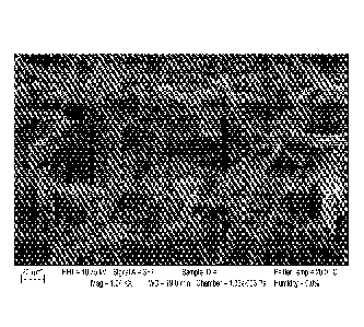
Figure 1 is an SEM image of the MPS rods. The random stacking and self
assembly of these rods generate a 3D space that allows for cell infiltration.
Figure 2 is a bar graph showing surface marker expression dependent on MPS
rods of different lengths. 100 pg of the MPS rods of different lengths were
incubated
with bone marrow derived dendritic cells for 18 hour. As a marker of
inflammation,
the percentage of activated cells was determined by staining for cell surface
receptors
CD1 lc (DC marker), MHC11 (antigen presentation marker) and CD86
(costimulatory
receptor). The inflammatory property of the rods increased with increasing
length.
Since a desired scaffold exhibits inflammatory properties (similar to the PLO
scaffold
control), rods between 30 and 100 pm or 120 p.m were used in subsequent
experiments.
Figure 3 is a series of images showing the effect of MPS rods in a mouse.
Figure 3A is a picture depicting a scaffold injection site excised after 7
days after 5
nig of rbodamine labeled MPS rod was injected subcutaneously into a C57b1/6j
mouse. The subcutaneous pocket indicates that the rods were localized and have
self-
assembled to create a 3D microenvironment. Figure 3B is an SEM image of the
excised scaffold site demonstrating cell infiltration into the scaffold site.
Figure 3C
depicts live/dead imaging of the cells detached from the MPS scaffold,
demonstrating
the recruitment of living cells.
Figure IA is a line graph showing GM-CSF release from MPS rods. 1 ue of
GM-CSF was loaded into the MPS rods and incubated in 0.1%BSAJPBS. Supernatant
was collected periodically and measured for GM-CSF using ELISA (R&D). GM-
CSI; is being continuously released from the MPS rods. Figure 4B is a bar
graph
showing surface marker expression in an in vitro bone marrow derived dendri
tic cell
5
CA 2870309 2019-09-30
CA 02870309 2014-10-10
WO 2013/158673
PCT/US2013/036827
(BMDC) co-culture with MPS. 10014 of the unmodified, amine-, thiol-, chloro-
and
phosphonate- modified MPS rods were incubated with 106/m1BMDCs for 18 hours.
The cells were then analyzed for the expression of MHC class-II and the
costimulatory molecule CD86. An increased expression of MHCII and CD86 of the
BMDCs stimulated by MPS indicates that the MPS rods are inflammatory, and this
property was modulated by surface-modifying the MPS rods.
Figure 5A is a bar graph depicting recruitment of CDs by GM-CSF loaded
MPS rods. 5 mg/mouse of MPS rods loaded with or without 1 fig/mouse GM-CST
were injected subcutaneously. Cells from the scaffold site were retrieved and
stained
for the CD lie receptor (DC marker). The MPS scaffold loaded with GM-CSF was
capable of recruiting more DCs than the blank MPS scaffold. Figure 5B is a bar
graph depicting surface marker expression of DCs. The MPS scaffold loaded with
GM-CSF and CpG-ODN activated more DCs than without CpG-ODN.
Figures 6A-B are bar graphs showing (Figure 6A) percentage of B220+ cells
or (Figure 6B) total number of B220+ cells at the scaffold site. 5 mg/mouse of
MPS
rods loaded with or without 1 .1g/mouse GM-CSF, 100 lug CpG-ODN and 130 lug
ovalbumin were injected subcutaneously. Cells from the scaffold site were
retrieved
periodically and stained for the B220 receptor (B cell marker). The MPS
scaffold
loaded with GM-CSF, CpG-ODN and Ova was capable of recruiting more B cells by
day 3.
Figures 7A-C are flow cytometry scatterplots (Figure 7A) and bar graphs
(Figures 7B-C) depicting the presence of the MHC-I-SIINFEKL (SEQ ID NO:1)
marker on the surface of DCs. The MPS scaffold loaded with GM-CSF, CpG-ODN
and the model antigen ovalbumin induced the expansion of antigen specific DCs
homed to the draining lymph node (dLN). dLN cells are stained with the
dendritic
marker CD11c and the marker MHC-I-SIINFEKL, which indicates that a part of the
ovalbumin protein, the peptide sequence SIINFEKL, is being presented on the
surface
of the cell in the MHC-I complex.
Figures 8A-B are bar graphs showing formation of the germinal center due to
the MPS scaffold. The MPS scaffold loaded with GM-CSF, CpG-ODN and the
model antigen ovalbumin was able to induce the germinal center, home to
activated
and antigen primed B cells, in the dLN. The dLN cells are stained with B220 (B
cell
marker) and GL7 (germinal center marker) to investigate the formation of the
6
CA 02870309 2014-10-10
WO 2013/158673
PCT/US2013/036827
geiminal center, which hosts only B cells that are activated and in the
process of
somatic hypermutation and isotype switching.
Figures 9A-B are bar graphs depicting that the MPS scaffold loaded with GM-
CSF, CpG-ODN and the model antigen ovalbumin was able to induce the expansion
of CD8+ cytotoxic T lymphocytes (CTLs) that can specifically recognize the
model
antigen. Splenocytes were stained for CD8 (killer T cell marker) and with a
MHCI-
tetramer-SIINFEKL antibody, which indicates whether a T cell is able to
recognize a
specific antigen presented on the MHCI complex.
Figures 10A-C are histograms showing that the MPS scaffold loaded with
GM-CSF, CpG-ODN and the model antigen ovalbumin was able to induce the clonal
expansion of antigen specific CD8+ CTLs in the dLN and the spleen. Injected
splenocytes from the OT-I mouse were stained the cell tracker dye CFSE, whose
fluorescence halves every time a cell divides. The lower fluorescence in the
CFSE
stained splenocytes in the spleen and the dLN in the fully loaded vaccine
group
indicates ova-specific CTL proliferation.
Figures 11 A-C are histograms showing that the MPS scaffold loaded with
GM-CSF, CpG-ODN and the model antigen ovalbumin was able to induce the clonal
expansion of antigen specific CD4+ THs in the dLN and the spleen. Injected
splenocytes from the OT-II mouse were stained the cell tracker dye CFSE, whose
fluorescence halves every time a cell divides. The lower fluorescence in the
CFSE
stained splenocytes in the spleen and the dLN in the fully loaded vaccine
group and
the MPS loaded with only the antigen group indicates ova-specific CD4+ TH cell
proliferation.
Figures 12 A-B are histograms showing that antibody titer is defined as the
degree to which the antibody-serum solution can be diluted and still contain a
detectable amount of the antibody. The anti-ovalbumin serum antibodies were
titrated. The MPS scaffold loaded with GM-CSF, CpG-ODN and ova elicited a
strong
and durable TH1 and TH2 antibody response. MPS loaded with OVA alone can
elicit
a strong TH2 response, indicating the adjuvant potential of the MPS material
itself.
Figures 13 A- B are line graphs showing that GM-CSF is released from the
MPS microparticles in a controlled and sustained manner. A) liug of GM-CSF was
loaded into 5mg of MPS rods and incubated in 0.1%BSA/PBS. Supernatant was
collected periodically and measured for GM-CSF using ELISA (R&D). GM-CSF was
continuously released from the MPS rods, although in small quantities. B) 1p g
of
7
CA 02870309 2014-10-10
WO 2013/158673
PCT/US2013/036827
GM-CSF was loaded into 5mg of MPS containing OVA and CpG. It was then
injected subcutaneously into C57BL/6j mice. At various time points, the tissue
surrounding the injected particle scaffold was harvested and analyzed for GM-
CSF
level. The level of GM-CSF was maintained throughout a week.
Figures 14 A-B are photographs. and Figure 14C is a bar graph. Higher aspect
ratio MPS microparticles recruit more cells. Figure 14A shows SEM images of
MPS
microparticles either between lOpm and 20 pm or between 80pm and 120pm in
length. The longer rods resulted in a composition with greater area or space
between
the rods. Figure 14B shows MPS compositions that was harvested from mice. Slug
of
MPS microparticles were injected subcutaneously into mice, and the scaffold
site was
harvested on day 7 post injection. Figure 14 C is a bar graph showing
enumeration of
cells that were isolated from the scaffold. MPS microparticles of higher
aspect ratio
recruited more cells.
Figures 15 A- B are bar graphs. Dendritic cells (DCs) are recruited to the MPS
microparticle as a response to GM-CSF. MPS microparticles were loaded with GM-
CSF (Ong, 500ng, 1000ng, 3000ng) and injected subcutaneously into mice. On day
7
post injection, the scaffold was harvested and analyzed using flow cytometry.
Figure
15A shows that recruited CD11c+ CD11b+ DCs increases with increasing GM-CSF
dose. Fig. 15B shows that recruited mature CD11c+ MHCII+ DCs also DCs
increases
with increasing GM-CSF dose.
Figure 16 is a bar graph showing that GM-CSF increases recruited DC
trafficking to the draining LN. MPS microparticles were loaded with Alexa 647
labeled ovalbumin (OVA), or Alexa 647 labeled OVA with 1p g GM-CSF, and
injected subcutaneously into mice. On day 7 post injection, the draining lymph
node
(dLN) was harvested and analyzed using flow cytometry. With the addition of
antigen (OVA), there is a modest increase of Alexa 647+ CD11c+ DCs that have
trafficked to the scaffold and up-taken the OVA. However, the addition of GM-
CSF
drastically increases DC trafficking from the scaffold to the dLN.
Figures 17 A-C are scatterplots and Figure 17D is a bar graph. Local delivery
of CpG-ODN from the MPS microparticle scaffold increases circulation of
activated
DCs. MPS microparticles were loaded with lug of GM-CSF, 300ug of OVA and
100ug of CpG-ODN. They were then injected subcutaneously into mice. The dLNs
were harvested and analyzed after 7 days post injection. 1,N cells were
stained with
CD11 c and CD86. The addition of CpG-ODN, which is locally released from the
8
CA 02870309 2014-10-10
WO 2013/158673
PCT/US2013/036827
MPS scaffold, further increases the percentage and number of activated, mature
DCs
in the dLN.
Figures 18 A-C are photographs and Figure 18 D is a bar graph. Proteins are
released from the MPS microparticle scaffold in a controlled and sustained
manner.
Alexa 647 labeled ovalbumin was injected subcutaneously into the flank of a
mouse
either in a buffer solution or loaded onto the MPS microparticles. Relative
fluorescence was measured using the IVIS at various time points. If injected
in a
buffer solution, the protein diffuses away from the injection site in less
than 1 day.
However, if loaded onto the MPS microparticles, the protein is released from
the local
scaffold site in a sustained manner, e.g., over the course of a week (2, 3, 4,
5, 7 or
more days).
Figures 19 A-B are line graphs showing antibody titers. Antibody titer is
defined as the degree to which the antibody-serum solution can be diluted and
still
contain a detectable amount of the antibody. Mice were vaccinated with 300 g
OVA,
100pg CpG-ODN and littg GM-CSF in the soluble form or loaded in Sing of MPS
microparticles. The anti-ovalbumin serum antibodies are titrated. The MPS
scaffold
loaded with GM-CSF, CpG-ODN and ova can elicit a strong and durable TH1 and
TH2 antibody response, as indicated by a high titer of the IgG2a and IgG1
antibody,
respectively. More impressively, this response is evident beyond 200 days
after
vaccination with a single injection. This response was compared to OVA
delivered
using aluminum hydroxide, the only adjuvant approved for human use in the US.
While the MPS vaccine is capable of inducing both TH1 and TH2 antibody
responses,
due to its capability to load small cytokines, proteins and DNAs, the response
induced
by alum is completely TH2 biased. The MPS vaccine is very versatile and is
readily
fine tuned and controlled to induce specific immune responses.
Figures 20 A-D are scatterplots showing that vaccination with the MPS
vaccine induces the expansion of T follicular helper cells. OT-II splenocytes
were
stained with CFSE and adoptively transferred into Thy1.1+ recipient mice. The
recipient mice were then vaccinated with MPS and lysozyme, MPS and OVA, and
the
full form of the vaccine containing GM-CSF and CpG. Three days after
vaccination,
dLN were harvested and the adoptively transferred cells were analyzed. It is
shown
here that mice that were vaccinated with MPS and ova, and the full form of the
vaccine, generated a strong population of follicular T helper cells, which are
the cells
9
CA 02870309 2014-10-10
WO 2013/158673
PCT/US2013/036827
directly responsible for "helping- B cells to differentiate and mature into
full,
functional antigen specific antibody secreting plasma cells.
Figures 21 A-D are line graphs showing that vaccination with the MPS
vaccine induces the clonal expansion of CD4+ T helper cells. OT-II splenocytes
were
stained with CFSE and adoptively transferred into Thy1.1+ recipient mice. The
recipient mice were then vaccinated with MPS and lysozyme, MPS and OVA, and
the
full form of the vaccine containing GM-CSF and CpG. Three days after
vaccination,
dLN were harvested and the adoptively transferred cells were analyzed. It is
shown
here that both MPS and OVA, and the full vaccine, induced strong clonal
expansion
of the CD4+ T helper cells, indicating a systemic antigen specific response.
Figure 22 A-B are line graphs showing that glycolic acid and lactic acid
modification of the MPS microparticles aids the release of bioactive GM-CSF.
1p g
of GM-CSF was loaded into 5mg of glycolic acid or lactic acid modified MPS
rods
and incubated in 0.1%BSA/PBS. Supernatant was collected periodically and
measured for GM-CSF using ELISA (R&D). Compared to GM-CSF released from
unmodified MPS microparticles, glycolic acid and lactic acid modification
increasecumulative release of bioactive GM-CSF by more than 20 fold.
Figures 23 A-D are scatterplots showing that glycolic acid modified MPS
increases the percentage of recruited DCs and mature DCs. 5mg of unmodified or
glycolic acid modified MPS microparticles were loaded with 1pg of GM-CSF for 1
hour at 37C, and injected subcutaneously into mice. The scaffold was harvested
at
day 7 and analyzed. It is evident here that the modified MPS almost doubles
the
percentage of recruited CD11c+ CD11b+ DCs, and more drastically increases the
percentage of CD11c+ CD86+ mature DCs. These results indicate that the great
surface modification potential of the MPS microparticles permit further
manipulation
of the phenotype of recruited cells to induce more potent immune responses.
DETAILED DESCRIPTION
Injectable MPS-based micro-rods randomly self-assemble to form 3D scaffold
in vivo. This system is designed such that it releases a cytokine to recruit
and
transiently house immune cells, present them with an antigen, and activate
them with
a danger signal. After recruitment and temporary housing or presence of the
cells in
the structure, these immune cells migrate out of the device structure and
homed to a
lymph node. Thus, the composition is one in which cells traffic/circulate in
and out
CA 02870309 2014-10-10
WO 2013/158673
PCT/US2013/036827
of, their status of immune activation being altered/modulated as a result of
the
trafficking through the device..
A markedly expanded population of antigen specific dendritic cells was found
in the lymph node as well as the formation of the geiminal center, which is
aided by
the involvement of follicular T helper cells in the lymph node as a result of
the
administration of the device. A population of antigen-recognizing CD8+ CTLs in
the
spleen was also found to be significantly enhanced. The system also induces
the
clonal expansion of both antigen specific CD4 and CD8 T cells. In addition to
the
significant amplification of the cellular immune response, a high and durable
antibody
production and a balanced T111/T112 response was detected.
Key elements of the injectable mesoporous silica micro-rod based scaffold
system for modulating immune cell trafficking and reprogramming include:
= Aspect ratio of the MPS micro rods (10 nm cross sectional area by 100
micron
length) prevents their uptake and therefore allows for local foimation of a 3D
structure.
= 8 nm pore size allows for adsorption of small molecules that can be
delivered
via diffusion in a controlled and continuous manner.
= High surface area to volume ratio allows for the control of the loading
of
various cytokines, danger signal and antigen.
= Inflammatory property of the MPS micro rods promotes immune cell
recruitment without the need of other inflammatory cytokines.
= Versatility in ability of surface chemical modification of the MPS rods
that
modulate properties of the rods and ways that proteins are bound to the rods.
= Controlled release of GM-CSF can modulate the trafficking of immune cells
to the site of the scaffold.
= Controlled release of CpG-ODN activates the recruited dendritic cells.
= Anchored protein antigens are taken up by the recruited immune cells at
the
site of the scaffold.
A pore size of approximately 5-10, e.g., 8 nm, was found to be optimal for
loading efficiency for small molecules as well as larger proteins, e.g., GM-
CSF
(which molecule is about 3 nm in diameter).
Upon subcutaneous injection of the micro rods suspended in a buffer, the rods
randomly stack into a 3D structure; the ends of the rods form physical contact
with
each other, and a micro space is formed between the contacts.
11
MPS
Mesoporous silica nanoparticles are synthesized by reacting tetraethyl
orthosilicate with a template made of micellar rods. The result is a
collection of nano-
sized spheres or rods that are filled with a regular arrangement of pores. The
template
can then be removed by washing with a solvent adjusted to the proper pH. In
another
technique, the mesoporous particle could be synthesized using a simple sol-gel
method or a spray drying method. Tetraethyl orthosilicate is also used with an
additional polymer monomer (as a template). Other methods include those
described
in U.S. Patent Publication 20120264599 and 20120256336.
Granulocyte Macrophage Colony Stimulating Factor (GM-CSF)
Granulocyte-macrophage colony-stimulating factor (GM-CSF) is a protein
secreted by macrophages, T cells, mast cells, endothelial cells and
fibroblasts.
Specifically, GM-CSF is a cytokine that functions as a white blood cell growth
factor.
GM-CSF stimulates stem cells to produce granulocytes and monocytes. Monocytes
exit the blood stream, migrate into tissue, and subsequently mature into
macrophages.
Scaffold devices described herein comprise and release GM-CSF polypeptides
to attract host DCs to the device. Contemplated GM-CSF polypeptides are
isolated
from endogenous sources or synthesized in vivo or in vitro. Endogenous GM-CSF
polypeptides are isolated from healthy human tissue. Synthetic GM-CSF
polypeptides
are synthesized in vivo following transfection or transformation of template
DNA into
a host organism or cell, e.g. a mammal or cultured human cell line.
Alternatively,
synthetic GM-CSF polypeptides are synthesized in vitro by polyinerase chain
reaction
(PCR) or other art-recognized methods Sambrook, J., Fritsch, E.F., and
Maniatis, T.,
Molecular Cloning: A Laboratory Manual. Cold Spring Harbor Laboratory Press,
NY,
Vol. 1, 2, 3 (1989).
GM-CSF polypeptides are modified to increase protein stability in vivo.
Alternatively, GM-CSF polypeptides are engineered to be more or less
immunogenic.
Endogenous mature human (.3M-CSF polypeptides are glycosylated, reportedly, at
amino acid residues 23 (leucine), 27 (asparagine), and 39 (glutaniic acid)
(see US
Patent No. 5,073,627). GM-CSF polypeptides of the present invention are
modified at
one or more of these amino acid residues with respect to glycosylation state.
12
CA 2870309 2019-09-30
CA 02870309 2014-10-10
WO 2013/158673
PCT/US2013/036827
GM-CSF polypeptides are recombinant. Alternatively GM-CSF polypeptides
are humanized derivatives of mammalian GM-CSF polypeptides. Exemplary
mammalian species from which GM-CSF polypeptides are derived include, but are
not limited to, mouse, rat, hamster, guinea pig, ferret, cat, dog, monkey, or
primate. In
a preferred embodiment, GM-CSF is a recombinant human protein (PeproTech,
Catalog # 300-03). Alternatively, GM-CSF is a recombinant murine (mouse)
protein
(PeproTech, Catalog #315-03) . Finally, GM-CSF is a humanized derivative of a
recombinant mouse protein.
Human Recombinant GM-CSF (PeproTech, Catalog # 300-03) is encoded by
the following polypeptide sequence (SEQ ID NO:2):
MAPARSPSPS TQPWEHVNAI QEARRLLNLS RDTAAEMNET VEVISEMFDL QEPTCLQTRL
ELYKQGLRGS LTKLKGPLTM MASI IYKQIICP PTPETSCATQ IIFILSFKEN LKDFLEVIPF
DCWEPVQE
Murine Recombinant GM-CSF (PeproTech, Catalog # 315-03) is encoded by
the following polypeptide sequence (SEQ Ill NO: 3):
MAPTRSPITV TRPWKHVEAI KEALNLLDDM PVTLNEEVEV VSNEFSFKKL TCVQTRLKIF
EQGLRGNFTK LKGALNMTAS YYQTYCPPTP ETDCETQVTT YADFIDSLKT FLTDIPFECK
KPVQK
Human Endogenous GM-CSF is encoded by the following mRNA sequence
(NCBI Accession No. NM_000758 and SEQ ID NO: 4):
1 acacagagag aaaggctaaa gttctctgga ggatgtggct gcagagcctg ctgctcttgg
61 gcactgtggc ctgcagcatc tctgcacccg cccgctcgcc cagccccagc acgcagccct
121 gggagcatgt gaatgccatc caggaggccc ggcgtctcct gaacctgagt agagacactg
181 ctgctgagat gaatgaaaca gtagaagtca tctcagaaat gtttgacctc caggagccga
241 cctgcctaca gacccgcctg gagctgtaca agcagggcct gcggggcagc ctcaccaagc
301 tcaagggccc cttgaccatg atggccagcc actacaagca gcactgccct ccaaccccgg
361 aaacttcctg tgcaacccag attatcacct ttgaaagttt caaagagaac ctgaaggact
421 ttctgcttgt catccccttt gactgctggg agccagtcca ggagtgagac cggccagatg
481 aggctggcca agccggggag ctgctctctc atgaaacaag agctagaaac tcaggatggt
541 catcttggag ggaccaaggg gtgggccaca gccatggtgg gagtggcctg gacctgccct
601 gggccacact gaccctgata caggcatggc agaagaatgg gaatatttta tactgacaga
661 aatcagtaat atttatatat ttatattttt aaaatattta tttatttatt tatttaagtt
721 catattccat atttattcaa gatgttttac cgtaataatt attattaaaa atatgcttct
781 a
Human Endogenous (IM-CSF is encoded by the following amino acid
sequence (NCBI Accession No. NP_000749.2 and SEQ ID NO: 5):
MWLQSLLLLGTVACSISAPARSPSPSTQPWEHVNAIQEARRLLNLSRDTAAEM
NETVEVIS EMFDLQEPTCLQTRLELY KQGLRGS LTKLKGPLTMMAS HY KQH C
PPTPETSCATQUTFESFKENI,KDFI J,VIPFDCWEPVQE
13
Cyrosine-Guanosine.(CpC) Oligonudeotide (CpG-ODN) Sequences
CpG sites are regions of deoxyribonucleic acid (DNA) where a cysteine
nucleotide occurs next to a guanine nucleotide in the linear sequence of bases
along
its length (the "p" represents the phosphate linkage between them and
distinguishes
them from a cytosine-guanine complementary base pairing). CpCI sites play a
pivotal
role in DNA methylation, which is one of several endogenous mechanisms cells
use
to silence gene expression. Methylation of CpG sites within promoter elements
can
lead to gene silencing. In the case of cancer, it is known that tumor
suppressor genes
are often silenced while oncogenes, or cancer-inducing genes, are expressed.
CpG
sites in the promoter regions of tumor suppressor genes (which prevent cancer
formation) have been shown to be methylated while CpG sites in the promoter
regions
of oncogenes are hypornethylated or unmethylated in certain cancers. The TLR-9
receptor binds unmethylated CpG sites in DNA.
The vaccine composition described herein comprises CpG oligonucleotides.
CpG oligonucleotides are isolated from endogenous sources or synthesized in
vivo or
in vitro. Exemplary sources of endogenous CpG oligonucleotides include, but
are not
limited to, microorganisms, bacteria, fungi, protozoa, viruses, molds, or
parasites.
Alternatively, endogenous CpG oligonucleotides are isolated from mammalian
benign
or malignant neoplastic tumors. Synthetic CpG oligonucleotides are synthesized
in
vivo following transfection or transformation of template DNA into a host
organism.
Alternatively, Synthetic CpG oligonucleotides are synthesized in vitro by
polymerase
chain reaction (PCR) or other art-recognized methods (Sambrook, 3., Fritsch,
E.F.,
and Maniatis, T., Molecular Cloning: A Laboratory Manual. Cold Spring Harbor
Laboratory Press, NY, Vol. 1, 2, 3 (l989),
CpG oligonucleotides are presented for cellular uptake by dendritic cells. For
example, naked CpG oligonucleotides are used. The term "naked" is used to
describe
an isolated endogenous or synthetic polynucleotide (or oligonucleotide) that
is free of
additional substituents. In another embodiment, CpG ohgonucleotides are bound
to
one or more compounds to increase the efficiency of cellular uptake.
Alternatively, or
in addition, cpu oligonucleotides are bound to one or more compounds to
increase
the stability of the oligonucleotide within the scaffold and/or dendritic
cell. CpG
oligonucleotides are optionally condensed prior to cellular uptake. For
example, CpG
oligonucleotides are condensed using polyethylimine (PEI), a cationic polymer
that
increases the efficiency of cellular uptake into dendritic cells.
14
CA 2870309 2019-09-30
CA 02870309 2014-10-10
WO 2013/158673
PCT/US2013/036827
CpG oligonucleotides can be divided into multiple classes. For example,
exemplary CpG-ODNs encompassed by compositions, methods and devices of the
present invention are stimulatory, neutral, or suppressive. The teini
"stimulatory"
describes a class of CpG-ODN sequences that activate TLR9. The term "neutral"
describes a class of CpG-ODN sequences that do not activate TLR9. The term
"suppressive" describes a class of CpG-ODN sequences that inhibit TLR9. The
term
"activate TLR9" describes a process by which TLR9 initiates intracellular
signaling.
Stimulatory CpG-ODNs can further be divided into three types A, B and C,
which differ in their immune-stimulatory activities. Type A stimulatory CpG
ODNs
are characterized by a phosphodiester central CpG-containing palindromic motif
and a
phosphorothioate 3' poly-G string. Following activation of TLR9, these CpG
ODNs
induce high IFN-a production from plasmacytoid dendritic cells (pDC). Type A
Cp0
ODNs weakly stimulate TLR9-dependent NF-KB signaling.
Type B stimulatory CpG ODNs contain a full phosphorothioate backbone with
one or more CpG dinucleotides. Following TLR9 activation, these CpG-ODNs
strongly activate B cells. In contrast to Type A CpG-ODNs, Type B CpG-ODNS
weakly stimulate IFN-a secretion.
Type C stimulatory CpG ODNs comprise features of Types A and B. Type C
CpG-ODNs contain a complete phosphorothioate backbone and a CpG containing
palindromic motif. Similar to Type A CpG ODNs, Type C CpG ODNs induce strong
IFN-a production from pDC. Simlar to Type B CpG ODNs, Type C CpG ODNs
induce strong B cell stimulation.
Exemplary stimulatory CpG ODNs comprise, but are not limited to, ODN
1585, ODN 1668, ODN 1826, ODN 2006, ODN 2006-G.5, ODN 2216, ODN 2336,
ODN 2395, ODN M362 (all InvivoGen). The present invention also encompasses any
humanized version of the preceding CpG ODNs. In one preferred embodiment,
compositions, methods, and devices of the present invention comprise ODN 1826
(the
sequence of which from 5' to 3' is tccatgacgttcctgacgtt, wherein CpG elements
are
bolded, SEQ ID NO: 10).
Neutral, or control, CpG ODNs that do not stimulate '1'LR9 are encompassed
by the present invention. These ODNs comprise the same sequence as their
stimulatory counterparts but contain GpC dinucleotides in place of CpG
dinucleotides.
Exemplary neutral, or control, CpG ODNs encompassed by the present
invention comprise, but are not limited to, ODN 1585 control, ODN 1668
control,
ODN 1826 control, ODN 2006 control, ODN 2216 control, ODN 2336 control, ODN
2395 control, ODN M362 control (all InvivoGen). The present invention also
encompasses any humanized version of the preceding CpG ODNs.
Antitens
Compositions, methods, and devices described herein comprise tumor antigens
or other antigens. Antigens elicit protective immunity or generate a
therapeutic
immune response in a subject to whom such a device was administered. Preferred
tumor antigens are tumor cell lysates (see, e.g., Ali et. AL, 2009, Nature
Materials 8,
151 158). For example, a
whole tumor or tumor
biopsy sample is extracted from a human patient (or non-human animal), and
digested
using collagenase to degrade the extra cellular matrix. Then the tumor cells
then
undergo three cycles of the freeze thaw process, in which the cells are frozen
in liquid
N2 and then thawed in a water bath. This process generates the tumor antigens,
which
are then loaded onto the MPS particles at the same time with GM-CSF and CpG
ODN. For example, a vaccine dose of tumor cell lys ate antigen is the amount
obtained from 1 x 10⁶ tumor cells. After fabrication of the MPS particles, the
particles are suspended in a physiologically accepted buffer, e.g., PBS, and
the
recruitment compound (e.g., GM-CSF), activating compound (e.g., CpG), and
antigen
added to the suspension of rods. The mixture is shaken at room temperature
overnight and then lyophilized for about 4 hours. Prior to administration, the
rods are
again suspended in buffer and the suspension loaded into a 1 ml syringe (18
gauge
needle). A typical vaccine dose is 150 pl of the mixture per injection.
Exemplary cancer antigens encompassed by the compositions, methods, and
devices of the present invention include, but are not limited to, tumor
lysates extracted
from biopsies, irradiated tumor cells, MACE series of antigens (MACE-1 is an
example), MART- Umelana, tyrosinase, ganglioside, gp100, GD-2, 0-acetylated OD-
3, GM-2, MIK:- I, Sosl, Protein kinase C-binding protein, Reverse
transcriptase
protein, AKAP protein, VRKI, KIAA1735, 17-1, T11-3, T11-9, Homo Sapiens
telomerase ferment (hTRT), Cytokeratin-19 (CYFRA21-1), SQUAMOUS CELL
CARCINOMA ANTIGEN 1 (SCCA-1), (PROTEIN T4-A), SQUAMOUS CELL
CARCINOMA ANTIGEN 2 (SCCA-2), Ovarian carcinoma antigen CA125 (1A1-3B)
16
CA 2870309 2019-09-30
CA 02870309 2014-10-10
WO 2013/158673
PCT/US2013/036827
(KIAA0049), MUCIN 1 (TUMOR-ASSOCIATED MUCIN), (CARCINOMA-
ASSOCIATED MUCIN), (POLYMORPHIC EPITHELIAL
MIJCIN),(PEM),(PEMT),(EPISIALIN), (TUMOR-ASSOCIATED EPITIIELIAL
MEMBRANE ANTIGEN),(EMA),(H23AG), (PEANUT-REACTIVE URINARY
MUCIN), (PUM), (BREAST CARCINOMA- ASSOCIATED ANTIGEN DF3),
CTCL tumor antigen se1-1, CTCL tumor antigen se14-3, CTCL tumor antigen se20-
4, CTCI, tumor antigen se20-9, CTCI. tumor antigen se33-1, CTCI, tumor antigen
se37-2, CTCL tumor antigen se57-1, CTCL tumor antigen se89-1, Prostate-
specific
membrane antigen, 5T4 oncofetal trophoblast glycoprotein, Orf73 Kaposi's
sarcoma-
associated heipesvirus, MAGE-Cl (cancer/testis antigen CT7), MAGE-Bl
ANTIGEN (MAGE-XP ANTIGEN) (DAM10), MAGE-B2 ANTIGEN (DAM6),
MAGE-2 ANTIGEN, MAGE-4a antigen, MAGE-4b antigen, Colon cancer antigen
NY-CO-45, Lung cancer antigen NY-LU-12 variant A, Cancer associated surface
antigen, Adenocarcinoma antigen ART1, Paraneoplastic associated brain-testis-
cancer
antigen (onconeuronal antigen MA2; paraneoplastic neuronal antigen), Neuro-
oncological ventral antigen 2 (NOVA2), IIepatocellular carcinoma antigen gene
520,
TUMOR-ASSOCIATED ANTIGEN CO-029, Tumor-associated antigen MAGE-X2,
Synovial sarcoma, X breakpoint 2, Squamous cell carcinoma antigen recognized
by T
cell, Serologically defined colon cancer antigen 1, Serologically defined
breast cancer
antigen NY-BR-15, Serologically defined breast cancer antigen NY-BR-16,
Chromogranin A; parathyroid secretory protein 1, DUPAN-2, CA 19-9, CA 72-4, CA
195, Carcinoembryonic antigen (CEA). Purified tumor antigens are used alone or
in
combination with one another.
The system is also useful to generate an immune response to other antigens
such as microbial pathogens (e.g., bacteria, viruses, fungi).
The following materials and methods were used to generate the data described
herein.
MPS scaffold fabrication
The Pluronic P-123 (Sigma-Aldrich) surfactant was dissolved in 1.6M HCI at
room temperature, and heated 40 degrees C. 42 mmol of Tetraethyl orthosilicate
(TEOS) (Sigma-Aldrich) was added and heated for 20 hours at 40 degrees C under
stifling (600 rpm). The composition was then heated to 100 degrees C for 24
hours.
The rod particles were collected by filtration and air dried at room
temperature. The
particles were extracted in ethanol/HC1 (5 parts HCl to 500 parts Et0H)
overnight at
17
CA 02870309 2014-10-10
WO 2013/158673
PCT/US2013/036827
80 degrees C. Alternatively, the particles were calcined at 550 degrees C for
4 hours
in 100% ethanol. The MPS composition may be stored and shipped for use before
or
after adding recruitment and/or activation compounds and before or after
adding
antigen. For example, if antigen is a tumor cells lysate made from a biopsy
sample
taken from a patient, it may be processed and added to MPS particles shortly
before
administration to the patient.
For full vaccine composition, 1 lug/mouse GM-CSF, 100 jug/mouse CpG-
ODN and 130 lug/mouse of the ovalbumin protein were incubated with 5 mg/mouse
MPS in dH20 for 12 hours, lyophilized for 12 hours, resuspended in 150
1/mouse
PBS and injected subcutaneously using a 18G needle.
To determine the release kinetics of GM-CSF, and CpG-ODN from the MPS
rods, radioactive I-labeled recombinant human GM-CSF was used as a tracer, and
standard release studies were carried out. Similarly, the amount of CpG-ODN
released into PBS was determined by the absorbance readings using a Nanodrop
instrument (ND1000, Nanodrop Technologies).
Modification of MPS microparticles
Standard 1-ethyl-3-(3-dimethylaminopropyl)carbodiimide (EDC) chemistry is
used to activate a carboxylic acid on glycolic acid or lactic acid. The MPS
particles
are amine modified using amine propyl silane. The two components are then
mixed
together at room temperature overnight to react. The particles are then
allowed to air
dry. "[he resulting glycolic acid and/or lactic acid modified particles are
then
contacted with GM-CSF or other compounds as described above.
In vitro DC activation assay
Murine Dendritic Cells (DCs) were differentiated from the bone marrow cells
using 20 ng/ml GM-CSF. Differentiated DCs, at 106/ml, were incubated with 100
pg/m1 of the MPS particles for 18 hours, and analyzed for the expression of
CD1 lc
(ebiosciences, San Diego, CA), CD86 (ebiosciences, San Diego, CA) and MHC
class-fl (ebiosciences, San Diego, CA) using flow cytometry.
Analysis of DC recruitment to MPS scaffold and emigration to lymph nodes
The MPS scaffold was retrieved from the animal and digested by
mechanically separating the cells from the rods. APC conjugated CD11c
(dendritic
cell marker), FITC conjugated MHC class-II, and PE conjugated CD86 (B7,
costimulatory molecule) stains were used for DC and leukocyte recruitment
analysis.
Cells were gated according to positive fluorescein isothiocyanate (FITC),
18
Allophycocyanin (APC) and phycoerythrin (PE) using isotype controls, and the
percentage of cells staining positive for each surface antigen was recorded.
To determine the presence of antigen specific DCs in the lymph nodes, the
draining lymph node (dLN) was harvested and analyzed using APC conjugated
CD11c and PE conjugated SIINFEKL-MHC class 1 (ebioseiences, San Diego, CA).
Analysis of antigen specific CD8+ spleen T cells
The spleen was harvested and digested. After lysing the red blood cells, the
splenocytes were analyzed using PE-CY7 conjugated CD3 (ebiosciences, San
Diego,
CA), APC conjugated CD8 (ebioscienees, San Diego, CA) and the PE conjugated
SIINEEKL (SEQ ID NO: ) MI-1C class4 tetramer (Beckman Coulter). SIINFF,KL is
an ovalbumin derived peptide lOVA(257-264)].
Analysis of CD4+ or CD8+ T cell clonal expansion
The spleens from the 0T41 (for CD4) or OT-1 (for C08) transgenic C57bI/6
mice (Jackson Laboratories) were harvested, digested, pooled and stained with
the
cell tracer Carboxyfluorescein succinirnidyl ester (CFSE). 20x106 stained
splenocytes/mouse were IV injected into the C57b1/6 (Jackson Laboratories)
mice two
days post immunization. The dENs and spleens were retrieved after four days
post IV
injection and analyzed using PE conjugated CD8 or CD4 marker.
Characterization of anti-OVA humoral response
Blood sera were analyzed for IgGl, and IgG2a antibodies by EL1SA using
ovalbumin-coated plates. Antibody titration was defined as the lowest senun
dilution
at which the ELISA OD reading is >0.3 (blank).
OTHER EMBODIMENTS
While the invention has been described in conjunction with the detailed
description thereof, the foregoing description is intended to illustrate and
not limit the
scope of the invention, which is defined by the scope of the appended claims.
Other
aspects, advantages, and modifications are within the scope of the following
claims.
The patent and scientific literature referred to herein establishes the
knowledge
that is available to those with skill in the art.
19
CA 2870309 2019-09-30
While this invention has been particularly shown and described with
references to preferred embodiments thereof, it will he understood by those
skilled in
the art that various changes in form and details may be made therein without
departing from the scope of the invention encompassed by the appended claims.
CA 2870309 2019-09-30