Note : Les descriptions sont présentées dans la langue officielle dans laquelle elles ont été soumises.
MULTI-LUMEN CATHETER
CROSS REFERENCE TO RELATED APPLICATIONS
[0001] This application claims priority to U.S. Patent Application No.
13/453,663,
filed April 23, 2012, which is a continuation-in-part of U.S. Patent
Application No.
12/479,257, filed June 5, 2009, which claims priority as a continuation of
U.S. Patent
Application No. 11/103,778, filed April 12, 2005, now U.S. Patent No.
7,569,029, which
claims the benefit of -U.S. Provisional Application Serial No. 60/561,430,
filed April 12,
2004. This application also claims the priority benefit of U.S. Provisional
Application No.
61/477,815, filed April 21, 2011, entitled "Dialysis Catheter."
BACKGROUND
[0002] The present invention relates to a multi-lumen catheter and, more
specifically, to a dual-lumen catheter with entry and exit ports having curved
or angled walls
to direct the flow of fluids therethrough.
[0003] Dual-lumen catheters have been available for many years for a variety
of
medical purposes. It is only in recent years, however, that such catheters
have been
developed for use in hemodialysis and other treatments which involve the
removal and
replacement of blood. The general form of dual-lumen catheters goes back to as
early as
1882 when Pfarre patented such a catheter in the United States under Ser. No.
256,590. This
patent teaches a flexible dual-lumen catheter which is used primarily for
cleaning and
drainage of, for example, the bladder, rectum, stomach and ear. In this type
of catherization,
CA 2871329 2018-09-12
the catheter is introduced into an existing body orifice without the use of
any puncturing
needle or guide wire.
[0004] More recently, a catheter was developed and patented by Blake et al.
under
U.S. Pat. No. 3,634,924. This patent teaches a double lumen cardiac balloon
catheter which
is introduced into a large vein and the balloon is inflated to control the
flow in the vein. The
catheter can be placed by using the balloon as a "sail" to move with the blood
from an ante-
cubital or other peripheral vein through for example, the right heart chambers
into the smaller
radicals of the pulmonary artery where the catheter takes up its intended
function. This
patent teaches the use of two lumina in a single body and explains how to make
a tip for a
dual-lumen structure of the type which has become common for a variety of
purposes
including hemodialysis. The structure uses a plug to seal the end of one lumen
and a wire
which retains the shape of the other lumen during formation of the tip in a
heated die.
[0005] Further patents which teach dual-lumen or multiple lumen catheters for
general use include the following: U.S. Pat. Nos. 701,075; 2,175,726;
2,819,718; 4,072,146;
4,098,275; 4,134,402; 4,180,068; 4,406,656; 4,451,252; 5,221,255; 5,380,276;
5,395,316;
5,403,291; 5,405,341; 6,001,079; 6,190,349; 6,719,749; 6,758,836; and
6,786,884.
100061 Vascular catheter access techniques have been known to the medical
profession for many years and, in fact, can be traced back to the 17th
century. However, it
was only with the introduction of the Seldinger technique in the early 1950s
that a new
approach could be used to improve vascular access. This technique was taught
in an article
published by Dr. Sven Ivar Seldinger resulting from a presentation made at the
Congress of
the Northern Association of Medical Radiology at Helsinki in June of 1952. The
technique
essentially involves the use of a hollow needle to make an initial puncture,
and a wire is then
entered through the needle and positioned in the vessel. The needle is
withdrawn and the
2
CA 2871329 2018-09-12
CA 02871329 2014-10-22
WO 2013/163172
PCT[US2013/037783
catheter is entered percutaneously over the wire which is itself later
withdrawn. With this
technique it became possible to make less traumatic vascular access and this
has now become
the accepted method of performing access in numerous medical techniques. One
of these
techniques which been the subject of much research and development is
hemodialysis.
[0007] Hemodialysis can be defined as the temporary removal of blood from a
patient for the purpose of extracting or separating toxins therefrom and the
return of the
cleansed blood to the same patient. Hemodialysis is indicated in patients
where renal
impairment or failure exists, that is, in cases where the blood is not being
properly or
sufficiently cleansed, particularly to remove waste materials and water, by
the kidneys.
[0008] In the case of chronic renal impairment or failure, hemodialysis has to
be
carried out on a repetitive basis. For example, in end-stage kidney disease
where
transplantation of kidneys is not possible or for medical reasons is contra-
indicated, the
patient may have to be dialyzed about 100 to 150 times per year. This can
result in several
thousand accesses to the blood stream to enable hemodialysis to be performed
over the
remaining life of the patient.
[0009] Towards the end of 1960, Dr. Stanley Shaldon and colleagues developed,
in
the Royal Free Hospital in London, England, a technique for hemodialysis by
percutaneous
catheterization of deep blood vessels, specifically the femoral artery and
vein. The technique
was described in an article published by Dr. Shaldon and his associates in the
Oct. 14, 1961
edition of The Lancet at Pages 857 to 859. Dr. Shaldon and his associates
developed single
lumen catheters having tapered tips for entry over a Seldinger wire to be used
in
hemodialysis. Subsequently, Dr. Shaldon and his colleagues began to insert
single lumen
inlet and outlet catheters in the femoral vein and this was reported in the
British Medical
Journal of Jun. 19, 1963. The purpose of providing both inlet and outlet
catheters in the
femoral vein was to explore the possibility of a "self-service" approach to
dialysis. Dr.
3
CA 02871329 2014-10-22
WO 2013/163172
PCT[US2013/037783
Shaldon was subsequently successful in doing this and patients were able to
operate
reasonably normally while carrying implanted catheters which could be
connected to
hemodialysis equipment periodically.
[0010] An advantage of dual-lumen catheters in hemodialysis is that only one
vein
access need be affected to establish continued dialysis of the blood. One
lumen serves as the
conduit for blood flowing from the patient to the dialysis unit and the other
lumen serves as a
conduit for treated blood returning from the dialysis unit to the patient.
This contrasts with
prior systems where either two insertions were necessary to place two separate
catheters as
was done by Dr. Shaldon, or a single catheter was used with a complicated
dialysis machine
which alternately removed blood and returned cleansed blood.
[0011] The success of Dr. Shaldon in placing catheters which will remain in
place
for periodic hemodialysis caused further work to be done with different sites.
Dr. Shaldon
used the femoral vein, and in about 1977 Dr. P. R. Uldall, in Toronto Western
Hospital,
Canada, began clinical testing of a subclavian catheter that would remain in
place between
dialysis treatments. An article describing this was published by Dr. Uldall
and others in
Dialysis and Transplantation, Volume 8, No. 10, in October 1979. Subsequently
Dr. Uldall
began experimenting with a coaxial dual-lumen catheter for subclavian
insertion and this
resulted in Canadian Patent No. 1,092,927 which issued on Jan. 6, 1981.
Although this
particular form of catheter has not achieved significant success in the
marketplace, it was the
forerunner of dual-lumen catheters implanted in the subclavian vein for
periodic
hemodialysis.
[0012] The next significant step in the development of a dual-lumen catheter
for
hemodialysis is Canadian Patent No. 1,150,122 to Martin. A subsequent
development is
shown in U.S. Pat. No. 4,451,252 also to Martin. This catheter utilizes the
well-known dual-
.. lumen configuration in which the lumina are arranged side-by-side separated
by a diametric
4
septum. The structure shown in this patent provides for a tip making it
possible to enter a
Seldinger wire through one of the lumina and to use this wire as a guide for
inserting the
catheter percutaneously. This type of structure is shown in a European Patent
Application to
Edelman published under No. 0 079 719, and in U.S. Pat. Nos. 4,619,643;
4,583,968;
4,568,329; 4,543,087; 4,692,141; 4,568,329, and U.S. Des. Pat. No. 272,651.
100131 In order to insert a catheter over a guide wire using the Seldinger
technique,
or indeed any similar technique, the tip of the catheter must possess
sufficient rigidity so that
it does not concertina as it contacts the skin because this would enlarge the
skin puncture as
the catheter is being entered over the wire. To some extent this is at odds
with the desirable
material qualities of the main body of catheter which should be soft and
flexible for patient
comfort. In an effort to solve this problem, a variety of tips have been
formed within the
limitations of using a single extrusion from which the body and tip are
formed. The result is
that the tips have in general been made by using some of the excess material
found in the
shorter intake lumen. This has led to other problems such as very stiff tips
which are
unsuitable for prolonged placement in a vein; voids which can accumulate
stagnant blood;
and short stubby tips which are less desirable for insertion than longer more
gradual tips.
Also, because there is not always sufficient material to form the tip, plugs
have been added
with a varying degree of success because if the plug is not placed accurately
the resulting
structure may have unacceptable spaces where blood can stagnate.
100141 It must also be recognized that the degree of rigidity in the tip
becomes more
important if the catheter is to reside in the patient for prolonged periods,
as is becoming more
common in many treatments, notably hemodialysis. This is because although
ideally the
catheter lies in the middle of the vein, in practice it will often bear
against the vessel wall. in
5
CA 2871329 2018-09-12
CA 02871329 2014-10-22
WO 2013/163172
PCT[US2013/037783
such circumstances it is possible that a stiff tip could damage or even embed
itself in the
vessel wall when left in place for extended periods.
[0015] Hemodialysis, as practiced today, normally employs one of two types of
catheters to remove blood from the patient for processing and return processed
blood to the
patient. Most commonly, a dual-lumen catheter is used, each lumen having
either a generally
cylindrical or semi-cylindrical configuration. Alternatively, two separate
tubes, each usually
having a full cylindrical configuration, are used separately to remove blood
for dialysis and
return the processed blood.
[0016] Flow rates possible with conventional dual-lumen catheters are usually
lower
than those achievable where separate tubes are used to remove blood from a
vein for dialysis
and then return processed blood back to the vein. Thus, the use of two tubes
has become
more and more popular as the capacity (maximum flow rate) of hemodialysis
membranes has
increased.
[0017] Hemodialysis membranes are now able to process blood at over 500 ml of
flow per minute. Even higher processing rates are foreseeable. However,
problems occur
with both the line introducing purified blood back into the vein and the line
removing blood
for purification at flow rates above 300 ml per minute. A high flow rate from
the venous line
may cause whipping or "firehosing" of the tip in the vein with consequent
damage to the vein
lining. A corresponding high flow rate into the arterial line may cause the
port to be sucked
into the vein wall, resulting in occlusion.
[0018] The rate of flow through a catheter lumen, whether it be in a single
lumen or
a dual-lumen catheter, is controlled by a number of factors including the
smoothness of the
wall surface, the internal diameter or cross-sectional area of the tube lumen,
and the length of
the tube lumen. The most important factor is the cross-sectional area of the
tube lumen. The
force or speed of the fluid flow in a tube lumen for a given cross-sectional
area is controlled
6
CA 02871329 2014-10-22
WO 2013/163172
PCT[US2013/037783
by the external pumping force. This is a positive pressure pushing processed
blood through
the venous lumen and a negative (suction) pressure pulling unprocessed blood
through the
arterial lumen.
[0019] Problems encountered in providing for a high flow rate through a
catheter are
magnified in a dual-lumen catheter construction. Because each of the lumina in
a dual-lumen
catheter normally has a D-shape, it has been assumed that flow rates are
limited.
Furthermore, such dual-lumen catheters are, to a great extent, catheters with
a main port
which opens at the end of a lumen substantially on the axis of the lumen.
Thus, firehosing
frequently results. Firehosing may damage the vein wall, triggering the build-
up of fibrin on
the catheter tip. Fibrin build-up may further result in port occlusion.
[0020] There arc dual lumen-catheters which utilize side ports for both
outflow and
inflow. An example is the catheter disclosed in U.S. Pat. No. 5,571,093 to
Cruz et al.
However, such catheters have not been entirely successful in solving problems
related to
hemodialysis with dual lumen catheters, e.g., high incidences of catheter port
occlusion as
.. well as some degree of fire-hosing. Further, the abrupt change in direction
of the flow of
blood from the vein into the catheter can result in trauma and damage to red
blood cells,
especially at higher flow rates.
SUMMARY
[0021] This disclosure is not limited to the particular systems, devices and
methods
described, as these may vary. The terminology used in the description is for
the purpose of
describing the particular versions or embodiments only, and is not intended to
limit the scope.
[0022] As used in this document, the singular forms "a," "an," and "the"
include
plural references unless the context clearly dictates otherwise. Unless
defined otherwise, all
technical and scientific terms used herein have the same meanings as commonly
understood
7
CA 02871329 2014-10-22
WO 2013/163172
PCT[US2013/037783
by one of ordinary skill in the art. Nothing in this document is to be
construed as an
admission that the embodiments described in this document are not entitled to
antedate such
disclosure by virtue of prior invention. As used in this document, the term
"comprising"
means "including, but not limited to."
[0023] An object of the present invention is to provide an improved multi-
lumen
catheter for use in hemodialysis, plasmapheresis, and other therapies which
require removal
of blood from one lumen of the catheter and return of treated blood through
the other lumen.
[0024] Another object is to provide a multi-lumen catheter which is capable of
accommodating high flow rates.
[0025] Yet another object is to provide a more efficient multi-lumen catheter
for use
in hemodialysis, plasmapheresis, and other therapies which require removal of
blood from
one lumen of the catheter and return of treated blood through the other lumen.
[0026] Still another object is to provide a multi-lumen catheter which permits
high
flow rates while reducing trauma to vessel walls and red cell damage.
[0027] Yet another object of the present invention is a multi-lumen catheter
having a
tip configuration which minimizes recirculation by maximizing the control and
direction of
blood flow into and out from the lumen ports.
[0028] The foregoing and other objects are realized in accord with the present
invention by providing an apparatus which comprises an elongated catheter body
for
placement within a vessel, a septum that runs longitudinally through the
interior of the
catheter body so as to divide the interior of the catheter body into a first
lumen and a second
lumen each having a distal end having curved or angled internal walls that
terminate at ports
located on opposing sides of the catheter body. The curved or angled internal
walls at the
distal end of the lumina provide for a transition zone in which the flow of
blood into and out
8
CA 02871329 2014-10-22
WO 2013/163172
PCT[US2013/037783
from the catheter travels a path that gradually changes the direction of the
flow of fluids
between the direction of flow in the lumen and the direction of flow in the
vessel.
[0029] In one embodiment, the change in direction of the flow pattern into and
out
of the catheter body is substantially helical. In other embodiments, the
direction of the flow
pattern into and out of the catheter body is curved, and in still other
embodiments the
direction of the flow pattern into and out of the catheter body is angled. In
this manner, the
flow patterns of these embodiments provide for more efficient exchange of
blood by creating
an alternate pattern of blood dynamics through the catheter lumina and the
vessel.
[0030] In another embodiment, the ports of the lumina are longitudinally
spaced. In
this manner the withdrawal of blood to be treated and the return of treated
blood are further
separated so as to advantageously minimize the recirculation of treated blood
with untreated
blood. The length of separation may vary according with specific application,
and is
preferably from about 2 to about 3 centimeters. Preferably, the lumen port
associated with
the withdrawal of blood from the patient is "upstream" of the lumen port
associated with the
return of treated blood.
[0031] These and other features of the invention will be more fully understood
by
reference to the following drawings and the detailed description set forth
below.
BRIEF DESCRIPTION OF THE DRAWINGS
[0032] FIG. 1 illustrates a perspective view of a dual-lumen catheter of the
present
invention attached to inflow and outflow tubing.
[0033] FIG. 2 illustrates a cross-sectional view of a dual lumen catheter of
FIG. 1.
[0034] FIG. 3 illustrates a perspective view of a dual-lumen catheter of the
present
invention shown apart from inflow and outflow tubing.
9
CA 02871329 2014-10-22
WO 2013/163172
PCT[US2013/037783
[0035] FIG. 4 illustrates another perspective view of the dual-lumen catheter
of FIG.
3.
[0036] FIG. 5 illustrates a third perspective view of the dual-lumen catheter
of FIG.
3.
[0037] FIGS. 6A-6D illustrate an alternative embodiment of a split tip multi-
lumen
catheter.
[0038] FIGS. 7A-7E illustrate an alternative embodiment of a step tip multi-
lumen
catheter.
[0039] FIGS. 8A-8E illustrate an alternative embodiment of a symmetrical tip
multi-
lumen catheter.
[0040] FIGS. 9A-9D illustrate an alternative embodiment of a symmetrical tip
multi-lumen catheter having a spiral twisting septum.
DETAILED DESCRIPTION
[0041] In accordance with the apparatus of the present invention, there is
provided a
catheter body that is adapted for insertion into a vessel of a patient such
as, for example, a
vein. The catheter body comprises an external wall and a septum extending
longitudinally
along the internal length of the catheter body to define two substantially
parallel lumina each
having an internal wall and a port located at the side of the distal end
thereof
[0042] At the distal end of each lumen, at least a portion of the internal
walls of the
lumina are curved or angled to define a transition zone terminating at the
port. So
configured, the transition zone permits the flow of fluids traveling the
length of the lumen to
be gradually deflected from the longitudinal direction of the lumen to the
transverse direction
of the side-facing port at the distal end thereof.
CA 02871329 2014-10-22
WO 2013/163172
PCT[US2013/037783
[0043] For fluids entering the side-facing port of the lumen, the transition
zone
permits the flow to be gradually deflected from the direction of travel
through the vessel
through the lateral direction of entry to the longitudinal direction of travel
in the lumen. In
this fashion, the transition zone provides a gradual change in the direction
of flow into and
out of the lumina. This smoother and more physiologic change of direction of
the fluids
traveling through the lumina decreases firehosing of the catheter tip during
high rates of fluid
exchange, reduces stresses experienced by the fluid, and permits a more
efficient and higher
rate of flow into and out from the catheter. This more gradual change in
direction of blood
flow also results in less prolongation of blood residence time within the
catheter tips which
can decrease the likelihood of thrombus formation within the catheter. In the
context of
hemodialysis, plasmapheresis, and other therapies which require the transport
of blood,
decrease in stress provided by the transition zones at the distal end of the
lumina decreases
the incidence of platelet activation, hemolysis and trauma to the vessel
lining.
[0044] In one embodiment, a first lumen of the catheter terminates in a first
bolus
cavity, which is formed into one side of a bolus tip at a position between the
interfacing
section of the lumen and a nose section of the bolus tip. The nose section of
the terminal
portion of the first lumen may be formed through an injection molding process
to create a
helical shape of the nose section and first bolus cavity so that fluids such
as blood traveling
through the lumen have a smooth transition of its direction of flow as it
enters the first lumen.
The second lumen of the catheter terminates in a second bolus cavity oriented
180 degrees
from the first bolus cavity, and possesses a nose section which has a similar
injection molded-
configuration as the nose tip of the first lumen. The nose sections of the
first and second
lumina direct blood flow in directions opposite to each other thereby reducing
the admixture
of treated blood with non-treated blood. Preferably, the second lumen in this
embodiment
extends beyond the first lumen by about 2 to about 3 centimeters so that the
nose section of
11
CA 02871329 2014-10-22
WO 2013/163172
PCT[US2013/037783
the terminal portion of the second lumen is longitudinally spaced from the
nose section of the
terminal portion of the first lumen.
[0045] In another embodiment, the terminations of the first and second lumina
within the nose section are partly recessed, to enable the overhanging aspect
of the nose
.. section to serve as a barrier with the vessel wall. This design is intended
to reduce the
phenomenon of partial or total occlusion of the lumina of the catheter.
[0046] In yet another embodiment, an additional lumen is provided within the
catheter body to allow introduction of a guide wire. The guide wire inserted
into this
additional lumen and used to assist in the introduction and proper placement
of the catheter
tip into a vessel of a patient. The additional lumen for the guide wire may
terminate at the
same position as the first and/or second lumina, or may terminate distally
beyond the distal
ends of the lumina to provide additional stability to the catheter body. In
such a
configuration, the substantially helical transition zones of the first and
second lumina may be
created through mechanically or thermally (e.g., with a laser) skiving the
apertures within the
.. terminal shafts of the catheter.
[0047] In still another embodiment, the first and second lumina may be split
apart
along the distal portion of the septum by, for example, a splittable membrane
in the septum.
In this manner, the lumina may be partially longitudinally separated from each
other.
[0048] As used herein, reference to curvature or angularity with regard to the
internal walls of the lumina includes a wide range of configurations in which
at least a
portion of the internal walls of the lumen at the distal ends thereof
undergoes a transition in
direction from the longitudinal direction of the lumen to a direction angled
from such
longitudinal direction. In this fashion, fluids traveling in either direction
through the lumen
will bear against the curved or angled wall in the transition zone in changing
direction from
or to a longitudinal orientation.
12
CA 02871329 2014-10-22
WO 2013/163172
PCT[US2013/037783
[0049] This change in the direction of the internal wall of the lumen in the
transition
zone may be constant or may vary along some or all of the transition zone, and
may extend
along two dimensions in which the flow path changes direction substantially
within a single
plane, or through three dimensions. Preferably, the curvature or angularity of
the internal
walls of the lumina extends in three dimensions and is substantially helical.
As used herein,
helical patterns includes patterns that are regular and irregular and with
constant or varying
diameters along their length. So configured, the movement of fluids through
the transition
zone imparts a helical flow pattern to such fluids. This helical flow pattern
reduces the
incidence of in-plane recirculation. In the context of hemodialysis,
plasmapheresis, and other
therapies which require the transport of blood, this helical flow pattern
reduces the incidence
of treated blood that is delivered to the patient through the catheter to re-
enter the catheter at
the intake port. The reduction of this type of recirculation allows for more
efficient blood
exchange and, consequently, reduced treatment time.
[0050] The cross-sectional area and geometry of the lumina may be similar to
or
different from each other. Preferably, the cross-sectional area of each lumen
is similarly
sized in order to accommodate similar flow volumes and rates into and out from
the catheter.
In preferred from, the cross-sectional area of each lumen is from about 3.5mm
to about 5mm,
and more preferably from about 4.5mm to about 5mm. The cross-section geometry
of the
lumina may assume a variety of shapes including circular, semi-circular (D-
shaped),
elliptical, semi-elliptical, teardrop-shaped, or curved teardrop-shaped
resembling a yin-yang
symbol.
[0051] The ports provided in the side walls of the distal ends of the lumina
may
accommodate a range of sizes and shapes including circular, semi-circular (D-
shaped),
elliptical, semi-elliptical, teardrop-shaped. Preferably, the ports are semi-
elliptical and are
from about 3 to about 6 mm in maximal diameter. The terminating cavities of
the first and
13
CA 02871329 2014-10-22
WO 2013/163172
PCT[US2013/037783
second channels have a greater surface area than prior designs, such us that
shown in U.S.
Pat. No. 4,808,155 by Mahurkar. This results in more efficient exchange of
fluids and blood.
[0052] The catheter of the present invention may be constructed from materials
that
are commonly used for multi-lumen catheters such as silicone or polyurethanes
including
polyurethanes sold under the trademark Carbothane by Carboline Company of St.
Louis,
Mo.
[0053] In another aspect of the present invention, there is provided a method
for
exchanging fluids in a patient comprising the step of positioning a catheter
of the present
invention in communication with a fluid-containing vessel of a patient. In
prefen-ed
embodiments, the exchanged fluid comprises blood and the fluid-containing
vessel of the
patient is a vein. The method of the present invention is particularly well-
suited to the
performance of hemodialysis, plasmapheresis, and other therapies which require
removal and
return of blood from a patient. The method of the present invention may
further comprise the
steps of ultrafiltration and/or venous sampling.
[0054] Turning now to the embodiment that is shown in the drawings and
referring
to FIGS. 1-5, there is shown a catheter 10 having a septum 12 bisecting the
interior of
catheter 10 to form two lumina 14 and 16. At the distal end of catheter 10,
the lumina 14 and
16 have with curved walls 18 and 20 which terminate at ports 22 and 24
disposed on opposite
sides of catheter 10. As shown in FIG. 2, the cross-section shape of lumina 14
and 16 are
semicircular. In the catheter shown in FIGS. 1 and 3-5, lumen 16 extends
beyond the end of
lumen 14 so as to further separate the intake port 24 from outflow port 22. In
operation,
fluid, such as blood, enters intake port 24, changes direction as the flow of
fluid passes
curved wall 20 through lumen 14 to tube 32 which conveys the fluid for
treatment to a device
such as a dialysis machine (not shown). After treatment, the treated fluid is
returned to a
14
CA 02871329 2014-10-22
WO 2013/163172
PCT/US2013/037783
patient through tube 30 to lumen 16. At the end of lumen 16, the fluid is
deflected by curved
wall 18 and out port 22 into the vessel of the patient.
[0055] FIGS. 6A-6D illustrate an alternative embodiment of a multi-lumen
catheter.
The catheter 60 includes a split tip to form two lumina 61 and 62. The split
design eliminates
the shared septum used to bisect the interior of the catheter. Each lumen 61
and 62 has a
unique wall that defines their shape. At the distal end of the catheter 60,
the lumina 61 and
62 may have with curved walls 63 and 64 which terminate at ports 65 and 66
disposed on
opposite sides of catheter 60. As shown by way of example only, the lumen 61
extends
beyond the end of lumen 62 so as to further separate the intake port 65 from
outflow port 66.
In operation, fluid, such as blood, enters intake port 65, changes direction
as the flow of fluid
passes curved wall 63 through lumen 61 to tube 67 which conveys the fluid for
treatment to a
device such as a dialysis machine (not shown). After treatment, the treated
fluid is returned
to a patient through tube 68 to lumen 62. At the end of lumen 62, the fluid is
deflected by
curved wall 64 and out port 66 into the vessel of the patient.
[0056] In operation, the lumina 61 and 62 of the catheter 60 may be attached
with a
weak adhesive to maintain structural integrity of the catheter during
operation. The adhesive
may be water-soluble such that blood flow around the catheter 60 causes the
adhesive to
dissolve, thereby causing at least a portion of the lumina 61 and 62 to split.
For example, the
catheter 60 may be manufactured such that the last 5cm of the catheter have
the water-soluble
adhesive and thus split when the adhesive dissolves.
[0057] Based upon the intended fluid flow and acceptable disruption of the
fluid,
one or more side holes 69 may be included in the catheter 60. The side holes
69 may assist in
providing additional means of blood or other fluid exchange as well as serve
as a mounting
point for attaching the catheter 60 on a guide wire during
insertion/removal/exchange from a
.. patient.
CA 02871329 2014-10-22
WO 2013/163172
PCT[US2013/037783
[0058] The side holes 69 may be sized and positioned such that fluid flow is
optimized about the lumen tips. For example, the side holes 69 may be
approximately lmm
in diameter, and be positioned approximately lcm from the ports 65 and 66.
Alternatively,
the side holes may be smaller (e.g., .5mm), larger (e.g., 1.5mm), or
positioned in another
location (e.g., 1.5cm from the ports). The size and location of the side holes
69 may produce
changes in shear stress and blood cell residence time at the catheter tips,
and thus the optimal
balance may incorporate side holes being approximately .75mm to 1.2mm in
diameter.
[0059] Using properly spaced and sized side holes, such as side holes 69, may
result
in a highly optimized catheter. For example, by using a similar catheter to
the catheter 60 as
shown in FIGS. 6A-6D, fluid flow through the catheter may be optimized while
recirculation
is greatly reduced.
[0060] It should be noted that side holes 69 as shown in FIGS. 6A-6D are shown
by
way of example only. Additional or alternative apertures such as slits, flaps,
semicircular
cuts, and other similar shapes may be used. Additionally, the side holes may
have a helical
contour to produce additional deflection to any fluid flowing therethrough.
[0061] As shown in FIGS. 6A-6D, the curved walls 63 and 64 define an area of
deflection such that fluid flowing through the catheter 60 is deflected.
Depending on the
application of the catheter 60, and the amount of desired deflection, an angle
of the curved
walls 63 and 64 may vary accordingly. For example, the curved walls 63 and 64
may be
.. approximately 54 . Alternatively, the curved walls 63 and 64 may be between
the range of 00
and 90 . Typically, 00 and 900 may not be used as they both have inherent
drawbacks. 00
would cause no deflection to the fluid. 90 would result in the port being
perpendicular to the
axial flow path of the catheter. On the input port, this could cause a vacuum
force which
attaches the associated lumina to the side of a blood vessel. While a side
hole would help to
16
CA 02871329 2014-10-22
WO 2013/163172
PCT[US2013/037783
alleviate any potential vacuum pressure, overall performance of the catheter
would still
decrease.
[0062] FIGS. 7A-7E illustrate an alternative embodiment of a multi-lumen
catheter.
The catheter 70 includes a stepped tip to form two lumina 71 and 72. Like the
split design,
this design results in one lumen being longer than the other. The stepped tip
design also
eliminates a septum at the tip to bisect the interior of the catheter to form
two ports where the
septum extends beyond the ports. Rather, each lumina 71 and 72 has a unique
curved wall
that defines their shape and the flow of fluid therethrough such that no
extended septum is
required to maintain fluid separation.
[0063] At the distal end of the catheter 70, the lumina 71 and 72 may have
with
curved walls 73 and 74 which terminate at ports 75 and 76 disposed on opposite
sides of
catheter 70. As shown by way of example only, the lumen 71 extends beyond the
end of
lumen 72, thereby forming a stepped tip design and further separating the
intake port 75 from
outflow port 76. As before, in operation, fluid, such as blood, enters intake
port 75, changes
direction as the flow of fluid passes curved wall 73 through lumen 71 to tube
77 which
conveys the fluid for treatment to a device such as a dialysis machine (not
shown). After
treatment, the treated fluid is returned to a patient through tube 78 to lumen
72. At the end of
lumen 72, the fluid is deflected by curved wall 74 and out port 76 into the
vessel of the
patient.
[0064] Similar to the discussion above in reference to FIGS. 6A-6D, one or
more
side holes (not shown in FIGS. 7A-7E) may be included in the catheter 70. The
side holes 79
may assist in providing additional means of blood or other fluid exchange as
well as serve as
a mounting point for attaching the catheter 70 on a guide wire during catheter
insertion and/or
removal and/or exchange from a patient.
17
CA 02871329 2014-10-22
WO 2013/163172
PCT[US2013/037783
[0065] As shown in FIGS. 7A-7E, the curved walls 73 and 74 define an area of
deflection such that fluid flowing through the catheter 70 is deflected.
Depending on the
application of the catheter 70, and the amount of desired deflection, an angle
of the curved
walls 73 and 74 may vary accordingly. For example, the curved walls 73 and 74
may be
approximately 54 . Alternatively, the curved walls 73 and 74 may be between
the range of 00
and 90 . Typically, 00 and 90 may not be used as they both have inherent
drawbacks. 00
would cause no deflection to the fluid. 90 would result in the port being
perpendicular to the
axial flow path of the catheter. On the input port, this could cause a vacuum
force which
attaches the associated lumina to the side of a blood vessel. While a side
hole would help to
.. alleviate any potential vacuum pressure, overall performance of the
catheter would still
decrease.
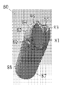
[0066] FIGS. 8A-8E illustrate an alternative embodiment of a multi-lumen
catheter.
The catheter 80 includes a symmetrical tip where two lumina 81 and 82
terminate at the same
point such that the ports are adjacent. Additionally, unlike common prior art,
the
symmetrical tip design as shown herein also eliminates the septum extending
beyond the
distal ends of the lumina 81 and 82 such that the septum extends beyond the
ports. Rather,
the septum is trimmed such that it terminates concurrent to the ports. Each
lumen 81 and 82
has a unique curved wall that defines their shape and the flow of fluid
therethrough such that
no extended septum is required to maintain fluid separation. As shown in FIG.
8D, the cross-
section shape of lumina 81 and 82 are semicircular.
[0067] At the distal end of the catheter 80, the lumina 81 and 82 may have
with
curved walls 83 and 84 which terminate at ports 85 and 86 disposed on opposite
sides of
catheter 80. In this symmetrical tip embodiment, the lumen 81 extends to same
point as the
lumen 82, thereby forming a symmetrical tip design. As before, in operation,
fluid, such as
blood, enters intake port 85, changes direction as the flow of fluid passes
curved wall 83
18
CA 02871329 2014-10-22
WO 2013/163172
PCT[US2013/037783
through lumen 81 to tube 87 which conveys the fluid for treatment to a device
such as a
dialysis machine (not shown). After treatment, the treated fluid is returned
to a patient
through tube 88 to lumen 82. At the end of lumen 82, the fluid is deflected by
curved wall 84
and out port 86 into the vessel of the patient. By providing the curved walls
83 and 84 to
deflect the fluid, the design eliminates the septum extending beyond the ports
while still
maintaining a low level of fluid from the out port 86 mixing with fluid
entering the intake
port 85.
[0068] Similar to the discussion above in reference to FIGS. 6A-6D, one or
more
side holes 89 may be included in the catheter 80. The side holes 89 may assist
in providing
additional means of blood or other fluid exchange as well as serve as a
mounting point for
attaching the catheter 80 on a guide wire during insertion and/or removal
and/or exchange
from a patient.
[0069] The side holes 89 may be sized and positioned such that fluid flow is
optimized about the lumen tips. For example, the side holes 89 may be
approximately lmm
in diameter, and be positioned approximately lcm from the ports 85 and 86.
Alternatively,
the side holes may be smaller (e.g., .5mm), larger (e.g., 1.5mm), or
positioned in another
location (e.g., 1.5cm from the ports). The size and location of the side holes
89 may produce
changes in shear stress and blood cell residence time at the catheter tips,
and thus the optimal
balance may incorporate side holes being approximately .75mm to 1.2mm in
diameter.
[0070] Using properly spaced and sized side boles, such as side holes 89, may
result
in a highly optimized catheter. For example, by using a similar catheter to
the catheter 80 as
shown in FIGS. 8A-8E, fluid flow through the catheter may be optimized while
recirculation
is greatly reduced.
[0071] It should be noted that side holes 89 as shown in FIGS. 8A-8E are shown
by
way of example only. Additional or alternative apertures such as slits, flaps,
semicircular
19
CA 02871329 2014-10-22
WO 2013/163172
PCT[US2013/037783
cuts, and other similar shapes may be used. Additionally, the side holes
themselves may have
a helical contour to produce additional deflection to any fluid flowing
therethrough.
[0072] As shown in FIGS. 8A-8E, the curved walls 83 and 84 define an area of
deflection such that fluid flowing through the catheter 80 is deflected.
Depending on the
application of the catheter 80, and the amount of desired deflection, an angle
of the curved
walls 83 and 84 may vary accordingly. For example, the curved walls 83 and 84
may be
approximately 54 . Alternatively, the curved walls 83 and 84 may be between
the range of 0
and 90 . Typically, 0 and 90 may not be used as they both have inherent
drawbacks. 0
would cause no deflection to the fluid. 90 would result in the port being
perpendicular to the
axial flow path of the catheter. On the input port, this could cause a vacuum
force which
attaches the associated lumina to the side of a blood vessel. While a side
hole would help to
alleviate any potential vacuum pressure, overall performance of the catheter
would still
decrease.
[0073] FIGS. 9A-9D illustrate an alternative embodiment of a multi-lumen
catheter.
The catheter 90 includes a symmetrical tip where two lumina 91 and 92
terminate at the same
point such that the ports are adjacent. Additionally, unlike common prior art,
the
symmetrical tip design as shown herein also eliminates the septum extending
beyond the
distal ends of the lumina 91 and 92 such that the septum extends beyond the
ports. Rather,
the septum is trimmed such that it terminates concurrent to the ports. Each
lumen 91 and 92
has a unique curved wall that defines their shape and the flow of fluid
therethrough such that
no extended septum is required to maintain fluid separation. As shown in FIG.
9D, the cross-
section shape of lumina 91 and 92 are semicircular.
[0074] Unlike the catheter 80 as shown in FIGS. 8A-8E, the catheter 90
includes a
spiral twist at the distal end. As shown in FIG. 9C, the twisted portion 100
of the catheter 90
with the spiral twist may be rotated between 10 and 359 , however, an optimum
value would
CA 02871329 2014-10-22
WO 2013/163172
PCT[US2013/037783
be somewhere in the middle of this range, depending on the viscosity and
volume of fluid
being moved through the catheter. For example, for a catheter to be used for
human dialysis,
a rotation of about 135 within about 4 cm extending from the distal end of
the catheter may
be optimal. Alternatively, a range of about 120 to about 1500 within an
approximate lcm to
5cm extending from the distal end of the catheter may be optimal.
[0075] At the distal end of the catheter 90, the lumina 91 and 92 may have
with
curved walls 93 and 94 which terminate at ports 95 and 96 disposed on opposite
sides of
catheter 90. In this symmetrical tip embodiment, the lumen 91 extends to same
point as the
lumen 92, thereby forming a symmetrical tip design. As before, in operation,
fluid, such as
blood, enters intake port 95, changes direction as the flow of fluid passes
curved wall 93 and
through the twisted portion 100 of lumen 91 to tube 97 which conveys the fluid
for treatment
to a device such as a dialysis machine (not shown). After treatment, the
treated fluid is
returned to a patient through tube 98 to lumen 92. At the end of lumen 92, the
fluid is
deflected by the twisted portion 100 and the curved wall 94, and passes
through out port 96
into the vessel of the patient. By providing the curved walls 93 and 94 and
the twisted
portion 100 to deflect the fluid, this design eliminates the septum extending
beyond the ports
while still maintaining a low level of fluid from the out port 96 mixing with
fluid entering the
intake port 95.
[0076] Similar to the discussion above in reference to FIGS. 6A-6D, one or
more
side holes 99 may be included in the catheter 90. The side holes 99 may assist
in providing
additional means of blood or other fluid exchange as well as serve as a
mounting point for
attaching the catheter 90 on a guide wire during insertion and/or removal
and/or exchange
from a patient.
[0077] The side holes 99 may be sized and positioned such that fluid flow is
increased about the lumen tips. For example, the side holes 99 may be
approximately lmm
21
CA 02871329 2014-10-22
WO 2013/163172
PCT/1JS2013/037783
in diameter, and be positioned approximately lcm from the ports 95 and 96.
Alternatively,
the side holes may be smaller (e.g., .5mm), larger (e.g., 1.5mm), or
positioned in another
location (e.g., 1.5cm from the ports). The size and location of the side holes
99 may produce
changes in shear stress and blood cell residence time at the catheter tips,
and thus the optimal
balance may incorporate side holes being approximately .75mm to 1.2mm in
diameter.
[0078] Using properly spaced and sized side holes, such as side holes 99, may
result
in a highly optimized catheter. For example, by using a similar catheter to
the catheter 90 as
shown in FIGS. 9A-9D, fluid flow through the catheter may be optimized while
recirculation
is greatly reduced.
[0079] It should be noted that side holes 99 as shown in FIGS. 8A-8E are shown
by
way of example only. Additional or alternative apertures such as slits, flaps,
semicircular
cuts, and other similar shapes may be used. Additionally, the side holes
themselves may have
a helical contour to produce additional deflection to any fluid flowing
therethrough.
[0080] As shown in FIGS. 9A-9D, the curved walls 93 and 94 define an area of
deflection such that fluid flowing through the catheter 90 is deflected.
Depending on the
application of the catheter 90, and the amount of desired deflection, an angle
of the curved
walls 93 and 94 may vary accordingly. For example, the curved walls 93 and 94
may be
approximately 54 . Alternatively, the curved walls 93 and 94 may be between
the range of 00
and 90 . Typically, 00 and 90 may not be used as they both have inherent
drawbacks. 0
would cause no deflection to the fluid. 90 would result in the port being
perpendicular to the
axial flow path of the catheter. On the input port, this could cause a vacuum
force which
attaches the associated lumina to the side of a blood vessel. While a side
hole would help to
alleviate any potential vacuum pressure, overall performance of the catheter
would still
decrease.
22
CA 02871329 2014-10-22
WO 2013/163172
PCT[US2013/037783
10081] Although the invention has been described in language specific to
structural
features and/or methodological acts, it is to be understood that the invention
defined in the
appended claims is not necessarily limited to the specific features or acts
described. Rather,
the specific features and acts are disclosed as exemplary forms of
implementing the claimed
invention.
[0082] Various of the above-disclosed and other features and functions, or
alternatives thereof, may be combined into many other different systems or
applications.
Various presently unforeseen or unanticipated alternatives, modifications,
variations or
improvements therein may be subsequently made by those skilled in the art,
each of which is
.. also intended to be encompassed by the disclosed embodiments.
23