Note : Les descriptions sont présentées dans la langue officielle dans laquelle elles ont été soumises.
CA 02871759 2014-10-27
WO 2013/165835
PCT/US2013/038360
ANTIPROLIFERATIVE SURFACE MODIFICATIONS AND METHODS OF USE
CROSS-REFERENCE TO RELATED APPLICATIONS
The present application claims the benefit of U.S. Provisional Patent
Application No.
61/640,843, filed on. May 1, 2012, which is incorporated herein by reference
[1].
FIELD OF THE INVENTION
This invention is in the field of implantable medical devices. The present
invention
relates to a device constructed from metals, polymers or other materials that
are amenable to
precise surface modifications and coupling with erodible agents methods for
its use, wherein (1)
the erodible agents, which contain active ingredients (i.e., for example,
medications) provide for
acute control of cellular proliferation and (2) a pattered surface having
micron-, and/or
nano-sized micro-patterning characteristics that imparts anti-proliferative
properties.
BACKGROUND OF THE INVENTION
A major problem in implanted devices is the proliferation of fibroblasts and
other cells on
the device surface and the foiniation of inflammation, scar tissue and
encapsulation. The
disorganized growth of fibroblasts and the inflammatory response elicited by
the presence of the
implant alter the function of implants. These responses most often result in
formation of a dense,
fibrous capsule surrounding the implant. In many applications, this fibrous
capsule can
negatively impact proper functioning of the implanted device, for example by
preventing
diffusion of molecules between the impant and its environment or by generally
altering the local
1
CA 02871759 2014-10-27
WO 2013/165835
PCT/US2013/038360
physiological environment. As wound modulation has both an acute and chronic
phase, it is
important to address both phases to result in optimal medical and surgical
outcomes. What is
needed is an implant that would reduce or mitigate the proliferation of cells
through both primary
(acute and short term) and secondary means (chronic/long-term).
SUMMARY OF THE INVENTION
This invention is in the field of implantable medical devices. The present
invention
relates to a device constructed from metals, polymers or other materials that
are amenable to
precise surface modifications and coupling with erodible agents methods for
its use, wherein (1)
the erodible agents, which contain active ingredients (i.e., for example,
medications) provide for
acute control of cellular proliferation and (2) a pattered surface having
micron-, and/or
nano-sized micro-patterning characteristics that imparts anti-proliferative
properties.
In one embodiment, the invention relates to a device comprising an anti-
proliferative
surface, wherein said surface comprises a micro-patterned geometrical pattern,
said pattern
having a plurality of grooves between a plurality of raised surfaces. In one
embodiment, said
pattern is selected from the group consisting of vertical, horizontal,
circular, intersecting grid,
and concentric rings. In one embodiment, said grooves comprise a plurality of
medication depots
such that the top of said depots are below said plurality of raised surfaces.
In one embodiment,
said plurality of raised surfaces are separated by a distance of approximately
10-50 um. In one
embodiment, said plurality of raised surfaces are separated by a distance of
approximately 20-35
um. In one embodiment, said plurality of raised surfaces are separated by a
distance of
approximately 20-25 um. In one embodiment, said grooves are at least as deep
as the distance
separating said plurality of raised surfaces. In one embodiment, the depths of
said grooves are
deeper than the distance separating said plurality of raised surfaces wherein
said grooves
2
CA 02871759 2014-10-27
WO 2013/165835
PCT/US2013/038360
comprise a plurality of medication depots. In one embodiment, said medication
depots are at
least 25 pm below said raised surfaces. In one embodiment, said medication
depot comprises an
anti-proliferative material. In one embodiment, said geometrical pattern
inhibits cellular
proliferation, cell attachment, cell migration or release of specific factors.
In one embodiment,
said device is an implanted medical device. In one embodiment, said implanted
medical device is
in an ocular region. In one embodiment, said ocular region is selected from
the group consisting
of the sclera, Schlemm's canal and the suprachoroidal space. In one
embodiment, said surface
further comprises silicone, polyimide (PI), polysulfone (PES),
polyetheretherketone (PEEK),
polypropylene, polyetherimide (PEI), titanium, nitinol, stainless steel, gold,
hydrophilic or
hydrophopic polymers, shape memory polymers or alloys, ceramics, alloys,
silicates, or other
materials. In one embodiment, said device has shape selected from the group
consisting of
spherical, non-spherical (egg-shaped), cylindrical, rectangular, cubic,
toroidal, conical, cuboidal,
pyramidal, prism, and planar shapes. In one embodiment, said device has a
cylindrical shape.
In one embodiment, said device contains at least one lumen. In one embodiment,
said lumen
contains a depot. In one embodiment, said medication depot contains at least
one medication. In
one embodiment, said medication is selected from the group comprising anti-
fibrotic agent,
anti-inflammatory agent, immunosuppressant agent, anti-neoplastic agent,
migration inhibitors,
anti-proliferative agent, rapamycin, triamcinolone acetonide, everolimus,
tacrolimus, paclitaxel,
actinomycin, azathioprine, dexamethasone, cyclosporine, bevacizumab, an anti-
VEGF agent, an
anti-IL-1 agent, canakinumab, an anti-IL-2 agent, viral vectors, beta
blockers, alpha agonists,
muscarinic agents, steroids, antibiotics, non-steroidal anti-inflammatory
agents, prostaglandin
analogues, ROCK inhibitors, nitric oxide, endothelin, matrixmetalloproteinase
inhibitors, CNPA,
corticosteroids, and/or antibody-based immunosuppresants. In one embodiment,
said medication
is combined with a polymer. In one embodiment, wherein said polymer is
selected from the
3
CA 02871759 2014-10-27
WO 2013/165835
PCT/US2013/038360
group comprising poly(lactic-co-glycolic acid), polyethylene glycol,
poly(lactic acid),
poly(glycolic acid), poly(amido ester), polyethylene terephthalate,
poly(caprolactone),
poly(hydroxy butyrate), poly(butylene succinate), poly(vinyl alchohol),
poly(hydroxybutyrate),
poly(methyl acrylate), poly(methyl methylmethacrylate), poly(sebacic acid),
carboxymethyl
cellulose, ethyl cellulose, cellulose acetate, polydioxanone, or polymers from
the categories:
polyesters, polyanhydrides, polyamides, polycyanoacrylates, polyurethanes,
polyorthoesters,
silicones, acrylic polymers, cellulose derivatives and/or poloxamers. In one
embodiment, said
grooves are patterned in a vertical orientation. In one embodiment, said
grooves are patterned in
a horizontal orientation. In one embodiment, said grooves are patterned in a
diagonal orientation.
In one embodiment, said grooves are patterned in a helical orientation. In one
embodiment, said
geometrical pattern further comprises a columnar structure. In one embodiment,
said device is
a catheter. In one embodiment, said device is a stent. In one embodiment, said
catheter comprises
a defibrillation device. In one embodiment, said device is an intravenous
catheter. In one
embodiment, said device is a Hickman catheter. In one embodiment, said device
is a mesh
prosthesis. In one embodiment, said device is a hernia mesh. In one
embodiment, said device is a
Baerveldt glaucoma implant. In one embodiment, said device is a dental
implant. In one
embodiment, said device is a glaucoma shunting device. In one embodiment, said
geometrical
pattern prevents encapsulation. In one embodiment, said geometrical pattern
prevents disorderly
growth of fibroblasts. In one embodiment, said geometrical pattern prevents
the formation of
scar tissue. In one embodiment, said geometrical pattern prevents cellular
proliferation. In one
embodiment, said geometrical pattern inhibits cellular attachment. In one
embodiment, said
geometrical pattern provides fluid drainage.
In one embodiment, the invention relates to a method of treating a subject in
need of
inhibiting cellular proliferation comprising: a) providing a drug delivery
device comprising an
4
CA 02871759 2014-10-27
WO 2013/165835
PCT/US2013/038360
anti-proliferative surface, wherein said surface comprises a micro-patterned
geometrical pattern,
said pattern having a plurality of grooves between a plurality of raised
surfaces, wherein said
grooves comprise a plurality of medication depots such that the top of said
depots are below said
plurality of raised surfaces; and b) delivering a medication from said
medication depot to inhibit
cellular proliferation. In one embodiment, said pattern is selected from the
group consisting of
vertical, horizontal, circular, intersecting grid, and concentric rings. . In
one embodiment, said
plurality of raised surfaces are separated by a distance of approximately 10-
50 1AM. In one
embodiment, said plurality of raised surfaces are separated by a distance of
approximately 20-35
1AM. In one embodiment, said plurality of raised surfaces are separated by a
distance of
approximately 20-25 um. In one embodiment, said grooves are at least as deep
as the distance
separating said plurality of raised surfaces. In one embodiment, the depths of
said grooves are
deeper than the distance separating said plurality of raised surfaces wherein
said grooves
comprise a plurality of medication depots. In one embodiment, said medication
depots are at
least 25 1..tm below said raised surfaces. In one embodiment, said wherein
said medication
comprises an anti-proliferative material. In one embodiment, said geometric
pattern inhibits
cellular proliferation, cell attachment, cell migration or release of specific
factors. In one
embodiment, said device is an implanted medical. In one embodiment, said
implanted medical
device is in an ocular region. In one embodiment, said ocular region is
selected from the group
comprising the sclera, Schlemm's canal and the suprachoroidal space. In one
embodiment, said
device further comprises silicone, polyimide (PI), polysulfone (PES),
polyetheretherketone
(PEEK), polypropylene, polyetherimide (PEI), titanium, nitinol, stainless
steel, gold, hydrophilic
or hydrophopic polymers, shape memory polymers, ceramics, alloys, silicates,
or other materials.
In one embodiment, said device has shape selected from the group consisting of
spherical,
non-spherical (egg-shaped), cylindrical, rectangular, cubic, toroidal,
conical, cuboidal, pyramidal,
5
CA 02871759 2014-10-27
WO 2013/165835
PCT/US2013/038360
prism, and planar shapes. In one embodiment, said device has a cylindrical
shape. In one
embodiment, said device contains at least one lumen. In one embodiment, said
lumen contains a
depot. In one embodiment, said medication is selected from the group
comprising anti-fibrotic
agent, anti-inflammatory agent, immunosuppressant agent, anti-neoplastic
agent, migration
inhibitors, anti-proliferative agent, rapamycin, triamcinolone acetonide,
everolimus, tacrolimus,
paclitaxel, actinomycin, azathioprine, dexamethasone, cyclosporine,
bevacizumab, an anti-VEGF
agent, an anti-IL-1 agent, canakinumab, an anti-IL-2 agent, viral vectors,
beta blockers, alpha
agonists, muscarinic agents, steroids, antibiotics, non-steroidal anti-
inflammatory agents,
prostaglandin analogues, ROCK inhibitors, nitric oxide, endothelin,
matrixmetalloproteinase
inhibitors, CNPA, corticosteroids, and antibody-based immunosuppresants. In
one embodiment,
said medication is combined with a polymer. In one embodiment, said polymer is
selected from
the group comprising poly(lactic-co-glycolic acid), polyethylene glycol,
poly(lactic acid),
poly(glycolic acid), poly(amido ester), polyethylene terephthalate,
poly(caprolactone),
poly(hydroxy butyrate), poly(butylene succinate), poly(vinyl alchohol),
poly(hydroxybutyrate),
poly(methyl acrylate), poly(methyl methylmethacrylate), poly(sebacic acid),
carboxymethyl
cellulose, ethyl cellulose, cellulose acetate, polydioxanone, or polymers from
the categories:
polyesters, polyanhydrides, polyamides, polycyanoacrylates, polyurethanes,
polyorthoesters,
silicones, acrylic polymers, cellulose derivatives and/or poloxamers. In one
embodiment, said
grooves are patterned in a vertical orientation. In one embodiment, said
grooves are patterned in
a horizontal orientation. In one embodiment, said grooves are patterned in a
diagonal orientation.
In one embodiment, said grooves are patterned in a helical orientation. In one
embodiment, said
geometrical pattern further comprises a columnar structure. In one embodiment,
said device is
a catheter. In one embodiment, said device is a stent. In one embodiment, said
device is a
catheter for a defibrillation device. In one embodiment, said device is an
intravenous catheter. In
6
CA 02871759 2014-10-27
WO 2013/165835
PCT/US2013/038360
one embodiment, said device is a Hickman catheter. In one embodiment, said
device is a mesh
prosthesis. In one embodiment, said device is a hernia mesh. In one
embodiment, said device is a
Baerveldt glaucoma implant. In one embodiment, said device is a dental
implant. In one
embodiment, said device is a glaucoma aqueous shunting device. In one
embodiment, said
device is a device that shunts fluid from one area to another. In one
embodiment, said
geometrical pattern prevents encapsulation. In one embodiment, said
geometrical pattern
prevents disorderly growth of fibroblasts. In one embodiment, said geometrical
pattern prevents
the foimation of scar tissue. In one embodiment, said geometrical pattern
prevents cellular
proliferation. In one embodiment, said geometrical pattern inhibits cellular
attachment. In one
embodiment, said geometrical pattern provides fluid drainage.
In one embodiment, the invention relates to a method of treating a subject in
need of
inhibiting cellular proliferation comprising: a) providing an implanted device
comprising an
anti-proliferative surface, wherein said surface comprises a micro-patterned
geometrical pattern,
said pattern having a plurality of grooves between a plurality of raised
surfaces; and b) using said
device to inhibit cellular proliferation. In one embodiment, said pattern is
selected from the
group consisting of vertical, horizontal, circular, intersecting grid, and
concentric rings. In one
embodiment, said plurality of raised surfaces are separated by a distance of
approximately 10-50
um. In one embodiment, said plurality of raised surfaces are separated by a
distance of
approximately 20-35 um. In one embodiment, said plurality of raised surfaces
are separated by a
distance of approximately 20-25 pm. In one embodiment, said grooves are at
least as deep as
the distance separating said plurality of raised surfaces. In one embodiment,
said geometric
pattern inhibits cellular proliferation, cell attachment, cell migration or
release of specific factors.
In one embodiment, said device is an implanted medical device. In one
embodiment, said
implanted medical device is in an ocular region. In one embodiment, said
ocular region is
7
CA 02871759 2014-10-27
WO 2013/165835
PCT/US2013/038360
selected from the group consisting of the sclera, Schlemm's canal and the
suprachoroidal space.
In one embodiment, said device further comprises silicone, polyimide (PI),
polysulfone (PES),
polyetheretherketone (PEEK), polypropylene, polyetherimide (PEI), titanium,
nitinol, stainless
steel, gold, hydrophilic or hydrophopic polymers, shape memory polymers,
ceramics, alloys,
silicates, or other materials. In one embodiment, device has shape selected
from the group
consisting of spherical, non-spherical (egg-shaped), cylindrical, rectangular,
cubic, toroidal,
conical, cuboidal, pyramidal, prism, and planar shapes. In one embodiment,
said device has a
cylindrical shape. In one embodiment, said device contains at least one lumen.
In one
embodiment, said grooves are patterned in a vertical orientation. In one
embodiment, said
grooves are patterned in a horizontal orientation. In one embodiment, said
grooves are patterned
in a diagonal orientation. In one embodiment, said grooves are patterned in a
helical orientation.
In one embodiment, said geometrical pattern further comprises a columnar
structure. In one
embodiment, said device is a catheter. In one embodiment, said device is a
stent. In one
embodiment, said device is a catheter for a defibrillation device. In one
embodiment, said device
is an intravenous catheter. In one embodiment, said device is a Hickman
catheter. In one
embodiment, said device is a mesh prosthesis. In one embodiment, said device
is a hernia mesh.
In one embodiment, said device is a Baerveldt glaucoma implant. In one
embodiment, said
device is a dental implant. In one embodiment, said device is a glaucoma
aqueous shunting
device. In one embodiment, said device is a device that shunts fluid from one
area to another. In
one embodiment, said geometrical pattern prevents encapsulation. In one
embodiment, said
geometrical pattern prevents disorderly growth of fibroblasts. In one
embodiment, said
geometrical pattern prevents the formation of scar tissue. In one embodiment,
said geometrical
pattern prevents cellular proliferation. In one embodiment, said geometrical
pattern inhibits
cellular attachment. In one embodiment, said geometrical pattern provides
fluid drainage.
8
CA 02871759 2014-10-27
WO 2013/165835
PCT/US2013/038360
It is not intended that embodiments of the invention be limited to any
particular method,
medical target, or device confirmation; however, it is believed that the
device may be optimally
designed to inhibit the proliferation of fibroblasts, smooth muscle cells and
other cells on the
surface of the implant in both the acute and chronic phases of wound
modulation.
DEFINITIONS
To facilitate the understanding of this invention, a number of terms are
defined below.
Terms defined herein have meanings as commonly understood by a person of
ordinary skill in
the areas relevant to the present invention. Terms such as "a", "an" and "the"
are not intended
to refer to only a singular entity, but include the general class of which a
specific example may
be used for illustration. The terminology herein is used to describe specific
embodiments of the
invention, but their usage does not delimit the invention, except as outlined
in the claims.
As used herein, the term "patient" or "subject" refers to a living mammalian
organism,
such as a human, monkey, cow, sheep, goat, dog, cat, mouse, rat, guinea pig,
or transgenic species
thereof. In certain embodiments, the patient or subject is a primate. Non-
limiting examples of
human subjects are adults, juveniles, infants and fetuses.
"Prevention" or "preventing" includes: (1) inhibiting the onset of a disease
in a subject or
patient which may be at risk and/or predisposed to the disease but does not
yet experience or
display any or all of the pathology or symptomatology of the disease, and/or
(2) slowing the onset
of the pathology or symptomatology of a disease in a subject or patient which
may be at risk and/or
predisposed to the disease but does not yet experience or display any or all
of the pathology or
symptomatology of the disease.
9
CA 02871759 2014-10-27
WO 2013/165835
PCT/US2013/038360
As used herein, the terms "medication" or "therapeutic agent" refer to
something that treats
or prevents or alleviates the symptoms of disease or condition, a drug or
pharmaceutical
composition. Medication is considered to be delivered or present in
therapeutically effective
amounts or pharmaceutically effective amounts.
The present invention contemplates the above-described compositions in
"therapeutically
effective amounts" or "pharmaceutically effective amounts", which means that
amount which,
when administered to a subject or patient for treating a disease, is
sufficient to effect such
treatment for the disease or to ameliorate one or more symptoms of a disease
or condition (e.g.
ameliorate pain).
As used herein, the teal's "treat" and "treating" are not limited to the case
where the
subject (e.g. patient) is cured and the disease is eradicated. Rather, the
present invention also
contemplates treatment that merely reduces symptoms, improves (to some degree)
and/or delays
disease progression. It is not intended that the present invention be limited
to instances wherein
a disease or affliction is cured. It is sufficient that symptoms are reduced.
As used herein, the terms "medical device," "implant," "device," "medical
device,"
"medical implant," "implant/device," and the like are used synonymously to
refer to any object
that is designed to be placed partially or wholly within a patient's body for
one or more
therapeutic or prophylactic purposes such as for tissue augmentation,
contouring, restoring
physiological function, repairing or restoring tissues damaged by disease or
trauma, and/or
delivering therapeutic agents to normal, damaged or diseased organs and
tissues. While medical
devices are normally composed of biologically compatible synthetic materials
(e.g.,
medical-grade stainless steel, titanium and other metals; exogenous polymers,
such as
polyurethane, silicon, PLA, PLGA, PGA, PCL), other materials may also be used
in the
construction of the medical implant. While not limiting the present invention
to any particular
CA 02871759 2014-10-27
WO 2013/165835
PCT/US2013/038360
device, specific medical devices and implants that are particularly relevant
to this invention
include stents, catheters, implanted defribrillators, defribillator leads,
cardiac, cerebral,
lumbar-peritoneal, peritoneovenous, pulmonary, ocular or other shunts, drug
delivery systems,
implanted electronic devices, and implanted, microelectromechanical (MEMS)
devices . Other
devices contemplated include dental implants, hernia mesh devices, encircling
bands (beriatic
surgery and scleral buckles) and any implant that might be placed in or around
the body.
As used herein, the term "medication depot" refers to medication deposited on
the bottom
level of a micro-patterned geometric pattern, such as a groove.
As used herein, the term "anti-proliferative" refers to refer to agents used
or tending to
inhibit cell growth.
As used herein, the terms "fibrosis" or "scarring" refers to the formation of
fibrous (scar)
tissue in response to injury or medical intervention. Therapeutic agents which
inhibit fibrosis or
scarring can do so through one or more mechanisms including inhibiting
inflammation,
inhibiting angiogenesis, inhibiting migration or proliferation of connective
tissue cells (such as
fibroblasts, smooth muscle cells, vascular smooth muscle cells), reducing
extracellular matrix
(ECM) production or encouraging ECM breakdown, arresting and/or inhibiting
cell cycle
progression, arresting and/or inhibiting DNA synthesis, and/or inhibiting
tissue remodeling. In
addition, numerous therapeutic agents described in this invention will have
the additional benefit
of also reducing tissue regeneration (the replacement of injured cells by
cells of the same type)
when appropriate.
As used herein, the terms "inhibit fibrosis," "inhibit scar," "reduce
fibrosis," "reduce
scar," "fibrosis-inhibitor," "anti-scarring" and the like are used
synonymously to refer to the
action of agents or compositions which result in a statistically significant
decrease in the
foimation, deposition and/or maturation of fibrous tissue that may be expected
to occur in the
11
CA 02871759 2014-10-27
WO 2013/165835
PCT/US2013/038360
absence of the agent or composition.
As used herein, the term "antifibrotic agent" refers to chemical compounds
which have
antifibrotic activity in mammals. This takes into account the abnotinal
foimation of fibrous
connective tissue, which is typically comprised of collagen to a greater or
lesser degree. These
compounds may have different mechanisms of action, some reducing the formation
of collagen
or another protein, others enhancing the metabolism or removal of collagen in
the affected area
of the body. All such compounds having activity in the reduction of the
presence of fibrous
tissue are included herein, without regard to the particular mechanism of
action by which each
such drug functions.
As used herein, the terms "encapsulation" as used herein refers to the
foimation of a
fibrous connective tissue capsule (containing fibroblasts, myofibroblasts,
inflammatory cells,
relatively few blood vessels and a collagenous extracellular matrix) encloses
and isolates an
implanted prosthesis or biomaterial from the surrounding body tissue. This
fibrous tissue capsule,
which is the result of unwanted scarring and inflammation in response to an
implanted prosthesis
or biomaterial, has a tendency to progressively contract, thereby tightening
around the
implant/biomaterial and causing it to become very firm and disfigured. Further
implications of
encapsulation and associated contracture include tenderness of the tissue,
pain, erosion of the
adjacent tissue as well as other complications.
As used herein, the terms "contracture" as used herein refers to permanent or
non-permanent scar tissue formation in response to an implanted prosthesis or
biomaterial. In
general, the condition of contracture involves a fibrotic response that may
involve inflammatory
components, both acute and chronic. Unwanted scarring in response to an
implanted prosthesis
or biomaterial can form a fibrous tissue capsule around the area or
implantable prosthesis or
biomaterial that encloses and isolates it from the surrounding body tissue (as
described for
12
CA 02871759 2014-10-27
WO 2013/165835
PCT/US2013/038360
encapsulation). Contracture occurs when fibrous tissue capsule matures and
starts to shrink
(contract) forming a tight, hard capsule around the implant/biomaterial that
can alter the anatomy,
texture, shape and movement of the implant. In some eases, contracture also
draws the overlying
skin in towards the implant and leads to dimpling of the skin and
disfuguration. Contracture and
chronic inflammation can also contribute to tenderness around the implant,
pain, and erosion of
the adjacent tissue. Fibrotic contractures related to implantation of soft
tissue
implant/biomaterials may be caused by a variety of factors including surgical
trauma and
complications, revisions or repeat procedures (the incidence is higher if
implantation is being
attempted where contractures have occurred previously), inadequate hemostasis
(bleeding
control) during surgery, aggressive healing processes, underlying or pre-
existent conditions,
genetic factors (people prone to hypertrohic scar or keloid formation), and
immobilization.
As used herein, the terms "implanted" refers to having completely or partially
placed a
device within a host. A device is partially implanted when some of the device
reaches, or extends
to the outside of, a host.
As used herein, the tetra "erodible agent" refers to materials such as polymer
or
semi-solid gel or the like which are eroded by physiological or chemical
processes such that the
mass of said agents decreases over the course of implantation. The erodible
agent, can be made
out of PLGA, Polymers, erodible gels and other materials capable of carrying
or containing
medications and eroding over time.
As used herein, the term "micro-patterning" preferably refers to milimeter,
micrometer,
and/or nanometer scale surface modifications including but not limited to
laser etching, chemical
etching, photo-etching, photolithography, machining, stamping, deposition
processes,
mechaninal drilling, molding, 3D printing, Atomic Layer Deposition or other
means of
modifying surfaces.
13
CA 02871759 2014-10-27
WO 2013/165835
PCT/US2013/038360
As used herein, the term "anti-inflammatory agent" refers to substance or
treatment that
reduces inflammation.
As used herein, the term "immunosuppressant agents" refers to drugs that
inhibit or
prevent activity of the immune system.
As used herein, the term "anti-neoplastic agents" refers to drugs that prevent
or inhibit
the development, maturation, or spread of neoplastic cells.
As used herein, the term "migration inhibitors" refers to agents that alter
the movement
of cells in a given environment or that inhibit the migration of specific cell
types or cells
generally.
As used herein, the term "butylated hydroxy toluene" (abbreviated BHT) refers
to a
lipophilic (fat-soluble) organic compound, chemically a derivative of phenol,
that is useful for its
antioxidant properties. BHT is also known as 2,6-bis(1,1-dimethylethyl)-4-
methylphenol,
2,6-di-tert-butyl-4-methylphenol, 2,6-di-tert-butyl-p-cresol (DBPC),
and
OH
3,5-di-tert-butyl-4-hydroxytoluene. Butylated hydroxy toluene has the
structure:
As used herein, the tem' "rapamycin" refers to an immunosuppressant drug used
to
prevent rejection in organ transplantation.
As used herein, the term "triamcinolone acetonide" refers to a synthetic
corticosteroid.
As used herein, the term "everolimus" refers to an immunosuppressant to
prevent
rejection of organ transplants and treatment of renal cell cancer.
As used herein, the term "tacrolimus" (also FK-506 or fujimycin, trade names
Prograf,
Advagraf, Protopic) refers to an immunosuppressive drug that is mainly used
after allogeneic
organ transplant to reduce the activity of the patient's immune system and so
lower the risk of
14
CA 02871759 2014-10-27
WO 2013/165835
PCT/US2013/038360
organ rejection. It is also used in a topical preparation in the treatment of
atopic dermatitis
(eczema), severe refractory uveitis after bone marrow transplants,
exacerbations of minimal
change disease, and the skin condition vitiligo.
As used herein, the term "paclitaxel" refers to a mitotic inhibitor used in
cancer
chemotherapy.
As used herein, the term "actinomycin" refers to a class of polypeptide
antibiotics
isolated from soil bacteria of the genus Streptomyces, of which the most
significant is
actinomycin D.
As used herein, the term "azathioprine" refers to a purine analogue
immunosuppressive
drug. It is used to prevent rejection following organ transplantation, and to
treat a vast array of
autoimmune diseases, including rheumatoid arthritis, pemphigus, inflammatory
bowel disease
(such as Crohn's disease and ulcerative colitis), multiple sclerosis,
autoimmune hepatitis, atopic
dermatitis, myasthenia gravis, neuromyelitis optica or Devic's disease,
restrictive lung disease,
and others.
As used herein, the term "dexamethasone" refers to a potent synthetic member
of the
glucocorticoid class of steroid drugs. It acts as an anti-inflammatory and
immunosuppressant.
As used herein, the term "cyclosporine" refers to an immunosuppressant drug
widely
used in organ transplantation to prevent rejection.
As used herein, the term "bevacizumab" refers to a drug that blocks
angiogenesis, the
growth of new blood vessels.
As used herein, the term "anti-VEGF agent" refers to a drug that inhibits the
action of
vascular endothelial growth factor (VEGF).
As used herein, the term "anti-IL-1 agent" refers to a drug that inhibits the
action of
Interleukin 1 protein.
CA 02871759 2014-10-27
WO 2013/165835
PCT/US2013/038360
As used herein, the term "canakinumab" refers to a human monoclonal antibody
targeted
at interleukin-1 beta.
As used herein, the Willi "anti-IL-2 agent" refers to a drug that inhibits the
action of
Interleukin 2 protein.
As used herein, the term "viral vectors" refers to a tool commonly used by
molecular
biologists to deliver genetic material into cells. A viral vector is modified
in such a way as to
minimize the risk of handling them. This usually involves the deletion of a
part of the viral
genome critical for viral replication. Such a virus can efficiently infect
cells but, once the
infection has taken place, requires a helper virus to provide the missing
proteins for production
of new virions.
As used herein, the term "beta blockers" (beta-adrenergic blocking agents,
beta-adrenergic antagonists, beta-adrenoreceptor antagonists or beta
antagonists) refer to a class
of drugs used for various indications. They are particularly for the
management of cardiac
arrhythmias, cardioprotection after myocardial infarction [2] (heart attack),
and hypertension [3].
As beta adrenergic receptor antagonists, they diminish the effects of
epinephrine (adrenaline) and
other stress hormones.
As used herein, the term "alpha agonists" or "a-adrenergic-antagonists" refers
to
pharmacological agents that act as receptor antagonists of a-adrenergic
receptors
(a-adrenoceptors).
As used herein, the term "muscarinic agents" refers to a muscarinic receptor
agonist or an
agent that enhances the activity of the muscarinic acetylcholine receptor.
As used herein, the term "steroids" refers to a type of organic compound that
contains a
characteristic arrangement of four cycloalkane rings that are joined to each
other. Examples of
steroids include, but are not limited to, the dietary fat cholesterol, the sex
hormones estradiol and
16
CA 02871759 2014-10-27
WO 2013/165835
PCT/US2013/038360
testosterone, and the anti-inflammatory drug dexamethasone.
As used herein, the term "antibiotics" refers to a compound or substance that
kills or
slows down the growth of bacteria, fungus, or other microorganism.
As used herein, the term "non-steroidal anti-inflammatory agents,"
"nonsteroidal
anti-inflammatory drugs," usually abbreviated to NSAIDs or NAIDs, but also
referred to as
nonsteroidal anti-inflammatory agents/analgesics (NSAIAs) or nonsteroidal Anti-
inflammatory
medicines (NSAIMs), refers to drugs with analgesic and antipyretic (fever-
reducing) effects and
which have, in higher doses, anti-inflammatory effects.
As used herein, the term "prostaglandin analogues" refers to molecules that
are made to
bind to a prostaglandin receptor.
As used herein, the tetin "ROCK inhibitors" refers to a drug that inhibits the
action of the
rho-associated protein kinase (ROCK).
As used herein, the term "nitric oxide" also known as "nitrogen monoxide"
refers to a
binary diatomic molecule with chemical folinula NO.
As used herein, the term "endothelin" refers to proteins that constrict blood
vessels, raise
blood pressure, in other emobidements, decrease eye pressure, and protect
neuronal tissues from
degeneration.
As used herein, the term "matrixmetalloproteinase i" (MMPs) refers to zinc-
dependent
endopeptidases (capable of degrading all kinds of extracellular matrix
proteins, but also can
process a number of bioactive molecules); other family members are
adamalysins, serralysins,
and astacins. The MMPs belong to a larger family of proteases known as the
metzincin
superfamily. MMPs are also thought to play a major role on cell behaviors such
as cell
proliferation, migration (adhesion/dispersion), differentiation, angiogenesis,
apoptosis, and host
defense.
17
CA 02871759 2014-10-27
WO 2013/165835
PCT/US2013/038360
As used herein, the term "matrixmetalloproteinase inhibitors" (MMPs) refers to
drugs
that inhibit zinc-dependent endopeptidases; other family members are
adamalysins, serralysins,
and astacins.
As used herein, the term CNP refers to C-Type Natriuretic Peptide.
As used herein, the term "corticosteroids" refers to a class of chemicals that
includes
steroid hounones naturally produced in the adrenal cortex of vertebrates and
analogues of these
hormones that are synthesized in laboratories. Corticosteroids are involved in
a wide range of
physiologic processes, including stress response, immune response, and
regulation of
inflammation, carbohydrate metabolism, protein catabolism, blood electrolyte
levels, and
behavior.
As used herein, the term "antibody-based immunosuppresants" refers to
immunosuppressant agents that are anti-body based.
As used herein, the term "release of an agent" refers to a statistically
significant presence
of the agent, or a subcomponent thereof, which has disassociated from the
implant and/or
remains active on the surface of (or within) the device/implant.
As used herein, the temis "analogue or analog" refer to a chemical compound
that is
structurally similar to a parent compound but differs slightly in composition
(e.g., one atom or
functional group is different, added, or removed). An analogue may or may not
have different
chemical or physical properties than the original compound and may or may not
have improved
biological and/or chemical activity. For example, the analogue may be more
hydrophilic, or it
may have altered reactivity as compared to the parent compound. The analogue
may mimic the
chemical and/or biological activity of the parent compound (i.e., it may have
similar or identical
activity), or, in some cases, may have increased or decreased activity. The
analogue may be a
naturally or non-naturally occurring (e.g., recombinant) variant of the
original compound. An
18
CA 02871759 2014-10-27
WO 2013/165835
PCT/US2013/038360
example of an analogue is a mutein (i.e., a protein analogue in which at least
one amino acid is
deleted, added, or substituted with another amino acid). Other types of
analogues include isomers
(enantiomers, diasteromers, and the like) and other types of chiral variants
of a compound, as
well as structural isomers. The analogue may be a branched or cyclic variant
of a linear
compound. For example, a linear compound may have an analogue that is branched
or otherwise
substituted to impart certain desirable properties (e.g., improve
hydrophilicity or bioavailability).
As used herein, the term "derivative" refers to a chemically or biologically
modified
version of a chemical compound that is structurally similar to a parent
compound and (actually
or theoretically) derivable from that parent compound. A "derivative" differs
from an "analogue"
in that a parent compound may be the starting material to generate a
"derivative," whereas the
parent compound may not necessarily be used as the starting material to
generate an "analogue."
An analogue may have different chemical or physical properties of the parent
compound. For
example, the derivative may be more hydrophilic or it may have altered
reactivity as compared to
the parent compound. Derivatization (i.e., modification) may involve
substitution of one or more
moieties within the molecule (e.g., a change in functional group). For
example, a hydrogen may
be substituted with a halogen, such as fluorine or chlorine, or a hydroxyl
group (¨OH) may be
replaced with a carboxylic acid moiety (¨COOH). The Wan "derivative" also
includes
conjugates, and prodrugs of a parent compound (i.e., chemically modified
derivatives which can
be converted into the original compound under physiological conditions). For
example, the
prodrug may be an inactive form of an active agent. Under physiological
conditions, the prodrug
may be converted into the active form of the compound. Prodrugs may be formed,
for example,
by replacing one or two hydrogen atoms on nitrogen atoms by an acyl group
(acyl prodrugs) or a
carbamate group (carbamate prodrugs). More detailed infoituation relating to
prodrugs is found,
for example, in Fleisher et al., Advanced Drug Delivery Reviews 19 (1996) 115
[4] incorporated
19
CA 02871759 2014-10-27
WO 2013/165835
PCT/US2013/038360
herein by reference. The term "derivative" is also used to describe all
solvates, for example
hydrates or adducts (e.g., adducts with alcohols), active metabolites, and
salts of the parent
compound. The type of salt that may be prepared depends on the nature of the
moieties within
the compound. For example, acidic groups, for example carboxylic acid groups,
can form, for
example, alkali metal salts or alkaline earth metal salts (e.g., sodium salts,
potassium salts,
magnesium salts and calcium salts, and also salts with physiologically
tolerable quaternary
ammonium ions and acid addition salts with ammonia and physiologically
tolerable organic
amines such as, for example, triethylamine, ethanolamine or tris-(2-
hydroxyethyl)amine). Basic
groups can form acid addition salts, for example with inorganic acids such as
hydrochloric acid,
sulfuric acid or phosphoric acid, or with organic carboxylic acids and
sulfonic acids such as
acetic acid, citric acid, benzoic acid, maleic acid, fumaric acid, tartaric
acid, methanesulfonic
acid or p-toluenesulfonic acid. Compounds that simultaneously contain a basic
group and an
acidic group, for example a carboxyl group in addition to basic nitrogen
atoms, can be present as
zwitterions. Salts can be obtained by customary methods known to those skilled
in the art, for
example by combining a compound with an inorganic or organic acid or base in a
solvent or
diluent, or from other salts by cation exchange or anion exchange.
As used herein, the term "inhibitor" refers to an agent that prevents a
biological process
from occurring or slows the rate or degree of occurrence of a biological
process. The process
may be a general one such as scarring or refer to a specific biological action
such as, for example,
a molecular process resulting in release of a cytokine.
As used herein, the term "antagonist" refers to an agent that prevents a
biological process
from occurring or slows the rate or degree of occurrence of a biological
process. While the
process may be a general one, typically this refers to a drug mechanism by
which the drug
competes with a molecule for an active molecular site or prevents a molecule
from interacting
CA 02871759 2014-10-27
WO 2013/165835
PCT/US2013/038360
with the molecular site. In these situations, the effect is that the molecular
process is inhibited.
As used herein, the term "agonist" refers to an agent that stimulates a
biological process
or rate or degree of occurrence of a biological process. The process may be a
general one such as
scarring or refer to a specific biological action such as, for example, a
molecular process
resulting in release of a cytokine.
As used herein, the term "anti-microtubule agent" should be understood to
include any
protein, peptide, chemical, or other molecule that impairs the function of
microtubules, for
example, through the prevention or stabilization of polymerization. Compounds
that stabilize
polymerization of microtubules are referred to herein as "microtubule
stabilizing agents." A wide
variety of methods may be utilized to determine the anti-microtubule activity
of a particular
compound, including for example, assays described by Smith et al. (Cancer
Lett. 79(2):213-219,
1994) [5] and Mooberry et al., (Cancer Lett. 96(2):261-266, 1995) [6] both
incorporated herein
by reference.
Any concentration ranges, percentage range, or ratio range recited herein are
to be
understood to include concentrations, percentages or ratios of any integer
within that range and
fractions thereof, such as one tenth and one hundredth of an integer, unless
otherwise indicated.
Also, any number range recited herein relating to any physical feature, such
as polymer subunits,
size or thickness, are to be understood to include any integer within the
recited range, unless
otherwise indicated. It should be understood that the terms "a" and "an" as
used above and
elsewhere herein refer to "one or more" of the enumerated components. For
example, "a"
polymer refers to both one polymer or a mixture comprising two or more
polymers. As used
herein, the term "about" means 15%.
As discussed above, the present invention provides compositions, methods and
devices
relating to medical and reconstructive devices and implants, which greatly
increase their ability
21
CA 02871759 2014-10-27
WO 2013/165835
PCT/US2013/038360
to inhibit the formation of reactive scar tissue on, or around, the surface of
the implant. In one
aspect, the present invention provides for the combination of an anti-scarring
agent and a soft
tissue implant for use in medical intervention, continuing medical therapy,
and/or cosmetic or
reconstructive surgery. In one aspect, the present invention is an
antifibrotic device for use in
medical intervention, continuing medical therapy, and/or cosmetic or
reconstructive surgery. In
yet another aspect, soft tissue implants are provided that can reduce the
development of
surrounding scar capsules that harden and contract (also referred to herein as
capsular or fibrous
contracture), discomfort, leakage of fluid from the implant, infection,
asymmetry, and patient
dissatisfaction. Described in more detail below are methods for constructing
soft tissue implants,
compositions and methods for generating medical implants that inhibit
fibrosis, and methods for
utilizing such medical implants.
As used herein, the term "sclera", also known as the white of the eye, referes
to the
opaque, fibrous, protective, outer layer of the eye containing collagen and
elastic fiber.
As used herein, the tem). "stent" refers to an artificial 'tube' inserted into
a natural
passage/conduit in the body to prevent, or counteract, a disease-induced,
localized flow
constriction. The term may also refer to a tube used to temporarily hold such
a natural conduit
open to allow access for surgery.
As used herein, the teal' "shunt" refers to an artificial 'tube' inserted into
the body to
create a hole or passage to allow movement of fluids between two areas. Said
tube may be
implanted temporarily or may be permanent.
As used herein, the term "catheter" refers to a tube that can be inserted into
a body cavity,
duct, or vessel. Catheters thereby allow drainage, administration of fluids or
gases, or access by
surgical instruments. The process of inserting a catheter is catheterization.
In most uses, a
catheter is a thin, flexible tube ("soft" catheter), though in some uses, it
is a larger, solid ("hard")
22
CA 02871759 2014-10-27
WO 2013/165835
PCT/US2013/038360
catheter. A catheter left inside the body, either temporarily or permanently,
may be referred to as
an indwelling catheter. A permanently inserted catheter may be referred to as
a permcath.
As used herein, the term "glaucoma valve" refers to a medical shunt used in
the treatment
of glaucoma to reduce the eye's intraocular pressure (I0P). There are also
several different
glaucoma drainage implants. These include the original Molteno implant (1966),
the Baerveldt
tube shunt, or the valved implants, such as the Ahmed glaucoma valve implant
and the later
generation pressure ridge Molteno implants. These are indicated for glaucoma
patients not
responding to maximal medical therapy, with previous failed guarded filtering
surgery
(trabeculectomy). The flow tube is inserted into the anterior chamber of the
eye and the plate is
implanted underneath the conjunctiva to allow flow of aqueous fluid out of the
eye into a
chamber called a bleb.
As used herein, the term "Hickman line" refers to an intravenous catheter most
often used
for the administration of chemotherapy or other medications, as well as for
the withdrawal of
blood for analysis. Some types of Hickman lines are used mainly for the
purpose of apheresis or
dialysis. Hickman lines may remain in place for extended periods and are used
when long-term
intravenous access is needed.
As used herein, the temi "PLGA or poly(lactic-co-glycolic acid)" refers to a
copolymer
which is used in a host of Food and Drug Administration (FDA) approved
therapeutic devices,
owing to its biodegradability and biocompatibility. PLGA has been studied for
slow drug
release [7].
As used herein, the term "polyethylene glycol" (abbreviated PEG) refers to is
a polyether
compound with many applications in medicine. It has also been known as
polyethylene oxide
(PEO) or polyoxyethylene (POE), depending on its molecular weight, and under
the tradename
Carbowax.
23
CA 02871759 2014-10-27
WO 2013/165835
PCT/US2013/038360
As used herein, the teitus "raised surfaces or flat peaks" refer to the
section of surface
that are not the grooves, but are at an elevated position relative to the
bottom of the grooves.
DESCRIPTION OF THE FIGURES
The accompanying figures, which are incorporated into and form a part of the
specification, illustrate several embodiments of the present invention and,
together with the
description, serve to explain the principles of the invention. The figures are
only for the purpose
of illustrating a preferred embodiment of the invention and are not to be
construed as limiting the
invention.
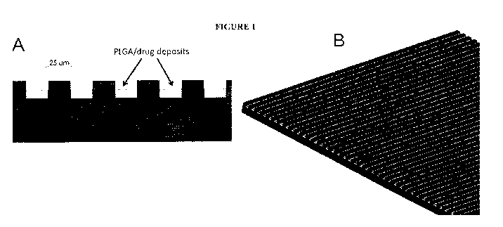
Figure 1 shows a diagram illustrating both the one embodiment of the micro-
patterned
grooves A) with PLGA/drug deposited within the grooves and B) a micro-
patterned surface of
one device.
Figure 2 shows another embodiment of the current invention wherein the micro-
patterned
grooves are 50 tun deep and 25 1.1m wide, the peaks are 25 lam wide, and the
deposited
therapeutic agent fills the grooves leaving 25 ium of the groove. The grooves
are created at
right angles to the peaks.
Figure 3 shows the the surface of one embodiment of the device (1)
Figure 4 shows a side cut view of a cylindrical embodiment of the device (1)
with a
central lumen (5) with the micro-patterned surface. The cylindrical embodiment
of the device
(1) and indicates the micro-patterned grooves containing medication (3) at the
bottom of the
grooves (4). The non-modified top surface (2) is at least 10-50 lam above the
bottom of the
grooves (4). This diagram also demonstrates the deposition of PLGA/drug within
the grooves
(4) of the surface modifications.
Figure 5 shows another view of a cylindrical embodiment of the device (1) with
a central
24
CA 02871759 2014-10-27
WO 2013/165835
PCT/US2013/038360
lumen with the micro-patterned surface.
Figure 6 shows a diagram of the one embodiment of a cylindrical device (1)
with
micro-patterned grooves (containing erodible material with medication) (3) on
the surface of said
device. This diagram was drawn with a cross-section taken out of the body of
the device to
illustrate the depth and geometry of the surface patterns. In this embodiment,
the grooves are 25
iu,m wide and 25 lam deep and contain a 10 Jim film of PLGA/drug within the
grooves.
Figure 7 shows a diagram of the one embodiment of a cylindrical device (1)
with
micro-patterned grooves (containing erodible material with medication) (3) on
the surface of said
device as well as micro-patterned grooves (containing erodible material with
medication) (7) on
the inner surface of the the lumen of said device (5). The non-grooved top
surface (2) and within
the lumen (6) are at least 10-50 !Ina above the bottom of the grooves (4 and
8). This diagram
was drawn with a cross-section removed from the body of thes device to
illustrate the deposition
of PLGA/drug within the grooves of the surface (4) and lumen (8).
Figure 8 shows use of triamcinolone acetonide (TA), a synthetic
corticosteroid, in ocular
tissue [8].
Figure 9 shows the use of PLGA + rapamycin + BHT as a therapeutic agent in
ocular
tissue. BHT is an antioxidant and acts as a stabilizer to prevent oxidative
degradation of
rapamycin.
Figure 10 shows a diagram of the manufacturing process.
Figure 11 shows a diagram of the process workflow.
LIST OF REFERENCE NUMERALS
1 the device
2 non-grooved top surface
CA 02871759 2014-10-27
WO 2013/165835
PCT/US2013/038360
3 micro-patterned grooves (containing erodible material with
medication)
4 bottom of the micro-patterned grooves
the lumen of said device
6 non-grooved top surface within the lumen
5 7 micro-patterned grooves on the inner surface of the lumen
8 bottom of the micro-patterned grooves within the lumen
DESCRIPTION OF THE PREFFERED EMBODIMENT
1. BACKGROUND:
One previous example of anti-fouling technology is found in Banerjee, I. et
al. (2011)
Adv. Mater. 23(6), 690-718 [9] incorporated herein by reference. This
reference teaches several
strategies for prevent fouling due to proteins, bacteria, and marine
organisms. "Several design
patterns, including channels, ridges, pillars, pits, and ribs (Sharklet AF,
biomimetic topography
inspired by shark skin), were fabricated on PDMS elastomer using standard
photolithography
techniques. Based on their studies of the performance of several
microtopographies, they
concluded that an effective coating should possess topographical features that
are smaller than
either the dimension of marine organisms or the parts of organisms that
explore the surface while
settling." The reference does not contemplate a device comprising micro-
patterned geometric
pattern haying anti-biofouling properties or material and do not combine a
micro-patterned
surface with a drug eluting material that controls both acute and chronic
aspects of inflammation
and cellular proliferation.
Another anti-fouling device is described in Ainslie, K. M. and Desai, T. A.
(2008), Lab
Chip 8(11), 1864-1878 [10] incorporated herein by reference. This review
mentions that by
adapting microfabrication techniques originally developed in the
microelectronics industry novel
26
CA 02871759 2014-10-27
WO 2013/165835
PCT/US2013/038360
device for drug delivery, tissue engineering and biosensing have been
engineered for in vivo use
and that implant microfabrication uses a broad range of techniques including
photolithography,
and micromachining to create devices with features ranging from 0.1 to
hundreds of microns
with high aspect ratios and precise features. With respect to biosensors,
methods mentioned to
prevent or limit capsule formation include anti-fouling polymers like PEG,
biomimics such as
phospholipids, flow based systems, membranes, and nanostructured surface
topography, like
nanowires. The reference does not disclose a device comprising an anti-fouling
material having
a micro-patterned geometric pattern and do not combine a micro-patterned
surface with a drug
eluting material that controls both acute and chronic aspects of inflammation
and cellular
proliferation.
Other antifouling materials are described by Vladkova, T. G. (2010)
International
Journal of Polymer Science 2010 (Article ID 296094), 22 pages [11]
incorporated herein by
reference. This reference discloses that many biocontact problems of polymer-
based medical
device may be solved using surface engineering that creates nanosize layers
with controlled
chemical composition, topography and roughness, and hydrophilic/hydrophobic
balance. The
reference teaches a variety of wafer coatings to prevent the adherence of
cells and/or proteins
following implantation. The reference also suggests that the effect of surface
topography and
chemistry on cellular response is of fundamental importance, especially where
living systems
encounter device surfaces in medical implants. Improved thrombo-resistance may
be achieved by
using: i) micro heterogeneous surfaces (e.g., polymers with micro phase
separated structure and
segmented polyurethanes); or ii) simulation of blood vessel properties (e.g.,
surfaces with
hydrophilic nature and high mobility, negatively charged surfaces). For
example, biomaterials
with micro-domain surfaces allow adsorbed proteins to self-organize.
Accordingly, surface
microheterogeneity provides bioinert biomaterials. For example, low-
trombogeneity of block
27
CA 02871759 2014-10-27
WO 2013/165835
PCT/US2013/038360
co-polymers of the type ABA with a hydrophilic/hydrophobic micro-domain
structure is due to a
significant oppress of adhering platelets activation. Typical representative
of this group are the
segmented poly(etherurethanes). The reference does not contemplate a device
comprising
micro-patterned grooves having anti-biofouling properties and do not combine a
micro-patterned
__ surface with a drug eluting material that controls both acute and chronic
aspects of inflammation
and cellular proliferation.
Another anti-fouling strategy is described in Chen, S. et at. (2010) Polymer
5/(23),
5283-5293 [12] incorporated herein by reference. This reference discloses that
there are two
major classes of biological anti-fouling materials, namely polyhydrophilic and
polyzwitterionic
__ materials. These materials are broadly grouped into PEG polymer-based
materials, polybetaine
materials, and polyampholyte materials. PEG anti-fouling materials have
been well
demonstrated to resist nonspecific protein adsorption and cell adhesion, but
suffer from the
disadvantage of biochemically-mediated oxidation. The reference teaches that
hydrogen
bonding and/or ionic interactions between these materials and the surrounding
water molecules
__ forms a hydration layer that is responsible for the anti-fouling
properties. The reference does
not disclose a device comprising an anti-fouling material having an etched
geometric pattern and
do not combine a micro-patterned surface with a drug eluting material that
controls both acute
and chronic aspects of inflammation and cellular proliferation.
Another anti-fouling strategy is described in Desai, T. A. et at. (2000)
Biosens.
__ Bioelectron. 15(9-10), 453-462 [13] incorporated herein by reference. This
reference discloses
the construction of implantable biosensors using anti-fouling materials. The
reference describes
several disadvantages of conventionally used anti-fouling coatings placed on
biosensors that
ultimately result in flaking, peeling, cracking and chipping. The reference
discloses the
construction of a nanopore biosensor chip comprising a plurality of filtration
pores passing
28
CA 02871759 2014-10-27
WO 2013/165835
PCT/US2013/038360
through conventional silicon wafers using conventional micro-patterning
techniques. The
reference teaches that micromachined membranes may be advantageous for in
vitro and in vivo
applications requiring membrane biostability and non-fouling over time. The
data presented
showed that little or no protein adhered to the silicon wafer nanopore
membrane channels during
the performance of a glucose diffusion test, whereas protein did adhere to ion-
track etched
(Millipore) or porous alumina (Whatman) compositions. The reference does not
contemplate a
device comprising micro-patterned grooves that provide a drug delivery
platform and do not
combine a micro-patterned surface with a drug eluting material that controls
both acute and
chronic aspects of inflammation and cellular proliferation.
Another anti-fouling strategy is described in Leoni, L. et al. (2002) Sensors
2(3), 111-120
[14] incorporated herein by reference. This reference discloses monodisperse
nanoporous,
biocompatible, silicon membranes as a platfoiin for cell and/or drug delivery
that remains free of
fibrotic deposition following a two week implantation into a rat peritoneal
cavity. Further, the
wafers were compatible for in vitro growth of insulinoma and/or neurosecretory
(PC12) cells that
grew to confluence and differentiated within the nanoporous wells. The
reference does not
contemplate a device comprising micro-patterned grooves that provide a drug
delivery platform
and do not combine a micro-patterned surface with a drug eluting material that
controls both
acute and chronic aspects of inflammation and cellular proliferation.
Another anti-fouling strategy is described in Messersmith, P. B. et al.
"Peptidomimetic
Polymers for Antifouling Surfaces," United States Patent 7,618,937 [15]
incorporated herein by
reference. This reference discloses polymer-peptide composition that have anti-
biofouling
properties. These polymers include but not limited to polyethylene glycol
(PEG), polyethylene
oxide (PEO), polypropylene oxide (PPO), PEO-PPO-PEO block copolymers,
polyphenylene
oxid, PEG/tetraglyme, PMEMA, polyMPC, and perfluorinated-polyethers. The
references
29
CA 02871759 2014-10-27
WO 2013/165835
PCT/US2013/038360
suggests that it is the peptide portion of the composition that is responsible
for the anti-fouling
properties. The polymers are suggested for use as a coating to prevent protein
and cellular
adhesion to devices for medical and research applications. These devices may
encompass
medical implants, surgical devices, biological sample containers, diagnostic
devices and/or
biosensors. The reference does not contemplate a device comprising micro-
patterned grooves
having anti-biofouling properties and do not combine a micro-patterned surface
with a drug
eluting material that controls both acute and chronic aspects of inflammation
and cellular
proliferation.
Another anti-fouling strategy is described in Mirzadeh, H. et al. (1998)
Iranian Polymer
Journal 7(1), 5-13 [16] incorporated herein by reference. This reference
describes the creation of
super-hydrophobic polymer surfaces by laser treatment and turns them into
hydrophilic ones
grafting hexamethylacrylate (HEMA) after their preactivation by CO2-pulse
laser treatment. The
data from in vitro investigations demonstrate significantly reduced platelet
adhesion and
aggregation on the two type modified surfaces but the best regarding the blood
compatibility
appears to be the super-hydrophobic one. The reference does not contemplate a
device
comprising micro-patterned grooves having anti-biofauling properties and do
not combine a
micro-patterned surface with a drug eluting material that controls both acute
and chronic aspects
of inflammation and cellular proliferation.
Another anti-fouling strategy is described in Acikgoz, C. et al. (2011) Eur.
Cell. Mater.
21(Suppl. 2), 39 [17] incorporated herein by reference. This reference
describes a polymer,
poly(2-methyl-2-oxazoline) (PMOXA), with an antibiotic moiety to kill bacteria
adhering onto
the surface. The reference does not contemplate a device comprising micro-
patterned grooves
having anti-biofouling properties and do not combine a micro-patterned surface
with a drug
eluting material that controls both acute and chronic aspects of inflammation
and cellular
CA 02871759 2014-10-27
WO 2013/165835
PCT/US2013/038360
proliferation.
Another anti-fouling strategy is described in Stofko Jr., J. J. and Yarwood,
J. M.
"Antimicrobial and Antifouling Polymeric Materials," United States Patent
Application
13/120293 [18] incorporated herein by reference. The reference describes
polymeric material
that can be used, for example, to provide coatings that can be antifouling,
antimicrobial, or both.
The reference teaches that the polymeric material described has a plurality of
different pendant
groups that include a first pendant group containing a ¨COOH group or a salt
thereof, a second
pendant group containing a poly(alkylene oxide) group, a third pendant group
containing a
silicon-containing group, and a fourth pendant group containing a quaternary
amino group. The
reference does not contemplate a device comprising micro-patterned grooves or
a geometric
pattern having anti-biofouling properties and do not combine a micro-patterned
surface with a
drug eluting material that controls both acute and chronic aspects of
inflammation and cellular
proliferation.
One patent application, Nguyen et al. "Bare Metal Stent with Drug Eluting
Reservoirs,"
United States Patent Application 13/010869 [19], incorporated herein by
reference, describes
therapeutic agents released under controlled and directional conditions from a
stent. The
reference does not contemplate a device comprising micro-patterned grooves or
a geometric
pattern having anti-biofouling properties and do not combine a micro-patterned
surface with a
drug eluting material that controls both acute and chronic aspects of
inflammation and cellular
proliferation.
Effects of a grooved surface on cell morphology are described by Chou, L. et
al. (1995) J.
Cell Set. 108(4), 1563-1573 [20] incorporated herein by reference. Human
gingival fibroblasts
were cultured on titanium coated grooved surfaces of 3 },tm in depth. Cells on
grooved surfaces
were significantly elongated and orientated along the grooves of the
substratum, while cell height,
31
CA 02871759 2014-10-27
WO 2013/165835
PCT/US2013/038360
measured using confocal scanning laser microscopy, was ¨1.5-fold greater than
that of cells on
smooth surfaces. The surface modifications described here are on single
micromter scale and
does not discuss the acute/chronic concept with etched/ micro-patterned
surfaces combined with
medications.
Other effects of cells grown on nanopatterned surfaces, such as `nanopost' and
`nanograte' structures, are described in Choi, C.-H. et al. (2007)
Biomaterials 28(9), 1672-1679
[21] incorporated herein by reference. Human foreskin fibroblasts exhibited
significantly
smaller cell size and lower proliferation on needle-like nanoposts, and
enhanced elongation with
alignment on blade-like nanogrates. These phenomena became more pronounced as
the
nanotopographical three dimensionality (structural height) increased. The
nanopost and
nanograte architectures provided the distinct contact guidance for both
filopodia extension and
the formation of adhesion molecules complex, which was believed to lead to the
unique cell
behaviors observed. The surface modifications described here are on single
nanomter scale and
does not discuss the acute/chronic concept with etched/ micro-patterned
surfaces combined with
medications.
Other effects of cells grown on nanopattemed surfaces, such as how
nanotopology can
affect cell adhesion and spreading, are described in Tay, C. Y. et al. (2011)
Micro-/Nano-engineered Cellular Responses for Soft Tissue Engineering and
Biomedical
Applications, Small 7(10), 1361-1378 [22] incorporated herein by reference.
The surface
modifications described here are on single nanomter scale and does not discuss
the acute/chronic
concept with etched/ micro-patterned surfaces combined with medications.
Other effects of cells grown on nanopattemed surfaces, such as how
nanotopology can
affect cell adhesion and spreading, are described in Bettinger, C. J., Langer,
R., and Borenstein, J.
T. (2009) Angewandte Chemie International Edition in English 48(30), 5406-5415
[23]
32
CA 02871759 2014-10-27
WO 2013/165835
PCT/US2013/038360
incorporated herein by reference. Three basic nanotopography geometries
include nanograting,
nanopost array, and nanopit array are also described. The surface
modifications described here
are on single nanomter scale and does not discuss the acute/chronic concept
with etched/
micro-patterned surfaces combined with medications.
2. DESCRIPTION OF THE INVENTION
This invention is in the field of implantable medical devices. The present
invention
relates to a device constructed from metals, polymers or other materials that
are amenable to
precise surface modifications and coupling with erodible agents methods for
its use, wherein (1)
the erodible agents, which contain active ingredients (i.e., for example,
medications) provide for
acute control of cellular proliferation and (2) a pattered surface having
micron-, and/or
nano-sized micro-patterning characteristics that imparts anti-proliferative
properties.
Further, the device comprises a drug delivery platform by placing erodible or
non-erodable
medication depots within the grooves of the constructed patterns. In other
embodiments, device
is created from a material wherein a pattered surface having micron-sized
micro-patterned
characteristics imparts anti-proliferative or anti-fibrotic properties.
Further, the device comprises
a drug delivery platform by placing medication depots (i.e., a plastic, or a
semi-solid gel) within
the grooves of the micro-patterned pattern.
For example, the device may have an etching pattern that forms a grid pattern
or
geometric pattern. Different devices can therefore be constructed with
different grid
dimensions or geometric patterns. The current invention contemplates that an
implanted 10-50
preferrably 20-35 pm, grid shows: i) a decrease in fibroblast or other cells
number: and ii) an
increase in cell alignment (i.e., improved organization of adhered cells).
This is in comparison
33
CA 02871759 2014-10-27
WO 2013/165835
PCT/US2013/038360
to a blank (non-micro-patterned or non-etched) device control that displays a
disorganized
pattern of more densely adhered cells. The current invention contemplates that
the optimal
dimension of the geometric patterns might depend on the specific material.
Data, such as Table
1, shows that devices, made of various materials, having specific surface
etching patterns can
control fibroblast proliferation.
The invention further contemplates that medications may be placed in the
grooves of the
micro-patterned grid or geometric pattern such that the benefit of the micro-
patterned surfaces
preventing fibroblast growth and promoting organizations of the micro-
patterned surfaces is
maintained or supplemented/accentuated. The medications can include but are
not limited to a
steroid, rapamycin, everolimus, tacrolimus, paclitaxel or other antifibrotic
medications as well as
biologics or targeted therapeutics for specific diseases like glaucoma,
macular degeneration or
neurodegenerative diseases. Preferably, the medication would be placed in a
slow release depot
comprising a polymer including but not limited to PLGA, PLA, PGA or PCL.
Methods of the present invention are contemplated as implanting the devices
within
tissues for the treatment of various medical conditions without inducing
fibrosis. For example,
the medical condition may be inflammation and/or swelling wherein the
implanted device
facilitates drainage of a tissue. Once the device is placed, the depot slowly
releases a
medication (e.g., an antifibrotic) to prevent/lessen encapsulation of the
device with fibroblasts or
other cell types. Secondarily, once the depot has released all the
medication, the
micro-patterned surface of the device continues to inhibit the encapsulation
process. Another
method contemplated by the present invention is related to precisely
depositing the medication
depots within the geometric pattern grooves by using by precise means. In one
embodiment,
said medication depots are deposited by an inkjet printer or other precision
dispensing
instrument. The placement and amount of the medication depots are such that
the antifibrotic
34
CA 02871759 2014-10-27
WO 2013/165835
PCT/US2013/038360
properties of the micro-patterned grid or geometric pattern surface are
maintained and
contributes to an antifibrotic environment even before the complete release of
the medication
from the depot. Specifically, if the medication depot is deposited on the
raised portions of the
geometric pattern, the anti-fibrotic properties of the device are impaired.
3. THERAPEUTIC AGENTS
Triamcinolone acetonide (TA), a synthetic corticosteroid, has been used to
treat ocular
tissue delivered using a number of controlled release systems such as PVA,
PEVA/PBMA
(SurModics), PLGA, PCL and PMM. The SurModics system claims up to 2 yrs of
delivery
whereas more typical durations of release are around 4-12 weeks.
There is previous use of rapamycin, a synthetic corticosteroid, in ocular
tissue using
PLGA drug-eluting stents. One particular formulation was PLGA + rapamycin +
butylated
hydroxy toluene (BHT) as described in the Eurpean Patent Application EP2361593
[24]. BHT
is an antioxidant and acts as a stabilizer to prevent oxidative degradation of
rapamycin. Release
from most thin film reservoir systems is somewhere in the 30-40 day range (see
Figure 9 from
reference [25]; NEVO is a PLGA/rapamycin system). However, some studies
suggest longer
release rates are possible by switching to higher L:G ratios of PLGA (higher
L:G ratio means
more hydrophobic polymer; as a result, less water can swell into the system so
diffusion of the
drug out of the polymer is slower; however, higher L:G ratios also mean slower
biodegradation
of the PLGA).
Bevacizumab has been conjugated with PEG and encapsulated in PLGA
nanoparticles
Pan et al. (2011) [26]. Conjugation is an actual chemical bonding of the
polymer to the drug
where encapsulation is just a physical barrier so these represented very
different delivery
strategies. Neither system showed very favorable release control. Another
study, formulated
CA 02871759 2014-10-27
WO 2013/165835
PCT/US2013/038360
PLGA nanoparticles with bevaciz-umab and achieved 4 weeks of sustained
delivery, Boddu et al.
(2010) [27]. Stability of the protein is of concern since as PLGA degrades its
byproducts are
lactic acid and glycolic acid so the PLGA matrix can become an acidic
environment.
4. USE OF THE DEVICE:
In one embodiment, the present invention contemplates a drug delivery device
wherein
medication (i.e., for example, antifibrotics and other medications or
therapeutic agents) is placed
withinin a plurality of grooves such that the medication does not rise above
the top surface of the
grooves. Although it is not necessary to understand the mechanism of an
invention, it is
believed that this medication placement maintains the benefit of the micro-
patterned surfaces for
preventing fibroblast growth and/or promoting organizations. It is also
believed that such
medication placement inhibits organized cell proliferation along the micro-
patterned geometric
grooves so that encapsulation and scar tissue formation is minimized,
eliminated, or appreciably
reduced. Further, a distinct reduction in cell proliferation may result. In
one embodiment,
medication can be a steroid, rapamycin, or other antifibrotic medications as
well as biologics or
targeted therapeutics for specific diseases including, but not limited to
cataract, diabetic
manifestations in the eye, systemic disease manifestations in the eye,
inherited retina and
choroidal diseases, glaucoma, neuropathies/neurodegenerative disease, uveitic
diseases, or
macular degeneration (wet and dry). In one embodiment, the medication may be
placed in a slow
release depot such as PLGA or PEG systems (or other). The devices can then be
implanted inside
of tissues and benefit from the action of both the depot/medication and the
micro-patterned
surfaces. Once the device is placed, primarily, the depot/medication will
slowly release
anti-fibrotic or other medications to prevent/lessen encapsulation of the
device with fibroblasts or
other cell types. This might benefit the device action, which could be for
drainage or for other
36
CA 02871759 2014-10-27
WO 2013/165835
PCT/US2013/038360
purposes unique to any given implant. Secondarily, once the medication and the
depot have
diffused, the micro-patterned surface remains so that it will still lessen the
encapsulation process
independent of the depot medication. The medication depot may be placed in
such a way (with
precise inkjet deposition, for example) that may allow the micro-patterned
surface qualities to be
maintained and provide an antifibrotic environment even before the
medication/depot empties
from the bottom of the grooved space.
In one embodiment, PLGA matrices release drug over immediate period after
implantation, preventing initial cell proliferation response, (other depot
mechanisms other than
PLGA might also be used). In another embodiment, micro-patterned surface may
provide
initial and long-term inhibition of fibrosis, ensuring long-temi prevention of
capsulation. In
one embodiment, precise inkjet printing may provide the avenue of deposition
for the medication
in the bottom of the micro-patterned grooved space. Inkjet printing is able to
accurately fill
such micron-scale features with flexibility in the solution to be dispensed
and high throughput
capability. In another embodiment, PLGA/drug matrix must be contained within
"channels" of
surface as a conformal polymer coating may counteract beneficial effect of
micro-patterned
surface.
5. DETAILED DESCRIPTON OF THE INVENTION
The following detailed description, and the drawings to which it refers, are
provided for
the purpose of describing and illustrating certain preferred embodiments or
examples of the
invention only, and no attempt has been made to exhaustively describe all
possible embodiments
or examples of the invention. Thus, the following detailed description and the
accompanying
drawings shall not be construed to limit, in any way, the scope of the claims
recited in this patent
application and any patent(s) issuing there from.
37
CA 02871759 2014-10-27
WO 2013/165835
PCT/US2013/038360
Distinguishing features compared to other devices:
1. No mechanically moving parts
2. Micro-patterned grooved valleys (i.e., for example, approximately 10-50um
wide and
at least 25 um deep)
3. Flat peaks (i.e., for example, at least 10-50 um wide)
4. The angle between the peaks and the valleys are at a right angle (e.g., 90
degrees) in
some materials but curved in other materials (i.e., for example, between
approximately 95 to 120
degrees, as a frame of reference 180 degrees would be a line crossing all the
peaks). The curve is
the slope between the peak and the wall going down to the bottom of the groove
(like a mountain
rather than a cliff).
5. The dimensional ratios of peaks and valleys and angles of the surface
modifications are
specific to each given material used as it relates to the interaction of
material to dimensions to
proliferating cells
6. The coupling of micro-patterned surfaces with eluting medication depots is
at the core
of the present invention and addresses both acute and chronic aspects of the
biologic response to
implanted materials.
In one embodiment, the invention contemplates implanted medical device in or
on the
eye. In one embodiment, the invention contemplates implanted medical device in
the sclera. In
one embodiment, the invention contemplates implanted medical device in
Schlemm's canal.
The present invention considers a device made by various materials. In one
embodiment, the
material is polymeric such as silicone, polyimide (PI), polysulfone (PES),
polyetheretherketone
(PEEK), polyetherimide (PEI), or metallic materials such as titanium or
aluminum or ceramic
such as titanium oxide, calcium phosphate or hydroxyapatite or alloys such as
nickel-titanium
38
CA 02871759 2014-10-27
WO 2013/165835
PCT/US2013/038360
(NiTi), stainless steel or titanium alloys.
In one embodiment, the present invention contemplates an implanted medical
device
capable of having a variety of shapes. In one embodiment, the device has shape
selected from
the group consisting of spherical, non-spherical (egg-shaped), cylindrical,
rectangular, cubic,
toroidal, conical, cuboidal, pyramidal, prism, and planar shapes. In one
embodiment, the device
has a cylindrical shape. In one embodiment, the device has a cylindrical shape
contains at least
one lumen. In one embodiment, the lumen contains a depot. In one embodiment,
said depot
contains at least one medication. In one embodiment, said medication includes,
but is not limited
to, anti-fibrotic agent, anti-inflammatory agent, immunosuppressant agent,
anti-neoplastic agent,
migration inhibitors, anti-proliferative agent, rapamycin, triamcinolone
acetonide, everolimus,
tacrolimus, paclitaxel, actinomycin, azathioprine, dexamethasone,
cyclosporine, bevacizumab, an
anti-VEGF agent, an anti-IL-1 agent, canakinumab, an anti-IL-2 agent, viral
vectors, beta
blockers, alpha agonists, muscarinic agents, steroids, antibiotics, non-
steroidal anti-inflammatory
agents, prostaglandin analogues, ROCK inhibitors, nitric oxide, endothelin,
matrixmetalloproteinase inhibitors, CNP/BMP, corticosteroids, and/or antibody-
based
immunosuppresants. In one embodiment, said medication is combined with a
polymer. In one
embodiment, said polymer is selected from the group consisting of poly(lactic-
co-glycolic acid),
polyethylene glycol, poly(lactic acid), poly(glycolic acid), poly(amido
ester), polyethylene
terephthalate, poly(caprolactone), poly(hydroxy butyrate), poly(butylene
succinate), poly(vinyl
alchohol), poly(hydroxybutyrate), poly(methyl acrylate), poly(methyl
methylmethacrylate),
poly(sebacic acid), carboxymethyl cellulose, ethyl cellulose, cellulose
acetate, polydioxanone, or
polymers from the categories: polyesters, polyanhydrides, polyamides,
polycyanoacrylates,
polyurethanes, polyorthoesters, silicones, acrylic polymers, cellulose
derivatives or poloxamers.
It is not intended that the current device be limited to one particular shape
or be limited to
39
CA 02871759 2014-10-27
WO 2013/165835
PCT/US2013/038360
any particual geometrical pattern. In one embodiment, said device has shape
selected from the
group consisting of spherical, non-spherical (egg-shaped), cylindrical,
rectangular, cubic, toroidal,
conical, cuboidal, pyramidal, prism, and planar shapes. In one embodiment,
said device has
micro-patterned grooves in a grid pattern. In one embodiment, said device has
micro-patterned
grooves in a vertical orientation. In one embodiment, said device has micro-
patterned grooves in
a horizontal orientation. In one embodiment, said device has micro-patterned
grooves in a
diagonal orientation. In one embodiment, said micro-patterned grooves
intersect. In the case of
a spherical shape, the micro-patterned grooves could be in the form of
circular pattern about the
body of the device. In the case of a toroidal shape, the micro-patterned
grooves could be in the
form of circular pattern about the body of the device. In the case of
cylindrical, rectangular, cubic,
cuboidal, and planar shapes, the micro-patterned grooves could be in the form
of vertical,
horizontal, diagonal, or intersecting grids. In the case of c conical,
pyramidal, prism shapes, the
micro-patterned grooves could be in the form of vertical, horizontal,
diagonal, intersecting grids
in the foul' of circular pattern about the body of the device. In one
embodiment, the device with
a cylindrical shape has micro-patterned grooves in a vertical orientation. In
one embodiment, the
device with a cylindrical shape has micro-patterned grooves in a horizontal
orientation. In one
embodiment, the device with a cylindrical shape has micro-patterned grooves in
a diagonal
orientation.
In one embodiment, said device is a catheter. In one embodiment, said device
is a stent.
In one embodiment, said device is a catheter for a defibrillation device. In
one embodiment, said
device is a catheter for a defibrillation device. In one embodiment, said
device is a intravenous
catheter. In one embodiment, said device is a Hickman line. In one embodiment,
said device is a
mesh prosthesis. In one embodiment, said device is a hernia mesh. In one
embodiment, said
device is a Baerveldt glaucoma implant. In one embodiment, said device is a
glaucoma
CA 02871759 2014-10-27
WO 2013/165835
PCT/US2013/038360
shunting device. Baerveldt glaucoma implants feature a surface area plate. In
one embodiment,
the current invention contemplates micro-patterning of the plate of the
Baerveldt device to
prevent encapsulation.
In one embodiment, the use of the implant prevents disorderly growth of
fibroblasts. In
one embodiment, the use of the implant prevents the formation of scar tissue.
In one
embodiment, the micro-patterned grooves provide an avenue for drainage.
In one embodiment, the current invention uses an inkjet loading system to
deposit
therapeutic agents into the micro-patterned grooves. The drop volumes produced
with the
inkjet dispensing system are in the range of 1.5 pL to 4.2 nL. The system
provides precise
control of filling volumes, typically 1-3% repeatability (drop-to-drop,
depending on dispensing
solution properties), with a drop firing rate up to about 30,000 per second.
Such a system has
high throughput, simple operation, high versatility, and is relatively
inexpensive. Error!
Reference source not found. shows stent loading with an injection loading
system. The entire
stent could be loaded in a very rapid and preceise process. The system is
largely automated
with machine vision-based mapping of deposition locations and accurately
ejected drops to those
locations appropriately, as illustrated in Figure 10. Figure 10 shows a
diagram of the
manufacturing process. The system employs a real-time camera or pre-programmed
image
recognition to accurately target reservoirs/depots.
In one embodiment, a solution containing approximately 10% 75:25
lactide:glycolide
PLGA, 10% rapamycin and 0.5% BHT in DMSO are prepared as the dispensing
solution. This
solution is loaded into the inkjet dispenser, which is heated to 50 C to
facilitate the dispensing
process. The control software uses the reservoir location map to translate the
devices underneath
the inkjet dispenser such that the dispensing locations pass under the inkjet
tip. Translation is
accomplished by three-axis motion stages and controllers connected to a
computer via a
41
CA 02871759 2014-10-27
WO 2013/165835
PCT/US2013/038360
hardware interface. The inkjet dispenser is triggered in a "drop-on-demand"
mode by software
control to dispense a set number of droplets of the solution into the grooves,
filling the grooves
completely but not overflowing them so as to prevent deposition of the matrix
onto the raised
surfaces. After dispesing the solution into the grooves in multiple devices in
a batch process, the
devices are transferred to a vacuum oven and the DMSO is driven off leaving
only PLGA,
rapamycin and BHT. This process is repeated, successively filling and drying
the reserviors until
the solids comprise the desired depot dimensions. This process workflow is
shown
diagrammatically in Error! Reference source not found..
Figure 11 shows a diagram of the process workflow. Intially one manually loads
one
batch of devices into "holder." Secondly, one creates digital "map" of
reservoir locations using
machine vision image recognition. Subsequently, one dispenses therapeutic
agent or
drug/polymer solution into reservoirs by translating cassette under inkjet.
Followed by a dry
cycle to remove solvent from solution (volume limitation of reservoir), then
the process is
repeated with fill/dry steps until reservoir is filled with solids
42
CA 02871759 2014-10-27
WO 2013/165835
PCT/US2013/038360
6. SUPPORTING DATA
Table 1: Cell growth associated with micro-patterned surfaces in various
materials. Each
data point was compared to a non micro-patterned surface as the control (data
not shown). All
non micro-patterned surfaces resulted in diffuse growth of cells. The patterns
were created with
equivalent width and depth.
0.5 1.0 5.0 10.0 20.0 25.0 30.0 50.0 100.0
Material
m pm 1.tm pm pm pm pm pm [tm
Silicone D D D D Mo Mo Mi D
PEEK D D D Mo Mi Mi Mi D
Titanium D D D Mo Mi Mi Mi Mo
Stainless Steel D D D D Mo Mo Mi
Mo
PTFE D D D D Mo
Mi Mi Mi
D = Diffuse Cell Growth PEEK = Polyether ether ketone
Mo = Moderate Cell Growth PTFE = Polytetrafluoroethylene
Mi = Minimal Cell Growth
Thus, specific compositions and configurations of antiproliferative surface
modifications
and methods of use have been disclosed. It should be apparent, however, to
those skilled in the
art that many more modifications besides those already described are possible
without departing
from the inventive concepts herein. The inventive subject matter, therefore,
is not to be restricted
except in the spirit of the disclosure. Moreover, in interpreting the
disclosure, all terms should be
interpreted in the broadest possible manner consistent with the context. In
particular, the terms
"comprises" and "comprising" should be interpreted as referring to elements,
components, or
steps in a non-exclusive manner, indicating that the referenced elements,
components, or steps
43
CA 02871759 2014-10-27
WO 2013/165835
PCT/US2013/038360
may be present, or utilized, or combined with other elements, components, or
steps that are not
expressly referenced.
All publications mentioned herein are incorporated herein by reference to
disclose and
describe the methods and/or materials in connection with which the
publications are cited. The
publications discussed herein are provided solely for their disclosure prior
to the filing date of
the present application. Nothing herein is to be construed as an admission
that the present
invention is not entitled to antedate such publication by virtue of prior
invention. Further, the
dates of publication provided may be different from the actual publication
dates, which may need
to be independently confirmed.
44
CA 02871759 2014-10-27
WO 2013/165835
PCT/US2013/038360
REFERENCES:
1. Kahook, M. Y. "Antiproliferative surface modifications and methods of
use," United
States Provisional Patent Application 61/640,843, filed 5/1/2012. (published
N/A).
2. Freemantle, N. et al. (1999) "beta Blockade after myocardial infarction:
systematic
review and meta regression analysis," BMJ 3/8(7200), 1730-1737.
3. Cruickshank, J. M. (2010) "13 blockers in hypertension," The Lancet
376(9739), 415.
4. Fleisher, D., Bong, R., and Stewart, B. H. (1996) "Improved oral drug
delivery: solubility
limitations overcome by the use of prodrugs," Adv. Drug Delivery Rev. 19(2),
115-130.
5. Smith, C. D. etal. (1994) "A sensitive assay for taxol and other
microtubule-stabilizing
agents," Cancer Lett. 79(2), 213-219.
6. Mooberry, S. L., Stratman, K., and Moore, R. E. (1995) "Tubercidin
stabilizes
microtubules against vinblastine-induced depolymerization, a taxol-like
effect," Cancer
Lett. 96(2), 261-266.
7. Ro, A. J., Falotico, R., and Dave, V. (2012) "Morphological and
degradation studies of
sirolimus-containing poly(lactide-co-glycolide) discs," Journal of Biomedical
Materials
Research Part B: Applied Biomaterials 100B(3), 767-777.
8. Eperon, S. etal. (2008) "A biodegradable drug delivery system for the
treatment of
postoperative inflammation," Int. J. Pharm. 352(1-2), 240-247.
9. Banerjee, I., Pangule, R. C., and Kane, R. S. (2011) "Antifouling
Coatings: Recent
Developments in the Design of Surfaces That Prevent Fouling by Proteins,
Bacteria, and
Marine Organisms," Advanced Materials 23(6), 690-718.
10. Ainslie, K. M. and Desai, T. A. (2008) "Microfabricated Implants for
Applications in
Therapeutic Delivery, Tissue Engineering, and Biosensing," Lab Chip 8(11),
1864-1878.
11. Vladkova, T. G. (2010) "Surface Engineered Polymeric Biomaterials with
Improved
CA 02871759 2014-10-27
WO 2013/165835
PCT/US2013/038360
Biocontact Properties," International Journal of Polymer Science 20/0(Article
ID
296094), 22 pages.
12. Chen, S. et al. (2010) "Surface hydration: Principles and applications
toward
low-fouling/nonfouling biomaterials," Polymer 51(23), 5283-5293.
13. Desai, T. A. et al. (2000) "Nanoporous anti-fouling silicon membranes
for biosensor
applications," Biosens. Bioelectron. 15(9-10), 453-462.
14. Leoni, L., Attiah, D., and Desai, T. (2002) "Nanoporous Platforms for
Cellular Sensing
and Delivery," Sensors 2(3), 111-120.
15. Messersmith, P. B. et al. "Peptidomimetic polymers for antifouling
surfaces," United
States Patent 7,618,937, Application 11/280107, filed 11/16/2005. (issued
11/17/2009).
16. Mirzadeh, H., Khorasani, M., and Sammes, P. G (1998) "Laser surface
modification of
polymers. A novel technique for the preparation of blood compatible materials
(II): in
vitro assay," Iranian Polymer Journal 7(1), 5-13.
17. Acikgoz, C. et al. (2011) "Implant Infection (Antifouling and
Antimicrobial Surface
Coatings through Poly(2-methyl-2-oxazoline))," Eur. Cell. Mater. 21(Suppl. 2),
39.
18. Stotko Jr., J. J. and Yarwood, J. M. "Antimicrobial and Antifouling
Polymeric Materials,"
United States Patent Application Publication Number 20110171158, Application
13/120293, filed 8/25/2009. (published 7/14/2011).
19. Nguyen, T. M., Parker, T. L., and Shanley, J. F. "Bare Metal Stent with
Drug Eluting
Reservoirs," United States Patent Application Publication Number 20110137407,
Application 13/010869, filed 1/21/2011. (published 6/9/2011).
20. Chou, L. et al. (1995) "Substratum surface topography alters cell shape
and regulates
fibronectin mRNA level, mRNA stability, secretion and assembly in human
fibroblasts," J.
Cell Sci. 108(4), 1563-1573.
46
CA 02871759 2014-10-27
WO 2013/165835
PCT/US2013/038360
21. Choi, C.-H. et al. (2007) "Cell interaction with three-dimensional
sharp-tip
nanotopography," Biomaterials 28(9), 1672-1679.
22. Tay, C. Y. et al. (2011) "Micro-/Nano-engineered Cellular Responses for
Soft Tissue
Engineering and Biomedical Applications," Small 7(10), 1361-1378.
23. Bettinger, C. J., Langer, R., and Borenstein, J. T. (2009) "Engineering
substrate
topography at the micro- and nanoscale to control cell function," Angewandte
Chemie
International Edition in English 48(30), 5406-5415.
24. Nguyen, T. M., Parker, T. L., and Shanley, J. F. "Bare metal stent with
drug eluting
reservoirs," European Patent Application EP 2361593, Application 11152966.5,
filed
2/1/2011. (published 8/31/2011).
25. Falotico, R. et al. (2009) "NEVOTm: a new generation of sirolimus-
eluting coronary
stent," EuroIntervention 5(Supplement F), F88-F93.
26. Pan, C. K. et al. (2011) "Comparison of Long-Acting Bevacizumab
Formulations in the
Treatment of Choroidal Neovascularization in a Rat Model," J. Ocul. Pharmacol.
Ther
27(3),219-224.
27. Boddu, S. H. S. et al. (2010) "Novel Nanoparticulate Gel Folinulations
of Steroids for the
Treatment of Macular Edema," .1 Ocul. Pharmacol. Ther. 26(1), 37-48.
47