Note : Les descriptions sont présentées dans la langue officielle dans laquelle elles ont été soumises.
CA 02872025 2014-10-29
WO 2013/181005 PCT/US2013/041784
CONTROL FOR BIOPSY DEVICE
BACKGROUND
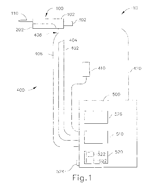
[0001] Biopsy samples have been obtained in a variety of ways in various
medical
procedures using a variety of devices. Biopsy devices may be used under
stereotactic
guidance, ultrasound guidance, MRI guidance, PEM guidance, BSGI guidance, or
otherwise. For instance, some biopsy devices may be fully operable by a user
using a
single hand, and with a single insertion, to capture one or more biopsy
samples from a
patient. In addition, some biopsy devices may be tethered to a vacuum module
and/or
control module, such as for communication of fluids (e.g., pressurized air,
saline,
atmospheric air, vacuum, etc.), for communication of power, and/or for
communication
of commands and the like. Other biopsy devices may be fully or at least
partially
operable without being tethered or otherwise connected with another device.
[0002] Merely exemplary biopsy devices are disclosed in U.S. Pat. No.
5,526,822,
entitled "Method and Apparatus for Automated Biopsy and Collection of Soft
Tissue,"
issued June 18, 1996; U.S. Pat. No. 6,086,544, entitled "Control Apparatus for
an
Automated Surgical Biopsy Device," issued July 11, 2000; U.S. Pat. No.
6,626,849,
entitled "MRI Compatible Surgical Biopsy Device," issued September 11, 2003;
U.S.
Pat. No. 7,442,171, entitled "Remote Thumbwheel for a Surgical Biopsy Device,"
issued
October 8, 2008; U.S. Pat, No. 7,854,706, entitled "Clutch and Valving System
for
Tetherless Biopsy Device," issued December 1, 2010; U.S. Pat. No. 7,938,786,
entitled
"Vacuum Timing Algorithm for Biopsy Device," issued May 10, 2011; U.S. Pat.
Pub.
No. 2006/0074345, entitled "Biopsy Apparatus and Method," published April 6,
2006;
U.S. Pat. Pub. No. 2008/0214955, entitled "Presentation of Biopsy Sample by
Biopsy
Device," published September 4, 2008; U.S. Pat, Pub. No. 2008/0221480,
entitled
"Biopsy Sample Storage," published September 11, 2008; U.S. Pat. Pub. No.
2009/0131821, entitled "Graphical User Interface For Biopsy System Control
Module,"
published May 21, 2009; U.S. Pat. Pub. No. 2009/0131820, entitled "Icon-Based
Uswer
Interface On Biopsy System Control Module," published May 21, 2009; U.S. Pat.
Pub.
- 1 -
CA 02872025 2014-10-29
WO 2013/181005 PCT/US2013/041784
No. 2010/0152610, entitled "Hand Actuated Tetherless Biopsy Device with Pistol
Grip,"
published June 17, 2010; U.S. Pat. Pub. No. 2010/0160819, entitled "Biopsy
Device with
Central Thumbwheel," published June 24, 2010; U.S. Pat, Pub. No. 2010/0317997,
entitled "Tetherless Biopsy Device with Reusable Portion," published December
16,
2010; U.S. Non-Provisional Patent App. No. 12/953,715, entitled "Handheld
Biopsy
Device with Needle Firing," filed November 24, 2010; U.S. Non-Provisional
Patent App.
No. 13/086,567, entitled "Biopsy Device with Motorized Needle Firing," filed
April 14,
2011; U.S. Non-Provisional Patent App. No. 13/150,950, entitled "Needle
Assembly and
Blade Assembly for Biopsy Device," filed June 1, 2011; U.S. Non-Provisional
Patent
App. No. 13/205,189, entitled "Access Chamber and Markers for Biopsy Device,"
filed
August 8, 2011; U.S. Non-Provisional Patent App. No. 13/218,656, entitled
"Biopsy
Device Tissue Sample Holder with Bulk Chamber and Pathology Chamber," filed
August
26, 2011; and U.S. Provisional Patent App. No. 61/566,793, entitled "Biopsy
Device
With Slide-In Probe," filed December 5, 2011. The disclosure of each of the
above-cited
U.S. Patents, U.S. Patent Application Publications, and U.S. Non-Provisional
Patent
Applications is incorporated by reference herein.
[0003] While several systems and methods have been made and used for
obtaining a
biopsy sample, it is believed that no one prior to the inventor has made or
used the
invention described in the appended claims.
BRIEF DESCRIPTION OF THE DRAWINGS
[0004] While the specification concludes with claims which particularly
point out and
distinctly claim this technology, it is believed this technology will be
better understood
from the following description of certain examples taken in conjunction with
the
accompanying drawings, in which like reference numerals identify the same
elements and
in which:
[0005] FIG. 1 depicts a schematic view of an exemplary biopsy system;
[0006] FIG. 2 depicts a perspective view of an exemplary biopsy device;
- 2 -
CA 02872025 2014-10-29
WO 2013/181005 PCT/US2013/041784
[0007] FIG. 3 depicts a perspective view of the biopsy device of FIG. 2
showing an
exemplary probe decoupled from an exemplary holster;
[0008] FIG. 4 depicts a rear perspective view of the holster of FIG. 3;
[0009] FIG. 5 depicts a rear perspective view of the holster of FIG. 4
with a top housing
cover omitted;
[0010] FIG. 6 depicts an exploded perspective view of the holster of FIG.
5;
[0011] FIG. 7 depicts a perspective view of the probe of FIG. 3;
[0012] FIG. 8 depicts a top plan view of the probe of FIG. 7 with a top
probe cover
omitted;
[0013] FIG. 9 depicts an exploded perspective view of the probe of FIG.
8;
[0014] FIG. 10 depicts a perspective view of an exemplary tissue sample
holder;
[0015] FIG. 11 depicts a front view of an exemplary user interface for
the biopsy device
of FIG. 2;
[0016] FIG. 12 depicts a flowchart of an exemplary control for the biopsy
device of FIG.
2;
[0017] FIG. 13 depicts a flowchart of another exemplary control for the
biopsy device of
FIG. 2;
[0018] FIG. 14 depicts a flowchart of another exemplary control for the
biopsy device of
FIG. 2; and
[0019] FIG. 15 depicts a flowchart of another exemplary control for the
biopsy device of
FIG. 2.
[0020] The drawings are not intended to be limiting in any way, and it is
contemplated
that various embodiments of the technology may be carried out in a variety of
other ways,
including those not necessarily depicted in the drawings. The accompanying
drawings
- 3 -
CA 02872025 2014-10-29
WO 2013/181005 PCT/US2013/041784
incorporated in and forming a part of the specification illustrate several
aspects of the
present technology, and together with the description serve to explain the
principles of
the technology; it being understood, however, that this technology is not
limited to the
precise arrangements shown.
DETAILED DESCRIPTION
[0021] The following description of certain examples of the technology
should not be
used to limit its scope. Other examples, features, aspects, embodiments, and
advantages
of the technology will become apparent to those skilled in the art from the
following
description, which is by way of illustration, one of the best modes
contemplated for
carrying out the technology. As will be realized, the technology described
herein is
capable of other different and obvious aspects, all without departing from the
technology.
Accordingly, the drawings and descriptions should be regarded as illustrative
in nature
and not restrictive.
[0022] I. Overview of Exemplary Biopsy System
[0023] FIG. 1 depicts an exemplary biopsy system (10) comprising a biopsy
device
(100), a plurality of conduits (400) and a control module (500). Biopsy device
(100)
comprises a holster (202) and a probe (102). A needle (110) extends distally
from probe
(102) and is inserted into a patient's tissue to obtain tissue samples as will
be described in
greater detail below. These tissue samples are deposited into a tissue sample
holder (302)
that is coupled to a proximal end of probe (102), as will also be describe in
further detail
below. Of course needle (110) and tissue sample holder (302) may be coupled to
probe
(102) at a range of locations. For instance, needle (110) may extend from the
top of
probe (102), from a side of probe (102), from the bottom of probe (102), or,
may be
omitted from probe (102) entirely. Tissue sample holder (302) may be coupled
to the top
of probe (102), to a side of probe (102), to the bottom of probe (102), or,
may be omitted
from probe (102) entirely. Probe (102) of the present example is separable
from holster
(202), though this is merely optional. It should also be understood that the
use of the
term "holster" herein should not be read as necessarily requiring any portion
of probe
(102) to be inserted into any portion of holster (202). While an notched upper
control
- 4 -
CA 02872025 2014-10-29
WO 2013/181005 PCT/US2013/041784
unit (220) of the holster (202) and a latch (190) of probe (102) are used to
cooperatively
removably secure probe (102) to holster (202), as shown in FIGS. 2-4 and 7 and
described in greater detail below, it should be understood that a variety of
other types of
structures, components, features, etc. (e.g., bayonet mounts, prongs, clamps,
clips, snap
fittings, etc.) may be used to provide removable coupling of probe (102) and
holster
(202). Furthermore, in some biopsy devices (100), probe (102) and holster
(202) may be
of unitary or integral construction, such that the two components cannot be
separated. By
way of example only, in versions where probe (102) and holster (202) are
provided as
separable components, probe (102) may be provided as a disposable component,
while
holster (202) may be provided as a reusable component. Still other suitable
structural and
functional relationships between probe (102) and holster (202) will be
apparent to those
of ordinary skill in the art in view of the teachings herein.
[0024] Biopsy system (10) shown in FIG. 1 further includes a control
module (500) that
is fluidly coupled to biopsy device (100) via one or more conduits (400). In
the present
example, control module (500) comprises a vacuum n source (510) operable to
provide a
vacuum to biopsy device (100). Control module (500) further comprises a user
interface
(526) that allows a user adjust the level of vacuum provided to biopsy device
(100). It
may be desirable for a user to adjust the level of vacuum depending on the
characteristics
(hardness, thickness, etc.) of the tissue to be sampled by biopsy device
(100). User
interface (526) will be discussed in more detail below. By way of example
only, vacuum
source (510) is contained within control module (500) and is fluidly coupled
to probe
(102) via a first conduit (402), such as flexible tubing. Of course, in
addition or in the
alternative, vacuum source (510) may be incorporated into probe (102),
incorporated into
holster (202), and/or be a separate component altogether. One merely exemplary
biopsy
device (100) having a vacuum source (510) incorporated therein is disclosed in
U.S. Non-
provisional Patent Application 12/953,715, entitled "Handheld Biopsy Device
with
Needle Firing," filed November 24, 2010, the disclosure of which is
incorporated by
reference herein. As shown in FIG. 1, vacuum source (510) is in fluid
communication
with probe (102) and, as will be described in greater detail below, with
needle (110).
Thus, vacuum source (510) may be activated to draw tissue into a lateral
aperture (112)
of needle (110), described in more detail below. Vacuum source (510) is also
in fluid
- 5 -
CA 02872025 2014-10-29
WO 2013/181005 PCT/US2013/041784
communication with tissue sample holder (302) and a cutter (120). Vacuum
source (510)
of control module (500) may thus also be activated to draw severed tissue
samples
through a cutter lumen (136) of cutter (120) and into tissue sample holder
(302). Of
course other suitable configurations and uses for vacuum source (510) will be
apparent to
those of ordinary skill in the art in view of the teachings herein. It should
also be
understood that vacuum source (510) may simply be omitted, if desired.
[0025] In some versions, vacuum source (510) is provided in accordance
with the
teachings of U.S. Pat, Pub. No. 2008/0214955, entitled "Presentation of Biopsy
Sample
by Biopsy Device," published September 4, 2008, the disclosure of which is
incorporated
by reference herein. As yet another merely illustrative example, vacuum source
(510)
may be provided in accordance with the teachings of U.S. Pat. Pub. No.
2011/0208086,
entitled "Biopsy Device with Auxiliary Vacuum Source," published August 25,
2011, the
disclosure of which is incorporated by reference herein. Still other suitable
ways in
which vacuum source (510) may be provided will be apparent to those of
ordinary skill in
the art in view of the teachings herein.
[0026] A. Exemplary Control module and Conduits
[0027] Control module (500) of the present example is further fluidly
coupled to biopsy
device (100) by a second conduit (404) and a third conduit (406), such as
flexible tubing,
though one or both may be omitted. Third conduit (406) is in fluid
communication with a
saline bag (410) via control module (500). Saline bag (410) comprises saline
fluid,
though it should be understood that other fluids, gels, solids suspended in
fluid, and/or
other fluid-like materials may be used as will be apparent to one of ordinary
skill in the
art in view of the teachings herein. Of course it should be understood that
saline bag
(410) may be directly coupled to third conduit (406) and/or to biopsy device
(100).
Furthermore, in some versions, third conduit (406) is not coupled to control
module
(500), but may instead include a luer lock end (not shown) to which syringes
(not shown)
or other items may be coupled to deliver fluids, medicaments, and/or other
items, Second
conduit (404) is also fluidly coupled to control module (500) and provides
filtered
atmospheric air to the biopsy device (100) via a filter (not shown) in control
module
- 6 -
CA 02872025 2014-10-29
WO 2013/181005 PCT/US2013/041784
(500). As with third conduit (406), in some versions second conduit (404) is
not coupled
to control module (500), but and instead includes a luer lock end (not shown)
or a filter
(not shown). In the present example, second conduit (404) and third conduit
(406) are
joined together by a connector (408) prior to coupling to probe (102).
Connector (408)
may comprise a valve to seal either second or third conduit (404, 406) while
the other
conduit (404, 406) is in fluid communication with probe (102). Of course in
other
versions, connector (408) may comprise a Y-shaped connector to permit both
second
conduit (404) and third conduit (406) to be coupled to probe (102).
[0028] In some versions, conduits (400) may be coupled to a retraction
system (520) of
control module (500) such that first, second, and/or third conduit (402, 402,
406) may be
retracted into control module (500) when not in use. By way of example only,
retraction
system (520) may comprise one or more spring-loaded spools (522) each sized to
coil
first, second, and/or third conduit (402, 404, 406) about spools (522). Spools
(522) may
be coupled to a ratchet assembly (not shown) such that when a user pulls on
conduits
(402, 404, 406), the ratchet assembly prevents spring-loaded spools from
retracting
conduits (402, 404, 406). A retraction button (524) is mounted to a casing of
control
module (500) and is operable to release the ratchet assembly to retract
conduits (402, 404,
406). In addition, or in the alternative, spools (522) may be coupled to hand
cranks (not
shown) to manually retract conduits (402, 404, 406) about spools (522). In
some
versions, retraction button (524) is operated from biopsy device (100), for
example, by a
button (228) on notched upper control unit (220), such that a user can retract
conduits
(402, 404, 406) while using the device. By way of example only, a button (not
shown)
on biopsy device (100) may activate a solenoid to release the ratchet
assembly.
Accordingly, the user can reduce the amount of potential tangling and/or any
excess
conduit (402, 404, 406) around where the user is using biopsy device (100). In
addition,
or in the alternative, such remote retraction may be selectively braked or
controlled
(either by a brake or a motor) to slowly retract the conduit (402, 404, 406).
Such slowed
retraction may prevent conduit (402, 404, 406) from rapidly retracting and
pulling biopsy
device (100) out of the user's hands.
- 7 -
CA 02872025 2014-10-29
WO 2013/181005 PCT/US2013/041784
[0029] While conduits (402, 404, 406) are shown as separate conduits, it
should be
understood that conduits (402, 404, 406) may be combined into a single tube
subdivided
into any number of suitable conduits. In some versions, conduits (402, 404,
406) may be
longitudinally fused together to form a rectangular unitary three conduit
tube. Of course
still further configurations for conduits (402, 404, 406) will be apparent to
one of
ordinary skill in the art in view of the teachings herein. In some versions
conduits (402,
404, 406) may not retract, or only part of conduits (402, 404, 406) may
retract. In such a
configuration, conduits (402, 404, 406) may be separable from a connector (not
shown)
operable to couple to one or more receptacles (not shown) on control module
(500).
Accordingly, after conduits (402, 404, 406) are used in a procedure, conduits
(402, 404,
406) may be detached from the connector and disposed of. New conduits (402,
404, 406)
may be coupled to the connector for the next procedure. In one merely
exemplary
configuration, a reusable conduit portion may be coupled to a disposable
conduit portion.
The reusable conduit portion of this example may be coupled to the retraction
system
(520). Accordingly, the reusable conduit portion may have a predetermined
size, such as
five feet, and one or more disposable conduits may be coupled to the reusable
conduit
portion to provide various lengths of conduit for a procedure. When the
procedure is
finished, the disposable conduit portions are disposed of and the reusable
conduit portion
is retracted into control module (500) for storage. In addition, or in the
alternative,
retraction system (520) and conduits (402, 404, 406) may be constructed as a
selectively
insertable device that may be inserted or removed from control module (500).
By way of
example only, such a selectively insertable retraction system (520) may be
configured
similarly to the vacuum canisters described in U.S. Pat. No. 7,938,786,
entitled "Vacuum
Timing Algorithm for Biopsy Device," issued May 10, 2011 the disclosure of
which is
incorporated by reference herein. Accordingly, in some versions the entire
retraction
system (520) may be disposable or, in some versions, reclaimable to be
resterilized for
reuse.
[0030] In the present example, a power cord (420) extends from vacuum
control unit
(500) to electrically couple and power biopsy device (100). Power cord (420)
may be
configured to supply DC or AC power to biopsy device (100). In addition, or in
the
alternative, power cord (420) may also be operable to transmit data between
control
- 8 -
CA 02872025 2014-10-29
WO 2013/181005 PCT/US2013/041784
module (500) and biopsy device (100). Power cord (420) includes an end
connector (not
shown) configured to selectively couple to an end connector (298) of cable
(290), shown
in FIGS. 2-6. Accordingly, power cord (420) of control module (500) may be
separable
from holster (202) such that each may be stored separately, though this is
merely
optional. Power cord (420) of the present example is also coupled to a spring-
loaded
spool (522) that may be retracted by retraction system (520) described above.
It should
be understood that spool (522) to which power cord (420) is coupled may be a
separate
spool from the spools for conduits (402, 404, 406). In addition, the
retraction system
(520) for spool (522) to which power cord (420) is coupled may be a separate
retraction
system as well. For instance, control module (500) may have a removable
retraction
system (520) for conduits (402, 404, 406) that may be removed and disposed of
while a
permanent retraction system (520) is provided for power cord (420). Of course,
some
versions of biopsy device (100) may be internally powered such that power cord
(420)
may be omitted. In some versions, spools (522) may comprise a single spool
having
multiple discrete spools such that conduits (402, 404, 406) and power cord
(420) are
retracted and extended at the same time and rate. In some versions, power cord
(420)
may be incorporated into the singular tube conduit described above such that a
single
cord, having three subdivisions for fluid flow and one subdivision to transmit
power,
extends from vacuum control unit (500). Still further configurations for power
cord
(420), control module (500), and/or retraction systems (520) will be apparent
to one of
ordinary skill in the art in view of the teachings herein.
[0031] B. Exemplary Biopsy Device Overview
[0032] Biopsy device (100) of the present example is configured to be held
by a user
against a patient and guided by an ultrasound imaging device. Of course,
biopsy device
(100) may instead be used under stereotactic guidance, MRI guidance, PEM
guidance,
BSGI guidance, or otherwise. It should also be understood that biopsy device
(100) may
be sized and configured such that biopsy device (100) may be operated by a
single hand
of a user. In particular, a user may grasp biopsy device (100), insert needle
(110) into a
patient's breast, and collect one or a plurality of tissue samples from within
the patient's
breast, all with just using a single hand. Alternatively, a user may grasp
biopsy device
- 9 -
CA 02872025 2014-10-29
WO 2013/181005 PCT/US2013/041784
(100) with more than one hand and/or with any desired assistance. In some
settings, the
user may capture a plurality of tissue samples with just a single insertion of
needle (110)
into the patient's breast. Such tissue samples may be pneumatically deposited
in tissue
sample holder (302), and later retrieved from tissue sample holder (302) for
analysis.
While examples described herein often refer to the acquisition of biopsy
samples from a
patient's breast, it should be understood that biopsy device (100) may be used
in a variety
of other procedures for a variety of other purposes and in a variety of other
parts of a
patient's anatomy (e.g., prostate, thyroid, etc.). Various exemplary
components, features,
configurations, and operabilities of biopsy device (100) will be described in
greater detail
below; while other suitable components, features, configurations, and
operabilities will
be apparent to those of ordinary skill in the art in view of the teachings
herein.
[0033] Biopsy device (100) of the present example comprises a separable
probe (102)
and holster (202) as shown in FIGS. 2-6. In the present example, probe (102)
is
configured to initially slide onto holster (202) laterally until a distal
probe portion (120)
enters and abuts a potion of notched upper control unit (220), then probe
(102) is slid
distally to secure probe (102) to holster (202). Once slide distally, latch
(190) of probe
(102) engages a latch member (238) of holster (202) to securely couple probe
(102) to
holster (202). Tissue may then be severed and transported proximally into
tissue sample
holder (302). Biopsy device (100) and tissue sample holder (302) may be
further
constructed in accordance with at least some of the teachings of U.S. Pat. No.
7,938,786,
entitled "Vacuum Timing Algorithm for Biopsy Device," issued May 10, 2011;
U.S. Pat.
Pub. No. 2008/0221480, entitled "Biopsy Sample Storage," published September
11,
2008; U.S. Pat. Pub. No. 2010/0317997, entitled "Tetherless Biopsy Device with
Reusable Portion," published December 16, 2010; U.S. Non-Provisional Patent
App. No.
12/953,715, entitled "Handheld Biopsy Device with Needle Firing," filed
November 24,
2010; U.S. Non-Provisional Patent App. No. 13/086,567, entitled "Biopsy Device
with
Motorized Needle Firing," filed April 14, 2011; and/or U.S. Non-Provisional
Patent App.
No. 13/205,189, entitled "Access Chamber and Markers for Biopsy Device," filed
August
8, 2011, the disclosures of which are incorporated by reference herein. Of
course still
further configurations for biopsy system (10) will be apparent to one of
ordinary skill in
the art in view of the teachings herein.
- 10 -
CA 02872025 2014-10-29
WO 2013/181005 PCT/US2013/041784
[0034] II. Exemplary Holster
[0035] Holster (202) comprises a top housing cover (210), a housing base
(260), and a
cable (290). Cable (290) comprises a plurality of wires (292), shown in FIG.
6, to
provide power and/or control signals to various components contained within
housing
base (260). Cable (290) further includes an end connector (298) operable to
selectively
couple holster (202) to a connector of power cord (420), described above, or,
in some
versions, end connector (298) may be directly coupleable to control module
(500).
Housing base (260) comprises a biocompatible rigid plastic material, such as
polycarbonate, that is molded to include a distal upwardly bending arcuate
portion (262),
shown in FIGS. 2-3, such that housing base (260) may be positioned closer to a
patient's
body during use. By way of example only, arcuate portion (262) is sized to
permit a
portion of a patient's anatomy, such as a breast or other part of the
patient's thorax, to at
least partially occupy the curved cavity formed by arcuate portion (262) such
that biopsy
device (100) may be readily positioned at various orientations near to the
patient's body.
By way of example only, the configuration of arcuate portion (262) may permit
greater
access to a patient's breast than might otherwise be provided by a generally
rectangular
or cylindrical shaped biopsy device. Arcuate portion (262) extends proximally
for
approximately one-fifth the length of holster (202), though this is merely
optional. In
some versions, arcuate portion (262) may extend proximally for approximately
half, less
than half, or more than half of the longitudinal length of holster (202). In
addition, or in
the alternative, arcuate portion (262) may comprise a padded portion (not
shown), such as
a gauze pad, to reduce the "mechanical" feel of arcuate portion (262) in the
event that
arcuate portion (262) comes into contact with the patient's skin.
Still further
arrangements for arcuate portion (262) will be apparent to one of ordinary
skill in the art
in view of the teachings herein.
[0036] Referring now to FIGS. 3-4, top housing cover (210) also is formed
of a
biocompatible rigid plastic material, such as polycarbonate, and includes a
notched upper
control unit (220), a gear slot (230), a mid rail (240), a front rail (242), a
latch member
(238), and a gear aperture (250). As best seen in FIG. 3, holster gear (272)
is exposed
through gear aperture (250) and is configured to mesh with probe gear (170) of
probe
- 11 -
CA 02872025 2014-10-29
WO 2013/181005 PCT/US2013/041784
(102) when probe (102) is coupled to holster (202). Accordingly, rotation of
holster gear
(272) rotates probe gear (170) to drive a cutter actuation assembly (150) in
probe (102),
described in greater detail below. Gear slot (230) is a recessed portion of
top housing
cover (210) configured to permit probe gear (170) to travel along gear slot
(230) as probe
(102) is slide onto holster (202). Gear slot (230) comprises a lateral portion
(232) and a
longitudinal potion (234). Accordingly, when probe (102) is coupled to holster
(202),
probe gear (170) first enters lateral portion (232) and travels along lateral
slot (232) until
probe (102) is substantially longitudinally aligned with holster (202). Once
probe (102)
is longitudinally aligned with holster (202), probe (102) is pushed forward by
the user,
causing probe gear (170) to travel within longitudinal portion (234) of gear
slot (230)
until probe gear (170) meshes with holster gear (272). Of course gear slot
(230) is
merely optional and may be omitted. In addition, or in the alternative, a
similar gear slot
(not shown) may be formed on a bottom portion of probe (102).
[0037] As probe (102) is slid distally, a mid slot (108) of probe (102)
slides onto mid rail
(240) of top cover (210) and a front slot (128) slides onto front rail (242).
The
combination of mid slot (108), mid rail (240), front slot (128), and front
rail (242)
provide additional alignment for coupling probe (102) to holster (202). In
addition, rails
(240, 242) may also be sized such that rails (240, 242) resist lateral
displacement of probe
(102) relative to holster (202) once probe (102) is coupled to holster (202).
Of course
still further configuration for rails (240, 242) and slots (108, 128) will be
apparent to one
of ordinary skill in the art in view of the teachings herein.
[0038] Notched upper control unit (220) initially extends upwardly and
then inwardly
from a first surface of top cover (210), thereby forming an inverted L-shaped
component
having an overhang (222). In the example shown, notched upper control unit
(220)
comprises an upwardly extending portion (224) coupled to an overhang (222),
thereby
forming an upper boundary to secure probe (102) against holster (202).
Accordingly,
overhang (222) retains probe (102) against holster (202) even if biopsy device
(100) is
inverted or positioned in any other orientation. In addition, while notched
upper control
unit (220) increases the height of holster (202), it will be appreciated by
one of ordinary
skill in the art in view of the teachings herein that the width of holster
(202) is narrowed
- 12 -
CA 02872025 2014-10-29
WO 2013/181005 PCT/US2013/041784
by providing upper control unit (220). Accordingly, this narrowed width may
permit a
user to grasp holster (202) and/or the assembly biopsy device (100) in a
similar manner to
holding a pencil or other narrow-bodied object.
[0039] Notched upper control unit (220) further includes a control panel
(226) having a
plurality of buttons (228) thereon. In the present example, buttons (228)
comprise a
rocker button (228a), a first button (228b), and a second button (228c). In
the present
example, second button (228c) is operable to selectively activate biopsy
device (100) to
take a biopsy sample of tissue. First button (228b) is operable to selectively
apply a
vacuum from control module (500) to one or more portions of biopsy device
(100), such
as to cutter lumen (136). Rocker button (228a) is operable to selectively
advance or
retract cutter (152), thereby opening or closing lateral aperture (118).
Buttons (228a,
228b, 228c) may of course have other uses, as will be apparent to one of
ordinary skill in
the art in view of the teachings herein. Moreover, additional buttons (228)
may be
provided to provide additional functionality. For instance, as noted above,
one such
additional button (228) may include a button to trigger retraction of conduits
(402, 404,
406) and/or power cord (420) into vacuum control unit (500). In addition, or
in the
alternative, indicators (not shown) may be included on notched upper control
unit (220)
to provide visual feedback to the user. In yet a further configuration,
notched upper
control unit (220) may comprise a touch panel, such as a resistive touch
screen,
capacitive touch screen, piezoelectric touch screen, acoustic pulse
recognition, and/or any
other type of touch screen as will be apparent to one of ordinary skill in the
art in view of
the teachings herein.
[0040] As noted previously, latch member (238) engages latch (190) to
selectively couple
probe (102) to holster (202). In the present example, latch member (238) snaps
into a gap
(192), shown best in FIG. 8, of probe (102) and is secured via latch (190)
when probe
(102) is slid onto holster (202). When probe (102) is to be decoupled, latch
(190) is
depressed inwardly by a user to permit latch member (238) to clear latch (190)
and exit
gap (192). The user can then decouple probe (102) from holster (202).
- 13 -
CA 02872025 2014-10-29
WO 2013/181005 PCT/US2013/041784
[0041] Top cover (210) further includes a proximal end (212) having a
sample holder cog
(214) and a peg (216) extending proximally therefrom. Sample holder cog (214)
is
operable to rotate a rotatable manifold (310) of tissue sample holder (302) to
rotate a
plurality of tissue sample chambers into alignment with a cutter lumen (136),
as will be
discussed in more detail below. Peg (216) is operable to decouple a parking
pawl (not
shown) when probe (102) is coupled to holster (202). Sample holder cog (214)
and peg
(216) may be further constructed and/or configured in accordance with at least
some of
the teachings of U.S. Pat. No. 7,938,786, entitled "Vacuum Timing Algorithm
for Biopsy
Device," issued May 10, 2011 and/or U.S. Non-Provisional Patent App. No.
13/205,189,
entitled "Access Chamber and Markers for Biopsy Device," filed August 8, 2011,
the
disclosures of which are incorporated by reference herein.
[0042] Still further configurations for top cover (210) of holster (202)
will be apparent to
one of ordinary skill in the art in view of the teachings herein.
[0043] FIGS. 5-6 depict holster (202) with top cover (210) removed,
showing the
components (270, 280, 288) contained within housing base (260). In the present
example, holster (202) includes a cutter drive motor (270), a sample holder
motor (280),
and a controller (288). In the present example, cutter drive motor (270) is
coupled to
holster gear (272), a top portion of which extends out of top cover (210)
through gear
aperture (250). Cutter drive motor (270) is operable to engage and drive
cutter actuation
assembly (150) within probe (102), as will be discussed in greater detail
below. In the
present example, cutter drive motor (270) is mounted with one or more rubber
bushings
(274) and/or rubber gaskets (276) to isolate vibrations from cutter drive
motor (270).
Sample holder motor (280) is coupled to sample holder cog (214) and includes
an
encoder assembly (282) operable to transmit the rotational position of sample
holder cog
(214) to controller (288). Controller (288) of the present example is
electrically coupled
to cutter drive motor (270), sample holder motor (280), encoder assembly
(282), control
panel (226) and control module (500). Controller (288) is operable to output
control
signals to cutter drive motor (270) and/or sample holder motor (280) in
response to one
or more control or input signals from encoder assembly (282), control panel
(226) and
control module (500). Controller (288) may be further constructed or
configured in
- 14-
CA 02872025 2014-10-29
WO 2013/181005 PCT/US2013/041784
accordance with at least some of the teachings of U.S. Pat. No. 7,938,786,
entitled
"Vacuum Timing Algorithm for Biopsy Device," issued May 10, 2011; U.S. Pat.
Pub.
No. 2010/0317997, entitled "Tetherless Biopsy Device with Reusable Portion,"
published
December 16, 2010; U.S. Non-Provisional Patent App. No. 12/953,715, entitled
"Handheld Biopsy Device with Needle Firing," filed November 24, 2010; and/or
U.S.
Non-Provisional Patent App. No. 13/086,567, entitled "Biopsy Device with
Motorized
Needle Firing," filed April 14, 2011, the disclosures of which are
incorporated by
reference herein.
[0044] Still further constructions and/or configurations for holster (202)
will be apparent
to one of ordinary skill in the art in view of the teachings herein.
[0045] III. Exemplary Probe
[0046] FIGS. 2-3 and 7-9 depict an exemplary probe (102) configured to
couple to
holster (202) described above. Probe (102) of the present example comprises a
probe
body (104), a needle (110) extending distally from probe body (104), and a
tissue sample
holder (302) detachably coupled to a proximal end of probe (102). Probe body
(104) of
the present example comprises a biocompatible rigid plastic material, such as
polycarbonate, divided into a chassis portion and a top probe cover, though
this is merely
optional. Indeed, in some versions, probe body (104) may be of unitary
construction. As
shown in FIGS. 3 and 7, probe body (104) includes a main portion (106) and a
distal
probe portion (120). Main portion (106) includes a mid slot (108) configured
to slide
onto mid rail (240) of top cover (210), as described above. Latch (190) of the
present
example is integrally formed as part of main portion (106), though this is
merely optional
and latch (190) may comprise a separate component mechanically coupled to main
portion (106). As best shown in FIG. 8, latch (190) is molded such that a gap
(192)
receives latching member (238) of holster (202) when probe (102) is coupled to
holster
(202). A first indicator (194) is also included on main body (106) to indicate
to the user
the first step, sliding probe (102) laterally, to couple probe (102) to
holster (202). Of
course still other configurations and/or constructions for main portion (106)
and/or latch
(190) will be apparent to one of ordinary skill in the art in view of the
teachings herein.
- 15 -
CA 02872025 2014-10-29
WO 2013/181005 PCT/US2013/041784
[0047] Distal probe portion (120) of the present example extends from main
portion
(106) and includes a top surface (122), a lateral surface (124), an outer
surface (126), and
a front slot (128). Top surface (122) and lateral surface (124) of the present
example are
formed substantially perpendicular to each other and are sized such that
distal probe
portion (120) nests beneath overhang (222) and adjacent to upwardly extending
portion
(224). Accordingly, as seen in FIG. 2, lateral surface (124) abuts upwardly
extending
portion (224) and top surface (122) is enclosed by overhang (222). In the
present
example, top surface (122) includes a second indicator (123) that instructs
the user of the
second step, sliding the probe longitudinally, to assemble probe (102) with
holster (202).
Outer surface (126) of the present example is shaped to provide a smooth
transition from
distal probe portion (120) to notched upper control unit (220) when probe
(102) is
coupled to holster (202), though this is merely optional.
[0048] Needle (110) is secured within probe body (104) by manifold (140),
shown in
FIG. 8, and extends distally therefrom. Needle (110) terminates with blade
assembly
(350) coupled to distal end (130) of needle (110). In the present example,
needle (110)
comprises an ovular two-piece needle having an ovular tube (112) with a notch
(114)
formed at a distal end of ovular tube (112) and an inset (116). Notch (114) is
sized to
receive inset (116) such that inset (116) and ovular tube (112) are flush at
distal end (130)
and form a two tiered needle having a longitudinal lumen (132) and a lateral
lumen (134).
In the present example, inset (116) comprises a cylindrical tube having a
plurality of
openings (119) formed in a sidewall of inset (116). As will be apparent to one
of
ordinary skill in the art in view of the teachings herein, openings (119)
allow fluid
communication between lateral lumen (134) and longitudinal lumen (132). Needle
(110)
may be further constructed in accordance with at least some of the teachings
of U.S. Non-
Provisional Patent App. No. 13/150,950, entitled "Needle Assembly and Blade
Assembly
for Biopsy Device," filed June 1, 2011 and/or in any other configuration as
will be
apparent to one of ordinary skill in the art in view of the teachings herein.
[0049] Manifold (140) of the present example receives needle (110) into an
ovular
aperture formed in manifold (140) to fixedly secure needle (110) into distal
probe portion
(120). While the present example depicts manifold (140) anchoring needle (110)
within
- 16 -
CA 02872025 2014-10-29
WO 2013/181005 PCT/US2013/041784
distal probe portion (120), it should be understood that manifold (140) may be
anchored
anywhere within probe (102). Manifold (140) further includes a plurality of
hex tabs
(142) and square tabs (144) to fixedly secure manifold (140) within distal
probe portion
(120). Hex tabs (142) include a hexagonal protrusion (not shown) extending
from hex
tabs (142) and configured to insert into complementary hex shaped recesses
formed in
distal probe portion (120) while the portion from which the hexagonal
protrusions extend
rests atop the framework within distal probe portion (120). Square tabs (144)
insert into
square recesses formed in distal probe portion (120). Accordingly, hex tabs
(142) and
square tabs (144) cooperatively secure manifold (140) within distal probe
portion (120).
It should be understood from the present example that manifold (140)
substantially
secures needle (110) to probe body (104) such that longer needles may be used
with
biopsy device (100) due to the anchoring provided by manifold (140). Of course
it
should be understood that manifold (140), hex tabs (142), and square tabs
(144) are
merely optional. By way of example only, tabs other than hex tabs (142) and/or
square
tabs (144) may be used, or, in some versions, manifold (140) may be integrally
formed
with distal probe portion (120) such that tabs (142, 144) may be omitted
entirely. Still
further configurations for manifold (140) will be apparent to one of ordinary
skill in the
art in view of the teachings herein.
[0050] In the example shown in FIGS. 8-9, a fluid junction member (146) is
coupled to a
proximal end of manifold (140) to fluidly couple lateral lumen (134) with one
or more of
conduits (400) described above. Fluid junction (146) is substantially sealed
at a proximal
end by distal sealing cylinder (156) of cutter overmold (154), as will be
described below.
Cutter (152) is inserted into inset (116) such that longitudinal lumen (132)
is substantially
fluidly coupled and sealed with cutter (152) and cutter lumen (136).
Accordingly, the
portion of ovular tube (112) extending proximally from inset (116) fluidly
couples lateral
lumen (134) to manifold (140) and fluid junction member (146). As seen in
FIGS. 8-9,
fluid junction (146) includes a Y-joint that couples fluid junction (146) to
an inlet tube
(196) that is subsequently coupled to one or more conduits (400), described
above. By
way of example only, inlet tube (196) may be selectively fluidly coupled to a
vacuum
source, a saline source, and/or an atmospheric source to selectively supply
vacuum,
saline, and/or atmospheric air through lateral lumen (134). Such selective
supply of
- 17-
CA 02872025 2014-10-29
WO 2013/181005 PCT/US2013/041784
vacuum, saline, and/or atmospheric air may be controlled by control module
(500) and/or
through other valving assemblies, as will be apparent to one of ordinary skill
in the art in
view of the teachings herein. Of course other valving assemblies and/or vacuum
systems
may be provided in such as those disclosed in U.S. Pat. No. 7,854,706,
entitled "Clutch
and Valving System for Tetherless Biopsy Device," issued December 1, 2010;
U.S. Pat.
No. 7,938,786, entitled "Vacuum Timing Algorithm for Biopsy Device," issued
May 10,
2011; and/or otherwise.
[0051] As noted above, cutter (152) is inserted into inset (116) to
fluidly couple cutter
lumen (136) with longitudinal lumen (132). A proximal end (168) of cutter
(152) is also
fluidly coupled to connector tube (182) of tissue sample holder seal (180), as
will be
described below, thereby providing a fluid passageway for tissue to travel
from
longitudinal lumen (132) into tissue sample holder (302). In the present
example, cutter
(152) comprises an elongate tubular member having a honed distal end operable
to sever
tissue as cutter (152) is advanced distally within inset (116). Accordingly,
when tissue is
prolapsed into lateral aperture (118) (such as by providing a vacuum through
lateral
lumen (134)) cutter (152) may be advanced by cutter actuation assembly (150)
to sever
the tissue. A vacuum may then be applied through tissue sample holder (302) to
draw the
tissue proximally through cutter lumen (136) and into a sample holder of a
tissue sample
tray (306) (shown in FIGS. 2 and 7). Thus, tissue may be harvested from a
location
proximate to lateral aperture (118) and deposited within tissue sample holder
(302). Of
course tissue may be deposited at other locations, as will be apparent to one
of ordinary
skill in the art in view of the teachings herein.
[0052] Cutter (152) of the present example includes a cutter overmold
(154) that is
operable to rotate and translate cutter (152) within needle (110). In the
present example,
cutter overmold (154) is formed of plastic molded about cutter (152) to
fixedly secure
cutter overmold (154) to cutter (152), though any other suitable materials may
be used,
and cutter overmold (154) may be secured relative to cutter (152) using any
other suitable
structures or techniques (e.g., set screws, etc.). Cutter overmold (154)
comprises a distal
sealing cylinder (156), a proximal hex end (158) and threading (159)
interposed
therebetween. As noted above, distal sealing cylinder (156) is inserted into
fluid junction
- 18-
CA 02872025 2014-10-29
WO 2013/181005 PCT/US2013/041784
(146) to fluidly seal the proximal end of fluid junction (146). In some
versions, an o-ring
(not shown) or other gasket (not shown) may be disposed about distal sealing
cylinder
(156) to assist in fluidly sealing the proximal end of fluid junction (146).
Of course other
configurations for distal sealing cylinder (156) and/or components to seal the
proximal
end of fluid junction (146) will be apparent to one of ordinary skill in the
art in view of
the teachings herein.
[0053] Threading (159) of cutter overmold (154) is configured to engage
and thread into
internal threading (166) of nut member (160). In the present example, nut
member (160)
is fixedly secured relative to probe (102) such that rotation of cutter (152)
engages
threading (159) and internal threading (166) to longitudinally advance or
retract cutter
(152) relative to needle (110) and probe (102). For instance, as shown in
FIGS. 8-9, nut
member (160) comprises a distal square end (162) and a proximal square end
(164) each
of which anchors nut member (160) to probe (102) such that nut member (160)
does not
rotate or translate relative to probe (102). Of course it should be understood
that in some
versions nut member (160) may be integrally formed or affixed to probe (102).
By way
of example only, threading (159, 166) may be configured to have a pitch that
provides
approximately 40-50 threads per inch. Such a thread pitch may provide a ratio
of cutter
(152) rotation to cutter (152) translation that is ideal for severing tissue.
Alternatively,
any other thread pitch may be used. Still further configurations of nut member
(160) will
be apparent to one of ordinary skill in the art in view of the teachings
herein.
[0054] Cutter overmold (154) also includes a proximal hex end (158)
configured to insert
into and engage with hex recess (172) formed through probe gear (170).
Accordingly,
when probe gear (170) is rotated, the proximal hex end (158) is rotated. This
rotation
causes threading (159) to engage internal threading (166) of nut member (160),
thereby
actuating cutter (152) proximally or distally depending upon the rotation
direction of
probe gear (170). As noted above, probe gear (170) extends out of the bottom
of probe
(102) and is configured to mesh with holster gear (272). When probe (102) is
coupled to
holster (202), cutter drive motor (270), described above, is operable to drive
cutter (152)
to actuate proximally or distally as threading (159) threads within nut member
(160).
Hex end (158) is further configured such that cutter (152) and cutter overmold
(154) may
- 19 -
CA 02872025 2014-10-29
WO 2013/181005 PCT/US2013/041784
translate longitudinally relative to probe gear (170) while probe gear (170)
is still
operable to rotate cutter (152) and cutter overmold (154). Accordingly, probe
gear (170)
remains engaged with holster gear (272) while cutter (152) and cutter overmold
(154)
actuate longitudinally.- Of course it should be understood that proximal hex
end (158)
and hex recess (172) are merely optional and may comprise any other
complementary
components that mesh to transfer rotational movement, including stars, teethed
gears,
squares, triangles, etc.
[0055] Tissue sample holder (302), shown in FIG. 10, is coupled to a
proximal end of
probe (102) and is fluidly coupled to cutter (152) such that tissue samples
are transported
proximally through cutter lumen (136) and into a sample holder (not shown) of
tissue
sample trays (306). Tissue sample holder (302) may be constructed in
accordance with at
least some of the teachings of U.S. Pat. No. 7,938,786, entitled "Vacuum
Timing
Algorithm for Biopsy Device," issued May 10, 2011; U.S. Pat. Pub. No.
2008/0221480,
entitled "Biopsy Sample Storage," published September 11, 2008; U.S. Non-
Provisional
Patent App. No. 13/205,189, entitled "Access Chamber and Markers for Biopsy
Device,"
filed August 8, 2011; U.S. Non-Provisional Patent App. No. 13/218,656,
entitled "Biopsy
Device Tissue Sample Holder with Bulk Chamber and Pathology Chamber," filed
August
26, 2011; and/or otherwise.
[0056] Tissue sample holder (302) of the present example comprises a cover
(304)
containing a rotatable manifold (310) with a plurality of tissue sample trays
(306)
inserted into rotatable manifold (310). Rotatable manifold (310) comprises a
plurality of
longitudinal chambers extending therethrough and annularly disposed about
rotatable
manifold (310). Accordingly, each chamber can be selectively aligned with
cutter (152)
and connector tube (182), described below, such that tissue samples can be
transported
from lateral aperture (118) into each chamber. Each chamber comprises an upper
longitudinal tray portion and a lower fluid portion that is parallel and
offset from the
upper tray portion. Merely exemplary chambers are shown and described in U.S.
Pat.
Pub. No. 2008/0221480, entitled "Biopsy Sample Storage," published September
11,
2008; U.S. Non-Provisional Patent App. No. 13/205,189, entitled "Access
Chamber and
Markers for Biopsy Device," filed August 8, 2011, the disclosure of which is
- 20 -
CA 02872025 2014-10-29
WO 2013/181005 PCT/US2013/041784
incorporated by reference herein. The tray portion is configured to receive a
sample
holder (308) of tissue sample trays (306) such that sample holder (308) is
configured to
receive a severed tissue sample therein. Each sample holder (308) of tissue
sample trays
(306) comprises a floor, a pair of sidewalls, and a proximal wall forming a
cavity that is
configured to receive a tissue sample therein. The floor, sidewalls, and/or
proximal wall
include a plurality of holes (not shown) such that fluid may be communicated
from
within each sample holder, (308) to the lower portion of the corresponding
chamber
formed in the rotatable manifold. When a vacuum is applied to the lower fluid
portion,
the vacuum is transmitted through sample holder (308), through connector tube
(182),
into cutter (152) and to lateral aperture (118). Accordingly, when the vacuum
is applied,
a severed tissue sample is transported proximally by the vacuum into a
corresponding
sample holder (308). Of course other configurations for tissue sample holder
(302) will
be apparent to one of ordinary skill in the art in view of the teachings
herein. In some
versions, tissue sample trays (306) and/or sample holders (308) comprise a
high-contrast
color compared to the color of the tissue samples, for instance, green, red,
blue, etc., such
that a user may visually detect the presence of a tissue sample within tissue
sample trays
(306). In the example shown, a dedicated passage does not receive a sample
holder
(308); instead, a plug (310) is provided to selectively seal a dedicated
passage.
[0057] Referring back to FIG. 9, tissue sample holder (302) is coupled to
cutter (152) by
a tissue sample holder seal (180). Seal (180) comprises a proximal wall (184)
formed as
a cylindrical disk that is configured to seal a distal end of tissue sample
holder (302) to a
proximal end of probe (102). By way of example only, proximal wall (184) may
comprise a resilient silicon rubber disk against which tissue sample holder
(302) may be
compressed to form a fluid-tight seal. In some versions, proximal wall (184)
may include
an annular recess (not shown) sized to receive and form an interference or
compression
fit with a rim of tissue sample holder (302) to further seal tissue sample
holder (302) to
seal (180). Tissue sample holder seal (180) of the present example also
includes a
connector tube (182) that extends distally into probe (102) to fluidly couple
to proximal
end (168) of cutter (152). Connector tube (182) is integrally formed with a
proximal wall
(184) and includes an internal passageway (183) into which proximal end (168)
of cutter
(152) is inserted. In the example shown, connector tube (182) has a sufficient
-21 -
CA 02872025 2014-10-29
WO 2013/181005 PCT/US2013/041784
longitudinal length such that cutter (152) can actuate via cutter actuation
assembly (150)
proximally and/or distally within connector tube (182) without decoupling from
connector tube (182). In the present example, connector tube (182) is
configured to
fluidly seal with proximal end (168) of cutter (152). By way of example only,
connector
tube (182) may be sized to form an interference fit with proximal end (168) of
cutter
(152). In addition, or in the alternative, connector tube (182) may include
one or more
interior seals (not shown), such as wiper seals, dome seals, domed-wiper
seals, etc. to
fluidly couple connector tube (182) to proximal end (168) of cutter (152).
[0058] Seal (180) also includes an aperture (186) formed through seal
(180) to fluidly
couple to an outlet tube (198). In the present example, aperture (186) is
parallel to and
offset from connector tube (182). Aperture (186) is configured to align with a
lower
portion of a corresponding chamber of rotatable manifold (310), described
above. Outlet
tube (198) is inserted into aperture (186) at a first end and is coupled to
one or more
conduits (400) at a second end to fluidly couple aperture (186) to the one or
more
conduits (400). For instance, outlet tube (198) may be coupled to a vacuum
source such
that a vacuum is provided through rotatable manifold (310), cutter (152), and
to lateral
aperture (118). In addition, or in the alternative, outlet tube (198) may be
coupled to a
saline source to provide saline through cutter (152) to flush the system.
Further still,
outlet tube (198) may be coupled to a medicine delivery system to provide
medicine out
of lateral aperture (118) (e.g., anti-inflammatory medicines, pain medicines,
etc.).
[0059] A central opening (187) also extends through seal (180) and is
configured to
permit sample holder gear (188) to extend therethrough. In some versions,
central
opening (187) may include seals (not shown), such as wiper seals, dome seals,
domed-
wiper seals, etc. to fluidly seal sample holder gear (188) and seal (180). In
the present
example, sample holder gear (188) is configured to engage a portion of
rotatable
manifold (310), such as a T-shaped axle, to rotate rotatable manifold (310)
when sample
holder gear (188) is rotated. As noted above, sample holder motor (280), shown
in FIG.
5-6, is operable to engage and rotate rotatable manifold (310) via the meshing
of sample
holder cog (214) and sample holder gear (188) when probe (102) is coupled to
holster
(202). Still other constructions for tissue sample holder seal (180) and/or
sample holder
- 22 -
CA 02872025 2014-10-29
WO 2013/181005 PCT/US2013/041784
gear (188) will be apparent to one of ordinary skill in the art in view of the
teachings
herein.
[0060] IV. Exemplary Operation Modes
[0061] As discussed above, user interface (526) on control module (500)
allows a user to
adjust several operational modes to selectively control operation of biopsy
device (100).
Exemplary operation modes and interfaces will be described below in further
detail,
while others will be apparent to those of ordinary skill in the art in view of
the teachings
herein. Additional exemplary operational modes and interfaces are disclosed in
U.S. Pat.
Pub. No. 2009/0131821, entitled "Graphical User Interface For Biopsy System
Control
Module," published May 21, 2009 and U.S. Pat. Pub, No. 2009/0131820, entitled
"Icon-
Based User Interface On Biopsy System Control Module," published May 21, 2009,
the
disclosures of which are incorporated by reference herein.
[0062] FIG. 11 depicts user interface (526) comprising selection bars
(527, 528, 529).
Each selection bar (527, 528, 529) comprises an operation mode that a user may
selectively control by using icons (530, 534, 536, 540, 542, 546). A user may
touch the
screen of user interface (526) on the selected icon to adjust and/or select an
operational
mode. Other suitable methods of adjusting and/or selecting operational modes,
such as
providing a button or a switch on user interface (526) or remotely, will be
apparent to one
with ordinary skill in the art in view of the teachings herein. Indicators
(532, 538, 544)
on selection bars (527, 528, 529) display the current or selected operation
mode of biopsy
device (100).
[0063] Cutter selection bar (527) allows a user to select various
sequences for cutter
(120). Cutter (120) is initially in a distal position to close lateral
aperture (112). Cutter
(120) is then retracted proximally to open at least a portion of aperture
(112) to allow to
tissue to prolapse into aperture (112). After tissue enters aperture (112),
cutter (120)
advances to the distal position. As cutter (120) advances distally, cutter
(120) severs the
tissue prolapsed into aperture (112) and closes aperture (112). Operation of
cutter (120)
may be varied by a user. Cutter selection bar (527) comprises an aperture icon
(530),
speed icon (534), and aperture indicator (532). A user may use aperture icon
(530) to
-23 -
CA 02872025 2014-10-29
WO 2013/181005 PCT/US2013/041784
adjust the size of aperture (112) with cutter (120) in a manner such that
aperture (112)
will not open further than a preselected size. It may be desirable to not
allow cutter (120)
to fully retract proximally in order to acquire tissue samples of a relatively
shorter length,
to acquire tissue samples that are relatively close to the surface of a
patient's skin, or for
other purposes. A user may adjust this effective needle aperture (112) by
activating
aperture icon (530). Each time the user activates aperture icon (530), biopsy
system (10)
will make a corresponding adjustment to the effective needle aperture (112),
such as
through control module (500). Such adjustments may be incremental, such as to
provide
an aperture (112) that is 50%, 75%, or 100% open, though other increments may
be used.
In addition, each time the user activates aperture icon (530), the cutter
portion of aperture
icon (530) moves relative to the needle portion of aperture icon (530). Arrows
are shown
above the cutter portion of aperture icon (530) to emphasize the maximum
proximal
position of cutter (120) selected by the user. A text representation (e.g.,
"Sm" for small
aperture (112), "Lg" for large aperture (112), etc.) may be included to
further indicate the
effective aperture (112) size selected by the user.
[0064] It may also be desirable to vary the speed of cutter (120). A user
may use speed
icon (534) to adjust the translational speed of cutter (120) in a manner such
that cutter
may retract proximally and advance distally at a preselected speed. A user may
adjust
cutter (120) speed by activating speed icon (534). Each time the user
activates speed icon
(534), biopsy system (10) will make a corresponding adjustment to cutter (120)
speed,
such as through control module (500) (e.g. higher or lower). Such adjustments
may be
incremental. Each time a user activates speed icon (534), the arrow in speed
icon (534)
may move relative to the hash marks to indicate the relative cutter (120)
speed. Cutter
(120) may also dwell at the proximal position to allow a sufficient amount of
tissue to
prolapse into aperture (112). The amount of time cutter (120) dwells at the
proximal
position may be adjusted based on the selected cutter (120) speed. As cutter
(120) speed
is increased, cutter (120) dwell time may be reduced. As cutter (120) speed is
decreased,
cutter (120) dwell time may be increased. Cutter (120) dwell time may be
adjusted
simultaneously with cutter (120) speed when a user activates speed icon (534)
or cutter
(120) dwell time may be adjusted separately from cutter (120) speed using a
different
-24-
CA 02872025 2014-10-29
WO 2013/181005 PCT/US2013/041784
icon, button, switch, etc. as will be apparent to one with ordinary skill in
the art based on
the teachings herein.
[0065] Aperture indicator (532) may also be provided on user interface
(526) screen. As
shown in FIG. 11, aperture indicator (532) includes a display of a needle
(110) end with a
brightly lit cutter (120). Aperture indicator (532) may indicate the current
position of
cutter (120) within needle (110). As shown in FIG. 11, aperture indicator
(532) shows
cutter (120) in a fully distal position to close aperture (112). As cutter
(120) is retracted
proximally, aperture indicator (532) may display cutter (120) retracting
proximally on
user interface (526). This allows a user to view the actual cutter (120)
position and cutter
(120) speed to ensure that the selected settings adjusted by aperture icon
(530) and speed
icon (534) have been properly applied.
[0066] Manifold selection bar (528) allows a user to select various
sequences for tissue
sample holder (302). Manifold (310) of tissue sample holder (302) may be
configured to
rotate after a tissue sample is acquired, to present the tissue sample to the
user for
viewing before the user acquires the next tissue sample. As merely an
illustrative
example, a tissue sample may be drawn into a chamber in manifold (310) that is
in the
twelve o'clock position when the tissue sample is initially acquired. Manifold
(310) is
then rotated until the tissue sample is at the three o'clock position, thereby
permitting a
user to easily view the tissue sample from the side of biopsy device (100).
Such rotation
may occur substantially immediately after tissue sample is drawn to manifold
(310), or
biopsy system (10) may wait to see if any user inputs occur within a certain
time period
(e.g., 2 seconds) after the tissue sample has been acquired, then rotate the
tissue sample to
the three o'clock position only if no user inputs have occurred within that
time period.
The rotational position of manifold (310) may be maintained such that tissue
sample is
kept at the three o'clock position until some other user input is provided. A
user may
provide input indicating a desire to obtain another tissue sample, biopsy
system (10) may
rotate manifold (310) to align the next available chamber (e.g., a chamber
that is
immediately adjacent to the chamber in which the most recently acquired tissue
sample
resides). As an alternative to waiting for user input, tissue sample may be
kept in the
three o'clock position for a certain time (e.g., 5 seconds), with manifold
(310) being
-25 -
CA 02872025 2014-10-29
WO 2013/181005 PCT/US2013/041784
automatically rotated to align the next available chamber with cutter (120),
regardless of
whether a user has provided an input.
[0067] Manifold selection bar (528) comprises a manifold icon (536), an
advance icon
(540), and a manifold indicator (538). A user may use manifold icon (536) to
adjust the
rotation of manifold (310) to view the acquired tissue sample such that
manifold (310)
rotates to a predetermined position. It may be desirable to rotate manifold
(310) to
various positions to view the tissue sample depending on the user orientation
of biopsy
device (100) or where the user is positioned relative to biopsy device (100).
A user may
adjust the rotation of manifold (310) to view the sample by activating
manifold icon
(536). Each time the user activates manifold icon (536), biopsy system (10)
will make a
corresponding adjustment to the rotation of manifold (310), such as through
control
module (500). Such adjustments may be incremental, such as to provide a
rotation that is
at 90 degree increments, though other increments may be used. In addition,
each time the
user activates manifold icon (536), an arrow in manifold icon (536) may light
up to
indicate the corresponding 90 degree increment that the user has selected to
position
manifold (310) in the tissue viewing position.
[0068] It may also be desirable to select a predetermined chamber in
manifold (310) in
which to transport the acquired tissue sample. A user may use advance icon
(540) to
rotate manifold (310) incrementally to the immediately adjacent chamber. Each
time the
user activates advance icon (540), biopsy system (10) will make a
corresponding
adjustment to the rotation of manifold (310), such as through control module
(500). Such
adjustments may be incremental to correspond to each chamber in manifold
(310),
though other increments may be used. Manifold (310) may advance more than one
chamber at time, such as in 90 degree or 180 degree increments. Advance icon
(540)
comprises a display of the chambers in manifold (310) with a dot to illustrate
the initial
chamber selected to receive a tissue sample. Each time the user activates
advance icon
(540), the dot may rotate either clockwise or counterclockwise to indicate the
corresponding chamber of manifold (310) that the user has selected to receive
a tissue
sample.
- 26 -
CA 02872025 2014-10-29
WO 2013/181005 PCT/US2013/041784
[0069] As shown in FIG. 11, manifold selection bar (528) comprises
manifold indicator
(538). Manifold indicator (538) comprises a display of the chambers of
manifold (310).
A shaded region covers the currently selected chamber of manifold (310) to
receive a
tissue sample. As manifold (310) is rotated, other chambers will rotate on
manifold
indicator (538) under the shaded region. Each chamber of manifold (310) may be
numbered on manifold (310) to easily identify a specific chamber. Accordingly,
a text
representation of the number of the selected chamber of manifold (310) to
receive the
tissue sample may be indicated on manifold indicator (538) in the center of
manifold
indicator (538).
[0070] Vacuum selection bar (529) comprises level icon (542), clear icon
(546), and level
indicator (544). Once needle (110) is inserted into a patient with cutter
(120) in the distal
position, vacuum may be applied to lateral lumen (134) and/or longitudinal
lumen (132).
With the vacuum applied as described above, cutter (120) is retracted
proximally to open
aperture (112), which results in tissue prolapsing into aperture (112) under
the influence
of the above-described vacuum. Cutter (120) may dwell in a retracted position
for a
certain period of time to ensure sufficient prolapse of tissue. Cutter (120)
may then
advance distally such that cutter (120) closes aperture (112), the prolapsed
tissue is
severed and at least initially contained within cutter lumen (136). With
vacuum applied
and communicated through cutter lumen (136), severed tissue sample may be
drawn
proximally through cutter lumen (136) and into the selected chamber of
manifold (310).
[0071] It may be desirable to adjust the vacuum level applied to biopsy
device (100)
depending on the characteristics (hardness, thickness, etc.) of the tissue to
be sampled. A
user may adjust the vacuum level by activating level icon (542). Each time the
user
activates level icon (542), biopsy system (10) will make a corresponding
adjustment to
the amount of vacuum applied to biopsy device (100), such as through control
module
(500). Such adjustments may be incremental, such as to provide a selected
amount of
increase or decrease to the amount of vacuum, though other increments may be
used.
Level icon (542) may include a set of ascending bars, to indicate the vacuum
level of
biopsy system (10). To adjust the vacuum level of biopsy system (10), the user
may
activate level icon (542). Each time the user activates level icon (542), the
vacuum level
-27 -
CA 02872025 2014-10-29
WO 2013/181005 PCT/US2013/041784
of biopsy system (10) may increase incrementally. Such incremental increase
may be
indicated by illuminating an additional bar in the set of ascending bars of
level icon
(542). The number of bars that are illuminated in level icon (542) may be
indicative of
the vacuum level of biopsy system (10). If the user activates level icon (542)
when all of
the bars are illuminated (e.g., which may indicate that the vacuum level is at
its highest),
the level of vacuum may be significantly decreased to the lowest level, such
that only the
first bar in the set of bars is illuminated. Thus, a user may cycle through
various
incremental vacuum levels by repeatedly activating level icon (542).
[0072] At some point during use of biopsy device (100), biopsy device
(100) may exhibit
signs of being jammed with tissue or other debris. Such signs will be apparent
to one
with ordinary skill in the art in view of the teachings herein. During such
times, or
otherwise, it may be desirable to initiate a sequence that may clear such
tissue or debris in
order to improve performance of biopsy device (100). Clear icon (546) may be
activated
to initiate such sequence. When a user activates the clear icon (546) a
maximum amount
of vacuum may be applied to biopsy device (100) for a certain period of time.
Other
suitable clearing methods (e.g., translating cutter back and forth, flushing
saline, etc.) will
be apparent to one with ordinary skill in the art based on the teachings
herein.
[0073] As shown in FIG. 11, vacuum selection bar (529) comprises level
indicator (544).
Level indicator (544) comprises a set of bars in ascending heights to indicate
the actual
vacuum level applied to biopsy device (100). As vacuum is applied to biopsy
device
(100), a corresponding bar may be illuminated to indicate the level of vacuum
applied to
biopsy device (100). Each ascending bar indicates a level of vacuum such that
illumination of a higher bar corresponds to a higher level of vacuum, while
illumination
of a lower bar corresponds to a lower level of vacuum. Accordingly, as actual
vacuum is
applied to biopsy device (100), the set of bars on level indicator (544) may
illuminate to
depict the level of vacuum applied to biopsy device (100) to the user. Other
suitable
methods of indication will be apparent to one with ordinary skill in the art
in view of the
teachings herein.
[0074] IV. Exemplary Control
- 28 -
CA 02872025 2014-10-29
WO 2013/181005 PCT/US2013/041784
[0075] An exemplary control to operate biopsy system (10) is shown in FIG.
12. Step
(700) comprises applying a vacuum to biopsy device (100). Control module (500)
may
be activated to fluidly apply vacuum to lateral lumen (134) and/or
longitudinal lumen
(132) at the vacuum level preselected by level icon (542). In step (710),
cutter (120) is
retracted to a proximal position to open at least a portion of lateral
aperture (112). Cutter
(120) is retracted to the preselected proximal position by aperture icon (530)
at the
preselected speed by speed icon (534). Cutter (120) may be retracted while
vacuum is
being generated to biopsy device (100) or cutter (120) may be retracted after
vacuum has
been generated to a desired level to biopsy device (100). Once lateral
aperture (112) is
open to the predetermined position, tissue may be prolapsed into lateral
aperture (112).
Cutter (120) may dwell at a proximal position, as shown in step (720), to
allow a
sufficient amount of tissue to be prolapsed into lateral aperture (112). Step
(730)
comprises advancing cutter (120) distally to close lateral aperture (112) at
the preselected
speed by speed icon (534). As cutter (120) advances distally, the prolapsed
tissue in
lateral aperture (112) is severed by cutter (120) within needle (110). Cutter
(120) may
optionally dither by oscillating proximally and distally to ensure that tissue
is fully
severed within needle (110), as shown in step (740). With vacuum applied to
biopsy
device (100) at the preselected level by level icon (542), the severed tissue
may then be
transported through cutter lumen (136) from lateral aperture (112) to tissue
sample holder
(302) in step (750). The tissue sample is deposited into the preselected
chamber of
manifold (310) by advance icon (540).
[0076] Cycle time is measured by the amount of time required to take a
tissue sample.
The cycle begins when vacuum is activated in step (700) and ends when the
tissue sample
is deposited in tissue sample holder (302) after step (750). The size of the
tissue sample
may depend on the amount of cycle time to take a tissue sample. A longer cycle
time
may allow for a larger tissue sample, while a shorter cycle time may result in
a smaller
tissue sample. The size of the tissue sample may depend on cycle time due to
such
factors as the translational and/or rotational speed of cutter (120), the
dwell time of cutter
(120), the amount of dithers cutter (120) performs, the amount of vacuum
pressure
applied to biopsy device (100), the amount of time to transport tissue from
lateral
aperture (112) to tissue sample holder (302), etc. Other suitable factors will
be apparent
-29 -
CA 02872025 2014-10-29
WO 2013/181005 PCT/US2013/041784
to one with ordinary skill in the art in view of the teachings herein. It may
be desirable to
optimize the cycle time and/or the tissue sample size by adjusting such cycle
time factors
as discussed below.
[0077] FIG. 13 depicts an exemplary control for vacuum regulation to
biopsy device
(100). The control shown in FIG. 13 is similar to the control shown in FIG.
12, except
that the control of FIG. 13 has an additional step (832). Step (832) comprises
applying
maximum vacuum regulation to biopsy device (100) after advancing cutter (120)
distally.
As discussed above, a user may vary vacuum regulation to biopsy device (100)
by user
interface (526) on control module (500) to selectively increase or decrease
vacuum
pressure. If a user has adjusted the vacuum pressure to biopsy device (100)
below the
available maximum amount of vacuum pressure, the amount of time required to
transport
the severed tissue sample from lateral aperture (112) to tissue sample holder
(302) may
increase because of the lower amount of vacuum pressure applied to biopsy
device (100),
thus increasing the cycle time. Step (832) overrides the vacuum pressure
adjustment
=
made by a user with user interface (526) to apply maximum vacuum pressure to
biopsy
device (100) after cutter (120) has advanced distally. By waiting to apply
maximum
vacuum pressure after cutter (120) advances distally, a user may still
selectively adjust
vacuum pressure to biopsy device (100) by user interface (526) to prolapse
tissue at
selected levels into lateral aperture (112). Step (832) may also be performed
after cutter
(120) dithers, as shown in step (840) if a dither step is performed.
[0078] The available maximum vacuum pressure may be determined by
comparing the
ambient pressure with the maximum pressure that may be generated with control
module
(500). Ambient pressure may be measured with a pressure sensor in control
module
(500), on biopsy device (100), or at other suitable sensor locations on biopsy
system (10)
as will be apparent to one with ordinary skill in the art based on the
teachings herein.
Maximum pressure generated by control module (500) may be determined by fully
activating vacuum source (510) until the vacuum levels to a substantially
steady pressure.
Pressure generated by control module (500) may also be measured by a pressure
sensor in
control module (500), on biopsy device (100), or at other suitable locations
on biopsy
system (10) as will be apparent to one with ordinary skill in the art. The
pressure may be
- 30 -
CA 02872025 2014-10-29
WO 2013/181005 PCT/US2013/041784
substantially steady when the pressure substantially remains within a
predetermined
range for a predetermined amount of time. Once the pressure generated by
control
module (500) reaches a substantially steady maximum pressure, the difference
between
the maximum pressure and ambient pressure may be determined for the maximum
vacuum pressure available to biopsy device (100). The maximum vacuum pressure
may
vary depending on ambient pressure, etc. A minimum amount of vacuum pressure
may
be determined by the minimum amount of vacuum pressure required to transport a
tissue
sample through cutter lumen (136) to tissue sample holder (302). The control
may
disable biopsy device (100) from use if the determined maximum vacuum pressure
is
below the minimum amount of vacuum pressure required to transport tissue to
tissue
sample holder (302).
[0079] Another exemplary control for vacuum regulation to biopsy device
(100) is shown
in FIG. 14. The control shown in FIG. 14 is similar to the control shown in
FIG. 12,
except that the control of FIG. 14 measures actual vacuum pressure as depicted
in step
(912). Actual vacuum pressure may be measured with a pressure sensor in
control
module (500), on biopsy device (100), or at other suitable sensor locations on
biopsy
system (10) as will be apparent to one with ordinary skill in the art based on
the teachings
herein. Various elements of biopsy device (100) (such as cutter (120)
rotational and/or
translational speed, dwell time of cutter (120), amount of dithers of cutter
(120), amount
of time to transport tissue to tissue sample holder (302), etc.) may then be
adjusted based
on the measured vacuum pressure.
[0080] Once vacuum pressure is measured in step (912), the level of vacuum
pressure
may be categorized. As shown in FIG. 14, vacuum pressure may be categorized
into
three levels such as relatively low, relatively medium, and relatively high.
Other suitable
number of level categories and type of categories will be apparent to one with
ordinary
skill in the art based on the teachings herein. Based on the category, biopsy
system (10)
may be adjusted from the nominal user preselected settings. If the vacuum
level is
categorized as relatively low, step (920) may be followed to dwell cutter
(120) for an
increased amount of time above the nominal amount of time. Next, cutter (120)
may be
advanced distally at a decreased speed below the nominal speed as in step
(930). Step
-31 -
CA 02872025 2014-10-29
WO 2013/181005 PCT/US2013/041784
(940) comprises dithering cutter (120) an increased amount of cycles above the
nominal
amount of cycles. At relatively low vacuum pressure, the amount of time to
transport
tissue from lateral aperture (112) to tissue sample holder (302) may be
increased above a
nominal transport time as depicted in step (950). Dwelling cutter (120) for an
increased
amount of time, advancing cutter (120) at a decreased speed, dithering cutter
(120) an
increased amount of times, and allowing an increased amount of tissue
transport time
allows tissue to be sufficiently prolapsed into lateral aperture (112) and
transported to
tissue holder (302) with a sufficient tissue sample size with a relatively low
vacuum
pressure. It should be noted that any one of steps (920, 930, 940, 950) may be
applied
individually or in combination to optimize the desired tissue sample size and
cycle time
at a relatively low vacuum pressure.
[0081] After step (912), if vacuum pressure is at a relatively nominal
level, the nominal
or preselected settings may be used. Cutter (120) may dwell at a proximal
position for a
nominal amount of time as shown in step (922). Cutter (120) may then advance
distally
at a nominal speed, step (932), and dither for a nominal amount of cycles,
step (942).
Tissue may be transported to tissue sample holder (302) in a nominal amount of
time. It
should be noted that any one of steps (922, 932, 942, 952) may be applied
individually or
in combination to optimize the desired tissue sample size and cycle time at a
relatively
medium vacuum pressure.
[00821 If vacuum pressure is categorized as relatively high, step (924)
may be applied,
which comprises dwelling cutter (120) at a proximal position for a decreased
amount of
time, or a decreased amount of time below the nominal cutter (120) dwell time.
Step
(934) comprises advancing cutter (120) distally at an increased speed above
the nominal
advancement speed. The amount of cutter (120) dither cycles may be decreased
below
nominal, as shown in step (944). The amount of cutter (120) dither cycles may
also be
bypassed as will be apparent to one with ordinary skill in the art based on
the teachings
herein. Tissue may be transported from lateral aperture (112) to tissue sample
holder
(302) in a decreased amount of time below nominal, as in step (954). Dwelling
cutter
(120) for a decreased amount of time, advancing cutter (120) at an increased
speed,
decreasing cutter (120) dither cycles, and decreasing the amount of tissue
transport time
-32 -
CA 02872025 2014-10-29
WO 2013/181005 PCT/US2013/041784
allows tissue to be sufficiently prolapsed into lateral aperture (112) and
transported to
tissue sample holder (302) with a sufficient tissue sample size at a
relatively high vacuum
pressure and a lower cycle time. It should be noted that any one of steps
(924, 934, 944,
954) may be applied individually or in combination to optimize the desired
tissue sample
size and cycle time at a relatively high vacuum pressure.
[0083] Vacuum pressure may be measured after step (910), or selectively
after each step
shown in FIG. 14. If vacuum pressure levels vary between steps, vacuum
pressure may
be recategorized after any step in FIG. 14 to adjust control of biopsy device
(100) after
each step or a selected number of steps. As an illustrative example, if vacuum
pressure is
measured in step (912) and categorized as relatively high, step (924) may be
applied to
dwell cutter (120) for an increased amount of time. If vacuum pressure is
measured again
after step (924), it may be recategorized as relatively medium and step (932)
may be
applied to advance cutter (120) at a nominal speed. Vacuum pressure may be
measured
and/or recategorized after any selected step. Other suitable variations will
be apparent to
one with ordinary skill in the art in view of the teachings herein.
[0084] Another exemplary control for biopsy device (100) is depicted in
FIG. 15. The
exemplary control shown in FIG. 15 is similar to the exemplary control of FIG.
14,
except that maximum vacuum regulation is applied after closing lateral
aperture (112).
Similarly to the control of FIG. 14, the control of FIG. 15 measures vacuum
pressure in
step (1012) and categorizes the level of vacuum pressure into three
categories, such as
relatively low, relatively medium, and relatively high. Other suitable number
of level
categories and type of categories will be apparent to one with ordinary skill
in the art
based on the teachings herein. As described above, if vacuum pressure is
relatively low,
cutter (120) may dwell at a proximal position for an increased amount of time
(step 1020)
and/or cutter (120) may be advanced distally at a decreased speed (step 1030).
If vacuum
pressure is relatively medium, cutter (120) may dwell for a nominal amount of
time (step
1022) and/or cutter (120) may be advanced proximally at a nominal speed (step
1032). If
vacuum pressure is relatively high, cutter (120) may dwell at a proximal
position for a
decreased amount of time (step 1024) and/or cutter (120) may be advanced
distally at an
increased speed (step 1034). Any one of steps (1020, 1022, 1024, 1030, 1032,
1034) may
-33 -
CA 02872025 2014-10-29
WO 2013/181005 PCT/US2013/041784
be applied individually or in combination to optimize the desired tissue
sample size and
cycle time.
[0085] Vacuum pressure may be measured after step (1010), or selectively
after each step
shown in FIG. 15. If vacuum pressure levels vary between steps, vacuum
pressure may
be recategorized after any step in FIG. 14 to adjust control of biopsy device
(100) after
each step or a selected number of steps. As an illustrative example, if vacuum
pressure is
measured in step (1012) and categorized as relatively high, step (1024) may be
applied to
dwell cutter (120) for an increased amount of time. If vacuum pressure is
measured again
after step (1024), it may be recategorized as relatively medium and step
(1032) may be
applied to advance cutter (120) at a nominal speed. Vacuum pressure may be
measured
and/or recategorized after any selected step. Other suitable variations will
be apparent to
one with ordinary skill in the art in view of the teachings herein.
[0086] Once cutter (120) is advanced to a distal position to close lateral
aperture (112)
and sever tissue, maximum vacuum regulation may be applied to biopsy device
(100)
(step (1040). As discussed above, a user may vary vacuum regulation to biopsy
device
(100) by adjusting user interface (526) on control module (500) to selectively
increase or
decrease vacuum pressure. If a user has adjusted the vacuum pressure to biopsy
device
(100) below the available maximum amount of vacuum pressure, the amount of
time
required to transport the severed tissue sample from lateral aperture (112) to
tissue
sample holder (302) may increase because of the lower amount of vacuum
pressure
applied to biopsy device (100), thus increasing the cycle time. Step (1040)
overrides the
vacuum pressure adjustment made by a user with user interface (526) to apply
maximum
vacuum pressure to biopsy device (100) after cutter (120) has advanced
distally. With
maximum vacuum pressure applied to biopsy device (100), cutter (120) dither
may be
decreased or bypassed (step 1044) and/or a minimum amount of time to transport
tissue
from lateral aperture (112) to tissue sample holder (302) may be applied (step
1050).
Steps (1044) and (1050) may decrease cycle time while still allowing a desired
tissue
sample size. By waiting to apply maximum vacuum pressure after cutter (120)
advances
distally, a user may still selectively adjust vacuum pressure to biopsy device
(100) by
user interface (526) to prolapse tissue at selected levels into lateral
aperture (112). Any
- 34 -
CA 02872025 2014-10-29
WO 2013/181005 PCT/US2013/041784
one of steps (1044, 1050) may be applied individually or in combination to
optimize the
desired tissue sample size and cycle time.
[0087] It should be appreciated that any patent, publication, or other
disclosure material,
in whole or in part, that is said to be incorporated by reference herein is
incorporated
herein only to the extent that the incorporated material does not conflict
with existing
definitions, statements, or other disclosure material set forth in this
disclosure. As such,
and to the extent necessary, the disclosure as explicitly set forth herein
supersedes any
conflicting material incorporated herein by reference. Any material, or
portion thereof,
that is said to be incorporated by reference herein, but which conflicts with
existing
definitions, statements, or other disclosure material set forth herein will
only be
incorporated to the extent that no conflict arises between that incorporated
material and
the existing disclosure material.
[0088] Embodiments of the present invention have application in
conventional
endoscopic and open surgical instrumentation as well as application in robotic-
assisted
surgery.
[0089] By way of example only, embodiments described herein may be
processed before
surgery. First, a new or used instrument may be obtained and if necessary
cleaned. The
instrument may then be sterilized. In one sterilization technique, the
instrument is placed
in a closed and sealed container, such as a plastic or TYVEK bag. The
container and
instrument may then be placed in a field of radiation that can penetrate the
container,
such as gamma radiation, x-rays, or high-energy electrons. The radiation may
kill
bacteria on the instrument and in the container. The sterilized instrument may
then be
stored in the sterile container. The sealed container may keep the instrument
sterile until
it is opened in a medical facility. A device may also be sterilized using any
other
technique known in the art, including but not limited to beta or gamma
radiation, ethylene
oxide, or steam.
[0090] Embodiments of the devices disclosed herein can be reconditioned
for reuse after
at least one use. Reconditioning may include any combination of the steps of
disassembly of the device, followed by cleaning or replacement of particular
pieces, and
- 35 -
CA 02872025 2014-10-29
WO 2013/181005 PCT/US2013/041784
subsequent reassembly. In particular, embodiments of the devices disclosed
herein may
be disassembled, and any number of the particular pieces or parts of the
devices may be
selectively replaced or removed in any combination. Upon cleaning and/or
replacement
of particular parts, embodiments of the devices may be reassembled for
subsequent use
either at a reconditioning facility, or by a surgical team immediately prior
to a surgical
procedure. Those skilled in the art will appreciate that reconditioning of a
device may
utilize a variety of techniques for disassembly, cleaning/replacement, and
reassembly.
Use of such techniques, and the resulting reconditioned device, are all within
the scope of
the present application.
[0091] Having shown and described various embodiments of the present
invention,
further adaptations of the methods and systems described herein may be
accomplished by
appropriate modifications by one of ordinary skill in the art without
departing from the
scope of the present invention. Several of such potential modifications have
been
mentioned, and others will be apparent to those skilled in the art. For
instance, the
examples, embodiments, geometries, materials, dimensions, ratios, steps, and
the like
discussed above are illustrative and are not required. Accordingly, the scope
of the
present invention should be considered in terms of the following claims and is
understood
not to be limited to the details of structure and operation shown and
described in the
specification and drawings.
- 36 -