Note : Les descriptions sont présentées dans la langue officielle dans laquelle elles ont été soumises.
CA 02874177 2016-12-01
VASCULAR OCCLUSION AND DRUG DELIVERY DEVICES, SYSTEMS, AND
METHODS
CROSS REFERENCE TO RELATED APPLICATIONS
[0001] This Patent Application claims priority to and the benefit of
Provisional
Patent Application Serial Number 61/660,615, entitled VASCULAR OCCLUSION
AND DRUG DELIVERY DEVICES, SYSTEMS, AND METHODS, filed June 15,
2012.
BACKGROUND
Field
[0002] The present disclosure relates to occlusion and drug delivery devices,
systems, and methods. Such devices and methods can be useful for tissue
ablation,
tissue and/or vascular drug delivery, and temporary and/or permanent vessel
occlusion.
Discussion of the Related Art
[0003] The systemic administration of therapeutic agents treats the body as a
whole even though the disease to be treated may be localized. In some cases of
localized condition or disease, systemic administration may not be desirable
because the drug agents may have deleterious or unwanted effects on parts of
the
body which are not to be treated or because treatment of the diseased part of
the
body requires a high concentration of drug agent that may not be achievable by
systemic administration. It is therefore often desirable to administer
therapeutic
agents to only localized sites within the body. Common examples of where this
is
needed include cases of localized disease (e.g., heart disease and saphenous
vein
incompetence) and occlusions or lesions in body lumens. Several devices and
methods for localized drug delivery are known.
[0004] Typically, with these types of treatments, an elongate member, such as
a
catheter, traverses the vasculature with a drug containing device mounted on
the
end. Once the target area is reached, the drug containing device delivers the
drug.
While the specifics of the drug containing device and the mode of delivery can
vary,
the problems encountered with these devices are usually the same.
1
CA 02874177 2014-11-19
WO 2013/188581
PCT/US2013/045490
[0005] Some of the problems encountered include dilution of the therapeutic
agent with body fluids, migration away from the treatment area, and adverse
effects
caused by the migration. For example, in a method of treating an incompetent
saphenous vein, chemical ablation involves treating the target vessel with a
sclerosant that actually injures the contacted tissue. As expected by its
effect,
sclerosants are highly toxic and thus migration should be avoided to the
extent
possible to minimize unwanted side effects. Sclerosant migration through the
vasculature has been linked with deep venous thrombosis, pulmonary embolism,
ulceration and neurological events such as migraines, transient ischemic
attacks and
cerebrovascular accidents. In addition, sclerosants can have a high price per
unit,
so minimizing the amount utilized to effect treatment is also desirable.
[0006] Complicating the ability of designing drug delivery devices and modes
of
treatment that minimize the issues discussed above is the tortuosity of the
vessel,
both traversing a tortuous, narrow vessel and treating a tortuous section of a
vessel.
For example, tortuosity often occurs in the Greater Saphenous Vein (GSV) and
can
pose difficulty. In the case of the GSV, the treatment site may be, for
example, 30-
40 cm or more of a tortuous vein.
[0007] As can be appreciated by the example of saphenous vein sclerotherapy,
improvements in vascular drug delivery that improve delivery rates or
efficacy,
minimize dilution, and/or minimize migration are desired.
SUMMARY
[0008] The present disclosure is directed to devices and methods for use in
connection with drug delivery and/or vessel occlusion, useful in the treatment
of
numerous conditions, such as saphenous vein incompetency. Disclosed devices
can be operable for providing close proximity to a surrounding tissue defining
a
lumen along a length of the device and further, applying a therapeutic agent,
to the
surrounding tissue along this length. Stated differently, the therapeutic
agent can be
intimately applied to at least a majority portion of the surrounding tissue
along this
length.
[0009] Additionally, disclosed devices can displace at least a portion of a
fluid,
such as blood, along the length of a vessel and thus, substantially occlude
the vessel
along this length. In effect, the close proximity to the surrounding tissue
and the
displacement of blood can reduce the amount of therapeutic agent required for
an
2
CA 02874177 2014-11-19
WO 2013/188581
PCT/US2013/045490
effective treatment as well as the amount of therapeutic agent migrating away
from
the treatment site.
[0010] In accordance with an aspect of the present disclosure, drug delivery
and/or occlusion devices and methods comprise an expandable member and a drug
delivery component that facilitate the application of a therapeutic agent to a
surrounding tissue defining a lumen along a length. In some embodiments, a
device
is operable to evert and thereby extend along the length of the vessel to be
treated.
Once in position, a device can be operable to deliver a therapeutic agent to
the
surrounding tissue, upon pressurization of the expandable member at pressures
less
than 20 psi. The drug delivery component can be infused with a therapeutic
agent,
while located in the vasculature prior to pressurization, or in some
embodiments, the
drug delivery component can be infused or imbibed with a therapeutic agent
prior to
the introduction of the device into the vasculature. Once therapeutic agent
has been
transferred to the surrounding tissue, the expandable member is collapsed and
then
the expandable member and the drug delivery component are retracted.
[0011] In accordance with another aspect of the disclosure, drug delivery
and/or
occlusion devices and methods can comprise a bioabsorbable, lumen-occluding
implant (bioabsorbable implant) member. Bioabsorbable implants can have an
occlusive or flow stasis effect and also contribute to augment healing.
Embodiments
can be implanted via an implantation guide, such as a hollow needle or
catheter, into
the lumen of a vessel or into a tissue or body cavity. In some embodiments,
the
bioabsorbable, implant can be extended and retracted on demand to adjust the
position of the bioabsorbable implant. In some embodiments, the bioabsorbable
implant can be anchorable. In some embodiments, the bioabsorbable implant can
have a narrow delivery profile and a wider implantation profile.
[0012] Bioabsorbable implant embodiments can further be imbibed with a
therapeutic agent. The same or different embodiments can be configured to
cause a
thrombogenic response and/or a spasmodic response to have an occlusive effect.
In
some embodiments, imbibing can be performed on demand, e.g., with the use of a
pressurizable capsule. The pressurizable capsule or other imbibing mode can be
integrated into the delivery device.
3
CA 02874177 2014-11-19
WO 2013/188581
PCT/US2013/045490
BRIEF DESCRIPTION OF THE DRAWINGS
[0013] The accompanying drawings are included to provide a further
understanding of the disclosure and are incorporated in and constitute a part
of this
specification, illustrate embodiments of the disclosure, and together with the
description serve to explain the principles of the disclosure, wherein:
[0014] FIG. 1A illustrates a top view of a hub comprising an expansion port,
an
infusion port, and a ventilation port;
[0015] FIG. 1B illustrates a perspective, schematic view of a vascular drug
delivery device;
[0016] FIG. 2A illustrates a layered, cross-sectional view of a vascular drug
delivery device;
[0017] FIG. 2B illustrates a layered, cross-sectional view of a vascular drug
delivery device;
[0018] FIG. 2C illustrates side views of a vascular drug delivery device
embodiment inserted into lumen of a vessel at a reduced pressure for drug
infusion
and at an increased pressure for drug delivery;
[0019] FIG. 2D illustrates side views of a vascular drug delivery device
embodiment retracted within lumen of elongate member and then extending
outward
as the expandable member is pressurized;
[0020] FIG. 2E illustrates a side view of a vascular drug delivery device
embodiment configured to traverse along a guidewire;
[0021] FIG. 2F illustrates a side view of a non-everting vascular drug
delivery
device embodiment mounted on the end of elongate member;
[0022] FIGS. 2G-1 to 2G-2 provide two variously scaled illustrations of a
porous
microstructure suitable for use in the drug infusible layer;
[0023] FIG. 3A illustrates an occluding device embodiment;
[0024] FIG. 3B illustrates a cross-sectional view of a pre-loaded delivery
capsule
embodiment;
[0025] FIG. 3C-1 to FIG. 3C-5 illustrate the steps of implanting an occluding
device embodiment into a vessel;
[0026] FIG. 4A-1 illustrates an occluding device embodiment;
[0027] FIG. 4A-2 illustrates an occluding device embodiment during release
from
a implantation guide;
4
CA 02874177 2014-11-19
WO 2013/188581
PCT/US2013/045490
[0028] FIG. 4B-1 illustrates a proximal end of an occluding device embodiment
inserted into a distal end of an implantation piston member embodiment;
[0029] FIG. 4B-2 illustrates the control end of a delivery device embodiment;
[0030] FIGS. 4C-1 and 4C-2 illustrate a distal end of a delivery device
embodiment comprising a cutter mechanism;
[0031] FIG. 5A-1 illustrates a first component of a bioabsorbable implant
embodiment delivered to a treatment site via a guidewire;
[0032] FIGS. 5A-2 and 5A-3 illustrates a implantation guide inserted into the
first
component of the bioabsorbable implant; and
[0033] FIGS. 5A-4 and 5A-5 illustrate an implantation guide injecting the
second
component of bioabsorbable implant into the first component of the
bioabsorbable
implant.
DETAILED DESCRIPTION
[0034] Persons skilled in the art will readily appreciate that various aspects
of the
present disclosure can be realized by any number of methods and apparatuses
capable of performing the intended functions. Stated differently, other
methods and
apparatuses can be incorporated herein to perform the intended functions. It
should
also be noted that the accompanying drawing figures referred to herein are not
all
drawn to scale, but can be exaggerated to illustrate various aspects of the
present
disclosure, and in that regard, the drawing figures should not be construed as
limiting.
[0035] Although the present disclosure can be described in connection with
various principles and beliefs, the present disclosure should not be bound by
theory.
For example, the present disclosure can be described herein in connection with
occlusion and drug delivery in the context of the vasculature. However, the
present
disclosure can be applied toward any space-filling and/or chemical agent
delivery
devices or methods of similar structure and/or function. Furthermore, the
present
disclosure can be applied in nonvascular applications and even non-biologic
and/or
non-medical applications.
[0036] The terms "proximal" and "distal," when used herein in relation to a
device
or device component refer to directions closer to and farther away from the
operator
of the device, respectively. Since the present disclosure is not limited to
peripheral
or central approaches, the device should not be narrowly construed when using
the
CA 02874177 2014-11-19
WO 2013/188581
PCT/US2013/045490
terms proximal or distal since device features can be slightly altered
relative to the
anatomical features and the device position relative thereto.
[0037] The term "lumen" or "body lumen", as used herein in the context of the
treatment site, comprises any vessel lumen or body cavity. "Vessel," as used
herein,
can include an artery or vein or any other body conduit such as a gastro-
intestinal
tract, fallopian tube, or the like.
[0038] The term "infuse" as used herein, refers to spreading over, through, or
in
between something, and includes to permeate, fill, suffuse, infuse, or the
like.
Similarly, the term "infusible" as used herein, refers to the ability to be
infused.
Embodiments described herein can be infused with a therapeutic agent for
purposes
of applying the therapeutic agent to a surrounding area or tissue.
[0039] The term "imbibe" as used herein, refers to absorbing, saturating,
bonding,
and/or coating something. Embodiments described herein can be imbibed with a
therapeutic agent for purposes of applying the therapeutic agent to a
surrounding
tissue.
[0040] The term "permeability" as used herein, refers to the ability to
transmit
fluids (liquid or gas) through the pores of a membrane or filter material when
the
material is subjected to a differential pressure across it. Permeability can
be
characterized by Gurley number, Frazier number, or water flux rate.
Embodiments
described herein can be configured to transmit a fluid at low differential
pressures.
[0041] The term "bioabsorbable" or "absorption" refers to the physiological
process in which at least a portion of a material hydrolyzes, degrades,
dissolves,
absorbs, resorbs, or otherwise assimilates into the body.
[0042] The term "therapeutic agent" or "drug" as used herein, refers to any
substance that aids in any procedure, e.g., diagnostic or therapeutic
procedures, or
that aids in providing a therapeutic and/or curative effect.
[0043] Such agents include, but are not limited to, sclerosants, such as
polidocanol (Aethoxysklerol), sodium teradecylsuflate (STS, Sotradecol),
ethanolamine oleate (ethamolin), Sodium morrhuate (Scleromate), concentrated
ethanol (>90%), concentrated phenol (-3%), hypertonic saline, hypertonic
dextrose
solutions (e.g. Sclerodex produced by Omega Laboratories), chromated glycerin
(Sklermo or Chromee), and glycerin-based sclerosants; anti-thrombotic agents
such as heparin, heparin derivatives (low molecular weight heparins,
danaparoid,
and fondaparinux), thrombolytics (urokinase, etc.), and dextrophenylalanine
proline
6
CA 02874177 2014-11-19
WO 2013/188581
PCT/US2013/045490
arginine, chloromethylketone, Coumadin, Coumarin, and direct thrombin
inhibitors
such as argatroban; anti-inflammatory agents such as dexamethasone,
prednisolone, corticosterone, budesonide, estrogen, sulfasalazine and
mesalamine,
sirolimus and everolimus (and related analogs), anti-
neoplastic/antiproliferative/antimiotic agents such as paclitaxel and
analogues
thereof, paclitaxel protein-bound particles such as ABRAXANE (ABRAXANE is a
registered trademark of ABRAMS BIOSCIENCE, LLC), paclitaxel complexed with an
appropriate cyclodexdrin (or cyclodextrin like molecule), rapamycin and
analogues
thereof, rapamycin (or rapamycin analogs) complexed with an appropriate
cyclodexdrin (or cyclodextrin like molecule), beta-lapachone and analogues
thereof,
5-fluorouracil, cisplatin, vinblastine, vincristine, epothilones, endostatin,
angiostatin,
angiopeptin, monoclonal antibodies capable of blocking smooth muscle cell
proliferation, and thymidine kinase inhibitors; anesthetic agents such as
lidocaine,
bupivacaine and ropivacaine; anti-coagulants such as D-Phe-Pro-Arg
chloromethyl
ketone, an RGD peptidecontaining compound, AZX1 00 a cell peptide that mimics
HSP20 (Capstone Therapeutics Corp., USA), heparin, hirudin, antithrombin
compounds, platelet receptor antagonists, anti-thrombin antibodies, anti-
platelet
receptor antibodies, aspirin, prostaglandin inhibitors, platelet inhibitors
and tick
antiplatelet peptides; vascular cell growth promoters such as growth factors,
transcriptional activators, and translational promotors; vascular cell growth
inhibitors
such as growth factor inhibitors, growth factor receptor antagonists,
transcriptional
repressors, translational repressors, replication inhibitors, inhibitory
antibodies,
antibodies directed against growth factors, bifunctional molecules consisting
of a
growth factor and a cytotoxin, bifunctional molecules consisting of an
antibody and a
cytotoxin; protein kinase and tyrosine kinase inhibitors (e.g., tyrphostins,
genistein,
quinoxalines); prostacyclin analogs; cholesterol-lowering agents;
angiopoietins;
antimicrobial agents such as triclosan, cephalosporins, aminoglycosides and
nitrofurantoin; cytotoxic agents, cytostatic agents and cell proliferation
affectors;
vasodilating agents; agents that interfere with endogenous vasoactive
mechanisms;
inhibitors of leukocyte recruitment, such as monoclonal antibodies; cytokines;
hormones or a combination thereof. In an embodiment, therapeutic agent can
comprise a biocompatible glue or tissue adhesive. Similarly, a therapeutic
agent can
comprise pro-coagulants, such as fibrin glue and/or thrombin administration.
In one
embodiment, said therapeutic agent is a hydrophilic agent. In another
embodiment,
7
CA 02874177 2014-11-19
WO 2013/188581
PCT/US2013/045490
said therapeutic agent is a hydrophobic agent. In another embodiment, said
therapeutic agent is paclitaxel.
[0044] The therapeutic agents useful in conjunction with the present
disclosure
can be delivered to the tissue in various physical forms, including but not
limited to
nanospheres, microspheres, nanoparticles, microparticles, crystallites,
inclusion
complexes, emulsions, gels, foams, creams, suspensions, and solutions or any
combination thereof. In one embodiment, the agent is delivered to the tissue
in a
solubilized form. In another embodiment, the agent is delivered to the tissue
in a gel.
[0045] The present disclosure is directed to devices and methods for use in
connection with drug delivery and/or vessel occlusion, useful in the treatment
of
numerous conditions, such as saphenous vein incompetency. Disclosed devices
can be operable for providing close proximity to a surrounding tissue defining
a
lumen along a length of the device and applying a therapeutic agent, to the
surrounding tissue along this length. For example, the therapeutic agent can
be
intimately applied to at least a majority portion of the surrounding tissue
along this
length.
[0046] Additionally, disclosed devices can displace at least a portion of a
fluid,
such as blood, along the length of a vessel and thus, substantially occlude
the vessel
along this length. In effect, the close proximity to the surrounding tissue
and the
displacement of blood can reduce the amount of therapeutic agent required for
an
effective treatment as well as the amount of therapeutic agent migrating away
from
the treatment site.
[0047] In accordance with an aspect of the present disclosure, drug delivery
and/or occlusion devices and methods comprise an expandable member and a drug
delivery component that facilitate the application of a therapeutic agent to a
surrounding tissue defining a lumen along a length. In some embodiments, a
device
is operable to evert and thereby extend along the length of the vessel to be
treated.
Once in position, a device can be operable to deliver a therapeutic agent to
the
surrounding tissue, upon pressurization of the expandable member at pressures
less
than 20 psi. The drug delivery component can be infused with a therapeutic
agent,
while located in the vasculature prior to pressurization, or in some
embodiments, the
drug delivery component can be infused or imbibed with a therapeutic agent
prior to
the introduction of the device into the vasculature. Once therapeutic agent
has been
8
CA 02874177 2014-11-19
WO 2013/188581
PCT/US2013/045490
transferred to the surrounding tissue, the expandable member is collapsed and
then
the expandable member and the drug delivery component are retracted.
[0048] In accordance with another aspect of the disclosure, drug delivery
and/or
occlusion devices and methods can comprise a bioabsorbable, lumen-occluding
implant (bioabsorbable implant) member. Bioabsorbable implants can have an
occlusive or flow stasis effect and also contribute to augment healing.
Embodiments
can be implanted via an implantation guide, such as a hollow needle or
catheter, into
the lumen of a vessel or into a tissue or body cavity. In some embodiments,
the
bioabsorbable, implant can be extended and retracted on demand to adjust the
position of the bioabsorbable implant. In some embodiments, the bioabsorbable
implant can be anchorable. In some embodiments, the bioabsorbable implant can
have a narrow delivery profile and a wider implantation profile.
[0049] Bioabsorbable implant embodiments can further be imbibed with a
therapeutic agent. The same or different embodiments can be configured to
cause a
thrombogenic response and/or a spasmodic response to have an occlusive effect.
In
some embodiments, imbibing can be performed on demand, e.g., with the use of a
pressurizable capsule. The pressurizable capsule or other imbibing mode can be
integrated into the delivery device.
[0050] With reference to FIGS. 1A and 1B, in accordance with various
embodiments, a vascular drug delivery device 100 can comprise an inner
expandable member 110 and an outer drug delivery component 120, inner and
outer
being in reference to the relative location when device 100 is in an extended
configuration. In such configuration, drug delivery component 120 can
circumscribe
or be mounted around at least a portion of the length of the expandable member
110.
[0051] Drug delivery component 120 is any structural component suitable for
transferring a therapeutic agent from component to a surrounding tissue that
defines
a lumen. Drug delivery component 120 is configured to be in close proximity to
the
surrounding tissue along a length of device 100 and permit application of a
therapeutic agent to the surrounding tissue along this length. In an
embodiment,
drug delivery component 120 can intimately transfer a therapeutic agent to at
least a
majority portion of the surrounding tissue along this length. In some
embodiments,
drug delivery component 120 can be imbibed or infused with a therapeutic
agent.
9
CA 02874177 2014-11-19
WO 2013/188581 PCT/US2013/045490
[0052] Expandable member 110 is any structural component or material suitable
for expanding into close proximity to the surrounding tissue along a length of
the
device. For example, expandable member 110 can be an inflatable device, such
as
a balloon wherein an inflation medium can be a fluid, such as a saline
solution,
contrast agent, or any other flowable material.
[0053] Expandable member 110 can be mounted to the distal end of an elongate
member 130. Elongate member 130 is any structural component suitable for
traversing the vasculature and having a distal and a proximal end with at
least one
lumen there through. For example, elongate member 130 can comprise a catheter
or ,
a plurality of catheters. In other embodiments, elongate member 130 can
comprise a
needle, e.g. a hypodermic needle. Elongate member 130 can be rigid or
flexible.
[0054] In an embodiment, elongate member 130, as used herein, comprises an
expandable lumen for purposes of expanding, inflating and/or everting
expandable
member. Elongate member 130 can also comprise an infusion lumen for purposes
of infusing drug delivery component 120 with a fluid and ventilating drug
delivery
component 120. Elongate member can further comprise a ventilation lumen can be
useful to purge air in drug delivery component 120 and to indicate to a
clinician when
a drug delivery component 120 has been infused. Elongate member 130 can be
configured to be bendable to traverse through tortuous vasculature, and can
further
be configured to minimize or eliminate kinking. Elongate member 130 can
comprise
an inner diameter of sufficient size to permit passage of an inflation medium.
Elongate member 130 can comprise any medical-grade material. Elongate member
130 can comprise polymeric or metallic materials or combinations thereof. For
example, elongate member 130 can comprise a polymeric film tube with spiral or
braided nitinol reinforcements.
[0055] Typical materials used to construct elongate member 130 can comprise
commonly known materials such as Amorphous Commodity Thermoplastics that
include Polymethyl Methacrylate (PMMA or Acrylic), Polystyrene (PS),
Acrylonitrile
Butadiene Styrene (ABS), Polyvinyl Chloride (PVC), Modified Polyethylene
Terephthalate Glycol (PETG), Cellulose Acetate Butyrate (CAB); Semi-
Crystalline
Commodity Plastics that include Polyethylene (PE), High Density Polyethylene
(HDPE), Low Density Polyethylene (LDPE or LLDPE), Polypropylene (PP),
Polymethylpentene (PMP); Amorphous Engineering Thermoplastics that include
Polycarbonate (PC), Polyphenylene Oxide (PPO), Modified Polyphenylene Oxide
CA 02874177 2014-11-19
WO 2013/188581
PCT/US2013/045490
(Mod PPO), Polyphenylene Ether (PPE), Modified Polyphenylene Ether (Mod PPE),
Thermoplastic Polyurethane (TPU); Semi-Crystalline Engineering Thermoplastics
that include Polyamide (PA or Nylon), Polyoxymethylene (POM or Acetal),
Polyethylene Terephthalate (PET, Thermoplastic Polyester), Polybutylene
Terephthalate (PBT, Thermoplastic Polyester), Ultra High Molecular Weight
Polyethylene (UHMW-PE); High Performance Thermoplastics that include Polyimide
(PI, lmidized Plastic), Polyamide lmide (PAI, Imidizecl Plastic),
Polybenzimidazole
(PBI, Imidized Plastic); Amorphous High Performance Thermoplastics that
include
Polysulfone (PSU), Polyetherimide (PEI), Polyether Sulfone (PES), Polyaryl
Sulfone
(PAS); Semi-Crystalline High Performance Thermoplastics that include
Polyphenylene Sulfide (PPS), Polyetheretherketone (PEEK); and Semi-Crystalline
High Performance Thermoplastics, Fluoropolymers that include Fluorinated
Ethylene
Propylene (FEP), Ethylene Chlorotrifluroethylene (ECTFE), Ethylene, Ethylene
Tetrafluoroethylene (ETFE), Polychlortrifluoroethylene (PCTFE),
Polytetrafluoroethylene (PTFE), expanded Polytetrafluoroethylene (ePTFE),
Polyvinylidene Fluoride (PVDF), Perfluoroalkoxy (PFA). Other commonly known
medical grade materials include elastomeric organosilicon polymers, polyether
block
amide or thermoplastic copolyether (PEBAX) and metals such as stainless steel
and
nickel/titanium alloys.
[0056] At the proximal end of elongate member 130, a hub 140 can be coupled
thereto. Hub 140 can comprise any structural component suitable for
facilitating
introduction of an inflation medium into expandable member 110. For example,
hub
140 can comprise an expansion port 141 in fluid communication with expandable
member 110 via expansion lumen. In addition, in some infusible embodiments,
hub
140 can further be configured to facilitate infusion and/or ventilation of
drug delivery
component 120. For example, hub 140 can comprise an infusion port 142 in fluid
communication with drug delivery component 120 via an infusion lumen, and a
ventilation port 143 in fluid communication with drug delivery component 120
via a
ventilation lumen.
[0057] In an embodiment, with reference to FIGS. 2A to 2F, vascular drug
delivery device 200 can comprise expandable member 210 and drug delivery
component 220. Drug delivery component 220 can circumscribe, be situated
about,
and/or be mounted around inner expandable member 210. Drug delivery component
220 can be configured to deliver a therapeutic agent upon pressurization of
the
11
CA 02874177 2016-12-01
expandable member 210 to a pressure less than about 3 to about 20 psi. Drug
delivery component 220 can be configured to be infused with a therapeutic
agent,
and can further be configured to be ventilated via a ventilation port. In some
embodiments, device 200 can be configured to evert into position.
[0058] Expandable member 210 can comprise any inflatable device. Expandable
member 210 can be any shape suitable to expand and substantially occupy the
lumen at the treatment site. Expandable member 210 can be a generally
compliant
and/or bendable material to facilitate substantially occupying the vessel
lumen along
a length and further to enable eversion and expansion at low pressures, e.g.,
pressures about of 1 psi to about 10 psi above the ambient pressure. In an
embodiment, expandable member 210 can have a generally tubular shape.
[0059] In addition, in various embodiments, expandable members can also be
radially compliant thereby permitting device 200 being capable of expanding
into a
range of diameters. In addition, expandable members can be radially compliant
across a length. In the instance of treating the entire length of a tapered
lumen, as is
often the case for the greater saphenous vein, device 200 can be capable of
treating
a lumen that varies in diameter across its length by about 5mm, about lOmm, or
about 15 mm. For example, device 200 can treat a lumen that tapers from a
diameter of about lOmm to a diameter of about 5mm.
[0060] In some embodiments, expandable member 210 can be liquid-tight or
impermeable. In this manner, the lumen of expandable member 210 is not in
fluid
communication with drug infusible layer. In other embodiments, expandable
member 210 can be permeable and/or be configured to permit a fluid to weep to
its
outer surface. The fluid weep-ability can facilitate the transfer of a
therapeutic agent,
e.g., by solvating or diluting the therapeutic agent.
[0061] In some embodiments, expandable member 210 can comprise a balloon.
Balloon formation can be carried out in any conventional manner using known
extrusion, blow molding and other molding techniques. Typically, three major
steps
in the process include extruding a tubular preform, molding the balloon and
annealing the balloon. Depending on the balloon material employed, the preform
can
be axially stretched before it is blown. Techniques for balloon formation are
described in U.S. Pat. Nos. 4,490,421 to Levy; RE32,983 to Levy; RE33,561 to
Levy;
and 5,348,538 to Wang et al.
12
CA 02874177 2016-12-01
[0062] The balloon can be attached to elongate member 230 by various bonding
techniques known to the skilled artisan. Examples include, but are not limited
to,
solvent bonding, thermal adhesive bonding and heat shrinking or sealing. The
selection of the bonding technique is dependent upon the materials from which
the
expandable element and tubular body are prepared. Refer to U.S. Pat. No.
7,048,713 to Wang.
[0063] According to the present disclosure, the balloon can be formed using
any
materials known to those of skill in the art with the desired physical
properties.
Commonly employed materials include the thermoplastic elastomeric and non-
elastomeric polymers and the thermosets.
[0064] In various embodiments configured to evert, a thin, strong and
impermeable version of PTFE membrane is useful because PTFE membranes
possess a low coefficient of friction, are strong, and are very flexible,
allowing device
200 to turn upon itself while everting. Expandable members made of PTFE can
also
be radially compliant.
[0065] Examples of suitable materials include but are not limited to,
polyolefins,
polyesters, polyurethanes, polyamides, polyether block amides, polyimides,
polycarbonates, polyphenylene sulfides, polyphenylene oxides, polyethers,
silicones,
polycarbonates, styrenic polymers, copolymers thereof, and mixtures thereof.
Some
of these classes are available both as thermosets and as thermoplastic
polymers.
For example, see U.S. Pat No. 5,500,181, to Wang et al.
As used herein, the term "copolymer" shall
be used to refer to any polymer formed from two or more monomers, e.g. 2, 3,
4, 5
and so on and so forth.
[0066] Useful polyamides include, but are not limited to, nylon 12, nylon 11,
nylon
9, nylon 6/9 and nylon 6/6. The use of such materials is described in U.S.
Pat. No.
4,906,244 to Pinchuk et al., for example.
Examples of some copolymers of such materials include the polyether-
block-amides, available from Elf Atochem North America in Philadelphia, Pa.
under
the tradename of PEBAXO. Another suitable copolymer is a
polyetheresteramide.materials available for use are vast and too numerous to
be
listed herein and are known to those of ordinary skill in the art.
[0067] Suitable polyester copolymers, include, for example, polyethyelene
terephthalate and polybutylene terephthalate, polyester ethers and polyester
13
CA 02874177 2016-12-01
elastomer copolymers such as those available from DuPont in Wilmington, Del.
under the tradename of HYTREL .
[0068] Block copolymer elastomers such as those copolymers having styrene end
blocks, and midblocks formed from butadiene, isoprene, ethylene/butylene,
ethylene/propene, and so forth can be employed herein. Other styrenic block
copolymers include acrylonitrile-styrene and acrylonitrile-butadienestyrene
block
copolymers. Also, block copolymers wherein the particular block copolymer
thermoplastic elastomers in which the block copolymer is made up of hard
segments
of a polyester or polyamide and soft segments of polyether can also be
employed
herein. Specific examples of polyester/polyether block copolymers are
poly(butylene
terephthalate)-block-poly(tetramethylene oxide) polymers such as ARNITEL EM
740, available from DSM Engineering Plastics and HYTREL polymers available
from DuPont de Nemours & Co, already mentioned above.
[0069] Suitable materials which can be employed in balloon formation are
further
described in, for example, U.S. Pat. Nos. 6,406,457 to Wang et al.; 6,284,333
to
Wang et at; 6,171,278 to Wang et at; 6,146,356 to Wang et al.; 5,951,941 to
Wang
et at; 5,830,182 to Wang et al.; 5,556,383 to Wang et al.; 5,447,497 to Sogard
et al.;
5,403,340 to Wang et al.; 5,348,538 to Wang et at.; and 5,330,428 to Wang et
al.
[0070] The above materials are intended for illustrative purposes only, and
not as
a limitation on the scope of the present disclosure. Suitable polymeric
materials
available for use are vast and too numerous to be listed herein and are known
to
those of ordinary skill in the art.
[0071] In various embodiments, vascular drug delivery device 200 can comprise
an inner expandable member 210 and an outer drug delivery component 220
wherein the drug delivery component 220 comprises a drug infusible layer 221
located on the outer surface of expandable member 210. Drug infusible layer
221
can be infused via infusion lumen 222. In accordance with specific
embodiments, no
therapeutic agent is present in drug delivery component 220 until drug
delivery
component 220 is in position for treatment. Once in position, a therapeutic
agent can
then be infused into drug delivery component 220. In addition to, drug
delivery
component 220 can comprise an outer barrier 225. Outer barrier 225
circumscribes,
is mounted around, or is situated about infusible layer 221 and prevents the
14
CA 02874177 2016-12-01
macroscopic transfer of a therapeutic agent until re-expansion of expandable
member 210. Outer barrier 225 is described in further detail below.
[0072] In various embodiments, drug delivery component 220 can be configured
to customize the amount of therapeutic agent released per unit area. For
example,
the infusion volume and/or saturation capabilities can be varied by varying
the
thickness and/or distensibility of infusible layer 221 and/or by selecting
infusible layer
221 materials with desired saturation properties. In this manner, the
therapeutic
agent release volumes can be tailored. By tailoring to utilize an effective
but
minimum amount of therapeutic agent, costs as well as unwanted secondary
effects
could potentially be reduced.
[0073] In an embodiment, with reference to FIG. 2A, infusible layer 221 can
comprise any material or structural configuration which facilitates
distribution of a
therapeutic agent throughout a majority or substantial portion of infusible
layer 221.
For example, infusible layer 221 can comprise a wicking material, a porous
wall or
layer, and/or a material that provides sufficiently low resistance to fluid
flow. In
addition, infusible layer can comprises a sufficiently crush resistant
material so that it
does not kink when extended across a tortuous vasculature. In an embodiment,
infusible layer 221 can comprise a highly nodal, low-density or open pore
membrane
of PTFE such as that described in U.S. Pat. No. 5,814,405 by Branca et al.
entitled
"Strong, Air Permeable Membranes of Polytetrafluroethylene".
FIGS. 2G-1 to 2G-2 illustrate a a porous
microstructure (at two scales of magnification) suitable for use in drug
infusible layer
221. Other suitable materials can include open cell polyurethane foam, open
cell
silicone foam, open cell fluoropolymers, or any other pliable materials
comprising
micro or macro channels to allow infusion
[0074] The material(s) utilized in infusible layer 221 can also be surface
treated to
vary the hydrophobic or hydrophilic properties of infusible layer 221. Such
treatments can vary based on the therapeutic agent to be infused. In an
embodiment, infusible layer 221 comprising ePTFE can be coated with
polyvinylalcohol (PVA) to render layer 221 more hydrophilic.
[0075] In various embodiments, with reference to FIG. 2B, infusible layer 221
can
comprise a film-like sleeve circumscribing the expandable member 210 and
defining
at least a portion of interstitial space, such interstitial space being the
region to be at
least partially filled with a therapeutic agent. To facilitate distribution of
a therapeutic
CA 02874177 2014-11-19
WO 2013/188581
PCT/US2013/045490
agent, an infusible layer 221 can comprise at least one seam 224 or a
plurality of
seams 224 which outlines an infusion path. In an embodiment, seam 224 can be a
spiral shape. In other embodiments, drug infusible layer can comprise a
plurality of
seams 224 arranged in a substantially parallel manner.
[0076] In order to infuse, again with reference to both FIGS. 2A and 2B,
device
200 can comprise an infusion lumen 222 that transports a therapeutic agent to
infusible layer 221 via an inflation port. In order to ensure and facilitate
distribution,
drug delivery component 220 can further comprise a ventilation lumen 223, also
in
fluid communication with infusible layer 221.
[0077] Infusion lumen 222 and ventilation lumen 223 can be in fluid
communication with infusible layer 221 at any location on infusible layer 221.
In an
embodiment, infusion lumen 222 is in fluid communication on a proximal end and
ventilation lumen 223 is in fluid communication on a distal end, or vice
versa.
Embodiments can also comprise infusion lumen 222 in fluid communication at a
first
end of an infusion path and ventilation lumen 223 in fluid communication at a
second
end of the infusion path.
[0078] In an embodiment, infusion can occur prior to or after expansion of
expandable member 210. Infusing while expandable member 210 is at a minimal or
negligible pressure can enhance the degree of distribution of the therapeutic
agent
throughout infusible layer 221. Once infused, expandable member 210 can be
expanded to occupy the lumen and facilitate the transfer of the therapeutic
agent.
[0079] In addition, as referenced above, drug delivery component 220 can
comprise outer barrier 225. Outer barrier 225 can partially or substantially
block the
transfer of an infused therapeutic agent until drug delivery component 220 is
approaching or in close proximity to the surrounding tissue. In some
embodiments,
drug delivery component 220 can be configured to transfer the therapeutic
agent
across outer barrier 225 once expandable member 210 is pressurized above a
specific pressure threshold. As expandable member 210 is pressurized and
expanded (as illustrated in FIG. 2C), infusible layer 221 can be compressed
between
expandable member 210 and a vessel wall 99, causing transfer of the
therapeutic
agent to vessel wall 99.
[0080] Pressure thresholds can be as low as about 0.5 psi to about 20 psi
above
ambient pressure of the lumen. (Ambient pressure can be the normal pressure
within the lumen, or in other embodiments where compression is to be applied
to the
16
CA 02874177 2016-12-01
surrounding tissue before and/or during a procedure, ambient pressure can be
the
pressure within the lumen under compression.) For example, outer barrier 225
can
comprise any film or membrane material that does not permit macroscopic
transfer
of the therapeutic agent in a vein below a pressure of about 15 psi, about 5
psi, or
about 3 psi and does permit such transfer above the threshold pressure. In an
embodiment, outer barrier 225 can comprise a fluoropolymer film, e.g., a PTFE
film.
Other suitable film materials include polyurethane, polyester, PEBAX and other
nylons, PVC, PVDF, polyethylene, and or other biocompatible polymer used in
medical applications.
[0081] In other or the same embodiments, outer barrier 225 can be configured
to
transfer the therapeutic agent upon radial expansion, which alters the
permeability of
the microstructure of outer barrier 225. In an embodiment, outer barrier 225
material
can comprise a fibrillated structure, such as expanded fluoropolymers (e.g.,
ePTFE)
or polyethylene, fibrous structures (such as woven or braided or non-woven
mats of
fibers, microfiber, or nanofibers), or films with openings created during
processing
(such as laser or mechanically drilled holes or foams or microporous
membranes,
etc.). In another embodiment, the material comprises micropores between nodes
interconnected by fibrils, such as in ePTFE. In another embodiment, the
material
comprises micropores in an essentially nodeless ePTFE, as described in U.S.
Pat.
No. 5,476,589 to Bacino.
[0082] Outer barrier 225, on its outer surface, can be modified with textures,
protrusions, depressions, grooves, coatings, particles, and the like. These
can serve
various purposes such as to modify tissues into which therapeutic agents will
be (or
have been) delivered, control placement of the system of the disclosure, and
direct
fluid transfer. In another embodiment, outer barrier 225 can contain or be
marked
with radiopaque markers or be constructed to be radiopaque in its entirety.
To properly track and place vascular drug delivery device 200, clinicians can
use
such radiopaque indicators.
[0083] To further control the delivery of a therapeutic agent, outer barrier
225 can
comprise sections or areas that remain impermeable to the therapeutic agent
throughout the treatment process. For example, having an impermeable end
cap(s)
can further mitigate undesired migration of the agent. Areas of outer barrier
225 can
be made impermeable by coating or imbibing with polyurethane, silicone, or any
other material that can render the outer barrier 225 impermeable where
applied.
17
CA 02874177 2014-11-19
WO 2013/188581
PCT/US2013/045490
[0084] In some embodiments, with reference to FIG. 2D, vascular drug delivery
device 200 can comprise an inner expandable member 210 and an outer drug
delivery component 220 which can extend and retract through the lumen of a
vessel
by eversion. Everting permits expandable member 210 and drug delivery
component 220 to extend through and substantially occupy a lumen, even a
tortuous
lumen, e.g. a saphenous vein, along lengths up to and greater than 200 cm.
Expandable member 210 and drug delivery component 220 together can form a
layered tubular member, wherein expandable member 210 is a base layer and drug
delivery component 220 is a surface layer that covers at least a section of
the
expandable member 210. The proximal end of layered tubular member can be
mounted on the distal end of elongate member 230. In an extended
configuration,
drug delivery component 220 is located about the outer surface of expandable
member 210 and both extend from the end of elongate member 230. In the initial
position, expandable member 210 and drug delivery component 220 can be folded
or longitudinally compressed about elongate member 230 or retracted within
lumen
of elongate member 230 (as illustrated in FIG. 2C). Upon pressurization of
expandable member 210, expandable member 210 and drug delivery component
220 can evert and/or extend through the lumen of a vessel or the like. In some
embodiments, with reference to FIG 2D, the distal end of layered tubular
member
can be sealed and can extend into lumen unguided. In other embodiments, with
reference to FIG. 2E, a distal end of the layered tubular member can be
coupled to
a distal end of second elongate member 235, which is slideable with the lumen
of
elongate member 230.' This embodiment can facilitate extending along a path
provided by a guidewire.
[0085] Once expandable member 210 and drug delivery component 220 are in
the desired location, the position of the components can be fixed to prevent
further
extension. To facilitate fixation, device 200 can further comprise a length
fixation
mechanism, i.e., a mechanism to prevent further extension of device 200 once
desired location is reached. For example, length fixation mechanism can
comprise a
tethering device 250, such as a tube, guidewire, filament, thread, or the like
coupled
to the distal end of expandable member 210. Tethering device 250 can slideably
extend from the distal end of expandable member 210 to the proximal end of
device
200. In some embodiments, tethering device 250 can extend through inflation
lumen.
During a treatment procedure, once the device is located along the treatment
area,
18
CA 02874177 2014-11-19
WO 2013/188581
PCT/US2013/045490
the proximal end of tethering device 250 can be secured, and thereby, the
length of
expandable member 210 and drug delivery component 220 can be fixed. Other
length fixation mechanisms can comprise a clamp, a fastener, or the like which
secures a proximal section of expandable member 210 and/or drug delivery
component 220 to elongate member 230, thereby preventing further extension.
[0086] Once the therapeutic agent has been delivered to the surrounding
tissue,
expandable member 210 and drug delivery component 220 can be collapsed and
then retracted. To facilitate retraction, tethering device 250 can be pulled
and the
expandable member 210 and drug delivery component 220 can evert into the lumen
of expandable member 210 and eventually into the lumen of elongate member 230.
Optionally, tethering device 250 can be twisted and pulled during re-eversion
to
retract. Another mode of retraction can include simply pulling, or twisting
and pulling,
in the proximal direction on the proximal end of elongate member 230. In an
embodiment, retraction of expandable member 210 and drug delivery component
220 can be conducted in conjunction with extrinsic manipulation (e.g., manual
compression) of the treated tissue.
[0087] In an alternative configuration, with reference to FIG. 2F, vascular
drug
delivery device 200 can comprise inner expandable member 210 and outer drug
delivery component 220 mounted on a distal portion of elongate member 230.
Elongate member 230 extends through the length of the lumen to be treated.
Once
in position, drug delivery component 220 can be infused and expandable member
210 can be pressurized.
[0088] In accordance with another embodiment, a method of delivery can
comprise the steps inserting drug delivery device 200 into a vessel; advancing
to a
location proximate the treatment site; and everting expandable member 210 and
drug delivery component 220 by increasing the pressure within expandable
member
210. Pressure can be increased by injecting an inflation medium into
expandable
member 210. During eversion, the compliant layered tubular combination of drug
delivery component 220 and expandable member 210 will advance along the path
of
least resistance, thereby avoiding any minor side branches.
[0089] Once drug delivery component 220 is extended along the desired
treatment site, reduce the pressure within expandable member 210; e.g., reduce
the
pressure to a minimal or negligible pressure; and secure proximal end of
tethering
19
CA 02874177 2014-11-19
WO 2013/188581
PCT/US2013/045490
device 250. Securing tethering device 250 will "lock" the length of device
200, i.e.,
not allow further eversion during subsequent pressurizations.
[0090] A further step can comprise infusing a therapeutic agent into drug
infusible
layer 221. During injection of the therapeutic agent, the agent can displace
any air
entrapped in drug infusible layer 221; this air can be expelled through
ventilation
lumen 223 and exit the ventilation port located on the hub. Once a drop of
therapeutic agent exits the ventilation port, the clinician can be sure that
drug
infusible layer 221 is substantially infused or full. Upon observing a certain
amount
of the therapeutically agent exiting the ventilation port, the infusion and
ventilation
ports can be closed.
[0091] Another step can comprise increasing the pressure within expandable
member 210. Increasing pressure within expandable member 210 will apply
pressure to drug infusible layer 221 and outer barrier 225. When a pressure
threshold is exceeded, outer barrier 225 will begin to allow trans-mural
perfusion of
the therapeutic agent. In this manner, the agent can be delivered directly to
the
surrounding tissue.
[0092] Additional steps can comprise slightly reducing the pressure within the
expandable member 210 an amount to permit device 200 to evert into elongate
member 230 and applying tension to tethering device 250. Tension applied to
tethering device 250 will force device 200 to evert back within itself, and
ultimately,
back within the elongate member 230. As device is re-everted, i.e., retracted,
the
inflation medium within expandable member 210 will be slowly forced out of the
unlocked expansion port.
[0093] A further step can require repeating the aforementioned steps because
once expandable member 210 and drug deliver component 220 are completely
retracted, elongate member 230 can be repositioned and the procedure can be
repeated. Once the therapy is complete, elongate member 230 can be removed
from the vasculature.
[0094] In the above described method of delivery, it is contemplated that
compression to the surrounding tissue can be applied before, during, and/or
after a
treatment procedure. Compression, which reduces the volume of the lumen, can
be
applied across the length of the lumen. For example, in the case of a
treatment
procedure involving an arm or a leg, such as a saphenous vein treatment,
compression socks or the like can be utilized. In addition, the tissue can be
CA 02874177 2014-11-19
WO 2013/188581
PCT/US2013/045490
monitored with non-invasive imaging, e.g., ultrasound to assess the efficacy
of the
treatment.
[0095] In another aspect of the present disclosure, an occluding device
comprises
a bioabsorbable, lumen-occluding implant, which can optionally be imbibed with
a
therapeutic agent. Bioabsorbable, lumen-occluding implants (also referred to
herein
as "bioabsorbable implants") comprise an implant made of a bioabsorbable
material
that has an occlusive effect. In some embodiments, the bioabsorbable implant
can
expand to occupy a width or cross-section approximately the width of the lumen
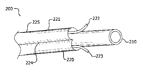
or
body cavity to be occluded. In the same or different embodiments, based on the
selection of the bioabsorbable material or the therapeutic agent, the
bioabsorbable
implant can cause a thrombogenic response to form an occlusion and/or a
spasmodic response to form an occlusion by causing the surrounding tissue to
shrink or collapse around the implant. To facilitate the occlusive effect,
manual
compression techniques about the surrounding tissue to collapse and/or reduce
to
volume of the lumen can be utilized. In some embodiments, bioabsorbable
implants
have lengths which extend along a length of a lumen and/or conform to the
shape of
the lumen.
[0096] Bioabsorbable, lumen-occluding implants can be useful for the treatment
of saphenous vein incompetency, endoleaks, perivalvular leaks, patent ductus,
patent foramen ovale, aortic dissection, growing aneurysms, gastro-esophageal
reflux, and obesity (by shrinking the gastro-esophageal junction or pylorus),
tumors,
or any disease or condition where local drug delivery and/or an occlusive or
spasmodic effect is desired. Some embodiments could be used as an alternative
to
tubal ligation or vasectomies, and embodiments causing a spasmodic response
could also be useful in cosmetic wrinkle reduction applications.
[0097] Embodiments herein can be adapted for use in cell based therapy such as
cell seeding. Embodiments can be imbibed with cells and further imbibed with
nutrients and/or other therapeutic agents.
[0098] Further embodiments described herein include systems or kits comprising
a bioabsorbable implant and a delivery device. In some embodiments, delivery
devices comprise an implantation guide which facilitates delivery of the
bioabsorbable implant or a bioabsorbable implant component to an implantation
site
by providing a delivery path. In other embodiments, delivery devices comprise
an
implantation guide and a translating member, wherein the translating member
21
CA 02874177 2014-11-19
WO 2013/188581
PCT/US2013/045490
facilitates translation of the bioabsorbable implant along the delivery path
defined by
the implantation guide. Translating member embodiments include a syringe, an
implantation piston member, or any other device that facilitates translation
of the
bioabsorbable implant along the delivery path.
[0099] Bioabsorbable lumen-occluding implants describe herein can comprise
any shape suitable for introduction into a lumen. For example, in occluding a
lumen,
bioabsorbable implant can comprise any space-filling member with a generally
round
or polygonal cross-section such as a spherical, ovoidal, cylindrical,
ellipsoidal or
prismoidal shape, or combinations and/or repetitions of the foregoing.
Bioabsorbable implant can have a generally open framework or a hollow center,
or
alternatively, it can be generally solid. In addition, in some embodiment,
bioabsorbable implants can comprise a generally elongated dimension, i.e.,
having a
greater length than width or height. Bioabsorbable implants can be made to be
permeable and/or can also be fashioned into bioabsorbable conduits through
which
a therapeutic agent can be infused.
[00100] In an embodiment, the bioabsorbable implant can be configured to
conform approximately to the dimensions of the lumen to be occluded. In the
same
or different embodiments, the bioabsorbable implant can facilitate the
surrounding
tissue conforming to the dimensions of the bioabsorbable implant, such as
through
the use of therapeutic agents like spasmodic agents, pro-coagulants, and/or
biocompatible glues/tissue adhesives, as well as manual compression.
[00101] Bioabsorbable, lumen-occluding implants describe herein can
optionally be imbibed or infused with a therapeutic agent and/or comprise a
modified
outer surface to have an improved or additional bioactive response. In an
embodiment, modifying the surface can comprise any modification that increases
the
surface area of the bioabsorbable implant. In some embodiments, surface
modifications can enhance the thrombogenic response caused by bioabsorbable
implant. One modification comprises adhering small fiber particles to at least
a
portion of the surface of bioabsorbable implant, creating an at least
partially flocked
surface. These small fiber particles can also be bioabsorbable. Another
modification can comprise an abraded or roughened surface.
[00102] Bioabsorbable elements referred to herein, namely, bioabsorbable,
lumen-occluding implants, anchoring mechanisms, radiopaque markers, occlusive
material, and occlusive members, comprise bioabsorbable material(s).
22
CA 02874177 2016-12-01
Bioabsorbable materials, as used herein, comprise any material capable of
biological
absorption. Such materials include copolymers of lactic acid and glycolic acid
(PLA/PGA) adjusted in the desired ratio to achieve the desired rate of
biological
absorption. Other potentially useful bioabsorbable materials include
polyglycolic acid
(PGA), poly-L-lactic acid (PLA), polydiaoxanone (PDS), polyhydroxybutyrate,
copolymers of hydroxybutyrate and hydroxyvalerate, copolymers of lactic acid
and E-
caprolactone, oxidized regenerated cellulose and various forms of collagen. A
most
preferred material is polyglycolide: trimethylene carbonate tri-block
copolymer
(PGA:TMC), e.g., the non-woven, bioabsorbable web material described in U.S.
Pat.
No. 7,659,219 by Biran et al. entitled "Highly porous self-cohered web
materials
having hemostatic properties".
This material has a history of use as bioabsorbable sutures; it is described
in detail by U.S. Pat. No. 4,429,080 to Casey et al.
The proportions of this or any other selected copolymer or
blends of polymers can be adjusted to achieve the desired absorption rate.
Other
potentially useful bioabsorbable, non-autologous materials including porous
forms
are described by U.S. Pat. Nos. 4,243,775 to Rosencraft et al.; 4,300,565 to
Rosencraft et al.; 5,080,665 Jarrett et al.; 5,502,092 Barrows et al.;
5,514,181 to
Light et al. and 5,559,621 to Minato et al., and published PCT application WO
90/00060 to Chu et al.
[0103] Implantation guide, referred to herein, can comprise any tubular
member or hollow needle having a lumen through which an occluding device can
pass through to be implanted into a lumen. In some embodiments, the
implantation
guide can be sufficiently flexible to extend along or traverse a curved or
tortuous
section of vasculature. In other embodiments, implantation guide does not need
to
extend along a curved or tortuous section of vasculature and thus, flexibility
is not
required.
[0104] The bioabsorbable implant embodiments described herein can further
comprise bioabsorbable radiopaque markers to facilitate monitoring of the
bioabsorbable implant in situ with non-invasive imaging techniques (e.g.,
ultrasound
imaging). For example, markers can be mounted on a proximal and/or distal end
of
the implant. In an embodiment, markers may be useful to ensure the device is
properly positioned at a vessel junction.
23
CA 02874177 2016-12-01
[0105] In one embodiment, with reference to FIG. 3A, an occluding device
300
comprises a bioabsorbable, lumen-occluding implant 360 having an anchoring
mechanism 365 coupled thereto. Anchoring mechanism 365 is any device suitable
for maintaining the position of the bioabsorbable implant 360 in situ. For
example,
when implanted into a blood vessel for purposes of creating a permanent
occlusion,
the flow of blood should not dislodge the bioabsorbable implant 360. Anchoring
mechanisms 365 can include a barb, a suture line 366, stent, or the like.
Anchoring
mechanism 365 can be coupled to implant at a proximal or distal end of implant
360
or any other suitable location on implant 360.
[0106] In one embodiment, occluding device 300 comprises a bioabsorbable,
lumen-occluding implant 360 securely coupled to bioabsorbable suture line 366.
Suture line 366 can be any length sufficient to extend from implant 360 to a
point
where suture line 366 can be secured, such as through the insertion path to
the skin
surface. A first end of suture line 366 can be securely coupled to
bioabsorbable
implant 360 at a proximal or distal end of bioabsorbable implant 360 or any
other
suitable location on implant 360. In an embodiment where suture line 366 exits
the
surface of the skin, suture line 366 can be knotted or, taped or cut flush
with the
surface of the skin.
[0107] In another embodiment, an occluding device 300 comprises a
bioabsorbable, lumen-occluding implant 360 coupled to at least one
bioabsorbable
barb, hook, or stent (collectively referred to as "barb). Barb can be any
structural
component that can penetrate a tissue making it difficult to become naturally
dislodged from the implantation site. In an embodiment, barb can be self-
setting
such that as implant 360 is inserted or injected into position, the barb will
radially
extend away from implant 360 and into surrounding tissue. The barb can be
coupled
to implant 360 at a proximal or distal end of the implant or any other
suitable location
on implant 360. In an embodiment, at least a portion of the barb can be
oriented to
generally curve, point or extend in the direction of blood flow to facilitate
engagement
with the surrounding tissue.
[0108] With reference to FIG. B and FIGS. 3C-1 to 3C-5, bioabsorbable
implant 360 can be pre-loaded into a capsule 370. The pre-loaded device
capsule
370 can be designed to connect to a syringe 382 and an implantation guide 380.
With the use of the syringe 382, capsule 370 and syringe 382 can be filled
with a
delivery fluid (such as saline or a therapeutic agent solution). With the use
of
24
CA 02874177 2016-12-01
implantation guide 380 (and optionally, an ultra sound device), access is
gained to
the implantation site, e.g., the lumen of a vessel. Once implantation guide
380 is in
position, capsule 370 and syringe 382 are connected to implantation guide 380.
The
plunger of the syringe 382 can then be depressed causing occluding device 300
to
be implanted and anchored into position.
[0109] In various embodiments, pre-loaded device capsule 370 comprises a
housing 371 defining at least one delivery chamber 372 in which at least one
occluding device 300 as described above is oriented for delivery. Delivery
chamber
372 is any pass-through cavity or compartment within housing 371 of
appropriate
dimensions for storing occluding device 300 in position for expulsion. As a
pass-
through, chamber 372 has an entrance end 376 and an exit end 375.
[0110] In order to connect to implantation guide 380 and syringe 382,
housing
371 comprises connectors 373, 374, such as a Luer taper fitting, about each
end
375, 376. For example, housing 371 can comprise a male-taper fitting about
exit
end 375 for connecting to implantation guide 380. About entrance end 376,
housing
371 can comprise a female taper fitting for connecting to syringe 382. Device
capsule 370 can further be hygienically sealed to maintain a sterilized
delivery
chamber 372 and occluding device 300. The caps or seals can be fitted onto the
connectors 373, 374 and can be removed or disrupted at the time of use.
[0111] In various embodiments, pre-loaded device capsule 370 can be
configured to receive an imbibing fluid containing at least one therapeutic
agent. In
this manner, occluding device 300 contained therein can be imbibed with a
therapeutic agent moments prior to implantation. In an embodiment, delivery
capsule 370 can comprise a pressure imbibing port and be configured to
withstand
positive pressures. The delivery chamber 372 can be filled with an imbibing
fluid,
liquid and/or gas, and held under positive pressure.
[0112] In other embodiments, pre-loaded device capsule 370 can comprise a
plurality of delivery chambers 372 configured to revolve, such as with the aid
of a
ratchet device. Each delivery chamber 372 is loaded with occluding device 300
as
described above. Such embodiments can help streamline the process of
implanting
multiple occluding devices 300 in a single treatment procedure. For example,
in
occluding a length of a vessel, a plurality of device can be implanted to
align end to
end as illustrated in FIG. 3C-5. In a further embodiment, pre-loaded device
capsule
CA 02874177 2016-12-01
370 can comprise a pressure imbibing port for purposes of simultaneously
imbibing a
plurality of occluding devices 300.
[0113] In accordance with another embodiment, an implant kit can comprise
(i) implantation guide 380 having a lumen through which occluding device 300
as
described above can pass through; (ii) pre-loaded device capsule 370 as
described
above, and (iii) at least one syringe which facilitates the expulsion of at
least one
occluding device 300 from chamber 372 through the lumen of implantation guide
380
out of distal tip. In an embodiment, a distal end of implantation guide 380
can
comprise an angled-cut tip and/or have a generally arced profile to facilitate
placement of occluding device 300. Syringe 382 can be manually operated or be
operated through automation.
[0114] In accordance with another embodiment, a method of delivery can
comprise the following steps. In any particular order, a clinician can connect
syringe
382 to pre-loaded device capsule 370 and insert implantation guide 380 into
the
lumen of a vessel (FIG. 3C-1). Placement of implantation guide 380 can be
guided
with the use of an ultrasound device. Once implantation guide 380 is in
position,
pre-loaded device capsule 370 is fitted onto implantation guide 380 (FIG. 3C-
2).
Bioabsorbable implant 360 can then be expulsed through implantation guide 380
via
the depression of syringe 382 plunger (FIG. 3C-3). Anchor mechanism 365 is
then
deployed (FIG. 3C-1).
[0115] In an embodiment where anchor mechanism 365 comprises suture line
366, suture line 366 remains within lumen of implantation guide 380, so upon
retraction of implantation guide 380, suture line 366 extends along the
insertion path
to the surface of the skin. Suture line 366 can then be cut flush with skin,
taped
down, and/or knotted.
[0116] Implantation guide can be withdrawn and the aforementioned steps
can be repeated at a neighboring location to implant a plurality of occluding
devices
300 (FIG. 3C-5).
[0117] In another embodiment, with reference to FIGS. 4A-1 and 4A-2, and
FIGS.4B-1 TO 4B-2 an occluding device 400 comprises bioabsorbable, lumen-
occluding implant 460 having a generally conformable conformation 461 when
loaded into implantation guide 480 and a convoluted conformation 462 when
released from implantation guide 480. Convoluted conformation 462 can comprise
26
CA 02874177 2016-12-01
coiled or spiral configuration, an undulating configuration, and/or a more
random
configuration of bends, twists, or whorls configuration.
[0118] In accordance with another aspect of the disclosure, with reference
to
FIGS. 4A-1 to 4A-2 and FIGS. 4B-1 to 4B-2, an occluding device and delivery
device
system comprises implantation guide 480 circumscribing an extendable and
retractable implantation piston member 496; and bioabsorbable lumen-occluding
implant 460 having a generally conformable conformation 461 when loaded into
implantation guide 480 and a convoluted conformation 462 when released from
guide 480, wherein bioabsorbable lumen-occluding implant 460 can be releasably
coupled to implantation piston member 496.
[0119] Implantation piston member 496 can comprise an elongated
component that passes through the lumen of implantation guide 480 and
releasably
couples to occluding device 400. In an embodiment, implantation piston member
496 comprises an outer tube 497 with a translatable inner core 498 located
within
and along the lumen of outer tube 497. Implantation piston member 496 has a
recessed distal end by way of outer tube 497 extending beyond translatable
inner
core 498 a certain distance. Along this distance or a portion thereof, outer
tube 497
is dimensioned to fit snugly over occluding device 400 Outer tube 497 is also
dimensioned to slideably extend and retract through implantation guide 480. In
this
manner, occluding device 400 can be loaded into implantation guide 480 and
then it
can be retracted and extended until occluding device 400 is released.
[0120] In order to release, translatable inner core 498 can be actuated to
slide
and extend within the lumen of outer tube 497 so that it is at least flush
with the distal
end of outer tube 497. In this manner, occluding device 400 is forced out of
the
lumen of outer tube 497 and released from implantation piston member 496.
[0121] In accordance with another embodiment, a method of loading
occluding device 400 into implantation guide 480 comprises the steps of
inserting a
proximal end of occluding device 400 into a distal end of implantation piston
member
496; retracting implantation piston member 496 and occluding device 400 into
implantation guide 480 lumen.
[0122] In accordance with another embodiment, a method of delivery can
comprise the following steps. First, implantation guide 480 is inserted into
lumen.
Once in position, implantation piston member 496 can be selectively extended
so
that occluding device 400 is released from implantation guide 480 and acquires
a
27
CA 02874177 2016-12-01
convoluted conformation 462. If desired, occluding device 400 can be again
retracted into the lumen of implantation guide 480 by retracting implantation
piston
member 496. The steps of extending and retracting occluding device 400 can be
repeated until occluding device 400 is in the desired implantation position.
Once in
the proper position, occluding device 400 can be released by actuating
implantation
piston member 496. For example, actuating implantation piston member 496 can
comprise sliding and extending translatable inner core 498 along the lumen of
the
outer tube 497.
[0123] In an embodiment, with reference to FIGS. 4C-1 to 4C-2, occluding
device and delivery system comprises a implantation guide 480, an actuatable
cutter
mechanism 484, and a bioabsorbable implant 460 having a conformable
conformation 461 when loaded into guide 480 and a convoluted conformation 462
when released from guide 480, wherein the bioabsorbable implant 460 can be
selectively extended and retracted, and then selectively severed with cutter
mechanism 484 after a sufficient length of bioabsorbable implant 360 is
implanted.
Similarly, in an embodiment, occluding device and delivery system comprise
guide
480, a cutter mechanism 484, and a bioabsorbable implant 460 having a
customizable length.
[0124] In order to sever bioabsorbable implant 460, cutter mechanism 484
can
comprise a blade 485 located on a distal end portion of implantation guide 380
and
an actuating component extending from the blade 485. Blade 485 can be oriented
so that the cutting edge faces toward the center of implantation guide 480
lumen.
Upon actuation, blade 485 moves across implantation guide 480 lumen and can
optionally reset itself. Blade 485 can comprise any material of suitable
hardness to
cut bioabsorbable implant 460. Blade 485 can be a hard polymer or a metallic
component. Blade 485 can comprise a shape memory material, such as nitinol.
[0125] In an embodiment, with reference to FIGS. 4C-1 to 4C-2, an occluding
device and delivery system comprise implantation guide 480, an actuatable
cutter
mechanism 484, and a bioabsorbable implant 460 having a conformable
conformation 461 when loaded into guide 480 and a convoluted conformation 462
when released from guide 480, wherein the bioabsorbable implant 460 can be
selectively extended and retracted, and then selectively severed with cutter
mechanism 484 after a sufficient length of bioabsorbable implant 360 is
implanted.
Similarly, in an embodiment, occluding device and delivery system comprise
guide
28
CA 02874177 2016-12-01
480, cutter mechanism 484, and a bioabsorbable implant 460 having a
customizable
length.
[0126] In order to sever bioabsorbable implant 460, cutter mechanism 484
can
comprise a blade 485 located on a distal end portion of implantation guide 380
and
an actuatable component extending from the blade 485. Blade 485 can be
oriented
so that the cutting edge faces toward the center of implantation guide 480
lumen.
Upon actuation, blade 485 moves across implantation guide 480 lumen, and can
optionally reset itself. Blade 485 can comprise any material of suitable
hardness to
cut bioabsorbable implant 460. Blade 485 can be a hard polymer or a metallic
component. Blade 485 can comprise a shape memory material, such as nitinol.
[0127] In accordance with another embodiment, a method of delivery
comprise the steps of extending and/or retracting occluding device 400 through
lumen of implantation guide 480; selectively severing occluding device 400 by
actuatable cutter mechanism 484.
[0128] In an embodiment, with reference to FIG. 5A-1 to 5A-5, occluding
device 500 can comprise a two-component bioabsorbable, lumen-occluding implant
560 wherein a first component comprises an occlusive member 590 defining a
lumen
or a cavity and a second component comprises an occlusive material 592. A
further
embodiment can also comprise a guidewire 594 and implantation guide 580
wherein
occlusive member 590 is mounted around the distal end of guidewire 594.
Implantation guide 580 can be configured to slide over guidewire 594 into the
lumen
or cavity of occlusive member 510, and after the retraction of guidewire 594,
guide
580 can inject occlusive material 592 therein. For example, occlusive member
590
can be a sleeve closed at a distal end and dimensioned, in its unexpanded
state, to
be situated around or circumscribe the distal end of implantation guide 580.
1[0129] In an embodiment, as occlusive material 592 is injected into the
lumen,
occlusive member 510 can distend or expand to occupy and approximately conform
to the lumen of a vessel or other surrounding empty space. For example,
occlusive
member 590 can have a pleated or knitted conformation which can expand upon
introduction of occlusive material 592. In other embodiments, occlusive member
590
can comprise a flexible material which stretches and/or expands upon
introduction of
occlusive material 592.
[0130] In other embodiments, occlusive member 590 can comprise an
elongated, compliant, and convoluted conformation, which permits occlusive
29
CA 02874177 2016-12-01
member 590 to be substantially straight when fitted or mounted around
implantation
guide 580 or guide wire 594, but as it 590 is released from implantation guide
580, it
590 takes on a convoluted conformation, which has a space-filling, occlusive
effect.
[0131] In the same or different embodiments, occlusive member 590 is not
distensible, but rather, bioabsorbable implant 500 facilitates the surrounding
tissue
conforming to the dimensions of the filled occlusive member (590 and 592),
such as
through the use of therapeutic agents like spasmodic agents, pro-coagulants,
and/or
biocompatible glues/tissue adhesives imbibed or infused into occlusive
material 592,
as well as manual compression.
[0132] Similar to other bioabsorbable implants described herein, occlusive
material 592 can be optionally imbibed with a therapeutic agent. Occlusive
member
590 can comprise a sufficiently porous or permeable material to permit
transfer of
the therapeutic agent to its outer surface.
[0133] Occlusive material 592 can comprise any bioabsorbable material that
is
suitable for filling occlusive member 590. In an embodiment, occlusive
material 592
can be an injectable material, i.e., suitable for transporting through the
lumen of
implantation guide 580. For example, occlusive material 592 can comprise
flowable
material like a liquid, small particle solid materials and/or materials having
loft, which
can include non-woven web materials, tufts of fibers, a plurality of small
spherical
particles, or an emulsion. In a preferred embodiment, occlusive material 592
can
comprise the non-woven, bioabsorbable web material made with poly(glycolide),
also
known as PGA, and poly(trimethylene carbonate), also known as TMC, described
in
U.S. Pat. No. 7,659,219 by Biran et al., entitled "Highly porous self-cohered
web
materials having hemostatic properties".
[0134] Occlusive member 590 comprises a bioabsorbable material formed into
any thin-walled structural component that defines a lumen or cavity and can be
expanded to occupy and approximately conform to a lumen vessel or a body
cavity.
Occlusive member 590 can comprise a bioabsorbable film or fabric that does not
permit passage of occlusive material 592. Occlusive member 590 can comprise a
distensible and compliant film or fabric to facilitate approximately
conforming to the
surrounding space.
[0135] Occlusive member 590 can be any shape suitable for occluding the
desired lumen or body cavity. As mentioned above, occlusive member 590 can
CA 02874177 2016-12-01
comprise an expandable sleeve. Expandable sleeve comprise a generally tubular
shape having a proximal and a distal end and a lumen there through. The distal
end
can be permanently closed to contain occlusive material 592 as it is injected.
Implantation guide 580 can be inserted through the proximal end to deliver
occlusive
material 592.
[0136] In order to close the occlusive member 590 so that occlusive
material
592 does not leak from occlusive member 590 once implantation guide 580 is
withdrawn, occlusive member 590 can be self-sealing or comprise a closure in
order
to at least substantially close the proximal end upon withdrawal of guide 580.
Closure can comprise any mechanism or configuration that will close the
proximal
end of occlusive member 590. For example, closure can comprise self-collapsing
section of occlusive member 590, such as an elastic band the will collapse
down and
close the proximal end of the occlusive member 590 upon retraction of
implantation
guide 580. Other closure embodiments can include a purse string, clip, or the
like.
[0137] In an embodiment, wherein occlusive material 592 is injected along
with a therapeutic agent, the transfer of the therapeutic agent may need to be
restricted to permeating through only certain portions of occlusive member
590.
According, occlusive member 590 can comprise sections or areas that remain
impermeable to a therapeutic agent at least during the initial absorbtion
phase. For
example, having an impermeable end cap(s) on occlusive member 590 can mitigate
undesired migration of a therapeutic agent. Areas of occlusive member 590 can
be
made impermeable by coating with a bioabsorbable sealant such as copolymers of
lactic acid and glycolic acid (PUVPGA), or varying the microstructure or
thickness in
these areas to make less permeable.
[0138] In accordance with another embodiment, a method of delivering
occluding device 500 can comprise the steps of extending occlusive member 590
on
guidewire 594 into a vessel; passing implantation guide 580 over guidewire 594
and
into lumen of occlusive member 590; and injecting occlusive material 592 into
lumen
of occlusive member 590. Implantation guide 380 can be retracted as it injects
occlusive material 592. Once implantation guide 580 is completely retracted,
occlusive member 590 can collapse around the opening of occlusive member 590
through which the implantation guide 580 entered. Collapsing can be
facilitated by a
closure.
31
CA 02874177 2016-12-01
[0139] While bioabsorbable implants are discussed in relation to providing
an
occlusive effect, implants can be modified to cause a thrombolytic effect and
can be
inserted into a tissue or vasculature where a thrombolytic response is
desired.
[0140] It will be apparent to those skilled in the art that various
modifications
and variations can be made in the present disclosure without departing from
the
spirit or scope of the disclosure. Thus, it is intended that the present
disclosure
cover the modifications and variations of this disclosure provided they come
within
the scope of the appended claims and their equivalents.
[0141] Likewise, numerous characteristics and advantages have been set
forth in the preceding description, including various alternatives together
with details
of the structure and function of the devices and/or methods. The disclosure is
intended as illustrative only and as such is not intended to be exhaustive. It
will be
evident to those skilled in the art that various modifications can be made,
especially
in matters of structure, materials, elements, components, shape, size and
arrangement of parts including combinations within the principles of the
disclosure,
to the full extent indicated by the broad, general meaning of the terms in
which the
appended claims are expressed. To the extent that these various modifications
do
not depart from the spirit and scope of the appended claims, they are intended
to be
encompassed therein.
32