Note : Les descriptions sont présentées dans la langue officielle dans laquelle elles ont été soumises.
PARTICLES, METHODS AND USES THEREOF
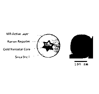
[0001]
Background
[0002] Nanoparticle systems that can incorporate dopant entities have
tremendous
potential and are useful in a wide variety of contexts. There is a continuing
need for
improved systems. One particular goal in developing such systems is to provide
imaging
nanoparticles that can be utilized in surgery to define resection boundaries.
Completeness of
surgical resection profoundly impacts morbidity and mortality. The challenges
and
significance are particularly acute in surgery to remove tumors. In trying to
achieve more
complete tumor resections, the surgeon encounters several hurdles, which
include irregular
and indistinct tumor margins as well as tumor growth adjacent to or invading
crucial
physiological structures. A wide variety of techniques have been explored to
date in an effort
to better visualize tumor margins. However, there remains a continuing need
for new and
better probes and/or methods. In particular, there is an important, unmet need
for a real-time
probe/method for accurately detecting residual tumor.
[0003]
Summary
[0004] The present invention provides technologies relevant to
particles (e.g., surface-
enhanced (resonance) Raman scattering (SE(R)RS)-active particles), including
technologies
for preparing particles, and/or for using particles, as well as particles
themselves. In general,
particles as described and/or utilized herein contain a nanoscale core, an
encapsulant, and a
plurality of dopant entities.
1
CA 2882388 2019-09-03
CA 02882388 2015-02-18
WO 2014/036470
PCMJS2013/057636
[0005] Provided compositions and methods are useful in a variety of
contexts. To give
but one example, in many embodiments, the present invention is particularly
useful for
particles wherein dopant entities are resonant agents, which experience
resonance at an
incident laser wavelength. In certain embodiments, a resonant agent is a
SE(R)RS-active
agent. As demonstrated herein, provided technologies achieve unprecedented
levels of
dopant entity density and/or surface localization, which, for a SE(R)RS-active
agent dopant,
results in dramatically improved signal intensity and/or imaging sensitivity.
[0006] Among other things, the invention provides technologies that permit
an
encapsulant coating without use of a surface primer. Such a surface primer is
often added to
enable encapsulant binding to the nanoscalc core surface. In some embodiments,
the
invention provides technologies that utilize a displaceable capping entity.
Features of
provided technologies include a higher density of dopant entities can be
located close to its
core surface. More traditional approaches that utilize a surface primer do not
permit such a
degree of density and/or surface localization of dopant entities.
[0007] In some embodiments, methodologies described herein include steps of
providing
a nanoscale core in association with a capping agent (e.g., surface-bound
stabilizing agent
present as a direct consequence of the nanoscale core synthesis); contacting
the capping-agent
associated nanoscale core with an encapsulant precursor and a dopant entity
under conditions
and for a time sufficient for the encapsulant precursor and/or dopant entity
to displace some
or all of the capping agent to produce a particle characterized by high
density of surface-
localized doping entity.
[0008] Provided technologies permit preparation of particles of previously
unachieved
structure and properties. In some embodiments, provided particles include a
nanoscale core,
an encapsulant, and a plurality of dopant entities, which particles are
characterized by: (i)
dopant entity density higher than typically observed for the relevant dopant
entity; and (ii)
localization of the dopant entity closer to the nanoscale core than typically
observed.
[0009] One remarkable feature of provided technologies for preparing
particles is that
they are applicable to and effective with a wide range of core materials, core
configurations,
encapsulant and entity materials, etc. In some embodiments, provided particles
comprise a
core of a metal material (e.g., gold, silver, copper, etc.). In some
embodiments, provided
particles comprise a nanoscale core, whose shape is or includes structural
elements selected
from the group consisting of spheres, rods, stars, shells, ellipses,
triangles, pyramids, cubes,
cages and combinations thereof. In some particular embodiments, provided
particles include
a nanoscale core having a central structure surrounded by satellite
structures.
2
CA 02882388 2015-02-18
WO 2014/036470
PCMJS2013/057636
[0010] The present disclosure, among other things, provides compositions
that include a
nanoscale core; a plurality of capping agent entities associated on the core;
an outer
encapsulant layer; and a plurality of dopant entities distributed at locations
selected from the
group consisting of: on or within the nanoscale core, on or between capping
agent entities, on
or within the encapsulating layer, and combinations thereof. Provided
technologies can
achieve unprecedented levels of dopant entity density and/or surface
localization, which, in
some embodiments, including for example in certain embodiments that utilize
one or more
SE(R)RS-active agent dopant(s), results in dramatically improved signal
intensity and/or
imaging sensitivity. In some embodiments, signal intensity and/or imaging
sensitivity is
improved relative to is improved relative to known imaging modalities,
including CT,
Ultrasound, or Fluorescence.
[0011] The present disclosure provides, among other things, methods of
applying an
encapsulant layer to a nanoscale core. In some embodiments, provided methods
include steps
of providing a capped composition including a nanoscale core substantially
coated with a
plurality of capping agent entities displaceably associated with the nanoscale
core's surface.
Alternatively or additionally, in some embodiments, provided methods include
steps of
contacting a capped composition with a plurality of dopant entities and a
plurality of
encapsulant precursor entities, the contacting being performed under
conditions and for a
time sufficient to permit i) accumulation of dopant entities onto or nearby
the core surface;
and ii) formation of an outer encapsulant layer by the encapsulant precursor
entities such that
a composition is generated that includes a nanoscale core; a plurality of
capping agent entities
associated on the core; an outer encapsulant layer; and a plurality of dopant
entities
distributed at locations selected from the group consisting of: on or within
the core, on
capping agent entities, within the encapsulating layer, on the encapsulating
layer and
combinations thereof.
[0012] The present disclosure, provides, among other things, methods
including steps of
administering to a subject a collection of particles including a nanoscale
core; a plurality of
capping agent entities associated on the core; an outer encapsulant layer; and
a plurality of
dopant entities distributed at locations selected from the group consisting
of: on or within the
nanoscale core, on capping agent entities, within the encapsulating layer, on
the
encapsulating layer and combinations thereof. In certain embodiments, such
particles further
include MRI agents, PET agents, SPECT agents, CT agents and/or combination
thereof In
certain embodiments, such methods also include one or more steps of imaging
localized
particles. In certain embodiments, a step of imaging localized particles
includes obtaining a
3
CA 02882388 2015-02-18
WO 2014/036470
PCMJS2013/057636
first signal selected from the group consisting of MRI signals, PET signals,
SPECT signals,
CT signals, and combinations thereof, wherein the first signal is used to
produce an image
corresponding to one or more of: tumor localization (e.g., of a whole tumor),
macroscopic
delineation of a tumor (e.g., of a whole tumor), and/or location, shape,
and/or size of residual
tumor; obtaining a photoacoustic signal, wherein the photoacoustic signal is
used to produce
an image corresponding to a tumor with deep tissue penetration; obtaining a
Raman
vibrational signal, wherein the Raman vibrational signal is used as a guide to
define tumor
margins; and producing an image of a tumor and its margins using the first
signal, the
photoacoustic signal, and the Raman vibrational signal.
Definitions
[0013] In order for the present disclosure to be more readily understood,
certain terms are
defined below. Additional definitions for the following terms and other terms
may be set
forth throughout the specification, or may otherwise be clear from context.
[0014] In this application, the use of "or" means "and/or" unless stated
otherwise. As
used in this application, the term "comprise" and variations of the term, such
as "comprising"
and "comprises," are not intended to exclude other additives, components,
integers or steps.
As used in this application, the terms "about" and "approximately" are used as
equivalents.
Any numerals used in this application with or without about/approximately are
meant to
cover any normal fluctuations appreciated by one of ordinary skill in the
relevant art. In
certain embodiments, the term "approximately" or "about" refers to a range of
values that fall
within 25%, 20%, 19%, 18%, 17%, 16%, 15%, 14%, 13%, 12%, 11%, 10%, 9%, 8%, 7%,
6%, 5%, 4%, 3%, 2%, 1%, or less in either direction (greater than or less
than) of the stated
reference value unless otherwise stated or otherwise evident from the context
(except where
such number would exceed 100% of a possible value).
[0015] 'Administration": The term "administration" refers to introducing a
substance
into a subject. In some embodiments, a route of administration is oral
administration.
Additionally or alternatively, a route is intravenous administration. However,
any route of
administration, such as topical, subcutaneous, peritoneal, intraarterial,
inhalation, vaginal,
rectal, nasal, introduction into the cerebrospinal fluid, or instillation into
body compartments
can be used.
[0016] 'Associated": As used herein, the term "associated" typically refers
to two or
more entities in physical proximity with one another, either directly or
indirectly (e.g., via
one or more additional entities that serve as a linking agent), to form a
structure that is
4
CA 02882388 2015-02-18
WO 2014/036470
PCMJS2013/057636
sufficiently stable so that the entities remain in physical proximity under
relevant conditions,
e.g., physiological conditions. In some embodiments, associated moieties are
covalently
linked to one another. In some embodiments, associated entities are non-
covalently linked.
In some embodiments, associated entities are linked to one another by specific
non-covalent
interactions (i.e., by interactions between interacting ligands that
discriminate between their
interaction partner and other entities present in the context of use, such as,
for example.
streptavidin/avidin interactions, antibody/antigen interactions, etc.).
Alternatively or
additionally, a sufficient number of weaker non-covalent interactions can
provide sufficient
stability for moieties to remain associated. Exemplary non-covalent
interactions include, but
are not limited to, affinity interactions, metal coordination, physical
adsorption, host-guest
interactions, hydrophobic interactions, pi stacking interactions, hydrogen
bonding
interactions, van der Waals interactions, magnetic interactions, electrostatic
interactions,
dipole-dipole interactions, etc.
[0017] "Biocompatible": The term "biocompatible", as used herein is
intended to
describe materials that do not elicit a substantial detrimental response in
vivo. In certain
embodiments, the materials are "biocompatible" if they are not toxic to cells.
In certain
embodiments, materials are "biocompatible" if their addition to cells in vitro
results in less
than or equal to 20% cell death, and/or their administration in vivo does not
induce
inflammation or other such adverse effects. In certain embodiments, materials
are
biodegradable.
[0018] "Biodegradable": As used herein, "biodegradable" materials are those
that, when
introduced into cells, are broken down by cellular machinery (e.g., enzymatic
degradation) or
by hydrolysis into components that cells can either reuse or dispose of
without significant
toxic effects on the cells. In certain embodiments, components generated by
breakdown of a
biodegradable material do not induce inflammation and/or other adverse effects
in vivo. In
some embodiments, biodegradable materials are enzymatically broken down.
Alternatively
or additionally, in some embodiments, biodegradable materials are broken down
by
hydrolysis. In some embodiments, biodegradable polymeric materials break down
into their
component polymers. In some embodiments, breakdown of biodegradable materials
(including, for example, biodegradable polymeric materials) includes
hydrolysis of ester
bonds. In some embodiments, breakdown of materials (including, for example,
biodegradable polymeric materials) includes cleavage of urethane linkages.
[0019] "Illuminating": The term "illuminating" as used herein refers to the
application of
a light source, including near-infrared (NIR), visible light, including laser
light capable of
CA 02882388 2015-02-18
WO 2014/036470
PCMJS2013/057636
exciting molecules and/or nanoscale cores of the embodiments of the particles
herein
disclosed.
[0020] "Magnetic Resonance Imaging": The term "magnetic resonance imaging
(MRI)"
as used herein refers to a medical imaging technique most commonly used in
radiology to
visualize the structure and function of the body. It provides detailed images
of the body in
any plane. MRI uses no ionizing radiation, but uses a powerful magnetic field
to align the
nuclear magnetization of (usually) hydrogen atoms in water in the body.
Radiofrequency
fields are used to systematically alter the alignment of this magnetization,
causing the
hydrogen nuclei to produce a rotating magnetic field detectable by the
scanner. This signal
can be manipulated by additional magnetic fields to build up enough
information to construct
an image of the body. When a subject lies in a scanner, the hydrogen nuclei
(i.e., protons)
found in abundance in an animal body in water molecules, align with the strong
main
magnetic field. A second electromagnetic field that oscillates at
radiofrequencies and is
perpendicular to the main field, is then pulsed to push a proportion of the
protons out of
alignment with the main field. These protons then drift back into alignment
with the main
field, emitting a detectable radiofrequency signal as they do so. Since
protons in different
tissues of the body (e.g., fat versus muscle) realign at different speeds, the
different structures
of the body can be revealed. Contrast agents may be injected intravenously to
enhance the
appearance of blood vessels, tumors or inflammation. MRI is used to image
every part of the
body, but is particularly useful in neurological conditions, disorders of the
muscles and joints,
for evaluating tumors and showing abnormalities in the heart and blood
vessels.
[0011] "Sample": The term "sample" refers to a volume or mass obtained,
provided,
and/or subjected to analysis. In some embodiments, a sample is or comprises a
tissue sample,
cell sample, a fluid sample, and the like. In some embodiments, a sample is
taken from a
subject (e.g., a human or animal subject). In some embodiments, a tissue
sample is or
comprises brain, hair (including roots), buccal swabs, blood, saliva, semen,
muscle, or from
any internal organs, or cancer, precancerous, or tumor cells associated with
any one of these.
A fluid may be, but is not limited to, urine, blood, ascites, pleural fluid,
spinal fluid, and the
like. A body tissue can include, but is not limited to, brain, skin, muscle,
endometrial, uterine,
and cervical tissue or cancer, precancerous, or tumor cells associated with
any one of these.
In an embodiment, a body tissue is brain tissue or a brain tumor or cancer.
Those of ordinary
skill in the art will appreciate that, in some embodiments, a "sample" is a
"primary sample"
in that it is obtained from a source (e.g., a subject); in some embodiments, a
"sample" is the
result of processing of a primary sample, for example to remove certain
potentially
6
CA 02882388 2015-02-18
WO 2014/036470
PCT/1TS2013/057636
contaminating components and/or to isolate or purify certain components of
interest.
[0012] "Substantially": As used herein, the term "substantially", and
grammatic
equivalents, refer to the qualitative condition of exhibiting total or near-
total extent or degree
of a characteristic or property of interest. One of ordinary skill in the art
will understand that
biological and chemical phenomena rarely, if ever, go to completion and/or
proceed to
completeness or achieve or avoid an absolute result.
[0013] "Subject": As used herein, the term "subject" includes humans and
mammals
(e.g., mice, rats, pigs, cats, dogs, and horses). In many embodiments,
subjects are be
mammals, particularly primates, especially humans. In some embodiments,
subjects are
livestock such as cattle, sheep, goats, cows, swine, and the like; poultry
such as chickens,
ducks, geese, turkeys, and the like; and domesticated animals particularly
pets such as dogs
and cats. In some embodiments (e.g., particularly in research contexts)
subject mammals will
be , for example, rodents (e.g., mice, rats, hamsters), rabbits, primates, or
swine such as
inbred pigs and the like.
Brief Description of the Drawing
[0021] The Drawing, which is comprised of at least the following Figures,
is for
illustration purposes only, not for limitation.
[0022] Figure 1 shows a schematic of a SE(R)RS particle in accordance with
the present
invention together with a transmission electron micrograph (TEM) of a
representative
SE(R)RS particle. At the center of the SE(R)RS particle is a gold nanostar
core coated with a
layer of (resonance) Raman-active molecules (reporters). The star shape
enables tuning of
the Localized Surface Plasmon Resonance (LSPR) towards the Near-Infrared
window and
incorporates several "hot-spots" (the tips) of incredibly concentrated
electric fields focused
on the (resonance) Raman reporters. A shell of silica encapsulates this core,
simultaneously
protecting the (resonance) Raman reporters, preventing reactions of the core
and reporters
with the environment, and providing a surface for further functionalization.
In this case, an
MR-active layer is bound to the outer surface of the silica.
[0023] Figure 2 illustrates direct comparison of the Raman spectral
intensity of the
SE(R)RS particles to the particles illustrated in Kircher et al., (2012) Nat
Med 18 (5):829-834
(Appendix A) currently considered to be the Raman gold standard. As shown in
the bar
graph, the SE(R)RS particles are 47-times more intense than the particles
previously
illustrated.
7
CA 02882388 2015-02-18
WO 2014/036470
PCT/ITS2013/057636
[0024] Figure 3 displays the output of a typical Nanoparticle Tracking
Analysis (NTA)
scan. NTA enables accurate quantification of particle concentration and size
distribution by
locking into the light scattered from individual particles and tracing their
paths in solution.
The concentration is determined by simply counting the number of particles in
a defined
volume, while size is calculated from the Brownian motion using the Einstein-
stokes
equation. When combined with the complete morphological information provided
by TEM,
NTA allows for thorough characterization of the SE(R)RS particles.
[0025] Figure 4 shows a series of images of a mouse with dedifferentiated
liposarcoma
implanted in the flank. Note that Raman signal outlines the tumor; there is
also Raman signal
visible beyond the margins of the tumor seen on the white light (the arrows).
[0026] Figure 5 shows a series of images of the same mouse as shown in
Figure 4, after
resection of the bulk tumor by a surgeon using his unaided eye (blinded to
Raman signal).
Note that there is a residual rim of Raman signal in the resection bed around
the resected
tumor. Histological evaluation confirmed tumor in the locations of the Raman
signal. The
arrow indicates tumor-associated macrophage having engulfed SE(R)RS particles.
[0027] Figure 6A shows images of a different mouse with liposarcoma,
multiple small
foci of Raman signal (1, 2, 3, 4, and 5) were found in the resection bed,
after the bulk tumor
had been resected by a surgeon. FIG. 6 shows detected microscopic metastases.
MPR-
Nanostars were injected intravenously (tail vein; 150 3nM) into a
dedifferentiated human
liposarcoma xenograft bearing mouse. Intraoperative Raman image (after 24 h)
was taken 1
cm adjacent to the margin of the bulk tumor. It correctly detects multiple
micrometastases.
[0028] Figure 7 shows images of the same mouse as shown in Figure 6 with
sarcoma,
multiple tiny foci of Raman signal are seen in the resection bed, after the
bulk tumor had been
resected by a surgeon using white light guidance only (blinded to Raman
signal). As
histological examination demonstrated, these foci of Raman signal represented
tumor-
associated macrophages.
[0029] Figure 8 demonstrates SE(R)RS particles are able to detect a variety
of different
tumors. Exemplary images are shown two spontaneous sarcomas in an Ink4A-/-
mouse
model, a brain tumor in the rcasitv-a model, and a breast cancer in the PyMT
model. In each
tumor, there was excellent depiction of the tumor by the Raman signal.
[0030] Figure 9 demonstrates the ability of SE(R)RS particles to outline
glioblastomas
(rcas/tv-a model). Note the high degree of correlation of Raman signal with
the presence of
tumor cells (HA-tag, Oligo-2 positive staining). RGD-MPR-Nanostars were
injected
8
CA 02882388 2015-02-18
WO 2014/036470
PCMJS2013/057636
intravenously via tail vein (150 .1, 3 nM). After 24 hours, mice were
sacrificed, perfused via
intracardial injection of PBS, brains embedded in paraffin, processed
histologically, and
imaged with the Renishaw Raman microscope. Adjacent sections were stained for
immunohistochemisty. Images are representative of n = 5 mice. When the Raman
signal is
compared to the immunohistochemical staining for glioblastoma cells (ct(=anti)-
HA-tag and
Oligo-2), a high degree of congruency is noted. Note the small Raman positive
focus outside
of the main tumor (eNOS=Endothelial cells; SMA=Smooth Muscle Cells;
IBA=Microglia;
GFAP=Astrocytes; NeuN=Neurons).
[0031] Figure 10 demonstrates the ability of SE(R)RS particles to depict a
single brain
tumor cell (micrometastasis away from the main tumor). Insert in Raman image
shows
magnification of single Raman positive voxel. Raman spectrum proves presence
of SE(R)RS
particles. Histology proves that this signal correlates to a signal brain
tumor cell. Correlation
of a single Raman positive pixel (red in upper left image, magnified within
the white square)
with immunohistochemistry in the RCAS/tv-a glioblastoma model. The same slide
as in
Figure 9 was examined at higher magnification. The presented Raman spectrum
confirms
that the Raman positive pixel truly represents the nanoparticle. HA-tag
positive staining
confirms that the MPR-Nanostars co-localize with the presence of a
glioblastoma cell.
Adjacent to the tumor cell are located a microglia cell (IBA-1 positive) and a
small blood
vessel (eNOS, SMA positive), explaining how the MPR-Nanostars could have been
transported to this location. Individual or small clusters of tumor cells
located outside of the
main tumor are often seen in this mouse model and in human glioblastomas.
[0032] Figure 11 shows a series of images illustrating using MPR-nanostars
to detect
submillimeter-sized dysplastic (premalignant) polyps and adenocarcinomas. The
illustrated
experiment was performed in an AFC'511 mouse, which is a mouse model known to
mimic
human "adenomatosis polyposis coli" syndrome, a genetic disorder that causes
many
dysplastic polyps and adenocarcinomas to develop simultaneously. Note that
Raman imaging
reveals many small foci (less than 1 mm in size) of SERRS-Nanostars uptake
within the
colon and small bowel of an APC'51'2 mouse (excised 24 hours after
nanoparticle injection).
These foci were then processed with histology (see Figure 12), which confirmed
that they
represented dysplastic polyps or adenocarcinomas.
[0033] Figure 12 shows a series of images illustrating using MPR-Nanostars
for
detecting submillimeter-sized dysplastic (premalignant) polyps and
adenocarcinomas ¨
histological confirmation. The presented images show two segments of colon
from the
9
CA 02882388 2015-02-18
WO 2014/036470
PCT/1JS2013/057636
mouse in Figure 11. Two histological cross-sections through the Raman positive
areas were
obtained and stained with Hematoxylin-Eosin (H&E) and anti-catenin IHC.
Section I
confirm that the lesion to represent an adenocarcinoma, section 2 ¨ a
dysplastic polyp, and
thereby also confirms MPR-Nanostars as described herein are able to detect not
only very
small colon cancers, but also their premalignant form ¨ dysplastic polyps ¨
which will
eventually develop into invasive adenocarcinomas. Among other things, these
data confirm
that, as described herein, MPR-Nanostars may be used as a new method for early
colon
cancer detection.
[0034] Figure 13 shows a series of images illustrating using MPR-Nanostars
nanoparticles for detecting prostate cancer. The depicted experiment was
performed in a
state-of-the-art genetic spontaneous (Hi-Myc) mouse model of prostate cancer.
Mice express
human c-Myc in the mouse prostate. The upper row of images shows a control
animal (same
mouse strain but without the Myc mutation) that was injected with MPR-
Nanostars: No
Raman signal is seen in this normal prostate. The lower row of images shows
images from a
prostate cancer bearing mouse (hi-Myc) with obvious deformity of the prostate
due to tumor
(photograph) that was injected with the same amount of MPR-Nanostars. The
Raman image
shows accumulation of MPR-Nanostars within the tumor areas.
[0035] Figure 14 shows a series of images illustrating using MPR-Nanostars
for
detecting microscopic residual tumor in resection bed in a transgenic mouse
model of
prostate cancer (Hi-Myc). A prostatectomy was performed in a tumor-bearing Hi-
Myc
mouse, and subsequently the resection bed was scanned with Raman imaging.
lmmunohistochemical correlation shows that small foci of Raman signal
correspond to
residual microscopic prostate cancer that could not have been visualized
otherwise and would
have been "missed". Note the excellent correlation between the histological
tumor markers
and the presence of the nanoparticles ("Raman nanoparticle staining" =
antibody against
PEGylated silica nanoparticle surface).
[0036] Figure 15 shows a series of images of the use of MPR-Nanostars for
detecting
breast cancer in a state-of-the-art genetic MMTV-PyMT breast cancer mouse
model. Mice
with this genetic mutation spontaneously develop multiple breast cancers in
different
mammary glands and closely mimic human breast cancer pathology. Note that the
Raman
signal from the MPR-Nanostars accurately depicts the extent of multiple 3-6 mm
sized breast
cancers in the same mice, including small submillimeter tumor extensions. The
upper row
shows images of breast cancers developed along the upper and middle mammary
glands of a
CA 02882388 2015-02-18
WO 2014/036470
PCMJS2013/057636
MMTV-PyMT mouse. The lower row shows breast cancers developed within the lower
mammary glands of a MMTV-PyMT mouse.
[0037] Figure 16 shows a series of images of the use of MPR-Nanostars to
detect
microscopic tumor infiltration into the skin. This experiment was performed in
an orthotopic
4T1 breast cancer mouse model. The 4T1 breast cancer cell line was transfected
to express
mCherry fluorescence. The photograph on the left shows the bulk tumor after
the overlying
skin was lifted off. Within the skin overlying the tumor, a subtle area of
thickening was
observed, with a central area of discoloration (arrows in dashed white box).
We then
performed Raman imaging of this area (middle image), which shows Raman signal
outlining
the area. The Raman signal matches closely the mCherry fluorescence (right
image) emitted
from the skin, proving the presence of breast cancer cells in this location.
[0038] Figure 17 illustrates the principle of hand-held Raman detection
method as
described in the Examples.
[0039] Figure 18 shows a IBM of a population of representative SE(R)RS
particles with
a core-satellite configuration.
[0040] Figure 19 shows a TEM of a representative fractal nanostar described
in the
present disclosure.
[0041] Figure 20 shows images of lymph nodes that were resccted from three
different
mice affected by metastatic breast cancer. The mice had been injected (via
tail vein) with the
SE(R)RS particles and mice were sacrificed after 24 hours and lymph nodes
excised.
"Clean" lymph nodes showed homogenous (resonant) Raman signal throughout the
lymph
node, while lymph nodes that contained metastatic breast cancer lesions
(confirmed by
histology) showed negative contrast.
[0042] Figure 21 illustrates exemplary particles with nanostar-based
configurations in
some embodiments of the present disclosure. A) solid gold star-shaped
nanoscale core coated
with a (resonant) agent-embedded encapsulant; B) solid gold star-shaped
nanoscale core
surrounded by (resonant) agent-embedded encapsulant, gold shell of a certain
thickness and a
encapsulant outer shell; C) gold star-shaped containing a (resonant) agent-
embedded
encapsulant collectively coated with an encapsulant; D) gold star-shaped shell
containing
(resonant) agent-embedded encapsulant surrounded by encapsulant (either with
or without a
resonant agent), a spherical gold shell and a encapsulant outer shell.
[0043] Figure 22 illustrates exemplary particles with nanolens-based
configurations in
some embodiments of the present disclosure. A) solid gold sphere inner core
surrounded by
at least 1 layer of smaller sized spheres separated by ¨5 nm (resonant) agent
embedded
11
CA 02882388 2015-02-18
WO 2014/036470
PCMJS2013/057636
encapsulant; B) gold spherical nanoshell inner core surrounded by at least 1
layer of smaller
sized spheres separated by ¨5 nm (resonant) agent embedded encapsulant; C)
gold spherical
nanoshell inner core surrounded by a smaller sized nanoshell separated by ¨10
nm (resonant)
agent embedded encapsulant with an outer shell consisting of solid particles
embedded in an
encapsulant; D) gold ellipse-shaped particles embedded in (resonant) agent
containing an
encapsulant.
[0044] Figure 23 illustrates exemplary particles in accordance with the
present
disclosure. A) solid gold star-shaped inner core surrounded by at least 1
layer of smaller
sized spheres separated by ¨10 nm (resonant) agent embedded encapsulant (and
variations
thereof); B) a nanorosctte consisting of a solid gold nanoscale core
surrounded by equally
sized solid gold particles embedded in (resonant) agent containing
encapsulant; C) gold
spherical inner core surrounded by at least 1 layer of smaller sized spheres
and 1 layer of
larger gold nanospheres separated by ¨10 nm (resonant) agent embedded
encapsulant; D)
nano-matryoshka with a solid gold nanoscale core surrounded by multiple (at
least 2)
alternating shells (resonant) agent containing encapsulant and gold or any
other noble metal.
[0045] Figure 24 illustrates exemplary particles with inverted nanostar
configurations in
some embodiments of the present disclosure. A) an inverted nanostar with a
(resonant) agent
embedded encapsulant core; B) an inverted nanostar with a solid spherical gold
core
embedded in (resonant) agent containing encapsulant; C) an inverted nanostar
with a solid
star-shaped gold core embedded in (resonant) agent containing encapsulant; D)
a fractal
nanostar embedded in (resonant) agent containing encapsulant.
[0046] Figure 25 illustrates methodologies for associating dopant entities
on a particle
surface with or without surface priming. In the top figure, a surface primer
(e.g. 3-
mercaptotrimethoxysilane, PEG-thiol, etc.) replaces the capping agent and in
this way it
provides stabilization to a particle, but more importantly it renders the
surface vitreophilic (it
acts as a primer for encapsulant to grow on). Since a surface primer has a
greater affinity for
the surface than a capping agent, the propensity of a dopant entity (e.g., a
(resonance) Raman
agent) to directly interact with the surface is decreased. In the bottom
figure, we illustrate a
method in which a capping agent is used stead of a surface primer. Since a
capping agent
interacts less strongly with the surface, the propensity of a dopant entity
(e.g., a (resonance)
Raman agent) to interact with the surface increases. Consequently, because the
overall
intensity of SE(R)RS signal generated by a particle depends on the number of
(resonance)
Raman reporter molecules near the particle surface, the signal is markedly
enhanced.
12
CA 02882388 2015-02-18
WO 2014/036470
PCMJS2013/057636
[0047] Figure 26 illustrates two examples of the methods described herein.
One uses a
silica-based surface primer (left); the other uses citrate or ascorbate as a
capping agent. A
partially hydrolyzed TEOS is used as an exemplary precursor of a silica
encapsulant.
[0048] Figure 27 presents a detection threshold chart of MPR-Nanostars for
three
imaging modalities ¨ MRI, photo-acoustic, and Raman. The indicated
concentrations of
MPR-Nanostars were embedded in 1% agarose in well-plates and imaged. The well
with the
lowest concentration of MPR-Nanostars that could still be detected is
indicated with a white
dashed box (adjusted window/level setting for improved visibility of
photoacoustic and
Raman data on the right) and represents the detection threshold for that
respective imaging
modality (0.9 nM for MRI, 600fM for Photoacoustic, 1.6 fM for Raman imaging).
[0049] Figure 28 presents a chart comparing the detection sensitivity
between MPR-
Nanostars and established imaging modalities, where the values for MPR-
Nanostars were
derived from data in Fig. 21; values for positron-emission-tomography (PET),
fluorescence,
MRI and CT were derived from Debbage P, Jaschke W. Molecular imaging with
nanoparticles: giant roles for dwarf actors. Histochemistry and cell biology.
2008;130(5):845-
75. Epub 2008/10/01. doi: 10.1007/s00418-008-0511-y. PubMed PMID: 18825403;
Lusic H,
Grinstaff MW. X-ray-Computed Tomography Contrast Agents. Chemical reviews.
2012.
Epub 2012112/06. doi: 10.1021/cr200358s. PubMed PMID: 23210836; and Massoud
TF,
Gambhir SS. Molecular imaging in living subjects: seeing fundamental
biological processes
in a new light. Genes & development. 2003;17(5):545-80. Epub 2003/03/12. doi:
10.1101/gad.1047403. PubMed PMID: 12629038. Note that MPR-Raman and MPR-PAT
are
approximately 6 and 4 orders of magnitude, respectively, more sensitive than
fluorescence
imaging. MPR-MRI (due to the clustering of ferumoxytol in the MPR core) is
approximately
4 orders of magnitude more sensitive than conventional MRI using clinically
approved small
molecule Gd-contrast agents (MPR-Nanostar-MRI approaches the sensitivity of
fluorescence).
[0050] Figure 29 illustrates certain strengths and weaknesses of different
imaging
modalities. The strengths of three imaging modalities incorporated in MPR-
Nanostars (top
three rows) are highly complementary to each other.
Detailed Description of Certain Embodiments
[0051] Embodiments of the present disclosure provide for particles, methods
of making
particles, methods of using particles and the like.
13
CA 02882388 2015-02-18
WO 2014/036470
PCT/ITS2013/057636
[0052] Various embodiments of the present invention employ surface-enhanced
(resonance) Raman scattering (SE(R)RS). The enhancement of a (resonance) Raman
signal
is the result of multiplicative effects of at least two phenomena, (resonance)
Raman scattering
((R)RS) and surface-enhanced Raman scattering (SERS).
[0053] Without wishing to be bound to any particular theory, particles
described in some
embodiments exhibit markedly improved Raman signals, than any that have been
reported,
resulting from one or more of the following parameters A) electromagnetic
enhancement; B)
chemical enhancement; C) dye resonance (e.g., a SE(R)RS-active agent is in
resonance with
an exciting laser wavelength). In some embodiments, such particles are
particularly useful
for in vivo imaging applications.
Particles
[0054] Particles used in accordance with the present disclosure, in theory,
can be of any
shape or design. In some embodiments, a particle can be or can comprise a
sphere.
Additionally or alternatively, a particle can be or can comprise a star, a
rod, a cube, a
rectangle, a cone, a pyramid, a cylinder, a tube, a ring, a tetrahedron, a
hexagon, a octagon, a
cage, or any irregular shapes.
[0055] In some embodiments, the greatest dimension or at least one
dimension of a
particle may be about or less than 10 pm, 5 pm, 1 pm, 800 nm, 500 nm, 400 nm,
300 nm, 200
nm, 180 nm, 150 nm, 120 nm, 110 nm, 100 nm, 90 nm, 80 nm, 70 nm, 60 nm, 50 nm,
40 nm,
30 nm, 20 nm, 10 nm, 5 nm, 2 nm, or even 1 nm. In some embodiments, the
greatest
dimension or at least one dimension of a particle may be more than 10 pm,
51.1m, 1 ipm, 800
nm, 500 nm, 400 nm, 300 nm, 200 nm, 180 nm, 150 nm, 120 nm, 110 nm, 100 nm, 90
nm, 80
nm, 70 nm, 60 nm, 50 nm, 40 nm, 30 nm, 20 nm, 10 nm, 5 nm, 2 nm, or even 1 nm.
In some
embodiments, the greatest dimension or at least one dimension of a particle
may be in a range
of about 1 pm to about 5 nm. In some embodiments, the greatest dimension or at
least one
dimension of a particle may be in a range of about 300 nm to about 5 nm. In
some
embodiments, the greatest dimension or at least one dimension of a particle
may be in a range
of any two values above. In some embodiments, the dimension of a particle is a
diameter,
wherein the diameter can be in a range as mentioned above. In some
embodiments, the
dimensions of a particle can be represented by a length, a width or a height
in X, Y and Z
axis, wherein each dimension can be in a range as mentioned above.
[0056] In certain embodiments, particle sizes and surface charges are tuned
to enter
tumors due to their leaky vasculature and are retained mostly via phagocytosis
by tumor
14
CA 02882388 2015-02-18
WO 2014/036470
PCMJS2013/057636
(associated) cells (known as "enhanced permeability and retention (EPR)"
effect). In certain
embodiments, particles do not wash out of a tumor, but are retained stably
within the tumor
(e.g., retention time at least 7 days).
[0057] An exemplary particle suitable for use in accordance with the
present disclosure is
illustrated in Figure 1. A particle may have an approximately spherical shape.
Such a
particle may have a diameter of approximately 50-300 nm.
[0058] In various embodiments, a particle described herein can comprise a
nanoscale
core, an encapsulant and one or more dopant entities. Referring to Figure 1
(left), in certain
embodiments, a nanoscale core is a gold nanostar, an encapsulant is silica and
a dopant entity
is a (resonance) Raman reporter and, in addition, an agent is an MRI agent.
Such a particle
may employ both surface-enhanced resonance Raman scattering (SE(R)RS) and MR
capabilities and/or positron emission tomography (PET), single photon emission
tomography
(SPECT), computed tomography (CT), or Ultrasound (US) capabilities.
Nanoscale core
[0059] A nanoscale core of a particle used in some embodiments of the
present invention
can be or can contain any metal or any other material capable of generating
localized surface
plasmon resonances (LSPRs).
[0060] In many embodiments, a metal is a SE(R)RS active metal. Such a metal
can be
any (metallic) substance capable of sustaining a (localized) surface plasmon
resonance. In
some embodiments, a SE(R)RS active metal is or comprises Au, Ag, Cu, Na, K,
Cr, Al, or Li.
A nanoscale core can also contain alloys of metals. In some embodiments, a
nanoscale core
is or contains Au, Ag or a combination thereof. In certain embodiments, a
nanoscale core can
provide a detectable photoacoustic signal.
[0061] A nanoscale core can be of any shape or design, and may contain one
or more
structural elements. In some embodiments, a nanoscale or at least one
structural element of it
is spherical. In some embodiments, a nanoscale core or at least one structural
element of it is
non-spherical. In some embodiments, a nanoscale core has structural elements
selected from
the group consisting of spheres, rods, stars, shells, ellipses, triangles,
cubes, cages, pyramids
and any combination thereof. For example, a nanoscale core can consist of or
can comprise a
star overlaid with at least one shell. To give another example, a nanoscale
core can consist of
or can comprise two or more concentric shells. In some particular embodiments,
a nanoscale
core can consist of or can comprise a central structure surrounded by
satellite structures.
Exemplary particles with various configurations are illustrated in Figures 15-
18.
CA 02882388 2015-02-18
WO 2014/036470
PCT/ITS2013/057636
[0062] In some embodiments, a nanoscale core comprises at least two
structural
elements, separated from one another within a distance suitable for a plasmon
hybridization
effect. A distance can be an average distance. In certain embodiments, a
distance between
two separated structural elements is less than 100 nm, 50 nm, 30 nm, 20 nm, 15
nm, 10 nm, 8
nm, 5 nm or 3 nm, or 1 nm. In certain embodiments, a distance between two
separated
structural elements is in a range of about 100 nm to about 50 nm, about 50 nm
to about 30
nm, about 30 nm to about 1 nm, or any two values above. In certain
embodiments, individual
structural elements are separated from one another or filled by an
encapsulant.
[0063] In some embodiments, a nanoscale core is star-shaped. As used
herein, the term
"star shaped" refers to a body portion from which a plurality of protrusions
extend. In some
embodiments, a star shape is a true star shape. A "true star shape", as that
term is used
herein, comprises a body portion from which a plurality of protrusions extend
radially. In
some embodiments, a true star shape has at least one access of symmetry. In
some
embodiments, a true star shape is substantially symmetrical. In some
embodiments,
protrusions in a true star shape have approximately the same length. In some
embodiments,
protrusions have approximately the same width. In some embodiments,
protrusions have
substantially identical structures. In some embodiments, a true star shape has
a body portion
that is substantially spherical. In some embodiments, a true star shape has a
body portion that
is substantially rectangular or square. In some embodiments, protrusions
substantially cover
the body surface. In some embodiments, protrusions are configured on the body
surface for
high polarizabilities, for example so that intense localized surface plasmons
can arise. It is
contemplated that when a particle contains radially-protruding spikes, the
coordinated
electron oscillation becomes corralled into narrow regions (i.e., the tips)
resulting in the
build-up of a lot of charge in a very small region. Thus, a certain number of
spikes results in
an electromagnetic enhancement over a geometry which does not contain any.
Nanoscale
cores with an excess of spikes or asymmetric features, on the other hand, have
smaller
polarizabilities and cannot sustain large surface plasmon resonances because
they encounter
strong damping from the significant increase in electron-electron collisions,
making
coordinated oscillations of electrons weak and short-lived. Figure 1
illustrates an example of
a star-shaped nanoscale core.
[0064] In some embodiments, the greatest dimension or at least one
dimension of a
nanoscale core or its each component may be about or less than 500 nm, 400 nm,
300 nm,
200 nm, 100 nm, 90 nm, 80 nm, 70 nm, 60 nm, 50 nm, 40 nm, 30 nm, 20 nm, 15 nm,
10 nm,
nm or 1 nm. In some embodiments, the greatest dimension or at least one
dimension of a
16
CA 02882388 2015-02-18
WO 2014/036470
PCMJS2013/057636
nanoscale core or its each component may be more than 500 nm, 400 nm, 300 nm,
200 nm,
100 nm, 90 nm, 80 nm, 70 nm, 60 nm, 50 nm, 40 nm, 30 nm, 20 nm, 15 nm, 10 nm,
5 nm or
1 nm. In some embodiments, the greatest dimension or at least one dimension of
a nanoscale
core or its each component may be in a range of about 500 nm to about 5 nm or
about 150 nm
to about 5 nm. In some embodiments, the greatest dimension or at least one
dimension of a
nanoscale core or its each component may be in a range of about 100 nm to
about 90 nm,
about 90 nm to about 80 nm, about 80 nm to about 70 nm, about 70 nm to about
60 nm, about
60 nm to about 50 nm, about 50 nm to about 40 nm, about 40 nm to about 30 nm,
about 30
nm to about 20 nm, about 20 nm to about 10 nm, about 10 nm to about 5 nm. In
some
embodiments, the greatest dimension or at least one dimension of a nanoscale
core or its each
component may be in a range of any two values above.
[0065] A nanoscale core with a desired size can be grown as metal colloids
by a number
of techniques well known in the art. For example, chemical or photochemical
reduction of
metal ions in solution using any number of reducing agents has been described.
Likewise,
syntheses of nanoscale cores can be carried out in constrained volumes, e.g.
inside a vesicle.
Nanoscale cores can also be made via electrical discharge in solution.
Nanoscale cores can
also be made by irradiating a metal with a high intensity pulsed laser.
Example 1
demonstrates, in certain embodiments, a metal nanoscale core can be made via
reduction with
citrate or ascorbic acid, and hydrogen peroxide
Encapsulant
[0066] Particles provided by the present invention may include an
encapsulant. In some
embodiments, the encapsulant will substantially cover the particle's surface.
[0067] According to various embodiments of the present disclosure, an
encapsulant can
be or can comprise oxides including silica (SiO2), titania (h02), alumina
(A1203), zirconia
(ZrO2), Germaniumdioxide (Ge02),etc., and non-oxides including pure metals or
metal
borides, carbides and nitrides, such as titanium and its combinations (Ti,
TiB2, TiC, TiN,
etc.). Additionally or alternatively, metal (e.g., gold, silver, and the like)
different from the
core material, polymers including PEG and PLGA/PEG, and polymeric chelators
(e.g., poly
DOTA, dendrimer backbone, poly DTPA, or dendrimer alone), (multiwalled) carbon
nanotubes, graphene, silicene.
[0068] An encapsulant in some embodiments is or comprises a dielectric. For
example,
an encapsulant such as silica can serve as a dielectric.
17
CA 02882388 2015-02-18
WO 2014/036470
PCMJS2013/057636
[0069] In some embodiments, an encapsulant is or includes silica. For
example, a silica
encapsulant can be synthesized from a silica precursor including, but not
limited to, sodium
silicate, alkylalkoxysilane; ethylpolysilicate; tetraethylorthosilicate
(TEOS);
tetramethylorthosilicate (TMOS); partially hydrolyzed TEOS; partially
hydrolyzed TMOS or
a combination thereof.
[0070] In some embodiments, an encapsulant is or includes one or more
polymers,
particularly polymers that which have been approved for use in humans by the
U.S. Food and
Drug Administration (FDA) under 21 C.F.R. 177.2600, including, but not
limited to,
polyesters (e.g. polylactic acid, poly(lactic-co-glycolic acid),
polycaprolactone,
polyvalerolactone, poly(1,3-dioxan-2-one)); polyanhydrides (e.g. poly(sebacic
anhydride));
polyethers (e.g., polyethylene glycol); polyurethanes; polymethacrylates;
polyacrylates;
polycyanoacrylates; copolymers of PEG and poly(ethylene oxide) (PEO).
[0071] In some embodiments, an encapsulant is or includes at least one
degradable
polymer. Such a degradable polymer can be hydrolytically degradable,
biodegradable,
thermally degradable, enzymatically degradable, and/or photolytically
degradable
polyelectrolytes. In some embodiments, degradation may enable release of one
or more
dopant entities (e.g., agent for delivery) associated with a particle
described herein.
[0072] Degradable polymers known in the art, include, for example, certain
polyesters,
polyanhydrides, polyorthoesters, polyphosphazenes, polyphosphoesters, certain
polyhydroxyacids, polypropylfumerates, polycaprolactones, polyamides,
poly(amino acids),
polyacetals, polyethers, biodegradable polycyanoacrylates, biodegradable
polyurethanes and
polysaccharides. For example, specific biodegradable polymers that may be used
include but
are not limited to polylysine, poly(lactic acid) (PLA), poly(glycolic acid)
(PGA),
poly(caprolactone) (PCL), poly(lactide-co-glycolide) (PLG), poly(lactide-co-
caprolactone)
(PLC), and poly(glycolide-co-caprolactone) (PGC). Another exemplary degradable
polymer
is poly (beta-amino esters), which may be suitable for use in accordance with
the present
application.
[0073] An encapsulant layer on a nanoscale core can have an average
thickness in various
ranges. In some embodiments, an average thickness is about or less than 300
nm, 200 nm,
100 nm, 90 nm, 80 nm, 70 nm, 60 nm, 50 nm, 40 nm, 30 nm, 20 nm, 15 nm, 10 nm,
5 nm, 1
nm, 0.5 nm, or 0.1 nm. In some embodiments, an averaged thickness is about or
greater than
300 nm, 200 nm, 100 nm. 90 nm, 80 nm, 70 nm, 60 nm, 50 nm, 40 nm, 30 nm, 20
nm, 15 nm,
nm, S nm, 1 nm, 0.5 nm, or 0.1 nm. In some embodiments, an averaged thickness
is in a
18
range from about 0.1 to about 200 nm, about 5 to about 50 nm or about 10 to
about 30 nm. In
some embodiments, an average thickness is in a range of any two values above.
[0074] In many embodiments of the present disclosure, an encapsulant can
have or be
modified to have one or more functional groups. Such functional groups (within
or on the
surface of an encapsulant layer) can be used for association with any agent
(e.g., MRI agent,
positron emission tomography (PET) tracer, single photon emission tomography
(SPECT)
tracer, fluorochrome, computed tomography (CT) agent, ultrasound (US) agent,
targeting
entity, or PEG).
Dovant Entity
[0075] Any entity of interest can be utilized as a dopant entity in
accordance with the
present invention. In some embodiments, dopant entities have sufficient
affinity for one or
more components of a particle to permit displacement of a capping agent and/or
to permit
high density and/or close surface localized loading of the dopant entity(ies)
into or onto the
particle.
[0076] In some embodiments, a dopant entity is or comprises a detectable
entity. In some
embodiments, a dopant entity is or comprises a dye, for example, a resonance
dye. In some
embodiments, a dopant entity is or comprises an agent useful in Raman
spectroscopy.
Exemplary dopant entities includes, but are not limited to, those agents
described in the art
such as in U.S. Pat. Nos. 5,306,403, 6,002,471, and 6,174,677.
[0077] In accordance with the present disclosure, dopant entities can be
located on a
nanoscalc core (e.g., in direct contact with the surface of a nanoscale core),
within a
nanoscale core (e.g., in between structural elements of a nanoscale core), on
or between
capping agent entities, on or within an encapsulating layer, and any
combination thereof.
Some embodiments are illustrated in Figure 25.
[0078] In some embodiments, at least some of a plurality of dopant
entities arc positioned
within a short distance from the surface of a nanoscale core. Such a distance
in various
embodiments can be about or less than 1 nm, 2 nm, 3 nm, 4 nm, 5 nm, 6 nm, 7
nm, 8 nm, 9
nm, 10 nm, 15 nm, 20 nm. In some embodiments, a distance between a dopant
entity and the
surface of a nanoscale core is in a range of 2 nm to 5 nm, 5 nm to 10 nm, or
10 nm or 15 nm.
In some embodiments, at least some of a plurality of dopant entities can be in
direct contact
with the surface of a nanoscale core.
19
CA 2882388 2019-02-21
CA 02882388 2015-02-18
WO 2014/036470
PCMJS2013/057636
[0079] In some particular embodiments, a dopant entity is a SE(R)RS-active
agent. In
some such embodiments, a high density of a SE(R)RS-active agent located close
to a
nanoscale core contributes to unprecedented Raman sensitivity achieved by a
particle
described herein. SE(R)RS-active agents generally benefit from signal
intensity
enhancement in the proximity of a metal surface. In accordance with the
present disclosure, a
skilled artisan in the art would be capable to choose a SE(R)RS-active agent,
to achieve
chemical enhancement and/or electromagnetic enhancement, considering factors
such as core
materials, core configurations, encapsulant material, etc. Such a SE(R)RS-
active agent can
have a charge transfer effect, from a metal to the molecule, or from the
molecule to the metal.
[0080] A SE(R)RS-active agent refers to a molecule that is capable of
generating a SERS
or SE(R)RS spectrum when appropriately illuminated. Non-limiting examples of
SE(R)RS-
active agents include phthalocyanines such as methyl, nitrosyl, sulphonyl and
amino
phthalocyanines, naphthalocyanines, chalcogen-based dyes, azomethines,
cyanines,
squaraincs, and xanthines such as the methyl, nitro, sulphano and amino
derivatives. Each of
these may be substituted in any conventional manner, giving rise to a large
number of useful
labels. It is noted that the choice of a SE(R)RS-active agent can be
influenced by factors
such as the resonance frequency of the molecule, the resonance frequency of
other molecules
present in a sample, etc.
[0081] Typically, detecting a SE(R)RS signal involves using incident light
from a laser.
The exact frequency chosen will depend on the SE(R)RS-active agent and on the
metal
surface (e.g., on the composition of the metal surface). Frequencies in
visible or near-
infrared spectrum tend, as a whole, to give rise to better surface enhancement
effects for
noble metal surfaces - such as those including silver and/or gold. However, it
is possible to
envisage situations in which other frequencies, for instance in the
ultraviolet range, might be
used. The selection and, if necessary, tuning of an appropriate light source,
with an
appropriate frequency and power, will be well within the capabilities of one
of ordinary skill
in the art, particularly with reference to available SE(R)RS literature.
[0082] The Raman enhancement generally is proportional to the density of a
SE(R)RS-
active agent associated (e.g., adsorbed) on a metal surface. A surprisingly
high density of a
SE(R)RS-active agent adsorbed on a core surface in accordance with the present
disclosure
may contribute to the superior sensitivity of particles disclosed herein.
CA 02882388 2015-02-18
WO 2014/036470
PCMJS2013/057636
Capping Agent
[0083] In some embodiments, a capping agent is an entity that can be or is
displaceably
associated with a nanoscale core. Without wishing to be bound by any
particular theory, it is
noted here that, in some embodiments, capping agents can play an important
role in
nanoscale core synthesis. In some embodiments, capping agent may control the
size and
geometry of a nanoscale core. In some embodiments, capping agents arc present
after
synthesis as an adsorbed monolayer on the synthesized nanoscale core. In some
embodiments, capping agents are strongly adsorbed to the surface of a
nanoscale core. In
some embodiments, capping agents provide stabilization and/or prevent
aggregation of
nanoscale cores.
[0084] Exemplary capping agents include organic agents such as citrate,
citric acid,
ascorbic acid, ascorbate, palmitoylascorbate,
tetrakis(hydroxymethyl)phosphonium chloride,
amino acids, and any combination thereof
[0085] Typically, capping agents are different from surface primers (e.g.,
a substance
(e.g., MPTMS, APTMS), or polymer (e.g., polyethyleneglycol-(PEG)-thiol)) in
that surface
primers are added after nanoscale core synthesis. In some such instances, some
or all of the
capping agents are ultimately removed from a nanoscale core by the surface
primers.
[0086] In contrast to traditional surface priming methods wherein capping
agents are
displaced by surface primers, in the present disclosure the capping agent
itself is employed to
enable core encapsulation.
[0087] By using the already-present capping agents to enable encapsulation
instead of
adding additional surface primers, a higher proximity and density of SE(R)RS-
active agents
on the nanoscale core is achieved.
[0088] In some embodiments, a capping agent is displaced by a dopant
entity. In many
embodiments, a capping entity does not form a covalent bond with a nanoscale
core's
surface.
Agents
[0089] Particles described herein can be prepared with dopant entities that
are agents
intended for administration or delivery. In some embodiments, such an agent
remains
associated with the particle after administration of the particle; in some
embodiments, such an
agent is released or otherwise disassociated from the particle after
administration
[0090] Any of a wide range of agents may be used in accordance with the
present
invention. Exemplary agents may include, but are not limited to, therapeutic
agents and/or
21
imaging agents. For example, agents may be or may comprise any therapeutic
agents (e.g.,
antibiotics, NSAIDs, angiogenesis inhibitors, neuroprotective agents, etc.),
cytotoxic agents,
diagnostic agents (e.g., contrast agents; radionuclides; and fluorescent,
luminescent, magnetic
moieties, etc.), targeting agents, prophylactic agents (e.g., vaccines),
and/or nutraceutical
agents (e.g., vitamins, minerals, etc.), or other substances that may be
suitable for
introduction to biological tissues, including pharmaceutical excipients and
substances for
cosmetics, and the like. In some embodiments, agents are selected from the
group consisting
of MRI agents, PET tracers, SPECT tracers, fluorochromes, CT agents US agents,
and any
combination thereof.
[0091] In some embodiments, an agent can be associated with a particle. In
certain
embodiments, agents are attached directly or indirectly to an encapsulant. In
certain
embodiments, agents are incorporated within an encapsulant.
MRI Agents
[0092] An agent can be an MRI agent. In some embodiments, the amount or
number of
MRI agents associated with an encapsulant can be about 1 to 10,000,000 MRI
agents or about
5000 to 500,000 MRI agents. In general, larger surface areas of encapsulant
contain larger
numbers of MRI agents. In some embodiments, all or a portion of MRI agents can
be
directly attached on the encapsulant surface. For example, an MRI agent can be
Gd(-salts),
and Gd may be directly attached to the surface of an encapsulant and not
attached via a linker
compound such as DOTA which in turn is conjugated to the surface. in some
embodiments,
all or a portion of MRI agents are indirectly attached on the encapsulant
surface via one or
more linkers. In certain embodiments, in addition to all of MRI agents being
directly
attached on an encapsulant or all being indirectly attached on the
encapsulant, the ratio of the
directly against indirectly attached MRI agent is about 1:10 to about 10:1 or
about 1:1. In
certain embodiments, the number of MRI agents directly attached and indirectly
attached can
be varied to achieve a certain signal. The amount of MRI agents associated
with an
encapsulant can be controlled by pH, temperature, ionic strength, and/or
identity of MRI
agent/encapsulant. Thus, the amount attached directly and indirectly can be
controlled and
selected to achieve desired results. U.S. Patent Application Publication No.
20120179029,
discusses, among
others, probes, methods of using probes, methods of making the probe, methods
of imaging a
condition (e.g., pre-cancerous tissue, cancer, or a tumor), methods of
planning resection of a
brain tumor, methods of imaging a brain tumor, and the like.
22
CA 2882388 2019-02-21
CA 02882388 2015-02-18
WO 2014/036470
PCMJS2013/057636
[0093] Some embodiments of a MRI agent can be or include Gd(-salts), iron
oxide,
paramagnetic chemical exchange saturation transfer (CEST) agents, 19F active
materials,
manganese, melanin, or a substance that shortens or elongates Ti or T2 and a
combination
thereof. In certain embodiments, a Gd MRI agent can be a compound such as DOTA-
Gd,
DTPA-Gd, Gd within a polymeric chelator, and Gd immobilized by negative
charges on an
encapsulant. In certain embodiments, an iron oxide MRI agent can be a compound
such as a
small paramagnetic iron oxide (SPIO) or an ultrasmall SPIO with or without a
dextran or
other stabilizing layer. In certain embodiments, a paramagnetic CEST MRI agent
can be a
compound such as lanthanide complexes.
[0094] In some embodiments, MRI agents can be linked to an encapsulant
surface via a
linkage such as a maleimide linkage, NHS ester, click chemistry, or another
covalent or non-
covalent approach or a combination thereof. In some embodiments, MRI agents
can also be
loaded without addition of any exogenous agent, for example, only encapsulant
and MRI
agent.
[0095] Alternatively or in addition to MRI agents, one or more other agents
can be
associated with a particle. Exemplary diagnostic agents including a PET (e.g.,
I SF, 64cu, HC,
13N, 150, and the like), SPECT (e.g., 99Tc, 67Ga, 1-921r and the like),
fluorochrome (e.g., Alexa
647, Alcxa 488 and the like), radio nuclide (e.g., alpha-emitting
radionuclides (e.g., At-211,
Bi-212, Bi-213, Ra-223, and Ac-225), beta-emitting radionuclides (e.g., Cu-67,
Y-90, Ag-
111, 1-131, Pm-149, Sm-153, Ho-166, Lu-177, Re-186, and Re-188)), and the
like, can be
associated with a particle and be detected using appropriate detection
systems. In certain
embodiments, the use of a radionuclide can be used to induce signal via
Cerenkov radiation.
Targeting Agents
[0096] An agent can be a targeting agent (e.g., a chemical or biological
agent) having an
affinity for a target in the living host, where the agent is associated with a
particle (e.g., an
encapsulant of the particle). In some embodiments, a particle can be used to
image, detect,
study, monitor, evaluate, and/or screen a disease, condition, or related
biological event
corresponding to the target.
[0097] In some embodiments, a targeting agent can function to cause a
particle to interact
with a molecule(s). In some embodiments, a targeting agent can have an
affinity for a cell, a
tissue, a protein, DNA, RNA, an antibody, an antigen, and the like, that may
be associated
with a condition, disease, or related biological event, of interest. In some
embodiments, a
targeting agent can function to target specific DNA, RNA, and/or proteins of
interest. In
23
some embodiments, a targeting agent can include, but is not limited to,
polypeptides (e.g.,
proteins such as, but not limited to, antibodies (monoclonal or polyclonal)),
antigens, nucleic
acids (both monomeric and oligomeric), polysaccharides, sugars, fatty acids,
steroids,
purincs, pyrimidincs, ligands, aptamers, small molecules, ligands, or
combinations thereof,
that have an affinity for a condition, disease, or related biological event or
other chemical,
biochemical, and/or biological events of the condition, disease, or biological
event. In some
embodiments, a targeting agent can include: sequence-specific DNA
oligonucleotides, locked
nucleic acids (LNA), and peptide nucleic acids (PNA), antibodies, and small
molecule
protein receptors.
Other Agents
[00981 In accordance with the present disclosure, a particle can include
one or more
agents for delivery after administration/implantation. Such an agent may be or
comprise
small molecules, large (i.e., macro-) molecules, or any combinations thereof.
Additionally or
alternatively, an agent can be a formulation including various forms, such as
liquids, liquid
solutions, gels, hydrogels, solid particles (e.g., microparticles,
nanoparticics), or
combinations thereof.
[0099] In representative, non-limiting, embodiments, an agent can be
selected from
among amino acids, vaccines, antiviral agents, nucleic acids (e.g., siRNA,
RNAi, and
microRNA agents), gene delivery vectors, interleukin inhibitors,
immunomodulators,
neurotropie factors, neuroprotective agents, antincoplastic agents,
chemotherapeutic agents,
polysaccharides, anti-coagulants, antibiotics, analgesic agents, anesthetics,
antihistamines,
anti-inflammatory agents, vitamins and/or any combination thereof In some
embodiments,
an agent may be selected from suitable proteins, peptides and fragments
thereof, which can
be naturally occurring, synthesized or recombinantly produced.
[00100] In some embodiments, an agent is or comprises a biologic. Examples of
biologics
including, but arc not limited to, monoclonal antibodies, single chain
antibodies, aptamcrs,
enzymes, growth factors, hormones, fusion proteins, cytokines, therapeutic
enzymes,
recombinant vaccines, blood factors, and anticoagulants. Exemplary biologics
suitable for
use in accordance with the present disclosure are discussed in S. Agganyal,
Nature
Biotechnology, 28:11, 2010.
[00101] In some embodiments, compositions and methods in accordance with the
present
application are particularly useful to deliver one or more therapeutic agents.
[00102] In some embodiments, a therapeutic agent is a small molecule and/or
organic
24
CA 2882388 2019-02-21
CA 02882388 2015-02-18
WO 2014/036470
PCMJS2013/057636
compound with pharmaceutical activity. In some embodiments, a therapeutic
agent is a
clinically-used drug. In some embodiments, a therapeutic agent is or comprises
an anti-
cancer agent, antibiotic, anti-viral agent, anesthetic, anticoagulant,
inhibitor of an enzyme,
steroidal agent, anti-inflammatory agent, anti-neoplastic agent, antigen,
vaccine, antibody,
decongestant, antihypertensive, sedative, birth control agent, progestational
agent, anti-
cholinergic, analgesic, anti-depressant, anti-psychotic, P-adrenergic blocking
agent, diuretic,
cardiovascular active agent, vasoactive agent, anti-glaucoma agent,
neuroprotectant,
angiogenesis inhibitor, etc.
[00103] Exemplary anticancer agents include, but are not limited to, a
cytokine, a
chemokine, a growth factor, a photosensitizing agent, a toxin, an anti-cancer
antibiotic, a
chemotherapeutic compound, a radionuclide, an angiogenesis inhibitor, a
signaling
modulator, an anti-metabolite, an anti-cancer vaccine, an anti-cancer
oligopeptide, a mitosis
inhibitor protein, an antimitotic oligopeptide, an anti-cancer antibody, an
anti-cancer agent,
antibiotic, an immunotherapeutic agent, hyperthermia or hyperthermia therapy,
a bacterium,
radiation therapy and a combination of such agents. In some examples, an
anticancer agent is
cisplatin, carboplatin, gemcitabine, irinotecan, an anti-EGFR antibody, an
anti-VEGF
antibody and any combinations thereof.
[00104] A therapeutic agent used in accordance with the present application
can be or can
comprise an agent useful in combating inflammation and/or infection. A
therapeutic agent
may be an antibiotic. Exemplary antibiotics include, but are not limited to, P-
lactam
antibiotics, macrolides, monobactams, rifamycins, tetracyclines,
chloramphenicol,
clindamycin, lincomycin, fusidic acid, novobiocin, fosfomycin, fusidate
sodium,
capreomycin, colistimethate, gramicidin, minocycline, doxycycline, bacitracin,
erythromycin,
nalidixic acid, vancomycin, and trimethoprim. For example, P-lactam
antibiotics can be
ampicillin, aziocillin, aztreonam, carbenicillin, cefoperazone, ceftriaxone,
cephaloridine,
cephalothin, cloxacillin, moxalactam, penicillin G, piperacillin, ticarcillin
and any
combination thereof. Other anti-microbial agents such as copper may also be
used in
accordance with the present invention. For example, anti-viral agents, anti-
protazoal agents,
anti-parasitic agents, etc. may be of use. Additionally or alternatively, a
therapeutic agent
may be an anti-inflammatory agent.
[00105] A therapeutic agent may be a mixture of pharmaceutically active
agents. For
example, a local anesthetic may be delivered in combination with an anti-
inflammatory agent
such as a steroid. Local anesthetics may also be administered with vasoactive
agents such as
epinephrine. To give but another example, an antibiotic may be combined with
an inhibitor
CA 02882388 2015-02-18
WO 2014/036470
PCMJS2013/057636
of the enzyme commonly produced by bacteria to inactivate the antibiotic
(e.g., penicillin and
clavulanic acid).
[00106] In some embodiments, a therapeutic agent may include a therapeutic
gene as
known in the art. In some embodiments, a therapeutic agent is a non-viral
vector. Typical
non-viral gene delivery vectors comprise DNA (e.g., plasmid DNA produced in
bacteria) or
RNA. In certain embodiments, a non-viral vectors is used in accordance with
the present
invention with the aid of a delivery vehicle. Delivery vehicles may be based
around lipids
(e.g., liposomes) which fuse with cell membranes releasing a nucleic acid into
the cytoplasm
of the cell. Alternatively or alternatively, peptides or polymers may be used
to form
complexes (e.g., in form of particles) with a nucleic acid which may condense
as well as
protect the therapeutic activity as it attempts to reach a target destination.
Uses and applications
[00107] Provided arc particles and methods that can be used in various
applications.
Embodiments of the present disclosure can be used to image, detect, study,
monitor, and/or
evaluate, any malignant or atypical cells or tissues, including a condition or
disease such as
pre-cancerous tissue, cancer, or a tumor. In some embodiments, compositions
and methods
described herein are particularly useful for solid tumors. Exemplary solid
tumors include, but
are not limited to, malignant tumors of brain, lung, breast, ovary, stomach,
pancreas, larynx,
esophagus, testes, liver, parotid, biliary tract, colon, rectum, cervix,
uterus, endometrium,
kidney, bladder, prostate, thyroid, head and neck, melanomas, gliomas,
neuroblastomas,
neuroendocrine tumors, and the like.
[00108] It is important to note that because of the unparalleled Raman signal
intensity
(which enables low detection threshold) and the unique pharmacokinetic
behavior (e.g.,
"enhanced permeability and retention" (EPR) effect) of a SE(R)RS particle
described here, in
some embodiments, any kind of cancer (not only one particular kind of cancer),
can be
imaged, detected and treated. In some embodiments, a SE(R)RS particle has a
detection
threshold as low as, or below about 100 fM, about 50 fM, about 20 fM, about 10
fM, about 5
fM or even about 1 fM. Example detection threshold results are shown in Figure
27.
[00109] In some embodiments, provided particles can be associated with a cell
(e.g.,
located within a cell or attached to cell surface) for cell tracking.
[00110] Exemplary administrations of particles include but are not limited to
oral,
intravenous, sublingual (i.e., under a tongue), respiratory, or intraoperative
administrations.
26
CA 02882388 2015-02-18
WO 2014/036470
PCMJS2013/057636
It is recognized in the present application that provided particles and
methods can be of
particular interest in and surprisingly useful for detecting residual tumor in
surgery.
[00111] In some embodiments, particles can be used to image, detect, study,
monitor,
evaluate, and/or screen a sample or subject (e.g., whole-body or a portion
thereof).
Embodiments of the present disclosure include a method of planning resection
of a tumor,
evaluating a tumor, intraoperative tumor resection guidance, verification of
clean margins in
vivo or ex vivo, or the like. In some embodiments, provided method can include
a pre-
operative and intra-operative procedure time frame and can also include the
post-operative
procedure time frame to study removed tissue. In some embodiments, a method
can include
administering an appropriate amount of a particle (e.g., an effective dose(s))
so that the
particle is detectable in a tumor for a few days to a week or ten days (or
other suitable time
period). If needed, larger doses can be administered to maintain a detectable
amount of the
particle in the tumor for a desired predetermined time period. In addition,
multiple doses of a
particle can be administered during the time frame of the procedure.
[00112] Now referring to methods of evaluating a tumor, after a particle has
been
administered to the subject, the following can be obtained during one or more
of the pre-
operative, intra-operative, and/or post-operative time frames of the
procedure: a MRI signal,
a photoacoustic signal, and a Raman signal. Each of the signals can be
included in an
information set (e.g., signal, location of the signal, time of the signal,
intensity of the signal,
and the like, wherein one or more of these or a combination can be referred to
as "data" as
discussed below) that can be analyzed. An appropriate energy can be used to
produce the
photoacoustic and Raman signals, as described in more detail in U.S. Patent
Application
Publication No. 20120179029.
[00113] In some embodiments, an MRI signal can be used to produce an image
corresponding to one or more of: the localization of the whole tumor,
macroscopic
delineation of the whole tumor, and residual portions of the tumor. The first
two can be
measured or detected during the pre-operative time frame of the procedure,
while the last is
measured or detected during the post-operative time frame of the procedure. A
MRI signal
can be measured or detected using an MRI system such as 15T, 11T, 9.4T, 7T,
3T, 1.5T, or
0.5T or less, which is well known in the art.
[00114] In some embodiments, a photoacoustic signal is used to produce an
image
corresponding to the tumor with deep tissue penetration (e.g., about 4 to 10
cm). A
photoacoustic signal can be measured using a photoacoustic system described in
U.S. Patent
Application Publication No. 20120179029.
27
CA 02882388 2015-02-18
WO 2014/036470
PCMJS2013/057636
[00115] In some embodiments, a Raman vibrational signal can be used as a guide
to
defining the tumor margins as well as to produce an image of a portion of the
brain (e.g.,
edges of transition from tumor to brain tissue). A Raman vibrational signal
can be measured
using a Raman system as described herein (e.g., raster scanning or point by
point scanning).
[00116] In some embodiments, an MRT signal, a photoacoustic signal, and a
Raman signal
(or the corresponding information set), can be used to image and/or determine
the location,
relative position, and/or the presence of a particle at a particular location,
of one or more of:
the tumor and the tumor margins, during the operative procedure. The signals
(or the
corresponding information set) can be used alone or in combination at any
given point during
the procedure. Signals (or the corresponding information set) can all be used
to facilitate a
superior resection procedure since at certain points of the procedure a single
type of particle
can be used to obtain each type of signal. This is advantageous because
repeated injection of
contrast agents can show decreased efficacy and may induce toxicity.
[00117] Now referring to methods of planning resection of a tumor as an
example, after a
particle has been administered to a subject, the following can be obtained
during one or more
of the pre-operative, intra-operative, and/or post-operative time frames of
the procedure: MR1
data, photoacoustic data, and Raman data. Data can be obtained by appropriate
processing of
each type of signal received to produce an image or monitored although not
processed into an
image. In some embodiments, one or more types data can be used to visualize
(e.g., image)
the tumor. Two or more of the types of data can be combined to visualize
(e.g., produce an
image) of the tumor. Processing of the signals to produce data is known in the
art (e.g., MRI
data processing).
[00118] In some embodiments, an MRI data corresponds to one or more of: tumor
localization and macroscopic delineation of the tumor. In some embodiments, an
MRI data
can be used to obtain the whole tumor in the pre-operative time frame as well
as obtain intra-
operative or post-operative data regarding any remaining tumor.
[00119] In some embodiments, a photoacoustic data corresponds to a tumor with
deep
tissue penetration (e.g., about 5 to 10 cm deep into the subject). In some
embodiments, a
photoacoustic data corresponds to the intra-operative time frame of the
procedure.
[00120] In some embodiments, a Raman data corresponds to the tumor margins. In
some
embodiments, a Raman data corresponds to the intra-operative time frame of the
procedure
and can also be used in the post-operative time frame of the procedure.
[00121] In some embodiments, MRI data, photoacoustic data, and Raman data can
be used
to determine the location of one or more of: the tumor and the tumor margins,
during an
28
CA 02882388 2015-02-18
WO 2014/036470
PCMJS2013/057636
operative procedure. A data can be used alone or in combination at any given
point during
the procedure. The data can all be used to facilitate a superior resection
procedure since at
certain points of the procedure a single type of particle can be used to
obtain each type of
data. This is advantageous because each of the three modalities has
complementary strengths
such as greater depth penetration, greater spatial resolution, greater
sensitivity, and greater
specificity.
[00122] Although the methods described above are directed to tumors, other
tissue types
can be substituted for the tumor. For example, pre-cancerous or cancerous
cells or even
noncancerous cells such as inflammation or infection can be treated in the
similar way.
Exemplification
[00123] The following examples demonstrate the development of a novel
multimodal
concept that allows straightforward detection of residual tumor in the
operating room. There
are three components: A) an injectable particle, B) an existing clinical MR
scanner, and C) a
hand-held Raman detector.
[00124] Simply put, the method uses a single intravenous injection of a
nanoscale
core/encapsulant particle, which can visualize both the tumor outline in 3D to
provide a
roadmap (with MRI, PET, SPECT, CT, US etc. preoperatively and/or
intraoperatively) and
determine residual tumor with microscopic resolution in real-time with a Raman
and/or
photoacoustic hand-held scanner.
[00125] The examples and many embodiments disclosed herein employ oscillating
electric
fields, which induce oscillations in the electron clouds of molecules,
referred to as induced
dipoles. These oscillating electron clouds in turn generate light. The light
generated by the
oscillating dipole is referred to as scattered light, and the intensity of the
scattered light is
dependent upon the amplitude of the dipole oscillation (the amplitude of the
electron cloud
oscillation).
[00126] The amplitude of a molecule's induced dipole is proportional to the
amplitude of
the electric field incident upon it (Eq. 1).
Eq. 1 P = aE
[00127] where P is the induced dipole, and E is the incident electric field.
The
proportionality constant a is called the molecule's polarizability, and it is
dependent upon the
nuclear coordinates for certain vibrational modes (the polarizability is a
function of the
position of the nuclei and will vary for certain vibrational modes as the
nuclei oscillate).
29
CA 02882388 2015-02-18
WO 2014/036470
PCMJS2013/057636
[00128] The time-dependent equation of a simple oscillating electric field is
Eq. 2 E = Eocos(avot)
where E0 is the [maximum] amplitude of the electric field, vo is the frequency
of the
oscillating electric field and t is the time. For a given vibrational mode,
the instantaneous
nuclear coordinate (position of the nuclei) is
Eq. 3 dQ = Q0cos(27i-vvibt)
where Qo is the maximum displacement of the nuclei relative to their
equilibrium position for
a given vibrational mode, Vvib is the vibrational frequency, and t is time.
Since the
polarizability is a function of the nuclear coordinates, it can be expressed
as a taylor series
about the equilibrium value of a in terms of the variable Q (the nuclear
coordinate). As a
simplification, which holds well for relatively small nuclear displacements,
the Taylor series
can be approximated by the first two tent's:
a
Eq. 4 a = ao + (---dQ)dQ
Inserting equations 2 and 4 into equation 1 yields:
Eq. 5 P = (a0 + /...)dQ)[E0cos(27Evot)]
Inserting equation 4 into equation 5 and simplifying gives:
,dct
Eq. 6 P = ao Eocos(27tv0t) + t¨eQ)[Qocos(22tvvibt)][Eocos(2zvot)]
Finally, applying the trigonometric identity cos(a)cos(b) = .,[cos (a + b) +
cos(a ¨ b)] yields:
ract dzoE
Eq. 7 P = aoEocos(avot) + t....7¨)(¨){cos[2rc(vo - vvib)t] +
cos[2E(vo +
vvib)t]l
Equation 7 demonstrates that an oscillating electric field will induce a
dipole in a molecule
which can oscillate at three frequencies: v0, vo - vvib, and vo + \Nib. The
first of these
frequencies is equal to the frequency of the incident electric field, and the
corresponding
equal-frequency scattering is referred to as elastic, or Rayleigh, scattered
light. The second
frequency is lower than the incident electric field's frequency, and
corresponding lower-
frequency inelastic scattering is referred to as Raman Stokes scattering. The
last of the three
frequencies is higher than the incident electric field frequency, and the
corresponding higher-
frequency inelastic scattering is referred to as Raman Anti-Stokes scattering.
[00129] In order to increase the intensity of the Raman scattered light, the
overall
scattering intensity can be increased, or the ratio of Raman scattering to
total scattering can
CA 02882388 2015-02-18
WO 2014/036470
PCMJS2013/057636
be increased. The overall scattering intensity is increased when the number
and/or amplitude
of induced dipoles is increased. From Equation 1 it can be seen that
increasing either the
polarizability or the incident electric field will increase the amplitude of
the induced dipole.
Both of these factors are enhanced for molecules close to the surface of a
noble-metal
particle. Specifically, molecules bound to the particle exist as a particle-
molecule hybrid
which is characterized by a dramatically increased polarizability relative to
the unbound
molecule, as the particle's (conduction band) electrons are much more
susceptible to incident
electric fields than are the molecule's. Furthermore, the dimensions of the
particle can be
tuned such that they are much smaller (e.g., < 10%) than the wavelength of
incident light, so
that the coordinated oscillation of conduction electrons ¨ called the
localized surface plasmon
resonance - effectively concentrates the electric field of incident
electromagnetic radiation at
its surface, providing a greatly-enhanced electric field for molecules
situated nearby (e.g.,
within approximately 20 nm of the particle surface). Further enhancement of
the electric
field can be achieved by tuning the morphology of the particle to incorporate
"hot-spots,"
such as the tip of a prolate-spheroid wherein large amounts of charge are
corralled into very
small regions.
[00130] The ratio of Raman-to-overall scattered light can be increased by
shifting the
incident electric field frequency to an electronic excitation frequency of the
molecule, or by
choosing a molecule which has an electronic excitation frequency at the fixed
electric field
frequency. The reason that this increases the ratio of Raman-to-overall
scattering is that the
lifetime of the "virtual" excited state that precedes emission of scattered
light is significantly
increased at excitation frequencies. Because of this longer excitation, the
initial and final
nuclear coordinates become further displaced than they would have for a non-
resonant
molecule (the nuclei have more time to move before the virtual state de-
excites). This large
change in nuclear coordinates results in a shift in the favored vibrational
state to which the
virtual excited state relaxes, as described by the Franck-Condon principle.
Thus, resonant
dyes yield much more intense Raman scattering. An important note is that a
similar
resonance effect can be achieved by means of a charge transfer between the
molecule and the
particle.
[00131] By combining resonant agents with particles that have been optimized
for electric
field enhancement, both the total scattering and ratio of Raman-to-total
scattering become
drastically increased, and the Raman scattering intensity per molecule is
maximized. The
final step toward generating the theoretically maximum-intensity SE(R)RS
particles is to
optimize the number of dye molecules at the surface of the particle. This
optimum
31
CA 02882388 2015-02-18
WO 2014/036470
PCMJS2013/057636
concentration of resonant dye molecules is generally much higher than that
which could be
achieved with current primer-based protocols for generating SE(R)RS particles.
In the latter
case, for silica in particular, the encapsulant is grown onto the particle
upon binding to
molecules, which prime the surface, typically amino- or mercaptosilanes. In
these cases, the
stabilizing or surface priming agents bind more tightly to the particle
surface than capping
agents, such as citrate or ascorbic acid, which do not contain nitrogen,
sulfur, or other
strongly-binding atoms. This means that the competitive equilibrium, which
exists for
surface binding, namely the equilibrium describing the displacement of capping
molecules
and the binding of Raman-active molecules to the particle surface, is strongly
disfavored
upon the addition of surface priming or stabilizing agents relative to their
absence. In
addition, previously unoccupied binding sites will be blocked by the surface
primers and
therefore not accessible for binding of the resonant agents. Therefore, it is
the present
disclosure that first recognizes that the number of Raman-active molecules
present on the
particle surface can be optimized by encapsulation or stabilization without
use of additional
surface primers or polymers.
Example 1: Synthesis of SE(R)RS Particles
[00132] Gold nanostar-shaped cores were synthesized by rapidly adding 20 mM
HAuC14
to 40 mM ascorbic acid at 4 C. The as-synthesized ascorbate-stabilized gold
nanostars (-75
nm, 1 nM) were collected by centrifugation (3,500 x g, 15 min) and dialyzed
overnight. The
dialyzed gold nanostars were coated with dye-embedded silica via a typical
Stotler method.
In brief, the dialyzed gold nanostars were added to ethanol to which the
resonant Raman dye,
TEOS and ammonia were added and allowed to react for 1 hour. The particles
were isolated
by centrifugation (3,500 x g, 15 min) and washed with ethanol. To enable
PEGylation, the
silica surface was modified with sulfhydryl-groups by heating the silica-
coated nanostars for
1 hour at 72 C in ethanol containing 1% (v/v) MPTMS. The nanostars were washed
with
ethanol to rid of the MPTMS and redispersed in 10 mM MES buffer (pH 7.1)
containing 1%
(w/v) methoxy terminated (m)PEG2000-maleimide. The maleimide-mPEG2000 was
allowed to
react with the sulfhydryl-modified silica surface for 2 hours at ambient
conditions. The
PEGylated resonant Raman active nanostars were washed and redispersed in
filter-sterilized
mM MES buffer (pH 7.3) and stored at 4 C prior to injection. A resultant
particle is
illustrated in Figure 1.
[00133] A SE(R)RS particle is unique in several ways: 1) It has the highest
detection
sensitivity of any similar particles reported worldwide. 2) It allows
visualizing tumors
32
WO 2914/936470
PCTRIS2013/057636
without the need for a specific targeting moiety on its surface, relying on
the "enhanced
permeability and retention" (EPR) effect. 3) It has a unique "fingerprint"
Raman spectrum
allowing detection with unequivocal specificity. 4) It combines a whole-body
3D imaging
method with an ultra-high sensitivity detection method for optimal
identification of tumor
margins. 5) It becomes stably trapped within the tumors, which allows pre-
operative staging
and intraoperativc resection with one single intravenous injection. 6)
Rigorous toxicity
evaluations of very similar gold-silica-based particles have found them to be
safe in vivo.
Example 2: Characterization
1001341 Ultra-high sensitivity: As shown in Figure 2, The SE(R)RS particles
synthesized
in Example 1 were characterized by transmission electron microscopy (TEM; JEOL
1200EX, USA), size distribution and concentration were determined by
nanoparticle tracking
analysis (NTA; Nanosighr, UK). Raman activities of equimolar amounts of
particles were
determined on a RenishaWmInVIA Raman microscope equipped with a 300 mW 785 nm
(near-IR) diode laser and a 1-inch charge-coupled¨device detector for a
spectral resolution of
1.07 cm-'. The Raman spectra were analyzed with WiRE) .4 software
(RenishaWN,'UK).
[00135] Nanopartiele Tracking Analysis (NTA): As shown in Figure 3, the size
distribution of 1 pM of particles in water is determined by NTA.
Example 3: Animal tests
[00136] Referring to Figures 4-10, tumor-bearing mice (Dedifferentiated
LipoSarcoma
model, PyMT-MMTV (fvb) transgenic breast cancer model, Hi-MYC transgenic
prostate
cancer model, RCAS/TV-a transgenic glioma model) were injected with 150 uL 2.5
nM
SE(R)RS particles synthesized in Example 1. Animals were sacrificed 18 hours
or later and
were scanned for Raman activity on the above described system. Tumor, organs
and lymph
nodes were harvested and subjected to ex vivo imaging additionally and
subsequently wax
embedded. The embedded tissues were processed for histology (H&E staining,
tumor marker
staining, macrophage staining).
[00137] Referring to Figures 11-16, MPR-Nanostars were used for detection of
colon
cancer, prostate cancer, and breast cancer in mice with histology correlation.
The
experiments further illustrated the use of MPR-Nanostars in a wide variety of
different cancer
models.
1001381 In vivo-ex vivo multimodal MRI-Raman-Histology correlation: We will
demonstrate that SE(R)RS particles arc able to depict the presence of tumor
reliably and with
33
CA 2882388 2019-09-03
WO 2014/036470
PCT/US2013/057636
microscopic precision in three different xenograft mouse sarcoma models (n-5
per model).
The cells implanted in these mouse models arc derived from actual human
tumors. Mouse
model #1 will be a dedifferentiated liposarcoma model, mouse model #2 a
myxofibrosarcoma
model, and mouse model #3 a plcomorphic malignant fibrous histiocytoma (FMH)
model.
All 3 models are known to produce local tumor infiltration and satellite
micrometastases
around the primary tumor. Model #2 and #3 are known to also produce metastases
to lung
and bone, and we will also assess the ability of our method to detect these
distant metastases.
We will inject the tumor bearing mice with the SE(R)RS partilces (150 I, 5
nM)
intravenously; perform MRI after 24 hours; then sacrifice the animals and
perform whole-
body histological slicing using a macrotorne (same slice thickness as MRI);
then image these
slices with our existing Raman microscope (Renishaw); and finally process the
same slices
histologically (H8LE staining, tumor marker staining, macrophage staining).
This will allow
us to assess the precision of this multimodal SE(R)RS particle method, as we
will be able to
compare, on the same slices, the Raman signal with the MRI signal and the
presence of tumor
cells as proven by histology.
1001391 Biodistribution and dose finding studies in mice: We will conduct in
vivo PET-
CT studies using SE(R)RS particles labeled with a PET tracer (zirconium-89, 89
Zr). The
labeling of SE(R)RS particles with 89Zr will be performed in collaboration
with the Lewis lab
at MSKCC. 89Zr-SE(R)RS particles will be injected intravenously into sarcoma
bearing mice
(n=3 for each tumor type above) and dynamic PET-CT imaging performed at 0, 1,
2, 4, 8, 12,
18, 24, 48 hours, 5 days, 7 days, 10 and 14 days. The PET data will provide A)
an exact
concentration of SE(R)RS particles within the tumors to allow calculation of
the particle
dosage used for aim 3, and B) determine the dynamics of intratumoral
accumulation and
retention of the SE(R)RS particles.
[00140] Testing of Raman-guided sarcoma surgery in dogs with osteosarcoma: We
will demonstrate how sarcomas can be resected in large animals using the
SE(R)RS particles
and a hand-held Raman detector. The hand-held scanner has specifications very
similar to
our Renishaw benchtop Raman microscope, including the use of a laser with the
same
wavelength in the near-infrared (785 nm) and the same laser power of 300 mW.
The hand-
held particle can be held directly against the tissue of interest, and
indicates with sound (or
optical signal, if preferred) when it detects our SE(R)RS particles.
[00141] This aim will be performed in collaboration with the Animal Medical
Center
(AMC) located on 62'd Street in Manhattan. This animal clinic is a
34
CA 2882388 201 9-0 9-03
CA 02882388 2015-02-18
WO 2014/036470
PCT/US2013/057636
highly specialized institution that routinely performs surgery on animals,
including sarcoma
surgeries. The incidence of osteosarcoma in dogs is high, and thus there will
be a sufficient
number of such diseased dogs where the owner will agree that surgery will be
performed in
conjunction with our image guidance method.
[00142] We will inject the SE(R)RS particles at the concentration determined
in Aim 2
intravenously in the dogs (n=10). After 24 hours, animals will be anesthetized
with
isofluorane anesthesia. After sterile prepping of the animals the tumors will
be surgically
exposed and the bulk of the tumor that can be clearly identified by the
surgeon with the naked
eye will be resected. When the resection has progressed close to tumor margin,
the hand-held
Raman particle will be used to verify the presence of residual tumor and to
search for the
presence of local micrometastases in the surgical bed. If SE(R)RS particles
are still present,
the Raman scanner will notify the surgeon with a "beep" sound (see Figure 17).
The
resection will then be continued until all Raman positive foci are resected;
the resected tissue
specimen will be sent for pathological evaluation (histology and tumor
markers).
Subsequently, another 5 cm rim of tissue (presumably tumor-free) will be
resected and also
sent for pathological evaluation in order to verify that the Raman guided
surgery had indeed
led to removal of all tumor cells.
[00143]
Other Embodiments and Equivalents
[00144] Those skilled in the art will recognize, or be able to ascertain using
no more than
routine experimentation, many equivalents to the specific embodiments of the
invention
described herein. The scope of the present invention is not intended to be
limited to the
above Description, but rather is as set forth in the following claims: