Note : Les descriptions sont présentées dans la langue officielle dans laquelle elles ont été soumises.
CA 02884097 2015-03-06
,
-1-
Title: MAGNETIC RESONANCE IMAGING (MRI) SYSTEM AND METHOD
FIELD OF THE INVENTION
The present invention relates to magnetic resonance imaging (MRI) systems
and methods of magnetic resonance imaging. More particularly, the present
invention relates to a magnetic field generator for an MRI device and methods
for making same.
BACKGROUND OF THE INVENTION
Conventional magnetic resonance imaging (MRI) systems while producing
excellent image quality, are heavy and immobile.
For example, conventional MRI systems utilizing superconducting magnet
systems which are now common-place, require a significant amount of helium
gas to be vented to the outside during emergencies. The reliance on a
superconducting magnet system, and the requirement that the helium gas be
vented to the outside are some of the main reasons why traditional MRI
systems are non-portable. In essence, such MRI systems need to be
permanently installed in dedicated room equipped with a suitable helium vent.
More recently, magnet designers have been focussing on creating ever stronger
and more homogenous magnets. By way of example, magnetic field strengths
of 7 T are now commercially available. Surprisingly, the field uniformity is
only
several parts per million. This is prompted by the desire to obtain evermore
signal that can then be traded for desirable imaging attributes, such as
increased spatial resolution or decreased scan time.
CA 02884097 2015-03-06
-2-
While stronger magnetic fields do improve signal, the latter condition, high
magnetic-field uniformity, has come under some strain. Sequence innovations
like short T2 imaging techniques disclosed in U.S. Pat. No. 5,025,216, for
example, allow an MRI system to live with greater field inhomogeneities than
ever before. However, one consequence of increasing field inhomogeneities
is increased demands for radio-frequency (RF) transmit power, as the
bandwidth one asks of the transmit system is increased. One attempt to work
around this issue is disclosed in U.S. Pat. No. 7,403,006, which teaches a
carefully designed frequency sweep.
A further problem that has received some attention over the years is of how to
arrange magnet segments to achieve a desired magnetic field profile. For
example, the circular Halbach array discovered by Klaus Halbach and
described for example in U.S. Pat. Nos. 4,538,130 and 5,148,138, consists of
a series of magnet segments arranged in a ring around the desired volume as
is shown in Fig. 1. As can be seen, each magnet segment has a magnetization
direction, and the magnet segments are arranged in a ring so that their
magnetization directions are all aligned with a plane defined by the ring.
Furthermore, the magnet segments are arranged to orient their magnetization
directions to progressively make two rotations through adjacent magnet
segments in one direction of the ring. As shown in Fig. 1, angle a is chosen
to
maximize its contribution to the magnetic field in the center of the ring,
which
turns out to be a = 213, where a is the angle the magnet segment's center
location makes to the magnetic field axis oriented at the center of the ring.
Multiple Halbach rings may be used as taught in for example U.S. Pat. No.
5,148,138 to extend the magnetic field profile.
Magnet assemblies using these principles have been built for the purposes of
MRI as taught in, for example Clarissa Zimmerman Cooley, Jason P.
CA 02884097 2015-03-06
-3-
Stockmann, Brandon D. Armstrong, Mathieu Sarracanie, Michael H. Lev,
Matthew S. Rosen, and Lawrence L. Wald, "Two-Dimensional Imaging in a
Lightweight Portable MRI Scanner without Gradient Coils", (2014) Magnetic
Resonance in Medicine 00:1-12, and U.S. Pat. App. Pub. No. No.
2014/0111202. However, at 45 kg, the magnet assembly is fairly heavy for
portable applications.
Other prior art documents of general interest include:
= Lustig et al., "Sparse MRI: The Application of Compressed Sensing for
Rapid MR Imaging", (2007) Magnetic Resonance in Medicine, 58:1182-
1195 (Lustig);
= Tony Tadic and B. Gino Fallone, "Design and Optimization of
Superconducting MRI Magnet Systems With Magnetic Materials", (2012)
IEEE Transactions on Applied Superconductivity,
22(2):4400107-4400107 (Tardic);
= Fletcher, R., and Reeves, C.M., "Function minimization by conjugate
gradients", (1964) The Computer Journal, 149-154 (Fletcher);
= U.S. Pat. No. 5,319,339 (Leupold);
= U.S. Pat. No. 5,320,103 (Rapoport);
= U.S. Pat. No. 5,550,472 (Richard);
= U.S. Pat. No. 5,621,324 (Ota);
= U.S. Pat. No. 5,631,616 (Ohta);
= U.S. Pat. No. 5,659,250 (Domigan);
= U.S. Pat. No. 5,717,333 (Frese);
= U.S. Pat. No. 5,825,187 (Ohashi);
= U.S. Pat. No. 5,900,793 (Katznelson);
= U.S. Pat. No. 6,163,154 (Anderson);
= U.S. Pat. No. 6,191,584 (Trequattrini);
= U.S. Pat. No. 6,275,128 (Aoki);
CA 02884097 2015-03-06
-4-
= U.S. Pat. No. 6,351,125 (Westphal);
= U.S. Pat. No. 7,023,309 (Laskaris);
= U.S. Pat. No. 7,245,128 (Ando);
= U.S. Pat. No. 7,345,479 (Park);
= U.S. Pat. No. 7,403,006 (Garwood);
= U.S. Pat. No. 7,760,059 (Higuchi);
= U.S. Pat. No. 8,148,988 (Blumich);
= U.S. Pat. No. 8,860,539 (Sakellariou); and
= U.S. Pat. App. Pub. No. 2013/0088225 (Weller).
Accordingly, there is continued need for improvements in MRI systems and MRI
methods.
SUMMARY OF THE INVENTION
What is desired is a lighter MRI system, which is preferably mobile, while
preserving image quality comparable to that obtainable with heavier
conventional MRI systems. Preferably, the magnetic field generator of the MRI
system is sufficiently lightweight to permit it to be carried by a person.
A magnetic resonance imaging (MRI) system according to the present invention
is preferably of lower weight and/or uses less power than conventional MRI
systems. Most preferably, the MRI system may also be portable. As described
in more detail below the features of reduced weight and power consumption of
embodiments of the MRI system according to the present invention, are a
function of one or more of magnetic field generator design, transmit and
receive
coil design, selection of RF pulse sequence, synchronization methods, and
image reconstruction methods, which features are discussed in more detail
below.
CA 02884097 2015-03-06
-5-
According to an embodiment of the present invention, a plurality of magnet
segments are arranged in two or more rings such that the magnet segments are
evenly spaced apart from adjacent magnet segments in the same ring, and
spaced apart from magnet segments in adjacent rings.
According to yet another embodiment of the present invention, a plurality of
magnet segments are arranged in two or more rings with the magnetization
directions of at least some of the magnet segments being unaligned with a
plane defined by their respective ring, to provide greater control over the
resulting magnetic field profile.
According to yet another embodiment of the present invention, the magnetic
field generator may be made with a 3D printer, allowing the magnetic field
profile to be controlled to a greater degree, than is possible with rigid
frames
made using conventional manufacturing processes.
According to another embodiment of the present invention, a fast pulse
sequence may be used to expand the MRI system's tolerance to reduced T2
and T2* parameters (and hence, tolerance to increased magnetic field
inhomogeneity), thereby allowing the magnetic field generator to trade system
weight for field in homogeneity.
According to yet another embodiment of the present invention, a unique coil
design may be used to allow for a fairly selective slice profile without the
need
for slice selective RF pulses. Additionally, the coil system may be
mechanically
tuned to minimize coupling between transmit and receive.
According to yet another embodiment of the present invention, the RF system
may be monitored by inserting sporadic low-power monitoring pulses into the
pulse sequence.
CA 02884097 2015-03-06
-6-
According to yet another embodiment of the present invention, a
pseudo-random synchronization scheme (akin to what is used in the global
positioning system (GPS)) may be used to synchronize transmit and receive
chains.
According to yet another embodiment of the present invention, an image
reconstruction technique may be developed to reconstruct images.
Therefore, according to one aspect of the present invention, there is provided
a magnet assembly for a magnetic resonance imaging (MRI) instrument, said
magnet assembly comprising:
a body having an opening through said body sized and shaped to receive
an object, said opening defining a longitudinal axis through a centre of said
opening; and
a plurality of permanent magnet segments, each having a magnetization
direction, attached to said body to generate a magnetic field profile within
said
opening, said plurality of permanent magnets being arranged in each of a first
ring and a second ring, said plurality of permanent magnet segments being
evenly spaced from adjacent permanent magnet segments in each of said
respective first and second rings;
wherein said permanent magnet segments define two rotations of said
magnetization directions in each of said respective first and second rings;
wherein at least some of said magnetization directions of said permanent
magnet segments in said first and second rings are oriented out of alignment
with a plane perpendicular to said longitudinal axis.
According to another aspect of the present invention there is provided an MRI
instrument comprising:
the above magnet assembly; and
a radio frequency coil for generating RF energy and receiving magnetic
CA 02884097 2015-03-06
-7-
resonance signals from an object positioned in said magnet assembly.
According to another aspect of the present invention there is provided a
method
for making a magnet assembly for a magnetic resonance imaging (MRI)
instrument, said method comprising the steps of:
a) determining a desired magnetic field profile;
b) determining center locations for each of a plurality of permanent
magnet segments evenly spaced apart from one another in each of at least two
spaced apart rings centred on a longitudinal axis and sized to accommodate a
desired imaging volume;
C) determining an initial magnetization direction for each of
said
permanent magnet segments based on a Halbach array arrangement;
d) estimating a magnetic field profile based on said initial
magnetization directions of said permanent magnet segments; and
e) applying a conjugate gradient algorithm using said estimated
magnetic field profile and said desired magnetic field profile to obtain
updated
magnetization directions for each of said plurality of permanent magnet
segments to generate a magnetic field profile having an acceptable error to
said
desired magnetic field profile.
BRIEF DESCRIPTION OF THE DRAWINGS
Reference will now be made to the preferred embodiments of the present
invention with reference, by way of example only, to the following drawings in
which:
Fig. 1 shows a prior art circular Halbach array, in which the desired
magnetic field direction is directed along the x-axis, and each permanent
magnet is oriented at an angle a to the center of the array and 13 is the
angle the
CA 02884097 2015-03-06
-8-
permanent magnet's center location makes to the desired magnetic field
direction, where a = 26;
Fig. 2 shows one part of a two part body of a magnet assembly with one
permanent magnet segment comprising a pair of permanent magnets attached
to one of seven magnet holders;
Fig. 3 shows a close-up of the permanent magnet segment attached to
the body of Fig. 2;
Fig. 4 shows magnetic field directions for a plurality of permanent magnet
segments attached to a magnet assembly according to an embodiment of the
present invention in an x/y plane;
Fig. 5 shows magnetic field directions for the plurality of permanent
magnet segments of Fig. 4 in a y/z plane;
Fig. 6 shows estimated magnetic field profiles generated by the plurality
of permanent magnet segments of Fig. 4 at z=-1 cm, z=0 cm and z=1 cm, and
a graph of the estimated magnetic field profile generated by the permanent
magnet segments of Fig. 4 at z=0 and the desired magnetic field profile;
Fig. 7 shows an example of a desired quadratic bowl magnetic field
profile;
Fig. 8 shows center locations of 24 permanent magnet segments
arranged in two rings according to another embodiment of the present
invention;
Fig. 9 shows magnetic field directions for the plurality of permanent
magnet segments of Fig. 8 in y/x and y/z planes;
Fig. 10 shows a graph of the differences in the magnetization directions
of the permanent magnet segments of Fig. 8 relative to magnetization
directions
of permanent magnets based on a circular Halbach array;
Fig. 11 shows estimated magnetic field profiles generated by the plurality
of permanent magnet segments of Fig. 8 at z=-1 cm, z=0 cm and z=1 cm;
Fig. 12 shows a graph of the estimated magnetic field profile generated
by the permanent magnet segments of Fig. 8 at z=0 and the desired magnetic
CA 02884097 2015-03-06
-9-
field profile;
Fig. 13 shows a perspective view of an MRI instrument according to
another embodiment of the present invention, including a magnet assembly
comprising a body attaching a plurality of permanent magnet segments in a
proximal ring, a center ring, and a distal ring;
Fig. 14 shows a view of the proximal end of the MRI instrument of Fig.
13;
Fig. 15 shows a side view of the MRI instrument of Fig. 13;
Fig. 16 shows a perspective view of the MRI instrument of Fig. 13 in use
and including an RF shield made from aluminum;
Fig. 17 shows estimated magnetic field profiles generated by the plurality
of permanent magnet segments of Fig. 13 at z=-1 cm, z=0 cm and z=1 cm;
Fig. 18 shows a graph of the estimated magnetic field profile generated
by the permanent magnet segments of Fig. 13 at z=0 and the desired magnetic
field profile;
Fig. 19 shows magnetic field directions for the plurality of permanent
magnet segments of Fig. 13 in y/x and y/z planes;
Fig. 20 shows center locations of the plurality of permanent magnet
segments of Fig. 13 on the left, and on the right a graph of the differences
in the
magnetization directions of the permanent magnet segments of Fig. 13 relative
to magnetization directions of permanent magnets based on a circular Halbach
array;
Fig. 21 shows a magnet assembly comprising 36 magnet holders for
attaching a plurality of permanent magnet segments arranged in 3 rings
according to another embodiment of the present invention;
Fig. 22 shows estimated magnetic field profiles generated by the plurality
of permanent magnet segments of Fig. 21 at z=-1 cm, z=0 cm and z=1 cm;
Fig. 23 shows a graph of the estimated magnetic field profile generated
by the permanent magnet segments of Fig. 21 at z=0 and the desired magnetic
field profile;
CA 02884097 2015-03-06
-10-
Fig. 24 shows magnetic field directions for the plurality of permanent
magnet segments of Fig. 21 in y/x and y/z planes;
Fig. 25 shows at the top the magnetic field directions for the plurality of
permanent magnet segments of Fig. 13 in the z/x plane, and at the bottom the
magnetic field directions for the plurality of permanent magnet segments of
Fig.
21 in the z/x plane;
Fig. 26 shows center locations of the plurality of permanent magnet
segments of Fig. 21 on the left, and on the right a graph of the differences
in the
magnetization directions of the permanent magnet segments of Fig. 21 relative
to magnetization directions of permanent magnets based on a circular Halbach
array;
Fig. 27 shows a method for determining magnetization directions for a
plurality of permanent magnets according to another embodiment of the present
invention;
Fig. 28 shows a magnet assembly comprising 36 magnet holders for
attaching a plurality of permanent magnet segments arranged in 3 rings
according to another embodiment of the present invention;
Fig. 29 shows estimated magnetic field profiles generated by the plurality
of permanent magnet segments of Fig. 28 at z=-1 cm, z=0 cm and z=1 cm;
Fig. 30 shows a graph of the estimated magnetic field profile generated
by the permanent magnet segments of Fig. 28 at z=0 and the desired magnetic
field profile;
Fig. 31 shows magnetic field directions for the plurality of permanent
magnet segments of Fig. 28 in y/x and y/z planes;
Fig. 32 shows center locations of the plurality of permanent magnet
segments of Fig. 28 on the left, and on the right a graph of the differences
in the
magnetization directions of the permanent magnet segments of Fig. 28 relative
to magnetization directions of permanent magnets based on a circular Halbach
array;
Fig. 33 shows at the left a magnet assembly comprising 36 magnet
CA 02884097 2015-03-06
-11-
holders for attaching a plurality of permanent magnet segments arranged in 3
rings according to another embodiment of the present invention, at middle the
initial magnetization directions for 3 of the plurality of permanent magnet
segments held in a row of magnet holders, and at right, the updated
magnetization directions for the 3 permanent magnet segments with an offset
angle a;
Fig. 34 shows a graph of the magnetic field strength at the center of the
imaging volume as a function of the offset angle a of Fig. 33;
Fig. 35 shows a graph of the field variation (inhomogeneity) of the
estimated magnetic field profile generated by the magnetization directions
updated with the offset angle a as a function of the offset angle a of Fig.
33;
Fig. 36 shows at left a radio frequency (RF) coil including a support
structure according to an embodiment of the present invention, and at right a
cad drawing showing a perspective view of the support structure, in which
flaps
on the RX side are curved slightly inward, to achieve a greater range of
positions by tightening the screw as needed to maximize TX-RX signal
isolation;
Fig. 37 shows the signal profile from the RF coil of Fig. 36, adapted for
a target slice thickness of 2 cm, which is achieved close to the coil;
Fig. 38 shows the RF pulse sequence according to an embodiment of
the present invention;
Fig. 39 shows at left a graph of RX/TX correlation vs. pixel shift without
pseudo-random synchronization, and at right the same graph where a
pseudo-random wait (up to a maximum of 40%) was added;
Fig. 40 shows the image scanning process, according to an embodiment
of the present invention, in which the magnet assembly rotates around the
object to be imaged, at an angle 0, information is collected along the dotted
line,
two receive coils are employed one on the top the other on the bottom to
collect
information along the entire line;
Fig. 41 shows a graph of the assumed signal radii vs. the magnetic field
strength; and
CA 02884097 2015-03-06
-12-
Fig. 42 shows a reconstructed magnetic resonance image according to
an embodiment of the present invention.
DETAILED DESCRIPTION OF THE PREFERRED EMBODIMENTS
The present invention is described in more detail with reference to exemplary
embodiments thereof as shown in the appended drawing. While the present
invention is described below including preferred embodiments, it should be
understood that the present invention is not limited thereto. Those of
ordinary
skill in the art having access to the teachings herein will recognize
additional
implementations, modifications, and embodiments which are within the scope
of the present invention as disclosed and claimed herein.
Magnetic Field Generator
An MRI system according to an embodiment of the present invention preferably
uses a magnetic field generator made with permanent magnets. Using
permanent magnets obviates the need for helium gas which is used as a
coolant in the superconducting magnet systems in conventional MRI systems,
and so overcomes the requirement for a helium vent.
Preferably, the permanent magnet system has a desired magnetic field profile
throughout the volume to be imaged. Traditionally, the desired magnetic
profile
has been a homogeneous, high field (1.5 Tesla/3 Tesla with homogeneity better
than 10 parts per million (ppm)). Since the magnetic field profile is very
inhomogeneous close to the magnets, in order to achieve the desired magnetic
field profile, the permanent magnets are often spaced far away from the
imaging volume. However, positioning the magnets further away from the
imaging volume requires using more magnets to compensate for the decrease
in the magnetic field strength that results from moving the permanent magnets
CA 02884097 2015-03-06
-13-
further away from the imaging volume. The disadvantage of using more
magnets is that it results in a heavier magnetic field generator.
A 10 ppm uniformity requirement is difficult to achieve. Surprisingly,
however,
as long as the magnetic field profile is known, a reliable magnetic resonance
(MR) image can be obtained. In fact, during a routine clinical MRI, a gradient
system is often used to purposely induce controlled field inhomogeneities for
the purposes of generating an image. These in homogeneities can be on the
order of 1000 ppm.
Embodiments of the present invention will now be described with reference to
the following examples.
Example 1
Referring now to Fig. 2, there is shown one part 10 of a two part body of a
magnet assembly 12 for a magnetic resonance imaging (MRI) instrument 14
according to an embodiment of the present invention. A permanent magnet
segment 16, comprising a pair of cuboid shaped permanent magnets 18, is
shown to be held in one of seven magnet holders 20 evenly spaced on the one
part of the body 10. Preferably, the two parts of the body 10 of the magnet
assembly 12 are made by 3D printing. However, it will be appreciated that the
magnet holder may be made by other methods available to the person skilled
in the art.
Fig. 3 shows a close-up of a pair of cuboid shaped permanent magnets 18
attached to the magnet holder 20 of Fig. 2. Preferably, the cuboid shaped
permanent magnets 18 may be 1x1 x1/2 inch NbFe magnets, which clamp to an
attachment member 22 of the magnet holder 20 by the force of their magnetic
attraction to one another. However, other shapes, sizes and compositions of
CA 02884097 2015-03-06
-14-
permanent magnets 18 may be used as will be appreciated by persons skilled
in the art. For example, cylinder-shaped, arc-shaped, and ring-shaped
permanent magnets are commercially available. Good results have been
obtained with an attachment member 22 in the form of a 3mm thick wall.
Preferably, the two parts of the body 10 are secured together with non-
magnetic
screws (not shown) to form the magnet assembly 12 having two symmetric
rings containing seven permanent magnet segments 16 each, for a total of 28
1x1x1/2 cuboid shaped permanent magnets. The resulting body 10 is a
cylinder with the opening from a proximal end of the cylinder to the distal
end
of the cylinder defining the imaging volume. The opening defines a
longitudinal
z-axis LZ through the centre of the opening, from the proximal end of the
cylinder to the distal end of the cylinder.
A representation of the arrangement of the 14 permanent magnet segments 16
on the body 10 of the magnet assembly 12 is shown in Figs. 4 and 5. The top
graphs of each figure represent views of the center positions and
magnetization directions of the permanent magnet segments 16 in the x/y
plane, as they appear facing the proximal end of the cylindrical body 10,
wherein left/right is represented by the x-axis, up/down is represented by the
y-
axis, and the 0,0 point is the centre of the imaging volume. The bottom graphs
of each figure represent views of the center positions and magnetization
directions of the permanent magnet segments in the y/z plane, as they appear
facing the right side of the cylindrical body 10, such that the proximal end
is
oriented on the left, and the distal end is oriented on the right. Taken
together,
these figures show the arrangement of the permanent magnet segments in the
two rings, namely a proximal ring and a distal ring, in terms of the positions
of
their center locations 24 and magnetization directions 26. As can be seen in
the x/y graphs, the permanent magnet segments 16 define two rotations of the
magnetization directions in each of the two rings (proximal ring and distal
ring),
which is consistent with the circular Halbach array, for example as shown in
Fig.
CA 02884097 2015-03-06
-15-
1. However, the y/z graphs for the rings shows that the magnetization
directions 26 of the permanent magnet segments 16 are oriented out of
alignment with their respective rings. In other words, the magnetization
directions 26 are out of alignment with a plane perpendicular to the
longitudinal
z-axis LZ, which is a surprising departure from the circular Halbach array.
As can be seen in Figs. 4 and 5, the permanent magnet segments 16 are
arranged in two spaced apart rings on the body 10 of the magnet assembly 12,
namely a proximal ring 28, and a distal ring 30, and within each ring the
permanent magnet segments 16 are evenly spaced from adjacent permanent
magnet segments 16. The permanent magnet segments16 define two rotations
of the magnetization directions 26 in each of the respective rings, and six of
the
seven permanent magnet segments 16 are arranged so that their magnetization
directions 26 are oriented out of alignment with a plane perpendicular to the
longitudinal z-axis LZ. In other words, the magnetization directions 26 of
some
of the permanent magnet segments 16 are oriented out of alignment with a
plane defined by the respective ring in which the permanent magnet segments
16 are positioned. The permanent magnet segments 16 are arranged in this
way to generate a desired magnetic field profile 32 within the opening 34
extending through the magnet assembly 12. Although, this example is directed
to an arrangement of permanent magnet segments 16 arranged in two rings on
the body 10 of the magnet assembly 12, as discussed in more detail below, the
present invention comprehends more than two rings of permanent magnet
segments 16.
Figure 6 shows an estimate of the magnetic field profile 32 generated within
the opening 34 by the above magnet assembly 12, both at the center of the
imaging volume (0,0), as well as 1 cm offset in either direction along the
longitudinal z-axis LZ through the opening 34. Furthermore, a graph of the
estimated magnetic field profile 32 through the center of the imaging volume
is
CA 02884097 2015-03-06
-16-
also shown.
The desired magnetic field profile 36 inside the imaging volume within the
opening 34 of the body 10 of the magnet assembly 12 can be selected based
on any number of considerations known to persons skilled in the art. For
example, a quadratic bowl magnetic field profile, shown in Fig. 7, which
provides a reasonable balance between magnetic field strength and uniformity,
can be used. As can be seen, at the center of the quadratic bowl magnetic
field
profile, magnetic field strength, II B II, reaches its lowest point, with
progressively higher II B II values closer to the periphery of the imaging
volume.
Preferably, II B II along the longitudinal z-axis LZ of the opening will also
be
reasonably uniform, and not showing any change at positions offset from the
center of the imaging volume along the z-axis LZ.
As can be seen in the graph in Fig. 6, there is reasonable agreement (i.e. to
within 10 G) between the estimated field profile 32 and the desired magnetic
field profile 36, particular along the y-axis. As discussed in more detail
below,
the sensitivity of the imaging radio-frequency (RF) coil 38 will require a
particularly controlled magnetic field along the z-axis LZ.
Once the desired magnetic field profile 36 is selected, the estimated magnetic
field profile 32 of a particular arrangement of permanent magnet segments 16
on the body 10 of the magnet assembly 12 is preferably compared to the
desired magnetic field profile 36. In this regard, good results have been
obtained using an eight-di-pole expansion, in which the pairs of permanent
magnets 18 making up each permanent magnet segment 16 are simulated as
a set of eight identical magnetic di-poles evenly spaced inside each of the
permanent magnet's volume. The time required to compute the estimated
magnetic field profile 32 using the eight-dipole-expansion is on the order of
a
fraction of a second, which, when compared to commercially available finite
CA 02884097 2015-03-06
,
-17-
element solvers, allows for the use of numerical optimization routines.
However, there are several other ways for estimating the magnetic field
profile
of the permanent magnet segments 16 that will be available to persons skilled
in the art, including for example, a finite element simulation discussed in
Tony
Tadic and B. Gino Fallone, "Design and Optimization of Superconducting MRI
Magnet Systems With Magnetic Materials", (2012) IEEE Transactions on
Applied Superconductivity, 22(2):4400107-4400107.
A comparison of the estimated magnetic field profile 32 shown in Fig. 6 and
the
desired magnetic field profile 36 shown in Fig. 7, reveals that the above
described magnet assembly 12 approximates the desired magnetic field profile
36 to a reasonable degree in the center two cm of the imaging volume. This
has been found to be adequate since the receive coil 38 of the MRI instrument
14 in this example is only sensitive to about the center two cm of the imaging
volume in the center of the opening 34 of the body 10 of the magnet assembly
12.
Example 2
In view of the above example, variants of the two ring magnet assembly will
now
be appreciated by persons skilled in the art. For example, Fig. 8 shows an
arrangement of 12 permanent magnet segments 16 evenly spaced apart in
each of two rings, for a total of 24 permanent magnet segments 16. The two
rings of permanent magnet segments 16 are spaced apart from each other, with
their center locations positioned on the longitudinal z-axis LZ of the opening
34.
As above, each permanent magnet segment 16 preferably comprises a pair of
permanent magnets 18 resulting in a total of 48 permanent magnets 18 being
attached to the body 10 of the magnet assembly 12.
Fig. 9 shows the arrangement of the permanent magnet segments 16 in the two
CA 02884097 2015-03-06
,
-18-
rings in terms of the positions of their center locations 24 and magnetization
directions 26. As can be seen in the x/y graphs, the permanent magnet
segments 16 define two rotations of the magnetization directions 26 in each of
the two rings, which is consistent with the circular Halbach array, for
example
as shown in Fig. 1. However, the y/z graphs for the rings shows that the
magnetization directions 26 of the permanent magnet segments 16 are oriented
out of alignment with their respective rings. In other words, the
magnetization
directions 26 are out of alignment with a plane perpendicular to the
longitudinal
z-axis LZ, which is a surprising departure from the circular Halbach array.
This departure from the circular Halbach array is more clearly seen in Fig.
10,
which shows a graph of the differences between the magnetization directions
26 of a magnet assembly 12 having the permanent magnet segments 16
arranged with the magnetization directions 26 shown in Fig. 9, and a magnet
assembly based on the circular Halbach array.
The permanent magnet segments 16 are attached to the body 10 to generate
a magnetic field profile within the opening similar to the quadratic bowl
magnetic
field profile shown in Fig. 6, although with a weaker magnetic field strength.
Figure 11 shows the estimated magnetic field profile 32 generated within the
opening 34 by the above magnet assembly 12, both at the center of the imaging
volume, as well as 1 cm offset in either direction along the longitudinal z-
axis
LZ of the opening 34. Fig. 12 shows a graph of the estimated magnetic field
profile 38 of a magnet assembly 12 having the permanent magnet segments 16
arranged with the magnetization directions shown in Fig. 9, as well as the
desired magnetic field profile 32, and magnetic field profiles using prior art
magnet arrangements based on the circular Halbach array, for comparison. As
can be seen in Fig. 12, the magnet assembly 12 is estimated to generate a
magnetic field profile having a center field of 240 G, which rises to about
270
CA 02884097 2015-03-06
,
,
-19-
G at the edges of the imaging volume. As can also be seen, there is good
agreement (i.e. to within 20 G) between the estimated magnetic field profile
and
the desired magnetic field profile.
Example 3
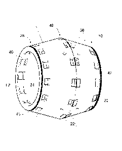
Referring now to Figs. 13 to 15, there is shown a magnetic resonance imaging
(MRI) instrument 14 according to another embodiment of the present invention.
The MRI instrument 14 includes a body 10 having an opening 34 from a
proximal end 40 to a distal end 42 of the body 10. The body 10 is barrel-
shaped, having an outside diameter of about 25.5 cm at the proximal and distal
ends 40,42 of the opening 34, and 27.3 cm in the region between the proximal
and distal ends 40,42. The wall of the body 10 is about 2.75 cm thick. A
rotatable RF coil 38 is positioned in the opening 34 at the center of the body
10.
The RF coil 38 has an internal diameter of about 18 cm, to provide a usable
imaging volume of about 16 cm in diameter, which is adequate for imaging
extremities such as legs, knees, ankles, feet, arms, elbows, wrists, and
hands,
etc. In the longitudinal z-axis LZ through the opening 34 of the body 10 of
the
magnet assembly 12, the usable slice thickness is about 2 cm, which may be
imaged as one slice with a single RF coil, or simultaneously as a plurality of
slices with multiple RF coils. For example, the 2 cm thick imaging volume may
be simultaneously imaged using four RF coils each sensitive to a 5 mm slice of
the 2 cm thick imaging volume. It will be appreciated that the entire magnet
assembly 12 may be moved along the longitudinal z-axis LZ, in a known
manner, if more coverage is needed.
Preferably, a stepper motor 44 is provided to engage and rotate the RF coil 38
in a known way. Additionally, the body 10 of the magnet assembly 12 may be
provided with a set of gear teeth 16 to allow the body to be rotated with a
motor
having a matching gear (not shown).
CA 02884097 2015-03-06
,
-20-
As will be appreciated by persons skilled in the art, the MRI instrument 14
will
include additional components such as one or more of a processor, a memory,
an input means, and an output means as may be required to operate the MRI
instrument to output magnetic resonance data of objects position in the
imaging
volume of the instrument, in a usable form.
36 permanent magnet segments 16, each with a magnetization direction 26, are
arranged on the body 10 in each of a proximal ring 28, a distal ring 30, and a
center ring 48 located between the proximal and distal rings 28,30. The
permanent magnet segments 16 are evenly spaced from adjacent permanent
magnet segments 16 in each of the respective proximal, center, and distal
rings
18,30,48. The three rings of permanent magnet segments 16 are evenly
spaced from each other, with their centers positioned on the longitudinal Z-
axis
LZ of the opening 34. Each permanent magnet segment 16 comprises a pair
of permanent magnets 18 having the same magnetization directions 26.
Although, the permanent magnet segments 16 are shown as comprising pairs
of permanent magnets 18, it will be understood that the permanent magnet
segments 16 may comprise one or more than two permanent magnets 18
having the same magnetization directions 26. Preferably, each of the pairs of
permanent magnets 18 are held in magnet holders 20 on the body 10. Each
magnet holder 20 has an attachment member 22 sized and shaped to permit
a pair of the permanent magnets 18 to clamp to the attachment member 22 by
the force of their magnetic attraction to one another. In this example, the
attachment members 22 are 3 mm thick strips of plastic. It has been found that
the clamping force of the permanent magnets 18 will hold them to the 3 mm
thick attachment member 22 provided that adjacent permanent magnets 18 are
spaced more than 3 mm apart. Additionally, 3 mm thick spacers 50 may be
used in conjunction with the attachment members 22 maintain the spacing
between the permanent magnets 18. Although, this example shows permanent
magnet segments arranged in three rings, it is contemplated that additional
CA 02884097 2015-03-06
,
-21-
rings of permanent magnet segments may be used to extend the magnetic field
profile along the longitudinal z-axis LZ of the opening 34. For example, at
least
one additional pair of rings may be positioned outwardly of the three rings
described above (not shown). All such embodiments are contemplated by the
present invention.
The permanent magnet segments 16 are attached to the body 10 to generate
a magnetic field profile within the opening 34. Fig. 17 shows an estimate of
the
magnetic field profile 32 generated by the magnet assembly 12 using a multi-
dipole expansion. As can be seen the estimated magnetic field 32 profile has
a substantially constant magnetic strength along the center most 2 cm portion
of the opening 34 along the longitudinal z-axis LZ of the opening 34. The
estimated magnetic field profile 34 is also substantially uniform radially
about
the longitudinal z-axis LZ of the opening 34 in a plane perpendicular to the
longitudinal z-axis LZ, over the 2 cm portion of the opening 34. As such, it
is
similar to the desired quadratic bowl magnetic field profile 36 shown in Fig.
7,
although at a weaker magnetic field strength.
Fig. 18 shows a graph of the estimated magnetic field profile 32 of the magnet
assembly 12 having the permanent magnet segments 16 arranged with the
magnetization directions 26 shown in Fig. 19, as well as the desired magnetic
field profile 36, and magnetic field profiles using prior art magnet
arrangements
based on the circular Halbach array, for comparison. As can be seen in Figs.
17 and 18, the magnet assembly 12 is estimated to generate a magnetic field
profile 32 having a center field of 380 G, which rises to about 400G at the
edges
of the imaging volume.
Fig. 19 shows the arrangement of the permanent magnet segments 16 in the
three rings on the body 10 in terms of the positions of their center locations
23
and magnetization directions 26 to generate the desired quadratic bowl
CA 02884097 2015-03-06
-22-
magnetic field profile 36. As can be seen in the x/y graphs, the permanent
magnet segments 16 define two rotations of the magnetization directions 26 in
each of the three rings 28,48,30, which is consistent with the circular
Halbach
array. The y/z graph for the center ring 48 also shows that the magnetization
directions 26 are aligned with the ring, which is also consistent with the
circular
Halbach array. However, the y/z graphs for the proximal and distal rings 28,30
show that the magnetization directions 26 of the permanent magnet segments
16 are oriented out of alignment with their respective rings. In other words,
the
magnetization directions 26 are out of alignment with a plane perpendicular to
the longitudinal z-axis LZ, which is a surprising departure from the circular
Halbach array, for example as shown in Fig. 1.
This departure from the circular Halbach array is more clearly seen in Fig.
20,
which shows the center locations of each of the permanent magnet segments
16 on the left, and on the right, a graph showing the differences between the
magnetization directions of a magnet assembly 12 having the permanent
magnet segments 16 arranged with the magnetization directions 26 shown in
Fig. 19, and a magnet assembly based on the circular Halbach array. As can
be seen, the differences in angles of the magnetization directions are most
pronounced in the X and y axis.
Since the MRI signal is typically weak, an RF shield 52 may preferably be used
to enclose the magnet assembly 12 to reduce interference from unwanted RF
sources, as shown in Fig. 16. Good results have been obtained using an RF
shield 52 made from aluminum. While typically in an MRI suite, the entire room
is shielded, for portability reasons, it is more practical to shield the
magnet
assembly 12, and placing the RF shield 52 on the outside of the magnet
assembly 12 provides a reasonable balance between ensuring sufficient shield
effectiveness and avoiding interference with the RF coil 38, since a grounded
RF shield in close proximity to a coil can significantly affect its operation.
CA 02884097 2015-03-06
-23-
As in the case of the first and second examples above, the body 10 of the
magnet assembly 12 in this third example may be made from plastic (such as
for example Polylactic acid) by 3D printing. By way of example, Fig. 21 shows
a one piece 3D printed body 10. However, it will be appreciated that the body
10 of the magnet assembly 12 may be made by other methods available to the
person skilled in the art.
Example 4
A problem with using a quadratic bowl magnetic field profile of the type shown
in Fig. 17, is that the derivative of the magnetic field strength vanishes in
the
center. This results in undesired artifacts in the image at the center, which
makes it difficult to accurately determine where the magnetic resonance signal
is coming from in the center of the imaging volume. The applicant has
overcome this problem by arranging the permanent magnet segments 16 on the
body 10 of the magnet assembly 12 to generate a linear magnetic field profile
54, which has a substantially constant magnetic strength along a portion of
the
opening 34 along the longitudinal z-axis LZ, and is substantially non-uniform
radially about the longitudinal z-axis LZ in a plane perpendicular to the
longitudinal z-axis LZ, along the portion of the opening 34. For example, as
shown in Figs. 22 and 23, the linear magnetic field profile 54 may increase in
strength from the left side of the opening 34 to the right side (i.e. along
the x-
axis, which is perpendicular to the longitudinal z-axis LZ through the opening
34). Although the linear magnetic field profile 54 has a reduced imaging area,
(i.e. 10 cm diameter compared to 16 cm diameter), and slightly reduced
magnetic strength in the center of the imagine volume (i.e. 370G vs 380G), it
can make it easier to determine accurately where the magnetic resonance
signal is coming from in the center of the imaging volume, as compared to the
quadratic bowl magnetic field profile 36.
CA 02884097 2015-03-06
,
-24-
Fig. 24 shows the arrangement of the permanent magnet segments 16 in the
three rings on the body 10 in terms of the positions of their center locations
24
and magnetization directions 26 to generate the linear magnetic field profile
54.
As can be seen in the x/y graphs, the permanent magnet segments 16 define
two rotations of the magnetization directions 26 in each of the three rings,
which
is consistent with the circular Halbach array. The y/z graph for the center
ring
48 also shows that the magnetization directions 26 are aligned with the ring,
which is also consistent with the circular Halbach array. However, the y/z
graphs for the proximal and distal rings 28,30 shows that the magnetization
directions 26 of the permanent magnet segments 16 are oriented out of
alignment with their respective rings. In this respect, the magnetization
directions 26 of the permanent magnet segments 16 are substantially the same
in the x/y and y/z planes for the linear magnetic field profile 54 in this
example
4, as compared to the quadratic bowl magnetic field profile 36 in example 3
discussed above. The difference is more evident in Fig. 25, which shows in the
top set of diagrams the magnetization directions 26 for generating the
quadratic
bowl magnetic field profile 36, and in the bottom set the magnetization
directions 26 for generating the linear magnetic field profile 54 in the zJx
plane.
As can be seen, the linear magnetic field profile 54 may be generated by
orienting some of the magnetization directions 26 of permanent magnet
segments 16 in the proximal and distal rings 28,30, in one region of the
magnet
assembly 12, away from the center of the imaging volume. For example,
comparing the graphs of the magnetization directions 26 in the proximal ring
of
the quadratic magnet assembly (Fig. 25, top, left) to those of the linear
magnet
assembly (Fig. 25, bottom, left) reveals that the magnetization direction 26
of
the leftmost 56 permanent magnet segment 16 is turned out more from the 0,0
point in the linear magnet assembly. The same is true for the leftmost magnet
in the distal ring 30, which also shows the magnetisation direction 26 of the
leftmost 56 permanent magnet segment 16 being turned out more from the 0,0
point in the linear magnet assembly.
CA 02884097 2015-03-06
,
,
-25-
It is also contemplated that a linear magnetic field profile may be generated
by
orienting some of the magnetization directions 26 of permanent magnet
segments 16 in the proximal and distal rings 28,30, in one region of the
magnet
assembly toward the center of the imaging volume.
It is also contemplated that a linear magnetic field profile may be generated
by
orienting some of the magnetization directions 26 of permanent magnet
segments 16 in the proximal and distal rings 28,30, in one region of the
magnet
assembly away from the center of imaging volume, and in an opposite region
of the magnet assembly, toward the center of the imaging volume.
This departure from the circular Halbach array is more clearly seen in Fig.
26,
which shows the positions of each of the permanent magnet segments 16 on
the left, and on the right, a graph showing the differences between the
magnetization directions 26 of a magnet assembly 12 having the permanent
magnet segments 16 arranged with the magnetization directions 26 shown in
Figs. 24 and 25, and a magnet assembly based on the circular Halbach array.
As can be seen, the differences in angles of the magnetization directions 26
are
most pronounced in the X and y axis. Furthermore, the differences are most
significant around permanent magnet segments 16 numbered 1, 6, 12 and 25,
and 36. This is because those magnet locations correspond to the proximal
and distal rings 28,30 on the extreme x-axis, where the linear magnetic field
profile 54 is dramatically different from the quadratic magnetic field profile
26.
25 Determining Magnetization Directions
With reference to Fig. 27, the following describes a preferred method for
determining the magnetization directions for a magnet assembly according to
Examples Ito 4 above.
CA 02884097 2015-03-06
-26-
As indicated in block 58, a desired magnetic field profile is selected. The
desired magnetic filed profile can be any magnetic filed profile. By way of
example, where a constant uniform field of 400 G is desired, the desired field
profile may simply be represented by the formula:
for all inside the imaging area.
As another example, the following formula represents a quadratic bowl
magnetic field profile 36:
iiit = TAC:
where ii7d0 is the desired magnetic field profile at a spatial location I:, at
the
center of the imaging volume = 0, A is a 3x3 matrix with coefficients that
define
how quadratic the desired magnetic field profile should be, such as for
example:
0 0-
A - ___________ ""7. 0 1 0
ridge'
where BEdv. is the desired field increment at the edge over the center (for
example 15 G), and rsdas is the radius of the circular imaging area.
As another example, the following formula represents a linear magnetic field
profile 54:
iqd (F) = kerter,
where gd.F.) is the desired magnetic field profile at a spatial location r, at
the
center of the imaging volume = 0, but now ti is a vector that controls the
linear
slope (in all three dimensions) of the desired field, such as for example:
=r4:47-44,13 g 017
where 361g6 is the desired field increment at the edge over the center (for
example 10 G), and redge is the radius of the circular imaging volume.
CA 02884097 2015-03-06
-27-
Regardless of how the desired magnetic field profile is determined, good
results
have been obtained by using an iterative process, whereby minor revisions to
the parameters are made resulting in a the desired magnetic field profile
having
a reasonable balance between magnetic field strength and uniformity.
The center locations of each of a plurality of permanent magnet segments 16
evenly spaced apart from one another in each of at least two spaced apart
rings
centered on a longitudinal z-axis LZ are also determined in block 58.
Preferably, the same number of permanent magnet segments are arranged in
each of the at least two rings. Preferably, the rings are sized to accommodate
a desired imaging volume, as well as the RF 38 coil and support structures
that
need to be fit between the imaging volume and the ring. For example, the RF
coil 38 may require an annular space of about 5-6 cm thick extending between
the imaging volume and the center most ring of permanent magnet segments
16, if the center ring is present. Preferably, the permanent magnet segments
16 are positioned as close as possible to the imaging volume.
Preferably, the diameter of the outermost rings may be reduced compared to
the center ring, if provided, to increase the magnetic field strength in the
center
of the imaging volume and/or increase homogeneity. By way of example, good
results have been obtained by determining the diameter of outer rings (d) at a
z-coordinate of z with the following formula:
d = 2
=44
where d2 is the diameter of the center ring and a is a number less than or
equal
to 1. Good results have been obtained with a being set to 1. However, in
larger
magnet assemblies, setting a to a number less than 1 may be necessary as the
smaller diameter of the outer rings limits the size of the object that can be
placed within the magnet assembly 12.
CA 02884097 2015-03-06
-28-
By way of example, in Example 3 above, the formula (with a=1) results in the
proximal and distal rings 28,30 having a diameter of 23.6 cm, when the center
ring 48 has a diameter of 27 cm, and provides an imaging volume of about 16
cm in diameter, which is adequate for imaging extremities, such as a legs,
knees, ankles, feet, arms, elbows, wrists, and hands. To image larger
anatomical structures such as the brain, the rings would need to be sized with
a larger diameter to accommodate an imaging volume of at least a 26 cm in
diameter.
Once the center locations for all of the permanent magnet segments 16 have
been determined, the next steps are used to determine their magnetization
directions to achieve the best match to the desired magnetic field profile.
At box 60, an initial magnetization direction is determined for each of the
permanent magnet segments 16 based on a circular Halbach array
arrangement, which as mentioned above has all of the magnetization directions
26 aligned with a plane defined by the ring, and progressively make two
rotations through adjacent permanent magnet segments 16 in one direction of
the ring. In other words, the magnetization directions 26 only rotate around
the
center of the ring, and the other two rotation angles are set to zero.
Next, at box 62 an estimate is made of the magnetic field profile based on the
initial magnetization directions, preferably using a multi-dipole expansion,
which
has shown good agreement (to within 1 G) of data acquired with a Gaussnneter.
For each permanent magnet segment, the orientation angles (S) are converted
into directional vectors. More preferably, the orientation angles (8) for each
permanent magnet of the permanent magnet segments are converted into
directional vectors. The directional vectors are then split eight dipole's
four for
CA 02884097 2015-03-06
-29-
each permanent magnet. The four dipoles for each permanent magnet are
then spaced uniformly within the permanent magnet. The dipole orientation
vector (R) is aligned with the directional vector for the permanent magnet,
and
its magnitude is a constant, carefully calibrated using gauss-meter
measurements. Preferably the magnitude of the dipole orientation vector is
re-calibrated later, once MRI data is available, to correct for minor
variations
(i.e. < 1%) in the magnetic field strength of the permanent magnets. While
more than eight diploes per magnetic block would have increased precision,
computation time would have also increased.
The magnetic field profile :6.(-43) was estimated by adding up all the
subfields
from all of the di-poles within all of the permanent magnets using the
following
formula:
"µ
IV if 3(c- CtL=j) 7-'11,i = (-L. j
= _
417 1.1 Eoits F¨ kr),
wherein 57'13 is the dipole orientation vector from the j-th dipole for the i-
th
permanent magnet, cif is the center location for the j-th diple for the i-th
permanent magnet, 77: is the spatial location where the magnetic field profile
is
being estimated, M is the number of di-poles per permanent magnet (i.e 8 in
this example), and N is the number of permanent magnets. The It operator
is the L2 vector norm, defined as OH = Et=lxkl, where L is the number of
elements in the vector, and A7k is the kth element of the vector.
Next a conjugate gradient algorithm is applied using the estimated magnetic
field profile and the desired magnetic field profile to obtain updated
magnetization directions 26 for each of the plurality of permanent magnets 16
to generate a magnetic field profile having an acceptable error to the desired
magnetic field profile. Preferably the acceptable error is a least mean
squared
CA 02884097 2015-03-06
-30-
error. However, it is contemplated that other error functions may be used,
such
as for example the max-norm defined aslixilmax = max xk (i.e. the largest
element in the vector -x).
Boxes 64 - 72 in Fig. 27 set out the steps for applying the conjugate gradient
algorithm. At box 64 a calculation is made of the mean squared error between
the desired magnetic field profile and the estimated magnetic field profile
based
on the initial magnetization directions.
If the mean squared error is acceptable or the maximum number of iterations
of the conjugate gradient algorithm has been reached, the updated
magnetization directions are returned in box 66. Otherwise, the conjugate
gradient algorithm continues at box 68 by determining the resulting mean
squared error between the desired magnetic field profile and the estimated
magnetic field profile for small changes in to obtain an estimate of the
gradient
of the error function.
What is considered an acceptable least mean squared error will be will be
appreciated by persons skilled in the art. By way of example, good results
have
been obtained by setting the least mean squared error to 10-6, as this value
is
the square root of the minimum representable floating point number. However,
in light of other errors inherent in the process of constructing the magnet
assembly a lower value may be warranted, such as about 10-3. For maximum
number of iterations, good results have been obtained using 10-100. However,
the maximum number of iterations may be set to any number. The purpose of
this limitation is to prevent the algorithm from continuing for a very long
time
making very minor adjustments to the magnetization directions.
Preferably, the gradient of the error function is computed numerically by
CA 02884097 2015-03-06
-31-
applying a small increment A (i.e. 1.8 ) to each of the orientation angles,
then
calculating how much the error changes using the formula:
vi(P) P61 f (11 -FH)-
where fCe) is the difference between the desired magnetic field profile (i.e.
the
gradient of the error function), and the estimated magnetic field defined as:
Vid0¨
Once the gradient of the error function is determined, the search direction
(SD)
is determined next at box 70. Preferably, the scalar beta is defined first
using
the following formula:
7(1977f t:73)
beta=
71(1v11:1L,
where, f( igzr is the gradient of the error function found in the previous
iteration.
In the case of the first iteration, where there is no previous iteration, the
scalar
beta is set to zero. Then the search direction is selected as follows:
SD f( A-1- beta SDL.,...n,
where SpLazt is the search direction from the last iteration.
Next at box 72 updated magnetization directions are obtained for each of the
permanent magnets with the formula:
= t5D,
where t provides the lowest mean squared error between the desired magnetic
field profile and the estimated magnetic field profile.
CA 02884097 2015-03-06
,
-32-
The conjugate gradient algorithm then repeats with the updated magnetization
directions obtained in box 72 being used in box 62 to determine the estimated
magnetic field profile based on the updated magnetization directions.
Example 5
A conventional magnet assembly based on the circular Halbach array
arrangement of permanent magnets generates a magnetic field profile, whose
magnetic field is oriented from one side of the magnet to the other (as seen
in
Fig. 1, along the x-axis). However, conventional MRI superconducting systems
have a different magnetic field orientation, going through the opening through
the body (along the longitudinal z-axis LZ). On a MRI system level, this
requires
the use different RE signal detection methods.
In this example 5, there is described a magnet assembly 12 for generating a
magnetic field profile 32 that is oriented through the opening 34 of the body
10
of the magnet assembly 12, which is compatible with conventional RE signal
detection methods developed for superconducting MRI scanners.
Fig. 28 shows the body 10 of a magnet assembly 12 according to another
embodiment of the present invention. The body 12 has an opening 34 from a
proximal end 40 to a distal end 42 of the body 10. The body 10 includes
magnet holders 20 for holding 36 permanent magnet segments 16, each with
a magnetization direction 26, arranged on the body 10 in each of a proximal
ring 28, a distal ring 30, and a center ring 48 located between the proximal
and
distal rings 28,30. The magnet holders 20 are arranged on the body 10 to hold
the permanent magnet segments 16 evenly spaced from adjacent permanent
magnet segments 16 in each of the respective proximal, center, and distal
rings
28,48,30. The three rings are evenly spaced from each other, with their center
locations 24 positioned on the longitudinal z-axis LZ of the opening 34 of the
CA 02884097 2015-03-06
,
-33-
body 10 of the magnet assembly 12. Each magnet holder 20 is adapted to hold
a pair of permanent magnet segments 16 having the same magnetization
directions 26. Each magnet holder 20 has an attachment member 22 sized and
shaped to permit a pair of the permanent magnets 18 to clamp to the
attachment member 22 by the force of their magnetic attraction to one another.
In this example, the attachment members 22 are 3 mm thick. It has been found
that the clamping force of the permanent magnets 18 will hold them to the 3
mm thick attachment member 22 provided that adjacent permanent magnets
18 are spaced more than 3 mm apart.
Preferably, the permanent magnet segments 16 are attached to the magnet
holders 20 on the body to generate a magnetic field profile within the
opening.
Fig. 29 shows an estimate of the magnetic field profile 32 generated by the
magnet assembly 12 using a multi-dipole expansion. As can be seen the
estimated magnetic field profile 32 has a magnetic strength that uniformly
increases, then uniformly decreases, along the center most 2 cm portion of the
opening 34 along the longitudinal z-axis LZ. The estimated magnetic field
profile is also substantially uniform radially about the longitudinal z-axis
LZ in
a plane perpendicular to the longitudinal z-axis LZ, over the 2 cm portion of
the
opening 34.
Fig. 30 shows a graph of the estimated magnetic field profile 32 of a magnet
assembly 12 having the permanent magnet segments 16 arranged with the
magnetization directions 26 shown in Fig. 35, as well as the desired magnetic
field profile 36.
Fig. 31 shows the arrangement of the permanent magnet segments 16 in the
three rings on the body 10 in terms of the center locations 24 of their
positions
and magnetization directions 26 to generate the quadratic bowl magnetic field
profile 36. As can be seen in the x/y graphs, in the proximal ring 28, all of
the
CA 02884097 2015-03-06
,
-34-
permanent magnet segments 16 have their magnetization directions 26
oriented to point radially towards the longitudinal z-axis LZ at the center of
the
opening. The x/y graph of the center ring 48 shows the magnetization
directions 26 of all of the permanent magnet segments 26 being parallel to the
longitudinal z-axis LZ. The x/y graph of the distal ring 30 shows the
magnetization directions 26 of all of the permanent magnet segments 16 being
oriented to point radially away from the longitudinal z-axis LZ at the center
of
the opening 34 of the body 10 of the magnet assembly 12. The y/z graphs
show the magnetization directions 26 of all of the permanent magnet segments
16 being oriented out of alignment with their respective rings, in the same
direction, namely the proximal direction.
Fig. 32, shows the positions of the center locations 24 of each of the
permanent
magnet segments 16 on the left, and on the right, a graph of the differences
between the magnetization directions 26 of a magnet assembly 12 having the
permanent magnet segments 16 arranged with the magnetization directions 26
shown in Fig. 31, and a magnet assembly based on the linear Halbach array.
The linear Halbach array is radially symmetric, such that in the 3 ring
example,
it would consist of 3 blocks arranged as shown in Fig. 32 (middle).
As in the case of the first example above, the body 10 of the magnet assembly
12 in this example may be made from plastic by 3D printing. By way of
example, Fig. 28 shows a one piece 3D printed body. However, it will be
appreciated that the magnet holder may be made by other methods available
to the person skilled in the art.
Determining Magnetization Directions
The following describes a preferred method for determining the magnetization
directions 26 for a magnet assembly 12 according to Example 5 above.
CA 02884097 2015-03-06
-35-
In this example, a stronger magnetic field strength is sought inside the
opening
34 of the magnet assembly 12 as compared to outside of the magnet assembly
12.
As shown in Fig. 33 the magnet holders 22 are positioned on the body 10 of the
magnet assembly 26 to hold the permanent magnet segments 16 in three rings
having their centers on the longitudinal z-axis LZ of the opening 34 of the
body
of the magnet assembly 12. Initially the permanent magnet segments 16
have their magnetization directions 26 oriented in radially symmetric rows,
such
10 that in the center ring 48 they point opposite to the desired magnetic
field
direction, and in the proximal and distal rings 28,30 they point in opposite
directions, perpendicular to the desired magnetic field direction, as shown in
the
middle of Fig. 33. As will be appreciated by persons skilled in the art, the
initial
magnetization directions are determined based on the linear Halbach array.
The linear Halbach array uses a specific permanent magnet arrangement to
focus the magnetic field to the one side of the magnet assembly. Expanding
this concept radially allows for a stronger magnetic field inside the opening.
The
magnetic field is oriented along the longitudinal z-axis LZ.
Next the initial magnetization directions are adjusted as shown in the right
of
Fig. 33. The offset angle a is adjusted and the magnetic field is re-computed
based on the updated magnetization directions using eight-di-pole expansion
to estimate the magnetic field profile. The resulting estimated magnetic field
profile 36 is then re-evaluated to determine which of the offset angles a to
choose. The estimated magnetic field strength is computed at the center of the
imaging volume within the opening 34, and the field inhomogeneity as a
function for a range of offset angles a. By way of example, Fig. 34 shows a
graph in which the magnetic field strength increases until it reaches a
plateau
at about 375 G as offset angles a increase from -60 to 20. Over the same
offset angles a the field variation (inhomogeneity), calculated as (max(B) -
CA 02884097 2015-03-06
,
-36-
min(B)), decreases from about 17 G to about 12 G and continues to rise to
about 82 G as shown in Fig. 35. Based on these two graphs, it will be
appreciated that an offset angle a of -8 degrees provides the strongest field
with
a field inhomogeneity of about 17 G. This field inhomogeneity is comparable to
what is seen in the above examples. On a system level, a more homogeneous
magnet assembly places less of a strain on the RF subsystem, hence a more
homogenous magnet assembly is desirable. Good results have been obtained
with inhomogeneities of about 20 G. Once the appropriate offset angle a is
determined, it may be used to determine the updated magnetization directions
for each of the permanent magnet segments 16.
Although the above example of the magnet assembly includes 3 rings of
permanent magnet segments 16, it is contemplated that in other embodiments
of the present invention the magnet assemblies 12 may contain two rings or
more than three rings of permanent magnet segments. All such embodiments
are contemplated by the present invention. Using more than three rings would
require considering additional offset angles in the same manner described
above with respect to the one offset angle a. However, as the number of offset
angles increases to more than 3 or 4, it becomes impractical to exhaustively
evaluate all combinations of offset angles as have been shown above with the
single offset angle a. In such instances it becomes necessary to resort to a
method similar to above where a conjugate-gradient algorithm is used to find
the best combination of offset angles to generate a desired magnetic field
profile.
Transmit and Receive Coil Design
Once placed in a magnetic field, protons start to interact with radio-waves.
The
frequency of these radio waves is proportional to the magnetic field strength.
According to an embodiment of the present invention, for example, the field
CA 02884097 2015-03-06
,
-37-
strength is 800G, which corresponds to a frequency of 3.4 MHz. A good
transmit and receive system is needed in order to interact with the protons
and
eventually construct an image. However, signal levels between transmit and
receive are vastly different. This is especially true when as in the
embodiments
of the present invention, the field inhomogeneity is quite significant. Table
1
below summarizes the signal levels observed on several MRI platforms
including an embodiment of the present invention.
Table 1: Transmit/receive power levels
System Field-strength Transmit (TX) Receive (RX)
signal
Pi signal
GE Discovery 750 3.0 50 to 70 dBm -50 to -30 dBm
Esaote C-Scan 0.2 40 to 50 dBm -70 to -50 dBm
MRI System 0.08 30 to 40 dBm -160 to -140 dBm
according to an
embodiment of the
present invention
All values in the above Table 1 are approximate based on experience and
measurements. Actual signal levels will vary depending on measurement
conditions (coil, objects in the imaging volume, pulse sequence, etc). All
values
are expressed in dBm (decibels relative to a mVV). The comparatively low
receive signal levels observed in the MRI system according to the embodiment
of the present invention tested are due to the inhomogeneities inherent in the
design, resulting in fewer protons being reachable at a given narrow frequency
band, and hence less signal.
MRI scanners must thus employ some form of protection against transmit
power levels seeping into the receive chain. Even in embodiments of the
present invention, with a relatively modest 40 dBm peak power level, the
CA 02884097 2015-03-06
,
,
-38-
corresponding RF powerwould easily ruin a sensitive low noise amplifier (LNA).
The industry standard solution to this problem is to employ some form of a
"blocking circuit". Both passive and active schemes are typically employed,
with
careful diode arrangements drastically reducing the observed RX RF power
during transmit. Unfortunately, for the purposes of the present invention, the
recovery time from these blocking schemes, which can be on the order of a
fraction of a millisecond, are much too slow. Due to the increased
inhomogeneity in embodiments of the present invention, the received signal
will
have nearly completely dephased in a fraction of a millisecond.
Therefore a faster recovery solution is desired. Preferably, the MRI system
according to the present invention may use two solutions to protect against
transmit power levels seeping into the receive chain. The first solution is to
utilize receive coils with small areas, therefore limiting exposure to the
strong
RF transmit field. The second solution is to place the RX coil symmetrically
over the TX coil, and adding adjustment flaps to the receive coil. These small
adjustment flaps allow the receive coil to be fine-tuned to minimize any TX
field
exposure. By carefully adjusting the screws on the RX coil structure shown in
Fig. 36, its exact spatial position is carefully controlled. While the
adjustment
itself is relatively minor (often no more than a millimeter or two), the
effect is
quite substantial. Increasing the TX-RX signal isolation from 30 dB to 60 dB
and beyond.
Thus the receive chain observes a much smaller peak RF power of -20 dBm,
even though the actual transmit signal is at 40 dBm. Unlike the active/passive
blocking approach, there is no transient period as the 60 dB level of
isolation
is always achieved. Furthermore, these two solutions are quite compatible, it
would be easy to combine the two approaches in an embodiment that has
active/passive blocking, in addition to the tunable, geometric scheme
described
above.
CA 02884097 2015-03-06
-39-
Another feature of the RF receive coil is that by shaping the RF receive coil
the
"slice-profile" of the coil can be altered, achieving slice selection with the
RF RX
coil. This greatly simplifies the reconstruction work, as the reconstruction
engine needs only to reconstruct two dimensional images. If needed,
information in the third dimension can be obtained by sliding the entire
magnet/coil unit along the axis of the bore. However, other more scan-time
efficient approaches are possible, by for example using a row of several coils
along the bore axis akin to multi-detector CT systems. Fig. 37 illustrates the
simulated RX coil profile.
RF Pulse Sequence Selection
The following will discuss two software components of the invention. First,
the
pulse sequence, which will use the hardware described above to query the
hydrogen atoms in the imaging volume, thus gathering information that will be
fed into the second component, namely the image reconstruction engine, which
is described later. As mentioned above, the transmit power levels are much
higher than those of the received signal, thus it is necessary to only
transmit,
or only receive during a given time period.
As is well known, when placed in a magnetic field, one can interact with the
hydrogen atoms by use of radio-waves. The following equation determines the
operating frequency,
f =y13,
where y is the gyronnagnetic ratio (42.58 MHz/T for hydrogen).
According to the present invention, the operating frequency will range a fair
bit,
as the magnet by design, has a field that varies from about 800 G to 860 G.
Therefore the preferred operating frequency varies from 3.4 MHz to 3.6 MHz.
One possible option is simply to design an RF pulse that is reasonably uniform
CA 02884097 2015-03-06
,
-40-
over the entire band (Bernstein et al. "Handbook of MRI pulse sequences"
Elsevier 2004, Chapter 2). However, such a pulse would have to be very short
and hence require a significant amount of RF transmit power, potentially
impacting the ability to make the MRI device portable.
Instead, the MRI system of the present invention preferably employs a
sweeping RF scheme, similar to that disclosed in U.S. Pat. No. 7,403,006, but
with several important differences. For example, instead of using a hyperbolic
secant sweep, the present invention preferably takes a more discrete approach
using a series of hard-pulses to query a narrow band of spectrum at a time.
This is followed by a RX period where no transmit activity takes place, so
that
the MR signal can be detected. This TX/RX block then repeats at a different,
randomly selected frequency as opposed to an orderly progression as taught
in U.S. 7,403,006. Hard-pulses ensure that all available RF power is used. The
sub-pulse duration, and the RX period duration need to be carefully determined
to ensure appropriate image contrast and good signal to noise ratio (SNR).
In addition, periodic system monitoring pulses are preferably inserted into
the
pulse chain. Hard RF pulses at full power cause compression in the receive
system, while lower power pulses do not. The MRI system of the present
invention preferably takes advantage of this, by utilizing a low-power system
monitoring RF pulses for system monitoring purposes. The resulting received
signal will inform the user of the total gain in the system, from transmit
amplifier
through the TX/RX coils, onwards to the low noise amplifiers LNAs. If, for
example, a receive amplifier was malfunctioning, the user would know from
consulting these system monitoring pulses, as the signal from the
malfunctioning amplifier channel would be abnormally low. Fig. 38 illustrates
an RF pulse sequence according to a preferred embodiment of the present
invention. As shown, the RF pulse operates at a narrow band around one
frequency at a time. Following is a brief receive period where the signal is
CA 02884097 2015-03-06
=
-41-
recorded. System monitoring is achieved with the system monitoring pulses
(M1 in Fig. 38). By measuring the transmit signal of the monitoring pulses
seen
at the receive end, changes in the receive and transmit coil profiles can be
measured. Furthermore, the receive period that follows allows for noise
characterization and thus eventual noise cancellation.
Advantageously, these pulses can be used to monitor coil loading, namely the
phenomenon that once items are placed near an RF coil, its signal profile
changes. Preferably, the system monitoring pulses can be used to determine
the total signal profile at a given frequency f. Assuming that the amount of
transmit power available limits the flip angle to less than 90 degrees or so
(small
flip angle approximation), the impact of coil loading on the receive signal is
approximately equal to the product of transmit and receive profiles.
Preferably,
the combined RF TX/RX profile may be corrected for via pilot pulses by simply
dividing out the observed TX/RX monitoring pulse power.
Preferably, the monitoring pulses may also be used to provide the signal free
receive period that follows (recall the system monitoring pulse is very low-
power
hence very little MR signal is generated). During this time, the system
preferably monitors the signal received. For some noise sources, in particular
narrow band interference sources, the unwanted noise can be predicted to a
great degree. This allows the present invention to remove its influence on the
signal, improving signal to noise ratio (SNR).
Preferably, once the signal has been corrected for coil loading, and strong
narrow-band interference has been removed, the total signal is averaged up at
the same frequency as the transmit pulse immediately preceding it to generate
the observed MR signal at that frequency. As discussed above, for
convenience, the observed MR signal is preferably converted to field-strength
using the equation above. Due to the relatively inhomogeneous magnetic-field
CA 02884097 2015-03-06
-42-
of the MRI system of the present invention, very little remnant signal from
the
previous receive period is observed leaking in, signal dephasing is such that,
more or less all signal is lost once the end of a receive window is reached.
TX-RX Synchronization Methods
Depending on the hardware used in the MRI system according to the present
invention, exact synchronization of transmit and receive waveforms may be
challenging. If left untreated, this could seriously hinder efforts in
attributing a
given receive waveform to a given transmit frequency, resulting in sub-par
images. This issue arises from latency uncertainties involved in buffered
communication between for example the computer and the physical transmit
and receive system, for example, a universal software radio peripheral (USRP
1), available from Ettus Research www.ettus.com. Many excellent ways exist
to resolve this problem, mostly by carefully controlling each latency
component
and calibrating appropriately.
According to a preferred approach, the USRP 1 uses the same clock to drive
the transmit and receive system. Under this approach only bulk shift, and
phase errors in the receive signal need to be addressed. The former is
preferably addressed first as it is more complex. By artificially lengthening
each
receive period by a pseudo-random amount, the overall magnitude correlation
between the transmit waveform and the received waveform, rapidly converges
to the optimal solution. This is illustrated in Fig. 39, where the pseudo-
random
receive window lengthening is varied from 0 (no artificial lengthening, all
receive
windows are the same length), to at most 40 % of the receive window length.
As can be seen, this slight lengthening ensures that there is only one optimal
shift estimate available corresponding to the actual shift seen, even in the
presence of substantial measurement noise. While 40 % may seem excessive
(this would lengthen scan time by about 20 %), the person skilled in the art
can
CA 02884097 2015-03-06
,
,
-43-
easily select an appropriate value based on observed measurement noise
ensuring consistent TX/RX synchronization. This approach is similar to that
used by the global positioning system, where the receiver is synchronized to
the
satellite constellation via a pseudo-random key (Misra et al. Global
Positioning
System, Signals, measurements and performance, available from
gpstextbook.com, chapter 8).
Once bulk shift correction is completed, phase correction is fairly
straightforward. Preferably, the frequency and phase of the received transmit
pulse is compared to that of the actual transmit pulse and corrected. This
helps
ensure appropriate phase of the receive window that is to follow.
Image Reconstruction Methods
Once the signal is received and corrected, the process of converting it to an
image begins. As discussed above, the RX coil effectively collects a "slice"
of
data, thus received data can be thought of belonging to a given image slice.
The image reconstruction problem is thus two dimensional. For convenience
the image reconstruction problem can be thought of in polar coordinates. As
discussed later, the polar coordinate system is a more natural coordinate
system for the image reconstruction problem. Therefore, to reconstruct an
image, a series of grayscale values must be found for the polar coordinates
(r,
A) in the image. Fig. 40 illustrates this concept.
As discussed above, the pulse sequence delivers information on how much MR
signal is present at a given narrow-band slice of magnetic field. This (along
with
which receive coil obtained the signal) allows for determining the radii
component of the polar coordinate (the magnet assembly's angle provides the
0). However, the magnetic-field must be transformed to radii components, for
example with a simple quadratic expression. As discussed above the magnetic
CA 02884097 2015-03-06
-44-
field profile goes quadratically from a low at the center to a high along the
edges. A sample transformation is shown in Fig. 41.
The discussion above assumes that each data-point (r, 0) corresponds only to
a single point in the image. This however, is not true as the coil according
to
the present invention "smears" the signal somewhat, primarily along the angle
direction. This is because the coil spans a 90 degree angle. Experiments with
a thin line phantom showed that this smearing is quite significant, expanding
over a 1500 span, although the strongest signal component is along the
center-line of the coil. Preferably, this may be modelled in the following
manner:
Signakr, = au Image(r, 8, ,)
where aõ is a weighting function (currently the Hanning window function works
well). Preferably, the method of least squares is then employed to retrieve
the
Image. It has been found to be helpful to employ a regularization function,
such
as for example, a simple Tikhonov regularization. A sample reconstructed MR
image according to an embodiment of the present invention is shown in Fig. 42.
While reference has been made to various preferred embodiments of the
invention other variations, implementations, modifications, alterations and
embodiments are comprehended by the broad scope of the appended claims.
Some of these have been discussed in detail in this specification and others
will
be apparent to those skilled in the art, such as, for example:
= Different
size: In the above examples the magnet assemblies were sized
to be suitable for imaging the extremities. However, the present
invention is not so limited, both larger and smaller magnet assemblies
can made using the teachings herein. Furthermore, depending on the
size of the magnet assembly, more or fewer rings may be used.
CA 02884097 2015-03-06
-45-
= Utilizing other magnetic block sizes. Many possible permanent magnet
sizes and shapes exist on the market today. The 1x1 x1/2 inch cuboid
shaped permanent magnets, mentioned above in association with
embodiments of the present invention, were selected as an example
only because it is cost effective, light-weight, available in a stronger
grade N52, and could be shipped by air. Depending on the desired
magnet geometry, a different permanent magnet size and/or shape may
be used. Furthermore, different sets of permanent magnets may be
used, with for example, a smaller size on the innermost ring(s) and a
larger size in rings space outwardly from the innermost ring(s).
= Elliptical opening through body of magnet assembly: While the above
examples show the permanent magnet segments arranged in circular
rings, it is contemplated that in other embodiments of the invention, the
rings may be elliptical. For example, as disclosed in U.S. Pat. No.
5,659,250, an elliptical shaped magnet assembly may have some
advantages in certain situations.
= Imaging into the third dimension. By adding other "slit" coils along the
bore axis, multiple image slices could be acquired at the same time,
alternatively the magnet assembly may be slid along the axis of the
magnet (in addition to the rotation done here).
= Increase the number of TX/RX coils. In embodiments of the present
invention discussed above the analog/digital system are preferably
limited to two receive channels. With custom receive systems, there is
no limit to the number of receive or transmit channels one could use.
For example, embodiments involving 2 TX and 4 RX channels are
feasible.
= Spatial location with transmit coil. Embodiments of the present invention
discussed above preferably employ a simple loop-coil for transmit,
resulting in uniform phase across the imaging volume. This need not be
the case, as work from Sharp et al. (Magnetic resonance in medicine,
CA 02884097 2015-03-06
-46-
issue 63: pg 151) shows, TX coils can be constructed to deposit varying
degrees of phase across the imaging volume, hence contributing to
spatial location of the signal. With clever coil construction techniques,
perhaps even independently rotating the TX coil, this could result in
improved spatial localization of the MR signal, eventually resulting in
improved image quality.
= Gradient systems. As gradient amplifiers and associated gradient
hardware consumes a fair amount of electrical power in traditional MRI
systems, embodiments of the present invention discussed above
preferably obtain images without a gradient system to permit a portable
system. However, gradient systems do have their advantages as they
allow tremendous flexibility on how an image is acquired. Karinni et al.
(Magnetic resonance in medicine, e-pub only DOI: 10.1002/mrm.24950)
published a mechanical gradient system that need not consume much
electrical power. Furthermore, low power gradient systems could boost
spatial localization and hence image quality close to the edge of the
bore. Accordingly, it is contemplated that a hybrid system with a limited
gradient system could be constructed.
= Improved image reconstruction methods. Recently, sparsity has been
proposed as a regularization parameter in medical imaging
reconstruction methods (Lustig et al. Magnetic resonance in medicine
58:1182). The image reconstruction method discussed above in relation
to embodiments of the present invention is much simpler, and could
benefit from considering sparsity to contain blurring as can be seen in
the reconstructed image. Although the results presented thus far in the
literature are impressive, long reconstruction times remain a major
barrier. Of particular interest is the absence of a Fourier transform in a
preferred image reconstruction method. This is a consequence of the
pulse sequence, in particular, the frequency sweep and random
frequency ordering scheme. In practice, this may enhance the speed of
CA 02884097 2015-03-06
-47-
compressed sensing image reconstruction, as the Lustig et al. paper
shows, a fair amount of computation time is spent on Fourier transforms.
= Increased spatial resolution. As can be seen in the sample
reconstructed MR image shown in Fig. 41, image resolution has some
ways to go to match the resolution available with clinical commercially
available MRI systems. It is contemplated that better image resolution
may be achieved by embodiments of the present invention discussed
above by increasing the amount of radial information obtained in the
pulse sequence, as well as fine-tune the modeling functions. It is
contemplated that one approach may be to utilize multi-tone hard pulses,
briefly combining several carefully selected frequencies into a single
pulse. While this does approach traditional Fourier-style pulses, the MRI
system according to the present invention need not interact with the
entire imaging volume at the same time, thus saving RF power.
Those of ordinary skill in the art having access to the teachings herein will
recognize these additional variations, implementations, modifications,
alterations and embodiments, all of which are within the scope of the present
invention, which invention is limited only by the appended claims.