Note : Les descriptions sont présentées dans la langue officielle dans laquelle elles ont été soumises.
CA 02888715 2015-04-17
WO 2014/060116 PCT/EP2013/003161
1
Medical device comprising a curved needle
Technical field of the invention
The invention relates generally to a medical device used in interventional
radiology.
Specifically, the invention relates to a medical device comprising a hollow
needle
extending longitudinally from a proximal end to a distal end and wherein the
distal end
of the hollow needle is formed as a cutting tip.
Background of the invention
Interventional radiology is a medical sub-specialty of radiology. It is a
minimally-
invasive image-guided procedure. It has been known to diagnose and treat
diseases in
almost every organ of the human or animal body. Today many medical conditions,
for
which conventional surgery would have been used in the past centuries, may be
treated by interventional radiology. An interventional radiologist uses, inter
alia X-ray
devices, computed tomography devices (CT), magnetic resonance imaging (MR1)
devices, and ultrasonic imaging devices in order to obtain images of a human
or animal
body. As a consequence of these images, the interventional radiologist is able
to
navigate an interventional instrument throughout the body to a targeted organ
or
other part of the human or animal. Flexible catheters are inserted through a
small nick
in the skin and thus may be guided through a patient's network of arteries or
veins.
Where tissues of organs are not within reach of a catheter, a biopsy device
comprising
a rigid hollow needle is used to penetrate from the outside of the patient's
body in a
direct way to the target. Special instruments may be guided through the hollow
needle
to extract samples of the tissue, inject fluids, or slide radiofrequency
ablation
instruments to the target, for example, to destroy cancerous tissue locally in
the
targeted zone.
CONFIRMATION COPY
CA 02888715 2015-04-17
WO 2014/060116 PCT/EP2013/003161
2
Prior Art
From US 5,749,889 a surgical access device for endoscopic surgeries comprising
biopsies or other surgical cutting procedures is known which comprises a
substantially
rigid channel extending to a curved distal end of the device. When a semi-
rigid
endoscope is inserted into the channel for viewing of the interior of the
patient's body,
the curves and bends direct the visualization area of the endoscope to
preferentially
view anatomical structures not on the axis of the insertion point in the body.
In
contrast to a hollow needle the known insertion device does not provide a
cutting tip.
Problem to be solved by the invention
Hitherto, the conventional use of hollow needles has been restricted to
targeted
organs or other parts of the human or animal body which are within reach of a
straight
trajectory. Often the target is behind a bone or organs that should not be
touched. In
such cases, the target is inaccessible for interventional radiology. The
objective of the
invention is to provide a hollow needle that permits access to targets that
are
inaccessible through use of conventional hollow needles.
Summary of the invention
In one aspect of the invention, the needle is pre-curved. As a consequence of
the use
of a curved needle, it is possible to effect a penetration via a curved
penetration canal.
This circumvents the obstacles which arise in the form of bones or other
organs or
elements or regions or zones of the body which must not be touched, such as
blood
CA 02888715 2015-04-17
WO 2014/060116 PCT/EP2013/003161
3
vessels, the heart, the pleura, the trachea, the bronchi, or the esophagus, to
name a
few as an example.
In another aspect of the invention, wherein a curvature point around which the
at
least one section of the hollow needle is curved is substantially in the
longitudinal axial
plane of the needle which extends through a culmination point of the cutting
tip (8),
and wherein in relation to the longitudinal axis of the hollow needle the
curvature
point is on the same side as the culmination point of the cutting tip. This
design of the
needle provides a bevel of the cutting tip to be located on the opposite side
of the
curvature center in respect to the longitutinal axis of the needle. The at
least one bevel
thus is on the convex side of the needle. When an appropriate force is applied
to the
needle the at least one bevel springs of the tissue and guides the cutting tip
in
direction of the end point of the cutting tip, e.g. in direction of the
virtual curvature
center. This configuration enables the operator to force the needle into a
smaller
curvature than that of the pre-arranged radius of the said needle.
In another aspect of the invention, the cutting tip is cut at an angle of at
least 45
degrees relative to the longitudinal axis of the hollow needle. An angle that
is at least
45 degrees enables the needle when a force on the handle, respectively the
proximal
end of the needle is applied into a single, specific direction to be forced
into a curved
trajectory. In another aspect of the invention, the needle is sufficiently
rigid to keep its
form when penetrating through body tissue, and at the same time is
sufficiently
flexible that it can be temporarily forced into another form.
Preferably the material properties of the needle are chosen such that the
needle
substantially returns to its curved form after the needle has been forced
temporarily
into another form. The rigidness depends on the material properties of the
needle,
such as, for example, the substance of which the needle is made, the thickness
of the
needle and the thickness of the needle walls. A needle that is flexible enough
to allow
small deformations but would return to its original shape once the deforming
forces
CA 02888715 2015-04-17
WO 2014/060116 PCT/EP2013/003161
4
are released, has the consequence of allowing the needle to be straightened
whilst
embedded in the body tissue by a simple turn of the needle by the operator.
Once the
needle is turned back to its original angle of attack, the needle takes
substantially its
original curved form, so that it is possible to continue the penetration with
a curved
trajectory. By applying appropriate turns the needle may be forced even into a
S-
shaped trajectory.
Description of the figures
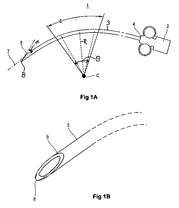
Fig. 1A shows a medical device with a curved needle
Fig. 1B shows the distal end of the needle
Fig. 2A ¨ 2C show the use of the curved needle in a treatment of the lung
Fig. 3A ¨ 3B show the use of the curved needle in another treatment of the
lung
Fig. 4A ¨ 4F show the use of two curved needle in a treatment of the lung
Detailed description of the invention
The invention will now be described on the basis of the drawings. It will be
understood
that the embodiments and aspects of the invention described herein are only
examples and do not limit the protective scope of the claims in any way. The
invention
is defined by the claims and their equivalents. It will be understood that
features of
one aspect or embodiment of the invention can be combined with a feature of a
different aspect or aspects and/or embodiments of the invention.
Fig. 1 shows a medical device 1 for percutaneous biopsy with a handle 2 and a
hollow
needle 3. The handle 2 facilitates manipulations by an operator of the needle
3 and
may for example include finger grips. The needle 3 extends from a proximal end
4 to a
distal end 5 and includes a lumen extending there through. It should be noted
that the
CA 02888715 2015-04-17
WO 2014/060116 PCT/EP2013/003161
terms "proximal" and "distal", as used herein, are intended to refer to a
direct toward
(proximal) and away from (distal) an operator of the biopsy device 1. The
distal end 5 is
cut at an angle a relative to the longitudinal axis 7 of the needle 3 forming
a distal tip 8
(Fig. 1b) and a bevel 9 (Fig. lb) extending there around. The distal tip 8 and
the bevel 9
5 facilitate the penetration of the distal end 5 into tissue.
At least a section 6 of the needle 3 has a curved shape around a virtual
curvature
center C. In case the section 6 corresponds substantially to a circular arc
the distance
between the virtual curvature center C and the needle 3 corresponds to a
radius R of
this circular arc and the section 6 of the needle 3 forms a segment of the
circular arc
extending over a central angle O. Alternatively the full length of the needle
may be
curved. As a function of the dimensions of the organs and bones of the person
to be
treated and the task to be achieved needles may be manufactured and offered
with
different curvature radius R and center angles B. Accordingly also the shape
of the
curved section 6 may vary. The shape could be, but is not limited to, a
segment of a
circle as described, a segment of an ellipse, a segment of a parable or a
segment of a
hyperbole. The shape may vary from any of these forms. The idea of the curved
section 6 of the needle 3 is to allow penetration of tissue in a curved
trajectory.
The needle may be formed of, for example, a polymer, stainless steel, alloys
or any
combination of materials that are suitable to achieve the appropriate
rigidness of the
needle. The needle is hollow, i.e. comprises a lumen through which cutting
devices or
other devices may be applied. The term rigid has to be understood to express
that the
needle is sufficiently rigid so as to be not deflected by effect of impact
upon the
human or animal tissue, when the needle is injected in and pushed through the
tissue.
The needle will keep its curved form to a large extent. For some applications,
the
material and the dimensions of the rigid needle may be chosen so that on the
one
hand it is able to yield appropriately under pressure, yet on the other hand
is
sufficiently flexible to regain at least partially its initial curvature when
the pressure is
released.
CA 02888715 2015-04-17
WO 2014/060116 PCT/EP2013/003161
6
Ideally the plane of the curvature is chosen such that the distal tip 8 of the
needle 3
substantially lies in the plane of the curvature and that the distal tip 8 is
on the same
side of the needle 3 as the curvature center C in respect to the longitudinal
axis 7 of
the needle 3. This configuration makes it easier to force the needle into a
curved
trajectory through the body tissue.
In preparation of the intervention, a patient is placed under an imaging
device (not
shown) such as an X-ray device, a Computer Tomography device (CT), or a
Magnetic
Resonance Imaging device (MRI). The term patient is used to describe a human
or an
animal that is treated by interventional radiology.
Fig. 2A to 2C show the use of a curved needle 10 for a treatment of a human
body. The
figures 2A ¨ 2C represent real images that are displayed to an operator on his
or her
screen. For formal requirements of patent drawings the colour of the real
images have
been inverted in Fig. 2, 3 and 4. The operator will see in a transversal
section of the
patient a vertebra 12, ribs 13, a heart 14, an aorta 15 and lung tissue 16.
Usually lung
tissue cannot be seen on an X-ray device. The lung tissue 16 that is depicted
on the
Figures 2A ¨ 2C has been made visible by applying a radiocontrast agent.
Therefore
only that part of the patient lung can be seen that is affected by the
radiocontrast
agent.
Fig. 2A shows the situation when an operator positions the needle 10 at the
skin 11 of
the patient. As the needle 10 is in front or behind the plane of the X-ray,
the needle 10
that is outside the patient's body cannot be seen on most of the figures. In
the
following, the term operator is used to describe the person who manipulates
the
medical device 1. In most cases this person would be a radiologist with
special
knowledge in intervention with needles. By pushing the handle 2 of the medical
device
1 the tip 8 of the needle 10 cuts through the skin 11 and penetrates through
the
patient's body. The operator follows the advancement of the needle 10 by
requesting
CA 02888715 2015-04-17
WO 2014/060116 PCT/EP2013/003161
7
images from the imaging device and watching these images on a screen. The
needle 10
usually gives a clear image on the screen.
Fig. 2B shows that the needle 10 was entered between the endothoracic fascia
and the
parietal pleural membrane. With a conventional needle, access would be
restricted to
tissue that is straight below this access point. By means of the curved needle
10 it is
however possible to direct the needle 10 to a region that would normally be
out of
reach (Fig. 2C).
As the distal tip 8 is on the same side of the longitudinal axis as the
curvature center C,
the needle can be pushed in a narrower curve than the actual radius of the
curvature.
In order to achieve this goal the operator has to guide the needle 10 such
that the
convex side, e.g. the tip 8 of the needle that is opposite to the curvation
center C in
respect of the longitudinal axis 7 of the needle, is pushed with its bevel 9
(Fig. 1B)
against the body tissue. The edge of the needle 10 props against this
counterforce and
is forced into a smaller curvature. In this respect it has been observed that
the cutting
angle, i.e. the angle a between the longitudinal axis 7 and the plane spanned
by the
, bevel 9 should be flat, e.g. substantially 45 degree or less. A cutting
angle a that is at
least 45 degrees facilitates the needle to be forced into a curved trajectory
when a
force on the handle 2, respectively the proximal end 4 of the needle 3 is
applied.
When the needle is in the intended place the operator in case the medical
device 1 is a
biopsy needle may either insert through the handle 2 a cutting tool to collect
a tissue
sample from the targeted region. The biopsy needle may also be used to insert
instruments for treatment. For example an electrode (not shown) by which the
region
around the needle tip is heated in order to destroy the tissue around the
needle tip,
for example by applying radio frequencies. In case the medical device is an
infiltration
needle it may be used to inject a toxic substance that locally kills the
cancer cells.
CA 02888715 2015-04-17
WO 2014/060116 PCT/EP2013/003161
8
Figures 3A ¨ 3C are photos taken at different instants. In reality the patient
is breathing
and especially the patient's lung is moving. In Figur 3A the curved needle has
approached a cancerous nodule 19 of the lung tissue 16. Due to the breathing
of the
patient the nodule 19 is moving. The operator takes the tip of the needle 10
closer to
the nodule 19 (Fig. 3B). By little turns of the curved needle 10 the operator
is able to
position the tip of the needle such that nodule 19 drives itself into the tip
of the needle
when the patient is breathing (Fig. 3C). With a straight needle it is only
possible to
retract or to push forward whereas the curved needle is able to turn like a
key in a
lock. With this kind of movement it is significantly easier to anticipate the
movement
10 of the nodule with the curved needle.
In another aspect of the invention the needle possesses sufficient rigidity to
reverse
back to its original curved form, even when it is forced temporarily into
another shape.
Fig. 4A ¨ Fig. 4F show another aspect of the invention making use of this
property of
the curved needle according to the invention. A first needle 17 is entered
between the
endothoracic fascia and the parietal pleural membrane so that a tip of the
first needle
17 is extending away from the vertebra (Fig. 4B). Fig. 4C shows the insertion
of a
second needle 18 through the canal of the first needle aiming between the
aorta 15
and the vertebra 12. Through the second needle 17 a volume of 15 ml of serum
is
injected. After the injection of the serum the second needle 18 is retracted.
The serum
creates a little volume of serum that pushes the aorta to the side, so that
more space
for the movement of the first needle is created. The first needle 17 is then
turned anti-
clockwise by 180 so that the curved part is now following the shape of the
vertebra
(Fig. 3D). The first needle is pushed deeper and then turned back clockwise by
180'
after the tip of the first needle has passed the aorta 15. Due to the curved
section of
the first needle 17, when the first needle 17 is pushed further into the body
tissue or
organ, it follows a trajectory that aims at a target that usually would have
been
blocked by the aorta 15.
CA 02888715 2015-04-17
WO 2014/060116 PCT/EP2013/003161
9
The treatment with curved needles is especially advantageous in cases of
mediastinum
lymphadenopathy, abdominal pulmonary masses, small pulmonary nodules and intra-
or retroperitoneal masses. The advantages in case of the pulmonary nodules has
been
already discussed above.
The medical device 1 comprising the curved needle 3 has been presented in the
course
of treatment of cancer. The person skilled in the art however will appreciate
that this is
an example only and that the curved needle may be used for other purposes and
is not
limited to treatment of cancer at all.