Note : Les descriptions sont présentées dans la langue officielle dans laquelle elles ont été soumises.
CA 02891369 2015-05-13
COIL ARRANGEMENT FOR A MAGNETIC
RESONANCE TOMOGRAPHY DEVICE
The invention relates to a magnetic resonance tomography device
for prostate examinations having a patient therein, comprising a
coil for generating a strong homogeneous magnetic field in the
direction of the longitudinal axis of the patient, at least one
transmitter coil for generating an electromagnetic alternating field,
three gradient coils and suitable receiver coils, individual ones of
which are arranged below in the lower back region and/or at the
backside and at least one is arranged above the patient, as well
as data processing for imaging from the signals of the transmitter
and receiver coils.
The early recognition of, in particular, prostate cancer is important
for successful treatment. The normal method of searching for
prostate cancer, such as manual examinations and blood tests,
fail in locating some malignant tumours or sometimes give a false
positive test result. Biopsy is the method of locating a tumour and
assessing its danger. Unfortunately this biopsy often misses the
tumour.
Magnetic resonance tomography is an imaging process with
excellent soft tissue contrast; bones, on the other hand, are not
distinctly imaged. It is therefore used for investigating the
prostate, wherein the best possible resolution is to be obtained.
Magnetic resonance tomography is based on the principles of
nuclear spin resonance (NMR), in particular pulsed field-gradient
NMR, and is therefore also known as nuclear spin tomography.
With the aid of magnetic resonance tomography, cross-sectional
images of the human (or animal) body can be produced, which
make possible an assessment of the organs and many
pathogenic organ changes. Magnetic resonance tomography
requires a very strong static magnetic field and electromagnetic
alternating fields in the radio frequency range, with which
particular atomic nuclei (actually always the hydrogen nuclei) in
2
the body are excited in resonance, which then emit and induce electrical
signals in the receiver electrical circuit. To achieve the local resolution,
gradient coils are used, which serve to generate the magnetic gradient
fields. The gradient coils are used in pairs with the same electrical current
strength but opposite polarity, so that one coil reduces the static magnetic
field, while the opposite coil increases it by the same amount. As a result,
the magnetic field is provided with a linear gradient. Such a device exists
for all three spatial directions. The background is that the local magnetic
field determines the resonance frequency and thereby makes possible
spatial localisation. In the prior art, planar receiver coils are normally
used,
that is to say at least one planar coil element lies beneath the torso of the
patient and a planar coil element lies on the patient. Typically these coil
elements consist in each case of six coils, wherein a preamplifier is
usually present for each coil. These so-called phase-array coils are a
combination of a plurality of surface coils in an array. The idea is, with
relatively small coils that have a good signal-to-noise ratio, nevertheless to
cover a large area. The noise is composed of the thermal noise of the
object to be measured and the thermal noise of the high-frequency coil.
However these receiver coils are, by virtue of the construction, relatively
remote from the prostate. As a result, only a relatively inadequate
resolution of the prostate is ensured. Furthermore, to improve the spatial
resolution, a so-called endorectal coil is already used, which permits a
better spatial resolution of the prostate, since in this case the coil can be
positioned in the direct vicinity of the prostate.
The published application document DE 103 17 629 Al discloses a
magnetic resonance tomography device for prostate examinations, which
consists of a coil for generating a strong homogeneous magnetic field in
the direction of the longitudinal axis of the patient, at least one
transmitter
coil for generating an electromagnetic alternating field, three gradient
coils, a data processing means for imaging from the signals of the
transmitter and receiver coils as well as suitable receiver coils, individual
CA 2891369 2019-07-15
2a
ones of which are arranged below in the lower back region and/or the
posterior and at least one is arranged above the patient.
The published application document DE 102 21 644 Al also discloses an
arrangement of local coils for a magnetic resonance device, which
comprises receiver coils, individual ones of which are arranged below in
the lower back region and/or the posterior and at least one is arranged
above the patient.
It is a feature of one embodiment of the invention to provide a device with
which, without the necessity to use an endorectal coil, the spatial
resolution of the prostate is nevertheless decisively increased.
According to one embodiment, a closed coil is provided, which is
positioned in the direct vicinity of the prostate and encloses the scrotum
and penis.
In accordance with one embodiment of the present invention, there is
provided a magnetic resonance-tomography device for prostate
examinations of a patient therein, the device comprises a coil for
generating a strong homogeneous magnetic field in a direction of a
longitudinal axis of the patient, at least one transmitter coil for generating
an electromagnetic alternating field, three gradient coils and receiver coils,
individual ones of which are arranged below in a lower back region and/or
at a posterior and at least one is arranged above the patient, as well as
data processing means for imaging from signals of the transmitter and
receiver coils, wherein a closed receiver coil is provided, which is
positioned in direct vicinity of the prostate and, bearing against the
patient,
encloses the scrotum and penis.
The coil is thereby positioned in the direct vicinity of the prostate
CA 2891369 2019-07-15
CA 02891369 2015-05-13
3
and thereby permits a better spatial resolution, since it can
intercept more signals from the volume element of the prostate.
This novel coil is used in combination with a conventional planar
coil element on the back of the patient. Actually the patient would
have to be surrounded on all sides with receiver coils to intercept
as much of the signal as possible, that is to say receiver coils
would also be appropriate at the sides. However, because of the
different anatomies of patients' bodies, this is difficult to
implement in practice. In the case of slim patients, a relatively
large circumferential angle of the standard coil element is
covered, that is to say the signal amount that is lost is smaller
since the distance between the upper and lower side of the
patient is smaller; a better resolution is thus achieved here. In the
case of relatively fat patients, the circumferential angle that is
covered by the standard coil element is smaller, that is to say
more signal is lost at the sides. Consequently, the resolution is
worse than in the case of the slim patient. The obvious solution
would be to provide different standard coil elements for different
patients, though this is not practicable.
An optimum image quality can then be achieved if the receiver
coil surrounding the testicles is oriented with its surface normal in
the direction of the prostate. The term "surface normal" is already
not unambiguous for flat coils, since there exists a multiplicity of
surface normals oriented parallel to one another. Since, in the
case of curved coils, it is still the case that the surface normals.
which are defined as perpendicular in the individual points of the
surface, that is to say perpendicular to the tangential plane
extending there, have different orientations, for the unambiguous
determination of the profile of the surface normal, that one should
be selected that runs through the centroid of the surface of the
receiver coil, so that, as a result, a clear instruction for action is
provided.
In a concrete embodiment, three coils are arranged in a triangle
above the patient, that is to say that two coils lie parallel side by
side on the abdomen of the patient and the third coil encloses the
CAA 02891369 2015-05-13
4
scrotum and penis. The third coil lies with the lower longitudinal
sides in a V-shape at the right and left strip, so that the scrotum
and penis pass through the opening of the coil. By spreading the
thighs and with a slight pressure of the coil, an optimum
orientation of the coil element is obtained. The two upper coil
elements are located between the strip and the lower abdominal
wall and can be oriented with their surface normal in the direction
of the prostate.
In an alternative embodiment, the magnetic resonance
tomography coil can also be constructed of more than three coils
on the upper side of the patient. Thereby, the resolution capacity
can be increased since the signal-to-noise ratio is better for
smaller coils. The number of coils, in its totality, defines the
surface area in which signals can be registered and thereby also
evaluated. Here, the outer perimeter of this area, which adds up
to the total of the surface areas of the individual coils, determines
the penetration depth of the entire arrangement, which is formed
from all coils. If a plurality of coils are used together and
simultaneously, a measurement result is obtained which
combines the high sensitivity of the individual coil on one hand
and the high penetration depth of the entire arrangement on the
other hand.
The relative assignment of the coils is in principle arbitrary within
the scope of the invention. The coils can thus be spaced from one
another, which may also be necessary from constructional
constraints. It is to be seen as disadvantageous that signals
emitted in the interstices of the coil cannot be used.
In a preferred case, the coils are directly adjacent to one another,
which has the advantage that as few emitted signals as possible
are lost. The higher the received intensity of the signals, the better
is the image quality.
The laying on and removal are greatly simplified if the coils are
accommodated in a flexible mat. This has the advantage that this
mat largely conforms to the individual body form of the patient.
CA 02891369 2015-05-13
The receiver coils thus lie as close as possible to the patient and
thus permit a better image quality. A passage for the penis and
scrotum must be present in the mat.
The aim is to distinctly image the volume element surrounding
and representing the prostate. For optimization, the radius of the
receiver coil is chosen such that it is larger than or equal to the
average distance of the coil plane from the organ to be examined,
in this case the prostate.
The distance varies from patient to patient; the term "average
distance" is therefore described here as the mean value of
anatomical conditions. The penetration depth depends directly on
the coil radius. The resolution is optimum when the distance of the
organ to be examined from the coil plane corresponds, at
maximum, to the radius of the coil. In principle, the image quality
is all the better the lower the distance from the coil to the prostate
is.
It was recognised as expedient, during the imaging phase, to
spatially position the patient in the region of the abdomen or of the
torso, with a fixture device at least partly enclosing these. The
patient is then retained with the aid of a corset so that no
movements that might cause blurring of the recording are
possible. Due to the fixing, a better image quality is achieved,
since the movement of the patient is restricted and fewer
movement artefacts can occur.
Finally, it is proposed to fasten the receiver coils via adjustment
devices, which permit, in the practical application, the receiver
coils to be optimally oriented in order to obtain a better image
quality in consequence. An expressly recommended possibility
consists in fastening the adjustment devices on the fixing device.
In an advantageous embodiment, a wedge-shaped pillow is used,
which is pushed beneath the pelvis of the patient such that the
pelvis is tilted slightly upwards, which requires a pushing of the
wedge-point in the direction of the longitudinal axis of the patient.
This cushion serves for orientation of the pelvis and therefore the
CAA 02891369 2015-05-13
= 6
orientation of the prostate with respect to the receiver coil.
Through an optimum orientation, a better image quality can be
achieved.
In a further embodiment, a pillow is used that can be charged with
liquid or gas in order thereby to change the shape of the pillow
and thereby optimize the orientation of the pelvis of the patient.
By an appropriate charging of the pillow, an arbitrary orientation of
the pelvis can be achieved in infinitesimal steps and in wide limits.
In one embodiment, the pillow can be subdivided into a plurality of
sectors or chambers, which can be differently charged with gas or
liquid. If a chamber is charged with higher pressure, it is enlarged;
with lower pressure, the chamber in each case is smaller. This
has the advantage that the pelvis of the patient can be oriented in
different spatial directions in an accurately targeted manner by
individually charging and thereby adjusting the individual
chambers. The number of sectors or chambers corresponds to
the number of the adjustment parameters that are available. The
aim is also to improve the image quality here.
Finally, receiver coils can also be accommodated on or in this
pillow. The installation of the receiver coils directly on or in the
pillow permits a closer placement on the patient; this is associated
with a higher resolution and a better signal-to-noise ratio.
In a further embodiment, electrical preamplifiers can be installed
for each coil in order to amplify the signal actually at the coil
where possible. Thereby, the additional relative noise amplitude
due to the wires to the electronics of the magnetic resonance
tomographic device becomes smaller, and thereby the signal
quality and ultimately also the image quality are improved.
Further details and features of the invention are explained below
in greater detail with reference to embodiments shown in the
drawing. In schematic views:
CA 02891369 2015-05-13
7
Figure 1 shows a coil element with patient according to the
invention
Figure 2 shows a coil element with patient as well as a fixing
device
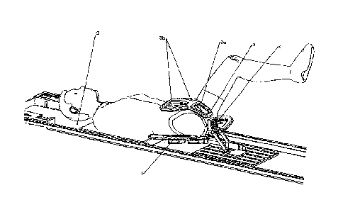
In the 3D representation of Figure 1, the patient (2) is shown
schematically lying on a table. Below the patient, in the lower back
region and on the backside, is located a standard receiver coil
element (1), which is slightly curved so that it adapts somewhat to
the torso of the patient. The standard coil element (1) typically
consists of six individual coils. This arrangement is also called a
phased array.
On the lower abdomen region and on the groin region of the
patient there is located the coil element (3) according to the
invention. This is subdivided into three partial coils, which are
arranged in a triangle. Two coils (3b) are located parallel next to
one another on the lower abdomen region of the patient and are
oriented with their surface normal in the direction of the prostate.
A third coil (3a), which is of decisive importance in conjunction
with the invention, was attached centrally below to these two coils
(3b) so that it encloses the scrotum and penis and bears against
the groin of the patient when the latter slightly spreads his thighs.
This third coils (3a) if V-shaped and is also oriented with its
surface normal in the direction of the prostate. The biopsy device
(4), which is also shown, does not play a role for the invention.
The actual magnetic resonsance tomographic device, that is to
say the coil generating the strong homogeneous magnetic field
and the transmitter coil, is not shown. The gradient coils are also
not illustrated.
In Figure 2, the patient (2) is shown schematically lying down from
an angle of view that is different from that in Figure 1. An
adjusting device (5) is additionally shown. Below the patient, there
is located a standard receiver coil element (1), on which the
patient lies with the lower back region and the backside. Directly
CA 02891369 2015-05-13
8
on the lower abdominal wall and in the groin region of the patient,
there is located the coil element (3) according to the invention.
Towards the head, the two coils (3b) are oriented on the abdomen
of the patient. In the opposite direction, there follows the third coil
(3a), which encloses the penis and scrotum. The coils located
above the patient are fastened on a fixing device (5), which partly
circumscribes the torso of the patient in an arc. Above the two
upper coils (3b), this device is adapted to the form of the coils.
Five adjusting screws (6) permit, by means of adjusting devices,
which are not illustrated, an optimization of the orientation of the
coils (3) relative to the patient (2).
In all the diagrammatic illustrations, essential functional
equipment elements are not shown for reasons of clarity. These
include the homogeneous coil, the gradient coils and the data
processing system necessary for evaluation.
CAA 02891369 2015-05-13
= 9
List of Reference Characters
1 Standard coil element
2 Patient
3 Coils
3a Receiver coil
3b Coils
4 Biopsy device
Fixing device
6 Adjusting screws