Note : Les descriptions sont présentées dans la langue officielle dans laquelle elles ont été soumises.
CALIBRATING ASSAYS USING REACTION TIME
Cross Reference to Related Applications
This patent application claims priority to United States Provisional
Application Number 61/726,626, filed November 15,2012.
Field of the Invention
[0001] The present invention relates to the field of diagnostic assays,
and in
particular to lateral flow assays where an analyte to be detected is present
in a
biological or non-biological sample.
Background
[0002] Diagnostic assays are widespread and central for the diagnosis,
treatment and management of many diseases. Different types of diagnostic
assays
have been developed over the years in order to simplify the detection of
various
analytes in clinical samples such as blood, serum, plasma, urine, saliva,
tissue
biopsies, stool, sputum, skin or throat swabs and tissue samples or processed
tissue
samples. These assays are frequently expected to give a fast and reliable
result,
while being easy to use and inexpensive to manufacture. Understandably it is
difficult to meet all these requirements in one and the same assay. In
practice, many
assays are limited by their speed. Another important parameter is sensitivity.
Recent
developments in assay technology have led to increasingly more sensitive tests
that
allow detection of an analyte in trace quantities as well the detection of
disease
indicators in a sample at the earliest time possible.
[0003] A common type of disposable assay device includes a zone or area
for
receiving the liquid sample, a reagent zone also known as a conjugate zone,
and a
reaction zone also known as a detection zone. These assay devices are commonly
known as lateral flow test strips. They employ a porous material, e.g.,
nitrocellulose,
defining a path for fluid flow capable of supporting capillary flow. Examples
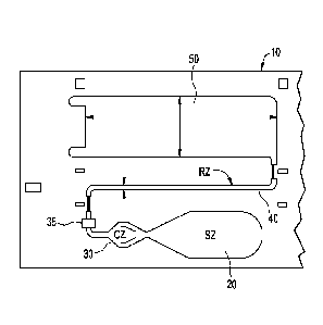
include
those shown in US Patent Nos. 5,559,041, 5,714,389, 5,120,643, and 6,228,660.
[0004] The sample-addition zone frequently consists of a more porous
material,
capable of absorbing the sample, and, when separation of blood cells is
desired, also
CA 2891509 2020-03-24
effective to trap the red blood cells. Examples of such materials are fibrous
materials,
such as paper, fleece, gel or tissue, comprising, e.g., cellulose, wool, glass
fiber,
asbestos, synthetic fibers, polymers, or mixtures of the same.
[0005]
Another type of assay device is a non-porous assay having projections to
induce capillary flow. Examples of such assay devices include the open lateral
flow
device as disclosed in WO 2003/103835, WO 2005/089082, WO 2005/118139, and WO
2006/137785.
[0006] A
known non-porous assay device is shown in Figure 1. The assay device
1, has at least one sample addition zone 2, a reagent zone 3, at least one
detection
zone 4, and at least one wicking zone 5. The zones form a flow path by which
sample
flows from the sample addition zone to the wicking zone. Also included are
capture
elements, such as antibodies, in the detection zone 4, capable of binding to
the analyte,
optionally deposited on the device (such as by coating); and a labeled
conjugate
material also capable of participating in reactions that will enable
determination of the
concentration of the analyte, deposited on the device in the reagent zone,
wherein the
labeled conjugate material carries a label for detection in the detection
zone. The
conjugate material is dissolved as the sample flows through the reagent zone
forming a
conjugate plume of dissolved labeled conjugate material and sample that flows
downstream to the detection zone. As the conjugate plume flows into the
detection
zone, the conjugated material will be captured by the capture elements such as
via a
complex of conjugated material and analyte (as in a "sandwich" assay) or
directly (as in
a "competitive" assay . Unbound dissolved conjugate material will be swept
past the
detection zone into the at least one wicking zone 5. Also shown in Figure 1
are
projections or micropillars 7. An
instrument such as that disclosed in US
20060289787A1, U520070231883A1, US 7,416,700 and US 6,139,800, are able to
detect the bound conjugated material in the detection zone. Common labels
include
fluorescent dyes that can be detected by instruments which excite the
fluorescent dyes
and incorporate a detector capable of detecting the fluorescent dyes.
[0007] In
order to produce a reportable result from a measured signal, e.g., a
fluorescent signal, a calibration curve needs to be formulated to correlate
the measured
signal to the concentration of the analyte of interest in the sample being
analyzed.
Developing a calibration curve is well known in the art and does not need
detailed
explanation. Briefly, multiple samples having known varying concentrations of
analyte
2
CA 2891509 2020-03-24
CA 02891509 2015-05-13
WO 2014/078679
PCT/US2013/070341
(also called calibrator fluids) are run on an assay device in a manner similar
to an end
user performing an assay on a sample having an unknown concentration of
analyte.
The signal produced by the analyte signal producing complex, such as an
analyte/labeled antibody conjugate, is read and recorded as a data point for
each of the
multiple samples. The data points are plotted on a curve of concentration
versus signal
strength. The data points can then be curve fit into an equation that provides
analyte
concentration as a function of signal strength for that particular assay. For
example, a
linear correlation can be represented by C = mS b, where C is the
concentration of
the analyte, S is the measured signal and m and b are experimentally
determined
constants. Non-linear correlations can be represented with non-linear
mathematical
models such as the logitI1og4 relationship.
[0008] For many commercially available assays, the calibration curve is
developed
at the factory where the assay is made. Due to variation in raw materials and
other
factors when an assay is made, a different lot-specific calibration curve may
be
developed for each lot of assays produced. The calibration curve data can then
be
included in each lot of assay sold to an end user. Alternatively, a
calibration curve is
automatically created by the customer's analyzer when the customer runs a
calibration
process with the lot of assay material, their analyzer, and a series of
calibrator materials
provided by the manufacturer.
[0009] When making a calibration curve at a factory, a standard calibrator
fluid is
used to approximate the characteristics of the actual samples that will be
used by the
end user of the assay device. For example, if the assay will be used with
plasma
samples, the calibrator fluid will generally be formulated to mimic the
characteristics of a
typical plasma sample. This should result in the unknown concentration of the
analyte
in the sample being assayed being the same as the concentration of the analyte
in the
calibrator fluid for equivalent measured signals.
[0010] A typical measured signal for lateral flow assays is a peak height
or peak
area of a trace of fluorescent intensity vs. distance along the detection
zone. However,
it is sometimes the case where the measured signal in sample does not depend
on
concentration alone. Other factors, particularly for capillary driven lateral
flow assays
devices, in addition to analyte concentration, can affect the measured signal.
If these
factors are not taken into account, then the measured signal for a sample
being
assayed will not accurately correlate to the true concentration of analyte in
the sample.
This of course, can have profound effects, e.g., on a patient's diagnosis or
prognosis.
3
CA 02891509 2015-05-13
WO 2014/078679
PCT/US2013/070341
[0011] Accordingly, there is a need for a method that can take into account
other
factors that affect the measured signal of a capillary driven lateral flow
assay device.
Summary of the Invention
[0012] The present invention is directed to an assay device that alleviates
one or
more the foregoing problems described above.
[0013] One aspect of the invention is directed to a method for performing
an assay
on a liquid sample for the detection of one or more analytes of interest in an
assay
device having a flow path which includes a sample zone and detection zone
thereon.
The method includes: dispensing the sample onto the sample zone; combining the
sample and a reagent, wherein the sample and reagent may be combined prior to
addition of the sample to the sample zone or on the assay device, flowing the
combined
sample/reagent by capillary action into and through the detection zone having
capture
elements bound thereto, wherein a signal at least partially representative of
the
presence or concentration of analyte(s) is produced and detected; determining
a
reaction time; and determining the concentration of the analyte by using both
the
detected signal and the reaction time.
[0014] Another aspect of the invention is directed to a method of
calibrating an
assay. The method includes: (a) providing multiple calibrator fluids having
known
concentrations of analyte therein, (b) providing an assay device having a
substrate that
include a sample zone and detection zone, wherein the method further includes:
(c)
dispensing one of the calibrator fluids onto the sample zone; (d) combining
the
calibrator fluid and a reagent, wherein the calibrator fluid and reagent may
be combined
prior to addition of the calibrator fluid to the sample zone or on the assay
device, (e)
flowing the combined calibrator fluid /reagent by capillary action into and
through the
detection zone having capture elements bound thereto, wherein a signal at
least
partially representative of the presence or concentration of analyte(s) is
produced and
detected; (f) determining a reaction time; (g) repeating steps (b)-(f) for
each calibrator
fluid; and (h) using the detected signal S, the reaction time t and the known
concentrations C to determine a calibration curve.
[0015] According to another aspect of the invention, there has been
provided a
method for performing an assay on a liquid sample for the detection of one or
more
analytes of interest in an assay device having a flow path which includes a
sample zone
and detection zone thereon. The method includes: dispensing the sample onto
the
4
sample zone; combining the sample and a reagent, wherein the sample and
reagent
may be combined prior to addition of the sample to the sample zone or on the
assay
device, flowing the combined sample/reagent by capillary action into and
through the
detection zone having capture elements bound thereto, wherein a signal at
least
partially representative of the presence or concentration of analyte(s) is
produced and
detected; determining a reaction volume; and determining the concentration of
the
analyte by using both the detected signal and the reaction volume.
[0016] According to still another aspect of the invention, there has
been provided a
method of calibrating an assay. The method includes: (a) providing multiple
calibrator
fluids having known concentrations of analyte therein; (b) providing an assay
device
having a substrate that include a sample zone and detection zone: (c)
dispensing one
of the calibrator fluids onto the sample zone; (d) combining the calibrator
fluid and a
reagent, wherein the calibrator fluid and reagent may be combined prior to
addition of
the calibrator fluid to the sample zone or on the assay device, (e) flowing
the combined
calibrator fluid /reagent by capillary action into and through the detection
zone having
capture elements bound thereto, wherein a signal at least partially
representative of the
presence or concentration of analyte(s) is produced and detected; (f)
determining a
reaction volume; (g) repeating steps (b)-(f) for each calibrator fluid; (h)
using the
detected signal S, the reaction volume and the known concentrations C to
determine a
calibration curve.
[0016A] In one aspect, there is provided a method for performing an assay
on a
liquid sample for the detection of one or more analytes of interest in an
assay device
having a flow path which includes a sample zone and detection zone thereon.
The
method comprises: dispensing the sample onto the sample zone; combining the
sample and a reagent, wherein the sample and reagent may be combined prior to
addition of the sample to the sample zone or on the assay device, flowing the
combined sample/reagent by capillary action into and through the detection
zone
having capture elements bound thereto that bind the one or more analytes,
wherein a
signal representative of the presence or concentration of the one or more
analytes is
produced by a detection element that is associated with the reagent or
generated
through a reaction with the reagent; determining a reaction time using signals
generated by detection elements at points in the flow path; and determining
the
concentration of the one or more analytes by dividing at least one of the
detected
signals by the reaction time.
CA 2891509 2020-03-24
[0017] Further objects, features and advantages of the present invention
will be
apparent to those skilled in the art from detailed consideration of the
preferred
embodiments that follow.
Brief Description of the Drawings
[0018] Figure 1 shows a schematic view of a known assay device usable
with the
present invention.
[0019] Figure 2 shows a schematic view of an assay device usable with
the
present invention.
[0020] Figure 3 shows a schematic view of an assay device usable with
the
present invention.
[0021] Figure 4 is a graph showing the relationship between total flow
time in an
assay device and viscosity.
5a
CA 2891509 2020-03-24
CA 02891509 2015-05-13
WO 2014/078679
PCT/US2013/070341
[0022] Figure 5 is a graph showing the relationship between signal strength
and
reagent dissolution time for a constant concentration of NT proBNP.
[0023] Figure 6 is a graph which shows signal strength as a function of
concentration and fluid types.
[0024] Figure 7 is a graph showing the relationship between reaction volume
and
reaction time.
[0025] Figure 8 is a graph showing the relationship between flow rate and
reaction volume.
[0026] Figure 9 is a graph showing the relationship between conjugate
(i.e.,
reagent) dissolution time and total flow time.
[0027] Figures 10A and 1013 are graphs which show the difference between
different fluids having no correction for reaction time and having correction
for reaction
time.
[0028] Figures 11A and 116 shows predicted concentration vs. actual
concentration for calibration models that account for reaction time and do not
account
for reaction time.
Detailed Description of Preferred Embodiments
[0029] As used in this specification and the appended claims, the singular
forms
"a", "an" and "the" include plural referents unless the context clearly
dictates
otherwise.
[0030] The term "about" as used in connection with a numerical value
throughout the description and the claims denotes an interval of accuracy,
familiar
and acceptable to a person skilled in the art. The interval is preferably 10
%.
[0031] The term "sample" herein means a volume of a liquid, solution or
suspension, intended to be subjected to qualitative or quantitative
determination of
any of its properties, such as the presence or absence of a component, the
concentration of a component, etc. Typical samples in the context of the
present
invention are human or animal bodily fluids such as blood, plasma, serum,
lymph,
urine, saliva, semen, amniotic fluid, gastric fluid, phlegm, sputum, mucus,
tears,
stool, etc. Other types of samples are derived from human or animal tissue
samples
where the tissue sample has been processed into a liquid, solution, or
suspension to
reveal particular tissue components for examination. The embodiments of the
6
CA 02891509 2015-05-13
WO 2014/078679
PCT/US2013/070341
present invention are applicable to all bodily samples, but preferably to
samples of
whole blood, urine or sputum.
[0032] In other instances, the sample can be related to food testing,
environmental testing, bio-threat or bio-hazard testing, etc. This is only a
small
example of samples that can be used in the present invention.
[0033] The determination based on lateral flow of a sample and the
interaction
of components present in the sample with reagents present in the device or
added to
the device during the procedure and detection of such interaction, either
qualitatively
or quantitatively, may be for any purpose, such as diagnostic purposes. Such
tests
are often referred to as lateral flow assays.
[0034] Examples of diagnostic determinations include, but are not limited
to, the
determination of analytes, also called markers, specific for different
disorders, e.g.
chronic metabolic disorders, such as blood glucose, blood ketones, urine
glucose
(diabetes), blood cholesterol (atherosclerosis, obesity, etc); markers of
other specific
diseases, e.g. acute diseases, such as coronary infarct markers (e.g. troponin-
T, NT-
ProBNP), markers of thyroid function (e.g. determination of thyroid
stimulating
hormone (TSH)), markers of viral infections (the use of lateral flow
immunoassays for
the detection of specific viral antibodies); etc.
[0035] Yet another important field is the field of companion diagnostics
where a
therapeutic agent, such as a drug, is administered to an individual in need of
such a
drug. An appropriate assay is then conducted to determine the level of an
appropriate marker to determine whether the drug is having its desired effect.
Alternatively, the assay device usable with the present invention can be used
prior to
administration of a therapeutic agent to determine if the agent will help the
individual
in need.
[0036] Yet another important field is that of drug tests, for easy and
rapid
detection of drugs and drug metabolites indicating drug abuse; such as the
determination of specific drugs and drug metabolites (e.g. THC) in urine
samples etc.
[0037] The term "analyte" is used as a synonym of the term "marker and
intended to encompass any chemical or biological substance that is measured
quantitatively or qualitatively and can include small molecules, proteins,
antibodies,
DNA, RNA, nucleic acids, virus components or intact viruses, bacteria
components or
intact bacteria, cellular components or intact cells and complexes and
derivatives
thereof.
7
CA 02891509 2015-05-13
WO 2014/078679
PCT/US2013/070341
[0038] The terms "zone", "area" and "site" are used in the context of this
description, examples and claims to define parts of the fluid flow path on a
substrate,
either in prior art devices or in a device according to an embodiment of the
invention.
[0039] The term "reaction" is used to define any reaction, which takes
place
between components of a sample and at least one reagent or reagents on or in
the
substrate, or between two or more components present in the sample. The term
"reaction" is in particular used to define the reaction, taking place between
an analyte
and a reagent as part of the qualitative or quantitative determination of the
analyte.
[0040] The term "substrate" means the carrier or matrix to which a sample
is
added, and on or in which the determination is performed, or where the
reaction
between analyte and reagent takes place.
[0041] Figures 2 and 3 show a schematic view of a preferred embodiment of
such
devices according to the invention. The assay device 10, has at least one
sample
addition zone 20, at least one reagent zone 30, at least one detection zone
40, and at
least one wicking zone 50. The zones form a flow path by which sample flows
from the
sample addition zone to the wicking zone. Also included are capture elements
in the
detection zone 40, capable of binding to the analyte, optionally deposited on
the device
(such as by coating); and a labeled reagent material also capable of binding
to the
analyte or the capture element, located on the device in the reagent zone,
wherein the
labeled reagent material carries a first label for detection in the detection
zone.
[0042] Components of an assay device (i.e., a physical structure of the
device
whether or not a discrete piece from other parts of the device) usable in the
present
invention can be prepared from copolymers, blends, laminates, metalized foils,
metalized films or metals. Alternatively, device components can be prepared
from
copolymers, blends, laminates, metalized foils, metalized films or metals
deposited
one of the following materials: polyolefins, polyesters, styrene containing
polymers,
polycarbonate, acrylic polymers, chlorine containing polymers, acetal
homopolymers
and copolymers, cellulosics and their esters, cellulose nitrate, fluorine
containing
polymers, polyamides, polyimides, polymethylmethacrylates, sulfur containing
polymers, polyurethanes, silicon containing polymers, glass, and ceramic
materials.
Alternatively, components of the device are made with a plastic, elastomer,
latex,
silicon chip, or metal; the elastomer can comprise polyethylene,
polypropylene,
polystyrene, polyacrylates, silicon elastomers, or latex. Alternatively,
components of
the device can be prepared from latex, polystyrene latex or hydrophobic
polymers;
8
the hydrophobic polymer can comprise polypropylene, polyethylene, or
polyester.
Alternatively, components of the device can comprise TEFLON , polystyrene,
polyacrylate, or polycarbonate. Alternatively, device components are made from
plastics which are capable of being embossed, milled or injection molded or
from
surfaces of copper, silver and gold films upon which may be adsorbed various
long
chain alkanethiols. The structures of plastic which are capable of being
milled or
injection molded can comprise a polystyrene, a polycarbonate, or a
polyacrylate. In a
particularly preferred embodiment, the assay device is injection molded from a
cyclo
olefin polymer, such as those sold under the name Zeonor . Preferred injection
molding techniques are described in U.S. Patent Nos. 6,372,542, 6,733,682,
6,811,736, 6,884,370, and 6,733,682.
[0043] In one embodiment, the flow path is non-porous and can include
open or
closed paths, grooves, and capillaries. In one preferred embodiment, the flow
path
comprises a lateral flow path of adjacent projections, having a size, shape
and
mutual spacing such that capillary flow is sustained through the flow path. In
one
embodiment, the flow path is in a channel within the substrate having a bottom
surface and side walls. In this embodiment, the projections protrude from the
bottom
surface of the channel. The side walls may or may not contribute to the
capillary
action of the liquid. If the sidewalls do not contribute to the capillary
action of the
liquid, then a gap can be provided between the outermost projections and the
sidewalls to keep the liquid contained in the flow path defined by the
projections.
Figure 1 shows projections 7.
[0044] In one embodiment the flow path is at least partially open. In
another
embodiment the flow path is entirely open. Open means that there is no lid or
cover at a
capillary distance. Thus the lid, if present as a physical protection for the
flow path,
does not contribute to the capillary flow in the flow path. An open lateral
flow path is
described for example in the following published applications: WO 2003/103835,
WO
2005/089082; WO 2005/118139; WO 2006/137785; and WO 2007/149042. The
projections have a height (H), diameter (D) and a distance or distances
between the
projections (t1, t2) such, that lateral capillary flow of the fluid, such as
plasma,
preferably human plasma, in the zone is achieved. These dimensions are shown
in US
2006/0285996. In addition to optimizing the above-mentioned height,
9
CA 2891509 2020-03-24
diameter and a distance or distances between the projections, the projections
may be
given a desired chemical, biological or physical functionality, e.g. by
modifying the
surface of the projections. In one embodiment, the projections have a height
in the
interval of about 15 to about 150 pm, preferably about 30 to about 100 pm, a
diameter
of about 10 to about 160 pm, preferably 30 to about 100 pm, and a gap or gaps
between the projections of about 3 to about 200 pm, preferably 5 to 50 pm from
each
other. The flow channel may have a length of about 2 to about 100 mm,
preferably
about 5 to about 50 mm, and a width of about 0.1 to about 5 mm, preferably
about 0.5
to 1.2 mm.
[0045] In another embodiment, the flow path is porous and includes a
porous
material, e.g., nitrocellulose, defining the flow path capable of supporting
capillary flow.
Examples include those shown in US Patent Nos. 5,559,041, 5,714,389,
5,120,643,
and 6,228,660.
[0046] The liquid sample zone 20, also referred to as the liquid sample
addition
zone, receives sample from a sample dispenser, such as a pipette. The sample
is
typically deposited onto the top of the zone. The sample addition zone is
capable of
transporting the liquid sample from the point where the sample is deposited to
the
reagent zone, through an optional filter and reagent addition zone, preferably
through
capillary flow. The capillary flow inducing structure can include porous
materials,
such as nitrocellulose, described above, or preferably through projections,
such as
micro-pillars, as shown in Figure 1 and also described above. In those devices
that
can use finger stick volumes of blood, the sample can be directly touched off
from the
finger, or by a capillary pipette.
[0047] A filter material (not shown) can be placed in the sample
addition zone to
filter particulates from the sample or to filter blood cells from blood so
that plasma
can travel further through the device.
[0048] Located between the sample addition zone and the detection zone
is a
reagent zone 30. The reagent zone can include reagent(s) integrated into the
analytical element and are generally reagents useful in the reaction¨binding
partners such as antibodies or antigens for immunoassays, substrates for
enzyme
assays, probes for molecular diagnostic assays, or are auxiliary materials
such as
materials that stabilize the integrated reagents, materials that suppress
interfering
reactions, etc. Generally one of the reagents useful in the reaction bears
a
detectable signal as discussed below. In some cases the reagents may react
with
CA 2891509 2020-03-24
CA 02891509 2015-05-13
WO 2014/078679
PCT/US2013/070341
the analyte directly or through a cascade of reactions to form a detectable
signal
such as, but not restricted to, a molecule detectable using spectroscopy such
as a
colored or fluorescent molecule. The amount of reagent in the reagent zone can
be
adjusted by the length of reagent deposited into the device while maintaining
the
same reagent width. The amount of reagent can also be adjusted by changing the
width while maintaining the length. The amount of reagent can further be
adjusted by
changing both width and length simultaneously. In one preferred embodiment,
the
reagent zone includes conjugate material. The term conjugate means any moiety
bearing both a detection element and a binding partner. Alternatively, the
reagents,
including the detection element and conjugate, can be added to the sample
prior to
addition to the sample addition zone. If all reagents are combined with the
sample
prior to the sample addition zone, then of course, a separate reagent zone
will not be
necessary.
[0049] The detection element is an agent which is detectable with respect
to its
physical distribution or/and the intensity of the signal it delivers, such as
but not
limited to luminescent molecules (e.g. fluorescent agents, phosphorescent
agents,
chemiluminescent agents, bioluminescent agents and the like), colored
molecules,
molecules producing colors upon reaction, enzymes, radioisotopes, ligands
exhibiting
specific binding and the like. The detection element also referred to as a
label is
preferably chosen from chromophores, fluorophores, radioactive labels, and
enzymes. Suitable labels are available from commercial suppliers, providing a
wide
range of dyes for the labeling of antibodies, proteins, and nucleic acids.
There are,
for example, fluorophores spanning practically the entire visible and infrared
spectrum. Suitable fluorescent or phosphorescent labels include for instance,
but are
not limited to, fluoresceins, Cy3, Cy5 and the like. Suitable chemoluminescent
labels
are for instance but are not limited to luminol, cyalume and the like.
[0050] Similarly, radioactive labels are commercially available, or
detection
elements can be synthesized so that they incorporate a radioactive label.
Suitable
radioactive labels are for instance but are not limited to radioactive iodine
and
phosphorus; e.g. 1251 and 32P.
[0051] Suitable enzymatic labels are, for instance, but are not limited to,
horseradish peroxidase, beta-galactosidase, luciferase, alkaline phosphatase
and the
like. Two labels are "distinguishable" when they can be individually detected
and
preferably quantified simultaneously, without significantly disturbing,
interfering or
11
quenching each other. Two or more labels may be used, for example, when
multiple
analytes or markers are being detected.
[0052] The binding partner is a material that can form a complex that
can be
used to determine the presence of or amount of an analyte. For example, in an
"sandwich" assay, the binding partner in the conjugate can form a complex
including
the analyte and the detection element and that complex can further bind to
another
binding partner, also called a capture element, integrated into the detection
zone. In
a competitive immunoassay, the analyte will interfere with binding of the
binding
partner in the conjugate to another binding partner, also called a capture
element,
integrated into the detection zone. Example binding partners included in
conjugates
include antibodies, antigens, analyte or analyte-mimics, protein, etc.
[0053] Optionally located in the fluid flow path, before or after the
reagent zone
and before the detection zone is a reagent addition zone. The reagent addition
zone
is shown as 35 in Figures 2 and 3. The reagent addition zone can allow
addition of a
reagent externally from the device. For example, the reagent addition zone may
be
used to add an interrupting reagent that may be used to wash the sample and
other
unbound components present in the fluid flow path into the wicking zone. In a
preferred embodiment the reagent addition zone 35 is located after the reagent
zone
30. According to a preferred embodiment, the reagent plume from the reagent
zone
should be as wide as possible to cover as much of the width of the detection
zone as
possible. One preferred embodiment for increasing the width of the reagent
plume is
described in co-pending application entitled "Assay Device Having Multiple
Reagent
Cells" (Serial No. 61/588738, Attorney Docket No. CDS5104USPSP, first named
inventor: Zhong Ding) filed January 20, 2012. In summary, multiple areas
having
reagent material (hereinafter referred to as "reagent cells") in a reagent
zone along
with elements to recombine multiple flow streams that result from the multiple
reagent cells into one flow stream results in a more desirably mixed, wider
reagent
plume as it leaves the reagent zone and enters the detection zone.
[0054] Downstream from the liquid sample zone and the reagent zone is
the
detection zone 40 which is in fluid communication with the sample addition
zone. The
detection zone 40 may include projections such as those described above. As
also
noted above, these projections are preferably integrally molded into the
substrate from
an optical plastic material such as Zeonor, such as injection molding or
embossing.
12
CA 2891509 2020-03-24
The width of the flow channel in the detection zone is typically on the order
of 2mm for
conventional size devices, however, some lower volume devices, such as those
described above and in co pending application entitled "Lower Volume Assay
Device
Having Increased Sensitivity" (Application No. 61/588758, Attorney Docket No.
CDS5111USPSP, first named inventor: Phil Hosimer) filed January 20, 2012, are
significantly narrower, e.g., 1.5 mm or less.
[0055] The
detection zone is where any detectable signal is read. In a preferred
embodiment attached to the projections in the detection zone are capture
elements.
The capture elements can include binding partners for the reagent or complexes
containing the conjugate, as described above. For example, if the analyte is a
specific protein, the conjugate may be an antibody that will specifically bind
that
protein coupled to a detection element such as a fluorescence probe. The
capture
element could then be another antibody that also specifically binds to that
protein. In
another example, if the marker or analyte is DNA, the capture molecule can be,
but
is not limited to, synthetic oligonucleotides, analogues thereof, or specific
antibodies.
Other suitable capture elements include antibodies, antibody fragments,
aptamers,
and nucleic acid sequences, specific for the analyte to be detected. A non-
limiting
example of a suitable capture element is a molecule that bears avidin
functionality
that would bind to a conjugate containing a biotin functionality. The
detection zone
can include multiple detection zones. The multiple detection zones can be used
for
assays that include one or more markers. In the event of multiple detection
zones,
the capture elements can include multiple capture elements, such as first and
second
capture elements. As noted above, the conjugate can be pre-deposited on the
assay
device, such as by coating in the reagent zone. Similarly the capture elements
can
be pre-deposited on the assay device on the detection zone. Preferably, both
the
detection and capture elements are pre-deposited on the assay device, on the
detection zone and reagent zone, respectively.
[0056] After the sample has been delivered to the sample zone, it will
encounter the
reagent zone. After the sample has flowed through and interacted with the
reagent zone
and optionally the reagent addition zone, the sample and a reagent plume will
be
contained in the fluid flow. The reagent plume can contain any of the reagent
materials
that have been dissolved in the reagent zone or those added through the
reagent
addition zone. The reagent plume can include the conjugate having both the
detection
element and binding partner, in which case it is often referred to as a
conjugate plume.
13
CA 2891509 2020-03-24
CA 02891509 2015-05-13
WO 2014/078679 PCT/US2013/070341
[0057] As described above, a problem facing the inventors and others in the
art was to
accurately and precisely correlate the measured signal in a lateral flow assay
to the
actual concentration of analyte in the sample. The
predicted concentration of an
analyte in the sample based on a calibration curve performed prior to the
actual analysis,
such as at the factory sometimes deviated from the actual concentration,
sometimes by
significant amounts. Something other than concentration of analyte was
affecting the
signal being measured by the instrument. As described above, in a lateral flow
assay
the analyte containing sample is combined with a detection element upstream
from a
detection zone, where it then flows into the detection zone and the detection
element or
a complex containing the detection element is captured and the signal produced
by the
detection element is measured by the instrument.
[0058] Samples that differ in viscosity will have differing flow rates or flow
times in
lateral flow devices relying on capillary forces to drive fluid flow. Figure 4
shows the
linear relationship between viscosity and total flow time for the lateral flow
device shown
in Figures 2 and 3. The present inventors have found that in lateral flow
assay device,
signal generated by two samples of equal concentration but having differing
flow times
will be different, assuming all other conditions are the same, .e.g.. same
device design,
amount of detection element deposited, etc. This is believed to be due to the
time the
detection element (generally conjugated with a binding partner as described
above) is
allowed to interact with analyte in the sample (for sandwich assays), and the
time the
detection element or a complex containing the detection element is allowed to
interact
with and bind with the capture element in the detection zone.
[0059] As explained above, in a lateral flow assay device the sample
containing the
detection element in the reagent plume flows into the detection zone. As the
sample
flows through the detection zone, the detection element, which is a function
of analyte
concentration, is brought into contact with the capture elements mainly
through diffusion.
This diffusion occurs as the detection element in the flow stream passes by
those
features (e.g., microposts or fibers) of the lateral flow device having the
capture element
bound thereto. After sample containing the detection element (e.g., the
conjugate
plume) passes through the detection zone, remaining sample or an added wash
passes
through the detection zone and washes unbound detection element out of the
detection
zone. The bound detection element is then read by the instrument and the
signal is
recorded. The ability of the detection element to diffuse from the flow stream
to the
bound capture element will depend on many factors, chief among them, is the
amount of
14
CA 02891509 2015-05-13
WO 2014/078679 PCT/US2013/070341
time the conjugated detection element is in proximity to the bound capture
element. As
noted above, the amount of time the detection element (directly or otherwise)
has to
interact with the analyte can also affect signal, all though to a much lesser
degree than
the interaction with the capture element. Thus, a longer flow time will put
the detection
element in proximity to the capture element for a longer time and thus,
provide more
opportunity for the conjugated detection element to diffuse to the bound
capture
element. The amount of detection element that binds to the capture element
will then
depend on the concentration of analyte, and the ability of the detection
element to
diffuse from the flow stream to the fixed capture element. The signal measured
or
detected by the instrument depends on the amount of conjugated detection
element in
the detection zone.
[0060] If the fluid characteristics (e.g., viscosity) between the calibrator
fluid used to
calibrate the assay and the sample fluid being assayed are different, a
different amount
of signal will result even for the same concentration of analyte. This being
due to the
differing ability of (i.e., amount of time) the conjugated detection element
to diffuse from
the flow stream to the fixed capture element as described above. Accordingly,
the
present inventors determined that a calibration curve needed to be able to
take into
account fluid flow time through the assay device and its effect on signal
strength, and not
only on analyte concentration. Thus, instead of detected signal S being a
function of
concentration C only (S=f(C)), the detected signal is a function of both
concentration and
flow time t (S=g(C,W. Accordingly, a broad aspect of the invention provides a
method
for determining the concentration of an analyte in a sample that accounts for
both
concentration and flow time.
[0061] The flow time can be represented by the term "reaction time" which is
broadly
defined as the time that a detection element has to bind with the capture
element in the
detection zone. In preferred embodiments, the reaction time itself can be
measured
using signal generated by the detection element at points in the flow path and
detected
by the same instrument used to detect signal used to generate a reportable
result.
However, other methods for determining reaction time that do not rely on
detecting
signal provided by the detection element can also be used.
[0062] There are several determinable methods for measuring a representation
of
reaction time. One is the "reagent dissolution time," which is the time the
combined
sample/reagent is first detected at a point along the substrate flow path
after the reagent
CA 02891509 2015-05-13
WO 2014/078679 PCT/US2013/070341
zone to the time when the combined sample/reagent is no longer detected at
that point
in the substrate.
[0063] Another time proportional to or representative of the reaction time can
be
obtained by measuring the total flow time, which is the time it takes the
sample to flow
from the sample zone to the end of the wicking zone.
[0064] Another time proportional to or representative of the reaction time can
also be
obtained by measuring the total wick time, which is the time between sample
entering
the wicking zone to reaching the end of the wicking zone. This can be done by
detecting
signal from detection element that is washed out of the detection zone.
[0065] Alternatively, a time proportional to the reaction time can determined
as the
wetting time, which is the time required for a sample to completely penetrate
throughout
the lateral flow device.
[0066] Finally, a rate inversely proportional to the reaction time can also be
determined
by simply measuring the flow rate of the sample moving through the assay
device. The
flow rate can be measured by any known method, such as measuring the time
required
for the flow front to pass from one point on the device to another point on
the device of
known distance from the first point. For any of the above techniques to
measure a
representation of the reaction time, the detection element that is measured to
generate a
reportable result, flowing through the assay device can also be used in the
measurement
of the reaction time. This has the advantages of the ability to use the same
instrument
that is used to measure the signal which is representative of the analyte in
the sample to
measure the reaction time as well. No additional detectable agents, such as
different
labels are required. However, in some embodiments, it may be desirable to read
another signal, such as using an infrared detector to detect when a portion of
the assay
device is wetted, etc.
[0067] Figure 5 graphically depicts the effect that reagent dissolution time
has on the
amount of signal generated by samples having the same concentration of NT-
proBNP (a
sandwich assay) but with varying reagent dissolution rates (i.e., reaction
times) caused
by differing fluid viscosities. In this example, samples containing the same
concentration
of NT-proBNP but of differing fluid viscosities were prepared by dissolution
of varying
amounts of the polymer polyvinylpyrrolidone (PVP) into different aliquots of
the fluid to
represent patient serum samples of differing viscosity. As Figure 5 shows,
increases in
reagent dissolution time results in an increased in measured signal for the
same
concentration of analyte.
16
CA 02891509 2015-05-13
WO 2014/078679 PCT/US2013/070341
[0068] Figure 6 shows another example of the effect of sample type on reaction
time
and hence on signal strength. In Figure 6, varying concentrations of intact
parathyroid
hormone (iPTI-1), a sandwich assay, in serum (having a higher viscosity and
hence
longer reaction time) and buffer (lower viscosity, shorter reaction time) are
plotted
against signal strength. As shown in the figure, a significantly smaller
signal is produced
in the buffer solution as compared to the serum for the same concentration.
Thus, as
evident in Figures 5 and 6, there is a need for a method of performing an
assay that
takes into account both concentration and reaction time. In other words, a
method is
needed that minimizes the impact of reaction time variability between samples
having
differing fluid characteristics on the calculated concentration of the
analyte.
[0069] In addition to reaction time having an effect on the detected signal,
the present
inventors also discovered further factors that can affect the detected signal
for a given
concentration of analyte. Specifically, in addition to the reaction time,
reaction volume
also affects the detected signal, especially for the competitive assay since a
change in
reaction volume leads to change in the dissolved reagent concentration (total
amount of
deposited reagent is the same). In a preferred embodiment, the reaction volume
is
determined as the product of flow rate and reaction time. When a sample is
applied to an
assay device, the sample will first contact the reagent in the reagent zone.
As the
sample flows through the reagent zone, it will dissolve reagent. Upon
dissolution of the
reagent, the remaining sample flowing through the reagent zone will no longer
encounter
reagent, and hence. will be reagent free. This portion of sample will be used
to wash
unbound reagent from the detection zone. Thus, there are two portions of
sample. The
sample used to dissolve the reagent in the reagent zone and the following
portion of
sample that is reagent free and is used as a wash. The portion of sample used
to
dissolve the reagent is called the reaction volume. As noted
above, the reaction
volume is preferably determined as a product of reaction time and flow rate
(reaction
volume = flow rate X reaction time). Thus, the discovery of reaction volume is
the
recognition that for some assays (e.g., competitive assay) the detected signal
is
dependent on reaction volume as well as analyte concentration.
[0070] Figure 7 shows the relationship between reaction volume and reaction
time. As
Figure 7 demonstrates an increased reaction volume results in a decreased
reaction
time.
[0071] Figure 8 shows the relationship between flow rate and reaction volume.
As
Figure 8 demonstrates, an increased flow rate results in an increased reaction
volume.
17
CA 02891509 2015-05-13
WO 2014/078679 PCT/US2013/070341
[0072] Figure 9 shows the relationship between total flow time and conjugate
dissolution time (i.e., reaction time). As Figure 9 demonstrates, an increased
total flow
time results in an increased conjugate dissolution time. Thus Figures 7-9 show
a slower
flow rate means longer flow time, longer reagent dissolution time and a
smaller reaction
volume.
[0073] As discussed above, the reaction volume is essentially the sample
volume that
is used to dissolve the deposited conjugate. described above, to create the
sample-
conjugate mixture. In a lateral flow assay, only a portion of the sample
dissolves and
mixes with the deposited conjugate, assuming no wash fluid is added. That
portion of
total sample that dissolves the conjugate (i.e., that volume of sample flowing
through the
reagent zone during the conjugate dissolution time) is the reaction volume.
[0074] When the flow rate is faster (i.e., viscosity is lower) the total
amount of sample
used to dissolve the conjugate will be larger (i.e., larger reaction volume).
Therefore, the
conjugate concentration will be smaller (i.e., a larger reaction volume). For
a given
concentration of analyte in the sample, the total amount of analyte in the
reaction volume
will be increased compared to a lower flow rate. The opposite applies when the
flow rate
is slower (i.e.. viscosity is higher). That is, the total amount of sample
used to dissolve
the conjugate will be smaller. For a given concentration of analyte in the
sample, the
total amount of analyte in the reaction volume will be decreased compared to a
higher
flow rate.
[0075] For a sandwich type assay, the labeled conjugate in the conjugate zone
is
typically in significant excess (typically 100 times or more) compared to what
is required
to bind with the analyte. Thus, although the labeled conjugate concentration
will be
somewhat lower due to the larger reaction volume, this will not affect the
binding of the
analyte to the labeled conjugate significantly, due to the significantly
larger excess of
labeled conjugate. The concentration of the analyte labeled conjugate complex
is
therefore dominated by the analyte concentration. Therefore slightly
increasing or
decreasing conjugate concentration, due to flow rate variation, has only a
small effect on
analyte-conjugate concentration. Given the same analyte concentration, a
longer
reaction time lead to more binding of the analyte-conjugate complex to the
pillar surface
to generate signal. Therefore the signal is affected by the reaction time.
Since the
reaction time is also related with the reaction volume as shown in Figure 7,
the reaction
volume can also be used to adjust the signal for the sandwich-type assay.
18
CA 02891509 2015-05-13
WO 2014/078679 PCT/US2013/070341
[0076] For a competitive type assay, a larger reaction volume will lead to a
lower
concentration of labeled analyte. Since the labeled analyte competes with the
unlabeled
analyte in the sample in the detection zone, the increased ratio of free
analyte/labeled
analyte in the sample results in less capture of the labeled analyte in the
detection zone.
Both concentration (and therefore the reaction volume) and reaction time
affects signal.
Since reaction volume and reaction time are correlated (longer reaction time
corresponding to lower reaction volume), both volume adjustment method (i.e.,
reaction
volume) and time adjustment method (i.e., reaction time) can be used.
[0077] The flow rate through a lateral flow device such as those shown in
Figures 2
and 3 is essentially constant through the wicking zone. Thus, if the volume of
the
wicking zone is known and the flow time in the wicking zone is known, the flow
rate can
be easily calculated. For example in Figure 3:
= t2 is the time for the sample fluid reaching the start of the wicking
zone
(location 2 in figure 3).
= t3 is the time for the sample fluid reaching the end of the wicking zone
(location 3 in figure 3).
= The volume of the wicking zone is Vwz
= The flow rate at the wicking zone is a constant and is calculated using
the
formula:
flow rate = Vw, (uL)/(trt2) (pUmin).
[0078] As noted abvove, once the flow rate is known the reaction volume is
determined
by reaction volume = flow rate X reaction time.
[0079] Using the reaction time and reaction volume, two adjustments to the
signal or
response of the assay can be made and are herein after called adjusted
responses (or
adjusted signals). As noted above, the signal itself is the peak area or the
peak height
obtained by scanning through the detection zone after sample flows to the end
of the
wicking zone.
[0080] There are two types of adjusted responses (also called adjusted
signals).
[0081] Type 1, the signal or response is adjusted by multiplying the
reaction
volume, i.e., adjusted response = response X (reaction volume) = response X
flow rate X reaction time
[0082] Type 2, the signal or response is divided by the reaction time,
i.e.,adjusted
response = response/reaction time. As the examples below show, using either
adjusted responses precision can be significantly improved.
19
CA 02891509 2015-05-13
WO 2014/078679 PCT/US2013/070341
[0083] One preferred method for taking reaction time and reaction volume
variability
into account in determining analyte concentration is to develop a calibration
curve that
also accounts for variability in reaction time or reaction volume, i.e.,
develop an equation
that recognizes that signal is both a function of concentration and reaction
time or
reaction volume. The calibration model (usually, but not always, carried out
in the
factory where the assay device is made) is determined using standard
calibrator fluids
having varying known concentrations and multiple assay devices. The calibrator
fluids
are applied to the assay devices. Signal and flow times at selected locations
are
measured for each calibrator fluid. The flow rate and reaction times are
calculated
based on the measurements. Each signal and reaction time is then used to
determine a
calibration model, which may be linear or a more complex non-linear as
desciibed
below.
[0084] One advantage of the present invention is that fluid conditions, e.g.,
viscosity,
are not required to be known in order to take into account the effect that
fluid conditions
have on reaction time. All that is required is to be able to determine some
indicator of
reaction time, e.g, reagent dissolution time or reaction volume.
[0085] According to one aspect of the invention, a method for performing an
assay
includes the steps for conducting an assay as described above, e.g.,
dispensing sample
on the assay device, flowing the sample through the device, reading the
resulting signal,
etc. In addition to the above steps, the method also includes determining a
reaction time
and optionally a flow rate as defined above, and using both the reaction time
or reaction
volume and detected signal to determine the concentration of the analyte in
the sample.
[0086] For the case of the type 2 adjusted response (i.e., using only reaction
time),
depending on the assay the function S=f(C.t) can be linear or more complex non-
linear
functions. In one embodiment the calibration model is linear and the function
can be
represented by:
S = 03t a)(kC b) (1)
[0087] In the above equation, S is the detected signal. t is the reaction
time, a is a first
constant, and 13 is second constant, and k is a third constant and b is the
4th constant.
These constants are determined by using calibrator fluids having at least two
different
concentrations and at least two different reaction times using techniques that
involve
solving simultaneous equations that are well known in the art.
[0088] In equation (1) above, the concentration can be represented by:
CA 02891509 2015-05-13
WO 2014/078679 PCT/US2013/070341
S
C -
k 4. a (2)
[0089] Once the constants are determined during the calibration process, the
constants
are included in the lot data for that particular lot of assays. In a preferred
embodiment
the instrument can automatically read the lot data, including the calibration
constants
when the assay is being performed. The instrument will also determine the
reaction time
in the same manner or in a manner proportional as was done during the factory
calibration, e.g., as reagent dissolution time, total wick time, flow rate,
etc., as well as the
resulting signal when the assay is performed. Using the above equation, the
instrument
will report a concentration that accounts for both concentration and reaction
time
variability.
[0090] In a similar manner, non-linear assays can be calibrated. For example,
concentration C of the analyte using the logit/1og4 math model can be
represented by
the equation:
A
fl2+111
fl3 flts+a fl 1
o j}
C = - (3)
wherein C is concentration of the analyte, S is the signal, and 80-83 are
first, second,
third and fourth constants, respectively. The constants [30433 are determined
during
the calibration procedure according to the present invention that measures
both
reaction time and signal strength.
[0091] In another preferred aspect of the invention, the reaction time I is
accounted for
in the calibration model by using the rate of signal to determine analyte
concentration.
The rate of signal R is defined as the signal S measured by the instrument
over the
reaction time t:
R = Slt (4)
This is the same as the Type 2 adjusted response described above. For an assay
that
has a linear relationship the concentration can be expressed as:
C = (R ¨ (5)
Where C is concentration of the analyte, R is the rate of signal, a is a first
constant,
and 13 is a second constant. The constants determined during the development
of
21
CA 02891509 2015-05-13
WO 2014/078679 PCT/US2013/070341
the calibration model, generally done at the factory, in a similar manner as
described
above. The advantages of using the rate of signal method to determine analyte
concentration is that fewer equations are needed during the calibration
process
because there are only two constants to solve for as opposed to four constants
for
equation (2) above.
[0092] Once the constants are determined during the calibration process, the
constants
are included in the lot data for that particular lot of assays. During use,
the instrument
can automatically read the lot data, including the calibration constants. The
instrument
will also determine the reaction time in the same manner as was done during
the factory
calibration. e.g., as reagent dissolution time, total wick time, flow rate,
etc., as well as the
resulting signal when the assay is performed. Using the above equation, the
instrument
will report a concentration that accounts for both concentration and reaction
time
variability.
[0093] In a similar manner, non-linear assays can be calibrated using the rate
of signal
R. For example, concentration C of the analyte using the logiti1094
relationship can be
represented by the equation:
fi -11-
th , R - j
C e' = (6)
wherein C is concentration of the analyte, R is the rate of signal, and 130-03
are first,
second, third and fourth constants, respectively. The constants 130-133 are
determined
during the calibration procedure according to the present invention that
measures
both reaction time and signal strength.
[0094] In a manner similar to the Type 1 adjusted response (R = Sit), the Type
2
adjusted response R=S X reaction volume, preferably R=(STIlow rate) can be
determined using the above calibration equations.
[0095] The device usable with the present invention is preferably a disposable
assay
device. The assay device may be contained in a housing for ease of handling
and
protection. If the assay device is contained in such a housing, the housing
will
preferably include a port for adding sample to the assay device.
[0096] The assay device usable in the method of the present invention can
be used
with a device for reading (a reader) the result of an assay device performed
on the assay
of the present invention. The reader includes means for reading a signal
emitted by, or
22
reflected from the detection element, such as a photodetector, and means for
computing
the signal and displaying a result, such as microprocessor that may be
included within
an integrated reader or on a separate computer. Suitable readers are described
for
example in US 2007/0231883 and US Patent No. 7,416,700.
[0097] Another aspect of the invention is directed to a method of performing
an assay
on a liquid sample for the detection of one or more analytes of interest. A
liquid sample
containing the analyte(s) of interest is deposited onto the sample zone of the
assay
device, such as through a port in the housing of the device, or by touching
off a finger
directly onto the sample addition zone in the case of a fingerstick blood
draw. The
sample moves by capillary action through an optional filter, and into the
reagent zone
where it dissolves the reagent material. Alternatively as described above, the
sample
and reagent material are combined at some point prior to the detection zone.
In a
preferred embodiment, the sample is reacted with a detection element in the
case of a
sandwich-type assay, either directly or indirectly, such as through an
antibody. The
sample flows away from the reagent zone having a dissolved reagent plume as it
flows
into the detection zone.
[0098] Next the sample moves by capillary action into the detection zone. In
the
detection zone, a signal representative of an analyte or control is produced.
In a
preferred embodiment the sample or the one or more reagents having a detection
element is captured in the detection zone, such as by antibodies on the
surface of the
detection zone and a signal representative of the presence or concentration of
the
analyte(s) or control(s) is produced. The reader or detection instrument as
described
above is then used to read the signal that is produced in the detection zone
to determine
the presence or concentration of the analyte(s) or control(s). The sample
moves from
the detection zone and into the wicking zone. The reader may read the signal
in the
detection zone immediately or a short time after the sample has moved through
the
detection zone. Also, one or more washes may follow the sample through the
device to
wash any unbound reagents, such as detection element, away from the detection
zone.
The reaction time is also determined using one of the techniques described
above.
[0099] The instrument, using the lot calibration data provided with the assay,
the signal
read in the detection zone and the reaction time, then automatically
determines the
concentration of the analyte. Alternatively, the calibration model can be
determined
outside of a factory setting. For example, the calibration curve may be
automatically
23
CA 2891509 2020-03-24
CA 02891509 2015-05-13
WO 2014/078679 PCT/US2013/070341
created by the analyzer when the assay lot is used in conjunction with
calibrators of
known analyzer concentration, generally provided by the assay manufacturer.
The
analyzer will then use the calibration curve it created to calculate the
concentration of
analyte when samples are tested. Alternatively, the concentration of the
analyte can be
calculated manually using the equations derived above.
[00100] The method, assay device, and reader according to an embodiment of the
invention have many advantages, mainly related to the improved reaction
kinetics of the
immunochemical reactions and the increased sensitivity of the assay.
[00101] It is to be understood that this invention is not limited to the
particular
embodiments shown here. The following examples are provided for illustrative
purposes
and are not intended to limit the scope of the invention since the scope of
the present
invention is limited only by the appended claims and equivalents thereof.
Examples
[00102] Example 1
[00103] Plastic substrate chips made of Zeonor (Zeon, Japan) having
oxidized
dextran on the surface for covalently immobilization of proteins via Schiff
base coupling
were used. Fluorescently labeled Anti-NT-proBNP monoclonal antibody was
deposited
and dried to create a reagent zone. Anti-NT-proBNP monoclonal antibody was
deposited and dried to create a detection zone. A small amount of Triton X-45
was
deposited on the device to increase wettability of the sample for better
capillary
flow. Sample having the concentration of sample set out in Table 1 below was
added to
the sample zone of the device and the capillary action of the micropillar
array distributed
the sample through the flow channel into the wicking zone. To provide
different
reaction times, different chip designs (A and B) were used that provided the
two
reaction times shown in Table 1 and in Figures 10A and 108. The signal
intensities
from the fluorescently labeled complexes in the detection zone were recorded
in a
prototype line-illuminating fluorescence scanner. The results are shown in
Figures 10A
and 10B described below.
Table 1
24
CA 02891509 2015-05-13
WO 2014/078679
PCT/1JS2013/070341
Chip
NT-1 NT-2 NT-3 NT-4 NT-5
...............................................................................
............................
i;otoom Reaction IIIMINERIENIME.1.ingliMP RIERI
11111rage Time 11115611111111112. illiiiii7 4ii
A 0:05:00 0.136 0.501 L552 8.993
44.76
[00104] 0:03:20 0.111 0.259 1.272 5.486 22.575
[00105] Figure 10A shows
a plot of signal intensity versus concentration of NT-
proBNP. As Figure 10A shows for equal concentrations of analyte, the detected
signal
is significantly greater for the assays having the longer reaction time (5
mins. vs. 3rnins
and 20 secs). Using conventional calibration models would result in
significantly
different report results for assays having the same concentration of NT-
proBNP. Figure
10B is a plot of rate of signal versus concentration of NT-proBNP. In this
graph, the
reaction time is accounted for by using rate of signal as opposed to signal,
per se. As a
result, the rate of signal for the different reaction times are roughly the
same.
Therefore, the reported result of analyte concentration between the two
different
samples having differing reaction times will be approximately the same.
[00106] Example 2
[00107] Assay devices
were prepared in a similar manner as Example 1, all using
the same chip design. Two sets of samples having differing viscosities but the
same
concentration of NT-proBNP were prepared. The samples in Table 2 had 43.3
pg/mL of
analyte. The samples in Table 3 had 750 pgirnL of NT-proBNP.
Table 2
Predicted Con.
Concentration CD Time Predicted Conc Rate of from Rate of
Viscosity (pgimL) Total Time \Nick Time (Min) Signal ,frorn
Signal F.igna
1.42 43.3 6.73 5.25 3.55 0.28 '
24.61 0.090 43.29
2.43 43,31 13.00 10.42 4.93 0.40 38.44 0.000
4352
3.71 43.31 27.13 22.30 8.02 0.63 67.65 0,075
42.60
mean 0.44 43.56 0.000 4315
SD 0.18 1 21.97 0.001 0.44
cv% 40.8%, 50.4% 1.0%
Table 3
CA 02891509 2015-05-13
WO 2014/078679
PCT/US2013/070341
Predicted Conc
Concentration CD TiMe Predicted Cone Rate of from Rata of
Viscosity (pg/mL.) Total Time Wick Time (Min) Signal
from Signal Signal signal
1.54 750 6.65 5.23 3.72 4.32 522.88 1.16
784.35
2.32 750 12.43 9.87 5.22 6.32 769 48 1.21
797.35
3.66 750 26.43 21.82 7.70 7.95 971.47
1.03 678.67
mean 6.20 754.61 1.14 746.16
SD 1.82 224.66 0.05 61.33
cv% 29.4% 29.8% 8.1% 8.2%
[00108] As Tables 2
and 3 show, differing viscosity samples yield differing reaction
times (CD time) and differing signal strength for the same concentration of
analyte. As
a result, the predicted concentration for samples containing 43.3 pg/mL of
analyte
(Table 2) using signal alone and a calibration model that uses signal alone
ranges from
24.61 to 67.65 which results in a coefficient of variation of 50.4%. For
samples
containing 750 mg/mt.. of NT-proBNP using signal alone and a calibration model
that
uses signal alone ranges from 522 to 971 mg/L, which results in a coefficient
of
variation of 29.8%. On the other hand, using rate of signal and a calibration
model that
uses rate of signal (signal/reaction time) yields concentrations range from
42.66 to
43.52 mg/mt.. (Table 2) for a cv% of 1.0%, or from 679 to 797 mg/mt.. (Table
3) for a
cv% of 8.2%.
[00109] Figures 11A
and 11B graphically represent the data from Table 3. As the
Figures show the predicted concentration from using signal alone with a
calibration
model using signal alone (Figure 11A) has a poor correlation with the actual
concentration (R2=0.8815), wherein the predicted concentration that uses
signal and
reaction time (i.e., rate of signal) and a calibration model that uses rate of
signal (Figure
118) has an excellent correlation with the actual concentration (R2=0.99)
[00110] Example 3
[00111] Plastic
substrate chips made of Zeonor (Zeon, Japan) having oxidized
dextran on the surface for covalently immobilization of proteins via Schiff
base coupling
were used. Two detection zones were provided to multiplex NT-proBNP and
Risperidone were provided Fluorescently labeled Anti-NT-proBNP monoclonal
antibody
was deposited and dried to create a reagent zone. Anti-NT-proBNP monoclonal
antibody was deposited and dried to create a detection zone. Fluorescently
labeled
BSA with covalently attached risperidone was deposited and dried to create a
reagent
zone. Anti-risperidone monoclonal antibody was deposited and dried to create a
second detection zone. A small amount of Triton X-45 was deposited on the
device to
26
CA 02891509 2015-05-13
WO 2014/078679 PCT/US2013/070341
increase wettability of the sample for better capillary flow. Sample having
the
concentration of sample set out in Table 4 below was added to the sample zone
of the
device and the capillary action of the micropillar array distributed the
sample through
the flow channel into the wicking zone. To provide different reaction times,
PVP was
used to modify the viscosity to provide different viscosity serum. The signal
intensities
from the fluorescently labeled complexes in the detection zone were recorded
in a
prototype line-illuminating fluorescence scanner. The results
are shown in Table 5
described below. For Tables 4 and 5, "mean CJ Diss time" is the reagent
dissolution
time, "approx. CJ diss volume" is the reaction volume, and "mean area" is an
indication
of signal strength.
[00112]
approx CJ
Measured N1proBNP Risperidone Mean Ci Diss Mean total
Flow diss volume
Fluid oioPVP Viscosity (cP) pg/mt. nem L time
time (W.)
Lv 1-1 0 1.42 30 28 0:03:37 0:06:44 4.83
Lv 1-2 0.5 2.43 30 28 0:04:51 0:13:00 3.36
Lv 14.1 1 3.71 30 28 0:08:11 0:27:08 2.71
LA, 2-1 0 1.54 945 2.8 0:03:43 0:06:39 5.03
Lv 2-2 0.5 2.32 945 2.8 0:05:13 0:12:26 3.78
Lv 2-3 1 3.66 945 2.8 0:07:42 0:26:26 2.62
Table 4 Higher viscosity with added PVP leads to slower flow (longer flow
time) and longer conjugate dissolution time (i.e., reagent dissolution time)
and less conjugate dissolution volume (i.e., reaction volume).
[00113] Table 5 shows
increasing response for both NTpro-NBP and Risperidone
as flow time increases.
Flow Time NI-pro-MP Risperidone
Measured
Sample Viscosity Flow to Me an Mean
Fluid EWZ SD %CV Area SD %CV Area SD %CV
Lv 1-1 1.42 0:06:44 0.0001 1.7892 0.25 0.09 38.61
15.79 2.86 18.10
Le 1-2 2.43 0:13:00 0.0002 2.1321 0.43 0.2 27.03
22.77 3.74 16.40
Lv 1-3 3.71 0:27:08 0.0010 5.3858 0.79 0.18 22.70
67.01 8.98 13.41
Lv 2-1 1.54 0:06:39 0.0000 0.9497 4.32 0.31 7.06
15.34 1.51 9.87
Le 2-2 2.32 0:12:26 0.0006 7.5103 6.32 0.43 6.75
24.64 1.47 5.97
Lv 2-3 3.66 0:26:26 0.0003 1.7393 7.95 1.51
18.97 _ 63.70 12.83 20.15
Table 5 Longer flow time leads to higher signal (i.e., response) for both
sandwich assay and competitive assay.
[00114] As Tables 4
and 5 show, differing viscosity samples yield differing reaction
times (CD time) and differing signal strength for the same concentration of
analyte.
27
CA 02891509 2015-05-13
WO 2014/078679
PCT/US2013/070341
[00115] Table 6
compares the precision (% CV) of original response from samples
with different viscosities vs. the two types of adjusted responses from the
data from
Example 3. NTproBNP is a sandwich assay and Risperidone is competitive assay.
Measured 141-Pro-BNP cj Digs NTpro-
Sample Viscosity Mean RISPMeas NTpro8NP
Risperidone Mean CJ vslume BEV X 13NP / Risp X Ri
Fluid (cP) Area Area pgfird. rigfrr4. Diss time (LE) Vol
Time Vo: lime
Lv 1-1 1.42 1123 15.79 30 28 0:0137 6.15 1.51 0.07
97.18 4.37
1v14 2.43 0.43 22.77 30 28 0:04:51 4.19 1.79
0.09 9547 4.70.
Lv 14 3.71 0.79 67.01 30 28 0:08:11 3.30 2.59
0.10 220.99 8.19
Average 0.49 11519 1.97 0.08
137.88 5.75
SD 0.27 27.77 0.56 0.01 71.98
2.12
CV 57% 79% 29% 17% 52%
37%
Lv 2.1 1.54 4.32 15.34 945 2.8 0:03:43 6.40 27 64
1.16 98.17 4.13 ,
Lv 2-2 2.32 6.32 24.64 945 2.8 0:05:13 4.76 30.06
1.21 117.23 4.72
Lv 24 346 7.95 63.70 945 2.8 007:47 3.19 25 41
1.03 203.50 8.27
Average 620 34.56 27.70 1.14
3796:3 5.71
SD 182 25.66 2.33 0.09 5632
2.24
CV 28% 74% 8% 8% 40% 3934
Table 6 Precision for the adjusted responses for both RISP and NTpro8NP are
improved with either volume or time adjustment as compared to the original
unadjusted response (peak area).
[00116] For sample
fluids Lvl, the uncorrected CV is 57%, whereas the Type 1
correction is 29% and Type 2 is 17%, a significant improvement. For
risperidone, the
uncorrected CV is 79%, whereas the Type 1 is 52% and Type 2 is 37%, again a
significant improvement. For sample fluids Lv2, the uncorrected CV is 29%,
whereas
the Type 1 correction is 8% and Type 2 is 8%, a significant improvement. For
risperidone, the uncorrected CV is 74%, whereas the Type 1 is 40% and Type 2
is 39%,
again a significant improvement.
[00117] In another
set of experiments, procalcitonin (PCT), a sandwich-type assay,
was tested.
[00118] Example 4
[00119] Plastic
substrate chips made of Zeonor (Zeon, Japan) having oxidized
dextran on the surface for covalently immobilization of proteins via Schiff
base coupling
were used. Fluorescently labeled Anti-PCT monoclonal antibody was deposited
and
dried to create a reagent zone. Anti-PCT monoclonal antibody was deposited and
dried to create a detection zone. A small amount of Triton X-45 was deposited
on the
device to increase wettability of the sample for better capillary flow. Sample
having the
concentration of PCT set out in Table 7' was added to the sample zone of the
device
and the capillary action of the micropillar array distributed the sample
through the flow
28
CA 02891509 2015-05-13
WO 2014/078679 PCT/US2013/070341
channel into the wicking zone. The viscosity was modified by adding
triglycerides (Trig)
in the concentration set out in Table 7 was also added to the samples
(referred to as
"Interferent" in Table 7). Increasing the concentration of the triglycerides
increased the
sample viscosity. The signal intensities (peak area) from the fluorescenfly
labeled
complexes in the detection zone were recorded in a prototype line-illuminating
fluorescence scanner.
PCTConcenetration I lnterferent Mean Flow Time to Peak Area
Fluid # of Reps II:Aerie rent ingiml ) Concentration End of WZ
Mean %CV
pp .wNtom: ax, 3000 0:15:32 S4.68
MI.I33M MVO:a
PP 1 3 Trig a7.6 at11S;;:;;; ;i:VACEfLU L52
DPI a Trig 0,26 50 07Z22 4.63 0i.5 393
711 3 Trig l. 250 002fifg:3:.;114 M=ltrm
PP3 3 Trig 2.64 3000 01355 47.72 1.17 2.44
PP3 3 Trig 2.64 1000 0:08:32 42.31 2.85 6.74
PP3 3 Trig 2.64 SOO 0:07:51 37.50 2.39 6.38
PP3 3 Trig 2.64 2 SO 00703 35.03 2.62 7.48
iii0:!!:*!;M:! .W40!:M$. 3000 fki4z4 4I76 141:;
PP4 3 TrigKM:MaltiMgM 1.000 00835
3485 MISD
PP4 3, Trig 2 SC0 0:0745liiiiniai314J5 1835 &8
Ppil. 3 Trig23:2fr"-- 250 0:1/70,M!!:::L4GMk8r2M
Table 7 Increasing triglyceride concentration in sample leads to slower flow,
longer flow time and higher PCT signal.
[00120] As Table 7
shows, increased viscosity (i.e.. increased concentration of Trig)
leads to a longer flow time (i.e., a measure of reaction time) and hence a
higher signal.
This is consistent with data for other assays (both sandwich and competitive)
that
demonstrate increased signal with increased reaction time. In another set of
experiments, total protein (from albumin and gamma-globulins) was added as a
viscosity modifier. Table 8 below shows that increasing total protein
concentration
leads to decreasing signal (peak area) although the flow time becomes longer.
This
was inconsistent with the inventors' other findings. Further experiments to
modify flow
time (i.e., reaction time) either by modifying viscosity or modifying the
assay device to
affect total flow time confirmed the inventors' original findings that
increased reaction
time leads to increased signal. It is believed that the protein somehow
interferes with
the PCT and subsequently gave erroneous results.
29
CA 02891509 2015-05-13
WO 2014/078679 PCT/US2013/070341
PCT Concenetration I nte rferent Meat Flow Time to Peak Area
Fluid #of Reps Interferent
ing/m1 ) Concentration End of WZ Mean
SD %CV
0.MH.A =Li=725 N:102 f)AC:
OWA14.gi
PPIN HE0.2HH
PP 3 3 TP 2.50 12 0:13:46 29.45 3.96
13.45
P13 3 TP 2.50 10 0:10:10 34.48 1.75
5.06
P P3 3 TP 2.50 8 0:07:53 42.30 1.44
3.40
PP3 3 TP 2.50 7 006:30 45.49 au 114
PP.1 W 22.0) 12 0:13:27
263.62 3131 1135
TP. HM22.00 10 0:09:40 268.13 21.64 &07
Dj-ON 4;?.k13 6.70
;.;;i;;A;s;c;.;1.18
Table 8 Increasing total protein concentration in sample increases the
inferences on PCT, leading to a decreasing signal (the peak area) although the
flow time increases with increased total protein concentration in sample.
[00121]
[00122] Additional Embodiments
[00123] 1. A method for performing an assay on a liquid sample for the
detection of
one or more analytes of interest in an assay device having a flow path which
includes a
sample zone and detection zone thereon, wherein the method comprises:
dispensing
the sample onto the sample zone; combining the sample and a reagent, wherein
the
sample and reagent may be combined prior to addition of the sample to the
sample
zone or on the assay device, flowing the combined sample/reagent by capillary
action
into and through the detection zone having capture elements bound thereto,
wherein a
signal at least partially representative of the presence or concentration of
analyte(s) is
produced and detected; determining a reaction time; and determining the
concentration
of the analyte by using both the detected signal and the reaction time.
[00124] 2. A method as disclosed in embodiment 1, further comprising
determining
flow rate and determining concentration by reaction time, flow rate and
detected signal.
[00125] 3. A method as disclosed in embodiment 2, wherein the flow rate is
represented by reaction volume which is the product of reaction time and flow
rate.
[00126] 4. A method as disclosed in embodiment 3, wherein the concentration
is
determined by adjusting the detected signal by: (1) multiplying the detected
signal by
reaction volume (adjusted signal (R) = detected signal X reaction volume); or
(2)
dividing the detected signal by the reaction time (adjusted signal (R) =
response/reaction time).
CA 02891509 2015-05-13
WO 2014/078679
PCT/US2013/070341
[00127] 5. A method as disclosed in embodiment 1, wherein the concentration
is
determined by adjusting the detected signal by dividing the detected signal by
the
reaction time (adjusted signal (R) = response/reaction time).
[00128] 6. A method as disclosed in embodiment 1, wherein the reaction time
is
the time it takes the combined sample/reagent to pass through the detection
zone.
[00129] 7. A method as disclosed in embodiment 6, wherein the reaction time
is
determined as the reagent dissolution time, which is the time the combined
sample/reagent is first detected at a point along the flow path to the time
when the
combined sample/reagent is no longer detected at that point in the flow path.
[00130] 8. A method as disclosed in embodiment 6, wherein the reaction time
is
determined by detecting a signal produced by a detection element.
[00131] 9. A method as disclosed in embodiment 1, wherein the sample and
reagent are combined prior to being dispensed onto the sample zone.
[00132] 10. A method as disclosed in embodiment 1, wherein the flow path
further
comprises a wicking zone located downstream from the detection zone and having
a
capacity to receive liquid flowing from the detection zone and further flowing
the sample
from the detection zone into the wicking zone.
[00133] 11. A method as disclosed in embodiment 10, wherein a time
proportional
to reaction time is obtained by measuring the total flow time, which is the
time it takes
the sample to flow from the sample zone to the end of the wicking zone.
[00134] 12. A method as disclosed in embodiment 10, wherein a time
proportional
to reaction time is obtained by measuring the total wick time, which is the
time between
sample entering the wicking zone to reaching the end of the wicking zone.
[00135] 13. A method as disclosed in embodiment 10, wherein a time
proportional
to the reaction time is determined as the wetting time, which is the time
required for a
sample to completely penetrate throughout the assay device.
[00136] 14. A method as disclosed in embodiment 1, wherein a rate inversely
proportional to the reaction time is determined as the flow rate.
[00137] 15. A method as disclosed in embodiment 5. wherein the
concentration is
determined by the adjusting the detected signal by a rate of signal R which is
defined as
Sit, wherein S is the detected signal and f is the reaction time.
[00138] 16. A method as disclosed in embodiment 15, wherein the
concentration
is determined by the equation:
[00139] C = (R 013
31
CA 02891509 2015-05-13
WO 2014/078679
PCT/US2013/070341
[00140] wherein C is concentration of the analyte, R is the rate of signal,
a is a first
constant, and 13 is a second constant.
[00141] 17. A method as disclosed in embodiment 16, wherein a and 13 are
determined during the calibration of the assay.
[00142] 18. A method as disclosed in embodiment 15, wherein the
concentration is
determined by the equation:
; _1 )
{ ;3 '32 +19 õ 1}
[00143] C=e 3 R...
- )-
[00144] wherein C is concentration of the analyte, R is the rate of signal,
and 13043
are first, second, third and fourth constants, respectively.
[00145] 19. A method as disclosed in embodiment 18, wherein 130-133 are
determined during the calibration of the assay.
[00146] 20. A method as disclosed in embodiment 1, wherein the
concentration is
determined by using S and t, wherein S is the detected signal and I is the
reaction time.
[00147] 21. A method as disclosed in embodiment 20, wherein the
concentration C
is determined by the equation:
I [00148] C S - k ,(flt + a) 1,1
[00149] wherein S is the detected signal, t is the reaction time, a is a
first constant,
and 13 is second constant, and k is a third constant.
[00150] 22. A method as disclosed in embodiment 21, wherein a, 13 and k are
determined during the calibration of the assay.
[00151] 23. A method as disclosed in embodiment 20, wherein the
concentration C
is determined by the equation:
/32 + hi S131 -1
/30
fit-t-cr
[00152] C =e' -
[00153] wherein C is concentration of the analyte, R is the rate of signal,
and 13043
are first, second, third and fourth constants, respectively.
[00154] 24. A method as disclosed in embodiment 23, wherein 130-133 are
determined during the calibration of the assay.
[00155] 25. A method as disclosed in embodiment 1, wherein the step of
combining the sample and a reagent further comprises, providing a reagent zone
32
CA 02891509 2015-05-13
WO 2014/078679
PCT/US2013/070341
between the sample zone and detection zone containing the reagent, wherein the
sample flowing from the sample zone dissolves the reagent and forms a reagent
plume
that comprises liquid sample and dissolved reagent.
[00156] 26. A method as disclosed in embodiment 25, wherein the reaction
time is
proportional to a reagent dissolution time, which is proportional to the time
the reagent
plume of first detected at a point along the substrate to the time when the
reagent
plume is no longer detected.
[00157] 27. A method as disclosed in embodiment 1, wherein the detection
zone
comprises projections which extend substantially vertically from the
substrate, wherein
the projections have a height, cross-section and a distance between one
another that
defines a capillary space between the projections capable of generating
capillary flow
parallel to the substrate surface.
[00158] 28. A method as disclosed in embodiment 1, wherein the reagent
comprises a labeled antibody conjugate which is capable of binding to the
analyte in the
sample in the case of a sandwich-type assay, or the reagent comprises an
analyte
having a labeled antibody bound thereto in the case of a competitive assay.
[00159] 29. A method as disclosed in embodiment 28, wherein the assay is a
sandwich-type assay.
[00160] 30. A method for performing an assay on a liquid sample for the
detection
of one or more analytes of interest in an assay device having a flow path
which includes
a sample zone and detection zone thereon, wherein the method comprises:
dispensing
the sample onto the sample zone; combining the sample and a reagent, wherein
the
sample and reagent may be combined prior to addition of the sample to the
sample
zone or on the assay device, flowing the combined sample/reagent by capillary
action
into and through the detection zone having capture elements bound thereto,
wherein a
signal at least partially representative of the presence or concentration of
analyte(s) is
produced and detected; determining a reaction volume; and determining the
concentration of the analyte by using both the detected signal and the
reaction volume.
[00161] 31. A method as disclosed in embodiment 30, wherein the reaction
volume is the product of reaction time and flow rate.
[00162] 32. A method as disclosed in embodiment 31, wherein the reaction
time is
the time it takes the combined sample/reagent to pass through the detection
zone.
[00163] 33. A method as disclosed in embodiment 33, wherein the reaction
time is
determined as the reagent dissolution time, which is the time the combined
33
CA 02891509 2015-05-13
WO 2014/078679
PCT/US2013/070341
sample/reagent is first detected at a point along the flow path to the time
when the
combined sample/reagent is no longer detected at that point in the flow path.
[00164] 34. A method of calibrating an assay comprising: (a) providing
multiple
calibrator fluids having known concentrations of analyte therein; (b)
providing an assay
device having a substrate that include a sample zone and detection zone: (c)
dispensing one of the calibrator fluids onto the sample zone; (d) combining
the
calibrator fluid and a reagent, wherein the calibrator fluid and reagent may
be combined
prior to addition of the calibrator fluid to the sample zone or on the assay
device, (e)
flowing the combined calibrator fluid /reagent by capillary action into and
through the
detection zone having capture elements bound thereto, wherein a signal at
least
partially representative of the presence or concentration of analyte(s) is
produced and
detected; (f) determining a reaction time; (g) repeating steps (b)-(1) for
each calibrator
fluid: (h) using the detected signal S. the reaction time t and the known
concentrations
C to determine a calibration curve.
[00165] 35. A method of calibrating an assay as disclosed in embodiment 34,
wherein the reaction time is the time it takes the combined sample/reagent to
pass
through the detection zone.
[00166] 36. A method of calibrating an assay as disclosed in embodiment 34,
wherein the detected signal S, the reaction time t and the known
concentrations C are
used to determine the function fin the relationship S = f(C,t).
[00167] 37. A method of calibrating an assay as disclosed in embodiment 34,
wherein the function f is linear and the calibration curve is defined by:
[00168] S = (pt a)(kC b)
[00169] wherein 13, a, k and b are constants.
[00170] 38. A method of calibrating an assay as disclosed in embodiment 35,
wherein the function f is non-linear and the calibration curve is defined by:
[00171] S = flt + a + __ ,fi2-83 int')
1 e
[00172] wherein 80-0,3 are first, second, third and fourth constants,
respectively.
[00173] 39. A method of calibrating an assay as disclosed in embodiment 34,
wherein a rate of signal change R is calculated as R = Sit and the rate of
signal and
concentration are used to determine the function f is the relationship R =
f(C).
[00174] 40. A method of calibrating an assay as disclosed in embodiment 39,
34
CA 02891509 2015-05-13
WO 2014/078679
PCT/US2013/070341
wherein the function is linear and the calibration curve is defined by:
[00175] R=n7C+b.
[00176] wherein 171 and b are constants.
[00177] 41. A method of calibrating an assay as disclosed in embodiment 39,
wherein the function is non-linear and the calibration curve is defined by:
[00178]
R = ilo _______________
1 4 e.
[00179] wherein (3043 are first, second, third and fourth constants.
respectively.
[00180] 42. A method of calibrating an assay as disclosed in embodiment 34,
further comprising providing a reagent zone, wherein the sample dissolves and
combines with the reagent in the reagent zone.
[00181] 43. A method as disclosed in embodiment 1, further comprising
providing
a reagent zone, wherein the sample dissolves and combines with the reagent in
the
reagent zone.
[00182] 44. A method of calibrating an assay comprising: (a) providing
multiple
calibrator fluids having known concentrations of analyte therein; (b)
providing an
assay device having a substrate that include a sample zone and detection zone:
(e)
dispensing one of the calibrator fluids onto the sample zone; (d) combining
the
calibrator fluid and a reagent, wherein the calibrator fluid and reagent may
be combined
prior to addition of the calibrator fluid to the sample zone or on the assay
device, (e)
flowing the combined calibrator fluid /reagent by capillary action into and
through the
detection zone having capture elements bound thereto, wherein a signal at
least
partially representative of the presence or concentration of analyte(s) is
produced and
detected; (f) determining a reaction volume; (g) repeating steps (b)-(f) for
each
calibrator fluid; (h) using the detected signal S, the reaction volume and the
known
concentrations C to determine a calibration curve.
[00183] 45. A method of calibrating an assay as disclosed in embodiment 44,
wherein the reaction volume is determined by the product of reaction time and
flow rate.
[00184] 46. A method of calibrating an assay as disclosed in embodiment 45,
wherein the detected signal S is adjusted by multiplying the detected signal
by the
reaction volume.
[00185] Those skilled in the art will appreciate that the invention and
embodiments
thereof described herein are susceptible to variations and modifications other
than
those specifically described. It is to be understood that the invention
includes all such
variations and modifications. The invention also includes all of the steps and
features
referred to in this specification, individually or collectively, and any and
all combinations
of any two or more of the steps or features.
36
CA 2891509 2020-03-24