Note : Les descriptions sont présentées dans la langue officielle dans laquelle elles ont été soumises.
FINE MEMBRANE FORCEPS WITH INTEGRAL SCRAPING FEATURE
FIELD AND BACKGROUND
The devices, systems, and methods disclosed herein relate generally to
surgical
instruments and techniques, and more particularly, to surgical instruments and
techniques for
treating an ocular condition.
Internal limiting membrane (ILM) removal and epi-retinal membrane (ERM)
removal are
useful surgical treatments of different macular surface diseases. However, the
surgical
techniques for ILM and ERM peeling require skill and patience. Precise and
carefully constructed
surgical instruments are used for each segment of the surgical technique.
The surgical treatment itself includes grasping an edge of the membrane, and
peeling the
membrane. The surgical technique itself is a two- step procedure. First, the
surgeon must gain an
edge of the membrane. Some surgeons use a scraper to gain the edge. Next, the
surgeon
introduces a special forceps to grasp and peel the membrane. However, since
each step requires
patience and precision, a surgeon may sometimes scrape and then attempt to
grasp the tissue
multiple times during a single surgical procedure. Each time that a different
tool is desired, the
surgeon removes the instrument being used from the surgical site, and
introduces the desired
surgical instrument. This can be time consuming, and often requires the
surgeon to make
judgment calls as to when an alternative instrument might or might not be
needed.
The present disclosure is directed to devices, systems, and methods that
address one or
more of the disadvantages of the prior art.
1
CA 2891775 2020-03-09
CA 02891775 2015-05-15
WO 2014/092956
PCT/US2013/071055
SUMMARY
In an exemplary aspect, the present disclosure is directed to
membrane forceps for performing an ILM or ERM peel procedure. The
membrane forceps include a handle, a tube extending from the handle, and
forceps jaws extending from the tube. The forceps jaws may be configured to
grasp an ILM or ERM, and may include an outer surface having a roughened
surface. The roughened surface may be structurally configured to aid in
gaining an edge of the ILM or ERM.
In one aspect, the forceps jaws may include a first jaw and a second
jaw, with the first jaw comprising a leading edge extending obliquely relative
to
a longitudinal axis extending between the first and second jaws. In one
aspect, the roughened surface comprises a series of ridges. The series of
ridges may be disposed substantially parallel to the leading edge.
In another aspect, the roughened surface may include surface features
having a peak to valley height within a range of about 3-40 microns. In yet
another aspect, the roughened surface comprises an array of points
extending away from the outer surface.
In another aspect, the outer surface is angled relative to a longitudinal
axis to lie substantially parallel to tissue within the eye that is to be
scraped
when the instrument is in the eye. The outer surface may be angled relative
to the longitudinal axis at an angle having a value between about 25 degrees
and 65 degrees. In one aspect, the forceps jaws comprise a first jaw and a
second jaw, each of the first and second jaws comprising a leg and a bend
that is made in an oblique direction relative to a longitudinal axis extending
between the first and second jaws.
In another exemplary aspect, the present disclosure is directed to a
surgical instrument for performing an ILM or ERM peel procedure that may
include a handle, a tube extending from the handle, and forceps jaws
extending from the tube. The forceps jaws may include a first jaw and a
2
CA 02891775 2015-05-15
WO 2014/092956
PCT/1JS2013/071055
second jaw, with the first and second jaws being asymmetrical and having a
leading edge extending obliquely relative to a longitudinal axis through the
surgical instrument. Each of the first jaw and the second jaw may include an
outer surface having roughening features. The outer surface may extend in a
proximal direction from the leading edge. The roughening features may be
structurally configured to aid in gaining an edge of the ILM or ERM.
In one aspect, the first jaw and the second jaw each comprise a leg
portion and bend between the leg portion and the outer surface portion having
the roughening features. In another aspect, the bend in the first jaw and the
second jaw is oblique relative to the longitudinal axis. In an aspect, the
outer
surface is angled relative to the longitudinal axis at an angle having a value
between about 25 degrees and 65 degrees. In an aspect, the outer surface
comprises surface features having a peak to valley height within a range of
about 3-40 microns.
In yet another exemplary aspect, the present disclosure is directed to a
surgical method comprising introducing membrane forceps into a globe of a
patient's eye for the purpose of performing an ILM or ERM peel procedure.
The membrane forceps may include an outer surface having a roughened
portion structurally configured to aid in gaining an edge of the ILM or ERM.
The method also may include scraping the ILM or ERM with the roughened
surface to gain an edge, and without removing the membrane forceps,
grasping a portion of the ILM or ERM between two jaws with the surgical
instrument.
In one aspect, scraping the ILM or ERM comprises engaging the ILM
or ERM with ridges forming the roughened portion of the outer surface. In
another aspect, the ridges forming the roughened portion of the outer surface
are disposed in lines substantially parallel with a leading edge of the
surgical
instrument. In yet another aspect, the roughened portion is a flat portion,
and
the method includes orienting the flat portion so that it is substantially
parallel
to the membrane or order to scrape the ILM or ERM with the roughened
portion. In another aspect, grasping a portion of the ILM or ERM comprises
3
CA 02891775 2015-05-15
WO 2014/092956
PCT/1JS2013/071055
squeezing a handle portion to bring the two jaws together. In another aspect,
grasping a portion of the ILM or ERM comprises gripping at least a portion of
the membrane between grip faces on each of the two jaws.
It is to be understood that both the foregoing general description and
the following detailed description are exemplary and explanatory in nature and
are intended to provide an understanding of the present disclosure without
limiting the scope of the present disclosure. In that regard, additional
aspects,
features, and advantages of the present disclosure will be apparent to one
skilled in the art from the following detailed description.
BRIEF DESCRIPTION OF THE DRAWINGS
The accompanying drawings illustrate embodiments of the devices and
methods disclosed herein and together with the description, serve to explain
the principles of the present disclosure.
Fig. 1 illustrates a perspective view of an exemplary surgical instrument
according to one embodiment consistent with the principles of the present
disclosure.
Fig. 2 illustrates a perspective view of a distal portion of the exemplary
surgical instrument according to an embodiment consistent with the principles
of the present disclosure.
Fig. 3 illustrates a side view showing surface features on the distal
portion of the exemplary surgical instrument according to an embodiment
consistent with the principles of the present disclosure.
Fig. 4 illustrates a portion of the exemplary surgical instrument of Fig. 1
disposed within an eye of a patient during a surgical procedure according to
an embodiment consistent with the principles of the present disclosure.
4
CA 02891775 2015-05-15
WO 2014/092956
PCT/1JS2013/071055
Fig. 5 illustrates a portion of the exemplary surgical instrument of Fig. 1
disposed within an eye of a patient during a surgical procedure according to
an embodiment consistent with the principles of the present disclosure.
Fig. 6 illustrates a portion of the exemplary surgical instrument of Fig. 1
disposed within an eye of a patient during a surgical procedure according to
an embodiment consistent with the principles of the present disclosure.
Fig. 7 illustrates a perspective view of a distal portion of an exemplary
surgical instrument according to an embodiment consistent with the principles
of the present disclosure.
Fig. 8 illustrates a top view showing surface features on the distal
portion of the exemplary surgical instrument of Fig. 7 according to an
embodiment consistent with the principles of the present disclosure.
Fig. 9 illustrates a portion of the exemplary surgical instrument of Fig. 7
disposed within an eye of a patient during a surgical procedure according to
an embodiment consistent with the principles of the present disclosure.
Fig. 10 illustrates a portion of the exemplary surgical instrument of Fig.
7 disposed within an eye of a patient during a surgical procedure according to
an embodiment consistent with the principles of the present disclosure.
Fig. 11 illustrates a portion of the exemplary surgical instrument of Fig.
7 disposed within an eye of a patient during a surgical procedure according to
an embodiment consistent with the principles of the present disclosure.
DETAILED DESCRIPTION
For the purposes of promoting an understanding of the principles of the
present disclosure, reference will now be made to the embodiments illustrated
in the drawings, and specific language will be used to describe the same. It
will nevertheless be understood that no limitation of the scope of the
5
CA 02891775 2015-05-15
WO 2014/092956
PCT/1JS2013/071055
disclosure is intended. Any alterations and further modifications to the
described devices, instruments, methods, and any further application of the
principles of the present disclosure are fully contemplated as would normally
occur to one skilled in the art to which the disclosure relates. In
particular, it is
fully contemplated that the features, components, and/or steps described with
respect to one embodiment may be combined with the features, components,
and/or steps described with respect to other embodiments of the present
disclosure. For the sake of brevity, however, the numerous iterations of these
combinations will not be described separately. For
simplicity, in some
instances the same reference numbers are used throughout the drawings to
refer to the same or like parts.
The present disclosure relates generally to devices, systems, and
methods for ILM and ERM peeling and removal. These types of procedures
require precision and can be difficult to perform for an unpracticed surgeon.
Because the procedure takes place inside the globe of the eye, the surgeon
must take great care to avoid any lasting trauma or tissue damage.
Introducing and removing instruments from the eye itself takes time. In
addition, because of convenience, some surgeons may be inclined to utilize
the instrument within the eyes rather than removing and introducing a
different, perhaps more effective, instrument to accomplish desired
objectives.
The devices, systems, and methods disclosed herein include a forceps
designed with a scraper feature to increase the efficiency of the surgical
procedure, potentially resulting in a better patient outcome. More
particularly,
the area on the jaws forming a forceps is roughened in a way that enables a
user to safely rupture the ILM or ERM using a scraping motion so that an
edge of the membrane can be more easily grasped by the forceps. In the
embodiments disclosed herein, the forceps feature still remains independent
of the scraper feature. That is, the forceps and the scraper features do not
impact the successful utilization of each other. In some embodiments, the
scraper area on the forceps is disposed to be fully exploited for all scraper
needs. Accordingly in use, the surgeon may no longer need to determine a
threshold for the insertion of a scraper to maintain a difficult peeling of an
6
CA 02891775 2015-05-15
WO 2014/092956
PCT/1JS2013/071055
adherent membrane. That is, whenever scraping would be opportune, the
feature is already there. This may increase the efficiency of the initiation,
performance, and maintenance of the peeling procedure.
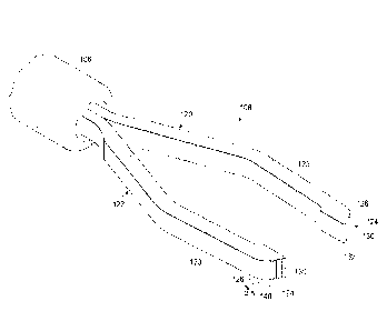
Fig. 1 illustrates a surgical instrument 100 shown as membrane forceps
having a handle 102, a probe actuation handle 104, a probe actuation tube
106, and a probe tip shown as forceps jaws 108. The handle 102 may be
made of any suitable material, and may be formed by any method, such as for
example, injection molding or machining. It may be made of a thermoplastic
or metal and may be textured or knurled to improve gripping. The actuation
handle 104 may be made from any suitable springy material having a
memory, such as titanium, stainless steel or suitable thermoplastic. The tube
106 may be any suitable medical grade tubing, such as titanium, stainless
steel, or suitable polymer and is sized so that the forceps jaws 108
reciprocate easily within. The forceps jaws 108 are generally made from
stainless steel or titanium, but other materials may also be used.
The surgical instrument 100 is designed so that in use, when the
actuation handle 104 is in its relaxed stated, the forceps jaws 108 protrude a
relatively large distance from the tube 106. Squeezing actuation handle 104
forces the front portion of the actuation handle 104 forward relative to the
handle 102. The forward movement of the front portion of the actuation
handle 104 is transferred to the tube 106, causing the tube 106 to slide
forward over a distal portion of the forceps jaws 108, thereby compressing
together the jaws 108. The amount of movement of tube 106 over the forceps
jaws 108 can be controlled easily by varying the outer diameter of the
actuation handle 104 in its relaxed stated.
Figs. 2 and 3 show the forceps jaws 108 in greater detail. The forceps
jaws 108 include two jaws, referred to herein as a jaw 120 and a jaw 122
extending from a distal end of the actuation tube 106. As shown in Fig. 2, the
two jaws 120, 122 extend along a longitudinal axis 118 that may be defined by
the tube 106.
7
CA 02891775 2015-05-15
WO 2014/092956
PCMJS2013/071055
Each of the jaws 120, 122 includes a projecting leg 123 and a distal
gripping tip 124. The legs 123 extend from the actuation tube 106. A bend
126 in the outer surface of the legs forms an intersection of the leg 123 and
a
leading side of the gripping tip 124.
The gripping tips 124 each include a leading edge 130 at the distal
ends and a grip face 132. In the embodiment disclosed, the leading edge 130
of each jaw 120, 122 lies in a plane substantially perpendicular to the
longitudinal axis 118.
The grip faces 132 of the two jaws 120, 122 are configured to abut
each other when the forceps jaws 108 are closed, and may be used to grip
tissue therebetween. In this example the grip faces 132 are formed to lie in
substantially parallel planes when the forceps jaws 108 are in a closed or
clamped position.
The gripping tips 124 also include a gripping tip distal surface 140 that
opposes the grip face 132. In this example, the gripping tip distal surface
140
is angled relative to the longitudinal axis at an angle 0. The angle 0 may be
an angle taken along a plane through the longitudinal axis 118 and an angle
having a value between about 25 degrees and 65 degrees. In other
embodiments, the angle 0 is between about 35 and 65 degrees. In yet
another embodiment, the angle 0 is formed between about 45 and 65 degrees
relative to the longitudinal axis. In addition, in some examples, the gripping
tip
distal surface 140 is formed as a substantially flat surface. In one example,
the distance between the leading edge 130 and the bend 126 is within a
range of about .1mm-.5mm, and the width across the gripping tip distal
surface is with a range of about .2mm-.9mm. The jaws 120, 122 may be
sized to fit within the tube 106 when the tube 106 is 20 gauge tube, a 23
gauge tube, a 25 gauge tube, or a 27 gauge tube. Other sizes are
contemplated.
The gripping tip distal surface 140 includes roughening features 142
that enable gaining the membrane by scraping the membrane in order to
rupture it so that an edge of the membrane may be grasped and peeled. In
8
CA 02891775 2015-05-15
WO 2014/092956
PCT/1JS2013/071055
this example, the roughening features 142 are manually formed using a file
drawn across the gripping tip distal surface 140 of the forceps to roughen the
surface. In some examples, the roughening features are formed in a series of
lines or grooves formed when the file is drawn in a lateral direction relative
to
the longitudinal axis, thereby creating a direction of grain extending in the
lateral direction relative to the longitudinal axis. Accordingly, these
grooves or
features may extend in a direction substantially parallel to the leading edge
130. In this example, substantially parallel is intended to mean having a
deviation of about ten degrees or less from parallel. In other examples, the
roughening features are formed by drawing the file in the longitudinal
direction
along the gripping tip distal surface 140, forming a grain extending
substantially in the longitudinal direction. Substantially in the longitudinal
direction is intended to mean having a deviation of about ten degrees or less
from the direction of the longitudinal axis. Other embodiments have the grain
of the roughening features formed in other oblique directions.
In some examples, the roughening features are formed on the gripping
tip distal surface 140 using a laser cutter. A laser cutter may be used to
form
roughening features 142 having a specific direction. In one embodiment, the
.. roughening features 142 include a series of ridges. These may include a
particular shape, such as a saw-tooth shape for example. Other
embodiments include knurled roughening features. Some embodiments
include roughening features formed as an array of peaks. In some
embodiments, these peaks each have a point, as may occur in diamond-
shaped knurls. Other roughening features are contemplated. Depending on
the embodiment, the roughening surface 140 may include features 142 having
a peak to valley height within a range of about 3-40 microns. In some
embodiments, the peak to valley height is within a range of about 3-20
microns, while in other embodiments, the peak to valley height is within a
range of about 5-10 microns.
In some embodiments, the ridges are formed in rows as in the example
shown in Figs. 2 and 3. Any of the roughening features may be formed in
rows and, depending on the embodiment, may be substantially parallel to the
9
CA 02891775 2015-05-15
WO 2014/092956
PCMJS2013/071055
leading edge 130 of the jaws 120, 122. In one example, the surface
roughening features 142 extend onto and form a part of the leading edge.
The roughening features 142 may be formed using any of a plurality of
methods including filing, grinding, scraping, machining, blasting, rolling,
etching, and laser cutting, among others. While shown with the roughening
features 142 on the outer surface 140, some embodiments include
roughening features on the edges and sides of the gripping tip 124.
Figs. 4-6 show an exemplary technique of using the surgical instrument
in an ILM or ERM peeling procedure. For purposes of this disclosure, the
technique will not describe cutting and removing the vitreous and posterior
hyaloid membrane.
During the procedure, the surgical instrument 100 is introduced into the
vitreous through an incision in the sclera, as shown in Fig. 4. The instrument
100 is advanced through the vitreous toward the macula. Some techniques
include introducing the distal tip with the jaws 120, 122 in a closed or
compressed condition into the globe and through the fluid within the globe
(this may be vitreous or may be, for example, a saline solution introduced
during removal of the vitreous). Other techniques include introducing the
distal tip with the jaws 120, 122 in an open condition through the vitreous,
and
then closing jaws prior to engaging the ILM or ERM. Additional instruments
such as a light pipe, also may be introduced in order to provide visualization
to the surgeon.
Peeling the ILM or ERM is a two-phase process. The first phase of the
procedure is to gain an edge of the ILM or ERM. The second phase is to
grasp and peel the membrane.
The first phase of the procedure may include placing the surgical
instrument gripping tip distal surface 140 in contact with the ILM or ERM as
shown in Fig. 5. As indicated above, the jaws are placed in a closed condition
prior to containing the ILM or ERM. The angle 0 of the gripping tip distal
surface 140 is formed so that the gripping tip distal surface 140 lies
CA 02891775 2015-05-15
WO 2014/092956
PCT/1JS2013/071055
substantially parallel to the surface of the membrane. Accordingly, the
gripping tip distal surface 140 may lie relatively flat on the membrane. In
some aspects, the instrument itself may extend at an angle, such as, for
example only, about 60 degrees from the membrane surface. At this angle, in
some examples, the angled roughened scraping surface 140 may be formed
to lie substantially flat against the membrane.
With the jaws in the closed condition, slight side-to-side or back-and-
forth movement may be used to scrape the ILM or ERM with the roughening
features 142 to gain an edge by rupturing the membrane, thereby creating an
edge of the membrane that can be grasped. Some techniques include
moving the jaws back and forth in a direction transverse to the grain of the
surface roughening features 142. This may permit the surface roughening
features to act against the membrane and result in easier membrane
rupturing. After the surgeon creates an edge by rupturing the membrane with
the roughened scraping surface 140 on the surgical instrument 100, the
surgeon may then proceed to phase two of the surgical procedure.
Because of its structural arrangement, grasping and peeling the
membrane can be accomplished without removing the surgical instrument 100
from the globe of the eye. Instead, the surgeon is able to grasp and peel the
membrane with the same surgical instrument used to scrape the membrane
and gain an edge. Using the same surgical instrument 100, after gaining the
edge of the membrane, the surgeon may immediately use the two jaws to
begin to grasp the membrane edge gained during the scraping procedure as
shown in Fig. 6. In order to grasp the tissue between the grip faces 132, the
surgeon may rotate the forceps 90 degrees so that the grip faces are
substantially perpendicular to the macula. This may permit the surgeon to
more easily grasp the membrane between the jaws. If necessary, the
surgeon may attempt to grasp the membrane between the leading tips 136
when very fine or minute segments of the tissue are available.
Advantageously, if the surgeon is unable to grasp the membrane with
the instrument 100, or if the membrane tears and the surgeon need to gain
11
CA 02891775 2015-05-15
WO 2014/092956
PCMJS2013/071055
another edge of the membrane, the surgeon may be able to use the
roughening features 142 on the instrument 100 again, without removing the
instrument 100 from the patient. Accordingly, the surgeon does not need to
make a conscious determination whether to withdraw the forceps and insert a
scraper to maintain a difficult peeling of an adherent membrane. Instead, the
scraping or roughening structure may be used anytime desired. This may
ease and simplify the initiation, performance, and maintenance of the peeling
procedure.
Fig. 7 shows a portion of another embodiment of forceps jaws
referenced herein by the numeral 200. Fig. 8 shows a top view of the forceps
jaws 200. This embodiment includes jaws formed so that the surgeon may
perform the scraping step and then grasp the membrane without rotating the
forceps. This will be explained further below.
The forceps jaws 200 include jaws 202 and 204, with each having a
projecting leg 206 and a distal gripping tip 208. The legs 206 extend from the
actuation tube 106 of a surgical instrument, such as the surgical instrument
100 in Fig. 1. A bend 210 in the outer surface of the legs forms an
intersection of the leg 206 and an opposing side of the gripping tip 208. In
this embodiment, the gripping tips 208 angle toward each other and include
an asymmetrical arrangement. Accordingly, while the jaws 202, 204 mirror
each other, the jaws form asymmetric forceps.
The gripping tips 208 each include a leading edge 220 at the distal
ends and a grip face 222. In the embodiment disclosed, the leading edge 220
of each jaw 202, 204 lies in a plane oblique to the longitudinal direction or
axis. Like the bend 210, the leading edge 220 at the distal end is formed at
an oblique angle, defined by the angle a. Accordingly, the leading edge 220
angles from a trailing tip 234 to a leading tip 236. This angled edge permits
the leading edge 220 to lie parallel to a surface, such a tissue surface, even
while the surgical instrument 100 is held at an oblique angle relative to the
tissue surface. In addition, because of the arrangement, a surgeon may use
any part of the entire leading edge 220 to grasp tissue since the whole
leading
12
CA 02891775 2015-05-15
WO 2014/092956
PCT/1JS2013/071055
edge may be parallel to the tissue during a procedure. In the embodiment
disclosed, the leading edge 130 is angled relative to the longitudinal axis
118
at an angle a having a value between about 35 degrees and 80 degrees. In
other embodiments, the angle a is between about 30 and 70 degrees. In yet
another embodiment, the angle a is formed between about 30 and 50
degrees. In some embodiments, the bend 210 at the intersection of the leg
206 and the gripping tips 208 is also formed parallel to the leading edge 220
and therefore is also formed at an oblique angle a.
Like the forceps jaws described with reference to Figs. 2 and 3, the grip
faces 222 in the forceps jaws 200 are configured to abut each other when the
forceps jaws are closed, and may be used to grip tissue therebetween. In this
example the grip faces 222 are formed to lie in substantially parallel planes
when the forceps jaws 108 are in a closed or clamped position.
The gripping tips 208 also include a gripping tip distal surface 228 that
opposes the grip face 222. In this example, the gripping tip distal surface
228
is formed to have at least a portion that is angled perpendicular to the
longitudinal direction so that when the grip faces 222 are pressed together,
the gripping tip distal surface 228 forms a leading plane as represented in
Fig.
8. As such, in some examples, the gripping tip distal surface 228 is formed as
a substantially flat surface. The jaws 120, 122 may be sized to fit within the
tube 106 when the tube 106 is 20 gauge tube, a 23 gauge tube, a 25 gauge
tube, or a 27 gauge tube. Other sizes are contemplated.
The gripping tip distal surface 228 includes roughening features 230
that enable gaining the membrane by scraping the membrane in order to
rupture it so that an edge of the membrane may be grasped and peeled. In
this example, the roughening features 230 are manually formed using a file
drawn across the gripping tip distal surface 228 of the forceps to roughen the
surface. The roughening features may be formed or shaped as discussed
above, and in some embodiments, are substantially parallel to the leading
edge 220.
13
CA 02891775 2015-05-15
WO 2014/092956
PCT/1JS2013/071055
Figs. 9-11 show an exemplary technique of using the surgical
instrument in an ILM or ERM peeling procedure. Only the differences from
the technique above will be described in detail, as much of the description
above applies to the technique using the forceps 200.
Referring to Fig. 9, the instrument is introduced into the globe of the
eye. In Fig. 10, the forceps jaws 200 are placed against the membrane.
Here, because the leading edge is formed at the angle a, and because the
gripping tip distal surface 228 is formed to be a flat leading surface, the
gripping tip distal surface 228 lies in a plane substantially parallel to the
membrane. In one embodiment, the surgeon may then scrape the membrane
using the gripping tip distal surface 228 on both of the jaws at the same
time.
In Fig. 10, the forceps are shown from the side angle. Because the
leading edge is angled, and the gripping tip distal surface 228 is angled, the
scraping may occurs while the forceps jaws are already positioned for
grasping scraped tissue. Fig. 11
shows the forceps jaws gripping a
membrane tissue. The grasping of the membrane occurs without rotating the
instrument 90 degrees. That is, during the scraping step in Fig. 10, the
leading edge of each jaw is already abutting against the membrane in the
closed position. The surgeon need only open the membrane and close it to
grasp the scraped tissue.
Because the ILM or ERM peel procedure may potentially be performed
without removing and introducing separate instruments into the eye to rupture
or scrape the membrane and to grasp the membrane, fewer instruments may
be used in the surgical procedure. This may increase efficiency of the
procedure, may result in less opportunity for error, and may provide better
patient outcome.
Persons of ordinary skill in the art will appreciate that the embodiments
encompassed by the present disclosure are not limited to the particular
exemplary embodiments described above. In that regard, although illustrative
embodiments have been shown and described, a wide range of modification,
14
CA 02891775 2015-05-15
WO 2014/092956
PCT/1JS2013/071055
change, and substitution is contemplated in the foregoing disclosure. It is
understood that such variations may be made to the foregoing without
departing from the scope of the present disclosure. Accordingly, it is
appropriate that the appended claims be construed broadly and in a manner
consistent with the present disclosure