Sélection de la langue

Search

Sommaire du brevet 2893035 

Énoncé de désistement de responsabilité concernant l'information provenant de tiers

Une partie des informations de ce site Web a été fournie par des sources externes. Le gouvernement du Canada n'assume aucune responsabilité concernant la précision, l'actualité ou la fiabilité des informations fournies par les sources externes. Les utilisateurs qui désirent employer cette information devraient consulter directement la source des informations. Le contenu fourni par les sources externes n'est pas assujetti aux exigences sur les langues officielles, la protection des renseignements personnels et l'accessibilité.

Disponibilité de l'Abrégé et des Revendications

L'apparition de différences dans le texte et l'image des Revendications et de l'Abrégé dépend du moment auquel le document est publié. Les textes des Revendications et de l'Abrégé sont affichés :

  • lorsque la demande peut être examinée par le public;
  • lorsque le brevet est émis (délivrance).
(12) Brevet: (11) CA 2893035
(54) Titre français: DISPOSITIF DE TOMODENSITOMETRIE DENTAIRE ET METHODE ASSOCIEE
(54) Titre anglais: DENTAL SCANNER DEVICE AND RELATED METHOD
Statut: Accordé et délivré
Données bibliographiques
(51) Classification internationale des brevets (CIB):
  • A61C 9/00 (2006.01)
  • A61C 19/00 (2006.01)
(72) Inventeurs :
  • FERNANDEZ PULIDO, ALFONSO (Espagne)
  • DE PABLOS GARCIA, DAVID (Espagne)
(73) Titulaires :
  • CARNOJAAL S.L.
(71) Demandeurs :
  • CARNOJAAL S.L. (Espagne)
(74) Agent: FIELD LLP
(74) Co-agent:
(45) Délivré: 2019-11-19
(86) Date de dépôt PCT: 2012-11-28
(87) Mise à la disponibilité du public: 2014-06-05
Requête d'examen: 2017-11-27
Licence disponible: S.O.
Cédé au domaine public: S.O.
(25) Langue des documents déposés: Anglais

Traité de coopération en matière de brevets (PCT): Oui
(86) Numéro de la demande PCT: PCT/ES2012/070834
(87) Numéro de publication internationale PCT: WO 2014083211
(85) Entrée nationale: 2015-05-27

(30) Données de priorité de la demande:
Numéro de la demande Pays / territoire Date
P201231853 (Espagne) 2012-11-28

Abrégés

Abrégé français

L'invention concerne un dispositif de scannage dentaire caractérisé en ce qu'il consiste essentiellement en deux corps, le premier corps ou corps de scanner (1) recevant un ensemble tête mobile de scannage (3) comportant essentiellement un élément longitudinal mobile se terminant au niveau d'une extrémité par la tête de scannage (4) à proprement parler, le second corps ou corps de morsure (2) formant une gaine destinée à recevoir la tête de scannage (4), ainsi que l'élément de fixation et de mise en place du dispositif par le patient au moyen de la force ou de la pression exercée par sa morsure.


Abrégé anglais


The invention relates to a dental scanning device characterized in that it is
essentially formed
by: two bodies, the first body or scanner body (1) housing a moving scan head
(3) consisting of
a longitudinal mobile element ending in the so-called scanning head (4), and
the second body or
bite body (2) consisting of a casing for receiving the scanning head (4); in
addition to the
element that allows the patient to hold and position the device by means of
the force or pressure
exerted by the patient's bite.

Revendications

Note : Les revendications sont présentées dans la langue officielle dans laquelle elles ont été soumises.


CLAIMS
1. A dental scanner device for 3D teeth and gingivae imaging, said device
comprising:
a scanner housing, which houses an elongate mobile element which juts
out the scanner housing and ends in a scanner head probe, the scanner head
probe comprising an element for emitting scanning energy; and
a bite fixture being held by a patient when biting said bite fixture during a
dental scan procedure wherein the dental scan procedure is fully automated and
generates a 3D image of the dental arch and gingivae,
wherein the elongate mobile element ending in the scanner head probe
moves within the bite fixture parallel to a long axis of the elongate rnobile
element
and perpendicular thereto,
and wherein the scanner housing, the elongate mobile element, the
scanner head probe, and the bite fixture can move with the patient's mouth,
the
patient's mouth being a fixed point of reference for the dental scan
procedure,
and thereby preserving the fixed point of reference at all moments while
scanning.
2. The dental scanner device of claim 1, wherein the bite fixture comprises
a
transparent material through which a scan can be performed.
3. The dental scanner device of claims 1 or 2, wherein the bite fixture is
capable of being coupled to and separable from the scanner housing.
4. The dental scanner device of any one of claims 1 to 3, wherein the bite
fixture is disposable.
- 7 -

5. The dental scanner device of any one of claims 1 to 4, wherein the
device
is portable and capable of being hand-held by the patient during the scanning
procedure.
6. A method for scanning a dental arch comprising the steps of:
a) providing a dental scanner of any one of claims 1 to 5;
b) placing the bite fixture in the patient's mouth; and
c) performing the dental scan procedure to produce a three-dimensional
image of the dental arch.
- 8 -

Description

Note : Les descriptions sont présentées dans la langue officielle dans laquelle elles ont été soumises.

DENTAL SCANNER DEVICE AND RELATED METHOD
FIELD OF THE INVENTION
[0001] The present disclosure generally relates to the field of stomatology
and
dental prosthetics manufacturing, and in particular to an intraoral dental
scanner
for 3D teeth and gingivae imaging and related method.
BACKGROUND OF THE INVENTION
[0002] Teeth and gingivae 3D diagnostic and therapeutic images have been
traditionally obtained by using replicas or models from alginate-impressed
molds.
Such replicas get gingiva and tooth negative images, which are later on
converted into positive, and then scanned. However, these mainstream
techniques pose a double problem: the patient is uncomfortable; and they are
not
very reliable and accurate; thus, the process is slow and costly.
[0003] There are several state-of-the-art devices that try to solve the
problems
posed by mainstream techniques; e.g.: panoramic dental X rays or computerized
dental tomographies.
[0004] EP 0825837, "Modular intra-oral imaging system video camera", for
instance, mainly provides a I-hand-held video camera to take images of the
patient's inner part of the mouth. The camera has a socket; in the inner part
of
the socket there is a base, and a visualizing device, as well as a socket's
long
axis optically aligned sensor that converts into data the images taken by the
camera.
- 1 -
CA 2893035 2019-05-01

[0005] US 2006154198 3D dental scanner is both an imaging method and a
system to get images of the dental structure in the inner part of the oral
cavity,
through the motion of at least one image capturer set on a fixed-reference-
system coupled arm, external to the mouth, to generate a 3D model of the
structure, based on the images captured.
[0006] ES2 383 220, "Intraoral dental imaging sensor and X-ray system, using
such sensor" is an intraoral dental radiological system equipped with a mouth-
insertable x-ray imaging sensor. It is made up of an image-detection matrix to
provide electronic signals. The system comprises a light source to receive the
matrix-generated signals; it emits binary light impulses corresponding to a
digital
image to be transmitted; it also has a light receiver placed at a certain
distance
from the patient; it can detect a light modulation triggered by the light
source; it
can transmit to the image-treating device the signal corresponding to such
modulation.
[0007] Lastly, E82324658 (T3), "Laser-digitalizing system for dental
applications"
is a laser digitilizer that has a light source with collimation optics to
generate a
collimated light beam; a scanner optically coupled with the light source
(configured to scan the collimated beam towards the object to be optically
represented in a predetermined pattern); an imaging instrument with an optical
axis in a e angle with the scanner; this is set up to detect a pattern
reflection from
the object, and generate the object surface representative data, based on the
pattern reflection; and a scanner-coupled processor, and the imaging system,
configured to render a data-based 3D image of the object; it is characterized
by
the fact that the scanner is set up to scan the collimated beam all along at
least
to axes in the predetermined pattern. Such pattern encompasses a series of
curvilinear segment.
- 2 -
CA 2893035 2019-05-01

[0008] These devices and systems have several inconveniences. First, in some
cases, a technician must manually operate them with a toothbrush-like motion,
which, obviously, is inaccurate. Likewise, they are based on photographs taken
by the various devices; usually, a software interprets and interpolates such
photographs to take a final 3D image. Thus, besides the time needed to get the
final image, these known systems depend on the operator's skill for
manipulating
the camera and inserting it in not-easy-to- reach parts of the mouth.
[0009] Other known devices do not depend so much on manual operation, but
have an external fixed-coordinate system, independent of the patient;
therefore,
the final image is exclusively based on the images captured with no extra
projection; thus, they are not very accurate, as the reference system changes
with the slightest movement of the patient.
SUMMARY OF THE INVENTION
[0010] This invention is a dental scanning device that overcomes at least some
of
the above-noted defects in certain embodiments. For example, the invention may
capture 3D images with respect to a fixed-reference system; i.e. the patient's
mouth; thus, images are accurate, as the device moves along with the patient;
i.e.; teeth-position point of reference, for instance, as related to the
device, is
preserved at all moments, and remains in place while scanning.
[0011] In one aspect, there is provided a dental scanner device for 3D teeth
and
gingivae imaging, said device including: a scanner housing, which houses an
elongate mobile element which juts out the scanner housing and ends in a
scanner head probe, the scanner head probe comprising an element for emitting
scanning energy; and a bite fixture being held by a patient when biting said
bite
fixture during a dental scan procedure wherein the dental scan procedure is
fully
automated and generates a 3D image of the dental arch and gingivae, wherein
- 3 -
CA 2893035 2019-05-01

the mobile element ending in the scanner head probe moves within the bite
fixture parallel to a long axis of the elongate mobile element and
perpendicular
thereto, and wherein the scanner housing, the elongate mobile element, the
scanner head probe, and the bite fixture can move with the patient's mouth,
the
patient's mouth being a fixed point of reference for the dental scan
procedure,
and thereby preserving the fixed point of reference at all moments while
scanning.
[0012] In another aspect, there is provided a method for scanning a dental
arch
including the steps of: a) providing a dental scanner described herein; b)
placing
the bite fixture in the patient's mouth; and c) performing the dental scan
procedure to produce a three-dimensional image of the dental arch.
[0013] In yet another aspect there is provided a dental scanning device made
up
of two components; one component lodges the scanner as such, as well as a
mobile head probe; the other component is to be held when the patient bites,
and
is to correspondingly house the mobile head probe. Once the head probe part is
introduced in the bite fixture - and therefore is held by the patient's mouth-
the
head probe moves in at least two directions. One direction following the long
axis
of the head probe; and the other direction perpendicular to it, so that
scanning is
the result of a combination of both movements, as related to the dental arcade
of
the patient. The component(s) to be held in the patient's mouth may be made of
an easy-to-disinfect / sterilize material.
[0014] Optionally, the aforementioned components can be coupled to one
another, and may be disassembled and joined again; in this case, either the
patient's mouth-held piece is made of an easy-to-disinfect I sterilize
material, or is
disposed after use. In this case, a scanning component and a bite fixture may
be
easily coupled to one another through specially placed devices in the coupling
ends.
- 4 -
CA 2893035 2019-05-01

BRIEF DESCRIPTION OF THE DRAWINGS
[0015] Below there is a description of the invention, based on one of the
versions
of choice, and as per attached drawings in which:
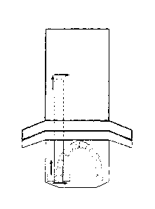
[0016] Figure 1 illustrates a scanner device as per one version of the
invention,
as seen from above;
[0017] Figure 2 illustrates a side view of the scanner device in figure 1,
showing
how it is placed in the patient's mouth; and
[0018] Figure 3 is a schematization of figure 1, showing device scanning
directions.
DETAILED DESCRIPTION OF THE INVENTION
[0019] As shown in figure 1, the dental scanner is made up of two components.
The first component, a scanner housing (1), houses an elongate mobile element
(3) which juts out of the scanner housing and ends in a scanner head probe
(4).
That is to say, the elongate mobile element (3) and the scanner head probe (4)
are partially kept inside the scanner housing (1). The scanner head probe (4)
may include an element for emitting scanning energy.
[0020]The scanner probe head (4) juts out the scanner housing (1) and is
adapted to be lodged in a pocket of the second embodiment or bite fixture (2).
Thus, the bite fixture (2) is, therefore, a scanner head probe lodging pocket
(4).
- 5 -
CA 2893035 2019-05-01

[0021] The scanner housing (1) and the bite fixture (2) may be coupled, and
separated and coupled again, the above is equally applied, just as when both
components (1 & 2) have been previously coupled to one another.
[0022] As indicated, the bite fixture (2) is introduced in the patient's
mouth; and it
is held in place when the patient bites it, so that the scanner head probe (4)
is
introduced in the mouth to scan the corresponding dental arcade.
[0023] Likewise, the bite fixture (2) may be mouth shaped to make it easier to
hold. Such bite fixture (2) is basically a protective pocket for the scanner
head
probe (4), and can be bitten by the user. The bite fixture (2) may be made of
a
suitable transparent material.
[0024] Once the part of the scanner head probe (4) that is housed in the bite
fixture (2) is introduced, as shown in figure 2, the scanner head probe moves
along in at least two directions; one parallel to a the long axis of the
elongate
mobile element (3), while the other one goes perpendicular to the long axis of
the
elongate mobile element, in such a way that scanning is the result of
combining
motions with regard the dental arcade, as shown in figure 3.
[0025] For the intraoral scanning, the device includes detection sensors,
laser
sensors or similar devices in the scanner head probe (4), as well as cameras
to
capture tooth-by-tooth images from the dental arcade and gingivae. These are
automatically generated and exact 3D images, as a result of their fixed and
constant reference system.
- 6 -
CA 2893035 2019-05-01

Dessin représentatif
Une figure unique qui représente un dessin illustrant l'invention.
États administratifs

2024-08-01 : Dans le cadre de la transition vers les Brevets de nouvelle génération (BNG), la base de données sur les brevets canadiens (BDBC) contient désormais un Historique d'événement plus détaillé, qui reproduit le Journal des événements de notre nouvelle solution interne.

Veuillez noter que les événements débutant par « Inactive : » se réfèrent à des événements qui ne sont plus utilisés dans notre nouvelle solution interne.

Pour une meilleure compréhension de l'état de la demande ou brevet qui figure sur cette page, la rubrique Mise en garde , et les descriptions de Brevet , Historique d'événement , Taxes périodiques et Historique des paiements devraient être consultées.

Historique d'événement

Description Date
Inactive : TME en retard traitée 2024-05-01
Paiement d'une taxe pour le maintien en état jugé conforme 2024-05-01
Lettre envoyée 2023-11-28
Inactive : Certificat d'inscription (Transfert) 2022-04-14
Inactive : Transfert individuel 2022-03-31
Représentant commun nommé 2020-11-07
Accordé par délivrance 2019-11-19
Inactive : Page couverture publiée 2019-11-18
Représentant commun nommé 2019-10-30
Préoctroi 2019-09-26
Inactive : Taxe finale reçue 2019-09-26
Un avis d'acceptation est envoyé 2019-06-12
Lettre envoyée 2019-06-12
Un avis d'acceptation est envoyé 2019-06-12
Inactive : QS réussi 2019-06-03
Inactive : Approuvée aux fins d'acceptation (AFA) 2019-06-03
Modification reçue - modification volontaire 2019-05-01
Inactive : Dem. de l'examinateur par.30(2) Règles 2018-11-14
Inactive : Rapport - CQ échoué - Mineur 2018-10-21
Lettre envoyée 2017-12-04
Exigences pour une requête d'examen - jugée conforme 2017-11-27
Requête d'examen reçue 2017-11-27
Toutes les exigences pour l'examen - jugée conforme 2017-11-27
Modification reçue - modification volontaire 2017-11-27
Lettre envoyée 2016-11-21
Inactive : Transfert individuel 2016-11-14
Inactive : Lettre officielle 2016-05-30
Inactive : Demandeur supprimé 2015-11-05
Inactive : Inventeur supprimé 2015-11-05
Demande de correction du demandeur reçue 2015-07-03
Inactive : Page couverture publiée 2015-06-29
Inactive : CIB en 1re position 2015-06-08
Inactive : Notice - Entrée phase nat. - Pas de RE 2015-06-08
Inactive : CIB attribuée 2015-06-08
Demande reçue - PCT 2015-06-08
Exigences pour l'entrée dans la phase nationale - jugée conforme 2015-05-27
Demande publiée (accessible au public) 2014-06-05

Historique d'abandonnement

Il n'y a pas d'historique d'abandonnement

Taxes périodiques

Le dernier paiement a été reçu le 2019-09-26

Avis : Si le paiement en totalité n'a pas été reçu au plus tard à la date indiquée, une taxe supplémentaire peut être imposée, soit une des taxes suivantes :

  • taxe de rétablissement ;
  • taxe pour paiement en souffrance ; ou
  • taxe additionnelle pour le renversement d'une péremption réputée.

Veuillez vous référer à la page web des taxes sur les brevets de l'OPIC pour voir tous les montants actuels des taxes.

Titulaires au dossier

Les titulaires actuels et antérieures au dossier sont affichés en ordre alphabétique.

Titulaires actuels au dossier
CARNOJAAL S.L.
Titulaires antérieures au dossier
ALFONSO FERNANDEZ PULIDO
DAVID DE PABLOS GARCIA
Les propriétaires antérieurs qui ne figurent pas dans la liste des « Propriétaires au dossier » apparaîtront dans d'autres documents au dossier.
Documents

Pour visionner les fichiers sélectionnés, entrer le code reCAPTCHA :


Pour visualiser une image, cliquer sur un lien dans la colonne description du document. Pour télécharger l'image (les images), cliquer l'une ou plusieurs cases à cocher dans la première colonne et ensuite cliquer sur le bouton "Télécharger sélection en format PDF (archive Zip)" ou le bouton "Télécharger sélection (en un fichier PDF fusionné)".

Liste des documents de brevet publiés et non publiés sur la BDBC .

Si vous avez des difficultés à accéder au contenu, veuillez communiquer avec le Centre de services à la clientèle au 1-866-997-1936, ou envoyer un courriel au Centre de service à la clientèle de l'OPIC.


Description du
Document 
Date
(aaaa-mm-jj) 
Nombre de pages   Taille de l'image (Ko) 
Description 2015-05-27 4 184
Abrégé 2015-05-27 1 12
Revendications 2015-05-27 1 40
Dessin représentatif 2015-05-27 1 11
Dessins 2015-05-27 3 21
Page couverture 2015-06-29 1 39
Description 2017-11-27 6 195
Revendications 2017-11-27 2 38
Revendications 2019-05-01 2 46
Description 2019-05-01 6 244
Abrégé 2019-06-10 1 12
Dessin représentatif 2019-10-22 1 5
Page couverture 2019-10-22 1 35
Paiement de taxe périodique 2024-05-01 3 86
Courtoisie - Réception du paiement de la taxe pour le maintien en état et de la surtaxe (brevet) 2024-05-01 1 434
Avis d'entree dans la phase nationale 2015-06-08 1 194
Courtoisie - Certificat d'enregistrement (document(s) connexe(s)) 2016-11-21 1 101
Rappel - requête d'examen 2017-07-31 1 116
Accusé de réception de la requête d'examen 2017-12-04 1 174
Avis du commissaire - Demande jugée acceptable 2019-06-12 1 163
Courtoisie - Certificat d'inscription (transfert) 2022-04-14 1 401
Avis du commissaire - Non-paiement de la taxe pour le maintien en état des droits conférés par un brevet 2024-01-09 1 541
Demande de l'examinateur 2018-11-14 4 276
PCT 2015-05-27 22 812
Modification au demandeur-inventeur 2015-07-03 3 141
Taxes 2015-11-02 1 26
Correspondance 2016-05-30 2 51
Taxes 2016-11-28 1 26
Requête d'examen / Modification / réponse à un rapport 2017-11-27 11 314
Modification / réponse à un rapport 2019-05-01 21 850
Taxe finale 2019-09-26 1 30
Paiement de taxe périodique 2021-11-26 1 27