Note : Les descriptions sont présentées dans la langue officielle dans laquelle elles ont été soumises.
CIS 02895652 2015-06-18
WO 2014/097289
PCT/IL2013/000096
1
VIRAL INACTIVATED BIOLOGICAL MIXTURE
FIELD OF THE INVENTION
Generally, the invention relates to a viral inactivated biological liquid or
dry mixture
and to its preparation. Principally, the invention relates, but is not
limited, to a viral
inactivated platelet extract and preparation thereof.
BACKGROUND OF THE INVENTION
Platelets are small, irregularly-shaped a-nuclear cells that play a
fundamental role in
hemostasis and healing. Platelets contain a complete array of pre-synthesized
proteins,
among which are signaling proteins, cyto skeletal proteins, membrane proteins
and
regulatory proteins. They are involved in key stages of tissue regeneration
and healing
processes at the site of injury, mainly due to the content of platelet
granules
comprising a multitude of bioactive molecules including growth factors (GFs),
cytokines and chemokines.
Platelet growth factors such as platelet-derived growth factor (PDGF),
transforming
growth factor (TGF), basic fibroblast growth factor (bFGF), vascular
endothelial
growth factor (VEGF) and others are key players in all the following phases of
the
wound healing cascade: inflammatory, proliferative and remodeling phase.
Studies have shown that platelet derived growth factors stimulate
angiogenesis,
mitogenesis, cell proliferation, neutrophils and macrophages, collagen
synthesis,
wound contraction, extracellular matrix synthesis, epithelialization and
chemotaxis.
Platelets are routinely used by transfusion e.g. to improve hemostasis.
Recently,
platelets are increasingly used in the form of Platelet Rich Plasma (PRP),
also referred
to as PRP gel, platelet gel, PRP-clot etc. Typically, PRP is an ex vivo
preparation
consisting of autologous platelets concentrated in a limited volume of plasma
(Lacci
KM, Dardik A. Platelet-rich plasma: support for its use in wound healing. Yale
J Biol
Med. 2010 Mar;83(1):1-9).
For topical application, PRP is usually activated by the addition of thrombin
and/or
CaCl2 resulting in the formation of fibrin gel by the interaction between
thrombin
(endogenous or exogenous) and fibrinogen. Upon activation, the platelets
undergo
active degranulation and release various mediators including GFs (Lacci KM,
Dardik
A, 2010). The use of PRP for injection currently comprises a small but rapidly
growing segment of the market. The rationale for using PRP in soft and hard
tissue
CA 02895652 2015-06-18
WO 2014/097289
PCT/IL2013/000096
2
augmentation is its potential to enhance tissue regeneration in non-healing
injuries,
accelerate wound maturity, vascularization and epithelialization, decrease
scar
formation, and reduce post operative complications and morbidity (Lacci KM,
Dardik
A, 2010).
Studies using activated PRP together with various cell types have shown that
factors
e.g. growth factors released from PRP can induce cell proliferation [(e.g.
Kanno et al.
Platelet-rich plasma enhances human osteoblast-like cell proliferation and
differentiation. J Oral Maxillofac Surg. 2005 Mar;63(3):362-9; Bertrand-
Duchesne et
al. Epidermal growth factor released from platelet-rich plasma promotes
endothelial
cell proliferation in vitro. J Periodontal Res. 2010 Feb;45(1):87-93; Kakudo
et al.
Proliferation-promoting effect of platelet-rich plasma on human adipose-
derived stem
cells and human dermal fibroblasts. Plast Reconstr Surg. 2008 Nov;122(5):1352-
60),
modulate the angiogenic capability of human endothelial cells (Sulpice et al.
Cross-
talk between the VEGF-A and HGF signalling pathways in endothelial cells. Biol
Cell. 2009 Sep;101(9):525-39; Rughetti et al. Platelet gel-released
supernatant
modulates the angiogenic capability of human endothelial cells. Blood
Transfus. 2008
Jan;6(1):12-7), and induce osteo-inductive properties (Intini G. The use of
platelet-
rich plasma in bone reconstruction therapy. Biomaterials. 2009 Oct;30(28):4956-
66)].
Moreover, activated PRP was found to support in vitro cell growth and
maintained
viability of a number of target cells including myelomas, hybridomas,
liepatocytes,
fibroblasts and epithelial cells, at a level comparable or superior to the
level supported
by fetal bovine serum (Johansson et al. Platelet lysate: a replacement for
fetal bovine
serum in animal cell culture? Cytotechnology. 2003 Jul;42(2):67-74).
PRP and released growth factors are currently used in various surgical tissue
regeneration procedures, predominantly in orthopedic and dental surgery
(Nurden et
al. Platelets and wound healing. Front Biosci. 2008 May 1;13:3532-48). In
orthopedic
surgery PRP is used mainly for knee arthroplasty, lumbar spinal fusion, and in
intervertebral disc degeneration (reviewed in Nurden et al, 2008). Dentistry
and
maxillofacial surgery PRP applications include mainly consolidation of
titanium
implants, maxillary sinus augmentation and bone remodeling (reviewed in Nurden
et
al, 2008). PRP is also increasingly used for tendon and ligament repair,
facial plastic
and reconstructive surgery, chronic skin wound healing, ophthalmology, facial
nerve
regeneration, as well as in cardiac and bariatric surgery (reviewed in Nurden
et al,
2008).
CA 02895652 2015-06-18
WO 2014/097289
PCT/IL2013/000096
3
However, a major disadvantage of the current use of autologous PRP and
released
factors resides in the lack of standardization. Of note, different manual,
semi-
automated and fully-automated systems for preparation of PRP are commercially
available that differ in parameters such as preparation time, platelet yield
and
collection efficiency (Mazzucco et al. Not every PRP-gel is born equal.
Evaluation of
growth factor availability for tissues through four PRP-gel preparations:
Fibrinet,
RegenPRP-Kit, Plateltex and one manual procedure. Vox Sang. 2009 Aug;97(2):110-
8).
Another important variable is the technique used for platelet activation
[autologous,
heterologous or recombinant thrombin, calcium chloride or batroxobin (Rozman
P,
Bolta Z. Use of platelet growth factors in treating wounds and soft-tissue
injuries.
Acta Dermatovenerol Alp Panonica Adriat. 2007 Dec;16(4):156-65)], which can
affect the efficiency of granule release and the amount of secreted GFs (Roman
P,
Bolta Z, 2007). Moreover, since platelets are very sensitive to mechanical
stress and
changes in the surrounding environment, they may be activated and GFs may be
released during processing, prior to the intended activation step (Mazzucco et
al,
2009). This uncontrolled activation may further increase the variability in
the
composition of the final product when using different PRP preparation systems.
Additionally, a major inherent weakness of autologous PRP preparation is that
the
platelets GFs content varies among individuals, and therefore may lead to sub-
optimal
results. Finally, the financial burden of dedicated machinery, disposable PRP
processing kits, and the need for trained personnel, should be taken into
consideration
when working with autologous PRP.
Background art includes Su et al. "A virally inactivated functional growth
factor
preparation from human platelet concentrates". Vox Sang. 2009 Aug;97(2):119-
128;
Burnouf et al. "A novel virally inactivated human platelet lysate preparation
rich in
TGF-beta, EGF and IGF, and depleted of PDGF and VEGF". Biotechnol App!
Biochem. 2010 Aug 6;56(4):151-60; and U.S. Patent Publication No. US 2012-
0156306.
SUMMARY OF THE INVENTION
In one aspect, the invention relates to a method for preparing a viral-safe
biological
liquid mixture, the method comprising the following steps: providing a
biological
liquid mixture; carrying out a solvent detergent (S/D) viral inactivation
treatment;
CA 02895652 2015-06-18
WO 2014/097289
PCT/IL2013/000096
4
contacting the S/D treated mixture with an amphiphilic polymer; removing the
S/D by
hydrophobic interaction chromatography (HIC) and/or by oil extraction;
collecting a
material comprising a flow through fraction from HIC and/or a liquid fraction
from oil
extraction; and subjecting the material to at least one more orthogonal viral
inactivation treatment.
In one embodiment of the invention, the amphiphilic polymer is non-toxic.
Yet, in a further embodiment of the invention, the amphiphilic polymer is a
hydrocarbon based surfactant.
Yet, in a further embodiment of the invention, the amphiphilic polymer has an
average molecular weight in the range of about 3.5 to lower than about 40
kilodalton.
In one embodiment of the invention, the average molecular weight is about 30
kilodalton.
Yet, in a further embodiment of the invention, the hydrocarbon based
surfactant is
polyvinylpyrrolidone (PVP).
In one embodiment of the invention, the PVP has an average molecular weight of
about 30 kilodalton (kDa).
In one embodiment of the invention, the HIC comprises the steps of: loading
the S/D
treated and polymer contacted mixture to HIC; washing with a solution
comprising an
organic solvent and/or a salt; and collecting a washed fraction.
In one embodiment of the invention, the organic solvent is ethanol.
In one embodiment of the invention, the salt is NaCl.
In a further embodiment of the invention, the at least one more orthogonal
viral
inactivation treatment comprises heat inactivation.
In one embodiment of the invention, the method further comprises a step of
concentrating the material.
In one embodiment of the invention, the method is for preparing a viral-safe
platelet
extract, and the biological liquid mixture is a platelet-enriched fraction.
In one embodiment of the invention, the collected material comprises the HIC
flow
through fraction combined with the HIC washed fraction.
In another aspect, the invention relates to a viral-safe biological liquid
mixture
obtainable according to the method of the invention.
In one embodiment of the invention, the concentration of PVP K25 is in the
range of
0.1% (vv/w) to lower than 1% (w/w).
CA 02895652 2015-06-18
WO 2014/097289
PCT/IL2013/000096
5 In certain embodiments of the invention, the concentration of PVP K25 is
in the range
of 0.1% (w/w) to 0.5 % (w/w).
In one embodiment of the invention, the PVP K17, K25, K30 concentration in the
viral-safe biological liquid mixture is in the range of 0.01-5% (w/w).
In certain embodiments of the invention, the biological liquid mixture is a
platelet
extract enriched with PDGF-AB, PDGF-BB, EGF, VEGF and/or bFGF.
In another aspect, the invention relates to a method for removing solvent-
detergent
(S/D) from a biological liquid mixture comprising the S/D, the method
comprises the
steps of: providing the mixture comprising the S/D; contacting the mixture
with an
amphiphilic polymer; removing the S/D from the mixture by hydrophobic
interaction
chromatography (HIC) and/or by oil extraction; and collecting a material
comprising a
flow through fraction from HIC and/or a liquid fraction from oil extraction.
In one embodiment of the invention, the amphiphilic polymer is non-toxic.
In one embodiment of the invention, the biological liquid mixture is a
platelet-
enriched fraction.
In another embodiment of the invention, the mixture comprises chemokines,
cytokines, growth factors, trophic factors or a mixture thereof.
In one embodiment of the invention, the HIC comprises the steps of: washing
with a
solution comprising an organic solvent and/or a salt; and collecting a washed
fraction.
In one embodiment of the invention, the amphiphilic polymer is a hydrocarbon
based
surfactant.
In one embodiment of the invention, the hydrocarbon based surfactant is
polyvinylpytTolidone (PVP).
In one embodiment of the invention, the organic solvent is ethanol.
In one embodiment of the invention, the salt is NaCl.
In one embodiment of the invention, the collected material comprises the flow
through fraction combined with the wash fraction.
In one embodiment of the methods, the source is contacted first with the S/D
and then
with the amphiphilic polymer.
In some embodiments of the methods, the PVP concentration in the S/D treated
source is in the range of about 0.01 to 0.9 mM, 0.01 to 0.3 mM, or 0.025 to
0.3 mM.
In some embodiments of the methods, the HPMC concentration in the S/D treated
source is in the range of about 0.01 to 0.3 mM.
6
In some embodiments, the methods further comprise a step of drying the
material,
thereby resulting in a biological dry mixture.
In a certain aspect, it is disclosed a method for preparing a biological
liquid mixture
composition from a biological source. The method comprises the following
steps:
providing the source; providing PVP and/or HPMC; treating the source with a
solvent
detergent (SID) to allow viral inactivation and with the PVP and/or HPMC;
removing
the S/D by contacting the treated source with a hydrophobic interaction
chromatography (HIC) resin; and collecting a material comprising an unbound
fraction from HIC.
In a certain aspect, it is disclosed a biological liquid mixture composition
obtainable
according to the disclosed methods.
In another aspect, it is disclosed a viral-safe biological liquid mixture
composition
obtainable according to the method of any one of claims 1 to 21, comprising
PVP at a
concentration in the range of about 0.07 to 6 mM, or HPMC at a concentration
in the
range of about 0.07 to 1.5 mM.
In one embodiment, the composition comprises a PVP concentration in the range
of
about 0.07 to 6 mM, 0.07 to 2 mM, or 0.17 to 2 mM.
In another embodiment, the composition comprises a HPMC concentration in the
range of about 0.07 to 1.5mM.
In a certain aspect, it is disclosed a pharmaceutical composition comprising
an
amphiphilic polymer; a platelet derived protein selected from the group
consisting of
a chemokine, a growth factor, a cytokine, a throphic factor and a mixture
thereof; and
a pharmaceutically acceptable carrier, wherein the amphiphilic polymer is PVP
at a
concentration in the range of about 0.07 to 6 mM or HPMC at a concentration in
the
range of about 0.07 to 1.5 mM.
In another aspect, it is disclosed a viral-safe pharmaceutical composition
comprising
an amphiphilic polymer; a platelet derived protein selected from the group
consisting
of a chemokine, a growth factor, a cytokine, a throphic factor and a mixture
thereof;
and a pharmaceutically acceptable carrier, wherein the amphiphilic polymer is
PVP at
a concentration in the range of about 0.07 to 6 mM or HPMC at a concentration
in the
range of about 0.07 to 1.5 mM.
In a certain aspect, it is disclosed a method for tissue healing; organ
reconstruction;
tissue regeneration and/or treating inflammation in a subject in need,
comprising
applying to the subject an effective amount of a composition described herein.
CAN_DMS \132430801\1
CA 2895652 2020-03-11
6a
In a certain aspect, it is disclosed sse of an effective amount of a
composition
described herein, for tissue healing; organ reconstruction; tissue
regeneration and/or
treatment of inflammation in a subject in need thereof
In one embodiment, the method or use comprises promoting skin flap adherence.
BRIEF DESCRIPTION OF THE DRAWINGS
Fig. 1 shows proliferation of 3T3 fibroblast cells treated with a platelet
extract
obtained by contacting the lysate with heparin prior to S/D removal (treatment
2 and
4). A platelet extract prepared without contacting the lysate with heparin
prior to S/D
removal served as the control (treatment 1 and 3).
Fig. 2 shows proliferation of 3T3 fibroblast cells treated with a platelet
extract
prepared by contacting the lysate with low molecular weight heparin (LMWH)
prior
to and during S/D removal (treatment 2). A platelet extract obtained following
S/D
removal in the absence of contacting the lysate with low molecular weight
heparin
(LMWH) prior to S/D removal served as the control (treatment 1).
CAN_DMS \132430801\ 1
CA 2895652 2020-03-11
CA 02895652 2015-06-18
WO 2014/097289
PCT/IL2013/000096
7
Fig. 3 shows proliferation of 3T3 fibroblast cells treated with extracts
obtained after
S/D removal in the presence of different molecular weight PVP polymers: PVP
1(25 ¨
treatment 3; or PVP K30 ¨ treatment 7.
Fig. 4 shows proliferation of 3T3 fibroblast cells treated with extracts
comprising
different concentrations of PVP. Sample 17 (treatment 17) and sample 19
(treatment
.. 19) differ in the second wash of the S/D removal, where 0.1% and 0.5% PVP
were
used, respectively.
Fig. 5 shows proliferation of 3T3 fibroblast cells treated with large scale
platelet
extracts obtained after S/D removal in the presence of PVP K25 (treatment 1)
or after
S/D removal in the presence of heparin (treatment 2).
In addition, all figures comprise R2 fit, median effective concentration
(EC50), and
95% Confidence Intervals EC50 values calculated by GraphPad Prism software.
Fig. 6 shows a rat dorsal flap (3x10 cm) at 2 weeks after surgery performance.
The
flap was elevated in cranial to caudal direction. A,B,C,D and E indicate
different areas
from where samples were taken for histological analysis. A is closest to the
caudal
.. flap attachment and therefore heals best, whereas E is in the cranial end
of the flap,
which shows highest leves of necrosis (dark color). The abdominal and thoracic
viscera were removed through ventral midline incision (line along the center
of the
flap).
Fig. 7 shows typical staining patterns for normal and healing skin: H&E
staining
(epidermal hyperplasia, score 1 and epidermis after completed healing process,
score
0), PCNA staining for dermal and epidermal proliferation (proliferating
tissue, score 1
and normal tissue, score 0) and Keratin 6 staining (suprabasal staining, score
1, for
healing and basal staining for regular skin, score 0).
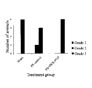
Fig. 8 shows the scores for the adherence grade of the rat dorsal flap after 2
weeks, as
they were tested by gently pulling the flap in the area A-C (see Fig. 6) away
from the
wound bed using a tissue forceps. The attachment of the skin flap to the
underlying
tissue was compared to the attachment of normal areas of skin and graded 1 to
3 as
follows: 1=no to low adherence, 2=below but nearly normal adherence or 3=about
normal adherence between skin flap and underlying tissue.
DESCRIPTION OF SOME EMBODIMENTS OF THE INVENTION
The invention relates to methods for preparing a viral-safe biological liquid
mixture
such as a viral safe platelet extract. Platelets contain a complete array of
factors
CA 02895652 2015-06-18
WO 2014/097289
PCT/IL2013/000096
8
involved in key stages of tissue regeneration and healing processes.
Currently, whole
autologous activated platelets (derived from the patient) are used for
facilitating
wound healing. However, there are multiple disadvantages of using whole
autologous
platelets, inter alia, the lack of standardization; the factors needed for
healing may be
scarce in the patient's own platelets; the special equipment needed for
preparing the
mixture of platelet factors; the procedure is time consuming and requires
additional
steps which are carried out on the patient itself; and the requirement of
medically
trained personnel. These problems can be solved e.g. by using a platelet
extract
prepared from multiple donors.
However, human blood-derived products may carry a risk of transmitting
infectious
agents such as viruses. Effective reduction of viral transmission risk can be
achieved
by including at least two orthogonal viral inactivation steps. Yet, including
additional
steps in the manufacture of a platelet extract may compromise the recovery and
activity of the factors contained therein.
One of these methods of viral inactivation is "Solvent detergent (S/D) viral
inactivation treatment".
This inactivation includes treatment with S/D and removal of the S/D. It was
found
according to the present invention that the recovery of certain growth factors
is
compromised after S/D removal by HIC.
It was found according to the present invention that recovery of certain
platelet factors
e.g. PDGF-AB; PDGF-BB; and bFGF, can be increased by contacting the S/D
treated
material prior to and/or during S/D removal with polyvinylpyiTolidone (PVP) or
Hydroxy Propyl Methyl Cellulose (HPMC), which are non-toxic amphiphilic
molecules.
It was found that contacting the S/D treated material with PVP and HPMC in
accordance to the invention resulted in increased recovery or enrichment of
PDGF-
AB and other platelet factors e.g. PDGF-BB and bFGF.
These findings are surprising, in view that contacting the S/D treated
material with
heparin and low molecular weight heparin (both known to bind certain growth
factors) during S/D removal increased the recovery of factors while contacting
S/D
treated material with PVP, which is a completely different compound (having
amphiphilic characteristics) had a similar beneficial effect on growth factor
recovery
during S/D removal.
CA 02895652 2015-06-18
WO 2014/097289
PCT/IL2013/000096
9
Also, the findings are surprising, since addition of PVP K30, K25, K17 and K12
under certain tested conditions did not compromise S/D removal.
It was found that using the method according to the invention to remove S/D
from a
source comprising platelet-derived mixture of factors results in high recovery
of
factors, high biological activity and efficient removal of S/D.
It was found that, using PVP K12, K17, K30 or PVP K25 during S/D removal
increases recovery of platelets growth/trophic factors. It was found that the
recovery
using K30 was higher than using K25. The material obtained using PVP K25, and
therefore comprising PVP K25, had a higher proliferative activity than
material
obtained using PVP K30 which comprised PVP K30.
The results also show that it is possible to reduce the PVP K25 concentration
contacted with the platelet factors mixture to below 0.5% or 0.17 mM (thereby
decreasing PVP to below 0.5% or 0.17 mM in the extract obtained after S/D
removal)
and still obtain an increase in factor recovery while maintaining the ability
of HIC to
efficiently remove S/D.
The results show that, the presence of different amounts of PVP 1(25, e.g.
0.1% (0.03
mM) and 0.5% (0.17 mM) in the extract did not affect its activity.
The results show that, unlike heparin and dextran sulfate at certain
concentration, the
presence of PVP in the final extract did not inhibit thrombin activity. This
property of
PVP is important especially when using fibrin sealant as a delivery agent for
the
platelet extract ("platelet extract" is one kind of biological liquid mixture
composition).
The results show that growth factors recovery and activity in the presence of
PVP
K25 in large scale process are comparable with those in small scale.
These results suggest that PVP can be advantageously used during S/D removal
in
order to obtain a final extract having increased biological potency, provided
that the
type of PVP used and its concentration (e.g. w/w or molarity of PVP in the
mixture)
does not compromise the S/D removal.
In one embodiment, a platelet extract is obtained, after contacting a
biological source
with PVP in combination with ethanol and NaCl during an S/D removal step. The
extract comprises PDGF-AB/TGF-Pl; PDGF-AB/VEGF; TGF-P 1 /bFGF; and VEGF/
bFGF ratios which are similar to the ratios in the Washed Aphaeresis Platelets
Leukocyte-Reduced (WAP-LR) starting material and in the material prior to S/D
removal.
CA 02895652 2015-06-18
WO 2014/097289
PCT/1L2013/000096
to
These findings paved the way to prepare a biological liquid mixture
composition
according to the invention.
The method of the invention enables to prepare a platelet extract with
increased
recovery of cytokines, growth factors, chemokines and/or trophic factors
following
removal of S/D.
It is disclosed a method for preparing a viral-safe biological liquid mixture
composition from a biological source, the method comprising the following
steps:
providing the source; providing an amphiphilic polymer; treating the source
with a
solvent detergent (S/D) to allow viral inactivation and with the amphiphilic
polymer;
removing the S/D by contacting the treated source with an hydrophobic
interaction
chromatography (HIC) resin; and collecting a material comprising an unbound
fraction from HIC; wherein the method comprises at least one more orthogonal
viral
inactivation treatment, thereby obtaining the viral-safe biological liquid
mixture
composition.
In one aspect, the invention provides a method for preparing a viral-safe
biological
liquid mixture, the method comprising the following steps:
providing a source; carrying out a solvent detergent (S/D) viral inactivation
treatment;
contacting the S/D treated material with a non toxic amphiphilic polymer;
removing
the S/D by hydrophobic interaction chromatography (HIC) and/or by oil
extraction;
and subjecting the material to at least one more orthogonal viral inactivation
treatment.
Examples of the source include, but are not limited to, body fluids such as
blood;
blood fractions, cryoprecipitate, cell cultures, lipophilic proteinaceous
agents; cells,
cell particles and/or cell organelles; cell lysate; platelet lysate; blood
buffy coat;
animal tissue extracts, such as bovine lungs, bovine intestines or animal bone
extracts
gelatin, bovine serum albumin, as well as animal derived water immiscible
fats, such
as lanoline. The source can be derived from a plurality of donors.
In one aspect it is disclosed a method for removing solvent-detergent (S/D)
from a
biological source comprising S/D, the method comprises the steps of: providing
the
source; providing an amphiphilic polymer; treating the source with S/D and
with the
amphiphilic polymer; removing the S/D from the biological source by contacting
the
treated source with an hydrophobic interaction chromatography (HIC) resin; and
collecting a material comprising an unbound fraction from HIC.
CA 02895652 2015-06-18
WO 2014/097289
PCT/IL2013/000096
11
In one embodiment the method of removing the S/D omits a further step of oil
extraction.
It has been found that the method of the invention can be used to remove S/D
in the
absence of a step of oil extraction partition.
In one embodiment, the invention relates to a method for preparing a viral-
safe
platelet extract, the method comprising the following steps: providing a
platelet-
enriched fraction from more than one donor; carrying out a solvent detergent
(S/D)
viral inactivation treatment; contacting the S/D treated material with a non
toxic
amphiphilic polymer; removing the S/D; and subjecting the material to at least
one
more orthogonal viral inactivation treatment.
The term "platelet extract" refers to a biological mixture comprising platelet-
derived
factors. Typically, extracts are cell free.
In one embodiment, the method comprises preparing a platelet lysate. The term
"lysate" refers to a solution produced when cells are destroyed by disrupting
their cell
membranes. Lysis of the platelets and release of the factors (e.g. various
platelet
growth factors and/or trophic factors) entrapped in the platelets, can be
carried out by
freezing and thawing the platelets enriched fractions, by S/D treatment, by
sonication
[Slezak et al., (1987) J. Exp. Med. V166 p489-505], by French press
[Salganicoff et
al., (1975) Biochem. Biophys. Acta v385 p394-411] and/or by any other method
known in the art.
In one embodiment of the invention, lysis of the platelets is carried out by
freezing
and thawing the platelets-enriched fractions followed by carrying out an S/D
treatment. Typically, lysis of the platelets produces a cell free platelet
lysate.
The term "viral-safe biological liquid mixture" refers to a mixture and/or
composition
which was subjected to at least two orthogonal viral inactivation treatments.
The term "viral-safe platelet extract" refers to an extract which was
subjected to at
least two orthogonal viral inactivation treatments.
The term "viral inactivation treatment" and "inactivating viruses" refers to a
situation
wherein viruses are maintained in the solution but are rendered non-viable
e.g. by
dissolving their lipid coat; and/or to the situation wherein viruses are
physically
removed from the solution e.g. by size exclusion techniques.
The term "orthogonal viral inactivation treatment" involves carrying out at
least two
different and independent treatments for inactivating viruses. A combination
of two or
more of the following non limiting treatment examples can be used: heat
inactivation,
CA 02895652 2015-06-18
WO 2014/097289
PCT/1L2013/000096
12
Solvent/Detergent (S/D), nanofiltration, Low pH treatment, UV irradiation and
Sodium thiocyanate treatment.
"Solvent detergent (S/D) viral inactivation treatment" typically refers to a
process that
inactivates enveloped or lipid-coated viruses by destroying their lipid
envelope. The
treatment can be carried out by the addition of detergents (such as Triton X-
45, Triton
X-100 or polysorbate 80) and solvents [such as tri(n-butyl) phosphate (TnBP),
di- or
trialkylphosphates].The solvent-detergent combination used to deactivate lipid
coated
viruses may be any solvent-detergent combination known in the art such as TnBP
and
Triton X-100; polysorbate 80 and Sodium cholate and other combinations.
The concentration of the solvent(s) detergent(s) used can be those commonly
used in
the art, for example as carried out in US5094960A, US4789545A. In one
embodiment
of the invention, a combination of >0.1% TnBP and >0.1% Triton X-100 is used.
In
another embodiment of the invention, a combination of 1% Triton X-100 and 0.3%
TnBP is used. Typically, the conditions under which the solvent-detergent
inactivates
the viruses consist of 10-100 mg/ml of solvent detergent at a pH level ranging
from 5-
8, and a temperature ranging from 2-37 C for 30 minutes to 24 hours. However,
other
solvent detergent combinations and suitable conditions will be apparent to any
person
versed in the art. This inactivation includes treatment with S/D and removal
of the
S/D.
"Heat inactivation" typically refers to a process by which heat destroys both
lipid-
enveloped and non-enveloped viruses. "Heat inactivation" is interchangeable
with the
term "Pasteurization". The heat inactivation can be carried out at a
temperature in the
range of 59.5 to 60.5 C for a period of 9 to 10.5 hours e.g. the inactivation
can be
carried out at 60 C for 10 hours. Stabilizers such as sucrose and glycine can
be added
into the material during the heat inactivation step.
"Nanofiltration" typically refers to a process by which lipid-enveloped and
non-
enveloped viruses are excluded from the sample by using nanometer-scale
filters such
as PlanovaTM 20N, 35N and 75N; Viresolve/70TM, Viresolve/180TM. The filters
can have a pore size of less than 70 nm, preferably between 15 and 50 nm.
However,
any membrane having a pore size sufficient to reduce or eliminate viruses from
the
sample can be employed in nanofiltration. Viruses removed by nanofiltration
can be
enveloped [e.g. HIV, hepatitis B virus, hepatitis C virus, West Nile Virus,
cytomegalovirus (CMV), Epstein-Barr virus (EBV), herpes simplex virus], and
non
enveloped (e.g. hepatitis A virus, paravirus B19, Polio virus).
CA 02895652 2015-06-18
WO 2014/097289
PCT/1L2013/000096
13
Low pH treatment is typically effective against enveloped viruses. In one
embodiment
of the invention, the platelet lysate is subjected to a low pH, typically to a
pH of 4,
and lasts anywhere between 6 hours and 21 days. "Low pH treatment" is
interchangeable with the term "acidic pH inactivation".
In one embodiment of the invention, the first viral inactivation step of the
extract
preparation comprises solvent-detergent (S/D) treatment of the platelets for
eliminating enveloped viruses. The S/D treatment also promotes lysis of the
platelets
and release of their content into the solution. For optimal envelope viral
inactivation, a
sub-step including aggregates removal (e.g. by filtration) can be carried out
during the
S/D treatment step.
The term "platelet-enriched fraction from more than one donor" refers to a
platelet-
enriched material which is obtained from at least two individuals. The
individuals can
be human or other mammalians. In some embodiments, platelets are collected
from 5
to 12 donors.
The term "platelet-enriched fraction" refers to a plasma composition having a
concentration of platelets above that of the concentration of platelets
normally found
in blood. In a particular embodiment, platelet concentration is above the
normal
baseline concentration of platelets, for example, about 200,000 platelets/A.
For
example, the platelet concentration may be at least 1.1, 1.2, 1.3, 1.4, 1.5,
1.6, 1.7, 1.8,
1.9, 2, 3, 4, 5, 6, 7, 8, 9, 10, 15, 20, 25, 30, 35, 40, 45, 50, 55, 60, 65,
70, 75, 80, 85,
90, 95, or 100 times or more the normal concentration in blood. In certain
embodiments, the platelet-enriched fraction has a platelet concentration of
greater
than about 200,000 platelets/A, 300,000 platelets/A, 400,000 platelets/A,
500,000
platelets/A, 600,000 platelets/A, 700,000 platelets/A, 800,000 platelets/A,
900,000 platelets/A, 1,000,000 platelets/A, 1,100,000 platelets/A, 1,200,000
platelets/A, 1,300,000 platelets/A, 1,400,000 platelets/A, 1,500,000
platelets/pt,
1,600,000 platelets/A, 1,700,000 platelets/A, 1,800,000 platelets/A, 1,900,000
platelets/A, or 2,000,000 platelets/pt.
Fractions from which the platelet-enriched material can be obtained from
include, but
are not limited to, blood fractions, plasma fractions, washed and leukocyte-
reduced
platelets from aphaeresis, and platelets from aphaeresis. In one embodiment,
washed
and/or leukocyte-reduced platelets pooled from multiple donors is used as the
starting
material for preparation of the platelet extract.
CA 02895652 2015-06-18
WO 2014/097289
PCT/1L2013/000096
14
Using washed platelets as the starting material for preparing the extract
enables
obtaining a non-clottable platelet extract with reduced plasma impurities
(e.g. reduced
IgG and fibrinogen levels).
Typically, the term "platelet starting material" relates to platelet-enriched
fractions
obtained from more than one donor for use in the method of the invention. The
platelet-enriched fractions can be, for example, separated from units of whole
blood,
from blood fractions and/or from plasma fractions. The platelet-enriched
fractions can
be obtained from aphaeresis donations. The starting material can be washed
and/or
leukocyte-reduced. In one embodiment of the invention, the platelet-enriched
fractions are washed and leukocyte-reduced and are obtained from aphaeresis
donations. In one embodiment, the minimal number of platelets in an aphaeresis
leukocyte-reduced collected unit is about or more than 3.0 x 10¹¹ as specified
in the
"Circular of Information for the Use of Human Blood and Blood Components".
The term "washed platelets" refers to platelets which were subjected to a
washing
step. During the washing procedure there can also be losses of platelets. The
washing
can be carried out using 0.9% sodium chloride with or without small amounts of
dextrose. The washing procedure can be carried out as elaborated in the
"Circular of
Information for the Use of Human Blood and Blood Components". In one
embodiment of the invention, the washing is carried out as follows: a platelet
material
unit is centrifuged under gentle conditions. Then, the supernatant is
discarded and the
platelet pellet is washed at least twice (with centrifugation between the
washes) with
saline under gentle conditions. The washed and re-suspended platelets can be
frozen
until used in the method of the invention.
The term "leukocyte-reduced" refers to a content of leukocyte which is lower
than the
content of leukocyte in whole blood (content in whole blood is about 1 to 10 x
109
white cells per blood unit). Any leukocytes reduction methods, e.g. by
filtration, can
be used to obtain a leukocyte-reduced unit. The reduction in leukocytes can be
carried
out during aphaeresis. Typically, a leukocyte-reduced unit of platelets which
contains
less than about 5 x 10⁶ leukocytes is used as the starting material for the
preparation
of the platelet extract.
CA 02895652 2015-06-18
WO 2014/097289
PCT/1L2013/000096
5 The term "aphaeresis" typically refers to the withdrawal of blood from a
single donor,
with a portion (e.g. platelets) being separated and retained and the remainder
retransfused into the donor. One unit of aphaeresis platelets obtained from a
single
donor can contain about or higher than 3.0 x 10¹¹ platelets. In one embodiment
of the
invention, one unit of aphaeresis platelets obtained from a single donor
contains up to
10 6.0 x 10¹¹ platelets. Oftentimes, when there are more than 6x10"
platelets in one
donation, the donation unit is split into two separate bags.
The term "amphiphilic polymer" or "amphipathic polymer" is a polymer
possessing
both hydrophilic (having an affinity for water, polar) and lipophilic (having
an affinity
for lipids) properties. The lipophilic group is typically a large hydrocarbon
moiety,
15 such as a long chain of the form CH3(CH2)n, with n> 4. In one
embodiment, the
hydrophilic group falls into one of the following categories:
1. Charged groups:
Anionic. Examples, with the lipophilic part of the molecule represented by an
R, are:
carboxylates: RCO2¨;
sulfates: RS04¨;
sulfonates: RS03¨.
phosphates: The charged functionality in phospholipids.
Cationic. Examples:
amines: RNH3+.
2. Polar, uncharged groups. Examples are alcohols with large R groups, such as
diacyl
glycerol (DAG), and oligoethyleneglycols with long alkyl chains.
Often, amphiphilic species have several lipophilic parts, several hydrophilic
parts, or
several of both. Proteins and some block copolymers are such examples.
Amphiphilic compounds have lipophilic (typically hydrocarbon) structures and
hydrophilic polar functional groups (either ionic or uncharged).
As a result of having both lipophilic and hydrophilic portions, some
amphiphilic
compounds may dissolve in water and to some extent in non-polar organic
solvents.
When placed in an immiscible biphasic system consisting of aqueous and organic
solvent the amphiphilic compound will partition in the two phases. The extent
of the
hydrophobic and hydrophilic portions determines the extent of partitioning.
CA 02895652 2015-06-18
WO 2014/097289
PCT/1L2013/000096
16
Non limiting examples of non toxic amphiphilic polymers are Polyethylene
glycol
(PEG), polyethylene oxides (PEO), Poly(2-acrylamidohexadecylsulfonic acid
(PAMC16S), lipopoly(2-methyl-2-oxazoline)s (LipoPOxs), Hydroxyethyl starch
(HES), amphiphilic polymers derived from Tris(hydroxymethyl)-acrylamidomethane
(THAM) Cationic polymers used for gene therapy like Poly-L-Lysin (PLL)- and
Polyethyleneimine (PEI)-based polymers.
Typically the term "non toxic" refers to a product, substance, or chemical
compound
that is non-toxic to a patient at the dosages and concentrations employed, and
will not
cause adverse health effects, either immediately or over the long-term. A non-
toxic or
physiologically safe compound is understood as a compound with an LD50 (rat)
of
500 mg/kg, better '950 mg/kg and best ?..-2000 mg/kg.
In one embodiment of the invention, the amphiphilic polymer is a hydrocarbon
based
surfactant.
The term "hydrocarbon based surfactant" is hydrocarbon compound that lowers
the
surface tension of a liquid, the interfacial tension between two liquids, or
that between
a liquid and a solid. Hydrocarbon surfactants may act as detergents, wetting
agents,
emulsifiers, foaming agents, and dispersants.
The term "contacting" is used herein in its broadest sense and refers to any
type of
combining action which e.g. brings the amphiphilic polymer into sufficiently
close
proximity with the factors of interest present (e.g. growth factors,
cytokines,
chemokines and/or throphic factors) in the S/D treated material or source such
that a
binding interaction will occur between the amphiphilic polymer and the
factors.
Contacting includes, but is not limited to, mixing, admixing and/or adding the
amphiphilic polymer into the S/D treated material and/or adding the
amphiphilic
polymer into the buffer used to wash the HIC column, and/or in the oil used to
extract
the S/D.
The polymer can have an average molecular weight of from 200 to below 50000
Daltons. In one embodiment of the invention, the amphiphilic polymer is
polyvinylpyrrolidone (PVP). PVP can be in a range of 12-30K, or about 12, 13,
14,
15, 16, 117, 18, 19, 20, 21, 22, 23, 24, 25, 26, 27, 28, 29, 30K.
In one embodiment of the invention, the amphiphilic polymer is
polyvinylpyrrolidone
having an average molecular weight in the range of 3500 to 40000 Dalton. E.g.
the
PVP used can have an average molecular weight of 3500 Dalton and/or a K-Value
in
CA 02895652 2015-06-18
WO 2014/097289
PCT/1L2013/000096
17
the range of 10.2-13.8; an average molecular weight of 8000 Dalton and/or a K-
Value
in the range of 16.0-18.0; an average molecular weight of 30000 Dalton and/or
a K-
Value in the range of 22.5-27.0; or an average molecular weight of 40000
Dalton
and/or a K-Value in the range of 27.0-32.4. A combination of different
amphiphilic
polymers and/or the same polymer having a different average molecular weight
can
be used to contact the S/D treated material. In one embodiment, PVP having an
average molecular weight of 30000 Daltons is added into the S/D treated
material
prior S/D removal e.g. prior to loading the material onto the column; and then
PVP
having a molecular weight of 30000 Daltons is added into the buffer used to
wash the
column.
It was found that PVP K25 concentration of (0.3mM) 1% or higher resulted in
the
presence of S/D material, namely Triton X-100, in the post-SDR material above
the
acceptable limit.
In one embodiment of the invention, the amphiphilic polymer is contacted with
the
S/D treated material within a concentration range of 0.01% (w/w) to lower than
1%
(w/w); in the range of 0.1% (w/w) to lower than 1% (w/w); or in the range of
0.1%
(w/w) to 0.5 % (w/w).
In a next step, an S/D removal step is carried out. The term "solvent-
detergent
removal (S/D removal)" refers to the removal of the bulk of the solvent-
detergent
used in the S/D treatment. The removal of solvent-detergent comprises using
hydrophobic interaction chromatography column (HIC) e.g. C-18 silica packing
material and SDR (Solvent-Detergent removal) HyperD; oil extraction; a
combination
thereof or any other method known in the art.
In one embodiment of the invention, oil extraction is used to remove the
solvent-
detergent.
Liquid¨liquid extraction, also known as "solvent extraction" and
"partitioning", or
"depletion partition" is a method to separate compounds based on their
relative
solubilities in two different immiscible liquids. It is an extraction of a
substance from
one liquid phase into another liquid phase. Two immiscible liquids can be oil
and an
aqueous liquid. Oftentimes in this case removal of a substance using oil and
aqueous
partition is referred as "oil extraction". Addition of oil to an aqueous
solution
comprising solvent detergent, mixing and allowing partition between water and
oil
will lead to leave a major part of the solvent detergent in the oil phase.
CA 02895652 2015-06-18
WO 2014/097289
PCT/1L2013/000096
18
In another embodiment of the invention, SDR HyperD, which is a chromatographic
packing made of silica beads in which the pore volume is filled with a three-
dimensional cross-linked hydrophobic acrylic polymer, is used to remove the
solvent-
detergent. The SDR HyperD advantageously involves a mixed-mode adsorption of
hydrophobic interaction and is associated with a molecular exclusion effect
[Guerrier
L et al. "Specific sorbent to remove solvent-detergent mixtures from virus-
inactivated
biological fluids". J Chromatogr B Biomed Appl. 1995 Feb 3;664(1):119-125].
The term "hydrophobic interaction chromatography (HIC)" refers e.g. to a
column
packed with a hydrophobic polymer resin. Generally the mixture is allowed to
travel
through the column comprising the packed resin at a certain flow rate, and the
S/D
material is being removed. HIC can be carried out batch-wise.
Hydrophobic resins are well known in the art. Non limiting examples are e.g. C-
18
silica packing material and SDR (Solvent-Detergent removal) HyperD.
The hydrophobic interaction chromatography can be carried out by a method
comprising the following steps: loading the S/D-treated and polymer-contacted
material to HIC; washing with an aqueous solution optionally comprising a low
concentration of organic solvent (e.g. ethanol at a concentration range of 5-
15%)
and/or a salt (e.g. NaCl at a concentration of 0.2-1.2M); and collecting the
wash
material.
Non limiting examples of salts are KC1, MgCl2, CaCl2 and the like.
Non limiting examples of organic solvents are isopropanol, glycerol, ethylene
glycol
and the like.
The term "loading to HIC" refers to applying the material to the column.
However, if
desired, the same resin can be used "batch¨wise" to remove the S/D material.
As used
herein, "batch-wise" generally refer to a technique in which the resin and the
mixture
are incubated together e.g. in a stirred tank, batch reactor or a vessel, and
the
adsorption is carried out in a continuous manner. In one embodiment of the
invention,
the mixture is contacted with the resin in a vessel e.g. a tube, and after an
incubation
period, the vessel is centrifuged and the supernatant comprising the platelet-
derived
factors is collected (the S/D material is present within the precipitate). The
batch
method can be carried out in a vessel or a batch reactor.
The term "S/D-treated and polymer-contacted material" means a substance that
was
subjected to an S/D for viral inactivation and contacted with an amphiphilic
polymer
as defined above.
CA 02895652 2015-06-18
WO 2014/097289
PCT/1L2013/000096
19
The material loaded to the HIC column can be dissolved in a binding buffer.
The
column can be equilibrated prior to loading the material e.g. by washing the
column
with the binding buffer.
The term "equilibrate" refers to allowing and/or adjusting the column to reach
a
specific buffer condition such as a specific pH level, specific amphiphilic
polymer
concentration and ionic strength. In one embodiment of the invention, the
adjustment
of the column is carried out by washing the column with an equilibration
buffer
having a predetermined pH level and ionic strength prior to loading the S/D-
treated
and polymer-contacted material onto the column. In one embodiment of the
invention,
the equilibration buffer comprises 20 mM sodium acetate and 10 mM glycine at
pH
6.8-7.4; 0.2% (w/w from the total volume) human serum albumin (HSA) and 0.1%
amphiphilic polymer.
In one embodiment of the invention, a method for removing solvent-detergent
(S/D)
from a biological liquid mixture comprises the steps of: contacting the S/D
treated
mixture with a non toxic amphiphilic polymer; removing the S/D from the
mixture by
subjecting the mixture to hydrophobic interaction chromatography (HIC) and/or
oil
extraction and collecting a material comprising a flow through fraction from
HIC
and/or liquid fraction from oil extraction.
In a further embodiment, the HIC comprises the steps of: washing with a
solution
comprising organic solvent and/or a salt.
In a further embodiment of the invention, the collected material includes the
flow
through fraction combined with the wash fraction of HIC.
The term "binding buffer" refers to the buffer used during loading of the S/D-
treated
and polymer-contacted material onto the chromatography column. Oftentimes, the
equilibration buffer used to adjust the column prior and/or during loading of
the
material is termed binding buffer. In one embodiment of the invention, the
binding
buffer comprises 20 mM sodium acetate and 10 mM glycine at pH 6.8-7.4; 0.2%
(w/w
from the total volume) human serum albumin (HSA) and 0.1% amphiphilic polymer.
HIC can also comprise the steps of: washing HIC with the equilibration buffer
and/or
the binding buffer; and collecting an unbound material.
Flow through or unbound material typically refers to the fraction collected
following
washing of the loaded column with the same buffer used for equilibration
and/or the
buffer used for loading the mixture onto the column ("binding buffer").
CA 02895652 2015-06-18
WO 2014/097289
PCT/IL2013/000096
5 .. The term "washing" refers to washing the column during an S/D removal
step with a
solution or condition equal or different from the solution or condition used
to load
and/or equilibrate the column, and/or equal or different from the solution
used in a
previous step. The washing conditions are such that S/D substantially remains
bound
to the column/resin whereas the factors are washed/unbound.
10 .. Washing conditions, may involve an increase in salt concentration and/or
including an
organic solvent within the solution.
The platelet extract may comprise a mixture of growth factors, trophic
factors,
chemokines and/or cytokines.
The term "growth factor" typically refers to an agent that promotes cellular
growth,
15 .. proliferation and/or differentiation. Examples of growth factors
include, but are not
limited to, transforming growth factor (TGF) e.g. TGF-b 1 , fibroblast growth
factor
(FGF) e.g. bFGF, vascular endothelial growth factors (VEGF), platelet-derived
growth factor (PDGF) e.g. PDGF-AB, and the like.
The term "trophic factors" typically refers to an agent that stimulates
differentiation
20 .. and/or survival of cells. Examples of trophic factor include, but are
not limited to,
adhesion molecules, bone morphogenetic proteins, cytokines, eph receptor
tyrosine
kinase, epidermal growth factors, fibroblast growth factors (FGF), GDNF,
heparin-
binding growth factors, insulin-like growth factors, neurotrophins,
semaphorins,
transforming growth factors (TGF)13, tyrosine kinase receptor ligands, and the
like.
The term "cytokines" typically refers to cell derived signaling protein
molecules that
are secreted by cells and are a category of signaling molecules used
extensively in
intercellular communication. Immune cells release cytokines.
A platelet factor may have a growth activity, a cytokine, a chemokine activity
and/or a
trophic activity.
.. In another aspect, the invention relates to an active and viral-safe (at
least double viral
inactivated) platelet extract derived from multiple donors obtainable
according to the
methods of the invention; and to its use. The viral-safe platelet extract
comprises a
mixture of biologically active platelet cell growth factors, chemokines,
cytokines
and/or trophic factors.
In another aspect, the invention relates to a method for removing an
amphiphilic toxic
molecule such as solvent-detergent (S/D) from a biological liquid mixture
comprising
the amphiphilic toxic molecule. The method comprises the steps of providing
the
biological liquid mixture comprising the amphiphilic toxic molecule;
contacting the
CA 02895652 2015-06-18
WO 2014/097289
PCT/1L2013/000096
21
mixture with a non toxic amphiphilic polymer such as PVP; and removing the
amphiphilic toxic molecule from the mixture.
An amphiphilic toxic molecule typically includes, but is not limited to,
Triton X-45,
polysorbate (e.g. polysorbate 20, polysorbate 80), Brij (polyethylene glycol
lauryl
ether, e.g. Brij 30, Brij 35, Brij 58), IGEPAL (octylphenoxypolyethoxyethanol,
e.g.
IGEPAL CA-630) and the like.
The term "biological liquid mixture" refers to any type of liquid substance
obtained
from a biological source and/or a liquid that comprises recombinant
ingredients and/or
recombinant platelet derived factors e.g. chemokines, growth factors,
cytokines
trophic factors or a combination thereof. "A biological source" typically
includes, but
is not limited to, preparations obtained from body fluids such as whole blood
plasma
or blood fractions e.g. cryodepleted plasma, cryoprecipitate, plasma or serum;
semen;
sputum; feces; sweat; saliva; nasal mucus; cerebrospinal fluid; a platelet
derived
fraction such as Platelet Rich Plasma releasate PRP-R (PRP-releasate); and
urine, as
well as liquids obtained from cell cultures, containing biological substances
secreted
by the cells into the preparation, or containing substances which originally
were
present inside the cells, and were released to the liquid preparation due to
various
manipulations such as lysing of the cells or activating of the cells.
The term "cryoprecipitate" refers to a blood component which is obtained from
frozen
plasma prepared from whole blood. A cryoprecipitate can be obtained when
frozen
plasma is thawed in the cold, typically at a temperature of 0-4 C, resulting
in the
formation of precipitated supernatant that contains fibrinogen and factor
XIII. The
precipitate can be collected, for example by centrifugation. The solution of
BAC
comprises further Factor VIII, fibronectin, von Willebrand factor (vWF),
vitronectin,
etc. for example as described in US-B-6,121,232 and W09833533.
In one embodiment, the method for removing S/D according to the invention can
be
used after a process for viral inactivation of a biological liquid
preparation.
Biologically derived liquid preparations such as blood and plasma preparations
are
used as raw materials from which a plurality of biologically useful compounds
can be
purified. Examples of such compounds include immunoglobulin, factor VIII,
albumin,
a 1 anti trypsine, Factor IX, factor XI, PPSB, fibrinogen, and thrombin
(prothrombin).
In addition, various biological products such as hormones, growth factors,
enzymes,
ligands and antibodies are isolated from biological preparations obtained from
cell
cultures.
CA 02895652 2015-06-18
WO 2014/097289
PCT/IL2013/000096
22
Yet, in another aspect, the invention relates to a pharmaceutical composition
comprising PVP at a concentration range of 0.07 to 6 mM and a platelet derived
protein composition comprising chemokines, growth factors, cytokines, throphic
factors or a mixture thereof.
It is shown here that performing removal of S/D using PVP in concentration of
0.9
mM (6 mM in the final product) resulted in removal of 98% of the triton to a
fmal
concentration of 170 ppm. The traces of Triton X-100 that remained in the
material
after the column were removed in the downstream process until no traces of
triton
were detected in the final product. Considering these results using PVP in
concentration higher than 0.9 mM may result in elution of triton from the
column at
concentration that cannot be removed downstream.
The term "pharmaceutical composition" refers to any compound or composition of
matter or combination of constituents, which when administered to a subject
induces a
physiologic and/or biological effect (e.g. induction of cell proliferation,
cell motility,
cell-cell interactions, and/or cellular morphological changes) by local and/or
systemic
action.
It was found that using PVP K12 and PVP K25 during SID removal it is possible
to
obtain a composition which has ratios of the factors which are comparable to
the
ratios in the WAP starting material.
It is disclosed a composition having PDGF-AB/TGF-13l in the range of about 0.3-
0.4;
PDGF-ABNEGF in the range of about 41 to about 102; TGF-131/bFGF in the range
of
about 1500 to about 1700; and/or VEGF/ bFGF in the range of about 6.0 to 12.5.
The term "subject", as used herein, includes animals of mammalian origin,
including
humans. In one embodiment, the subject is a human.
The viral-safe platelet extract prepared according to the invention can be
used for any
therapeutic purpose.
The extract of the invention is suitable for any therapeutic use e.g. for
promoting
healing of injured tissue in a subject. The platelet extract can be used as is
for
injection into a target area or for intravenous administration; applied
onto/administered into bandages, foams, pads and matrices and/or can be used
in
combination with fibrin sealant for topical applications. The extract can be
released
into/onto a desired location from different delivery agents such as bandages,
pads,
foams and matrices. The agents can be made of natural and/or synthetic
materials.
Examples of such materials include, but are not limited to, polymers,
hydrogels,
CA 02895652 2015-06-18
WO 2014/097289
PCT/IL2013/000096
23
Polyvinyl alcohol (PVA), polyethylene glycol (PEG), hyaluronic acid,
chondroitin
sulfate, gelatin, alginate, collagen matrices, carboxymethylcellulose,
dextran, poly(2-
hydroxyethylmethacrylate) [PHEMA], agar, oxidized regenerated cellulose (ORC),
self assembled peptides [SAPs], poly(glycolic) acid, poly(lactic) acid, fibrin
and
combinations thereof.
It was found that administering fibrin sealant in combination with a platelet
extract
obtained according to the invention which comprises PVP had a significant
positive
effect on healing in-vivo: it promoted skin flap adherence, and accelerated
the healing
process, as shown using histology, compared to fibrin sealant alone or the
sham group
(animals that underwent the same flap creation procedure, but did not have any
treatment applied prior to flap closure).
The term "any therapeutic purpose" refers to any curative or preventive
treatment; for
cosmetic use; and/or for any disease, disorder or condition in a subject.
Exemplary
therapeutic purposes include, but are not limited to, to improve graft
integration;
accelerating internal or external wound healing, i.e., causing the wound to
heal rapidly
as compared to an untreated wound or to other known wound treatments; treating
any
injury or condition that requires stimulating angiogenesis, mitogenesis, cell
proliferation, neutrophils and macrophages, collagen synthesis, migration,
wound
contraction, extracellular matrix synthesis, epithelialization and chemotaxis;
injury or
condition that requires tissue generation, regeneration or reorganization,
epithelialization, formation of new blood vessels, or angiogenesis; for
decreasing scar
formation; reducing post operative complications and morbidity; for healing
soft
tissue e.g. skin wounds e.g. for healing surgical skin flap failure, cuts or
ulcers. The
composition disclosed can be administered by topical or parenteral route.
The platelet extract can be used in various surgical fields such as, but not
limited to,
orthopedic surgery (e.g. bone repair, articular cartilage repair, knee
arthroplasty,
lumbar spinal fusion, and in intervertebral disc degeneration); dental
surgery;
dentistry and maxillofacial surgery (e.g. consolidation of titanium implants,
maxillary
sinus augmentation and bone remodeling); for muscle, tendon and ligament
repair;
facial plastic and reconstructive surgery; chronic skin wound healing, skin
burn
healing, ophthalmology; facial nerve regeneration, peripheral nerve repair,
central
nervous system (CNS) repair (spine and/or brain surgery), optic nerve repair,
nerve
compression syndrome repair, cranial nerve repair, sciatic nerve repair;
cardiac;
gastrointestinal surgery and bariatric surgery. The extract can be
administered onto a
CA 02895652 2015-06-18
WO 2014/097289
PCT/IL2013/000096
24
surface of a body part of a patient. The term "surface" refers to an external
surface
that can be seen by unaided vision and to a surface of an internal body part
which is a
part of the internal anatomy of an organism. External surfaces include, but
are not
limited to, the skin of the face, throat, scalp, chest, back, ears, neck,
hand, elbow, hip,
knee, and other skin sites. Examples of internal body parts include, but are
not limited
to, body cavity or anatomical opening that are exposed to the external
environment
and internal organs such as the nostrils; the lips; the ears; the genital
area, including
the uterus, vagina and ovaries; the lungs; the anus; the spleen; the liver;
the cardiac
muscle, and the gastrointestinal tract. The surface can be a bleeding or a non-
bleeding
site. Alternatively, the extract can be administered by injection e.g.
intradermally,
intraperitonealy, subcutaneously, intrathecally, intrasternally,
intracranially,
intramuscularly, and/or intravenously. The extract can also be administered by
infusion.
The invention also provides a method of treating inflammation; tissue healing;
organ
reconstruction and/or tissue regeneration comprising administering to a
subject in
need a therapeutically effective amount of an extract according to the
invention.
The extract according to the invention can also be used for facilitating
growth,
proliferation, differentiation and/or maintenance of various cell types e.g.
stem cells.
For this purpose, the extract can be used alone or in combination with fibrin
sealant in
in vivo and/or in vitro applications. In one embodiment, the extract can be
used
together with a biocompatible implant e.g. for tissue engineering in vivo, as
well as
for in vitro cell culturing.
The term "a therapeutically effective amount" refers to the dose required to
prevent or
treat (relieve a symptom or all of the symptoms) a disease, disorder or
condition. The
effective amount can be measured based on any change in the course of the
disease in
.. response to the administration of the composition. The effective dose can
be changed
depending on the age and weight of the subject, the disease and its severity
(e.g. early
or advanced stage) and other factors which can be recognized by the skilled in
the art.
The extract can also comprise a pharmaceutically acceptable excipient. As used
herein
the term "excipient" refers to an inert substance which is added into the
extract.
Typically, an excipient is a material used in the final formulation of a
pharmaceutical
composition. The excipients can be added, for example, in order to ensure that
the
active substances retain their chemical stability and/or biological activity
upon
CA 02895652 2015-06-18
WO 2014/097289
PCT/IL2013/000096
5 storage, to aid the manufacturing process and/or for aesthetic reasons
e.g. color. The
added excipient is generally safe and non-toxic.
The platelet extract according to the invention can be used in combination
with a
surgical sealant. Different types of surgical sealants can be used in
combination with
the platelet extract, including, but not limited to, a biological sealant
(such as a fibrin
10 sealant prepared with fibrinogen and thrombin components); a synthetic
sealant such
as acrylates, cyanoacrylates, and polyethylene glycol (PEG) polymers; and a
semisynthetic sealant e.g. made from a combination of biological and synthetic
materials such as gelatin-formaldehyde-resorcinol (GFR) glue. In one
embodiment of
the invention, the platelet extract is used in combination with fibrin sealant
15 components. In another embodiment of the invention, the platelet extract
is used with
a synthetic sealant.
If desired, the platelet extract obtained by the method of the invention can
be dried
e.g. by lyophilization, supercritical fluid technology, spray freeze drying,
spray
coating, modifications of spray coating such as drying with conventional
spouted bed,
20 and other drying methods based on solvent evaporation without
atomization (such as
vacuum drying, Xerovacl, foam drying, film drying) or spray drying. Prior to
drying,
the extract can be formulated with a cryoprotectant.
The term "cryoprotectant" refers to a substance which is added to solutions in
order to
retain the chemical stability and/or biological activity of the active
components (e.g.
25 growth factors, chemokines, cytokine and/or trophic factors) during
freezing. Non
limiting examples of cryoprotectant include, but are not limited to,
carbohydrates such
as Monosaccharides: include glucose (dextrose), fructose (levulose),
galactose, and
ribosedisaccharides Disaccharides: sucrose, lactose, maltose and trehalose and
Disaccharides oligosaccharides another group are the poliols Sugar alcohols:
Maltitol,
Mannitol, sorbitol, xylitol and isomalt. Apart of carbohydrates other polymers
such as
Polyethylene glycol (PEG) can also be used as cryoprotectants such as
polyethylene
oxide (PEO) or polyoxyethylene (POE), or amino acids and polyamines.
The term "lyophilization" typically refers to the process of freezing a
substance and
then reducing the concentration of water e.g. by sublimation to levels which
do not
support biological or chemical reactions. The resulting lyophilized biological
material
may be stored for a relatively long period of time. Following storage, the
lyophilized
material can be used as a powder or can be reconstituted by the addition of
various
volumes of an aqueous solution. The volume added during reconstitution can be
CA 02895652 2015-06-18
WO 2014/097289
PCT/IL2013/000096
26
similar to the volume of the solution before lyophilization, lower (resulting
in a
concentration of the extract compared to the volume of the starting material)
or higher
(resulting in a dilution of the extract compared to the volume of the starting
material).
If desired, the platelet extract can be kept frozen or as solid e.g.
lyophilized for
prolonged storage or for use as a powder.
For example, the platelet extract obtained by the method of the invention can
be kept
frozen e.g. at -18 C or at lower temperature, or as solid (e.g. lyophilized)
for
prolonged storage. The platelet extract can also be refrigerated e.g. at a
temperature of
2 C to 8 C.
The lyophilized extract can be used as solid or can be reconstituted in a
pharmaceutically acceptable carrier prior to use. The term a "pharmaceutically
acceptable carrier" refers to any diluent and/or a vehicle which is suitable
for human
administration or for animal administration. The carrier can be selected from
any of
the carriers known in the art such as, but not limited to, saline, sodium
chloride
solution, lactated ringers (LR), 5% dextrose in normal saline, and water for
injection.
If administered with fibrin sealant, the extract can be reconstituted in one
of the
sealant components (thrombin or fibrinogen) or can be reconstituted separately
in
another diluent or vehicle.
Of advantage, the lyophilization cycle and the formulation can allow for a
very fast
reconstitution of the extract e.g. within fibrin sealant e.g. to facilitate
hemostasis and
healing which calls for an emergency use, thus in this case the reconstitution
is
beneficially done within seconds. In one embodiment of the invention, albumin
is
used in the formulation to allow fast reconstitution.
The extract obtained according to the method of the invention can be
concentrated.
The concentration can be carried out at any step e.g. immediately after the
SID
removal step or at a later step. Concentration can be achieved by
diafiltration of the
material and/or reconstitution of a lyophilized extract in a lower volume
compared to
the volume of the extract prior to its lyophylization.
The invention provides a kit. The kit may comprise a recipient comprising the
extract
according to the invention. The extract can be in a solid form e.g.
lyophilized, as a
solution or in frozen form. In the case that the extract is provided in solid
form, the kit
can further comprise a recipient with a pharmaceutically acceptable carrier
for
reconstituting the solid extract. The kit may further comprise one or more
syringes
CA 02895652 2015-06-18
WO 2014/097289
PCT/IL2013/000096
27
and/or syringe needles for injecting the extract to the patient. The kit can
comprise
instructions for use. The instructions may describe how to administer the
extract to a
patient. The invention also relates to a kit comprising recipients containing
the
components of the fibrin sealant, the synthetic sealant, and/or another
possible
delivery agent; a recipient containing the extract of the invention; and
instructions for
use. Optionally, the extract of the invention can be in the recipient of one
component
of the fibrin sealant. Also, the invention relates to a kit comprising a
recipient
containing the lyophilized extract, a recipient containing a reconstitution
solution or
carrier and instructions for use.
The fibrin sealant components can be prepared from blood compositions. The
blood
composition can be whole blood or blood fractions, i.e. a product of whole
blood such
as plasma.
In one embodiment of the invention, the fibrinogen component is comprised from
a
biologically active component (BAC) which is a solution of proteins derived
from
blood plasma which can further comprise tranexamic acid and arginine or lysine
or
mixtures or arginine and lysine, or their pharmaceutically acceptable salts.
BAC can
be derived from cryoprecipitate, in particular concentrated cryoprecipitate.
The composition of BAC can comprise stabilizers such as arginine
hydrochloride.
Typically, the amount of fibrinogen in BAC is in the range of from about 40 to
about
60 mg/ml. The amount of tranexamic acid in the solution of BAC can be from
about
80 to about 110 mg/ml. The amount of arginine hydrochloride can be from about
15
to about 25 mg/ml.
Optionally, the solution is buffered to a physiological compatible pH value.
The
buffer can be composed of glycine, sodium citrate, sodium chloride, calcium
chloride
and water for injection as a vehicle. Glycine can be present in the
composition in the
amount of from about 6 to about 10 mg/ml, the sodium citrate can be in the
range of
from about 1 to about 5 mg/ml, sodium chloride can be in the range of from
about 5 to
about 9 mg/ml and calcium chloride can be in the concentration of about 0.1-
0.2
mg/ml. Optionally, the BAC component can be diluted to comprise 3- 60 mg/ml
fibrinogen. The dilution can be carried out e.g. using a solution comprising
glycine,
sodium citrate, sodium chloride, calcium chloride and water for injection.
In one embodiment, the thrombin component used can be in a range of 100-1200
IU/ml.
CA 02895652 2015-06-18
WO 2014/097289
PCT/IL2013/000096
28
During application of the liquid fibrin sealant formulation onto the desired
location,
the fibrinogen containing component and the thrombin containing component may
be
applied in any desired range of ratios. For example, when the concentration of
the
fibrinogen component is 3-60 mg/ml and the thrombin concentration is about 10-
1200
IU/ml the two components can be mixed in a ratio of 1:1, 2:1, 3:1, 4:1, 5:1,
6:1,
respectively, and so on. In one embodiment of the invention, the components of
the
liquid fibrin sealant are applied in a ratio of 3:1.
In another embodiment, the concentration of plasminogen and plasmin in the BAC
composition is lowered to equal or less than 15 pg/m1 like for example 5 pg/ml
or less
plasminogen e.g. using a method as described in US-B-7,125,569 and W002095019.
In this case addition of tranexamic acid, aprotinin or any other fibrinolytic
inhibitors
into the BAC is not needed.
It is also possible that the fibrin sealant comprises components which
encourage the
formation of the clot, such as Ca2+, Factor VIII, Factor XIII, fibronectin,
vitronectin,
von Willebrand factor (vWF) which can be provided as a separate component or
formulated with the fibrin sealant components.
Fibrin sealant components derived from blood compositions are typically
purified
from infective particles. The purification procedure can be carried out by
nanofiltration; solvent/detergent treatment and/or by any other method known
in the
art.
The term "infective particle" refers to a microscopic particle, such as micro-
organism
or a prion, which can infect or propagate in cells of a biological organism.
The
infective particles can be viral particles.
The platelet extract prepared according to the invention can be used in
combination
with various cell types e.g. fibroblast and stem cells e.g. endothelial stem
cells e.g.
HUVEC. The cell type can be determined according to the intended therapeutic
use.
For example, for regeneration of intervertebral disc, a cell composition
comprising
notochordal-derived cells can be used. For induction of angiogenesis,
endothelial stem
cells can be used.
The following examples are illustrative but not limiting.
EXAMPLES
Materials and Methods.
Washed aphaeresis platelets leukocyte-reduced (WAP) preparation.
CA 02895652 2015-06-18
WO 2014/097289
PCT/IL2013/000096
29
.. Platelet material units (platelets aphaeresis leukocyte-reduced units) were
collected
and processed according to the "Circular of Information for the Use of Human
Blood
and Blood Components" (Dec 2009) and conformed to applicable federal statuses
and
regulations of the FDA and US Department of Health. Each unit had a volume of
approximately 200 ml. Each unit was drawn from a single donor who was screened
and found acceptable for donation of a transfusable blood component based on
FDA
regulations, requirements and guidelines. Included were only units which were
found
non-reactive for red blood cell antibodies and negative for the following
viruses by
using FDA-approved kits and methods: Hepatitis B virus surface antigen;
Antibody to
hepatitis B virus core antigen; Hepatitis C virus antibody; Human T-cell
lymphotrophic virus type 1 and 2 antibody; Human immunodeficiency virus types
1
and 2 antibody; HIV-1 by nucleic acid technology testing (NAT); HCV RNA by
NAT; West Nile Virus RNA by NAT; and Serological test for syphilis. The
minimal
number of platelets in an aphaeresis leukocyte-reduced collected unit was as
specified
in the Circular of Information: > 3.0 x 10¹¹ (the number of platelets in a
single whole
blood unit is > 5.5 x 10¹⁰).
All units were maintained under the recommended conditions for transfusion
(see the
direction for use for the blood collection, processing, and storage system
approved by
the FDA) until the washing step.
Washing procedure.
Each unit was washed under aseptic conditions as follows:
1. Each unit was centrifuged at 4658 x g for 6 minutes (rotor break was not
used)
at room temperature. Under these gentle conditions breakage of the cells was
avoided.
2. The supernatant was discarded and the platelet pellet was re-constituted in
200
ml sterile docked saline (a way of transferring liquids between containers in
a
closed system to maintain aseptic conditions).
3. A second centrifugation was carried out in the same conditions specified in
step 1.
4. The saline was discarded and the pelleted platelets were re-suspended in
200
ml sterile docked saline.
5. The washed and re-suspended platelets were frozen at -20 to -30 C. The
freezing step was carried out within 4 hours from the previous step.
CA 02895652 2015-06-18
WO 2014/097289
PCT/IL2013/000096
5 The above
elaborated collection and washing procedures of the leukocyte-reduced
platelets aphaeresis unit were carried out in a blood collection center (Rock
River
Valley Blood Center, Rockford, IL/FDA License #249). The final WAP bags were
supplied for the experiments within two months from the date of preparation.
The
units were received frozen on dry ice.
10 Prior to
further processing the individuals frozen units (bags) were thawed and then
pooled together or pooled while frozen and thawed together. The thawing
procedure
was carried out at 25 C while stirring at 30 RPM using a stainless steel
propeller
connected to a RK1 overhead stirrer (Heidolph Instruments, Germany).
Growth factors recovery.
15 The concentrations of several growth factors in the samples were detected
and
measured using specific commercial ELISA kits (Quantikine by R&D Systems, MN
USA: Human TGF-131 Cat. DB100B, Human FGF basic Cat. HSFBOOD, Human
VEGF Cat. DVE00, Human PDGF-AB Cat. DHDOOB, Human PDGF-BB Cat.
DBBOO, and Human EGF Cat. DEG00). In all the experiments below, growth factors
20 content was
measured in the lysate before loading the sample to the chromatography
resin (pre-SDR material) and after collecting the sample from the resin
(following
solvent and detergent removal; post-SDR material), and the percentage of
recovery of
the growth factor following S/D removal was calculated.
Cell Count.
25 3T3-Swiss
Albino fibroblasts cells (ATCC, Cat. number CCL92) were grown as an
adherent monolayer. Every 2-3 days, the flasks were examined for confluence
using a
microscope.
The counting procedure was carried out as following:
- The culture medium was aspirated.
30 - The cell
layer was briefly rinsed with 10 ml PBS to remove all traces of
serum.
- 5 ml of Trypsin-EDTA (0.05% and 0.02%, respectively) solution were
added
per 175 cm2 flask and the cells were observed under an inverted microscope
until the cell layer was dislodged from the plate surface (usually within 3 to
5
minutes).
- 10 ml of complete growth medium [DMEM; Biological Industries,
Israel;
Cat. number 01-055-1A containing 4.5 gr/1 glucose and supplemented with 4
CA 02895652 2015-06-18
WO 2014/097289
PCT/1L2013/000096
31
mM glutamine (Biological Industries, Israel; Cat. number 03-020-1B), 10%
fetal calf serum (FCS; HyClone, USA; Cat. number SH30070.03), penicillin
(100 U/ml)/ streptomycin (0.1 mg/ml)/ amphotericine (0.25 g/m1) solution
(P/S/A; Biological Industries, Israel; Cat. number 03-033-1B)] were added,
and the floating cell clumps/clusters were dispersed by gently pipetting up
and down several times until a homogenous cell suspension was obtained.
- The cell suspension was transferred into a 50 ml tube and centrifuged in a
swinging bucket rotor at 1000 x g, for 4 minutes at room temperature.
- The supernatant (containing the medium) was discarded and the pellet
(containing the cells) was re-suspended in 3-10 ml of fresh complete growth
medium as above.
- 10 1 of the cell suspension were loaded in the haemocytometer for
cell
counting.
Proliferation assay.
On day 1:
.. The cells were counted (as elaborated above), diluted to a concentration of
25,000
cells/ml with complete growth medium (GM) and 100 1 from the cell solution
were
seeded and allow to adhere in rows B-G of a 96-well plate (a final cell
concentration
of 2500 cells per well). Rows A+H were left empty. The plates were incubated
for 24
hours at 37 C in a water-jacketed incubator with 5% CO2.
On day 2:
The growth medium was aspirated and the plate adhered cells were washed twice
with
starvation medium [DMEM containing 4.5 gr/1 glucose supplemented with 4 mM
glutamine, 1% MEM-EAGLE non-essential amino acids (Biological Industries,
Israel;
Cat. number 01-340-1B, 1% human serum albumin (Plasbumin 25, Talecris
Biotherapeutics, Germany) and P/S/A (in the concentrations listed above)] (SM)
(about 100 pil/well each wash) and fresh 100 ul/well SM were added to all
wells (rows
A-H). The plates were incubated for 24 hours at 37 C in a water-jacketed
incubator
with 5% CO2.
On day 3:
CA 02895652 2015-06-18
WO 2014/097289
PCT/IL2013/000096
32
10 of undiluted or serially diluted tested material/extract (prepared
according to a
given treatment) was added to a well (in three replicates) and incubated for
additional
48 hours at 37 C in a water-jacketed incubator with 5% CO2. Diluted samples
were
prepared by performing 6 or 9 serial dilutions of 1:2 or 1:3 with starvation
medium.
On day 4:
10 1.11 of a cell proliferation measuring reagent WST-1 (Roche Diagnostics,
Mannheim, Germany; Cat. number 11-644-807; this reagent is designed to be used
for
the non-radioactive, spectrophotometric quantification of cell proliferation,
growth,
viability and chemosensitivity in cell populations using a 96-well-plate
format) were
added to each well. After an additional incubation of 4 hours at 37 C in a
water-
jacketed incubator with 5% CO2, the 96-well plate was read at 450 nm and 650
nm in
an ELISA reader after blanking the instrument on the blank wells (rows A+H),
containing the medium only.
Evaluation of results.
The results obtained by ELISA reader at 650 nm were subtracted from the
results
obtained at 450 nm per each well separately. The values were further analyzed
by
Prism software (GraphPad Software, Inc). To reduce the background reading, the
results obtained for the untreated wells (containing cell that were not
treated with the
test materials) were subtracted from all the values of wells on the same 96-
wells plate.
The obtained results were used to plot a sigmoidal dose response curve against
the log
of the material concentration. In addition, R2 fit, median effective
concentration
(EC50), and 95% Confidence Intervals EC50 values were calculated by GraphPad
Prism software.
Thrombin activity.
Thrombin activity was assessed by clotting time measurements using STart4
Coagulation Instrument (Diagnostica Stago, Asnieres sur Seine, France). The
assay is
a modification of the European Pharmacopaiea Assay procedure, 1997, 0903, p.
858.
Briefly, a calibration curve was prepared by mixing thrombin standard with a
fibrinogen solution of 0.1% fibrinogen content (Enzyme Research Laboratories,
IN,
USA). Thrombin concentration in the different tested extract samples is then
calculated from the calibration curve by their clotting time (the
concentration is
interpolated from the calibration curve). Prior to the measurments the tested
extract
samples were mixed 1:1 (w/w) with thrombin 16 1U/m1 (Omrix, Israel) to reach a
final
CA 02895652 2015-06-18
WO 2014/097289
PCT/IL2013/000096
33
concentration of 8 IU thrombin /ml. For the calibration curve, a 8 IU
thrombin/ml
standard sample was prepared by mixing the same 16 IU thrombin /m1 solution
1:1
(w/w) with thrombin dilution buffer (0.4% tri-sodium citrate di-hydrate, 0.9%
sodium
chloride and 1% BSA, pH=7.5). A positive control sample for the treatment was
made
by mixing the same 16 IU thrombin /m1 solution 1:1 (w/w) with a platelet
extract
sample which was not SID treated and did not contain heparin, LMWH or PVP
(prepared as in Example 1). Thrombin activity was measured in each lysate
prior to
and after the column. The results are shown relative to the control sample
(considered
as 100% thrombin activity).
PVPs used in the experiments below.
In Table 30 below the concentration w/w of different PVPs and their
corresponding
concentration in mM are shown.
PVP is characterized by its K-value, or the Fikentscher's viscosity
coefficient, which
is a function of the average molecular weight, the degree of polymerization,
and the
intrinsic viscosity. The average molecular weight of the soluble Kollidon
grades is
expressed in terms of the K-value in the pharmacopoeias valid in Europe and
the
USA.
PVP polymer is made by a polymerization reaction in which monomers are joined
together. Different molecular weights of PVP are made by controlling the
termination
of the polymerization reaction. This gives rise to the different types of
PVPs, each
with its own range of MW. There are several methods to determine the MW of
PVP.
Of note, the K-value and viscosity are not interchangeable. K-value is
calculated from
the viscosity in water.
In all the experiments below the listed percentages of PVP are w/w or mM.
Also, the
acetate/glycine/HSA percentages are calculated as w/w.
In all platelet lysate preparations below, the osmolarity of the pooled WAP
was about
260-280 mOs [measured by using The AdvancedTm Micro Osmometer Model 3300
(Advanced Instruments Inc, Norwood, MA, USA)]. In order to keep the osmolarity
level as constant as possible throughout the process, buffer osmolarity was
monitored
and adjusted, if needed, to that of the WAP starting material using NaCl
(Sigma-
Aldrich, St. Louis, MO, USA).
CA 02895652 2015-06-18
WO 2014/097289
PCT/IL2013/000096
34
Example 1: Platelet extract prepared from pooled washed aphaeresis platelets
leukocyte reduced (WAP), treated with S/D, admixed with heparin, and
subjected to S/D removal.
In the following example the effect of including non-fractionated heparin
[Heparin
Sodium-Fresenius 5000 i.u./1 ml (injection) Bodene (PTY) Limited, South
Africa]
during S/D removal by hydrophobic interaction chromatography (HIC) on the
recovery of TGF-fl L PDGF-AB, PDGF-BB, bFGF, VEGF, and EGF was examined.
Heparin was tested since it is known to bind some growth factors. Heparin has
a wide
range of molecular weight, and by non-fractionated heparin it is meant that
there was
no isolation or selection of specific narrower range of molecular size.
S/D removal was carried out using SDR HyperD solvent-detergent removal
chromatography resin (Pall Corp). SDR HyperD is a chromatographic packing made
of silica beads in which the pore volume is filled with a three-dimensional
cross-
linked hydrophobic acrylic polymer. The SDR HyperD involves a mixed-mode
adsorption of hydrophobic interaction and is associated with a molecular
exclusion
effect [Guerrier L et al. "Specific sorbent to remove solvent-detergent
mixtures from
virus-inactivated biological fluids". J Chromatogr B Biomed Appl. 1995 Feb
3;664(1):119-125].
2 ml of the resin were packed in a 1 cm diameter Bio-Rad column (small scale
experiment). The S/D treated platelet lysate samples were prepared using 965-
2328 g
pooled washed apheresis platelets leukocyte reduced (WAP) obtained from 5-12
bags
(each bag is obtained from one donor). 20 mM sodium acetate, 10 mM glycine and
0.2% human serum albumin (HSA) W/W (from the final volume solution) were added
into the pooled WAP. In the next step, 1% Triton X-100 and 0.3% TnBP were
added
into the solution, and the solution was incubated and mixed (on a tube roller)
at room
temperature (22 2 C) for 2 hours for platelet lysis and antivirus treatment.
The stock
lysate was then aliquoted (14 ml) into vials, frozen and stored at -80 C until
use. Prior
to use, the aliquoted lysate was thawed in a 37 C water bath, filtered through
5 inn
syringe filter to remove any particulate matter and mixed on a roller mixer
for at least
5 minutes.
The packed columns with the resin were washed, prior to loading the S/D
treated
lysate, with 10 ml Purified Water, and equilibrated with 10 ml acetate-glycine-
HSA
buffer pH 6.8-7.4 (abbreviated as "AGA"; concentration as above; In the
experiments
below all AGA used was at a pH of 6.8-7.4) with or without heparin according
to the
CA 02895652 2015-06-18
WO 2014/097289
PCT/IL2013/000096
5 lysate's buffer (AGA with or without heparin). In the next step, 10 ml of
the SID-
treated lysate were loaded onto the column except for samples 2 and 4 which
were
incubated with 5 IU hepaiin /ml (50 I heparin was added into 50 ml of
platelet
lysate) prior to loading the lysate onto the column. The flow rate was kept up
to 0.4
ml/min. Prior to loading the column, all samples with or without heparin, were
mixed
10 on a tube roller for 20 minutes at room temperature (22 2 C). The residual
SID-
treated lysate material (which was not loaded onto the column) was transferred
into
1.5 ml vials and kept at -80 C for analysis for growth factor concentration
measurements and was regarded as loading control/pre-SDR material.
After loading the lysates, the columns were loaded with different buffers (as
shown in
15 the Table 1 below), 10 ml each. Each buffer contained different ingredients
at
different concentrations. In some of the buffers, heparin was combined with
NaCl/ethanol. The flow rate was kept at or below 0.8 ml/min. All fractions
obtained
from the column after loading, and after washing with the buffers were
collected,
combined, and the collected material was divided into 1 ml aliquots and was
kept
20 frozen under -80 C until proceeding with the measurements of growth factor
recovery. This material was regarded as post-SDR removal material.
The recovery of different growth factors (% relative to the pre-SDR material)
is
shown in Table 2.
30
CA 02895652 2015-06-18
WO 2014/097289
PCT/IL2013/000096
36
Table 1: A detailed description of samples and conditions used during the S/D
removal step.
Final volume
of post-SDR
Treatment Sample Buffer 1 Buffer 2 Buffer 3 material
loaded (ml)
AGA +
S/D 12.5% Et0H
1 treated AGA + 0.5M AGA + 40
Lysate NaC1 + 10% Et0H
51U/ml + 1M NaC1
heparin
S/D AGA +
treated 12.5% Et0H
2 Lysate + AGA + 0.5M AGA + 40
5 IU/ml NaC1 + 10% Et0H
Heparin 51U/m1 + 1M NaC1
heparin
AGA +
12.5% Et0H
3 S/D +0.5M AGA + 10% 30
treated NaC1 + Et0H + 1M
Lysate 5IU/m1 NaC1
heparin
AGA +
S/D 12.5% Et0H
4 treated + 0.5M AGA + 10% 30
Lysate + NaC1 + Et0H + 1M
5 IU/m1 51U/m1 NaC1
Heparin heparin
CA 02895652 2015-06-18
WO 2014/097289
PCT/IL2013/000096
37
Table 2: Recovery of various growth factors in a platelet extract following
S/D
removal step (% relative to pre-SDR column) according to the samples and
conditions in Table 1.
Growth factor Recovery (%)
Treatment PDGF- PDGF- bFGF VEGF EGF TGF-t11
AB BB
1 50.4 62.6 52.6 55.8 54.9 76.1
2 49.1 77.3 92.9 62.4 50.3 77.3
3 61.7 86.0 53.7 66.7 66.9 76.2
4 60.6 92.1 97.0 50.2 56.7 66.9
The results presented in Table 2 show that incubation of an S/D-treated
platelet lysate
with heparin prior to loading the material to an SDR column (treatments 2 and
4 vs. 1
and 3) resulted in a dramatic increase in the recovery of bFGF from about 53%
to
about 93 or 97%. The recovery of PDGF-BB also increased, but to a lesser
extent.
Treatments 2 and 4, which included a combination of incubation with heparin
prior to
loading to the SDR column and an additional washing step with heparin resulted
in a
significant enrichment of the resulting platelet extract with PDGF-AB and PDGF-
BB
which is further increased in treatment 4 (61% and 92% recovery, respectively)
by
washing the column, immediately after loading, with a buffer containing
heparin as
opposed to treatment 2 in which preceding the use of the heparin containing
buffer,
the column is washed with a buffer without heparin.
Therefore, the results show that contacting the S/D treated platelet lysate
with heparin
prior to the removal of the S/D, e.g. by HIC, can advantageously increase the
recovery
of certain growth factors, e.g. bFGF and PDGF-BB.
Example 2: The effect of platelet extract prepared by admixing the lysate with
heparin prior to S/D removal on fibroblast cell proliferation.
In the following example the biological effect of a platelet extract prepared
by
contacting the lysate with heparin prior to S/D removal on 3T3-Swiss Albino
fibroblast cell proliferation was examined. Four extracts prepared according
to the
CA 02895652 2015-06-18
WO 2014/097289
PCT/IL2013/000096
38
different four steps elaborated in Table 1 were examined. The results are
shown in
Fig. 1.
The cell proliferation results (Fig. 1) show that samples obtained by
treatments 2 and
4 including incubation with heparin prior to loading on the SDR column were
significantly more effective in inducing proliferation than samples obtained
by
treatments 1 and 3 that were not incubated with heparin. Samples obtained by
treatment 2 and 4 had EC50 of 0.99 and 0.89, respectively, whereas samples
obtained
by treatments 1 and 3 had EC50 of 2.93 and 2.4, respectively.
These results demonstrate that incubation with heparin prior to S/D removal,
which
resulted in higher GFs recovery, also improved biological potency as reflected
by the
fibroblasts proliferation assay.
Example 3: The effect of admixing an S/D-treated lysate with low molecular
weight heparin prior to S/D removal step.
Low molecular weight heparins (LMWHs) arc heparins having an average molecular
weight of about 4.5 kDa compared to 15 kDa of an unfractionated heparin. In
the
clinical setting, LMWH has several pharmacological and practical advantages
over
unfractionated heparin. LMWH have been shown to have less unspecific binding
to
cells and proteins, has a longer plasma half-life, and can therefore be
administered
subcutaneously (Hirsh and Raschke, Heparin and low-molecular-weight heparin:
the
Seventh ACCP Conference on Antithrombotic and Thrombolytic Therapy. Chest.
2004;126:188S-203S).
In the following example the effect of different conditions used during the
HIC S/D
removal step on the recovery of PDGF-AB, PDGF-BB, bFGF, VEGF, and EGF were
examined (see the different conditions in Table 3). In this experiment,
Enoxaparin
sodium (Clexane, Sanofi Aventis) was used as a low molecular weight heparin.
The lysates were prepared and then loaded to an SDR column as elaborated in
the
experiment above under the conditions elaborated in Table 3. Prior to loading,
equilibration was carried out using the buffer of the loaded lysate. All
fractions
obtained from the column after loading, and after washing with the buffers
were
collected, combined, and the total growth factors recovery was calculated.
Treatment
2 included incubation with 5 IU/ml Enoxaparin in the same manner as was
carried out
for heparin above. The results are shown in Table 4.
CA 02895652 2015-06-18
WO 2014/097289
PCT/1L2013/000096
39
Table 3: A detailed description of samples and conditions used during the S/D
removal step.
Treatment Sample Buffer 1 Buffer 2 Buffer 3
Final volume
loaded
of post-SDR
material
(m1)
AGA+
S/D treated 12.5% Et0H+ AGA+
1 lysate AGA 0.5M NaC1+ 10% Et0H+ 40
51U/m1 1M NaCl
Enoxaparin
AGA+ AGA+
S/D treated 12.5% Et0H+ 10% Et0H+
2 lysate+ 0.5M NaC1+ 1M NaC1+ 30
51U/m1 51U/m1 51U/m1
Enoxaparin Enoxaparin Enoxaparin
Table 4: Recovery of various growth factors in a platelet extract following
S/D
removal step (% relative to pre-SDR column) according to the samples and
conditions in Table 3.
Growth factor Recovery (%)
Treatment PDGF-AB PDGF-BB bFGF VEGF EGF
1 38.8 63.9 43.0 53.4 63.7
2 55.9 74.9 61.5 58.2 60.4
The results presented in Table 4 show that incubation of the S/D-treated
platelet lysate
with Enoxaparin prior to S/D removal step (treatment 2 vs. 1), and an
additional
Enoxaparin-containing washing step increased the recovery of PDGF-AB, PDGF-BB
and bFGF.
However, the recoveries following contact with 5 IU/ml unfractionated heparin
(treatment 4 in Table 1; Example 1 above) for PDGF-BB and bFGF (92% and 97%,
respectively) were higher than with 5 IU/ml Enoxaparin (75% and 62%,
respectively).
An additional experiment with increased Enoxaparin concentration (30 IU/ml;
data
not shown), did not result in any significant change in growth factor
recoveries.
CA 02895652 2015-06-18
WO 2014/097289
PCT/IL2013/000096
5 Example 4: The effect of platelet extract prepared by admixing the lysate
with
low molecular weight heparin prior to S/D removal on fibroblast cell
proliferation.
In the following example the effect of a platelet extract prepared as
described in
Example 3 on cell proliferation was examined. Cell count and proliferation
assay was
10 carried out as elaborated in the "MATERIALS and METHODS" section using
3T3-
Swiss albino fibroblast cells. The results are shown in Fig. 2.
The results (Fig. 2) show that fibroblasts proliferation was higher in the
sample that
was incubated with LMWH (sample 2/treatment 2, EC50 0.05) compared with the
sample that was not incubated with LMWH (sample 1/treatment 1, EC50 0.11).
15 .. These results demonstrate that incubation with low molecular weight
heparin prior to
S/D removal increases the recovery of some growth factors (Example 3) and also
improves the biological potency.
Example 5: The effect of admixing a platelet lysate with PVP prior to S/D
removal on growth factor recovery.
20 In the previous Example, prior to S/D removal, the lysate was contacted
with
unfractionated heparin and LMWH, which are known to bind certain growth
factors.
In the following Example, PVP, a completely different compound having
amphiphilic
characteristics, was explored on its effect on growth factor recovery
following S/D
removal by HIC column.
25 In this example different S/D removal conditions were tested with or
without (control)
the addition of PVP prior to or during the HIC S/D removal step. The recovery
of the
following growth factors was examined: TGF-01, PDGF-AB, PDGF-BB, bFGF,
VEGF, and EGF using the commercial ELISA kits listed above. Two types of PVP
were tested: 1(25 (Kollidon 25 having a K-Value of 22.5-27.0 and an average
30 molecular weight of 30000 Da, Cat. 02286 Sigma Life Sciences, Germany);
and K30
(Povidone K-30 having a K-Value of 27.0-32.4 and an average molecular weight
of
40000 Da, Cat. P1454 Spectrum chemical mfg corp. USA).
The lysates were prepared and loaded onto a SDR column as elaborated in
Example 1
above and by using the S/D removal conditions as elaborated in Table 5. Prior
to
35 loading, equilibration was carried out using the buffer of the loaded
lysate. All
fractions obtained from the column after loading, and after washing with
buffer 1 and
buffer 2 were collected, combined, and the growth factor recovery was
calculated.
Growth factor recovery results (calculated as explained above) are shown in
Table 6.
CA 02895652 2015-06-18
WO 2014/097289 PCT/1L2013/000096
41
All loaded samples that comprised PVP were also incubated with PVP in the
manner
discussed above.
Table 5: A detailed description of samples and conditions used during the S/D
removal step.
Treatment Sample Buffer 1 Buffer 2
loaded
7 S/D treated AGA+ AGA+
lysate + 1% 0.1% (0.025 mM) 1% PVP 30
(0.25 mM) PVP 30+ 0.5M
PVP 30 NaC1+
12.5% Et0H
3 S/D treated AGA+ AGA+
lysate + 1% 0.1% (0.03mM) 1% PVP 25
(0.3 mM) PVP 25+ 0.5M
PVP 25 NaC1+
12.5% Et0H
6 S/D treated AGA+ AGA+
lysate + 0.1% 0.1% PVP 25 0.1% PVP 25
PVP 25
8 S/D treated AGA+ AGA+
lysate + 0.1% 0.1% PVP 30+ 0.1% PVP 30+
PVP 30 0.5M NaC1+ 1M NaC1+
12.5% Et0H 12.5% Et0H
9 S/D treated AGA+ AGA+
lysate + 1% 0.1% PVP 30 1% PVP 30
PVP 30
5 S/D treated AGA+ AGA+
lysate + 1% 0.1% PVP 25 1% PVP 25
PVP 25
2 S/D treated AGA+ AGA+
lysate 0.5M NaCl + 1M NaC1+
12.5% Et0H 12.5% Et0H
4 S/D treated AGA+ AGA+
lysate + 0.1% 0.1% PVP 25+ 0.1% PVP 25+
PVP 25 0.5M NaC1+ 1M NaC1+
12.5% Et0H 12.5% Et0H
S/D treated AGA+ AGA+
lysate + 0.1% 0.1% PVP 30 0.1% PVP 30
PVP 30
1 S/D treated AGA+ AGA
lysate 0.5M NaCl +
12.5% Et0H
CA 02895652 2015-06-18
WO 2014/097289
PCT/IL2013/000096
42
Table 6: Recovery of various growth factors in a platelet extract following
S/D
removal step (% relative to pre-SDR column) according to the samples and
conditions in Table 5.
Growth factor Recovery (%)
Treatment PDGF- PDGF-
AB BB bFGF EGF VEV
7 90 90 100 65 81
3
83 86 88 67 72
6 80 94 59 59 60
8 15
70 87 66 57 49
9 58 34 89 69 82
5 47 29 83 67 82
2 48 65 28 59 51
4
29 30 35 65 70
41 45 53 62 63
1 49 45 20 50 55
The results show that contacting an S/D-treated platelet lysate with PVP prior
to and
25 during S/D removal by an SDR column greatly affected the recovery of growth
factors from an SDR column.
Also, the results show that, during the S/D removal step, PVP combined with
ethanol
and NaC1 was more effective in increasing growth factor recovery than PVP
alone
(compare treatments 7 vs. 9, and 3 vs. 5).
30 The results also show that using a lower molecular weight PVP resulted
in a further
increase in growth factor recovery (compare treatment 3 with 7).
It can be concluded that an increase in growth factor recovery, when
subjecting an
SID treated lysate to an S/D removal step, can be achieved by contacting the
S/D
treated lysate with PVP prior to and/or during the S/D removal step.
CA 02895652 2015-06-18
WO 2014/097289
PCT/IL2013/000096
43
Example 6: The effect of the molecular weight of the PVP polymer used prior to
and during S/D removal on fibroblast cell proliferation.
In the following example the effect of the molecular weight of the PVP
contacted with
an S/D treated lysate prior to and during S/D removal on cell proliferation
was
examined.
Platelet lys ate samples prepared according to the S/D removal procedure
elaborated
for treatments 3 and 7 in Table 5 (Example 5) were used.
Cell count and cell proliferation assay were carried out as elaborated in the
"MATERIALS and METHODS" section using 3T3-Swiss albino fibroblast cells.
The results are shown in Fig. 3.
The results show that sample 3 comprising PVP K25 had a higher proliferation
rate
(EC50 = 0.34) than sample 7 comprising PVP K30 (EC50 = 1.95). Although, as
indicated in Example 5, the recovery of growth factors resulting from
treatment 7 was
higher than the recovery of growth factors resulting from treatment 3, the
activity of
the growth factors recovered in treatment 3 is much more effective than the
activity of
the growth factors recovered in treatment 7.
These results suggest that in order to obtain an incresed biological potency,
a lower
molecular weight of PVP (e.g. PVP K25) can be advantageously used.
Example 7: The effect of different concentrations of PVP prior to and during
S/D
removal on growth factor recovery.
The following example aims to corroborate the previous results showing that
using
PVP, ethanol and/or NaCl during S/D removal step increases growth factor
recovery
from an SDR column. The effect of different conditions during the S/D removal
step
was examined and the recovery of several growth factors in the post-SDR
material
was measured as specified above (TGF-f3 1 , PDGF-AB, PDGF-BB, bFGF, VEGF, and
EGF).
PVP K25 (same as above) was used in these experiments. The lysates were
prepared
and loaded onto a SDR column as elaborated above in Example 1 using the S/D
removal conditions as elaborated in Table 7 below. Prior to loading,
equilibration was
carried out with AGA (concentrations as above) + 0.1% (0.03 mM) PVP 1(25. All
samples contained a final concentration of 0.1% (0.03 mM) PVP K25 in AGA
buffer
(concentrations as above). All loaded samples were incubated with PVP in the
manner
discussed above before loading. All fractions obtained from the column after
loading,
and after washing with the buffers were collected, combined, and the growth
factor
CA 02895652 2015-06-18
WO 2014/097289
PCT/IL2013/000096
44
recovery was calculated. The growth factor recovery results (% relative to the
pre-
SDR material) are presented in Table 8 and are listed from the highest to the
lowest
total growth factor recovery.
One aim was to try to reduce the PVP in the final product. In some experiments
described in the preceding Examples, the sample was incubated with a
relatively high
PVP final concentration of 1% (Example 5). In some cases, after loading the
column
with the sample volume (6 column volumes) the column was washed with a
relatively
low PVP concentration of 0.1%. However, since the largest fraction by volume
is the
sample loaded (6 column volumes), the best way to reduce the PVP in the
product is
by reducing PVP concentration in the sample to be loaded e.g. by incubating
the
sample to be loaded with a final concentration of 0.1% (0.03mM) PVP K25
instead of
1% (0.3mM) PVP K25.
25
35
CA 02895652 2015-06-18
WO 2014/097289
PCT/IL2013/000096
5 Table 7: A detailed description of samples and conditions used during the
S/D
removal step.
Treatment Buffer 1 Buffer 2
19 AGA+0.5% PVP AGA+0.5% PVP+1M
NaC1+12.5% Et0H
18 AGA+0.5% PVP+0.5M NaC1+12.5% AGA+0.1% PVP+1M
Et0H NaC1+12.5% Et0H
13 AGA+1% PVP AGA+0.1% PVP+1M
NaC1+12.5% Et0H
17 AGA+0.5% PVP AGA+0.1% PVP+1M
NaC1+12.5% Et0H
8 AGA+1% PVP+1M NaC1+12.5% AGA
Et0H
7 AGA AGA+1% PVP+1M
NaC1+12.5% Et0H
20 AGA+0.5% PVP+0.5M NaC1+12.5% AGA+0.5% PVP+1M
Et0H NaC1+12.5% Et0H
9 AGA+0.1% PVP AGA+1% PVP+1M
NaC1+12.5% Et0H
2 AGA+0.1% PVP AGA +0.1% PVP+1M
NaC1+12.5% Et0H
3 AGA+0.1% PVP+0.5M NaC1+12.5% AGA+0.1% PVP+1M
Et0H NaC1+12.5% Et0H
12 AGA+0.1% PVP+1M NaC1+12.5% AGA+1% PVP
Et0H
6 AGA+ 0.5M NaC1+12.5% Et0H AGA+1% PVP+1M
NaC1+12.5% Et0H
11 AGA+0.1% PVP+1M NaC1+12.5% AGA+1% PVP+1M
Et0H NaC1+12.5% Et0H
10 AGA+0.1% PVP+0.5M NaC1+12.5% AGA+ 1% PVP+1M
Et0H NaC1+12.5% Et0H
14 AGA+0.5% PVP AGA+0.5% PVP
16 AGA+0.5% PVP AGA+0.1% PVP
4 AGA AGA+1% PVP
5 AGA+1% PVP AGA
15 AGA+0.1% PVP AGA+0.5% PVP
1 AGA+ 0.1% PVP AGA+0.1% PVP
CA 02895652 2015-06-18
WO 2014/097289
PCT/IL2013/000096
46
Table 8: Recovery of various growth factors in a platelet extract following
S/D
removal step according to the samples and conditions in Table 7.
Growth factor Recovery (%)*
Treatment PDGF- bFGF VEGF EGF TGF-pl
AB
19 76 73 75 60 74
18 89 74 50 57 83
13 75 56 49 64 101
17 71 65 72 63 70
8 74 71 71 55 65
7 72 60 67 57 74
20 73 56 50 54 88
9 61 68 64 61 67
2 56 60 73 60 65
3 62 60 53 54 83
12 60 48 51 57 93
6 65 61 53 54 74
11 64 58 53 53 75
54 69 50 57 73
14 25 32 87 54 98
16 18 40 77 60 98
4 21 29 76 65 90
5 19 32 72 64 88
13 26 71 60 98
1 15 25 69 59 86
* % relative to pre-SDR column.
The results confirm the previous results and show that the highest growth
factor
recovery from the SDR column was obtained when the lysate was contacted with
PVP
10 .. in combination with ethanol and NaCl than when contacted with PVP alone
during the
S/D removal step.
The results also show that it is possible to reduce the PVP concentration in
the product
to below 0.5% (0.17 mM).
Example 8: The effect of different concentrations of PVP in a platelet extract
on
15 fibroblast cell proliferation.
The proliferative effect of samples prepared in Example 7 on cell
proliferation was
examined in the manner described in the "MATERIALS and METHODS" section using
3T3-Swiss albino fibroblasts cells.
CA 02895652 2015-06-18
WO 2014/097289
PCT/IL2013/000096
47
.. In this experiment, cell proliferation activities of sample 17 and 19,
which showed
comparable amounts of growth factor recovery, were examined. The main
difference
between the two samples was that sample 17 was prepared by a second washing
buffer
comprising 0.1% (0.03mM) PVP whereas sample 19 was prepared with a second
washing buffer comprising 0.5% (0.17mM) PVP (see Table 7). This difference
resulted
in two extract products having different PVP concentration.
The results show that cell proliferation activity in sample 17 which comprises
a low PVP
concentration (prepared with 0.1% PVP; EC50=2.3) was similar to that of sample
19
(prepared with 0.5% PVP; EC50 2.87) which comprises a higher PVP
concentration.
The results (Fig. 4) show that higher amount of PVP in the product did not
affect
proliferation level.
Example 9: The effect of PVP on thrombin activity.
The following example was aimed to examine whether a platelet extract prepared
using
PVP or heparin during S/D removal affects thrombin activity.
Thrombin is a key enzyme of coagulation and is activated as the first step of
the
coagulation cascade. Thrombin acts as a serine protease that converts soluble
fibrinogen
into insoluble strands of fibrin, as well as catalyzes many other coagulation-
related
reactions.
The effect of heparin and LMWH on blood coagulation has been known and it is
used
principally in medicine for anticoagulation [Machovich R et al. "Effect of
Heparin on
Thrombin Inactivation by Antithrombin - III". Biochem. J.1978; 173:869-875].
Thrombin activity was assessed by the clotting time measurement as described
above.
The results shown in Table 9 are relative to the control sample (which is
considered as
100% thrombin activity).
35
CA 02895652 2015-06-18
WO 2014/097289
PCT/IL2013/000096
48
Table 9: Thrombin activity (% relative to control sample) of platelet extract
samples comprising heparin, LMWH or PVP 25.
PVP K25-comprising
Heparin-comprising platelet extract LMWH- comprising platelet extract
sample** platelet extract sample***
sample****
pre-
SDR pre-SDR
post-SDR pre-SDR post-SDR
material post-SDR material material material material
material
Treatment Treatment Treatment Treatment
2 4 2 17
62* 52* 61* 96* 86* 100* 100*
* Thrombin activity (%) relative to control sample.
** Prepared according to treatment 2 and 4 in Example 1.
*** Prepared according to treatment 2 in Example 3.
**** Prepared according to treatment 17 in Example 7.
The results show that platelet extract samples comprising unfrationated
heparin or
LMWH had an inhibitory effect on thrombin activity in vitro, whereas no
inhibitory
effect was detected with platelet extract samples comprising PVP.
Example 10: Platelet extract prepared from pooled washed aphaeresis platelets
leukocyte reduced (VVAP), treated with S/D, incubated with PVP K25, and washed
from a SDR column with PVP K25 prepared in a large scale process.
In this experiment, the effect of PVP addition prior S/D removal on the
recovery of
growth factor was evaluated in higher scale (in the above experiments a small
scale
process was carried out). In this experiment PVP K25 was used.
The lysate was prepared as follows: Platelet lysate samples were prepared
using 2328g
pooled washed apheresis platelet leukocyte reduced (WAP) obtained from 12
bags. 255
ml acetate-glycine buffer to a final concentration of 20 mM sodium acetate, 10
mM
glycine; at pH 6.8-7.4 and Human Serum Albumin (HSA; Talecris USA) to a final
concentration of 0.2% W/W from the final volume solution were added into the
pooled
WAP. In the next step, S/D treatment was carried out by slowly adding 1%
Triton X-100
and 0.3% TnBP (w/w) into the pooled sample while mixing at 30 RPM. In order to
avoid sub-optimal viral inactivation due to the possible presence of
particulate matter,
the S/D treatment was split into two parts. First, the sample was continuously
stirred for
30 minutes, centrifuged at 5016 x g for 10 minutes at 23-27 C, and filtered
through 0.45
gm filter (Sartopore 2, Sartorius Stedim Biotech S.A., Aubagne, France). In
the second
CA 02895652 2015-06-18
WO 2014/097289
PCT/IL2013/000096
49
part, the filtered material was poured into a stainless steel pot immersed in
a water bath
adjusted to 25 C and mixed at 30 RPM for additional 2 hours to continue the
viral
inactivation process in a clear solution. PVP K25 was added to the S/D-treated
lysate to
a final concentration of 0.1% (w/w) (0.03mM) and incubated for 20 minutes at
25 C
while stirring at 30 RPM. The sample was filtered using 5 pm Sartopore PP2
filter to
remove particulate matter.
Next, S/D removal was carried out using XK50 liquid chromatography column
packed
with 300 ml SDR HyperD solvent-detergent removal chromatography resin (Pall
Corp)
in conjunction with a peristaltic pump and a UA-6 UW/WIS detector + Type 11
recorder
(ISCO, NE, USA). The column was equilibrated with 900 ml of Acetate-Glycine
buffer
containing 20 mM Na-acetate, 10 mM glycine, 0.1% (0.03mM) PVP and 0.2% HSA, pH
6.8-7.4. 1800 ml of S/D- and PVP-treated platelet lysate (which contained 1620
ml of
platelet material) were loaded onto the column followed by washing the column
with
600 ml acetate glycine buffer (same concentrations as above) containing 0.5%
(0.17mM)
PVP and 0.2% HSA. This was followed by washing with 1,200 ml of Acetate-
Glycine
buffer containing 12.5% ethanol, 1M NaCl, 0.1% (0.03mM) PVP and 0.2% HSA.
Next,
the column was washed with 300 ml of purified water. The extract included all
fractions
combined and collected from the column after loading, and after washing with
the
buffers. Growth factor recovery in the extract was measured and calculated.
A total volume of 3600 ml was collected from the column. The collected
material was
filtered using consecutively 3 and 1.2 pm Sartopure PP2 filters and a 0.45 p.m
Sartopore
2 filter (Sartorius Stedim Biotech S.A., Aubagne, France).
PDGF-AB, PDGF-BB, VEGF, TGF-131, bFGF and EGF recoveries were calculated as
described above.
Table 10: Recovery of the various growth factors in a platelet extract
following S/D
removal step according to the above conditions in a large scale process.
Growth factor Recovery (%)*
TGF-131 83
PDGF-AB 73
PDGF-BB 87
EGF 59
bFGF 61
VEGF 76
*% relative to pre-SDR column.
CA 02895652 2015-06-18
WO 2014/097289
PCT/IL2013/000096
5 The results
presented in Table 10 show that the relatively high growth factor recoveries
from the SDR column when using low concentration of PVP in combination with
ethanol and NaCl are maintained when carrying out a largeprocess scale. The
concentration of PVP in the post-SDR extract was 0.17% (0.057 mM).
10 Example 11:
The effect of a platelet extract prepared in a large scale process by
contacting the lvsate with low concentration of PVP 1(25 prior to and during
S/D
removal on fibroblast cell proliferation.
The effect of a sample prepared in the previous Example with PVP 1(25 on cell
proliferation was carried out as elaborated above using 3T3-Swiss albino
fibroblasts
15 cells. The
activity of the sample (marked as treatment 1 in Fig. 5) was compared to the
activity of a lysate prepared by contact with heparin as elaborated in Example
1 (marked
as treatment 2 in Fig. 5).
The proliferation results are shown in Fig. 5.
The results show that platelet extract samples prepared in a large scale
process had a
20 positive
proliferative effect (EC50=0.024) which is higher than the proliferative
effect of
a platelet extract sample prepared with heparin (EC50= 0.047).
These results demonstrate that a platelet extract collected from a large scale
SDR
column as disclosed has a high growth factor recovery and thus has a
biological activity
when carried out in a large process scale.
25 Example 12:
The effect of using PVP prior to and/or during S/D material removal
on the residual levels of S/D material in the post-SDR material.
In the previous set of experiments it was shown that increased growth factor
recovery
was obtained while contacting the platelet lysate with PVP, ethanol and/or
NaC1 prior to
and/or during the S/D removal step.
30 It is
important to verify that PVP does not reduce the S/D binding capacity of the
SDR
resin, to ensure efficient S/D removal from the lysate.
In the following set of experiments, the efficacy of S/D (TritonX-100 and
TnBP)
removal under the same conditions as elaborated in Example 10 was evaluated.
Of note, the acceptable limit of both Triton X-100 and TnBP in blood-derived
products
35 is <5 ppm.
The concentration of Triton X-100 and TnBP was measured prior (pre-SDR
material) to and following (post-SDR material) the S/D removal step. Triton X-
100 was
determined by reversed phase HPLC with a U.V. detector, and TnBP was
determined by
capillary gas chromatography using a Flame Ionization Detector.
CA 02895652 2015-06-18
WO 2014/097289
PCT/IL2013/000096
51
The results are shown in Table 11 below.
Table 11: S/D concentration in the lysate prior to and following S/D removal
according to the conditions elaborated in Example 10.
PPm
Material S/D
Triton X-100 9,136
pre-SDR material _______________________________________
TnBP 2,586
Triton X-100 <5
post-SDR material ______________________________________
TnBP <0.3
In the above experiment, incubation/equilibration/washing were carried out in
the
presence of PVP K25 in a concentration of up to 0.5% (0.17mM).
The results show that carrying out an S/D material removal in the presence of
PVP 1(25
in a concentration of up to 0.5%, which was found to be efficient for growth
factor
recovery in the previous experiments, did not affect the S/D removal
performance of the
column.
Additional experiments (carried out in small scale process as shown in Example
5),
wherein incubation/equilibration/washing were carried out in the presence of
PVP K25
and 1(30 at a concentration of 1% (0.3mM) resulted in the presence of Triton X-
100 in
the post-SDR. The results are shown in Table 12 below.
35
CA 02895652 2015-06-18
WO 2014/097289 PCT/IL2013/000096
52
Table 12: S/D concentration in the lysate prior to and following S/D removal
according to the conditions elaborated in Example 5.
Treatment Material S/D ppm
Triton X-100 8587
pre-SDR material ______________________________________________
TnBP 2480
3
Triton X-100 60.8
post-SDR material _____________________________________________
TnBP <0.3
Triton X-100 8587
pre-SDR material
TnBP 2480
5
Triton X-100 7.2
post-SDR material ____________________________________________
TnBP <0.3
Triton X-100 8142
pre-SDR material ______________________________________________
TnBP 2437
7
Triton X-100 7.9
post- SDR material ____________________________________________
TnBP <0.3
Triton X-100 8142
pre-SDR material
TnBP 2437
9
post-SDR material I Triton X-100 14.9
TnBP <0.3
It was concluded that, advantageously, carrying out an S/D removal in the
presence of
PVP K25 in a concentration of lower than 1% (0.3mM) results in increased
growth
factor recovery during S/D removal step and at the same time ensures efficient
S/D
removal from the lysate.
Example 13: The ratio between several growth factors in a platelet extract
prepared from WAP, treated with S/D, contacted with PVP K25, and subjected to
S/D removal.
The following Example shows the ratio between several growth factors in a
platelet
extract prepared as disclosed, and examines whether the obtained ratio is
comparable to
that in the starting material, and to that in the lysate before loading the
sample onto the
CA 02895652 2015-06-18
WO 2014/097289
PCT/IL2013/000096
53
chromatography resin (the pre-SDR material). The pre- and post-SDR material
(or
extract) were prepared as described in Example 10.
The levels of TGF-131, VEGF, bFGF, and PDGF-AB were measured in all three
tested
materials (WAP starting material, pre-SDR material, and post-SDR material)
using the
specific commercial ELISA kit described above, and the ratios between PDGF-
AB/TGF-f1; PDGF-AB/VEGF; TGF-131/bFGF; and VEGF/ bFGF were calculated. The
growth factors levels and ratios are shown in Table 13 and 14, respectively,
below.
Table 13: Levels of TGF-P1, VEGF, bFGF, and PDGF-AB in WAP starting
material, pre-SDR material, and post-SDR material.
Growth factor level (ng)
PDGF-
Tested material TGF-111 VEGF bFGF AB
WAP starting material 301445 2965 267 119520
Pre-SDR material 299228 2396 240 102308
Post-SDR material 249563 1818 147 74365
Table 14: Calculated growth factor ratio in WAP starting material, pre-SDR
material, and post-SDR material.
Tested
material PDGF-AB/TGF-131 PDGF-ABNEGF TGF-(31/bFGF VEGF/ bFGF
WAP starting
material 0.40 40 1129 11
pre-SDR
material 0.34 43 1247 10
post-SDR
material 0.30 41 1698 12.4
The results show that a platelet lysate contacted with PVP K25 in combination
with
ethanol and NaCl during a S/D removal step results in an extract having PDGF-
AB/TGF-I31; PDGF-AB/VEGF; TGF-131/bFGF; and VEGF/ bFGF ratios which are
similar to the ratios in the starting material and in the material prior to
S/D removal.
It can be concluded that carrying out an S/D removal as disclosed results in a
platelet
extract comprising a proportion of factors that is similar to the material
before S/D
removal.
CA 02895652 2015-06-18
WO 2014/097289
PCT/IL2013/000096
54
Example 14: Growth factor recovery in platelet extracts prepared by contacting
the lysate with heparin, dextran sulfate or PVP K25 prior to and/or during the
S/D
removal step.
The following Example compares the growth factor recovery in different
platelet
extracts prepared by contacting the lysate with heparin, dextran sulfate or
PVP during
the S/D removal step. The recovery was calculated as elaborated above. A
platelet
extract with heparin was prepared using 1900-2500g pooled washed apheresis
platelets
leukocyte reduced (WAP) obtained from 10-13 bags. 209-385 ml acetate-glycine
buffer
was added to a final concentration of 20 mM sodium acetate, 10 mM glycine; at
pH 6.8-
7.4 and 0.2% w/w (final concentration) Human serum albumin (HSA, Talecris USA)
were added into the pooled WAP. S/D treatment was carried out by slowly adding
1%
Triton X-100 and 0.3% TnBP (w/w) into the pooled sample while mixing at 50
RPM.
First, the sample was continuously stirred for 30 minutes, and then filtered
consecutively
through 20 and 3 gm Sartopure PP2 filters and 0.45 gm Sartopore 2 (Sartorius
Stedim
Biotech S.A., Aubagne, France). Then, the filtered material was returned to a
beaker
immersed in a water bath adjusted to 25 C and mixed at 50 RPM for additional 2
hours
for continuing the viral inactivation
process.
S/D removal was carried out using XK50 liquid chromatography column packed
with
295 ml SDR HyperD solvent-detergent removal chromatography resin (Pall Corp)
in
conjunction with a peristaltic pump and a UA-6 UW/WIS detector + Type 11
recorder
(ISCO, NE, USA). Equilibration was carried out with the respective buffer of
the loaded
sample. 1800 ml of S/D-treated platelet lysate (which contained 1620 ml of
platelet
material) were loaded onto the column followed by washing with 600 ml acetate
glycine
buffer (same concentrations as above) + 0.2% HSA. 600 ml of acetate glycine
buffer
(same concentrations as above) containing 12.5% ethanol, 0.5M NaC1, 5 IU/ml
Heparin
(Heparin Sodium-Fresenium 5000 IU/ml, Bodene (PTY) Ltd, South Africa) and 0.2%
HSA. This was followed by a second washing step carried out with 600 ml of
acetate
glycine buffer (same concentrations as above) containing 10% ethanol, 1M NaCl
and
0.2% HSA. The column was finally washed with 300 ml of purified water. The
flow-
through and all fractions obtained in the washing steps were collected and
pooled (about
3.6 liter). The collected material was filtered using consecutively 3 and 1.2
gm
Sartopure PP2 filters and 0.45gm Sartopore 2 (Sartorius Stedim Biotech S.A.,
Aubagne,
France).
CA 02895652 2015-06-18
WO 2014/097289
PCT/IL2013/000096
5 A platelet
extract with dextran sulfate was prepared using 1800-2050 g pooled washed
apheresis platelets leukocyte reduced (WAP) obtained from 10 bags. 197-224 ml
acetate-glycine buffer was added to a final concentration of 20 mM sodium
acetate, 10
mM glycine; at pH 6.8-7.4 and 0.2% w/w (from the final volume solution) Human
serum albumin (HSA, Talecris USA) were added into the pooled WAP. S/D
treatment
10 was carried out by slowly adding 1% Triton X-100 and 0.3% TnBP (w/w) into
the
pooled sample while mixing at 50 RPM. In order to avoid sub-optimal viral
inactivation
due to the possible presence of particulate matter, the S/D treatment was
split into two
parts. First, the sample was continuously stirred for 30 minutes and then
filtered through
20 and 3 pm Sartopure PP2 filters and 0.45 p.m Sartopore 2 (Sartorius Stedim
Biotech
15 S.A.,
Aubagne, France). Then, the filtered material was returned to a beaker,
immersed
in a water bath adjusted to 25 C and mixed at 50 RPM for additional 2 hours
for
continuing the viral inactivation process.
Dextran sulfate (Sigma-Aldrich, Canada; Cat. number D4911) was added to the
sample
to a final concentration of 1% (w/w) and incubated at 25 C while stirring at
50 RPM for
20 20 minutes. The sample was filtered using 5 p.m Sartopore PP2 filter to
remove
particulate matter.
Next, S/D removal was carried out using XK50 liquid chromatography column
packed
with 295 ml SDR HyperD solvent-detergent removal chromatography resin (Pall
Corp)
in conjunction with a peristaltic pump and a UA-6 UW/WIS detector + Type 11
recorder
25 (ISCO, NE,
USA). The column was equilibrated with 900 ml of acetate glycine buffer
containing 1% dextran sulfate and 0.2% HSA. 1800 ml of S/D- and dextran
sulfate-
treated platelet lysate (which contained 1620 ml of platelet material) were
loaded onto
the column followed by washing with 600 ml acetate-glycine buffer (same
concentrations as above) with 12.5% ethanol, 0.5M NaCl, 0.1% dextran sulfate
and
30 0.2% HSA.
This was followed by washing with 600 ml of acetate-glycine buffer (same
concentrations as above) containing 1% dextran sulfate and 0.2% HSA. Next, the
column was washed with 300 ml of purified water. A total volume of 3000 ml was
collected from the column. The collected material was filtered using 3 and 1.2
pm
Sartopure PP2 filters and 0.45 m Sartopore 2 (Sartorius Stedim Biotech S.A.,
Aubagne,
35 France).
A platelet extract with PVP was prepared as elaborated in Example 10.
The comparative growth factor recovery is shown in Table 15 below.
CA 02895652 2015-06-18
WO 2014/097289 PCT/IL2013/000096
56
Table 15: Growth factor recovery of the different extracts.
Growth factor Recovery (%)***
Extract-Dextran
Extract-Heparin Extract-PVP
Sulfate
(Average SD)* 1(25
(Average SD)**
TGF- f31 91 8 74 5 83
PDGF-AB 43 5 61 10 73
PDGF-BB 69 9 65 5 87
bFGF 56 10 44 10 61
EGF 56 3 52 5 59
VEGF 78 7 83 12 76
* An average between 4 independent extract preparations.
** An average between 6 independent extract preparations.
*** % relative to the pre-SDR material.
The results show that preparing an extract contacting with PVP 1(25 during the
S/D
removal step resulted in a similar and, with regards to some growth factors,
even
superior (e.g. PDGF-AB and PDGF-BB) growth factor recovery compared to
contacting
the lysate with dextran sulfate or heparin.
Advantageously, a platelet extract comprising PVP has no inhibitory effect on
thrombin
activity in-vitro as compared to heparin (shown in the Examples above) and
dextran
sulfate at certain concentrations (data not shown).
Example 15: The effect of a platelet extract prepared as disclosed on skin
healing in
an in-vivo model.
Tissue ischemia due to compromised blood flow is a major contributor to
surgical skin
flap failure.
In this experiment, a modified McFarlane rat pedicle skin flap model was used
(McFarlane et al., Plast Reconst Surg (1965) 35:177) to evaluate the ability
of a platelet
extract prepared as disclosed in promoting flap healing.
16 male Spargue-Dawley rats weighing 350-450 g (n=4 per treatment group) were
used
in this study.
Surgical procedure:
The dorsal hair was removed one day prior to surgery to uncover the skin. On
the day of
surgery, animals received pre-operative analgesic in the form of one dose of
CA 02895652 2015-06-18
WO 2014/097289
PCT/IL2013/000096
57
buprenorphine SQ injection (0.05 mg/Kg), and pre-operative antibiotics
(Enrofloxacin;
mg/Kg). Anesthesia was administered by isoflurane inhalation (about 1-4%). The
surgical site was prepared using chlorohexidine gluconate and 70% isopropyl
alcohol. A
three-sided rectangular shaped dorsal full thickness flap in the size of about
10x3 cm
was created. The flap margins were incised on three sides (cranially to
caudally) leaving
10 the flap
attached along the caudal edge. The flap was elevated by blunt dissection and
the exposed underlying dorsal surface of the flap was progressively drip-
coated with 1.5
ml of fibrin sealant (prepared from a BAC2 component comprising a final
concentration
of 3 mg/ml human fibrinogen, and a final concentration of 250 IU/ml human
thrombin)
with: i) an extract comprising PVP, ii) an extract comprising heparin or iii)
Water for
Injection (WFI; as control I). See preparation of the extract and of the
administered
material and the exact application procedure below. The flap was put back into
its
anatomical place as the sealant was applied.
The skin flap was repositioned to its correct anatomic location, approximating
the edges
of the skin surrounding the flap and avoiding dead space by applying gentle
pressure
during the initial time period required for the sealant to react and form a
gel-like
consistency. The flap incision was then sutured back in its correct anatomic
location
using a 4-0 non-absorbable monofilament suture material in a consistent simple
interrupted pattern.
A control group (referred to as "control II") underwent the same flap creation
procedure,
but did not have any treatment applied to the underlying dorsal surface prior
to flap
closure.
Extract preparation:
Two platelet extract preparations were examined: one comprising PVP (prepared
as in
Example 10 PVP); and the other comprising heparin (prepared as in Example 14).
Both
extracts were then subjected to a step of stabilization, pasteurization and
removal of the
stabilizers by diafiltration against acetate-glycine buffer as follows:
One gram sucrose per gram of sample was slowly added into the extract material
while
mixing (at about 22 C) until the sucrose was completely dissolved. Then, the
solution
was warmed to 37 1 C and 0.11 g glycine per g of extract material was slowly
added
into the solution while mixing and adjusting the pH to 6.8-7.4 using 0.5N
NaOH. pH
adjustment was carried out until the glycine was completely dissolved. This
was
followed by a gradual addition of 0.8 g sucrose per g extract material while
mixing at
37 C until completely dissolved. Sucrose and glycine were added into the
solution to
CA 02895652 2015-06-18
WO 2014/097289
PCT/IL2013/000096
58
serve as stabilizers during the pasteurization step. The solution was then
pasteurized by
heat treatment at 60 C for 10 hours with constant mixing (50 RPM). In order to
transfer
the resulting viscous solution (which was formed as a result of the
stabilizers addition)
into a clean vessel, it was diluted with acetate-glycine buffer (20 mM sodium
acetate,
and 10 mM glycine at pH 6.8-7.4) up to a total weight of 14,000-14,300 g
(about 12,000
m1). The stabilizers were removed from the solution by diafiltration against
acetate-
glycine buffer (20 mM sodium acetate, 10 mM glycine, pH 6.8-7.4) using
Centramate
System with 2 Omega 10kDa cassettes (Pall Corp, Port Washington, NY, USA). The
diafiltration step was carried out as follows: the sample was first
concentrated to a
volume of 1800 ml, and dialysis was carried out against a total volume of
10,800 ml
acetate-glycine buffer (20 mM sodium acetate, 10 mM glycine, pH 6.8-7.4) by a
gradual
addition of the buffer and keeping the solution volume at 1,800200 ml. The
dialyzed
solution was then concentrated to 410-445 ml.
For stabilization, Mannitol was added into the solution at a final
concentration of 2%
w/w. In order to remove aggregated material, the solution was filtered through
1.2 gm
Sartopure PP2 filter and 0.45gm Sartopore 2 filter. Sterile filtration was
carried out
under aseptic conditions using a 0.2 gm Sartopore 2 filter.
The obtained solution was then aliquoted into 4 ml portions into autoclaved
glass vials,
lyophilized and sealed with autoclaved rubber stoppers under nitrogen
atmosphere and
in partial vacuum (0.6 Bar).
Preparation of BAC2 (fibrinogen comprising component) + extract:
The lyophilized extract's vial cap was aseptically opened and 1 ml of sterile
Water for
Injection (WFI) was slowly added (without pipetting up or down or vortexing as
to not
create foaming) to the vial of lyophilized extract. The cap was aseptically
replaced back
on the vial and the vial was placed on a tube roller/rocker for approximately
5 minutes at
room temperature or until the extract powder became fully reconstituted.
Next, 2 ml BAC2 (fibrinogen component of EVICEL fibrin sealant; Omrix
Biopharmaceuticals Ltd.;
comprising 60 mg fibrinogen/ml as in EVICEL0) was aseptically mixed with 18 ml
BAC2 dilution buffer (to make 6 mg fibrinogen /m1) containing 120 mM sodium
chloride, 10 mM tri-sodium citrate, 120 mM glycine, 95 mM arginine
hydrochloride, 1
mM calcium chloride, pH-7.0-7.2. The vial was gently agitated for at least 5
minutes.
Then, 2 ml diluted BAC2 (6 mg fibrinogen/m.1) was drawn into a syringe without
a
needle and mixed with the above mentioned 1ml rehydrated extract-heparin;
extract-
CA 02895652 2015-06-18
WO 2014/097289
PCT/IL2013/000096
59
PVP or WFI. The vials were placed on a tube roller/rocker for approximately 5
minutes
at room temperature until use.
Preparation and Application of the tested article:
The yellow triple lumen catheter tip of the EVICEL application device was cut
at its
base and replaced with a 16G single lumen VenflonTM Intravenous Catheter
(without the
needle) or other appropriately sized catheter. The vial connectors and the
syringes were
removed from the device. One of the 3 ml syringes was replaced with a 1 ml
syringe,
and 1 ml of thrombin component (EVICEL fibrin sealant; Omrix
Biopharmaceuticals
Ltd.) was aseptically drawn up straight from the vial and into the 1 ml
syringe. Three ml
of BAC2+a platelet extract comprising heparin; BAC2+a platelet extract
comprising
.. PVP; or BAC2+WFI (prepared as described above) were aseptically drawn up
into the 3
ml syringe.
The two syringes (one containing 1 ml thrombin; and the other containing 3 ml
of BAC2
and extract or WFI solution were placed in the blue barreled syringe holder
(from a new
EVICEL application kit) and the provided plastic blue end connector was
placed over
the ends of both plungers such that both thrombin and the tested solution
could be
administered simultaneously. As mentioned above, a total volume of 1.5 ml
fibrin
sealant with: i) an extract comprising PVP, ii) an extract comprising heparin
or iii)
Water for Injection (WFI; as control I) was administered in a volume ratio of
3
(fibrinogen comprising component with or without extract):1 (thrombin
component)
[1.125 m1:0.375 ml].
The growth factor concentration per ml (measured using the ELISA kit described
above)
in the material administered to the rat was as shown in Table 16 (a volume of
1.5 ml was
administered).
Table 16: Growth factor concentration in the tested extracts.
Growth factor (pg/ml)
TGF-131 PDGF-AB PDGF-BB VEGF EGF
Extract-Heparin 217502 4555 1323 1147 3777
Extract-PVP 211153 7417 1279 1844 3790
CA 02895652 2015-06-18
WO 2014/097289
PCT/IL2013/000096
5 Evaluation: At 14 days after surgery the animals were anesthetized, and
then euthanized
by exposure to CO2. The adherence of a healthy (non-necrotic and soft tissue)
area of the
flap to the underlying tissue was evaluated according to the following ranking
(from
worse to best): 1 - no flap adherence; 2 - partial adherence; and 3 - near
normal or
normal flap adherence.
10 The scoring/grade for the various treatment groups are shown in Table
17.
Table 17: Adherence grade for normal appearing area of the skin flap (scale of
1-3)
for the various treatment groups.
Animal Adherence grade for normal appearing area of the skin
flap (scale of 1-3)
FS*+ FS+
a platelet a platelet
Control II FS* alone extract extract
comprising comprising
heparin PVP
1 2 2 3 3
2 2 2 3 3
3 2 2 3 3
4 2 1 2 3
AVERAGE 2 1.75 2.75 3
* FS- fibrin sealant.
The results show that administering fibrin sealant in combination with a
platelet extract
comprising PVP had a similar positive effect in promoting skin flap adherence
as
administering the fibrin sealant with a platelet extract comprising heparin,
both were
superior to FS alone or Control II.
Once the macroscopic evaluation was completed, the skin flaps were collected
including
about 0.5 cm of normal skin adjacent to the lateral edges. The abdominal and
thoracic
viscera were removed through ventral midline incision. The collected tissue
was placed
into 10% neutral buffered formalin. After adequate fixation, tissue sections
were taken
approximately every 2 cm starting from the caudal end and designated as areas
A-E, as
shown in Figure 6, perpendicular to the right side of the skin flap in such a
manner that
CA 02895652 2015-06-18
WO 2014/097289 PCT/1L2013/000096
61
normal tissue and the skin flap were included in each tissue section. The
tissue sections
were processed (infiltrated and embedded in paraffin) and the paraffin blocks
were then
sectioned using a microtome (5 micron). The sections were mounted on Super
Frost+ TM
slides, and assessed by histology and immunohistochemistry.
Hematoxylin & Eosin staining: Paraffin embedded skin/wound-section slides were
incubated at 60 C for 30 minutes and deparaffinized by washing the slides
twice with
xylene (100%) for 5 minutes, followed by rehydration in decreasing
concentrations of
ethanol in DDW (100-70%) for 5 minutes in each concentration. The slides were
stained
with hematoxylin (ready for use solution) for 8 minutes, rinsed with water,
immersed for a
few seconds in 1% HC1/70% ethanol and then stained with eosin (0.5% in DDW)
for 6
minutes. The sections were then washed with 70% ethanol by 2 quick immersions.
Thereafter, the slides were dehydrated by washing once with 95% ethanol for 5
minutes,
twice with absolute ethanol for 5 minutes, and twice with xylene (100%) for 3
minutes and
then mounted with Entellan mounting medium (MERCK Darmstadt Germany).
Immunohistochemistry staining: Paraffin embedded skin incision section slides
were
prepared as described above. The slides were incubated with blocking solution
(10%
normal serum) for 1 hour followed by incubation with one of the following
primary
antibody: directed against keratin 6, keratin 1, keratin 14 (Covance), PCNA
(Santa Cruz)
overnight at 4 C. The next day, slides were washed with 0.05% Tween in PBS and
incubated with the corresponding secondary biotin-conjugated antibody (Vector
Labs) for
1 h. Detection was carried out using ABC Elite kit (Vector Labs), following
manufacture
instructions. Slides were then rinsed with water, counter-stained with Mayer's
hematoxylin
for 30 seconds, and then washed with 70% ethanol by 2 quick immersions.
Thereafter, the
slides were dehydrated by washing once with 95% ethanol for 5 minutes, twice
with
absolute ethanol for 5 minutes, and twice with xylene (100%) for 10 minutes,
then
mounted with Entellan mounting medium (MERCK Darmstadt Germany).
Analysis criteria.
Epidermal hyperplasia: Hyperplastic response as measured by epidermal
thickness was
determined by H&E staining, where a sample was scored as one, when its
epidermal
thickness was observed in at least one field to contain >6 nucleated layers.
Samples where
.. 6 or less layers were observed on the entire section were scored as zero.
Dermal hyperproliferation: Hyper-proliferative granulation tissue was assessed
utilizing
PCNA staining of proliferating nuclei, where score 1 was assigned when >10
nuclei per
CA 02895652 2015-06-18
WO 2014/097289
PCT/IL2013/000096
62
field (X40) were counted at the dermal incision area. Score 0 was assigned
when 10 or less
nuclei were counted.
Suprabasal keratin 6: Suprabasal keratin 6 was scored 1 when wounds displayed
extensive
distribution of K6 staining presented by brown staining.
Suprabasal proliferation: Non healed wounds display proliferating cells in
several layers
above the basal layer at the wound gap, observed by PCNA staining. When
suprabasal
proliferation was observed in at least one area of the sample, it was scored
as 1. Upon
advanced healing, proliferation is observed only at the basal layer, which was
scored as 0.
Taken together, for all markers, a score of zero represents a more advanced
healing stage
relative to score 1.
Figure 7 shows representative stainings for the four tested markers from this
study.
Epidermal hyperplasia, dermal hyperproliferation, suprabasal keratin 6
staining and
suprabasal proliferation (left panel) were scored as 1. Representative fields
are presented
for H&E staining (epidermal hyperplasia), PCNA staining (dermal and epidermal
proliferation) and Keratin 6 staining. Yellow arrows in the epidermal
hyperplasia panel
portray epidermal thickness. Yellow arrows in the K6 panel indicated K6
keratin
distribution as presented in brown staining. Red arrows demonstrate PCNA
positive nuclei
of proliferating cells.
As shown in Table 18, flaps treated with FS+PEX-PVP showed more advanced
healing i.e.
more animals scored 0 (for definition of scores 0 and 1, see "Methods"), than
sham- or FS-
treated flaps for 4 different healing markers. FS+PEX-PVP treated wounds
displayed a
significant reduction in several characteristics of active wounds which
included reduction
in epidermal hyperplasia, reduction of dermal fibroblast and epidermal
keratinocyte
proliferation and diminished Keratin 6 staining. Reduced hyperplasia
represents the
thinning of the epidermis characteristic to normal skin. Reduction of dermal
fibroblast and
suprabasal keratinocyte proliferation (shown by PCNA staining) marks mature
matrix and
remodeling of the dermis and epidermis, and reduced Keratin 6 staining limited
to a single
cell layer at the basal epidermis marks normalization of skin characteristics.
In contrast,
control (FS) treated rat flaps displayed an early immature healing stage
CA 02895652 2015-06-18
WO 2014/097289 PCT/IL2013/000096
63
Table 18: Comparison of healing markers in PEX-PVP vs. control treatments in a
rat
dorsal flap model.
Area Sham FS Control FS+PEX-PVP
Epidermal A 2/4 2/4 0/4
Hyperplasia C 2/4 3/4 0/4
Dermal C 2/4 2/4 1/4
hyperproliferation D 3/4 3/4 2/4
Suprabasal A 2/4 3/4 0/4
proliferation
Suprabasal A 1/4 2/4 0/4
Keratin 6 C 2/4 3/4 1/4
Example 16: Large scale process of platelet lysate preparation from pooled
washed
aphaeresis platelets, leukocyte reduced (WAP), treated with S/D, incubated
with PVP
K12, subjected to SDR column at the presence of PVP K12, treated with
stabilizers,
concentrated with ultrafiltration/diafiltration (UF/DF) system and
lyophilized.
In this experiment, the effect of PVP K12 addition during S/D removal on the
recovery of
growth factors was evaluated in a large scale process (see example 10). Lower
molecular
weight (LMW)-PVP, e.g. PVP K12, is more suitable for formulation of parenteral
drugs
than higher molecular weight PVPs (PVP 25), since the first permits rapid
renal
elimination without storage. Also, in some countries in Europe, e.g. Germany
and Austria,
only such low-molecular PVP types with a K-value of up to 18 are approved for
injection.
Platelet lysate samples were prepared using 1958 g pooled washed apheresis
platelet
leukocyte reduced (WAP) obtained from 10 bags. 214 ml acetate-glycine final
concentration of 20 mM sodium acetate, 10 mM glycine; at pH 6.8-7.4 (AGA
buffer) and
Human Serum Albumin (HSA; Talecris USA, final concentration of 0.2% v/v) were
added
into the pooled WAP. In the next step, S/D treatment was carried out by slowly
adding
Triton X-100 and TnBP 1% and 0.3% (v/v) final concentration, respectively)
while mixing
at 30 RPM. In order to avoid potential sub-optimal viral inactivation due to
the possible
presence of particulate matter, the S/D treatment was split into two steps.
First, the sample
was continuously stirred for 30 minutes, centrifuged at 5016 x g for 10
minutes at 23-27 C,
and filtered through a 0.45 um filter (Sartopore 2, Sartorius Stedim Biotech
S.A., Aubagne,
France). Then, the filtered material was poured into a stainless steel pot
immersed in a
water bath adjusted to 25 C and mixed at 30 RPM for additional 2 hours to
continue the
viral inactivation process in a clear solution. PVP K12 (Polyvinylpyrrolidone
K12 with a
CA 02895652 2015-06-18
WO 2014/097289
PCT/IL2013/000096
64
K-Value of 12 and an average molecular weight of 3500 Da, Cat. 276142500 Acros
organics, Germany) was added to the S/D-treated lysate to a final
concentration of 0.3%
(w/w) i.eØ857 mM) and incubated for 20 minutes at 25 C while stirring at 30
RPM. The
sample was filtered using 5 gm Sartopore PP2 filter to remove particulate
matter.
Next, SID removal was carried out on a XK50 liquid chromatography column
packed with
300 ml SDR HyperD solvent-detergent removal chromatography resin (Pall Corp)
in
conjunction with a peristaltic pump and a UA-6 UV/VIS detector + Type 11
recorder
(ISCO, NE, USA). The column was equilibrated with 900 ml of AGA buffer (as
above)
containing 0.3% (0.857 mM) PVP K12. 1800 ml of SID- and PVP-treated platelet
lysate
(which contained 1620 ml of platelet material) were loaded onto the column
followed by
washing the column with 600 ml AGA buffer (as above) containing 0.3% (0.857
mM) PVP
K12. This was followed by washing with 600 ml of AGA buffer containing 12.5%
ethanol,
1M NaC1 and 0.3% (0.857 mM) PVP K12. Flow-through and washing fractions were
collected and combined for growth factor recovery calculations.
A total volume of 3000 ml was collected from the column. The collected
material was
filtered consecutively with 3 and 1.2 gm Sartopure PP2 filters and 0.45gm
Sartopore 2
filter (Sartorius Stedim Biotech S.A., Aubagne, France).
PDGF-AB, PDGF-BB, VEGF, TGF-131, EGF and bFGF recoveries were calculated as
described above and are shown in Table 19 below.
Table 19: Comparison between recoveries of growth factors in a platelet lysate
following S/D removal step in the presence of (PVP K12 or PVP K25).
Growth factor Recovery (%)*
Lysate-PVP 1(25 (0.1 [0.03 Lysate-PVP K12
mM]-0.5%[0.17 mM])** (0.3%(0.857 mM))
TGF-bl 83 65
PDGF-AB 73 75
PDGF-BB 87 73
EGF 59 60
bFGF 61 74
VEGF 76 96
* Compared to pre-SD step
** For detailed conditions, see example 10.
CA 02895652 2015-06-18
WO 2014/097289 PCT/IL2013/000096
5 The results presented in Table 19 show that the high recoveries of growth
factors from the
SDR column in the presence of PVP K25 are maintained when using lower
molecular
weight PVP e.g. PVP K12.
Next, the filtered material was poured into a stainless steel pot immersed in
a water bath
adjusted to 25 C and mixed at 30 RPM. In preparation for viral heat
inactivation, a
10 stabilizing procedure was carried out in three steps. First, 100%
sucrose (w/w) was added
to the material in small portions. Once the sucrose was completely dissolved,
the solution
was warmed to 37 C, and 10% glycine (w/w) was slowly added at pH 6.9-7.1. In
the last
step of stabilizer addition, 80% sucrose (w/w) was added and further mixed
until
completely dissolved. Then, the heat viral inactivation step was carried out
at 59.5-60.5 C
15 for 10 hours while stirring at 20 RPM.
At the end of the viral inactivation step, 40% acetate-glycine buffer (w/w)
was added to the
solution while mixing at 37 C at 30 RPM. The solution was then filtered using
3 gm
Sartopure PP2 filter (Sartorius Stedim Biotech S.A., Aubagne, France). Next,
concentration and stabilizers removal procedures were carried out using
CentramateTM UF
20 Holder system installed with 4 OmegaTM CentramateTM 10 kDa cassettes
(Pall corp) in 3
steps. First, the material was concentrated to a final volume of 1800 ml. In
the second step,
10800 ml of acetate-glycine buffer were gradually added while maintaining the
volume of
the solution between 1600 to 2000 ml in order to replace the stabilizing
buffer with a fresh
buffer. Third, at the end of the dialysis step, the sample was concentrated to
a final volume
25 of 560 ml.
Mannitol (Sigma-Aldrich, St. Louis, MO, USA) was added to the material to a
final
concentration of 2% (w/w). Next, the material was filtered using consecutively
1.2 gm
Sartopure 300 filter, 0.45 gm Sartopore 2 filter and 0.2 gm Sartopore 2 300
filter (Sartorius
Stedim Biotech S.A., Aubagne, France). 80 glass vials were then filled with 4
ml of the
30 solution and freeze-dried using Epsilon 2-8D lyophilizer (Martin Christ
GmbH).
The concentrations of Triton X-100 and TnBP were measured prior to (pre-SDR
material)
and after the SID removal step (post-SDR material) and at the end of the
production
process (Final). Triton X-100 was determined by reversed phase HPLC with a
U.V.
detector, and TnBP was determined by capillary gas chromatography using a
Flame
35 .. Ionization Detector.
The results are shown in Table 20 below.
CA 02895652 2015-06-18
WO 2014/097289
PCT/IL2013/000096
66
Table 20: S/D concentration in the lysate prior to, following S/D removal and
at the
end of the production process according to the conditions elaborated above.
Material 1 S/D ppm
= Triton X-100 8,795
pre-SDR material
TnBP 2,526
= Triton X-100 170
post-SDR material
TnBP _____________________________________________ <0.3
Triton X-100 <5
Final
TnBP <0.3
The results show that carrying out S/D removal in the presence of PVP K12 at a
concentration of 0.3% (0.857 mM) ,which was found to be efficient for growth
factor
recovery, resulted in efficient removal of TnBP and almost complete removal of
Triton
X-100 in the post-SDR intermediate (less than 2% of the starting material
remained).
The traces of Triton X-100 that remained in the material after the column were
removed
in the downstream process.
Example 17: The effect of contacting a platelet lysate with PVP with different
molecular weights at different concentrations during loading on S/D removal
column on growth factor recovery.
The aim of this example was to investigate the effect of PVPs with different
molecular
weights at different concentrations on growth factors recovery following S/D
removal.
In this example different S/D removal conditions were tested such as the
addition of four
different molecular weight PVPs prior to and during the HIC S/D removal step.
The
recovery of the following growth factors was examined: PDGF-AB, bFGF, VEGF,
and
EGF as determined by ELISA.
PVP K12 (Polyvinylpyrrolidone K12 having a K-Value of 12 and an average
molecular
weight of 3500 Da, Cat. 276142500 Acros organics, Germany), PVP K17
(Polyvinylpyrrolidone K16-18 having an average K-Value of 17 and an average
molecular weight of 8000 Da, Cat. 22746500 Acros organics, Germany), PVP K25
and
PVP K30 were used in this example.
The lysates were prepared and loaded onto a SDR column as elaborated in
Example 5
above. All S/D removal conditions are elaborated in Table 21. Prior to
loading,
equilibration was carried out using the buffer of the lysate to be loaded. All
loaded
samples were also incubated with PVP prior to SDR application, in the manner
described above. All fractions obtained from the column after loading, and
after washing
CA 02895652 2015-06-18
WO 2014/097289
PCT/IL2013/000096
67
with the wash buffer were collected, combined, and the growth factor recovery
was
calculated.
Growth factor recovery results (calculated as explained above) are shown in
Table 22.
Table 21: A detailed description of samples and conditions used during the S/D
removal step.
Treatment Sample loaded Washing buffer
1 S/D treated lysate + 0.3mM PVP K12 AGA + 0.3mM PVP K12
2 S/D treated lysate + 0.4mM PVP K12 AGA + 0.4mM PVP K12
3 S/D treated lysate + 0.9mM PVP K12 AGA + 0.9mM PVP K12
4 S/D treated lysate + 1.7mM PVP K12 AGA + 1.7mM PVP K12
5 S/D treated lysate + 0.2mM PVP K17 AGA + 0.2mM PVP K17
6 S/D treated lysate + 0.3mM PVP K17 AGA + 0.3mM PVP K17
7 S/D treated lysate + 0.8mM PVP K17 AGA + 0.8mM PVP K17
8 S/D treated lysate + 1.5mM PVP K17 AGA + 1.5mM PVP K17
9 S/D treated lysate + 0.2mM PVP 1(25 AGA + 0.2mM PVP K25
10 S/D treated lysate + 0.3mM PVP K25 AGA + 0.3mM PVP K25
11 S/D treated lysate + 0.4mM PVP K25 AGA + 0.4mM PVP K25
12 S/D treated lysate + 0.1mM PVP 1(30 AGA + 0.1mM PVP K30
13 S/D treated lysate + 0.3mM PVP K30 AGA + 0.3mM PVP K30
______ 14 S/D treated lysate + 0.4mM PVP K30 AGA + 0.4mM PVP 1(30
Table 22: Recovery of growth factors in a platelet lysate following S/D
removal step
at the presence of different MW PVPs (conditions in Table 21).
Growth Factor Recovery (%)*
Treatment Details PDGF-
bFGF EGF VEGF
AB
1 0.3mM PVP K12 17 28 62 78
2 0.4mM PVP K12 18 31 68 88
3 0.9mM PVP 1(12 38 83 67 102
4 1.7mM PVP K12 71 96 77 108
5 0.2mM PVP K17 41 52 64 96
6 0.3mM PVP K17 42 62 84 103
7 0.8mM PVP K17 77 95 71 111
8 1.5mM PVP K17 89 123 72 106
9 0.2mM PVP 1(25 51 68 61 98
10 0.3mM PVP K25 55 83 66 98
11 0.4mM PVP K25 53 102 68 95
12 0.1mM PVP K30 40 45 61 83
13 0.3mM PVP K30 62 91 67 104
14 0.4mM PVP K30 55 ! 89 67 104
* % relative to the pre-SDR column material.
CA 02895652 2015-06-18
WO 2014/097289
PCT/IL2013/000096
68
The results show that increasing the concentration of each PVP correlates
within
creasing growth factors recovery, especially for PDGF-AB and bFGF. Also,
increased
growth factor recovery is observed with increased molecular weight of PVP at
identical
molar concentration: For example, using PVP K12, K17, K25 or K30 at 0.3mM,
resulted
in PDGF-AB recovery of 17, 42, 55 and 62%, respectively.
In order to evaluate the efficacy of S/D (Triton X-100 and TnBP) removal, the
concentrations of Triton X-100 and TnBP were measured prior (pre-SDR material)
to
and following (post-SDR material) the S/D removal step. Triton X-100
concentration
was determined by reversed phase HPLC with a U.V. detector, and TnBP was
determined by capillary gas chromatography using a Flame Ionization Detector.
25
35
CA 02895652 2015-06-18
WO 2014/097289
PCT/IL2013/000096
69
Table 23: S/D concentration in the lysate prior to and following SID removal
according to the conditions elaborated above.
Treatment Details 1 PPm
Triton X-100 8228
Pre-SDR material for treatments 1, 5, 6, 9, 10
TnBP 2750
Triton X-100 7936
Pre-SDR material for treatments 2, 3, 11, 12, 14
TnBP 2168
Triton X-100 8095
Pre-SDR material for treatments 4, 7, 8
TnBP 2374
Triton X-100 7350
Pre-SDR material for treatment 13
TnBP 2181
0.3mM PVP post-SDR Triton X-100 <5
1
K12 material TnBP <0.3
0.4mM PVP post-SDR Triton X-100 13
2
K12 material TnBP <0.3
0.9mM PVP post-SDR Triton X-100 52
3
____________________ K12 ______ material TnBP <0.3
1.7mM PVP post-SDR Triton X-100 173
4
K12 material TnBP <0.3
0.2mM PVP post-SDR Triton X-100 <5
5
K17 material TnBP <0.3
0.3mM PVP post-SDR Triton X-100 18
6
K17 material
TnBP , <0.3
0.8mM PVP post-SDR Triton X-100 39
7
K17 material
TnBP <0.3
1.5mM PVP post-SDR Triton X-100 86
8
K17 material TnBP <0.3
0.2mM PVP post-SDR Triton X-100 <5
9
K25 material TnBP <0.3
0.3mM PVP post-SDR Triton X-100 7
K25 material
TnBP <0.3
0.4mM PVP post-SDR Triton X-100 82
11
K25 material TnBP <0.3
0.1mM PVP post-SDR Triton X-100 <5
12
K30 material TnBP <0.3
0.3mM PVP post-SDR Triton X-100 <5
13
K30 material
TnBP <0.3
0.4mM PVP post-SDR Triton X-100 56
14
K30 material
TnBP <0.3
CA 02895652 2015-06-18
WO 2014/097289
PCT/IL2013/000096
5 The results show an efficient removal of S/D during the S/D removal step by
HIC
column. While all TnBP is removed, some residuals of Triton X-100 are found in
post-
SDR material when using PVP in concentrations of 0.4 mM and above. It was
shown in
Example 16 Table 20 above that residual Triton X-100 in the intermediate
material, even
slightly higher than the concentrations presented here, was removed downstream
to the
10 presented production process.
Example 18: The effect of using PVP in different stages of S/D removal on
growth
factor recovery.
In order to identify at which stage in the multistep process of PVP
application to the S/D
removal column PVP's have the greatest effect on recovery of growth factors,
different
15 combinations of PVP addition to the three S/D removal steps were tested.
In this example the recovery of GFs during S/D removal step was tested with
the
addition of 0.5% (w/w) (0.63 mM) PVP K17 to the platelet lysate, to the
equilibration
buffer and to the washing buffer at different combinations as shown in Table
24.
The recovery of the following growth factors was examined: PDGF-AB, bFGF,
VEGF,
20 and EGF using ELISA. The lysates were prepared and loaded onto a SDR
column as
elaborated in Example 5, in larger scale process above, with the exception of
using
equilibration and washing buffers in each treatment as shown in Table 24. Flow
through
and washing fractions o were collected and combined and the growth factor
recovery
was calculated.
25 Growth factor recovery results (calculated as explained above) are shown
in Table 25.
Table 24: A detailed description of samples and conditions used during the S/D
removal step.
Treatment Sample loaded Equilibration buffer Washing buffer
1 S/D treated lysate + AGA AGA -
2 S/D treated lysate + AGA + AGA -
3 S/D treated lysate + AGA - AGA +
4 S/D treated lysate + AGA + AGA +
5 S/D treated lysate - AGA + AGA
6 S/D treated lysate - AGA + AGA +
7 S/D treated lysate - AGA- AGA +
30 +: PVP K17 was added at 0.5% (0.63mM).
-: No addition of PVP K17.
* % relative to the pre-SDR material.
CA 02895652 2015-06-18
WO 2014/097289 PCT/1L2013/000096
71
Table 25: Recovery of growth factors in a platelet lysate following S/D
removal step
according to the samples and conditions in Table 24. Results are sorted in a
descending order according to PDGF-AB recovery.
0.5% PVP K17 Growth factor Recovery (%)*
Treatment
presence PDGF-AB bFGF EGF VEGF
S/D treated
2 LysateEquilibration 74 96 77 133
buffer
S/D treated
LysateEquilibration
4 69 100 76 123
buffer
Washing buffer
Equilibration buffer
6 55 75 71 115
Washing buffer
5 Equilibration buffer 38 68 74 105
1 S/D treated Lysate 36 59 65 117
S/D treated Lysate
3 29 57 67 111
Washing buffer
7 Washing buffer 21 36 61 103
* % relative to pre-SDR material.
In three samples, 0.5% PVP K17 was added only to one step: to the platelet
lysate
(treatment #1), to the equilibration buffer (treatment #5) or to the washing
buffer
(treatment #7).
Addition of PVP K17 only to the platelet lysate or to the equilibration buffer
resulted in
similarly low growth factor recovery, whereas addition of PVP K17 only to the
washing
buffer resulted in the lowest improvement in GFs recovery, compared with the
two other
treatments.
Three additional treatments (treatment #2, #3 and #6) included combinations
between
two of the three conditions while a treatment #4 included a combination of all
three
conditions.
The results show that the most effective single step in increasing GFs
recovery from the
HIC column was addition of PVP to the equilibration buffer. Moreover, the
highest GFs
recovery was achieved using a combination of PVP in the equilibration buffer
with
addition of PVP to the lysate.
CA 02895652 2015-06-18
WO 2014/097289
PCT/IL2013/000096
72
In order to evaluate the efficacy of S/D (Triton X-100 and TnBP) removal, the
concentrations of Triton X-100 and TnBP were measured prior (pre-SDR material)
to
and following (post-SDR material) the S/D removal step. Triton X-100 was
determined
by reversed phase HPLC with a U.V. detector, and TnBP was determined by
capillary
gas chromatography using a Flame Ionization Detector.
Table 26: S/D concentration in the lysate prior to and following S/D removal
according to the conditions elaborated above.
0.5% (0.63 mM) PVP
Treatment Material S/D ppm
K17 presence
Triton X-100 8601
pre-SDR material for treatments 1-4
TnBP 2516
Triton X-100 8322
pre-SDR material for treatments 5-7
TnBP 2564
2 Lysate
post-SDR material Triton X-100 101
Equilibration buffer TnBP <0.3
Lysate Triton X-100 73
4 Equilibration buffer post-SDR material
Washing buffer TnBP <0.3
6 Equilibration buffer
post-SDR material Triton X-100 7
Washing buffer TnBP <0.3
5 Equilibration buffer post-
SDR material Triton X-100 30
TnBP <0.3
1 Lysate post-SDR material Triton X-100 <5
TnBP <0.3
3 Lysate
post-SDR material Triton X-100 7
Washing buffer TnBP <0.3
7 Washing buffer post-SDR material Triton X-100 <5
TnBP <0.3
The results show an efficient removal of S/D during the S/D removal step by
HIC
column. While all TnBP was removed in all treatments, some residuals of Triton
X-100
were found in post-SDR material when using PVP both in the equilibration
buffer and in
the platelet lysate. However, it was shown in example 16 above that higher
concentration of Triton X-100 in the intermediate material than the
concentrations
presented here were removed in the downstream process.
Addition of PVP to the equilibration buffer as well as to the washing buffer
resulted in
high recovery of growth factors, without significantly compromising the
efficacy of
triton X-100 removal by the HIC column.
CA 02895652 2015-06-18
WO 2014/097289
PCT/IL2013/000096
73
Example 19: The effect of admixing a platelet lysate with HPMC prior to S/D
removal on growth factor recovery.
In order to test if other amphiphilic polymers can be used to improve the
recovery of
growth factors during the S/D removal step by HIC column, Hydroxy Propyl
Methyl
Cellulose (HPMC) was explored in the following example.
HPMC is a natural multifunctional carbohydrate polymer currently widely used
as an
excipient and controlled-delivery component in oral medicaments and as an
emulsifier in
food industry.
In this example, two different concentrations of HPMC (10 KDa, Cat. 423238
Sigma-
Aldrich, USA) were tested prior to and during HIC S/D removal step. S/D
removal step
without additives was tested as control.
The lysates were prepared and loaded onto a SDR column as elaborated in
Example 5
above. In order to investigate the effect of HPMC alone (without NaC1 and
Et0H), in
this example only 1 buffer with volume of 15 ml was used for washing. All the
conditions are elaborated in Table 27. Prior to loading, equilibration was
carried out
using the same buffer as the one used in the lysate loaded. Flow through and
washings
were collected, combined, and the growth factor recovery was calculated.
Growth factor
recovery results (calculated as explained above) are shown in Table 28 All
loaded
samples that comprised HPMC were incubated with HPMC in the manner discussed
above.
The recovery of the following growth factors was examined: PDGF-AB, b(basic)
FGF,
VEGF, and EGF using ELISA .
Table 27: A detailed description of samples and conditions used during the S/D
removal step.
Treatment Sample loaded Washing Buffer
1 S/D treated lysate AGA
2 S/D treated lysate + AGA+
0.1% (0.1mM) HPMC 0.1% (0.1mM) HPMC
3 S/D treated lysate + AGA+
0.3% (0.3mM) HPMC 0.3% (0.3mM) HPMC
CA 02895652 2015-06-18
WO 2014/097289
PCT/IL2013/000096
74
Table 28: Recovery of various growth factors in a platelet extract following
S/D
removal step according to the samples and conditions in Table 27.
Growth factor Recovery (%)*
Treatment PDGF-AB bFGF EGF VEGF
1 7 13 44 48
2 22 25 55 66
3 28 39 63 89
* % relative to pre-SDR column.
The results show that contacting an S/D-treated platelet lysate with HPMC
prior to and
during S/D removal by an SDR column improved the recovery of growth factors
from
an SDR column.
Also, the results show that higher concentration of HPMC during the S/D
removal step
resulted in increased recovery of growth factors.
The concentrations of Triton X-100 and TnBP were measured prior (pre-SDR
material)
to and following (post-SDR material) the S/D removal step. Triton X-100 was
determined by reversed phase HPLC with a U.V. detector, and TnBP was
determined by
capillary gas chromatography using a Flame Ionization Detector.
The results are shown in Table 29 below.
Table 29: S/D concentration in the lysate prior to and following S/D removal
according to the conditions elaborated above.
Treatment Material S/D PPm
Triton X-100 8322
pre-SDR material
TnBP 2564
1
Triton X-100 <5
post-SDR material
TnBP <0.3
Triton X-100 8492
2 pre-SDR material
TnBP 2555
Triton X-100 <5
post-SDR material
TnBP <0.3
Triton X-100 8297
pre-SDR material
TnBP 2573
3
Triton X-100 <5
post-SDR material
TnBP <0.3
The results show that carrying out an S/D material removal in the presence of
HPMC in
a concentration of up to 0.3%, which was found to be efficient for growth
factor
recovery, did not affect the S/D removal performance of the column.
CA 02895652 2015-06-18
WO 2014/097289 PCT/IL2013/000096
5 Table 30: PVP and HPMC concentrations and MW.
MW average Conc. Conc.
PVP K-Value (Dalton) (% w/w) (mM)
K 12 10.2-13.8 3500 0.5 1.43
K 17 16.0-18.0 8000 0.5 0.63
K25 22.5-27.0 30000 0.5 0.17
K25 22.5-27.0 30000 1 0.3
K30 27.0-32.4 40000 0.5 0.13
HPMC 10000 0.5 0.5
Example 20: The ratio between several growth factors in a platelet extract
prepared from WAP, treated with S/D, contacted with PVP K12, and subjected to
S/D removal.
10 The following Example shows the ratio between several growth factors in a
platelet
extract prepared as disclosed, and examines whether the obtained ratio is
comparable to
that in the starting material, and to that in the lysate before loading the
sample onto the
chromatography resin (the pre-SDR material). The pre- and post-SDR material
(or
extract) were prepared as described in Example 16.
15 The levels of TGF-01, VEGF, bFGF, and PDGF-AB were measured in all three
tested
materials (WAP starting material, pre-SDR material, and post-SDR material)
using the
specific commercial ELISA kit described above, and the ratios between PDGF-
AB/TGF-I31; PDGF-AB/VEGF; TGF-131/bFGF; and VEGF/ bFGF were calculated. The
growth factors levels and ratios are shown in Table 31 and 32, respectively,
below.
20 Table 31: Levels of TGF-I11, VEGF, bFGF, and PDGF-AB in WAP starting
material, pre-SDR material, and post-SDR material.
Growth factor level (ng)
Tested material TGF-131 VEGF bFGF PDGF-AB
318740 1368 139 151810
WAP starting material
462757 1226 256 159791
Pre-SDR material
299010 1178 189 120126
Post-SDR material
CA 02895652 2015-06-18
WO 2014/097289 PCT/IL2013/000096
76
Table 32: Calculated growth factors ratio in WAP starting material, pre-SDR
material, and post-SDR material.
Tested PDGF-AB/ PDGF-AB/ TGF-pl/ VEGF/
material TGF-pl VEGF bFGF bFGF
WAP starting
material 0.48 111 I 2288 9.8
pre-SDR
material 0.34 130 1810 4.8
post-SDR
material 0.40 102 1582 6.2
The results show that a platelet lysate contacted with PVP K12 in combination
with
ethanol and NaCl during a S/D removal step results in an extract having PDGF-
AB/TGF-I31; PDGF-ABNEGF; TGF-I31/bFGF; and VEGF/ bFGF ratios which are
comparable to the ratios in the starting material and in the material prior to
S/D removal.
It can be concluded that carrying out an S/D removal as disclosed results in a
platelet
extract comprising a proportion of factors that is comparable to the material
before S/D
removal.
25