Note : Les descriptions sont présentées dans la langue officielle dans laquelle elles ont été soumises.
CA 02896589 2015-06-25
WO 2014/106186
PCT/US2013/078321
DEVICES, SYSTEMS, AND METHODS FOR ASSESSMENT OF VESSELS
TECHNICAL FIELD
The present disclosure relates generally to the assessment of vessels and, in
particular,
the assessment of the severity of a blockage or other restriction to the flow
of fluid through a
vessel. Aspects of the present disclosure are particularly suited for
evaluation of biological
vessels in some instances. For example, some particular embodiments of the
present
disclosure are specifically configured for the evaluation of a stenosis of a
human blood
vessel.
BACKGROUND
Currently, physiological measurements and angiographic images are displayed on
separate monitors or at least separate windows on a common monitor. As a
result, medical
personnel must attempt to determine what portion(s) of the angiographic image
corresponds
to the obtained physiological measurements while looking between separate
monitors and/or
different portions of a single monitor. In that regard, the black and white
nature of
angiographic images with "grayscale" contrast between the anatomy and
radiopaque elements
makes the radiopaque markers difficult to identify in many instances.
Further, the current focus of cardiovascular diagnostic techniques is to
identify
particular spots of the vasculature that may be suitable for treatment (e.g.,
stents, balloons,
and/or other treatment techniques). Each of these locations is referred to as
a focal stenosis.
However, in some instances it is desirable to identify what is referred to as
a diffuse stenosis.
In particular, it is desirable to identify diffuse stenoses that extend over
the length of the
vessel such that when considered over a majority or the entire length of the
diffuse stenosis
presents as a clinically significant stenosis, but when considered over
shorter, partial lengths
of the diffuse stenosis may appear as innocuous or at least less problematic.
By evaluating
the effects of both focal stenoses and diffuse stenoses a more complete
diagnosis of the
patient can be made, which leads to more appropriate treatments and,
therefore, better patient
outcomes.
Aspects of the present disclosure, address these and other issues surrounding
intravascular diagnostic techniques and treatment techniques. For example, in
some
instances the present disclosure is directed to the control and display of
intravascular images
-1-
CA 02896589 2015-06-25
WO 2014/106186
PCT/US2013/078321
augmented with co-registered focal and/or diffuse physiological measurements
(pressure,
flow, temperature, viscosity, etc.).
SUMMARY
Embodiments of the present disclosure are configured to assess the severity of
a
blockage in a vessel and, in particular, a stenosis in a blood vessel. In some
particular
embodiments, the devices, systems, and methods of the present disclosure are
configured to
provide visual depictions of vessel that allow assessment of the vessel and,
in particular, any
stenosis or lesion of the vessel.
In some embodiments, methods of evaluating a vessel of a patient are provided.
The
method includes obtaining intravascular data from an intravascular instrument
positioned
within a vessel of a patient while the intravascular instrument is moved
longitudinally
through the vessel from a first position to a second position; obtaining an
angiographic image
of the vessel while the intravascular instrument is moved longitudinally
through the vessel;
correlating the intravascular data from the intravascular instrument to
locations on the
angiographic image; and outputting an enhanced angiographic image of the
vessel on a
display, the enhanced angiographic image including the angiographic image
overlaid with
visualizations representing the intravascular data at the correlated
locations.
In some instances, the first position is distal of at least one stenosis of
the vessel and
the second position is proximal of the at least one stenosis of the vessel
such that moving the
second instrument longitudinally through the vessel comprises a pullback. The
intravascular
data can include an intravascular image. For example, the intravascular image
is at least one
of an intravascular ultrasound (IVUS) image and an optical coherence
tomography (OCT)
image in some instances. The intravascular data can also include a pressure
measurement.
To that end, the visualizations can include a representation of a calculated
pressure ratio. The
intravascular data can also include a flow measurement. Accordingly, the
visualizations can
include a representation of a calculated flow ratio. In some instances, the
angiographic image
is at least one of a two dimensional angiographic image, a three dimensional
angiographic
image, and a computed tomography angiographic (CTA) image. The visualizations
can
include an intensity map based on changes in the intravascular data as the
intravascular
instrument is moved longitudinally through the vessel. For example, a first
visual
characteristic of the intensity map is associated with intravascular data
above a threshold
value and a second visual characteristic of the intensity map is associated
with intravascular
data below the threshold value. In that regard, the first visual
characteristic can be a first
-2-
CA 02896589 2015-06-25
WO 2014/106186
PCT/US2013/078321
color and the second visual characteristic can be a second color visually
distinguishable from
the first color. Systems for performing such methods are also provided.
Additional aspects, features, and advantages of the present disclosure will
become
apparent from the following detailed description.
BRIEF DESCRIPTION OF THE DRAWINGS
Illustrative embodiments of the present disclosure will be described with
reference to
the accompanying drawings, of which:
FIG. 1 is a diagrammatic perspective view of a vessel having a stenosis
according to
an embodiment of the present disclosure.
FIG. 2 is a diagrammatic, partial cross-sectional perspective view of a
portion of the
vessel of Fig. 1 taken along section line 2-2 of Fig. 1.
FIG. 3 is a diagrammatic, partial cross-sectional perspective view of the
vessel of
Figs. 1 and 2 with instruments positioned therein according to an embodiment
of the present
disclosure.
FIG. 4 is a diagrammatic, schematic view of a system according to an
embodiment of
the present disclosure.
FIG. 5 is an enhanced angiographic image of a vessel according to an
embodiment of
the present disclosure.
FIG. 6 is an enhanced angiographic image of a vessel according to an
embodiment of
the present disclosure.
FIG. 7 is an enhanced angiographic image of a vessel according to an
embodiment of
the present disclosure.
FIG. 8 is a chart of intravascular information according to an embodiment of
the
present disclosure.
FIG. 9 is an enhanced angiographic image of a vessel according to an
embodiment of
the present disclosure.
FIG. 10 is an enhanced angiographic image of a vessel according to an
embodiment
of the present disclosure.
FIG. 11 is an enhanced angiographic image of a vessel according to an
embodiment
of the present disclosure.
FIG. 12 is an enhanced angiographic image of a vessel according to an
embodiment
of the present disclosure.
-3-
CA 02896589 2015-06-25
WO 2014/106186
PCT/US2013/078321
FIG. 13 is an enhanced angiographic image of a vessel according to an
embodiment
of the present disclosure.
FIG. 14 is an enhanced angiographic image of a vessel according to an
embodiment
of the present disclosure.
FIG. 15 is an enhanced angiographic image of a vessel according to an
embodiment
of the present disclosure.
-4-
CA 02896589 2015-06-25
WO 2014/106186
PCT/US2013/078321
DETAILED DESCRIPTION
For the purposes of promoting an understanding of the principles of the
present
disclosure, reference will now be made to the embodiments illustrated in the
drawings, and
specific language will be used to describe the same. It is nevertheless
understood that no
limitation to the scope of the disclosure is intended. Any alterations and
further
modifications to the described devices, systems, and methods, and any further
application of
the principles of the present disclosure are fully contemplated and included
within the present
disclosure as would normally occur to one skilled in the art to which the
disclosure relates. In
particular, it is fully contemplated that the features, components, and/or
steps described with
respect to one embodiment may be combined with the features, components,
and/or steps
described with respect to other embodiments of the present disclosure. For the
sake of
brevity, however, the numerous iterations of these combinations will not be
described
separately.
Referring to Figs. 1 and 2, shown therein is a vessel 100 having a stenosis
according
to an embodiment of the present disclosure. In that regard, Fig. 1 is a
diagrammatic
perspective view of the vessel 100, while Fig. 2 is a partial cross-sectional
perspective view
of a portion of the vessel 100 taken along section line 2-2 of Fig. 1.
Referring more
specifically to Fig. 1, the vessel 100 includes a proximal portion 102 and a
distal portion 104.
A lumen 106 extends along the length of the vessel 100 between the proximal
portion 102
and the distal portion 104. In that regard, the lumen 106 is configured to
allow the flow of
fluid through the vessel. In some instances, the vessel 100 is a blood vessel.
In some
particular instances, the vessel 100 is a coronary artery. In such instances,
the lumen 106 is
configured to facilitate the flow of blood through the vessel 100.
As shown, the vessel 100 includes a stenosis 108 between the proximal portion
102
and the distal portion 104. Stenosis 108 is generally representative of any
blockage or other
structural arrangement that results in a restriction to the flow of fluid
through the lumen 106
of the vessel 100. Embodiments of the present disclosure are suitable for use
in a wide
variety of vascular applications, including without limitation coronary,
peripheral (including
but not limited to lower limb, carotid, and neurovascular), renal, and/or
venous. Where the
vessel 100 is a blood vessel, the stenosis 108 may be a result of plaque
buildup, including
without limitation plaque components such as fibrous, fibro-lipidic (fibro
fatty), necrotic
core, calcified (dense calcium), blood, fresh thrombus, and mature thrombus.
Generally, the
composition of the stenosis will depend on the type of vessel being evaluated.
In that regard,
-5-
CA 02896589 2015-06-25
WO 2014/106186
PCT/US2013/078321
it is understood that the concepts of the present disclosure are applicable to
virtually any type
of blockage or other narrowing of a vessel that results in decreased fluid
flow.
Referring more particularly to Fig. 2, the lumen 106 of the vessel 100 has a
diameter
110 proximal of the stenosis 108 and a diameter 112 distal of the stenosis. In
some instances,
the diameters 110 and 112 are substantially equal to one another. In that
regard, the
diameters 110 and 112 are intended to represent healthy portions, or at least
healthier
portions, of the lumen 106 in comparison to stenosis 108. Accordingly, these
healthier
portions of the lumen 106 are illustrated as having a substantially constant
cylindrical profile
and, as a result, the height or width of the lumen has been referred to as a
diameter.
However, it is understood that in many instances these portions of the lumen
106 will also
have plaque buildup, a non-symmetric profile, and/or other irregularities, but
to a lesser
extent than stenosis 108 and, therefore, will not have a cylindrical profile.
In such instances,
the diameters 110 and 112 are understood to be representative of a relative
size or cross-
sectional area of the lumen and do not imply a circular cross-sectional
profile.
As shown in Fig. 2, stenosis 108 includes plaque buildup 114 that narrows the
lumen
106 of the vessel 100. In some instances, the plaque buildup 114 does not have
a uniform or
symmetrical profile, making angiographic evaluation of such a stenosis
unreliable. In the
illustrated embodiment, the plaque buildup 114 includes an upper portion 116
and an
opposing lower portion 118. In that regard, the lower portion 118 has an
increased thickness
relative to the upper portion 116 that results in a non-symmetrical and non-
uniform profile
relative to the portions of the lumen proximal and distal of the stenosis 108.
As shown, the
plaque buildup 114 decreases the available space for fluid to flow through the
lumen 106. In
particular, the cross-sectional area of the lumen 106 is decreased by the
plaque buildup 114.
At the narrowest point between the upper and lower portions 116, 118 the lumen
106 has a
height 120, which is representative of a reduced size or cross-sectional area
relative to the
diameters 110 and 112 proximal and distal of the stenosis 108. Note that the
stenosis 108,
including plaque buildup 114 is exemplary in nature and should be considered
limiting in any
way. In that regard, it is understood that the stenosis 108 has other shapes
and/or
compositions that limit the flow of fluid through the lumen 106 in other
instances. While the
vessel 100 is illustrated in Figs. 1 and 2 as having a single stenosis 108 and
the description of
the embodiments below is primarily made in the context of a single stenosis,
it is nevertheless
understood that the devices, systems, and methods described herein have
similar application
for a vessel having multiple stenosis regions.
-6-
CA 02896589 2015-06-25
WO 2014/106186
PCT/US2013/078321
Referring now to Fig. 3, the vessel 100 is shown with instruments 130 and 132
positioned therein according to an embodiment of the present disclosure. In
general,
instruments 130 and 132 may be any form of device, instrument, or probe sized
and shaped to
be positioned within a vessel. In the illustrated embodiment, instrument 130
is generally
representative of a guide wire, while instrument 132 is generally
representative of a catheter.
In that regard, instrument 130 extends through a central lumen of instrument
132. However,
in other embodiments, the instruments 130 and 132 take other forms. In that
regard, the
instruments 130 and 132 are of similar form in some embodiments. For example,
in some
instances, both instruments 130 and 132 are guide wires. In other instances,
both instruments
130 and 132 are catheters. On the other hand, the instruments 130 and 132 are
of different
form in some embodiments, such as the illustrated embodiment, where one of the
instruments
is a catheter and the other is a guide wire. Further, in some instances, the
instruments 130
and 132 are disposed coaxial with one another, as shown in the illustrated
embodiment of
Fig. 3. In other instances, one of the instruments extends through an off-
center lumen of the
other instrument. In yet other instances, the instruments 130 and 132 extend
side-by-side. In
some particular embodiments, at least one of the instruments is as a rapid-
exchange device,
such as a rapid-exchange catheter. In such embodiments, the other instrument
is a buddy
wire or other device configured to facilitate the introduction and removal of
the rapid-
exchange device. Further still, in other instances, instead of two separate
instruments 130
and 132 a single instrument is utilized. In some embodiments, the single
instrument
incorporates aspects of the functionalities (e.g., data acquisition) of both
instruments 130 and
132.
Instrument 130 is configured to obtain diagnostic information about the vessel
100.
In that regard, the instrument 130 includes one or more sensors, transducers,
and/or other
monitoring elements configured to obtain the diagnostic information about the
vessel. The
diagnostic information includes one or more of pressure, flow (velocity),
images (including
images obtained using ultrasound (e.g., IVUS), OCT, thermal, and/or other
imaging
techniques), temperature, and/or combinations thereof. The one or more
sensors, transducers,
and/or other monitoring elements are positioned adjacent a distal portion of
the instrument
130 in some instances. In that regard, the one or more sensors, transducers,
and/or other
monitoring elements are positioned less than 30 cm, less than 10 cm, less than
5 cm, less than
3 cm, less than 2 cm, and/or less than 1 cm from a distal tip 134 of the
instrument 130 in
some instances. In some instances, at least one of the one or more sensors,
transducers,
and/or other monitoring elements is positioned at the distal tip of the
instrument 130.
-7-
CA 02896589 2015-06-25
WO 2014/106186
PCT/US2013/078321
The instrument 130 includes at least one element configured to monitor
pressure
within the vessel 100. The pressure monitoring element can take the form a
piezo-resistive
pressure sensor, a piezo-electric pressure sensor, a capacitive pressure
sensor, an
electromagnetic pressure sensor, a fluid column (the fluid column being in
communication
with a fluid column sensor that is separate from the instrument and/or
positioned at a portion
of the instrument proximal of the fluid column), an optical pressure sensor,
and/or
combinations thereof. In some instances, one or more features of the pressure
monitoring
element are implemented as a solid-state component manufactured using
semiconductor
and/or other suitable manufacturing techniques. Examples of commercially
available guide
wire products that include suitable pressure monitoring elements include,
without limitation,
the PrimeWire PRESTIGE pressure guide wire, the PrimeWire pressure guide
wire, and
the ComboWire XT pressure and flow guide wire, each available from Volcano
Corporation, as well as the PressureWireTm Certus guide wire and the
PressureWireTm Aeris
guide wire, each available from St. Jude Medical, Inc. Generally, the
instrument 130 is sized
such that it can be positioned through the stenosis 108 without significantly
impacting fluid
flow across the stenosis, which would impact the distal pressure reading.
Accordingly, in
some instances the instrument 130 has an outer diameter of 0.018" or less. In
some
embodiments, the instrument 130 has an outer diameter of 0.014" or less.
Instrument 132 is also configured to obtain diagnostic information about the
vessel
100. In some instances, instrument 132 is configured to obtain the same
diagnostic
information as instrument 130. In other instances, instrument 132 is
configured to obtain
different diagnostic information than instrument 130, which may include
additional
diagnostic information, less diagnostic information, and/or alternative
diagnostic information.
The diagnostic information obtained by instrument 132 includes one or more of
pressure,
flow (velocity), images (including images obtained using ultrasound (e.g.,
IVUS), OCT,
thermal, and/or other imaging techniques), temperature, and/or combinations
thereof.
Instrument 132 includes one or more sensors, transducers, and/or other
monitoring elements
configured to obtain this diagnostic information. In that regard, the one or
more sensors,
transducers, and/or other monitoring elements are positioned adjacent a distal
portion of the
instrument 132 in some instances. In that regard, the one or more sensors,
transducers, and/or
other monitoring elements are positioned less than 30 cm, less than 10 cm,
less than 5 cm,
less than 3 cm, less than 2 cm, and/or less than 1 cm from a distal tip 136 of
the instrument
132 in some instances. In some instances, at least one of the one or more
sensors,
-8-
CA 02896589 2015-06-25
WO 2014/106186
PCT/US2013/078321
transducers, and/or other monitoring elements is positioned at the distal tip
of the instrument
132.
Similar to instrument 130, instrument 132 also includes at least one element
configured to monitor pressure within the vessel 100. The pressure monitoring
element can
take the form a piezo-resistive pressure sensor, a piezo-electric pressure
sensor, a capacitive
pressure sensor, an electromagnetic pressure sensor, a fluid column (the fluid
column being
in communication with a fluid column sensor that is separate from the
instrument and/or
positioned at a portion of the instrument proximal of the fluid column), an
optical pressure
sensor, and/or combinations thereof. In some instances, one or more features
of the pressure
monitoring element are implemented as a solid-state component manufactured
using
semiconductor and/or other suitable manufacturing techniques. Currently
available catheter
products suitable for use with one or more of Siemens AXIOM Sensis, Mennen
Horizon
XVu, and Philips Xper IM Physiomonitoring 5 and include pressure monitoring
elements can
be utilized for instrument 132 in some instances.
In accordance with aspects of the present disclosure, at least one of the
instruments
130 and 132 is configured to monitor a pressure within the vessel 100 distal
of the stenosis
108 and at least one of the instruments 130 and 132 is configured to monitor a
pressure
within the vessel proximal of the stenosis. In that regard, the instruments
130, 132 are sized
and shaped to allow positioning of the at least one element configured to
monitor pressure
within the vessel 100 to be positioned proximal and/or distal of the stenosis
108 as necessary
based on the configuration of the devices. In that regard, Fig. 3 illustrates
a position 138
suitable for measuring pressure distal of the stenosis 108. In that regard,
the position 138 is
less than 5 cm, less than 3 cm, less than 2 cm, less than 1 cm, less than 5
mm, and/or less than
2.5 mm from the distal end of the stenosis 108 (as shown in Fig. 2) in some
instances. Fig. 3
also illustrates a plurality of suitable positions for measuring pressure
proximal of the
stenosis 108. In that regard, positions 140, 142, 144, 146, and 148 each
represent a position
that is suitable for monitoring the pressure proximal of the stenosis in some
instances. In that
regard, the positions 140, 142, 144, 146, and 148 are positioned at varying
distances from the
proximal end of the stenosis 108 ranging from more than 20 cm down to about 5
mm or less.
Generally, the proximal pressure measurement will be spaced from the proximal
end of the
stenosis. Accordingly, in some instances, the proximal pressure measurement is
taken at a
distance equal to or greater than an inner diameter of the lumen of the vessel
from the
proximal end of the stenosis. In the context of coronary artery pressure
measurements, the
proximal pressure measurement is generally taken at a position proximal of the
stenosis and
-9-
CA 02896589 2015-06-25
WO 2014/106186
PCT/US2013/078321
distal of the aorta, within a proximal portion of the vessel. However, in some
particular
instances of coronary artery pressure measurements, the proximal pressure
measurement is
taken from a location inside the aorta. In other instances, the proximal
pressure measurement
is taken at the root or ostium of the coronary artery.
In some embodiments, at least one of the instruments 130 and 132 is configured
to
monitor pressure within the vessel 100 while being moved through the lumen
106. In some
instances, instrument 130 is configured to be moved through the lumen 106 and
across the
stenosis 108. In that regard, the instrument 130 is positioned distal of the
stenosis 108 and
moved proximally (i.e., pulled back) across the stenosis to a position
proximal of the stenosis
in some instances. In other instances, the instrument 130 is positioned
proximal of the
stenosis 108 and moved distally across the stenosis to a position distal of
the stenosis.
Movement of the instrument 130, either proximally or distally, is controlled
manually by
medical personnel (e.g., hand of a surgeon) in some embodiments. In other
embodiments,
movement of the instrument 130, either proximally or distally, is controlled
automatically by
a movement control device (e.g., a pullback device, such as the Trak Back II
Device
available from Volcano Corporation). In that regard, the movement control
device controls
the movement of the instrument 130 at a selectable and known speed (e.g., 2.0
mm/s, 1.0
mm/s, 0.5 mm/s, 0.2 mm/s, etc.) in some instances. Movement of the instrument
130 through
the vessel is continuous for each pullback or push through, in some instances.
In other
instances, the instrument 130 is moved step-wise through the vessel (i.e.,
repeatedly moved a
fixed amount of distance and/or a fixed amount of time). Some aspects of the
visual
depictions discussed below are particularly suited for embodiments where at
least one of the
instruments 130 and 132 is moved through the lumen 106. Further, in some
particular
instances, aspects of the visual depictions discussed below are particularly
suited for
embodiments where a single instrument is moved through the lumen 106, with or
without the
presence of a second instrument.
In some instances, use of a single instrument has a benefit in that it avoids
issues
associated with variations in pressure measurements of one instrument relative
to another
over time, which is commonly referred to as drift. In that regard, a major
source of drift in
traditional Fractional Flow Reserve (FFR) measurements is divergence in the
pressure
reading of a guidewire relative to the pressure reading of a guide catheter.
In that regard,
because FFR is calculated as the ratio of the pressure measurement obtained by
the guidewire
to the pressure measurement obtained by the catheter, this divergence has an
impact on the
resulting FFR value. In contrast, where a single instrument is utilized to
obtain pressure
-to-
CA 02896589 2015-06-25
WO 2014/106186
PCT/US2013/078321
measurements as it is moved through the vessel, drift is negligible or non-
existent. For
example, in some instances, the single instrument is utilized to obtain
relative changes in
pressures as it is moved through the vessel such that the time period between
pressure
measurements is short enough to prevent any impact from any changes in
pressure sensitivity
of the instrument (e.g., less than 500 ms, less than 100 ms, less than 50 ms,
less than 10 ms,
less than 5 ms, less than 1 ms, or otherwise).
Referring now to Fig. 4, shown therein is a system 150 according to an
embodiment
of the present disclosure. In that regard, Fig. 4 is a diagrammatic, schematic
view of the
system 150. As shown, the system 150 includes an instrument 152. In that
regard, in some
instances instrument 152 is suitable for use as at least one of instruments
130 and 132
discussed above. Accordingly, in some instances the instrument 152 includes
features similar
to those discussed above with respect to instruments 130 and 132 in some
instances. In the
illustrated embodiment, the instrument 152 is a guide wire having a distal
portion 154 and a
housing 156 positioned adjacent the distal portion. In that regard, the
housing 156 is spaced
approximately 3 cm from a distal tip of the instrument 152. The housing 156 is
configured to
house one or more sensors, transducers, and/or other monitoring elements
configured to
obtain the diagnostic information about the vessel. In the illustrated
embodiment, the
housing 156 contains at least a pressure sensor configured to monitor a
pressure within a
lumen in which the instrument 152 is positioned. A shaft 158 extends
proximally from the
housing 156. A torque device 160 is positioned over and coupled to a proximal
portion of the
shaft 158. A proximal end portion 162 of the instrument 152 is coupled to a
connector 164.
A cable 166 extends from connector 164 to a connector 168. In some instances,
connector
168 is configured to be plugged into an interface 170. In that regard,
interface 170 is a
patient interface module (PIM) in some instances. In some instances, the cable
166 is
replaced with a wireless connection. In that regard, it is understood that
various
communication pathways between the instrument 152 and the interface 170 may be
utilized,
including physical connections (including electrical, optical, and/or fluid
connections),
wireless connections, and/or combinations thereof.
The interface 170 is communicatively coupled to a computing device 172 via a
connection 174. Computing device 172 is generally representative of any device
suitable for
performing the processing and analysis techniques discussed within the present
disclosure. In
some embodiments, the computing device 172 includes a processor, random access
memory,
and a storage medium. In that regard, in some particular instances the
computing device 172
is programmed to execute steps associated with the data acquisition and
analysis described
-11-
CA 02896589 2015-06-25
WO 2014/106186
PCT/US2013/078321
herein. Accordingly, it is understood that any steps related to data
acquisition, data
processing, instrument control, and/or other processing or control aspects of
the present
disclosure may be implemented by the computing device using corresponding
instructions
stored on or in a non-transitory computer readable medium accessible by the
computing
device. In some instances, the computing device 172 is a console device. In
some particular
instances, the computing device 172 is similar to the s51'm Imaging System or
the s5ii'm
Imaging System, each available from Volcano Corporation. In some instances,
the
computing device 172 is portable (e.g., handheld, on a rolling cart, etc.).
Further, it is
understood that in some instances the computing device 172 comprises a
plurality of
computing devices. In that regard, it is particularly understood that the
different processing
and/or control aspects of the present disclosure may be implemented separately
or within
predefined groupings using a plurality of computing devices. Any divisions
and/or
combinations of the processing and/or control aspects described below across
multiple
computing devices are within the scope of the present disclosure.
Together, connector 164, cable 166, connector 168, interface 170, and
connection 174
facilitate communication between the one or more sensors, transducers, and/or
other
monitoring elements of the instrument 152 and the computing device 172.
However, this
communication pathway is exemplary in nature and should not be considered
limiting in any
way. In that regard, it is understood that any communication pathway between
the instrument
152 and the computing device 172 may be utilized, including physical
connections (including
electrical, optical, and/or fluid connections), wireless connections, and/or
combinations
thereof. In that regard, it is understood that the connection 174 is wireless
in some instances.
In some instances, the connection 174 includes a communication link over a
network (e.g.,
intranet, internet, telecommunications network, and/or other network). In that
regard, it is
understood that the computing device 172 is positioned remote from an
operating area where
the instrument 152 is being used in some instances. Having the connection 174
include a
connection over a network can facilitate communication between the instrument
152 and the
remote computing device 172 regardless of whether the computing device is in
an adjacent
room, an adjacent building, or in a different state/country. Further, it is
understood that the
communication pathway between the instrument 152 and the computing device 172
is a
secure connection in some instances. Further still, it is understood that, in
some instances,
the data communicated over one or more portions of the communication pathway
between
the instrument 152 and the computing device 172 is encrypted.
-12-
CA 02896589 2015-06-25
WO 2014/106186
PCT/US2013/078321
The system 150 also includes an instrument 175. In that regard, in some
instances
instrument 175 is suitable for use as at least one of instruments 130 and 132
discussed above.
Accordingly, in some instances the instrument 175 includes features similar to
those
discussed above with respect to instruments 130 and 132 in some instances. In
the illustrated
embodiment, the instrument 175 is a catheter-type device. In that regard, the
instrument 175
includes one or more sensors, transducers, and/or other monitoring elements
adjacent a distal
portion of the instrument configured to obtain the diagnostic information
about the vessel. In
the illustrated embodiment, the instrument 175 includes a pressure sensor
configured to
monitor a pressure within a lumen in which the instrument 175 is positioned.
The instrument
175 is in communication with an interface 176 via connection 177. In some
instances,
interface 176 is a hemodynamic monitoring system or other control device, such
as Siemens
AXIOM Sensis, Mennen Horizon XVu, and Philips Xper IM Physiomonitoring 5. In
one
particular embodiment, instrument 175 is a pressure-sensing catheter that
includes fluid
column extending along its length. In such an embodiment, interface 176
includes a
hemostasis valve fluidly coupled to the fluid column of the catheter, a
manifold fluidly
coupled to the hemostasis valve, and tubing extending between the components
as necessary
to fluidly couple the components. In that regard, the fluid column of the
catheter is in fluid
communication with a pressure sensor via the valve, manifold, and tubing. In
some
instances, the pressure sensor is part of interface 176. In other instances,
the pressure sensor
is a separate component positioned between the instrument 175 and the
interface 176. The
interface 176 is communicatively coupled to the computing device 172 via a
connection 178.
Similar to the connections between instrument 152 and the computing device
172,
interface 176 and connections 177 and 178 facilitate communication between the
one or more
sensors, transducers, and/or other monitoring elements of the instrument 175
and the
computing device 172. However, this communication pathway is exemplary in
nature and
should not be considered limiting in any way. In that regard, it is understood
that any
communication pathway between the instrument 175 and the computing device 172
may be
utilized, including physical connections (including electrical, optical,
and/or fluid
connections), wireless connections, and/or combinations thereof. In that
regard, it is
understood that the connection 178 is wireless in some instances. In some
instances, the
connection 178 includes a communication link over a network (e.g., intranet,
internet,
telecommunications network, and/or other network). In that regard, it is
understood that the
computing device 172 is positioned remote from an operating area where the
instrument 175
is being used in some instances. Having the connection 178 include a
connection over a
-13-
CA 02896589 2015-06-25
WO 2014/106186
PCT/US2013/078321
network can facilitate communication between the instrument 175 and the remote
computing
device 172 regardless of whether the computing device is in an adjacent room,
an adjacent
building, or in a different state/country. Further, it is understood that the
communication
pathway between the instrument 175 and the computing device 172 is a secure
connection in
some instances. Further still, it is understood that, in some instances, the
data communicated
over one or more portions of the communication pathway between the instrument
175 and the
computing device 172 is encrypted.
It is understood that one or more components of the system 150 are not
included, are
implemented in a different arrangement/order, and/or are replaced with an
alternative
device/mechanism in other embodiments of the present disclosure. For example,
in some
instances, the system 150 does not include interface 170 and/or interface 176.
In such
instances, the connector 168 (or other similar connector in communication with
instrument
152 or instrument 175) may plug into a port associated with computing device
172.
Alternatively, the instruments 152, 175 may communicate wirelessly with the
computing
device 172. Generally speaking, the communication pathway between either or
both of the
instruments 152, 175 and the computing device 172 may have no intermediate
nodes (i. e. , a
direct connection), one intermediate node between the instrument and the
computing device,
or a plurality of intermediate nodes between the instrument and the computing
device.
Diagnostic information within a vasculature of interest can be obtained using
one or
more of instruments 130, 132, 152, and 175. For example, diagnostic
information is obtained
for one or more coronaries arteries, peripheral arteries, cerebrovascular
vessels, etc. The
diagnostic information can include pressure-related values, flow-related
values, etc.
Pressure-related values can include FFR, Pd/Pa (e.g., a ratio of the pressure
distal to a lesion
to the pressure proximal to the lesion), iFR (e.g., a pressure ratio value
calculated using a
diagnostic window relative to a distance as a first instrument is moved
through a vessel
relative to a second instrument, including across at least one stenosis of the
vessel), etc.
Flow-related values can include coronary flow reserve or CFR (e.g., maximum
increase in
blood flow through the coronary arteries above the normal resting volume),
basal stenosis
resistance index (B SR), etc.
In some embodiments, the diagnostic information can include angiographic
images
and/or other two-dimensional or three-dimensional depictions of a patient's
vasculature. The
diagnostic information and/or data obtained by instruments 130, 132, 152,
and/or 175 are
correlated or co-registered to angiographic image(s) and/or other two-
dimensional or three-
dimensional depictions of a patient's vasculature. In some instances, the
location of the
-14-
CA 02896589 2015-06-25
WO 2014/106186
PCT/US2013/078321
intravascular instrument is depicted on the angiographic images. For example,
identifiable
markers of the intravascular instrument, such as radiopaque markers, can be
visually depicted
on the angiographic image to illustrate the location of the markers and,
thereby, the
intravascular instrument.
In some implementations, the radiopaque elements of an intravascular
instrument are
algorithmically located, differentiated, and enhanced such that the radiopaque
elements of the
intravascular device are more easily visible in the angiographic display. For
example, in
some instances the radiopaque elements are displayed in a color (e.g., red,
blue, yellow,
green, etc.) that contrasts to the standard grayscale of the angiographic
image display. Fig. 5
shows a representative display where an angiographic image 300 is shown with
markers 302
of an intravascular instrument positioned within a vessel 304 visually
accentuated. In some
implementations, the angiographic image 300 is enhanced along with
algorithmically
locating, differentiating, and enhancing the radiopaque elements of the
intravascular device.
For example, in some instances the angiographic image 300 is filtered,
processed, and/or
otherwise treated to enhance the contrast between various portions of the
anatomy and
intravascular device(s) displayed in the angiographic image. This allows a
user to more
easily identify the location of the intravascular device within the
vasculature and, therefore,
the corresponding portions of the vasculature relevant for physiological
measurements
obtained with the intravascular device. Fig. 6 shows an angiographic image 310
after such
enhancement treatments to the angiographic image 300 of Fig. 5.
In some instances, the physiological information is correlated or co-
registered to the
angiographic image(s) as a portion of the intravascular instrument is moved
through the
vasculature. For example, Fig. 7 shows an angiographic image 320 illustrating
a pathway of
an intravascular device being moved through a vessel having multiple focal
lesions. To
facilitate correlation/co-registration of the intravascular data with the
angiographic image, in
some implementations the following information is obtained and recorded at
least once every
heart-beat (or other regular interval), while moving the intravascular
instrument through the
vessel: pulse number; physiology measurement(s); angiographic image (and
orientation);
location on image of radiopaque elements; and/or vascular location of
measurement ¨ relative
to start/zero position. The information may be maintained in a chart or other
data structure
for subsequent use and/or processing. For example, Fig. 8 shows a
representative chart 330.
In some embodiments, the movement of the intravascular device is tracked with
a resolution
of about 1 mm per heart beat. The relative amount of movement of the
intravascular device
to the vasculature can be monitored using a linear encoder at the proximal
portion of the
-15-
CA 02896589 2015-06-25
WO 2014/106186
PCT/US2013/078321
device (e.g., outside of the patient). In other instances, the relative amount
of movement is
determined by image processing of the angiographic images.
Co-registration of the location of the intravascular instrument (and
corresponding data
acquisitions) with the angiographic image(s) can be completed using techniques
disclosed in
U.S. Patent No. 7,930,014, titled "VASCULAR IMAGE CO-REGISTRATION," which is
hereby incorporated by reference in its entirety, based on the known pullback
speed/distance,
based on a known starting point, based on a known ending point, and/or
combinations
thereof. In some embodiments, diagnostic information and/or data is correlated
to vessel
images using techniques similar to those described in one or more of U.S.
Patent No.
8,290,228, titled "LOCATION-SENSITIVE CURSOR CONTROL AND ITS USE FOR
VESSEL ANALYSIS," U.S. Patent Application No. 12/666,879, filed Dec. 28, 2009
and
titled "AUTOMATIC QUANTITATIVE VESSEL ANALYSIS," PCT Patent Application
No. PCT/IL2008/000316, filed on Mar. 9, 2008 and titled "IMAGING AND TOOLS FOR
USE WITH MOVING ORGANS," U.S. Patent Application No. 12/075,244, filed Mar.
10,
2008 and titled "IMAGING FOR USE WITH MOVING ORGANS," U.S. Patent Application
No. 12/075,214, filed Mar. 10, 2008 and entitled "TOOLS FOR USE WITH MOVING
ORGANS," and U.S. Patent Application No. 12/075,252, filed Mar. 10, 2008 and
titled
"IMAGING AND TOOLS FOR USE WITH MOVING ORGANS," each of which is hereby
incorporated by reference in its entirety. In some embodiments, co-
registration and/or
correlation can be completed as described in U.S. Provisional Patent
Application No.
61/856,509, filed July 19, 2013 and titled "DEVICES, SYSTEMS, AND METHODS FOR
ASSESSMENT OF VESSELS," which is hereby incorporated by reference in its
entirety.
Referring now to Fig. 9, shown therein is an enhanced angiographic image 340
of a
vessel based on intravascular measurements according to an embodiment of the
present
disclosure. As shown, the intravascular physiological measurements are
overlaid onto the
angiographic image based on the locations of the marker(s) of the
intravascular instrument
when the intravascular data is obtained by the intravascular instrument. As
shown in Fig. 9,
the angiographic images of vessels can be annotated with one or more
visualizations
configured to assist in identifying one or more lesions and/or stenoses,
and/or assess the
severity thereof. The visualizations are based on the physiology values
obtained from an
instrument (e.g., instrument 130) as the instrument is positioned within
and/or moved through
the vessel. For example, Fig. 9 illustrates pressure ratio calculations (such
as FFR or iFR) at
different points along the length of the vessel. The vessel can be colorized
and/or otherwise
visualized using a heat map that illustrates changes in pressure measurements
obtained as the
-16-
CA 02896589 2015-06-25
WO 2014/106186
PCT/US2013/078321
instrument is moved through the vessel. For example, Fig. 10 illustrates an
enhanced
angiographic image 350 representing the same pressure ratio calculations as
Fig. 9, but using
a heat map approach where different colors provide an indication of the
corresponding
pressure ratio. In this manner, the colors can provide an indication of the
severity of a lesion
and whether it is a focal or diffuse lesion.
In some instances the pressure measurements shown in the heat map are
representative of a pressure differential between a fixed location within the
vessel and the
moving position of the instrument as the instrument is moved through the
vessel. For
example, in some instances a proximal pressure measurement is obtained at a
fixed location
within the vessel while the instrument is pulled back through the vessel from
a first position
distal of the position where the proximal pressure measurement is obtained to
a second
position more proximal than the first position (i.e., closer the fixed
position of the distal
pressure measurement).
For clarity in understanding the concepts of the present disclosure, this
arrangement
of having one instrument pulled back while another is stationary will be
utilized to describe
many of the embodiments of the present disclosure. However, it is understood
that the
concepts are equally applicable to other arrangements. For example, in some
instances, the
instrument is pushed through the vessel from a first position distal of the
proximal pressure
measurement location to a second position further distal (i.e., further away
from the fixed
position of the proximal pressure measurement). In other instances, a distal
pressure
measurement is obtained at a fixed location within the vessel and the
instrument is pulled
back through the vessel from a first position proximal of the fixed location
of the distal
pressure measurement to a second position more proximal than the first
position (i.e., further
away from the fixed position of the distal pressure measurement). In still
other instances, a
distal pressure measurement is obtained at a fixed location within the vessel
and the
instrument is pushed through the vessel from a first position proximal of the
fixed location of
the distal pressure measurement to a second position less proximal than the
first position (i.e.,
closer the fixed position of the distal pressure measurement).
The pressure differential between the two pressure measurements within the
vessel
(e.g., a fixed location pressure measurement and a moving pressure
measurement) is
calculated as a ratio of the two pressure measurements (e.g., the moving
pressure
measurement divided by the fixed location pressure measurement), in some
instances. In
some instances, the pressure differential is calculated for each heartbeat
cycle of the patient.
In that regard, the calculated pressure differential is the average pressure
differential across a
-17-
CA 02896589 2015-06-25
WO 2014/106186
PCT/US2013/078321
heartbeat cycle in some embodiments. For example, in some instances where a
hyperemic
agent is applied to the patient, the average pressure differential across the
heartbeat cycle is
utilized to calculate the pressure differential. In other embodiments, only a
portion of the
heartbeat cycle is utilized to calculate the pressure differential. The
pressure differential is an
average over the portion or diagnostic window of the heartbeat cycle, in some
instances. In
that regard, in some embodiments a diagnostic window is selected using one or
more of the
techniques described in U.S. Patent Application No. 13/460,296, filed April
30, 2012 and
titled "DEVICES, SYSTEMS, AND METHODS FOR ASSESSING A VESSEL," which is
hereby incorporated by reference in its entirety. As discussed therein, the
diagnostic
windows and associated techniques are particularly suitable for use without
application of a
hyperemic agent to the patient. In general, the diagnostic window for
evaluating differential
pressure across a stenosis without the use of a hyperemic agent is identified
based on
characteristics and/or components of one or more of proximal pressure
measurements, distal
pressure measurements, proximal velocity measurements, distal velocity
measurements, ECG
waveforms, and/or other identifiable and/or measurable aspects of vessel
performance. In
that regard, various signal processing and/or computational techniques can be
applied to the
characteristics and/or components of one or more of proximal pressure
measurements, distal
pressure measurements, proximal velocity measurements, distal velocity
measurements, ECG
waveforms, and/or other identifiable and/or measurable aspects of vessel
performance to
identify a suitable diagnostic window.
In some embodiments, the determination of the diagnostic window and/or the
calculation of the pressure differential are performed in approximately real
time or live to
identify the section 212 and calculate the pressure differential. In that
regard, calculating the
pressure differential in "real time" or "live" within the context of the
present disclosure is
understood to encompass calculations that occur within 10 seconds of data
acquisition. It is
recognized, however, that often "real time" or "live" calculations are
performed within 1
second of data acquisition. In some instances, the "real time" or "live"
calculations are
performed concurrent with data acquisition. In some instances the calculations
are performed
by a processor in the delays between data acquisitions. For example, if data
is acquired from
the pressure sensing devices for 1 ms every 5 ms, then in the 4 ms between
data acquisitions
the processor can perform the calculations. It is understood that these
timings are for
example only and that data acquisition rates, processing times, and/or other
parameters
surrounding the calculations will vary. In other embodiments, the pressure
differential
calculation is performed 10 or more seconds after data acquisition. For
example, in some
-18-
CA 02896589 2015-06-25
WO 2014/106186
PCT/US2013/078321
embodiments, the data utilized to identify the diagnostic window and/or
calculate the
pressure differential are stored for later analysis.
By comparing the calculated pressure differential to a threshold or
predetermined
value, a physician or other treating medical personnel can determine what, if
any, treatment
should be administered. In that regard, in some instances, a calculated
pressure differential
above a threshold value (e.g., 0.80 on a scale of 0.00 to 1.00) is indicative
of a first treatment
mode (e.g., no treatment, drug therapy, etc.), while a calculated pressure
differential below
the threshold value is indicative of a second, more invasive treatment mode
(e.g., angioplasty,
stent, etc.). In some instances, the threshold value is a fixed, preset value.
In other instances,
the threshold value is selected for a particular patient and/or a particular
stenosis of a patient.
In that regard, the threshold value for a particular patient may be based on
one or more of
empirical data, patient characteristics, patient history, physician
preference, available
treatment options, and/or other parameters.
In that regard, the coloring and/or other visually distinguishing aspect of
the
physiology values (e.g., pressure differential measurements) can be based on
the threshold
value. An index or severity key showing the colors and their corresponding
physiological
values can be provided to the user. For example, a first color (e.g., green,
medium grey, or
otherwise) is utilized to represent values well above the threshold value
(e.g., where the
threshold value is 0.80 on a scale of 0.00 to 1.00, values above 0.85), a
second color (e.g.,
yellow, white, or otherwise) is utilized to represent values near but above
the threshold value
(e.g., where the threshold value is 0.80 on a scale of 0.00 to 1.00, values
between 0.82 and
0.84), a third color (e.g., orange, light grey, or otherwise) is utilized to
represent values near
the threshold value (e.g., where the threshold value is 0.80 on a scale of
0.00 to 1.00, values
between 0.79 and 0.81), and a fourth color (e.g., red, dark grey, or
otherwise) is utilized to
represent values equal to or below the threshold value (e.g., where the
threshold value is 0.80
on a scale of 0.00 to 1.00, values of 0.79 and below). It is appreciated that
any number of
color combinations, scalings, categories, and/or other characteristics can be
utilized to
visually represent the relative value of the pressure differential to the
threshold value.
However, for the sake of brevity, the numerous variations will not be
individually described.
In some embodiments, the heat map included in Fig. 10, for example, is based
on a
cumulative or total pressure differential, where the color selected for a
particular point is
determined based on the pressure differential between the instrument at that
point being
moved through the vessel and the stationary or fixed instrument. In other
embodiments, the
heat map is based on localized pressure differential, where the color selected
for a particular
-19-
CA 02896589 2015-06-25
WO 2014/106186
PCT/US2013/078321
point is determined based on differences between the pressure differential of
that point with
one or more of the surrounding points. In that regard, the localized pressure
differential is
calculated as the difference between the immediately preceding point in some
instances. For
example, the localized pressure differential for point Pr, is equal to the
cumulative or total
pressure differential for point Pr, minus the total or cumulative pressure
differential for point
Pr,_1. In other instances, the localized pressure differential is calculated
as the difference
between that point and a point a fixed amount of time (e.g., 10 ms, 5 ms, 2
ms, 1 ms, or
otherwise) or distance (e.g., 10 mm, 5 mm, 2 mm, 1 mm, or otherwise) away from
that point.
By utilizing a localized pressure differential the location of significant
changes in pressure
differential values, which are often associated with the presence of a lesion
or stenosis, can be
identified.
The enhanced angiographic images can also identify transition points or areas
of the
vessel wherein the physiology values between portions of the vessel change by
a threshold
amount. In some embodiments, the threshold amount can be fixed, while in other
embodiments, the threshold amount can vary between patients. The one or more
transition
points can be indicated by visualizations on the angiographic image. For
example, markings
such as tick marks extending transversely across the vessel can be utilized to
signify a
transition point. In other embodiments, the markings can take different shapes
(e.g., circles,
squares, etc.), be in different positions relative to the vessel (beside,
within, etc.), be
differently sized, etc. The transition points can be representative of a
boundary of a lesion or
stenosis of the vessel that results in an increased or decreased pressure
differential, which is
illustrated by the change in color of the vessel. As a result, the
visualizations of the
intravascular measurements (e.g., the numerical representation of the
intravascular
measurements, changes in color, markings, etc.) can be utilized to both
identify the location
of the lesion or stenosis within the vessel and assess the severity of the
lesion or stenosis.
Value indicators or numerical representations of the intravascular instruments
can also
be displayed on the intravascular image to indicate the location within the
patient's
vasculature to which the measurement corresponds. In that regard, the value
indicators can
be displayed proximate to the corresponding portion of the vessel or displayed
further away
from the corresponding portion of the vessel but with an additional visual
element (e.g., an
arrow, a straight line, a curved line, etc.) to indicate the location of the
measurement. For
example, Fig. 11 shows an enhanced angiographic image 360 with an FFR
calculation and
associated proximal and distal pressure measurements and ratio displayed with
an arrow
identifying the corresponding location on the angiographic image. Similarly,
Fig. 12 shows
-20-
CA 02896589 2015-06-25
WO 2014/106186
PCT/US2013/078321
an enhanced angiographic image 370 with FFR calculations and associated
proximal and
distal pressure measurements and ratios displayed with an arrow identifying
the
corresponding location at multiple locations along the length of the vessel
associated with the
pullback of the intravascular instrument. The color of the text or surrounding
box of the
intravascular data can be color coded in a similar manner to the heat map such
that the color
of the intravascular data in addition to the actual value can provided an
indication to the user
as to the severity of the lesion.
In some embodiments, the value indicators include only the value of the
physiological
measurement (e.g., "0.96"), while in other embodiments, the value indicators
204 include the
value and type of physiological measurement (e.g., "0.95 FFR"). In yet other
embodiments,
additional information, such as the time the measurement was taken, severity
of the stenosis
or lesion, etc. can also be provided. For example, a user may provide a user
input (e.g., a
selection from a drop-down menu, toggle through the available options, etc.)
selecting the
types of information that should be displayed in value indicators. Labels, for
each of the
value indicators, can also be provided. Labels can include alphabetical,
numeric, and/or other
symbolic characters. Labels may assist in identifying markings and/or value
indicators (e.g.,
to distinguish between different markings/value indicators and/or to
facilitate discussion of
the vessel depictions). The labels can be textual indications providing the
names of major
and/or minor vessels or segments thereof. The labels can include alphabetical,
numeric,
and/or other symbolic characters. In some embodiments, labels can correspond
to a listing of
parts of patient's vasculature.
In some embodiments, markings and/or value indicators can be positioned
automatically. The system can be configured to select locations within the
vessel that are
clinically significant based on the intravascular information obtained (e.g.,
locations where
the physiology value changes significantly). In some embodiments, markings can
be moved
along the length of the vessel. For example, a user may provide a user input
(e.g., click and
drag the marking, click the marking to select it and then click a new location
to which it
should move, etc.) to cause movement of the markings. Value indicators may be
correspondingly updated with data that is based on the new location and/or
move based on
new location. That is, value indicators can display diagnostic information
along the length of
the vessel. In this manner, a user may select a region of interest of the
vessel by moving
marking and/or value indicator to indicate an area of a vessel with a higher
pressure
differential, a lesion, and/or stenosis.
-21-
CA 02896589 2015-06-25
WO 2014/106186
PCT/US2013/078321
In some embodiments, visualizations to indicate a region of interest include
multiple
markings and a connector between the markings. In some embodiments, the
markings may
be individually moved and the connector corresponding lengthens or shortens to
span the
space between them. In other embodiments, the markings and connector are
collectively
translated along the vessel with a fixed length or spacing between them.
Referring now to Fig. 13, shown therein is an enhanced angiographic image 380
that
includes FFR calculations and associated proximal and distal pressure
measurements and
ratios along with a corresponding pressure graph of the underlying proximal
and distal
pressure measurements and the chart of intravascular information used to make
the
calculations and/or co-register the intravascular information to the
angiographic image. The
pressure graph and chart are exemplary in nature and simply represent the fact
that in some
embodiments the intravascular information is presented in a more traditional
format over a
portion of the angiographic image. As discussed below, in some instances, a
user is able to
select what intravascular information and in what format will be displayed on
the enhanced
angiographic image.
One or more images of a vessel, the visualizations in those images, and/or the
measured physiological values can be used to evaluate whether and/or how to
perform a
surgical procedure. For example, the measured physiological values and/or the
images of the
vessels, which indicate the location, extent, and severity of one or more
lesions or stenoses,
can be used to predict probabilities of different treatment options. The
regions of interest can
be used to determine how and/or where in the vasculature to intervene. For
example, the
location, extent, and severity of one or more lesions or stenoses, can be used
to estimate the
number of stents, the length of stents, etc. The physiological values can also
be used to
calculate a numerical or otherwise objective indication of risk/benefit, as
described herein.
The objective indication of risldbenefit can be used to evaluate whether
and/or how to
perform a surgical procedure.
The one or more visualizations of can include or be supplemented with
information
regarding characteristics of the lesion or stenosis and/or the vessel using
one or more other
vessel data-gathering modalities. The other representations of the lesion or
stenosis and/or
the vessel can include, e.g., IVUS (including virtual histology), OCT, ICE,
Thermal, Infrared,
flow, Doppler flow, and/or other vessel data-gathering modalities. The
additional
information can provide a more complete and/or accurate understanding of the
vessel
characteristics and/or assist in evaluating a risk associated with a lesion or
stenosis. For
example, in some instances the information can include the occlusive value of
the vessel.
-22-
CA 02896589 2015-06-25
WO 2014/106186
PCT/US2013/078321
The occlusive value of the vessel and/or other additional information may be
utilized to
calculate an objective measure of the risk associated with the stenosis or
lesion.
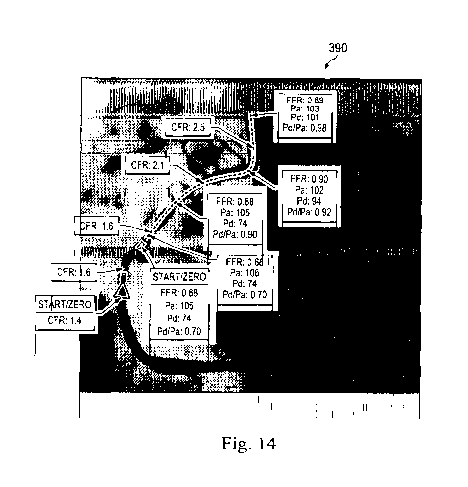
Referring to Figs. 14 and 15, shown therein are enhanced angiographic images
that
include visualizations based on both pressure measurements and flow
measurements. In
particular, Fig. 14 shows an enhanced angiographic image 390 having both CFR
and FUR
calculations displayed with an arrow identifying the corresponding location at
multiple
locations along the length of the vessel associated with the pullback of the
intravascular
instrument. Fig. 15 shows an enhanced angiographic image 400 with similar
features, but
also including a graph of corresponding pressure measurements, flow
measurements, and/or a
chart of the intravascular information used to make the calculations and/or co-
register the
intravascular information to the angiographic image.
It is understood that numerous other visualization techniques may be utilized
to
convey the information in the context of an angiographic image or other image
of the vessel
(including both intravascular and extravascular imaging techniques, such as
IVUS, OCT,
ICE, CTA, etc.) to help the user evaluate the vessel. In that regard, while
the examples of the
present disclosure are provided with respect to angiographic images, it is
understood that the
concepts are equally applicable to other types of vessel imaging techniques,
including
intravascular and extravascular imaging
In some instances, a user is able to select what information should be
included or
excluded from the displayed image. In that regard, it should be noted that
these visualization
techniques related to conveying the pressure measurement data in the context
of an
angiographic or other image of the vessel can be utilized individually and in
any
combinations. For example, in some implementations a user is able to select
what
visualization mode(s) and/or portions thereof will be utilized and the system
outputs the
display accordingly. Further, in some implementations the user is able to
manually annotate
the displayed image to include notes and/or input one or more of the measured
parameters.
In some instances, the user has the option to either show or hide the graphs,
tables, and/or
other data corresponding to the overlaid physiological data. To this end, the
user may click
or otherwise select to display the graphs, tables, and/or other data,
including zooming in on
particular relevant data or other information of interest to the user.
The images of vessels can include three-dimensional, two-dimensional,
angiographic,
a computed tomography angiographic (CTA), and/or other suitable forms of
images. In some
embodiments, a three-dimensional image may be rotated about a vertical axis.
In some
embodiments, a two-dimensional image may include multiple views about a
vertical axis
-23-
CA 02896589 2015-06-25
WO 2014/106186
PCT/US2013/078321
such that different two-dimensional views are shown when the image is rotated.
In some
implementations, the three dimensional model is displayed adjacent to a
corresponding two
dimensional depiction of the vessel. In that regard, the user may select both
the type of
depiction(s) (two dimensional (including imaging modality type) and/or three
dimensional)
along with what visualization mode(s) and/or portions thereof will be
utilized. The system
will output a corresponding display based on the user's preferences/selections
and/or system
defaults.
Those skilled in the art will recognize that the features described above can
be
implemented in a many different ways, dependent upon various factors such as
user
preference, targeted physiology, type(s) of intravascular instrument(s)
utilized, available
processing resources, procedure time, etc. However, an exemplary technique for
creating an
enhanced angiographic image with overlaid physiological measurements according
to
embodiments of the present disclosure will now be described. The result is a
compound
image (or a series of images) constructed to highlight the radiopaque
locations and/or the
attendant physiology measurements using selected symbol settings. It is
understood that the
steps described below may be performed in a different order, include
additional steps, omit
steps described, and/or otherwise be modified without departing from the scope
of the present
disclosure.
In some instances, the method begins by allowing a user to define the
physiology
overlay display settings. That is, a user defines what information and in what
format should
be displayed. The system may include default settings, group settings, and/or
individual
settings. The group settings allow a group of users to share display settings.
Multiple groups
can be defined, each with custom settings. Similarly, the individual settings
allow an
individual user to have custom display settings. Again, multiple individuals
can be defined,
each with custom settings. The overlay settings can be utilized to set the
display parameters
for any of the features described in the present application. For sake of
brevity, a few options
will be described. For example, an individual or group can select how to
enhance the
vascular location and display the physiology measurement(s) on the
angiographic image
using unique symbols (arrow, circle, Pd, FFR, CFR, FPR/CFR, etc.), symbol
combinations
(arrow/FPR, arrow/CFR, etc.), symbol colors, symbol actions (on, fade-in/fade-
out, strobe,
etc.), etc.
With the initial physiology overlay display settings defined, an intravascular
instrument is positioned within the anatomy and utilized to obtain
intravascular information.
In some instances, an optional hyperemic drug is administered. With the
physiological
-24-
CA 02896589 2015-06-25
WO 2014/106186
PCT/US2013/078321
overlay activated (via button, voice command, etc.), the system calculates the
pixel location
and uniquely identifies every radiopaque element of the intravascular
instrument relative to
the angiographic image(s) within the display. In some implementations,
multiple radiopaque
elements on the same guide wire can be uniquely identified (length,
radiopacity, etc.). The
intravascular information or physiology is measured with the intravascular
instrument and the
intravascular/physiology measurement is associated with the two-dimensional
display
location that corresponds to the radiopaque element that is the sensor
location (or sensor focal
point). Generally, duplicate images (i.e., where the location of radiopaque
elements is the
same or "equal") may generally be ignored. The pre-selected symbol(s) from the
selected
physiology overlay display settings is then superimposed on the angiographic
image at the
two-dimensional display location identified. Optionally, the compound image
and
associative elements (time-stamp, angiographic orientation, etc.) may be saved
as a new
single element separate from the separate underlying data. This procedure is
repeated for
each heartbeat (or other common interval) as the intravascular instrument is
moved through
the vessel. In some instances, data is captured once per heartbeat. The
compound image(s)
are then displayed to the user. In some instances, multiple compound images
are provided
using different underlying angiographic images obtained during movement of the
intravascular device. In some instances, the different angiographic images are
from different
orientations to allow alternative views of the vessel. For example, bi-plane
angiography is
utilized in some instances.
Aspects of the present disclosure provide: (1) a comprehensive map of focal
and/or
diffuse stenosis severity for a targeted vascular region; (2) a demonstration
that CFR is
linearly related to FFR for progressive stenosis superimposed on diffuse
narrowing; (3) a
demonstration that the relative contributions of focal and diffuse disease
define the slope and
values along the linear CFR and FFR relationship; and (4) a showing that
discordant CFR and
FFR values reflect divergent extremes of focal and diffuse disease, not
failure of either tool.
Persons skilled in the art will also recognize that the apparatus, systems,
and methods
described above can be modified in various ways. Accordingly, persons of
ordinary skill in
the art will appreciate that the embodiments encompassed by the present
disclosure are not
limited to the particular exemplary embodiments described above. In that
regard, although
illustrative embodiments have been shown and described, a wide range of
modification,
change, and substitution is contemplated in the foregoing disclosure. It is
understood that
such variations may be made to the foregoing without departing from the scope
of the present
-25-
CA 02896589 2015-06-25
WO 2014/106186
PCT/US2013/078321
disclosure. Accordingly, it is appropriate that the appended claims be
construed broadly and
in a manner consistent with the present disclosure.
-26-