Note : Les descriptions sont présentées dans la langue officielle dans laquelle elles ont été soumises.
CA 02902075 2017-01-05
1
A TENSIONING RING FOR RESTORING THE ANTERIOR CAPSULE CENTRIPETAL
FORCES LOST BY CAPSULORHEXIS AND EXEMPLARY CAPSULES CONTAINING
ACCOMMODATIVE INTRAOCULAR LENSES AND RESTORED BY SAME
Cross-Reference to Related Applications
This application claims priority to U.S. Provisional Patent
Application Serial No. 61/770,446, filed on February 28, 2013.
Field of the Invention
The present invention relates to tensioning rings for
anterior capsules and accommodative intraocular lenses for use
therewith, and to methods for implanting the tensioning rings and
intraocular lenses.
Specifically, the present invention relates
to tensioning rings for attachment to anterior capsules to restore
the centripetal forces that were provided by the portion of
anterior capsules removed by capsulorhexis, to accommodative
intraocular lenses that are directly or indirectly actuated by the
rings, and to methods for implanting the lenses and the rings.
Discussion of Background Art
Prior art intraocular lenses, whether accommodative or
single-focus, are typically implanted in the capsule of an eye
from which the crystalline lens has been removed via a procedure
that includes capsulorhexis. Because the capsulorhexis destroys
the natural accommodation mechanism of the eye
whereby the
crystalline lens is elastically reconfigured to a diopter power
appropriate for the visual task by the posterior forces exerted by
the anterior capsule and the anterior forces exerted by the
posterior capsule, the visual accommodation must be provided in
some other way.
Because the crystalline lens is also both a spacer between
the anterior and posterior capsules and a determinant of the
zonule-proximal capsule curvature, and thus a determinant of the
zonular load distribution in the natural eye, these crystalline
lens functions, which are lost by its extraction, must also be
addressed.
1
CA 02902075 2015-08-20
WO 2014/134302
PCT/US2014/019016
2
Most of the accommodative intraocular lenses in the prior art
assume the validity of the Helmholtz Theory of Accommodation -
i.e., that tension on the zonules increases the equatorial
diameter of the capsule and flattens the capsule, and thus
flattens the crystalline lens therein to a shape appropriate for a
distant vision, while contraction of the ciliary body muscle(s)
reduces zonular tension and allows the elastically reconfigurable
crystalline lens to assume a more convex (accommodative) shape.
Thus, lens assemblies implanted in capsules and having
accommodating mechanisms responsive to these changes in diameter
should be able to provide the visual accommodation despite the
capsulorhexis.
The prior art includes tens, if not hundreds, of examples of
lenses intended to provide accommodation on this basis. Most of
these lenses can be demonstrated to work as expected in vitro.
None of the lenses, however, work as expected in vivo.
For
example, the named inventors of U.S. Patent Application
Publication Nos. 2009/0234449 (De Juan, Jr., et al.) and
2007/0100445 (Shadduck), attribute this failure to "shrink-
wrapping," and disclose spacers intended to prevent this failure
by maintaining a separation of the anterior and posterior
capsules. While "shrink-wrapping" may be a contributing factor to
this failure, its elimination has not solved the problem.
There are, however, two kinds of prior art intraocular
lenses that can be implanted in capsulorhexis-crippled capsules
that provide some degree of accommodation.
One is a Fresnel
configuration that is disclosed in U.S. Patent Application
Publication No. 2007/0171362 (Simpson).
ReStorTM, ReZoomm and
Tecrism are known trade names for such lenses. These lenses are
not intended to respond to a change in capsular diameter, but
instead have zones of different diopter power, some of which are
appropriate for distance vision and others for reading. The well-
known shortcomings of such lenses, however, include loss of
contrast, halos, etc.
I i
CA 02902075 2015-08-20
WO 2014/134302
PCT/US2014/019016
3
The other is an intraocular lens assembly that is disclosed
in U.S. Patent No. 6,849,091 to Cumming and other U.S. Patents and
published U.S. Patent Applications by the same inventor
("Cumming").
CrystalensTM is a known trade name for such lens
assemblies. These intraocular lens assemblies are anchored
equatorially in capsulorhexis-crippled capsules by "shrink-
wrapping," and accommodation is provided by anterior movement of
the lens in response to forces exerted anteriorly upon the portion
of the lens assembly that is in contact with the posterior
capsule.
Cumming attributes these forces to "viscous pressure"
from the vitreous humor.
Thus the CrystalensTM, which achieves
some degree of visual accommodation, does so in direct opposition
to Helmholtz, who teaches disaccommodation via flattening of the
capsule - i.e., anterior translation of the posterior capsule. De
Juan, Jr. also discloses the use of "viscous pressure" to provide
the visual accommodation. [61
U.S. Patent Application Publication No. 2007/0032867 to
Cumming discloses translational accommodative intraocular lenses
having plate-type haptics with "T" shaped ends, and U.S. Patent
7,985,253 to the same inventor discloses hydraulic accommodative
intraocular lenses in which the "T" shaped ends are curly. Plate-
type haptics are familiar from commercially available intraocular
lenses, and both the "T" shaped and the curly ends are variations
of the "J" type haptics familiar from the prior art that, like the
"J" type haptics, secure the lens to the capsule by the "shrink-
wrapping" of the latter.
U.S. Patent No. 2,300,251 to Flint discloses variable focus
hydraulic lenses of the kind employed by Cumming's hydraulic lens
systems, and U.S. Patent No. 4,261,655 to Honigsbaum teaches
eyeglasses having adjustable focus hydraulic lenses in which a
part of the focusing mechanism is a bellows-like arrangement.
U.S. Patent Application Publication No. 2011/0035001 to
Woods discloses spacer-like "optics positioning members" that are
implanted into capsules and that are intended to provide the
I I
CA 02902075 2015-08-20
WO 2014/134302
PCT/US2014/019016
4
visual accommodation by appropriately positioning lens elements in
response to zonular tension-induced changes in capsule shape, and
are expected to do so despite the unaddressed crippling effects of
capsulorhexis.
U.S. Patent Application Publication No. 2012/0253459 to
Reich, et al. discloses a lens elastically reconfigured by means
in direct contact with the ciliary structure of the eye.
U.S. Patent Application Publication No. 2007/0123981 to
Tassignon ("Tassignon") discloses the use of a ring as a guide or
template for cutting an axisymmetric capsulorhexis as a part of a
procedure for intraocular lens implantation, and a two part
intraocular lens arrangement in which a "U" section haptic ring
that embraces the margin of both an anterior and posterior
capsulorhexis holds a lens that is separable from its haptic ring.
Tassignon also states that such an arrangement can provide
accommodation on the basis of changes in the capsular diameter.
U.S. Patent No. 4,822,360 to Deacon, U.S. Patent No.
7,156,101 to Terwee, and U.S. Patent Application Publication No.
2012/0226351 to Peyman disclose intracapsular lenses intended to
replace the natural crystalline lens. Because the implantation of
such lenses in a fully functional state would require an
unacceptably large corneal incision, the patents disclose
implantation of the lens as an uncured polymer that is to be cured
and shaped in vivo. The patents do not, however, disclose how the
lens-shaping function of the anterior capsule compromised by
capsulorhexis during the removal of the natural lens is restored.
U.S. Patent Application Publication No. 2012/0303118 to
DeBoer, et al. ("DeBoer") discloses a bag-type crystalline lens
replacement that is implanted uninflated via a small equator-
proximal anterior capsule incision that is also used for
lensectomy. The lens is then "inflated" with silicone oil to the
desired size and shape.
DeBoer also discloses a plurality of
self-sealing post-implantation bag access ports.
Such ports are
familiar from known art, such as spray-can valves.
I I
I I
CA 02902075 2015-08-20
WO 2014/134302
PCT/US2014/019016
U.S. Patent Application Publication No. 2007/0213817 to
Esch, et al. discloses hydraulic lens assemblies having tubular
ring haptics in which a portion of the actuating fluid is
contained.
5
Surgical glues are commercially available under trade names
such as BioGlueTM, TissueGlum, etc., and U.S. Patent Application
Publication No. 2011/0029074 to Reisin, et al. discloses
alternatives to the commercially available surgical glue products.
U.S. Patent Application Publication No. 2013/0013061 to
Coroneo ("Coroneo") discloses a "U" section ring for permanent
insertion into a capsulorhexis to apply centrifugal forces to
capsulorhexis-crippled anterior or posterior capsules to address
phimosis.
Tassignon [13] discloses a similar section ring for
centrifugal forces but for a different purpose.
While Coroneo
uses the term "capsular tension rings" (CTR), Coroneo's CTRs are
the familiar "C" shaped rings inserted into some capsules to apply
centrifugal forces to address phimosis. Such CTRs are available
commercially from FCI Opthalmics and others, and, like the Coroneo
ring, apply centrifugal forces to the capsule.
U.S. Patent Application Publication No. 2013/0304206 to
Pallikaris, et al. ("Pallikaris") discloses zonules that
originate at the ciliary body muscle and insert both anteriorly
and posteriorly at the equatorial region of the capsule, thus
centrifugally tensioning the capsule. Pallikaris also discloses
that, if the centripetal equatorial capsular tension lost by
capsulorhexis and crystalline lens removal were restored, the
decrease in zonular tension resulting from contraction of the
ciliary body muscle would decrease the capsule diameter, and
that this change in capsular diameter could actuate an
accommodative intraocular lens.
Pallikaris further discloses three mechanisms to restore
the centripetal tension: (1) a tensioning ring glued
equatorially to the capsule interior, (2) a comb-like equatorial
compression ring the tines of which are implanted between the
II
CA 02902075 2015-08-20
WO 201-1/134302 PCT/US2014/019016
6
zonules, and (3) an interior capsule equator tensioning ring
held in place by clamps and/or grooves that secure a further
surgically modified anterior capsule to the ring.
Capsules are of course soft tissue, and lens implants are
expected to serve the patient for the rest of his/her life -
i.e., for two decades or more, and there are no soft tissue
glues that can serve their intended purpose for anywhere near
this length of time.
There are about 72 zonules, roughly half anterior and half
posterior, and implanting anywhere near this complement of
compression ring tines between both anterior and posterior
zonules without damage to the zonules and/or the tines is a
virtually impossible task.
The three mechanisms for restoring centripetal tensions are
based upon the assumption that the zonules originate at the
ciliary body muscle and insert both anteriorly and posteriorly
at the equatorial region of the capsule. Both in-vivo studies
(e.g., "Extralenticular and Lenticular Aspects of Accommodation
and Presbyopia in Human vs. Monkey Eyes," IOVS Manuscript, IOVS
12-10846, June 6, 2013 by M.A. Croft, et al. ("Croft")) and in-
vitro studies (e.g., "Evidence for Posterior Zonular Fiber
Attachment on the Anterior Hyaloid Membrane," IOVS Vol. 47, No.
11, Nov. 2006 by Bernal, et al. ("Bernal")) confirm, however,
that at least some of the zonules originate elsewhere on the
ciliary body, and also confirm that at least some of the
posterior zonules insert at the hyaloid membrane before
inserting at the posterior capsule. Thus, the change in zonular
tension moves the capsule equator anteriorly and posteriorly
with respect to the tensioning device or rotates the tensioning
devise about the axis of its cross-section, or both.
This applies peel-type loading to the glued version (which
is the kind of loading to which glue bonds are most vulnerable),
a saw-type motion to the tines of the comb-like version (which
can cut the zonules and/or the tines), and accommodation when
I I
CA 02902075 2015-08-20
WO 2014/134302
PCT/US2014/019016
7
disaccommodation is expected (and vice versa) when a surgically
modified anterior capsule is attached to a capsule-equator-based
tensioning ring.
The Croft and Bernal studies are of particular interest
because they both offer evidence that at least some, if not all,
of the posterior zonules insert at the anterior hyaloid membrane
before inserting at the posterior capsule.
Thus, the anterior
translation of the posterior capsule is constrained while the
anterior capsule is free to translate posteriorly in response to
anterior zonule tension because the anterior zonules insert
directly at the anterior capsule.
This not only explains the
visual accommodation mechanism of the human eye, which is a
combination of translation and elastic reconfiguration, but it
also explains the need to restore the centripetal anterior capsule
forces lost by capsulorhexis if the accommodative intraocular
lenses of this invention and/or those of the prior art are to
serve their intended function.
Summary of the Invention
This invention addresses the accommodative failures of prior
art intraocular lenses with a tensioning ring that is attached to
the anterior capsule to restore the centripetal anterior capsular
forces lost by capsulorhexis.
These centripetal forces are
opposed by the centrifugal forces exerted by the zonules, and the
latter forces are increased and decreased by the relaxation and
contraction of the ciliary body muscle(s), respectively.
Thus,
the anterior capsular forces are restored to their pre-
capsulorhexis levels, and these restored forces are directly
and/or indirectly used to actuate an accommodative intraocular
lens assembly implanted in the capsule.
Suitably modified
accommodative intraocular lens assemblies can alternately be
implanted for actuation by the change in tensioning ring diameter
rather than by forces exerted by the capsule.
This invention also includes a spacer to maintain separation
between the anterior and the posterior capsule and to restore at
CA 02902075 2015-08-20
WO 2014/134302
PCT/US2014/019016
8
least some of the equator-proximal capsule curvature and thus the
zonular load distribution lost by crystalline lens extraction.
Spacers also actuate the accommodative mechanism of the lenses of
some versions of this invention, and serve as a mounting and
support structure for the lenses of others.
In one embodiment, a tensioning device configured for
attaching to the anterior capsule of an eye is provided.
The
tensioning device includes a biocompatible, elastically
reconfigurable ring for restoring at least a portion of anterior
capsule centripetal forces lost by capsulorhexis. The tensioning
device also includes a plurality of penetrators configured for
attaching the ring to the anterior capsule.
The plurality of
penetrators is biocompatible and partially embedded to a part of
the ring configured for facing the anterior capsule.
In another embodiment, a tensioning ring is configured for
attaching to the anterior face of a shrink-wrapped capsule for
securing a replacement intraocular lens or an augmentation
intraocular lens to the capsule. The tensioning ring includes a
plurality of penetrators that are partially embedded in a part of
the ring configured for facing the shrink-wrapped capsule.
The
tensioning ring also includes a substantially axisymmetric groove
in a part of the ring proximal to the ring's principal axis. The
groove is configured for initially holding an expander ring and
for permanently holding the haptics of a replacement or an
augmentation intraocular lens.
The ring is made of a
biocompatible, elastically reconfigurable polymer.
In yet another embodiment, a biocompatible accommodative
intraocular lens system is provided. The lens system includes a
tensioning ring that is configured for attaching to the anterior
capsule of an eye to restore at least a part of anterior capsule
centripetal forces lost by capsulorhexis.
The lens system also
includes a foldable accommodative intraocular lens assembly that
is configured for implantation into the capsule of the eye to
restore at least a portion of normal vision to the eye. The lens
11
CA 02902075 2015-08-20
WO 2014/134302
PCT/US2014/019016
9
system further includes a foldable spacer-actuator that is
configured for implantation in the capsule to maintain separation
between the anterior capsule and the posterior capsule of the eye
and to translate the accommodative intraocular lens system. The
foldable spacer-actuator, may be attached to the lens assembly.
It may also be an integral part of the lens assembly.
In another embodiment, a biocompatible accommodative
intraocular lens system is provided. The lens system includes a
grooved tensioning ring configured for attaching to the anterior
capsule of an eye to restore at least a portion of the
centripetal forces lost by capsulorhexis. The lens system also
includes a foldable hydraulic accommodative intraocular lens
assembly configured for implantation in the capsule to restore
at least a portion of normal vision to the eye and a ring-type
hydraulic actuator for implantation in the tensioning ring
groove to change the diopter power of the hydraulic intraocular
lens assembly. The lens system further includes a foldable
spacer configured for maintaining a separation between an
anterior portion and a posterior portion of the capsule of the
eye, and for holding the hydraulic intraocular lens assembly.
In another embodiment, a method is provided for implanting
an accommodative intraocular lens system into an eye to replace a
cataractous or otherwise compromised crystalline lens, wherein the
intraocular lens system comprises a grooved anterior capsule
tensioning ring for attaching to the anterior capsule of the eye
and one of a capsule-equator-anchored accommodative intraocular
lens assembly with a spacer-actuator and a spacer-based
accommodative intraocular lens assembly with a spacer. The method
includes the steps of preparing the eye for capsulorhexis and
injecting into the eye the tensioning ring. An expander ring has
been inserted into a groove of the tensioning ring. The method
also includes the steps of expanding the tensioning ring to a
diameter that provides the ring centripetal forces to be applied
to the anterior capsule after implantation of the lens assembly
11
II
CA 02902075 2015-08-20
WO 2014/134302
PCT/US2014/019016
and one of the spacer-actuator and spacer.
The method further
includes the steps of positioning the tensioning ring
substantially axisymmetrically on the anterior capsule and
attaching the tensioning ring to the anterior capsule by reducing
5 the expander ring pressure. The method also includes the steps of
cutting a substantially axisymmetric capsulorhexis, adjusting the
expander ring pressure to a level corresponding to anterior
capsule centripetal forces safe for the zonules of an empty
capsule, and extracting the crystalline lens.
The method also
10
includes the steps of purging the surgical debris from the capsule
and the eye, implanting one of the lens assembly with the spacer-
actuator and the lens assembly with the spacer into the capsule,
and depressurizing the expander ring and removing it from the eye.
In another embodiment, a method for securing one of a
replacement intraocular lens assembly and an augmenting
intraocular lens assembly to the anterior face of the shrink-
wrapped capsule of an eye is provided. The method includes the
steps of preparing the eye as for a cataract surgery, and
injecting a grooved tensioning ring having an expander ring into
the eye.
The method also includes the steps of expanding the
tensioning ring to a diameter that provides a desired ring
centripetal force and positioning the tensioning ring
axisymmetrically against the anterior face of the shrink-wrapped
capsule.
The method further includes the steps of reducing the
expander ring pressure to attach the grooved tensioning ring to
the anterior face of the shrink-wrapped capsule, depressurizing
the expander ring, and removing it from both the grooved
tensioning ring and the eye. The method also includes the steps
of injecting one of the replacement intraocular lens assembly and
the augmenting intraocular lens assembly into the eye, and
inserting the haptics of the lens assembly into the tensioning
ring groove.
Other features and advantages of the present invention will
become apparent from the following more detailed description,
II
I I
CA 02902075 2015-08-20
WO 2014/134302
PCT/US2014/019016
11
taken in conjunction with the accompanying drawings, which
illustrate, by way of example, the principles of the presently
described apparatus and methods of its use.
I 1
CA 02902075 2015-08-20
WO 2014/13-1302
PCT/US2014/019016
12
Brief Description of the Drawings
The following description, given with respect to the
attached drawings, may be better understood with reference to the
non-limiting examples of the drawings, wherein:
FIG. 1 is a cutaway perspective view of an eye.
FIG. 2 is the cutaway perspective view of an eye of FIG. 1
into which a tensioning ring has been implanted in accordance with
some embodiments of the disclosed subject matter;
FIGS. 3a-3g are plan and sectional views of rings in
accordance with some embodiments of the disclosed subject matter;
FIGS. 4a-4e are cutaway perspective and sectional views of
capsule-equator-anchored accommodative intraocular lenses in
accordance with some embodiments of the disclosed subject matter;
FIGS. 5a-5d are sectional views of spacer-anchored
accommodative intraocular lenses in accordance with some
embodiments of the disclosed subject matter; and
FIGS. 6a-6c are cutaway perspective, plan and sectional
views of a defective shrink-wrapped intraocular lens and the
details of its replacement in accordance with some embodiments of
the disclosed subject matter.
Definitions
The term "accommodation" as used herein means the ability of
a lens to change its focus from distant to near objects.
The term "accommodative" is used herein to describe a
replacement lens system and/or a component thereof for
implantation in an eye to provide distance vision and
accommodation.
The term "biocompatible" is used herein to describe a
substance (or an item made therefrom) that neither elicits an
unacceptable immune response when implanted in an eye nor elicits
an unacceptable change to itself, to other components of the
systems and/or the lens assemblies of this invention that have
been implanted in the eye, nor to the eye itself.
CA 02902075 2015-08-20
WO 2014/13-1302
PCT/US2014/019016
13
The terms "centripetal" and "centrifugal" are used herein in
lieu of "radially inward" and "radially outward" respectively, and
are so used in both classical mechanics and the medical
literature.
The terms "cut" and "cutting" when used herein with respect
to capsulorhexis are intended to include lasering and tearing as
well as actual cutting.
The term "foldable" as used herein includes the rolling,
folding, and otherwise elastically reconfiguring spacers, lens
assemblies and rings for insertion into an injector.
The term "translation" is used herein as in classical
mechanics to describe linear motion.
Ophthalmologists use the
term "vaulting" to describe the anterior translation of a
crystalline or implanted lens, and that is the meaning intended
herein.
Medical terms not specifically defined herein are as defined
in standard medical dictionaries such as Merriam-Webster's Medical
Dictionary.
Detailed Description of the Presently Preferred Embodiments
The description and the drawings to which the description
refers are for purposes of explanation and illustration and are
not for limiting the scope of the invention. The
scope of the
invention is defined by the claims.
FIG. 1 is a cutaway perspective view of an eye according to
the prior art, and from which a cataractous or otherwise vision-
compromising crystalline lens 101 is to be removed and its
function replaced by an intraocular lens. Lens 101 is contained
in capsule 102, which comprises anterior capsule 102a and
posterior capsule 102b. Anterior capsule 102a includes the part
of capsule 102 that is anterior to the equatorial plane of capsule
102, and posterior capsule 102b includes the part of capsule 102
that is posterior to the equatorial plane of capsule 102.
I I
CA 02902075 2015-08-20
WO 2014/134302
PCT/US2014/019016
14
Anterior capsule 102a is connected to ciliary body muscle(s)
104 by anterior zonules 103a and posterior capsule 102b is
connected to ciliary body muscle(s) 104 by posterior zonules 103b,
and the zonules, according to Helmholtz, act in unison to apply
tension to capsule 102, thereby flattening the capsule and the
lens for distant vision when muscle(s) 104 are relaxed.
Contraction of ciliary body muscle(s) 104, again according to
Helmholtz, relaxes the tension on zonules 103 allowing the
elastically reconfigurable lens and its capsule to assume the more
rounded shape appropriate for the visual accommodation (or at
least to do so in a healthy pre-presbyopic eye).
FIG. 1 also
shows cornea 105, sclera 106, and iris 107.
According to recent studies (e.g., in vivo studies of Croft
and in vitro studies of Bernal), at least some, if not all, of the
posterior zonules 103b insert at the hyalon membrane before
inserting at the anterior capsule, and originate elsewhere on the
ciliary body. Thus, the anterior-posterior translation of the
posterior capsule is more constrained than that of the anterior
capsule, and a decrease in anterior zonular tension not only
allows the crystalline lens to assume a more rounded
(accommodative) shape, but it also translates the optical center
of the crystalline lens anteriorly, and vice versa.
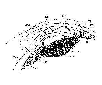
FIG. 2 shows substantially axisymmetric tensioning ring 211
that is attached under tension to anterior capsule 202a by at
least one of prongs, barbs, hooks, claws and scarring.
Ring
implantation before capsulorhexis is preferred because the
undisturbed capsule is a more stable platform for its
implantation, the ring function is best served by a capsulorhexis
that is concentric with the ring, and the ring is a convenient
template for making it so.
FIG. 2 illustrates a step in
practicing this invention that is different from the intraocular
lens implantation procedures of the prior art.
The steps following attachment of the actuating ring 211 of
FIG. 2 are capsulorhexis, extraction of the crystalline lens,
I I
I
CA 02902075 2015-08-20
WO 2014/13-1302
PCT/US2014/019016
purging of surgical debris and implantation of an intraocular
lens, and these steps are known and regularly practiced by
ophthalmologists, some of whom may prefer to apply a cell growth
inhibitor such as Fluorouracil 5 (fu5) to the cut edge of the
5 capsulorhexis.
Some of the accommodative intraocular lenses of this
invention also require the implantation of spacers, spacer-
actuators, etc.
Because extraction of crystalline lens 201 also removes the
10 ultraviolet (UV) protection provided by that lens to parts of the
eye (e.g., the retina) that are sensitive to UV, the two-optical-
surface lenses 4b26 of FIG. 4b, 4e26.2 of FIG. 4e, 5a26.2 of FIG.
5e and 6b26 of FIG. 6b and the membrane 5c26.2.2 of FIG. Sc
preferably provide the UV protection last by crystalline lens
15 extraction.
FIG. 3a illustrates a toroidal ring 3all.
Toroidal ring
3all comprises a biocompatible, elastically reconfigurable
material such as a silicone polymer. A plurality of substantially
uniformly spaced biocompatible penetrators 3a12 is partially
embedded in toroidal ring 3all at a uniform angle such that when a
portion of the centripetal anterior capsule forces are transferred
from the anterior capsule to the ring by capsulorhexis or other
means, penetrators 3a12 pierce the anterior capsule and secure the
ring to the anterior capsule.
That uniform angle is preferably
twenty to twenty five degrees because penetrators 3a12 having
significantly smaller angles will not be able to serve their
intended purpose, and those having significantly larger angles can
damage anterior capsule cells at the junction of the penetrators
and the ring. Significantly larger angles also increase the risk
of capsule damage if the ring is repositioned, replaced or
removed.
The penetrators are preferably made of a metal such as that
used for stents, and a listing of such metals is contained in
Levesque, J. et al., Materials and Properties for Coronary Stents,
CA 02902075 2015-08-20
WO 201-1/134302 PCT/US2014/019016
16
Advanced Materials and Properties, Sept. 2004 pp 45-48. Of these,
nitenol and stainless steel are presently preferred. The
penetrators can be hooks, barbs, pins and/or prongs, but the pin-
type penetrators shown in the figures are presently preferred
because the others can cause more anterior capsule damage not only
initially, but even more so if the ring is repositioned.
The pin-type penetrators are preferably sharp enough to
penetrate the anterior capsule but blunt enough to push aside the
elastic fibers of the anterior capsule rather than puncturing and
destroying them as they penetrate the anterior capsule to attach
the ring to the anterior capsule. The pin-type penetrators may be
also preferably be textured and/or coated to encourage ring
retention by the anterior capsule.
Suturing and gluing have also been considered as ways of
attaching the rings, but the former introduces an even greater
risk of tearing, and no known biocompatible soft tissue glues have
an in vivo adhesive life expectancy that is even a small fraction
of that of an intraocular lens.
Such means of attachment that
suitably address these concerns are not, however, excluded from
the invention.
FIG. 3b illustrates a toroid-like ring with a D-shape
cross-section. The
penetrators are distributed over the flat
part of the ring, thereby reducing the risk of tearing.
FIG. 3c illustrates a tubular ring 3c11 that has an inner
chamber 3c14 that is accessible via an access port 3c15. Ring
3c11 can be used to achieve a desired centripetal force if that
provided by rings 3all and 3b11 proves to be insufficient. Ring
3c11 also has penetrators but the penetrators have been omitted
from the drawing for purposes of clarity of the illustration.
Ring 3011 is inserted into the eye with inner chamber 3c14
filled with a biocompatible silicone oil, and at a pressure
slightly above atmospheric pressure to prevent the influx of
fluids from the eye. Once in the eye, it is pressurized with the
silicone oil to increase its major diameter to one that
CA 02902075 2015-08-20
WO 2014/134302 PCT/US2014/019016
17
corresponds to a desired centripetal force. The
ring is then
temporarily attached to the anterior capsule with viscoelastic
and/or a weak surgical glue and depressurized as appropriate to
embed the penetrators.
FIG. 3d illustrates an expander ring 3d11 for the anterior
capsule tensioning rings of FIGS. 3e and 3f. Expander ring 3d11
comprises a coil spring 3d16 that is closed upon itself, a
bladder 3d17 contained within the spring, and a pressurizing tube
3d18. The spring and bladder combination is somewhat analogous to
a tube and tire combination in that the bladder is the pressure
vessel as is the tube and the spring determines its pressurized
shape as does the tire. Thus, when the bladder is pressurized via
tube 3d18 or (less conveniently) via an access port such as 3c15
shown in FIG. 3c (not shown in FIG. 3d), the spring constrains the
response of the spring/bladder combination to an increase in the
major diameter of ring 3d11 (in practice, such spring would likely
have its coils wound more tightly than those in the drawing).
Suitable spring materials include those used for stents.
The material for bladder 3d17 is preferably an elastically
reconfigurable silicone polymer and its shape is ideally a
toroidal shell, but the manufacturing complications of winding a
spring about such a shell make it more convenient to use tubing or
a shell that has been cut and its ends sealed closed.. FIG. 3d
shows cut ends 3d17.1 and 3d17.2 that are so sealed .
FIGS. 3e and 3f are alternative cross-sections for the ring
shown in FIG. 3b. The embodiment of FIG. 3e is much like that of
FIG. 3b with the exception of a groove 3e19 for accommodating an
expander ring like shown in FIG. 3d. Spring 3e16 and bladder 3e17
are also shown, but the inflation tube corresponding to tube 3d18
of FIG. 3d is not shown. Groove
3e19 also has a lip 3e20, the
purpose of which is explained in the descriptions of FIGS. 5b and
6b.
The purpose of the expander ring is to preload the
tensioning ring so that it can replace the centripetal anterior
CA 02902075 2015-08-20
WO 2014/134302 PCT/US2014/019016
18
capsular forces lost by capsulorhexis.
Because bladder 3e17
pressure, and thus the ring tension and diameter, are under
control of the surgeon, the ring can be expanded to a diameter
appropriate for preliminary positioning on the anterior capsule
and reduced enough to implant penetrators 3e12 into the capsule,
and the capsulorhexis cut.
After the penetrator are implanted
into the capsule and the capsulorhexis is cut, the expander ring
pressure can be further adjusted to atmospheric pressure, and the
expander ring can be removed from both the ring and the eye.
FIG. 3f illustrates groove 3f21 and spring 3f22. The
purpose of spring 3f22 is to provide some of the centripetal
capsular force that is lost by capsulorhexis and that would
otherwise be provided by the ring. A
metal spring, however,
introduces some restrictions with respect to material selection
because both spring 3f22 and penetrators 3f12 are made of metal,
both remain in place and are immersed in the same electrolyte (the
aqueous humor), and differences in material introduce the
possibility of unintended electrochemical reactions. Thus, spring
3f22 and penetrators 3f12 are preferably made of the same metal
to address this possibility.
Ring 3g11 shown in FIG. 3g differs from the previously
described rings in that the radially inward capsular force is
provided not only by the D-shaped ring portion 3g13, which may be
modified in accordance with FIG. 3e, but also by transparent
elastically reconfigurable membrane 3g23 attached to it.
Ring 3g11 has penetrators along the flat face of its "D"
shaped perimeter as do the rings shown in FIGS. 3b, 3e and 3f, but
the penetrators have been omitted from the drawing because
including them would erroneously suggest that membrane portion
3g23 also has penetrators, when it does not.
Because ring 3g11 is intended as anterior capsule repair for
use with lenses that are elastically reconfigured by the capsule,
including lenses taught by Deacon, Terwee and Peyman, it restores
not only the radially inward capsular forces lost by
I i
CA 02902075 2015-08-20
WO 2014/134302
PCT/US2014/019016
19
capsulorhexis, but also the lens shaping function lost also by
capsulorhexis. Membrane 3g23 is for this second purpose thinner
at its center than it is at its edge, and has local mechanical
properties approximating those of the portion of the anterior
capsule it is intended to replace.
Typical membrane materials
include those used in accommodative intraocular hydraulic lenses.
Membrane 3g23 would, however, block access to the capsules
for purposes of capsulorhexis, crystalline lens extraction, and
the implantation of lenses, if ring 3g11 were attached to anterior
capsules before capsulorhexis.
For this reason, ring 3g11 is
attached to anterior capsules after some of the above-described
steps (e.g., capsulorhexis, removal of crystalline lens, etc.)
have been completed.
Regarding the size of the tensioning rings or the external
length of penetrators, capsularexii are typically about five
millimeters in diameter, crystalline lenses are about six
millimeter in diameter, and lens capsules are about ten
millimeters in diameter.
Thus, allowing for capsulorhexis
irregularities, the nominal half millimeter difference in capsule
diameter between visual accommodation and disaccommodation, and
the risk that the penetrators of rings having major outer
diameters approximating that of the capsules can damage the
anterior zonules, the presently preferred limits for rings after
implantation are major inner diameters of about six millimeters
and major outer diameters of about eight millimeters.
However,
this is not intended to suggest that rings must have these minimum
and maximum diameters if rings having smaller maxima and/or
different minima are more appropriate for their intended purpose.
Anterior capsules comprise curvilinear radial elastic fibers
and circular elastic fibers orthogonal to the radial elastic
fibers (an arrangement suggestive of the longitudinal and
latitudinal coordinates of a hemispherical shell), and the
penetrators are preferably sharp enough to penetrate these
capsules but dull enough to push aside the elastic fibers rather
CA 02902075 2015-08-20
WO 2014/134302 PCT/US2014/019016
than piercing and cutting or otherwise damaging them.
These
pushed aside elastic fibers will try to return to their previous
orthogonal configuration and will thus help to secure the
tensioning ring to the anterior capsule, and this securing can be
5 enhanced by contouring, texturing and/or coating the penetrators
for maximum effect.
Anterior capsules are also thickest at their equators and
thinnest at their poles, and penetrators need to have an external
length no more than a few micrometers greater than the portion of
10 the capsule they are intended to penetrate. Their optimal length
is best initially determined by in vitro studies and refined on
the basis of the experience.
FIG. 4a is a perspective view, partially cut away, of the
eye shown in FIG. 2 after capsulorhexis 4a25, the extraction of
15 crystalline lens (e.g., lens 101 shown in FIG. 1), the purging of
surgical debris from the eye, and the intracapsular implantation
of the accommodative intraocular lens assembly comprising lens
4a26 and haptics 4a27 having bending grooves 4a28.
Accommodation in the embodiment of FIG. 4a is effected by
20 the lens assembly in response to a decrease in tension on anterior
zonules 4a03a that results from contraction of the ciliary body
muscle(s) and the centripetal forces provided by anterior capsule
4a02a and tensioning ring 4all, all of which combine to apply
centripetal forces to haptics 4a27 that bend the haptics at
bending grooves 4a28 to translate (vault) lens 4a26 anteriorly.
The lens assembly of FIG. 4a is also intended as a stand-in
for other intraocular lens assemblies intended to be
accommodative.
Some lens assemblies also serve as shrink-wrap-
prevention spacers. No such spacers are shown in Fig 4a on the
assumption that tensioning ring 4a11 and anterior zonules 4a03a
will maintain separation between anterior capsule 4a02a and
posterior capsule 4a02b.
Even if separation could be so maintained, however, it would
be at the risk of the anterior zonule 4a03a stretching, tearing,
I I
CA 02902075 2015-08-20
WO 2014/134302
PCT/US2014/019016
21
and/or detaching as mentioned earlier.
These risks to the
anterior zonules can be addressed by limiting the amount of
centripetal force applied to the anterior capsule by tensioning
ring 4all, but this also limits the change in capsule diameter
when the ciliary body muscle contracts, thus limiting the change
in accommodation as well.
FIG. 4b shows an embodiment that addresses the risks to the
anterior zonules of FIG. 4a with a spacer 4b29, which serves as
both a spacer with respect to capsule 4b02a,b and an actuator of
lens assembly comprising lens 4b26 and haptics 4b27 having bending
grooves 4b28, and will be referred to as a
spacer-actuator.
Spacer-actuator 4b29 is shown in greater detail in FIG. 4c.
Also shown in FIG. 4b are tensioning ring 4b11, anterior
capsule 4b02a, anterior capsulorhexis 4b25 through which the
crystalline lens and its associated debris has been removed,
anterior zonules 4b03a, posterior capsule 4b02b, and posterior
zonules 4b03b, 4b03b1, 4b03b2. Some of the posterior zonules are
shown as discontinuous to emphasize the point made earlier herein
that at least some of the posterior zonules insert at the hyalon
membrane (not shown) before inserting at the posterior capsule.
The haptics 4b27 of the lens assembly shown in FIG. 4b have
a posterior bias (they are for this reason shown angled towards
the left with respect to lens 4b26 in the drawing) so that, in the
absence of other forces, the lens will lie in its accommodative
(anterior) position, i.e., to the right of the equator of capsule
4b02a,b as shown in the drawing.
When the ciliary body muscle(s) in the natural eye relax to
effect disaccommodation the anterior zonules tighten, the anterior
capsule flattens, and this flattening both flattens the
crystalline lens and translates it posteriorly. This translation
is possible in part because the response of the posterior capsule
to changes in zonular tension is constrained by the previously
mentioned posterior zonular insertion at the hyalon membrane.
I I
CA 02902075 2015-08-20
WO 2014/134302
PCT/US2014/019016
22
Because the centripetal anterior capsule 4b02a forces lost
to capsulorhexis have been restored by tensioning ring 4b11,
anterior capsule 4b02a will also respond to increased zonular
tension by flattening, and this flattening will move spacer-
actuator 4b29 posteriorly (to the left in the drawing), bend
haptics 4b27 posteriorly at bending grooves 4b28, overcome the
posterior bias of the haptics, translate lens 4b26 posteriorly,
and effect disaccommodation (and vice versa).
Bending haptic 4b27, however, also risks bending and thus
distorting lens 4b26, and while some of this risk is addressed by
positioning bending grooves 4b28 proximal to lens 4b26, it may
also be appropriate to attach the haptics to a lens support
structure as shown in FIG. 4e, instead of directly to the lens.
This support structure can also be modified to provide anterior
bias in addition to that provided by haptics 4b27 as explained
with respect to 4e31 of FIG. 4e.
The anterior face of spacer-actuator 4b29 is in contact with
anterior capsule 4b02a, and is contoured accordingly, as shown in
greater detail in FIG. 4c.
This contour, however, changes with
change in anterior zonular tension and this change in contour is
addressed by grooves that allow the face of spacer-actuator 4b29
in contact with anterior capsule 4b02a to flex accordingly.
The anterior face of spacer-actuator 4b29 is shown as face
4c29.1 in FIG. 4c, the flex portions of face 4c29.1 as 4c29.2 and
4c29.4, and the grooves defining them as grooves 4c29.3 and
4c29.5, respectively.
Depending on the amount of flexing
expected, it may also be appropriate to slit the flex portions
into flaps via slits 4c29.6.
Spacer-actuator 4b29 also has a
posterior face 4b29.7 (4c29.7 in FIG. 4c) which presses against
haptics 4b27 to effect disaccommodation.
Haptics 4b27 are plate-type haptics that are intended to
anchor the lens assembly comprising lens 4b26 and haptics 4b27
having bending grooves 4b28 to the equator of capsule 4b02a,b,
and to do so despite a change in capsule diameter resulting from a
CA 02902075 2015-08-20
WO 2014/134302 PCT/US2014/019016
23
change in zonular tension, and to do so without compromising the
accommodation-disaccommodation mechanism described earlier.
Thus, with reference to FIG. 4d, which is a plan view of the
anterior face of the haptic 4b27 in the upper part of FIG. 4b and
part of lens 4b26, the haptic is identified by leader line 4d27,
its lens-proximal bending groove by leader line 4d28, and the part
of the lens by leader line 4d26.
Also shown in FIG. 4d are
tapered haptic outriggers 4d27.1, 4d27.2 which, like the "J"
haptics of conventional intraocular lenses, both allow for
differences in capsular equatorial diameter and center the lens in
the capsule. The "J" haptics, however, do so only before "shrink-
wrapping," something which the spacer function of spacer-actuator
4b29 of FIG. 4b is intended to prevent.
Because the haptic bending mentioned with respect to
disaccommodation in the description of FIG.4b can distort not only
lens 4b26, but also capsule 4b02a,b and/or outriggers 4d27.1 and
4d27.2, an optional second bending groove 4d28.1, proximal to
outriggers and cut into the anterior face of haptic 4d27, is
included in the embodiment shown in FIG. 4b.
Because groove
4d28.1 is proximal to both the outriggers and the capsule, it
serves the same function with respect to distortion of both the
capsule and the outriggers that groove 4d28 does with respect to
the lens. While only the upper haptic of FIG. 4b is shown in FIG.
4d, it is clear that both haptics are intended to be the
substantially the same, so that if the upper haptic of FIG. 4b is
modified in accordance with FIG. 4d, the lower haptic of FIG. 4b
would be so modified as well.
While only two haptics spaced 180 degrees apart are shown in
FIG. 4b and elsewhere herein, an appropriately spaced greater
plurality could provide more reliable centering.
Outriggers
4d27.1 and 4d27.2 serve the same function, however, and do so at
the cost of less injector space than additional haptic(s) would
require.
I
CA 02902075 2015-08-20
WO 2014/134302
PCT/US2014/019016
24
The translation (vaulting) of lens 4b26 provides some degree
of visual accommodation as does translation in the human eye, but
translational space in both is limited, as is the range of
accommodation available therefrom, and most of the accommodative
range of the human eye is the result of elastic reconfiguration of
the crystalline lens (or by a hydraulic lens after implantation of
the embodiment of FIG. 4e).
The embodiment shown in FIG. 4e is intended as an
alternative to translational lens assembly of FIG. 4b, and
familiar on this basis are haptics 4e27 with, of course, bending
grooves 4e28 and haptic outriggers 4e27.1, 4e27.2, 4e27.3, 4e27.4.
Outrigger-proximal bending grooves corresponding to grooves 4d28.1
of FIG. 4d can also optionally be included in the lens assembly
shown in FIG. 4e.
The lens assembly shown in FIG. 4e is, however, both
translational and elastically reconfigurable as is the lens in the
human eye, and the elastically reconfigurable part of FIG. 4e is
hydraulic, having transparent elastically reconfigurable membrane
4e26.1, fixed focus lens 4e26.2, chamber 4e26.4 for hydraulic
fluid, a support structure for the optical elements of the lens,
shown as part of the haptics in the drawing, bellows 4e30,
hydraulic fluid passages 4e26.5 connecting bellows 4e30 to chamber
4e26.4, and fill/purge ports 4e26.6. Membrane 4e26.1, lens 4e26.2
and bellows 4e30 are bonded, glued or otherwise secured to the
support structure in ways that allow for their intended function
but prevent hydraulic fluid leakage.
Fixed focus lens 4e26.2 is shown as planoconvex in the
drawing, the plane surface defining the posterior face of chamber
4e26.4 and the convex surface providing most of the diopter power
needed for disaccommodation, correction of the spherical
aberration of a spherical lens of this diopter power, correction
of the spherical aberration induced by membrane 4e26.1, and, where
appropriate, correction of eye anomalies such as myopia, hyperopia
and astigmatism.
CA 02902075 2015-08-20
WO 2014/134302 PCT/US2014/019016
The fill/purge ports 4e26.6 are like access port 3c15 of
FIG. 3c, and are used to fill chamber 4e26.4 with a biocompatible
hydraulic fluid that also serves as a refractive medium, and to
purge air bubbles therefrom.
Because that fluid is in contact
5 with lens 4e26.2, membrane 4e26.1, haptic 4e27, and bellows 4e30,
it should also be compatible with them. Silicone oils used for
hydraulic lenses satisfy these requirements and are biocompatible
as well.
The diopter power of lens assembly shown in FIG. 4e is
10 changed by changing the refractive fluid volume in chamber 4e26.4,
and thus the curvature of elastically reconfigurable membrane
4e26.1, and this is effected by the flexing of haptics 4e27 which,
in turn, compresses bellows 4e30, transferring fluid from bellows
4e30 to chamber 4e26.4, and thus increasing the curvature of
15 membrane 4e26.1 and the diopter power of the lens assembly shown
in FIG. 4e (and vice versa).
Piston-and-cylinder arrangements
were also considered as alternatives to bellows as were
diaphragms, but bellows are presently preferred because the former
introduce the risk of leakage and the latter are of larger
20 diameter and thus require greater actuating force than do the
bellows.
The lens assembly shown in FIG. 4e is both translational and
hydraulically reconfigurable, and may be implanted in the capsule
of FIG. 4b in lieu of the translation-only lens assembly of FIG.
25 4b to provide a greater range of accommodation than that provided
by the latter. The lens assembly of FIG. 4e is accommodative when
maintained anteriorly within the capsule by the posterior bias of
haptics 4e27 and disaccommodative when the tension on anterior
zonules 4b02a increases,_ because anterior capsule 4b02a is
flattened by the tension, and this flattening moves both spacer
actuator 4b29 and the lens assembly of FIG. 4e posteriorly,
thereby flexing haptics anteriorly, expanding bellows 4e30,
removing fluid from chamber 4e26.4, decreasing the curvature of
membrane 4e26.1, and effecting disaccommodation. The
lens
I I
CA 02902075 2015-08-20
WO 2014/134302
PCT/US2014/019016
26
assembly shown in FIG. 4e can be considered a stand-in for other
haptic-actuated hydraulic lenses as well.
Hydraulic fluid chamber 4e26.4 is, however, also pressurized
by elastically reconfigurable membrane 4e26.1 as are bellows 4e30,
and bellows so pressurized apply forces to haptics 4e27 in
opposition to their intended bias. These hydraulic fluid bellows
forces can be addressed by bellows that have an inherent
contractional bias, by tension springs (not shown in the drawing)
internal to or parallel with the bellows, by forming a portion of
the lens assembly proximal to dashed line 4e31 as a bellows-type
compression spring and by cutting lens assembly shown in FIG. 4e
at dashed line 4e31 and interposing a compression spring (not
shown) between the parts separated by the spring.
The lens support structure of FIG. 4e is shown as extending
posteriorly with respect to lens 4e26.2 in order to prevent
contact between that lens and the posterior capsule, and the risk
that cells can migrate to the surface of that lens, multiply
there, and compromise vision. This risk can be further addressed
by lens treatments that inhibit cell growth but could compromise
posterior capsule cells if a lens so treated contacted that
capsule, and the lens structure posterior extension prevents this.
The lens support structure is shown having a posterior face
4e27.4, the curvature of which approximates that of the posterior
capsule. That structure also has channels 4e27.5 to allow for the
exchange of aqueous humor that would otherwise be trapped between
lens 4e26.2 and the posterior capsule during disaccommodation.
Posterior face 4e27.4 of FIG. 4e and the anterior face 4c29
of FIG. 4c are both in contact with capsules that are elastic and
can thus stretch and/or move with respect to those faces, and the
material and/or treatment of those faces should allow for this.
Such materials are familiar from their use as, e.g., glaucoma
shunts and vascular system repairs.
The lens assemblies of FIGS. 4b, 4e, and others that may be
actuated by tensioning rings such as 4b11 and spacer-actuators
I I
CA 02902075 2015-08-20
WO 2014/134302
PCT/US2014/019016
27
such as 4b29, are implanted into capsules to which tensioning
rings have been attached and capsulorhexii cut, and from which
crystalline lenses have been extracted and surgical debris purged.
The lenses are implanted via injectors that are inserted into the
same incision used for crystalline lens extraction, and the lens
assemblies are prepared for injection by being rolled and/or
folded for insertion into the injectors.
While this rolling and/or folding is a matter of routine for
the lens assemblies of FIG. 4b, the lenses of FIG. 4e are bulkier,
and injectors large enough to accommodate a full complement of
hydraulic fluid may require incisions large enough to require,
e.g., surgical corneal repair and correction for corneal
distortion.
If reducing the complement of hydraulic fluid to a
minimum and replacing it via a port readily accessible after
implantation, such as the port 4e26.6 nearest the bottom of the
lens assembly shown in FIG. 4e, does not adequately address the
injector size problem, a more readily foldable fixed focus lens
such as membrane lens 5c26.2.2 of FIG. 5c can be substituted for
planoconvex lens 4e26.2.
The presently preferred implantation sequence for the
embodiments of FIGS. 4b and 4e is lens assembly first and spacer-
actuator second, the preferred sequence for rings, capsulorhexii,
crystalline lens extraction, and so on, as listed above.
It is
not, however, intended to preclude alternate sequences that have
been practiced successfully by surgeons who prefer them.
Such alternatives include injecting the spacer-actuator
first and holding it in place temporarily with viscoelastic,
attaching the spacer-actuator to the haptics temporarily or
permanently before injection, and forming the spacer-actuator as
an integral part of the lens assembly. (If the connection between
the spacer-actuator and the lens assembly is permanent, the
junction between the two would include a flex groove or its
equivalent.)
I I
CA 02902075 2015-08-20
WO 2014/134302
PCT/US2014/019016
28
The crystalline lens in a normal eye remains centered in its
capsule because the tension on the elastic fibers of the capsule
is axisymmetric when it is centered and asymmetric in a way that
acts to center the lens when it is not.
The haptic outriggers
4e27.1-4 do the same thing for the lens assemblies of FIGS. 4b and
4e as explained earlier, and the tension on the elastic fibers of
the capsule will also center the spacer-actuator for the same
reason, but only if the capsulorhexis and the tensioning ring as
implanted are substantially axisymmetric with respect to the
capsule.
If otherwise, the spacer-actuators of FIGS. 4b and 4e can
tilt these lens assemblies and their axes with respect to that of
the capsule and thus the eye, and glasses with prismatic
correction may be required for proper vision. While connecting
the spacer-actuator to the haptics may help to keep the former
centered, it does not eliminate the risk of tilt and the need for
prismatic correction if the tensioning rings and/or the
capsulorhexis are axially asymmetric because the forces applied to
the spacer-actuator will still be axially asymmetric.
The accommodative intraocular lens system shown in FIG. 4b
may be used if the visual accommodation provided by a
translational lens is acceptable. If, however, a greater range of
visual accommodation is required, the lens system shown in FIG. 4e
may be used. The lens system of FIG. 4b or 4e may not be the best
choice, however, if there is a problem with respect to axisymmetry
of the tensioning ring and/or the capsulorhexis. If there is such
a problem, the embodiments of FIG. 5a (or of FIG. 5a with the lens
assembly of FIG. Sc in lieu of that shown in FIG. 5a) may be used.
The lens shown in FIG. 5a is less sensitive to capsulorhexis
and/or tensioning ring decentration than the lenses of FIGS. 4b
and 4e, but is so at the expense of losing the translational
component of the visual accommodation provided by the spacer-
actuator of FIG. 4c and haptics 4b27 and 4e27, and of additional
complication with respect to implantation.
I I
I i
CA 02902075 2015-08-20
WO 2014/134302
PCT/US2014/019016
29
With reference to FIG. 5a, tensioning ring 5all is shown
attached to anterior capsule 5a02a, the crystalline lens has been
extracted via capsulorhexis 5a25, and a spacer 5a29 has been
implanted to maintain a separation between anterior capsule 5a02a
and posterior capsule 5a02b (as did the crystalline lens). Spacer
5a29 also serves as a support structure for lens assembly 5a26.
Lens assembly 5a26 comprises an elastically reconfigurable
membrane 5a26.1 secured to optical element holder 5a26.3, which is
grooved to retain fixed lens element 5a26.2 and ridged to engage
the groove in the lens assembly support part 5a29.1 of spacer
5a29.
Thus, a chamber 5a26.4 is formed between membrane 5a26.1 and
fixed lens element 5a26.2, and this chamber is filled with a
biocompatible hydraulic fluid having an index of refraction higher
than that of aqueous humor, as are tubes 5a27.5 and hydraulic lens
actuator 5a27, so that when actuator 5a27 is pressurized by
tensioning ring 5all the curvature of membrane 5a26.1 is increased
and accommodation is effected by the increased curvature.
Fixed, plano-convex lens element 5a26.2, which defines one
of the boundaries of chamber 5a26.4, includes two optical
interfaces: plano interface 5a26.2.1 in contact with the
previously mentioned hydraulic fluid and convex interface 5a26.2.2
in contact with the aqueous humor. In one embodiment, the fixed
lens surface 5a26.2.2 is configured to provide, among other
things, (1) most of the disaccommodation diopter power previously
provided by the crystalline lens, (2) correction for corneal
distortions such as astigmatism, (2) correction for the spherical
aberration typical of hydraulic lenses, and (3) correction for the
spherical aberration of high diopter spherical lenses. The plano
interface can be replaced with a curved surface that provides at
least some of the magnification and/or correction, if appropriate.
Spacer 5a29 has an equatorial diameter less than that of
anterior and posterior capsules 5a02a, 5a02b, a contour
approximating that of the capsule of a natural eye with the
I
I I
CA 02902075 2015-08-20
WO 2014/13-1302
PCT/US2014/019016
exception of its diameter, and a stiffness adequate to maintain
that contour despite forces that are exerted by anterior zonules
5a03a, posterior zonules 5a03b (some of which are shown as
discontinuous for reasons mentioned earlier herein), equatorial
5 zonules 5a03e, and tensioning ring 5a11. Spacer 5a29 also has an
anterior circular axisymmetric opening 5a29.5 of a diameter that
is greater than that of capsulorhexus 5a25 and an optional
axisymmetric posterior opening 5a29.6.
FIG. 5b illustrates the interaction between tensioning ring
10 5b11 and actuator 5b27.
Actuator 5b27 is held in place with
respect to ring 5b11 by groove 5b19 and lip 5b20 and has a thin-
walled portion 5b27.1 and thick-walled portion 5b27.2. When ring
5b11 contracts, the thin-walled portion 5b27.1 of actuator 5b27 is
urged toward thick-walled portion 5b27.2, causing some of the
15 refractive hydraulic fluid to flow from the actuator to hydraulic
lens assembly 5a26 via tube(s) 5a27.5, increasing the curvature of
membrane 5a26.1, and effecting accommodation thereby. Tensioning
ring 5b11 also has actuator-contacting surfaces 5b11.1
appropriately curved to facilitate this.
20 Also shown in FIG. 5b are penetrators 5b12, a fill port
5b27.6 (which is analogous to access port 3c15 of FIG. 3c), a
connection nipple 5b27.4 for connection to a tube 5a27.5 of FIG.
5a (the connection nipple can, in the alternative, be the tubing
itself), and bypass channels 5b11.5 which address the possibility
25 that tensioning ring 5b11 may contact some part of the eye
anterior to ring 5b11, such as the iris, and obstruct the aqueous
humor drainage path.
Accommodation is at least in part effected by: (1) the
tension on anterior zonules 5a03a and thus the centrifugal forces
30 applied to anterior capsule 5a02a, both being reduced by
contraction of the ciliary body muscle(s) as is the tension on
tensioning ring Sall, the part of the anterior capsule not removed
by capsulorhexus serving as the coupling between the unaltered
parts of the eye and the man-made parts introduced to restore both
i
I i
CA 02902075 2015-08-20
WO 2014/134302
PCT/US2014/019016
31
distant and accommodative vision.
The changes in tension also
change the equatorial diameter of the capsule, and the equatorial
diameter of spacer 5a29 is smaller than that of capsule 5a02a,b to
allow for this.
Because the change in tension also involves
relative motion between anterior capsule 5a02a and spacer 5a29, it
is important (1) that penetrators 5b12 do not engage spacer 5a29
(the diameter of anterior opening 5a29.5 is made larger than that
of ring 5all for this reason), (2) that capsule 5a02a,b does not
adhere to spacer 5a29, and (3) that the material of spacer 5a29
and/or its coating is selected accordingly.
The lens assembly shown in FIG. 5c (which is an alternative
to 5a26) comprises support structure 5c26.3, refractive hydraulic
fluid chamber 5c26.4 and tubes 5c27.5 (which were previously
mentioned as an alternative to nipple(s) 5b27.4). The planoconvex
lens 5a26.2 of FIG. 5a has, however, been replaced by transparent
membrane 5c26.2.2, which, in conjunction with the refractive
hydraulic fluid in chamber 5c26.4, provides most, if not all, of
the diopter power needed for distance vision, and also correction
for the aberrations mentioned earlier. Because membrane 5c26.2.2
is intentionally made thicker than hydraulic lens membrane 5c26.1,
it is unyielding at the hydraulic pressures that actuate hydraulic
lens assembly of FIG. Sc and thus determines the curvature of a
fixed focus refractive hydraulic fluid lens, but allows for more
compact folding for insertion into an injector. The fixed focus
refractive hydraulic fluid lens defined by membrane 5c26.2.2 can
also provide the corrections mentioned with respect to lens
surface 5a26.2.2.
Because the lens assembly of FIG. 5c has no two-optical-
surface lenses corresponding to lens 5a26.2 of FIG. 5a to provide
UV filtration, that protection is provided by, e.g., suitably
modified membrane 5c26.2.2, hydraulic fluid 5c26.4, or both.
FIG. 5d shows actuator 5d27 interposed between capsule
5d02a,b and spacer 5d29, where it is intended to be actuated by
the change in capsule equatorial diameter.
I
I I
CA 02902075 2015-08-20
WO 2014/134302
PCT/US2014/019016
32
Assuming that capsule 5a02a,b of FIG. 5a has been prepared
for the implantation of spacer 5a29 and lens assembly 5a26 of FIG.
5a or that of FIG. 5c by attachment of tensioning ring 5all,
capsulorhexis, and the purging of surgical debris, the preferred
implantation sequence for these assemblies includes (1) implanting
spacer 5a29 first, (2) implanting a lens assembly that has been
prepared for implantation by attachment of tubes 5a27.5 or 5c27.5
and hydraulic lens actuator 5a27 and filling the assembly with
hydraulic fluid and the purging of bubbles therefrom, (4)
inserting hydraulic actuator 5a27 into the groove in tensioning
ring 5a11 corresponding to groove 5b19 of FIG. 5b, and (5)
adjusting the hydraulic fluid pressure, and thus the curvature of
hydraulic membrane 5a26.1 or 5c26.1, via the port corresponding to
5b27.6 of FIG. 5b. If spacer 5a29 can be folded small enough for
implantation with lens assembly 5a26 or that of FIG. 5c secured
thereto or an integral part thereof, they would be so implanted.
The preferred procedures for the implantation of the
accommodative intraocular lens systems of FIG. 4b, the system in
which the lens assembly of FIG. 4b is replaced with the lens
assembly of FIG. 4e, and those of FIG. 5 include the attachment of
the tensioning ring to the anterior capsule before capsulorhexis
and crystalline lens extraction for reasons explained with respect
to FIG. 2 (see paragraph 52), and these procedures thus introduce
the risks to the anterior zonules mentioned with respect to FIG.
4a (see paragraph 78).
While these risks are addressed by the spacer-actuators of
the systems shown in FIGS. 4a and 4e and the spacers of those of
FIG. 5, they are not so addressed until these spacer-actuators and
spacers are implanted.
It is therefore appropriate to employ
grooved tensioning rings such as those of FIGS. 4e, 4f, or 5b, and
to leave the expander rings, such as the one shown in FIG. 3d, in
those grooves after attaching the tensioning rings to the anterior
capsules, but at a pressure corresponding to tensioning ring
centripetal forces that are safe for the zonules of empty
I I
CA 02902075 2015-08-20
WO 2014/134302
PCT/US2014/019016
33
capsules, until the spacer-actuators or spacers are implanted.
The expander rings may then be depressurized and removed from both
the tensioning rings and the eye.
FIG. 6a shows an "off-label" use of the tensioning rings of
the invention in which a grooved tensioning ring 6all is attached
to shrink-wrapped capsule 6a02a,b to provide for the attachment of
a new intraocular lens to augment or to replace an existing
intraocular lens. Existing lens 6a26 is shown as clouded in FIG.
6a, and if the existing lens is to be removed for this or other
reasons it is removed by cutting haptics 6a27 at dashed lines
6a27.8 and extraction by means familiar to surgeons skilled in the
art.
FIG. 6b is a plan view of the shrink-wrapped capsule 6a02a,b
of the eye shown in FIG. 6a after clouded lens 6a26 has been
extracted, tensioning ring 6b11 has been attached to shrink-
wrapped capsule 6b02a with the aid of an expander ring and the
procedures described herein, and replacement intraocular lens
assembly comprising lens 6b26 and haptics 6b27 has been secured
to tensioning ring 6b11 by inserting haptics 6b27 into tensioning
ring groove 6b11.19 (analogous to grooves 3e19 or 3f19 of FIGS. 3e
and 3f, respectively).
Also shown in FIG. 6b are anterior and posterior zonules
6b03a,b, capsule 6b02a,b and the "J" haptics of FIG. 6a and their
cut ends (shown dashed in FIG. 6b and identified by leader lines
6b27.8).
The outboard ends of haptics 6b27 are contoured to engage
tensioning ring groove 6b11.19, and this haptic contour is shown
in greater detail in FIG. 6c, which is a sectional view of one of
the haptics 6b27 and of a part of lens 6b26. The inboard portion
of haptics 6b27 is a lens support structure identified by leader
line 6c27.9 in FIG. 6c, and that structure has a lens groove
6c27.10, a skirt portion 6c27.12, and aqueous humor channel 6c27.5
analogous to 4e27.5 of FIG. 4e.
I I
I I
CA 02902075 2015-08-20
WO 2014/134302
PCT/US2014/019016
34
Skirt 6c27.12 is preferably long enough to maintain a
separation between lens 6b26 and eye tissue to discourage cell
migration, and because of that separation, the lens can be coated
and/or impregnated with a cell growth inhibitor.
Skirt 6c27.12
can also be contoured as represented by cutaways 6c27.13 to allow
for irregularities such as those resulting from the remaining
portions of the haptics shown in FIG. 6a, and if so, cutaways
6c27.13 can also serve as drainage channels, and aqueous humor
channel(s) 6c27.5 can be omitted.
If lens assembly of 6b26 and 6b27 is intended to complement
rather than replace introcular lens 6a26 of FIG. 6a, its skirt can
be made long enough to allow clearance between the lenses as well.
The biocompatibilitv of the rings of this invention has been
earlier addressed.
One way to effect the compatibility of the
rings with respect to other components of the lens systems of this
invention, such as spacers, spacer-actuators, lenses, lens support
structures, haptics, hydraulic lens actuators, etc. (not only to
one another but also with respect to the eye when implanted
therein), is to make all of the lens system components and their
parts (with the exception of the ring penetrators and springs) of
a same class of biocompatible polymer, which may be compounded and
cured to provide, among others, the stiffness, elastic
reconfigurability, transparency, and index of refraction that are
appropriate for the component and the parts thereof.
The majority of intraocular lenses that are commercially
available have silicone polymer lenses and silicone polymer
haptics that have proven themselves to be viable in the eyes
millions of patients, and these polymers, suitably modified to
provide the optical and other physical properties mentioned above,
can be used for the components and parts of the lens systems of
this invention.
Thus, the rings of this invention, their uses, and
accommodative intraocular lens systems made functional by their
use have been illustrated and described.
The methods and
I I
CA 02902075 2015-08-20
WO 2014/134302
PCT/US2014/019016
procedures for the implantation of these rings and those peculiar
to the lens systems of this invention have also been described.
The methods and procedures for extraction of compromised
crystalline lenses are known, and are routinely practiced.
The
5 methods and procedures for extraction of compromised intraocular
lenses are also known.
While the tensioning rings of this invention are shown as
attached to the anterior faces of the anterior capsules in FIGS.
2, 4 and 5, there is nothing that inherently precludes their
10 attachment to the posterior face of the anterior capsules (which
can be done after capsulorhexis and crystalline lens extraction)
or a "U" section ring that attaches to both, and such embodiments
are within the scope of this invention.
t