Note : Les descriptions sont présentées dans la langue officielle dans laquelle elles ont été soumises.
CA 02906806 2015-09-14
WO 2014/144955 MECHANICAL IMAGE ROTATION FOR PCT/US2014/029572
RIGIDLY COUPLED IMAGE SENSOR AND ENDOSCOPE
BACKGROUND
[0001] Advances in technology have provided advances in imaging
capabilities for medical use. One area that
has enjoyed some of the most beneficial advances is that of endoscopic
surgical procedures because of the advances
in the components that make up an endoscope.
Conventional endoscopes used in, e.g., arthroscopy and laparoscopy are
designed such that the image sensors are
placed at the proximal end of the device, within the hand-piece unit. In such
a configuration, the endoscope unit
must transmit the incident light along its length toward the sensor via a
complex set of precisely coupled optical
components, with minimal loss and distortion. The cost of the endoscope unit
is dominated by the optics, since the
components are expensive and the manufacturing process is labor intensive.
Moreover, this type of scope is
mechanically delicate and relatively minor impacts can easily damage the
components or upset the relative
alignments thereof. This necessitates frequent, expensive repair cycles in
order to maintain image quality.
[0002] One solution to this issue is to place the image sensor within the
endoscope itself at the distal end,
thereby potentially approaching the optical simplicity, robustness and economy
that are universally realized within,
e.g., cell phone cameras. An acceptable solution to this approach is by no
means trivial, however, as it introduces its
own set of engineering challenges, not the least of which is the fact that the
sensor must fit within a highly confined
area.
[0003] Placing aggressive constraints on sensor area naturally pushes one
in the direction of fewer and/or
smaller pixels. Lowering the pixel count directly affects the spatial
resolution. Reducing the pixel area reduces the
available signal capacity and the sensitivity. Lowering the signal capacity
reduces the dynamic range i.e. the ability
of the camera to simultaneously capture all of the useful information from
scenes with large ranges of luminosity.
There are various methods to extend the dynamic range of imaging systems
beyond that of the pixel itself. All of
them have some kind of penalty however, (e.g. in resolution or frame rate) and
they can introduce undesirable
artifacts which become problematic in extreme cases. Reducing the sensitivity
has the consequence that greater light
power is required to bring the darker regions of the scene to acceptable
signal levels. Lowering the F-number will
compensate for a loss in sensitivity too, but at the cost of spatial
distortion and reduced depth of focus.
[0004] With an image sensor located in the distal end of an endoscopic
device, there are challenges present,
which are not at issue when the imaging sensor is located remotely from the
distal end of the endoscopic device. For
example, when a user or operator rotates or changes the angle of the
endoscopic device, which is common during a
surgery, the image sensor will change orientation and the image horizon shown
on screen will also change. What is
needed are devices and systems that accommodate an image sensor being located
in the distal end of the endoscopic
device without changing the orientation and maintaining a constant image
horizon for the user or operator. As will
be seen, the disclosure provides devices and systems that can do this in an
efficient and elegant manner.
BRIEF DESCRIPTION OF THE DRAWINGS
[0005] Non-limiting and non-exhaustive implementations of the disclosure
are described with reference to the
following figures, wherein like reference numerals refer to like parts
throughout the various views unless otherwise
specified. Advantages of the disclosure will become better understood with
regard to the following description and
accompanying drawings where:
1
SUBSTITUTE SHEET (RULE 26)
CA 02906806 2015-09-14
WO 2014/144955 PCT/US2014/029572
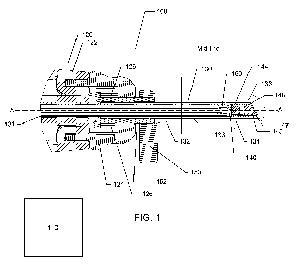
[0006] FIG. 1 is a side, cross-sectional view of an endoscopic system,
illustrating a rigidly coupled image
sensor located at a tip of the endoscope, and further illustrating a fixed
inner lumen and a rotatable outer lumen
according to one implementation;
[0007] FIG. 2 is a side, cross-sectional view of the endoscopic system of
FIG. 1, illustrating the inner lumen
and the outer lumen with their respective optical components in an exploded
view;
[0008] FIG. 3 is an enlarged, detailed view of the tip of the endoscope
illustrated in FIG. 1 according to one
implementation;
[0009] FIG. 4 is an enlarged, detailed view of the tip of the endoscope
according to one implementation;
[0010] FIG. 5 illustrates one implementation of the endoscopic device,
illustrating the ability of the outer
lumen, along with a distal lens and prism, of the endoscope to rotate while
maintaining the position of the image
sensor to create a wide angle field of vision;
[0011] FIG. 6 illustrates one implementation of the endoscopic device,
where the outer lumen has been rotated
one-hundred and eighty degrees with respect to the view in FIG. 5 and
illustrating a limited field of view in
comparison to FIG. 5 and according to one implementation;
[0012] FIGS. 7A and 7B illustrate a perspective view and a side view,
respectively, of an implementation of a
monolithic sensor having a plurality of pixel arrays for producing a three
dimensional image in accordance with the
teachings and principles of the disclosure;
[0013] FIGS. 8A and 8B illustrate a perspective view and a side view,
respectively, of an implementation of an
imaging sensor built on a plurality of substrates, wherein a plurality of
pixel columns forming the pixel array are
located on the first substrate and a plurality of circuit columns are located
on a second substrate and showing an
electrical connection and communication between one column of pixels to its
associated or corresponding column of
circuitry; and
[0014] FIGS. 9A and 9B illustrate a perspective view and a side view,
respectively, of an implementation of an
imaging sensor having a plurality of pixel arrays for producing a three
dimensional image, wherein the plurality of
pixel arrays and the image sensor are built on a plurality of substrates.
DETAILED DESCRIPTION
[0015] The disclosure extends to endoscopic devices and systems for image
rotation for a rigidly coupled image
sensor. The disclosure allows for a distal prism to rotate, which changes the
angle of view of the user or operator,
while the sensor remains fixed at a constant location. This allows the device
to be used in the same manner as
expected by a user or operator experienced in using conventional rigid
endoscopy systems. The user or operator
may rotate an outer lumen, thereby changing the angle of view, while the
sensor remains in a fixed position and the
image viewable on screen remains at a constant horizon. The prism may rotate
while the sensor does not rotate, such
that the user does not lose orientation.
[0016] In the following description of the disclosure, reference is made to
the accompanying drawings, which
form a part hereof, and in which is shown by way of illustration specific
implementations in which the disclosure
may be practiced. It is understood that other implementations may be utilized
and structural changes may be made
without departing from the scope of the disclosure.
[0017] It must be noted that, as used in this specification and the
appended claims, the singular forms "a," "an,"
and "the" include plural referents unless the context clearly dictates
otherwise.
2
CA 02906806 2015-09-14
WO 2014/144955 PCT/US2014/029572
[0018] As used herein, the terms "comprising," "including," "containing,"
"characterized by," and grammatical
equivalents thereof are inclusive or open-ended terms that do not exclude
additional, unrecited elements or method
steps.
[0019] Further, where appropriate, functions described herein can be
performed in one or more of: hardware,
software, firmware, digital components, or analog components. For example, one
or more application specific
integrated circuits (ASICs) can be programmed to carry out one or more of the
systems and procedures described
herein. Certain terms are used throughout the following description and Claims
to refer to particular system
components. As one skilled in the art will appreciate, components may be
referred to by different names. This
document does not intend to distinguish between components that differ in
name, but not function.
[0020] Referring now to the figures, it will be appreciated that FIG. 1
illustrates an example of an endoscopic
system 100 according to the disclosure. The endoscopic system 100 may comprise
a control unit 110, a handpiece
120, and an endoscopic device 130. It will be appreciated that the control
unit 110 may be located remotely from an
image sensor 140 (discussed more fully herein) and may be located in the
handpiece 120 in an implementation. In
one implementation the control unit 110 may be located remotely from the image
sensor 140 and may be housed at a
base unit without departing from the scope of the disclosure.
[0021] In one implementation, the handpiece 120 may comprise a body 122
that may be fixed relative and
attached to an inner lumen 131 of the endoscopic device 130. The handpiece 120
may also comprise a spring loaded
mechanism. The spring loaded mechanism may comprise a spring cap 124, which
may be located adjacent the body
122. The spring cap 124 may be fixed and attached to the inner lumen 131 of
the endoscope 130. At least one
spring 126 may be present in the spring cap 124 and may be part of the spring
loaded mechanism. This spring-
loaded mechanism may function to maintain constant contact between a distal
lens holder 148 and a proximal lens
holder 144, discussed more fully below in relation to FIG. 3. The system 100
may also comprise a rotation post 150
that is attached to a spring sleeve 152. The spring sleeve 152 may be attached
to the outer lumen 133, such that both
the rotation post 150 and the spring sleeve 152 may be rotated relative to the
inner lumen 131. As the rotation post
150 is moved, the spring 126 may operate to push against the spring cap 124
and spring sleeve 152 causing
consistent contact between the distal lens holder 148 and the proximal lens
holder 144. It will be appreciated that the
spring 126 may operate to maintain axial pressure and ensure that there is a
consistent distance between lens
elements 146, thereby allowing rotation without axial movement and a loss of
focus.
[0022] It will be appreciated that the outer lumen 133 may be in mechanical
communication with the handpiece
120. In an implementation, the outer lumen 133 may be spring-loaded at a
junction with the handpiece 120 to
provide consistent contact between the distal lens holder 148 and the proximal
lens holder 144, thus ensuring
consistent axial distance with the proximal lens elements 146 and the distal
lens elements 147 and retaining focus
while the outer lumen 133 rotates.
[0023] In an implementation, the handpiece 120 may comprise a focus
mechanism. The focus mechanism may
permit focal adjustments in the system and may be attached to the inner lumen
131, such that the inner lumen 131 is
movable axially as the focus mechanism may function to control the axial
distance between the proximal lens 146
and the distal lens 147. The focus mechanism may move the inner lumen 131 in
the axial direction only and may not
allow rotation.
3
CA 02906806 2015-09-14
WO 2014/144955 PCT/US2014/029572
[0024] The endoscopic device 130 may comprise a proximal portion 132, which
may be defined as the portion
nearest the handpiece 120, and a distal portion 134, which may be defined as
the portion farthest away from the
handpiece 120. The distal portion 134 may comprise a tip 136. The endoscopic
device 130 may house the image
sensor 140 for providing visualization of an area. In one implementation, the
image sensor 140 may be located
within the distal portion 134 at or near the tip 136 of the endoscopic device
130. The endoscopic device may also
comprise the inner lumen 131 and the outer lumen 133. In one implementation,
the image sensor 140 and the inner
lumen 131 may be fixed relative to the outer lumen 133. In the implementation,
the outer lumen 133 may be
rotatable about an axis A-A of the endoscope 130 and with respect to the image
sensor 140 and the inner lumen 131.
Thus, the disclosure extends to any endoscopic device and system for use with
a rigidly coupled image sensor 140.
[0025] Referring now to FIG. 2, which is an exploded, side cross-sectional
view of the endoscopic system of
FIG. 1, the inner lumen 131 and the outer lumen 133 are illustrated with their
respective optical components in an
exploded view. As noted, the inner lumen 131 may be fixed relative to the
handpiece 120. The image sensor 140
may be fixed to the inner lumen 131. In one implementation, the proximal lens
holder 144 holds the proximal lens
elements 146, the image sensor 140, and support hardware 142 and is fixed to
the inner lumen 131. The proximal
lens holder 144 may abut against the distal lens holder 148.
[0026] The distal lens holder 148 may be rotatable with respect to the
inner lumen 131. It will be appreciated
that the outer lumen 133 may be freely rotatable, such that any components
that are attached thereto may also be free
to rotate. The distal lens holder 148 may be attached to the outer lumen 133
and is freely rotatable. The distal lens
holder 148 may abut against an outer window 151. The outer window 151 may also
be attached to the outer lumen
133 and may be rotatable relative to the inner lumen 131 and the image sensor
140. The outer window 151 may be
in mechanical communication with the outer lumen 133 and may be located on the
terminal end of the tip 136 of the
endoscope 130.
[0027] The distal lens holder 148 may house a prism 145 and a distal lens
147, both of which may be located at
or near the tip 136 of the endoscope 130. It should be noted that the prism
145 as shown in the Figures and
referenced herein may be comprised of multiple elements as necessary to
properly change the direction of light
through the system. It should also be noted the proximal lens 146 and distal
lens 147 as shown in the Figures and
referenced herein together comprise a complete lens system that projects a
focused image on the image sensor 140.
The lens system may be comprised of multiple elements and any number of these
elements may be included in the
distal lens 147 with the remainder included in the proximal lens 146. The
prism 145 and the distal lens 147 may
both be fixed to the outer lumen 133 and may be rotatable relative to the
inner lumen 131 and the image sensor 140,
such that as the angle of view is changed the orientation of an image remains
constant within the viewing area of the
user. It will be appreciated that the distal lens holder 148 may comprise a
guide for aligning the prism 145 and the
distal lens 147 within the tip 136 of the endoscope 130. The distal lens
holder 148 may be fixed to the outer lumen
133 and may be rotatable relative to the inner lumen 131 and the image sensor
140. The distal lens 147 may be
located near the tip 136 of the endoscope 130 and the proximal lens 146 may be
located proximally with respect to
the distal lens 147. The proximal lens 146 may be fixed to the inner lumen
131, such that it remains fixed relative to
the outer lumen 133 as the outer lumen 133 is rotated.
[0028] As illustrated in FIGS. 3 and 4, which are detailed views of
alternative implementations of the distal
portion 134 and tip 136 of the endoscope 130, a channel 154 may be formed
between the inner lumen 131 and the
4
CA 02906806 2015-09-14
WO 2014/144955 PCT/US2014/029572
outer lumen 133, wherein the channel 154 may house fiber optics 156 for
providing a light source to the surgical
scene. The fiber optics 156 may be fixed to the outer lumen 133 and may be
rotatable relative to the inner lumen
131 and the image sensor 140. In an implementation, the endoscope 130 may
further comprise a friction reducing
layer formed between the outer lumen 133 and the inner lumen 131, such that
friction is reduced between the inner
lumen 131 and the outer lumen 133 to allow easy rotation. It will be
appreciated that the friction reducing layer may
be any material that provides lubrication to allow rotation of the outer lumen
133 with respect to the inner lumen
131.
[0029] The proximal lens holder 144 may comprise an inner guide wall 144a
that is formed at one end of the
proximal lens holder 144 and an outer guide wall 144b that is formed at the
other end of the proximal lens holder
144. The proximal lens holder 144 acts as a housing and guide for aligning the
proximal lens 146 with respect to the
distal lens 147, wherein the proximal lens holder 144 is fixed to the inner
lumen 131 and remains fixed relative to
the outer lumen 133 as the outer lumen 133 is rotated. In an implementation,
the inner guide wall 144a may engage
the guide of the distal lens holder 148, such that the distal lens holder 148
is rotatable with respect to the proximal
lens holder 144.
[0030] In one implementation, as illustrated in FIG. 3, the outer window
151 may be formed at an angle. The
angle may be any angle that may be useful in endoscopy and may fall within a
range of about zero degrees to about
ninety degrees, and may be about thirty degrees. However, it will be
appreciated that in one implementation the
outer window 151 may comprise a zero angle as illustrated in FIG. 4 without
departing from the scope of the
disclosure. It will be appreciated that all outer window angles that fall
within the above-noted range of about zero
degrees to about ninety degrees fall within the scope of the disclosure as if
each angle were independently identified
herein, such that the scope of the disclosure includes all angles within the
identified range. For example, angles of
about five degrees, about ten degrees, about fifteen degrees, about twenty
degrees, about twenty-five degrees, about
thirty degrees, about thirty-five degrees, about forty degrees, about forty-
five degrees, about fifty degrees, about
fifty-five degrees, about sixty degrees, about sixty-five degrees, about
seventy degrees, about seventy-five degrees,
about eighty degrees, and about eighty-five degrees and all angles in between
about zero and about ninety degrees
fall within the scope of the disclosure.
[0031] As illustrated best in FIGS. 3 and 4, the endoscopic device 130 may
further comprise an electrical
communication harness 160. The harness 160 may be fixed to and located within
the inner lumen 131. The electrical
communication harness 160 may be electrically connected to or in communication
with the image sensor 140,
thereby providing power to the image sensor 140. Because of its association
and connection to the inner lumen 131,
the electrical communication harness 160 may be fixed relative to the outer
lumen.
[0032] Referring now to FIGS. 5 and 6, there is illustrated the ability of
the outer lumen 133 and the distal lens
147 and prism 145 of the endoscope 130 to rotate while maintaining the
positioning of the image sensor 140. The
rotation ability provides the advantage of creating a wide angle field of
vision without creating distortion as seen in a
fisheye lens. It will be appreciated that because of the rotation of the
distal prism 145, the angle of view of the user
or operator is changed accordingly, while the sensor 140 remains fixed at a
constant location. This allows the
endoscopic device 130 to be used in the same manner as expected by a user or
operator using a traditional
endoscope. The user or operator may rotate the outer lumen 133, thereby
changing the angle of view, while the
CA 02906806 2015-09-14
WO 2014/144955 PCT/US2014/029572
sensor 140 remains in a fixed position and the image viewable on screen
remains at a constant horizon. The prism
145 may rotate while the sensor 140 does not rotate, such that the user does
not lose orientation.
[0033] Referring generally to the image sensor technology illustrated in
FIGS. 7A-9B, and referring to sensor
technology generally, it will be appreciated that CMOS image sensors have
largely displaced conventional CCD
imagers in modern camera applications such as endoscopy, owing to their
greater ease of integration and operation,
superior or comparable image quality, greater versatility, and lower cost.
[0034] Typically CMOS image sensors include the circuitry necessary to
convert the image information into
digital data and have various levels of digital processing incorporated
thereafter. This can range from basic
algorithms for the purpose of correcting non-idealities, which may, for
example, arise from variations in amplifier
behavior to full image signal processing (ISP) chains, providing video data in
the standard sRGB color space
(cameras-on-chip).
[0035] The desired degree of sensor complexity for a given camera system is
driven by several factors, one of
which is the available physical space for the image sensor. The most extreme
functionally minimal CMOS sensor
would have only the basic pixel array plus a degree of buffering to drive the
analog data off chip. All of the timing
signals required to operate and read out the pixels would be provided
externally. The need to supply the control
signals externally adds many pads, which consume significant real estate,
however. Therefore it doesn't necessarily
follow that minimal functionality equates to minimal area.
[0036] If the second stage is an appreciable distance from the sensor, it
becomes much more desirable to
transmit the data in the digital domain, since it is rendered immune to
interference noise and signal degradation.
There is a strong desire to minimize the number of conductors since that
reduces the number of pads on the sensor
(which consume space), plus the complexity and cost of camera manufacture.
Although the addition of analog to
digital conversion to the sensor is necessitated, the additional area is
offset to a degree, owing to a significant
reduction in the required analog buffering power. In terms of area
consumption, given the typical feature size
available in computer information systems technologies, it is preferable to
have all of the internal logic signals be
generated on chip via a set of control registers and a simple command
interface.
[0037] The disclosure contemplates and covers aspects of a combined sensor
and system design that allows for
high definition imaging with reduced pixel counts in a highly controlled
illumination environment. This is
accomplished by virtue of frame by frame pulsed color switching at the light
source in conjunction with high frames
capture rates and a specially designed monochromatic sensor. Since the pixels
are color agnostic, the effective
spatial resolution is appreciably higher than for their color (usually Bayer-
pattern filtered) counterparts in
conventional single-sensor cameras. They also have higher quantum efficiency
since far fewer incident photons are
wasted. Moreover, Bayer based spatial color modulation requires that the
modulation transfer function (MTF) of the
accompanying optics be lowered compared with the monochrome case, in order to
blur out the color artifacts
associated with the Bayer pattern. This has a detrimental impact on the actual
spatial resolution that can be realized
with color sensors.
[0038] The disclosure is also concerned with a system solution for
endoscopy applications in which the image
sensor is resident at the distal end of the endoscope. In striving for a
minimal area sensor based system, there are
other design aspects that can be developed too, beyond the obvious reduction
in pixel count. In particular, the area of
6
CA 02906806 2015-09-14
WO 2014/144955 PCT/US2014/029572
the digital portion of the chip should be minimized, as should the number of
connections to the chip (pads). This
involves the design of a full-custom CMOS image sensor with several novel
features.
[0039] It will be appreciated that the disclosure may be used with any
image sensor, whether a CMOS image
sensor or CCD image sensor, without departing from the scope of the
disclosure. Further, the image sensor may be
located in any location within the overall system, including, but not limited
to, the tip of the endoscope, the hand
piece of the imaging device or camera, the control unit, or any other location
within the system without departing
from the scope of the disclosure.
[0040] Implementations of an image sensor that may be utilized by the
disclosure include, but are not limited
to, the following, which are merely examples of various types of sensors that
may be utilized by the disclosure.
[0041] Referring now to FIGS. 7A and 7B, the figures illustrate a
perspective view and a side view,
respectively, of an implementation of a monolithic sensor 700 having a
plurality of pixel arrays for producing a three
dimensional image in accordance with the teachings and principles of the
disclosure. Such an implementation may
be desirable for three dimensional image capture, wherein the two pixel arrays
702 and 704 may be offset during
use. In another implementation, a first pixel array 702 and a second pixel
array 704 may be dedicated to receiving a
predetermined range of wave lengths of electromagnetic radiation, wherein the
first pixel array 702 is dedicated to a
different range of wave length electromagnetic radiation than the second pixel
array 704.
[0042] FIGS. 8A and 8B illustrate a perspective view and a side view,
respectively, of an implementation of an
imaging sensor 800 built on a plurality of substrates. As illustrated, a
plurality of pixel columns 804 forming the
pixel array are located on the first substrate 802 and a plurality of circuit
columns 808 are located on a second
substrate 806. Also illustrated in the figure are the electrical connection
and communication between one column of
pixels to its associated or corresponding column of circuitry. In one
implementation, an image sensor, which might
otherwise be manufactured with its pixel array and supporting circuitry on a
single, monolithic substrate/chip, may
have the pixel array separated from all or a majority of the supporting
circuitry. The disclosure may use at least two
substrates/chips, which will be stacked together using three-dimensional
stacking technology. The first 802 of the
two substrates/chips may be processed using an image CMOS process. The first
substrate/chip 802 may be
comprised either of a pixel array exclusively or a pixel array surrounded by
limited circuitry. The second or
subsequent substrate/chip 806 may be processed using any process, and does not
have to be from an image CMOS
process. The second substrate/chip 806 may be, but is not limited to, a highly
dense digital process in order to
integrate a variety and number of functions in a very limited space or area on
the substrate/chip, or a mixed-mode or
analog process in order to integrate for example precise analog functions, or
a RF process in order to implement
wireless capability, or MEMS (Micro-Electro-Mechanical Systems) in order to
integrate MEMS devices. The image
CMOS substrate/chip 802 may be stacked with the second or subsequent
substrate/chip 806 using any three-
dimensional technique. The second substrate/chip 806 may support most, or a
majority, of the circuitry that would
have otherwise been implemented in the first image CMOS chip 802 (if
implemented on a monolithic substrate/chip)
as peripheral circuits and therefore have increased the overall system area
while keeping the pixel array size constant
and optimized to the fullest extent possible. The electrical connection
between the two substrates/chips may be done
through interconnects 803 and 805, which may be wirebonds, bump and/or TSV
(Through Silicon Via).
[0043] FIGS. 9A and 9B illustrate a perspective view and a side view,
respectively, of an implementation of an
imaging sensor 900 having a plurality of pixel arrays for producing a three
dimensional image. The three
7
CA 02906806 2015-09-14
WO 2014/144955 PCT/US2014/029572
dimensional image sensor may be built on a plurality of substrates and may
comprise the plurality of pixel arrays and
other associated circuitry, wherein a plurality of pixel columns 904a forming
the first pixel array and a plurality of
pixel columns 904b forming a second pixel array are located on respective
substrates 902a and 902b, respectively,
and a plurality of circuit columns 908a and 908b are located on a separate
substrate 906. Also illustrated are the
electrical connections and communications between columns of pixels to
associated or corresponding column of
circuitry.
[0044] It will be appreciated that the teachings and principles of the
disclosure may be used in a reusable device
platform, a limited use device platform, a re-posable use device platform, or
a single-use/disposable device platform
without departing from the scope of the disclosure. It will be appreciated
that in a re-usable device platform an end-
user is responsible for cleaning and sterilization of the device. In a limited
use device platform the device can be
used for some specified amount of times before becoming inoperable. Typical
new device is delivered sterile with
additional uses requiring the end-user to clean and sterilize before
additional uses. In a re-posable use device
platform a third-party may reprocess the device (e.g., cleans, packages and
sterilizes) a single-use device for
additional uses at a lower cost than a new unit. In a single-use/disposable
device platform a device is provided
sterile to the operating room and used only once before being disposed of.
[0045] Additionally, the teachings and principles of the disclosure may
include any and all wavelengths of
electromagnetic energy, including the visible and non-visible spectrums, such
as infrared (IR), ultraviolet (UV), and
X-ray.
[0046] The foregoing description has been presented for the purposes of
illustration and description. It is not
intended to be exhaustive or to limit the disclosure to the precise form
disclosed. Many modifications and variations
are possible in light of the above teaching. Further, it should be noted that
any or all of the aforementioned alternate
implementations may be used in any combination desired to form additional
hybrid implementations of the
disclosure.
[0047] Further, although specific implementations of the disclosure have
been described and illustrated, the
disclosure is not to be limited to the specific forms or arrangements of parts
so described and illustrated. The scope
of the disclosure is to be defined by the claims appended hereto, any future
claims submitted here and in different
applications, and their equivalents.
8