Sélection de la langue

Search

Sommaire du brevet 2914756 

Énoncé de désistement de responsabilité concernant l'information provenant de tiers

Une partie des informations de ce site Web a été fournie par des sources externes. Le gouvernement du Canada n'assume aucune responsabilité concernant la précision, l'actualité ou la fiabilité des informations fournies par les sources externes. Les utilisateurs qui désirent employer cette information devraient consulter directement la source des informations. Le contenu fourni par les sources externes n'est pas assujetti aux exigences sur les langues officielles, la protection des renseignements personnels et l'accessibilité.

Disponibilité de l'Abrégé et des Revendications

L'apparition de différences dans le texte et l'image des Revendications et de l'Abrégé dépend du moment auquel le document est publié. Les textes des Revendications et de l'Abrégé sont affichés :

  • lorsque la demande peut être examinée par le public;
  • lorsque le brevet est émis (délivrance).
(12) Brevet: (11) CA 2914756
(54) Titre français: PROCEDES D'EVALUATION DE LONGUEURS DE FRAGMENT DE CHAINES MOLECULAIRES UTILISANT DES COLORANTS MULTIPLES
(54) Titre anglais: METHODS FOR ASSESSING FRAGMENT LENGTHS OF MOLECULAR CHAINS USING MULTIPLE DYES
Statut: Accordé et délivré
Données bibliographiques
(51) Classification internationale des brevets (CIB):
  • C12Q 1/68 (2018.01)
  • C12Q 1/6809 (2018.01)
  • G01N 33/52 (2006.01)
(72) Inventeurs :
  • SLOBODAN, JARED (Canada)
  • NESBITT, MATTHEW (Canada)
(73) Titulaires :
  • YOURGENE HEALTH CANADA INC.
(71) Demandeurs :
  • YOURGENE HEALTH CANADA INC. (Canada)
(74) Agent: TORYS LLP
(74) Co-agent:
(45) Délivré: 2022-05-31
(86) Date de dépôt PCT: 2014-06-11
(87) Mise à la disponibilité du public: 2015-02-19
Requête d'examen: 2019-02-27
Licence disponible: S.O.
Cédé au domaine public: S.O.
(25) Langue des documents déposés: Anglais

Traité de coopération en matière de brevets (PCT): Oui
(86) Numéro de la demande PCT: PCT/US2014/042019
(87) Numéro de publication internationale PCT: WO 2015023351
(85) Entrée nationale: 2015-12-07

(30) Données de priorité de la demande:
Numéro de la demande Pays / territoire Date
61/833,823 (Etats-Unis d'Amérique) 2013-06-11

Abrégés

Abrégé français

La présente invention concerne un procédé pour visualiser un fragment d'échantillon d'ADN/ARN séparément d'un marqueur interne dans l'échantillon, et qui est dans une ligne électrophorétique sur gel commune par marquage du fragment d'échantillon d'ADN/ARN avec un premier colorant ayant un premier spectre d'émission lorsqu'il émet une fluorescence sous l'effet d'un premier moyen d'excitation; et le marquage du marqueur interne avec un deuxième colorant ayant un deuxième spectre d'émission différent du premier colorant lorsqu'il émet une fluorescence sous l'effet d'un deuxième moyen d'excitation.


Abrégé anglais

A method for visualizing and discriminating between DNA/RNA fragment(s) of unknown length(s) and an internal marker(s) of known length in a sample that is disposed in a common electrophoresis gel laneway. The method comprises labeling the DNA/RNA fragment(s) with a first dye and labeling the internal marker(s) with a second dye. The first and second dyes have discrete fluorescent emission spectra, which may be used to visually discriminate the DNA/RNA fragment(s) and the internal marker(s).

Revendications

Note : Les revendications sont présentées dans la langue officielle dans laquelle elles ont été soumises.


What is claimed:
1. A method for visualizing at least one DNA sample fragment or at least
one RNA sample
fragment separately from at least one internal marker having a length of at
least 2500 base
pairs and being of known length, to assist in estimating DNA fragment lengths
or RNA fragment
lengths, where the at least one internal marker is in a common electrophoretic
laneway with the
at least one DNA sample fragment or the at least one RNA sample fragment, the
method
comprising:
labeling the at least one internal marker having a length of at least 2500
base pairs with
a plurality of first dye molecules that exclusively label the at least one
internal marker throughout
its length, each of the plurality of first dye molecules having a first
emission spectra when
fluoresced by a first excitation means; and
labeling the at least one DNA sample fragment or the at least one RNA sample
fragment
with a second dye molecule, the second dye molecule being different from the
plurality of first
dye molecules and having a second emission spectra when fluoresced by a second
excitation
means.
2. The method of claim 1, further comprising exciting the laneway with the
first excitation
means and the second excitation means, and visualizing the at least one DNA
sample fragment
or the at least one RNA sample fragment when fluoresced by the second
excitation means.
3. The method of claim 2, wherein the visualizing occurs separately from
the visualization
of the at least one internal marker.
4. The method of claim 2, wherein the laneway is concurrently excited by
the first and
second excitation means.
5. The method of claim 2 or 3, wherein the laneway is sequentially excited
by the first and
second excitation means.
6. The method of any one of claims 1-5, wherein each of the plurality of
first dye molecules
is the same.
8
Date Recue/Date Received 2021-06-11

7. The method of any one of claims 1-6 wherein the plurality of first dye
molecules that
exclusively label the at least one internal marker are covalently bonded to
the at least one
internal marker.
8. The method of any one of claims 1-7, wherein the first dye molecule is
Cy5.
9. The method of any one of claims 1-7, wherein the second dye molecule is
Ethidium
Bromide or Cy5.
10. The method of any one of claims 1-9, wherein the at least one DNA
sample fragment or
the at least one RNA sample fragment is at least one DNA sample fragment.
11 The method of any one of claims 1-9, wherein the at least one DNA sample
fragment or
the at least one RNA sample fragment is at least one RNA sample fragment.
12. The method of any one of claims 1-11, wherein the electrophoretic
laneway is in a slab
gel.
9
Date Recue/Date Received 2021-06-11

Description

Note : Les descriptions sont présentées dans la langue officielle dans laquelle elles ont été soumises.

CA 02914756 2015-12-07
WO 2015/023351 PCT/US2014/042019
METHODS FOR ASSESSING FRAGMENT LENGTHS OF MOLECULAR CHAINS
USING MULTIPLE DYES -
Background
Electrophoretic separation of nucleic acid (DNA/RNA) strands based on fragment
length (by means of a gel or capillary) is a widely used tool in laboratory
genetics. This
process is used to assess the distribution of fragment lengths in a DNA/RNA
sample.
Visualization of DNA/RNA after electrophoretic separation relies on the
attachment of
one or more dye molecules to the DNA/RNA. A dye molecule can be excited to
fluorescence in order to ascertain the location of DNA/RNA within a gel
column, channel
or other laneway (hereinafter collectively "laneway" unless otherwise
presented) along
which the DNA/RNA has traversed via electrophoresis. Interpretation of the
DNA/RNA
fragment length distribution generally relies upon a comparison between at
least one
sizing reference, and preferably a plurality of sizing references, which
consist of
DNA/RNA of discrete, known sizes ("markers"). The implementation of markers
can
come in varying forms, but a common method to optimize sizing accuracy is to
include
the markers in the same laneway for electrophoresis as the fragmented DNA/RNA.
One currently preferred method for estimating the size of DNA/RNA fragments
using electrophoresis comprises the inclusion of known markers within a sample
of such
fragments before commencing electrophoresis, i.e., use of internal markers.
However,
as both the DNA/RNA sample and markers usually incorporate the same dye to
facilitate visualization thereof within the laneway, it can be difficult or
impossible to
distinguish between the two if there is a significant concentration of DNA/RNA
fragments of the same size and with the same length(s) as the marker(s). In
such
situations, the fluorescence from the dye attached to the DNA/RNA sample
fragments
will often mask the fluorescence of the marker(s) dye. This consequence
results
inaccurate size estimation of the DNA/RNA fragment lengths insofar as the
internal
marker(s) cannot be distinguished to provide a reference for comparison.
1

CA 02914756 2015-12-07
WO 2015/023351 PCT/US2014/042019
The problem of masking the fluorescence signal of the internal marker can be
largely avoided through selection of internal markers that are of a size that
permits their
discrimination from the sample. The Bioanalyzer 21 00 capillary
electrophoresis system
(Agilent Technologies), for example, utilizes a two internal marker system
with one
being very small (-15 ¨ 50 bp) and the other being very large (-1.5 ¨ 17 kbp)
to
automatically assess the fragment length distribution of DNA/RNA. By selecting
internal
markers of divergent lengths, the chances of significant overlap of fragment
lengths
between the sample DNA/RNA and the markers are statistically minimized.
While the use of divergent length internal markers can circumvent the masking
problem described above, the results of fragment length estimation are
improved when
the internal marker(s) are of a size that more closely approximates that of
the sample:
more accurate estimations of DNA/RNA fragment lengths can be provided by using
more suitably sized internal markers, however, doing so increases the
likelihood of
undesirable fluorescence masking.
Another approach for estimating the size of DNA/RNA fragments using
electrophoresis comprises the use of external markers, i.e., introducing
markers into a
laneway adjacent to the laneway(s) loaded with the DNA/RNA sample(s). After
electrophoresis has been completed, estimations of fragment lengths in the
sample
laneway(s) can be made by comparing them to the external marker laneway.
External marker fragment sizes can be suitably chosen to match the fragment
size ranges of the sample; there is no chance for any overlap or fluorescence
saturation
between them. However, the comparison is not optimal as variances in applied
current
and the laneway matrix composition between the external marker(s) and the
DNA/RNA
samples can confound sizing approximations. Additionally, the actual sample
contents
can impact fragment mobility, such that the migration speed of identical
molecules can
differ between channels depending upon the remaining sample composition. To
account for variances and improve fragment length estimations, it is best to
include the
markers with the DNA/RNA sample in the same laneway.
2

CA 02914756 2015-12-07
WO 2015/023351 PCT/1JS2014/042019
Finally, another method for assessing fragment lengths involves labeling
internal
markers with fluorescent dyes with unique excitation/emission spectra on the
ends of
DNA/RNA molecules. This method has limitations though. Because dye molecules
are
only attached to the ends of the internal markers, any signal emitted by such
a marker
molecule is minimal, and can only be detected by highly sensitive, costly
detectors that
are not compatible with gel electrophoresis. Additionally, large markers
(i.e., larger than
2,500 bp) have exceedingly high DNA/RNA fragment/dye molecule ratios. And,
because the absolute mass of markers that can be loaded has a practical upper
limit,
this ratio prohibits the utilization of large internal markers even for
analytical systems
with highly sensitive detectors (i.e., capillary systems).
SUMMARY OF THE INVENTION
Mindful of the foregoing deficiencies associated with the prior art, the
instant
invention is directed to methods for separately visualizing DNA/RNA samples
from
internal markers in a common electrophoresis laneway, to assist in estimating
DNA/RNA
fragment lengths within the sample. As a consequence, one or more internal
markers
can be selected, which may be nested within the distribution of DNA/RNA sample
fragments during estimation of the fragment length distribution of a DNA/RNA
sample,
for maximum comparative value. Beneficially, the historical problem of
internal marker
fluorescence signal masking by that of the DNA/RNA fragments is eliminated,
yet
internal marker fluorescence signal strengths are sufficient for use even in
less sensitive
electrophoresis apparatus, such as those using CCD or MOSFET imaging hardware
(e.g., conventional digital camera technologies) and/or non-coherent
illumination
hardware (e.g., conventional LED technologies).
The invention comprises the use of a plurality of fluorescing dyes for DNA/RNA
sample fragments and at least one internal marker in a sample to enable
concurrent
fluorescence in an electrophoresis apparatus and yield non-competing emission
spectra, and/or sequential fluorescence to similarly yield non-competing
emission
spectra but that are temporally separated.
3

CA 02914756 2015-12-07
WO 2015/023351 PCT/US2014/042019
If enhanced fragment length resolution is desired, multiple internal markers
and/or dyes can be used according to the invention with respect to single dye-
marker
combinations. Thus, methods according to the invention include labeling of any
internal
marker with at least one, and preferably more than one, unique dye molecule
throughout the length of the internal marker. This makes the size estimation
approach
compatible with low-cost detector means (e.g., an optical camera) that are
often used
with gel electrophoresis. It also enables the utilization of large internal
markers (e.g., >
-2500 bp) with the multiple dye methodology advanced herein.
In addition to increased sensitivity and fragment length selectivity, the
ability to
exclusively visualize an internal marker when practicing various invention
embodiments
obviates the need to load an absolute mass of an internal marker that
otherwise would
be reliably distinguishable from the fluorescence signal of the DNA/RNA of
interest.
Consequently, generally less than 10% of the original internal marker is
necessary to
achieve desired elucidation that otherwise would be necessary without use of
the
multiple dye solution according to the invention. Such a reduction over prior
art single
dye solutions is projected to save more than 50% in operating costs in
addition to
increasing accuracy of DNA/RNA fragment evaluation when practicing dual dye
embodiments of the invention. Axiomatically, technicians are no longer
required to have
detailed advance knowledge of potential overabundant fragment lengths in the
DNA/RNA sample that could mask the signal from selected internal markers. The
multiple dye methods according to the invention will therefore also reduce the
time
needed to conduct fragment length distribution assessments.
In one series of invention embodiments, a first dye having a first emission
spectra when fluoresced by a first excitation means, such as limited
wavelength light
source, is associated with at least one marker, and a second dye having a
second
emission spectra different from the first dye when fluoresced by a second
excitation
means, such as limited wavelength light source different from the first
excitation means,
is associated with a plurality of DNA/RNA sample fragments, wherein the
DNA/RNA
sample fragments and the at least one marker are disposed in a common gel
4

CA 02914756 2015-12-07
WO 2015/023351 PCT/1JS2014/042019
electrophoresis laneway. At least a portion of the laneway is concurrently
excited by the
first and second excitation means such that both dyes simultaneously
fluoresce, and/or
the dyes are sequentially fluoresced. As a consequence of this methodology,
one or
more internal markers with a size that may be expected to overlap with an
overly
abundant fragment length (known as an adaptermer), which may be present in a
DNA/RNA sample being characterized, may be selected. Such overlap, which
permits
high precision comparisons between the marker(s) and the DNA/RNA sample
fragments
finds particular utility with respect to estimations of the fragment length
distributions for
a set of biologically important nucleic acid molecules known as miRNAs.
A method for accomplishing the foregoing dye associations comprises covalently
bonding one of the dyes to multiple sites on at least one, and preferably a
plurality of,
DNA/RNA sample fragments, and effecting a lesser strength bond between the
other
dye and multiple sites on at least one, and preferably a plurality of, marker
fragments.
Thus, dye molecules and compounds comprising the same such as Cy5, Alexa Fluor
647, and DyLight 650 may be used to first establish labeling of markers while
dye
molecules and compounds comprising the same such as SYBR Gold, SYBR Green,
Ethidium Bromide, and Pico Green may be used to then establish labeling of
DNA/RNA
sample fragments.
It should be noted that in the example referenced above, separate association
of
non-covalently bonded dye molecules is not necessary. Suitable results have
been
obtained from simultaneous exposure of the labeled markers and the unlabeled
DNR/RNA fragments to the non-covalently bonded dye molecule. While there may
still
be bonding sites available on the markers, the fact that the markers already
have a
plurality of dye molecules covalently bonded thereto minimizes available
bonding sites,
and the unique fluorescence signatures between the dyes enables appropriate
position
substitution during subsequent assay steps. Therefore and generally stated,
the
various invention embodiments are sufficiently enabled when at least one of
the
markers or the DNA/RNA sample fragments are exclusively labeled with one of
the
unique dyes.

CA 02914756 2015-12-07
WO 2015/023351
PCT/US2014/042019
In view of the increases in sensitivity, selectivity of fragment length
determination,
choice of DNA/RNA fragment sizes to use as internal markers, as well as the
decrease
in operating costs and time, the invention as exemplified in its various
embodiments
represents a significant advancement over prior art efforts in the field of
electrophoresis
in gel-type media using internal markers with dye-assisted visualization
opportunities.
DETAILED DESCRIPTION OF AN INVENTION EMBODIMENT
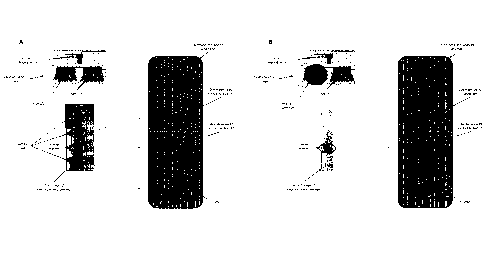
Referring generally to Figs. 1A and 1B, DNA/RNA is electrophoresed along a
laneway (see Electrophoretic laneway schematic) causing smaller fragments to
travel
through the medium at a faster rate than larger fragments and thus, the sample
is separated
by fragment length, as is well known in the art. As both DNA/RNA and internal
markers are
run together, they can be visualized together during electrophoresis. In Fig.
1A, double
stranded Sample DNA has been labeled with a first dye (Dye Molecule #1) and
when
excited by a Blue LED emits green light. Internal Marker has been labeled with
a second
dye (Dye Molecule #2) and when excited with a Red LED emits red light. A
Camera
captures a First image of the electrophoretic laneway and passes the data to a
software
image analysis package.
In Fig. 1B, the Blue LED is immediately turned off to extinguish excitation of
Dye
Molecule #1 and abrogate emission of the green light. With the Red LED still
exciting Dye
Molecule #2, the Internal Marker is exclusively visualized. The Camera then
captures a
Second image of the electrophoretic laneway and passes it to the software
image analysis
package. The package then compares the two images, and, knowing the size of
the
Internal Marker, assesses the size of the surrounding fragment lengths that
constitute the
=
Sample DNA. Note that in another embodiment of this process, the First image
of the
electrophoretic laneway can be captured when only the Blue LED is on to only
visualize the
Sample DNA. This ensures that visualization of the Internal Marker and Sample
DNA can
be mutually exclusive.
Referring next to Fig. 2, representative Excitation/Emission spectra for two
candidate
dye molecules that could be used in the dual dye system are shown. Dye
Molecule #1
6

CA 02914756 2015-12-07
WO 2015/023351
PCT/US2014/042019
occupies an excitation/emission wavelength range that is sufficiently
different from that of
Dye Molecule #2 to enable discrimination between the two.
7

Dessin représentatif
Une figure unique qui représente un dessin illustrant l'invention.
États administratifs

2024-08-01 : Dans le cadre de la transition vers les Brevets de nouvelle génération (BNG), la base de données sur les brevets canadiens (BDBC) contient désormais un Historique d'événement plus détaillé, qui reproduit le Journal des événements de notre nouvelle solution interne.

Veuillez noter que les événements débutant par « Inactive : » se réfèrent à des événements qui ne sont plus utilisés dans notre nouvelle solution interne.

Pour une meilleure compréhension de l'état de la demande ou brevet qui figure sur cette page, la rubrique Mise en garde , et les descriptions de Brevet , Historique d'événement , Taxes périodiques et Historique des paiements devraient être consultées.

Historique d'événement

Description Date
Inactive : Correspondance - Transfert 2023-01-24
Inactive : Correspondance - Transfert 2023-01-19
Lettre envoyée 2023-01-10
Inactive : Correspondance - Transfert 2022-12-08
Inactive : Transferts multiples 2022-11-22
Accordé par délivrance 2022-05-31
Inactive : Octroit téléchargé 2022-05-31
Lettre envoyée 2022-05-31
Inactive : Page couverture publiée 2022-05-30
Préoctroi 2022-03-16
Inactive : Taxe finale reçue 2022-03-16
Lettre envoyée 2022-02-18
Inactive : Transferts multiples 2022-01-28
Un avis d'acceptation est envoyé 2022-01-18
Lettre envoyée 2022-01-18
Un avis d'acceptation est envoyé 2022-01-18
Inactive : Approuvée aux fins d'acceptation (AFA) 2021-11-25
Inactive : QS réussi 2021-11-25
Modification reçue - réponse à une demande de l'examinateur 2021-06-11
Modification reçue - modification volontaire 2021-06-11
Rapport d'examen 2021-03-19
Inactive : Rapport - Aucun CQ 2021-02-11
Représentant commun nommé 2020-11-07
Inactive : COVID 19 - Délai prolongé 2020-08-19
Inactive : COVID 19 - Délai prolongé 2020-08-06
Inactive : COVID 19 - Délai prolongé 2020-07-16
Inactive : COVID 19 - Délai prolongé 2020-07-02
Inactive : COVID 19 - Délai prolongé 2020-06-10
Inactive : COVID 19 - Délai prolongé 2020-05-28
Inactive : COVID 19 - Délai prolongé 2020-05-14
Modification reçue - modification volontaire 2020-05-06
Inactive : COVID 19 - Délai prolongé 2020-04-28
Inactive : CIB désactivée 2020-02-15
Rapport d'examen 2020-01-06
Inactive : Rapport - Aucun CQ 2020-01-03
Représentant commun nommé 2019-10-30
Lettre envoyée 2019-03-07
Inactive : CIB attribuée 2019-03-05
Inactive : CIB en 1re position 2019-03-05
Inactive : CIB attribuée 2019-03-05
Requête d'examen reçue 2019-02-27
Exigences pour une requête d'examen - jugée conforme 2019-02-27
Toutes les exigences pour l'examen - jugée conforme 2019-02-27
Inactive : CIB expirée 2018-01-01
Exigences relatives à la révocation de la nomination d'un agent - jugée conforme 2016-09-29
Inactive : Lettre officielle 2016-09-29
Exigences relatives à la nomination d'un agent - jugée conforme 2016-09-29
Demande visant la révocation de la nomination d'un agent 2016-09-19
Demande visant la nomination d'un agent 2016-09-19
Inactive : CIB attribuée 2016-04-07
Inactive : CIB en 1re position 2016-04-07
Inactive : Page couverture publiée 2015-12-29
Inactive : CIB en 1re position 2015-12-15
Lettre envoyée 2015-12-15
Inactive : Notice - Entrée phase nat. - Pas de RE 2015-12-15
Inactive : CIB enlevée 2015-12-15
Inactive : CIB en 1re position 2015-12-15
Inactive : CIB attribuée 2015-12-15
Inactive : CIB enlevée 2015-12-15
Inactive : CIB attribuée 2015-12-15
Demande reçue - PCT 2015-12-15
Exigences pour l'entrée dans la phase nationale - jugée conforme 2015-12-07
Demande publiée (accessible au public) 2015-02-19

Historique d'abandonnement

Il n'y a pas d'historique d'abandonnement

Taxes périodiques

Le dernier paiement a été reçu le 2022-03-02

Avis : Si le paiement en totalité n'a pas été reçu au plus tard à la date indiquée, une taxe supplémentaire peut être imposée, soit une des taxes suivantes :

  • taxe de rétablissement ;
  • taxe pour paiement en souffrance ; ou
  • taxe additionnelle pour le renversement d'une péremption réputée.

Veuillez vous référer à la page web des taxes sur les brevets de l'OPIC pour voir tous les montants actuels des taxes.

Historique des taxes

Type de taxes Anniversaire Échéance Date payée
Enregistrement d'un document 2015-12-07
Taxe nationale de base - générale 2015-12-07
TM (demande, 2e anniv.) - générale 02 2016-06-13 2016-03-10
TM (demande, 3e anniv.) - générale 03 2017-06-12 2017-03-10
TM (demande, 4e anniv.) - générale 04 2018-06-11 2018-05-08
TM (demande, 5e anniv.) - générale 05 2019-06-11 2019-02-26
Requête d'examen - générale 2019-02-27
TM (demande, 6e anniv.) - générale 06 2020-06-11 2020-04-30
TM (demande, 7e anniv.) - générale 07 2021-06-11 2021-04-05
Enregistrement d'un document 2022-01-28
TM (demande, 8e anniv.) - générale 08 2022-06-13 2022-03-02
Taxe finale - générale 2022-05-18 2022-03-16
Enregistrement d'un document 2022-11-22
TM (brevet, 9e anniv.) - générale 2023-06-12 2023-04-19
TM (brevet, 10e anniv.) - générale 2024-06-11 2024-04-16
Titulaires au dossier

Les titulaires actuels et antérieures au dossier sont affichés en ordre alphabétique.

Titulaires actuels au dossier
YOURGENE HEALTH CANADA INC.
Titulaires antérieures au dossier
JARED SLOBODAN
MATTHEW NESBITT
Les propriétaires antérieurs qui ne figurent pas dans la liste des « Propriétaires au dossier » apparaîtront dans d'autres documents au dossier.
Documents

Pour visionner les fichiers sélectionnés, entrer le code reCAPTCHA :


Pour visualiser une image, cliquer sur un lien dans la colonne description du document. Pour télécharger l'image (les images), cliquer l'une ou plusieurs cases à cocher dans la première colonne et ensuite cliquer sur le bouton "Télécharger sélection en format PDF (archive Zip)" ou le bouton "Télécharger sélection (en un fichier PDF fusionné)".

Liste des documents de brevet publiés et non publiés sur la BDBC .

Si vous avez des difficultés à accéder au contenu, veuillez communiquer avec le Centre de services à la clientèle au 1-866-997-1936, ou envoyer un courriel au Centre de service à la clientèle de l'OPIC.


Description du
Document 
Date
(aaaa-mm-jj) 
Nombre de pages   Taille de l'image (Ko) 
Dessin représentatif 2022-05-04 1 87
Description 2015-12-07 7 307
Revendications 2015-12-07 2 71
Dessins 2015-12-07 2 125
Abrégé 2015-12-07 2 121
Dessin représentatif 2015-12-16 1 62
Page couverture 2015-12-29 1 93
Dessins 2020-05-06 3 856
Revendications 2020-05-06 2 59
Revendications 2021-06-11 2 58
Page couverture 2022-05-04 1 120
Paiement de taxe périodique 2024-04-16 33 1 320
Avis d'entree dans la phase nationale 2015-12-15 1 193
Courtoisie - Certificat d'enregistrement (document(s) connexe(s)) 2015-12-15 1 103
Rappel de taxe de maintien due 2016-02-15 1 110
Rappel - requête d'examen 2019-02-12 1 115
Accusé de réception de la requête d'examen 2019-03-07 1 174
Avis du commissaire - Demande jugée acceptable 2022-01-18 1 570
Courtoisie - Certificat d'inscription (changement de nom) 2022-02-18 1 386
Courtoisie - Certificat d'enregistrement (document(s) connexe(s)) 2023-01-10 1 354
Certificat électronique d'octroi 2022-05-31 1 2 527
Demande d'entrée en phase nationale 2015-12-07 9 261
Traité de coopération en matière de brevets (PCT) 2015-12-07 3 113
Rapport de recherche internationale 2015-12-07 2 85
Changement de nomination d'agent 2016-09-19 3 96
Courtoisie - Lettre du bureau 2016-09-29 1 19
Courtoisie - Lettre du bureau 2016-09-29 1 22
Paiement de taxe périodique 2017-03-10 1 25
Paiement de taxe périodique 2018-05-08 1 25
Paiement de taxe périodique 2019-02-26 1 25
Requête d'examen 2019-02-27 2 69
Demande de l'examinateur 2020-01-06 5 235
Paiement de taxe périodique 2020-04-30 1 26
Modification / réponse à un rapport 2020-05-06 17 1 448
Demande de l'examinateur 2021-03-19 4 207
Paiement de taxe périodique 2021-04-05 1 26
Modification / réponse à un rapport 2021-06-11 9 278
Paiement de taxe périodique 2022-03-02 1 26
Taxe finale 2022-03-16 4 104