Note : Les descriptions sont présentées dans la langue officielle dans laquelle elles ont été soumises.
PERIPHERAL TUMOUR TREATMENT
[0001] The present invention relates generally to hybrid Magnetic Resonance
Imaging-
Radiotherapy system and specifically to an apparatus and method for peripheral
tumour
treatment in such a system.
BACKGROUND
[0002] Most modern radiotherapy (RT) treatments are delivered
"isocentrically", where a
target volume in a patient is placed at an isocentre of the radiotherapy
apparatus. The target
volume can then be irradiated from multiple gantry angles without needing to
move the patient in
order to realign the target volume to the beam axis. The isocentre is often an
intersection of a
gantry axis and a beam axis of the radiotherapy apparatus. An example of a
radiotherapy
apparatus is a linear accelerator.
[0003] More recently, hybrid magnetic resonance (MR)-RT systems have been
used to
provide MR guided RT treatments. For example, the systems by ViewRay and
Elekta AB both
provide MR guided radiotherapy systems. However for these systems it is
difficult, if not
impossible, to position the patient so that a peripherally located tumour,
such as a breast or lung
tumour for example, is at the isocentre without coming into contact with the
magnet. This, in
turn, reduces the gantry angles from which the tumour may be irradiated
thereby inhibiting the
effectiveness of the treatment. Accordingly, it is an object of the present
invention to obviate or
mitigate this disadvantage.
SUM MARY
[0004] In accordance with an aspect of an embodiment, there is provided a
magnetic
resonance (MR)-radiotherapy (RT) hybrid system for treating a patient, the MR-
RT hybrid system
comprising: an MR imaging (MRI) apparatus comprising bi-planar magnets
configured to
generate a magnetic field; a radiation source configured to supply a radiation
beam to treat the
patient; a gantry configured to couple the MR apparatus and the radiation
source so that they
can rotate in unison; a treatment support configured to support the patient; a
motor configured to
move the treatment support; and a controller comprising: a processor; and
memory having stored
thereon instructions, which when executed by the processor, cause the motor to
move the
treatment support in order to avoid collision between the MRI apparatus and
the patient when the
MRI apparatus is rotated, wherein the instructions cause the motor to move the
treatment
support to a central location within the MR-RT hybrid system prior to rotation
of the MRI
apparatus and to a treatment position within the MR-RT hybrid system prior to
treatment of the
patient.
[0005] In accordance with another aspect of an embodiment, there is
provided a method for
positioning a treatment support upon which a patient is positioned within an
MR-RT hybrid
1
Date Recue/Date Received 2022-05-09
system, the method comprising: positioning the treatment support at a central
location; the
central location defined to avoid collision between the patient and the MR-RT
hybrid system;
rotating a gantry of the MR-RT hybrid system to a gantry angle; moving the
treatment support to
a treatment position prior to applying a treatment beam; and after applying
the treatment beam,
moving the treatment support to avoid collision between the MR-RT hybrid
system and the
patient when the gantry is rotated to a different gantry angle.
[0005a] In accordance with another aspect of an embodiment, there is
provided a magnetic
resonance (MR)-radiotherapy (RT) hybrid system for treating a patient, the MR-
RT hybrid system
comprising: an MR imaging (MRI) apparatus comprising bi-planar magnets
configured to
generate a magnetic field; a radiation source configured to supply a radiation
beam to treat the
patient; a gantry configured to couple the MRI apparatus at a first end and
the radiation source
so that they can rotate in unison; a treatment support configured to support
the patient; a motor
configured to move the treatment support; and a controller comprising: a
processor; and memory
having stored thereon instructions, which when executed by the processor,
cause the motor to:
move the treatment support in order to avoid collision between the MR1
apparatus and the patient
when the MRI apparatus is rotated; and move the treatment support to a central
location within
the MR-RT hybrid system prior to rotation of the MRI apparatus to position the
patient center
substantially in alignment with the MR-RT hybrid system isocenter and then
move the treatment
support to at least one a treatment position within the MR-RT hybrid system to
position a center
of a patient target volume, that is offset from the patient center and in
which a peripheral tumor is
located, substantially in alignment with the MR-RT hybrid system isocenter.
[0005b] In accordance with another aspect of an embodiment, there is
provided a method for
positioning a patient treatment support within an MR-RT hybrid system, the
method comprising:
(i) positioning the treatment support at a central location within the MR-RT
hybrid system defined
to avoid collision between a patient positioned on the treatment support and
the MR-RT hybrid
system, and to align substantially a center of the patient with an isocenter
of the MR-RT hybrid
system; (ii) rotating a gantry of the MR-RT hybrid system to a gantry angle;
(iii) moving the
treatment support from the central location to a treatment position to align
substantially a center
of a patient target volume, that is offset from the center of the patient,
with the isocenter of the
MR-RT hybrid system; (iv) following operation of a radiation source, returning
the treatment
support to the central location; and (v) repeating steps (ii) to (iv) for each
subsequent gantry
angle to which the gantry is rotated thereby to avoid collision between the MR-
RT hybrid system
and the patient positioned on the treatment support at each gantry angle.
2
Date Recue/Date Received 2022-12-19
BRIEF DESCRIPTION OF THE DRAWINGS
[0006] Embodiments of the invention will now be described by way of example
only with
reference to the following drawings in which:
Figure 1 is a block diagram of a conventional MR-RT hybrid system;
Figure 2 is a block diagram of an MR-RT hybrid system in accordance with an
embodiment of the
present invention;
Figure 3a is a flow chart illustrating operation of the MR-RT hybrid system;
Figure 3b is a flow chart illustrating pre-treatment processing;
Figure 4 is a block illustrating the MR-RT hybrid system of Figure 2 at a
different gantry angle;
and
Figure 5 is is a block diagram of an MR-RT hybrid system in accordance with an
alternative
embodiment of the present invention;.
DETAILED DESCRIPTION OF THE PREFERRED EMBODIMENTS
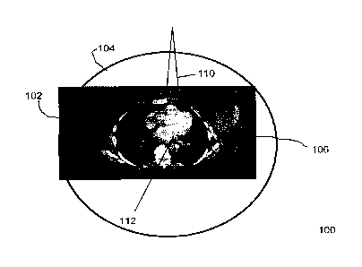
[0007] For convenience, like numerals in the description refer to like
structures in the
drawings. Referring to Figure 1, a block diagram of a cross-section portion of
an MR-RT hybrid
system is illustrated generally by numeral 100. The cross-section portion 100
illustrates an
image slice of a patient 102 within a magnetic resonance imaging (MRI)
apparatus 104. A target
volume 106 is peripherally located within the patient 102. In an embodiment,
the target volume
106 is a tumour. A radiation beam 110 for treating the tumour 106 passes
through the centre of
the MRI apparatus 104. Thus, the MR-RT hybrid system 100 has a centrally
located isocentre
112. The MRI apparatus 104 used for the MR-RT hybrid system 100 is typically
cylindrical and
has a bore of approximately 60 cm. An MR-RT hybrid system 100 using biplanar
magnets for
the MRI apparatus 104 has a similar pole to pole spacing. For patients that
are between 50 and
55 cm wide, of which there are many, there is very little room to laterally
move the patient 102.
Thus, it can be difficult, if not impossible to align the tumour 106 to the
isocentre 112 of the MR-
RT hybrid system.
[0 0 08] In order to allow the MR-RT hybrid system 100 to effectively treat
the tumour 106, it
is preferable to align the tumour 106 with the radiation beam 110 at all
gantry angles. Such an
alignment is straightforward for centrally located tumours, but for peripheral
tumors, such as
breast tumours and lung tumours for example, this would only be possible for
the
2a
Date Recue/Date Received 2022-12-19
CA 02920581 2016-02-10
smallest patients. However, the cost to build a magnet with a larger bore or
pole-to-pole
spacing becomes prohibitively expensive.
[0009] Accordingly, a peripheral tumour treatment positioning (PUP) system
and
method are described herein. The PUP system and method allow peripheral
tumours in
larger patients to be placed at, or proximal to, the isocentre 112 of the MR-
RT hybrid system
100 without needing a larger bore or larger pole-to-pole spacing. Thus, the
PUP system
and method facilitate treating large patients with peripheral tumours in the
MR-RT hybrid
system 100.
[0010] Referring to Figure 2, a block diagram of an MRI-RT hybrid system in
accordance
with an embodiment of the invention is illustrated generally by numeral 200.
The MRI-RT
hybrid system 200 includes an MRI apparatus 202, a rotating gantry (not
shown), a radiation
source 206, a treatment support 208, and a controller 210. In an embodiment,
the treatment
support 208 is a couch, table or the like configured to support the patient
102. The MRI
apparatus 202 is coupled to the radiation source 206 via the rotating gantry
to enable them
to rotate in unison about the treatment support 208. An example of such an MRI
apparatus
is described in U.S. Application Publication No. 2009/0149735, titled
"Integrated external
beam radiotherapy and MRI system" by Fallone et al. The treatment support 208
is
movable by a motor (not shown). Different motors capable of moving the
treatment support
208 as described below can be used. In an embodiment, the motor is configured
to move
the treatment support 208 in a direction parallel to a superior-inferior axis
of the patient 102
to move the patient 102 into and out of the MRI-RT hybrid system 200. As is
known in the
art, the superior-inferior axis runs the length of the patient 102. Further,
the motor is
configured to move the treatment support 208 substantially any direction
normal to the
cranial-caudal axis of the patient to position the patient 102 for treatment,
as will be
described below.
[0011] The MRI apparatus 202 is a bi-planar MRI apparatus comprising a pair
of spaced
apart magnets 202a. The radiation source 206 is directed at the patient 102
either parallel
or antiparallel to the direction of the main magnetic field of the MRI
apparatus 202 through a
hole 201 in the centre of one of the magnets 202a. In the MR-RT hybrid system
200 shown
in Figure 2, the patient 102 is 50 cm wide and the bi-planar magnets have a 60
cm pole to
pole spacing.
[0012] The bi-planar, space apart, configuration of the magnets 202a allows
each
magnet 202a to be individually connected to the gantry at a first end only.
Such a
configuration allows unrestricted lateral motion of the patient 106 in a
direction 212 parallel
to a face of the magnets 202a, and perpendicular to the radiation beam 110.
Such motion is
3
CA 02920581 2016-02-10
limited in current cylindrical magnets. The bi-planar configuration of the
magnets 202a also
allows some motion of the patient 102 in a direction 214 parallel to the
radiation beam 110,
and perpendicular to the face of the magnets 202a.
[0013] The controller 210 is a computing device that is configured to
control the motion
of the treatment support 208. The controller 210 is programmed to position the
MRI
apparatus 202, the radiation source 206, and the patient 102 so that the
target volume 106 is
as close to the isocentre of the radiation beam 110 as possible.
[0014] Prior to treating the patient using the MR-RI hybrid system 200, a
patient centre
is determined. The patient centre (x,,y,) can be calculated based on an
analysis of the
contours taken during a simulation process. The analysis determines the
patient centre
(x,,y,) such that a distance from the central point to the skin surface is
less than the bore
diameter or pole-to-pole spacing of the MRI apparatus 202 for all z positions.
Although this
analysis could be done from a computed tomography (CT) or MR scan as part of
the
simulation process, it may be inefficient or unethical, in the case of CT, to
scan well above
and below the treatment area just to get an external contour for this
analysis. Therefore, a
method of generating the patient contour from head to toe that does not
require a CT or MR
could also be used. Devices, such as laser contouring devices, are readily
available that
could do this in a quick and efficient manner.
[0015] Further, a treatment plan is calculated. Specifically, a 3D position
of the patient
centre (xc,Yc,zc) is calculated using contours obtained above. Using
techniques similar to
conventional isocentric radiotherapy, a 3D location of a pseudo isocentre
(xphypi,zpi), and
gantry angles for each field are defined. In an embodiment, the centre of the
target volume
is defined as the pseudo isocentre. Based on these two points, treatment
centres
(x-r(n),y-r(n),zT(n)) are calculated for each gantry angle, where n denotes a
radiation beam
number. As will be appreciated, since the grantry 204 rotates the MRI
apparatus 202 and
the radiation source 206 about the patient 102, different gantry angles will
likely be
associated with different treatment centres. For each of the different gantry
angles, the
machine isocentre would be relocated to the treatment centre position, and the
dose would
be calculated. As is well known to those knowledgeable in the art, as the
machine isocentre
112 is moved from the pseudo isocentre to the treatment centre, a field size
and multileaf
collimator (MLC) would need to be adjusted according to divergence. This could
be
accomplished either manually or through a computerized calculation that
adjusted each
parameter accordingly. Dose distributions could be calculated and optimized
through the
various tools normally available in the treatment planning system. If, for any
reason, any of
4
CA 02920581 2016-02-10
the treatment centres needed to be modified as part of the planning process,
the system
could check that the modified position would be valid and would not cause any
collisions.
[0016] Once the treatment plan has been calculated with the different
treatment centres
for each radiation beam 110, the patient 102 is ready to be treated with the
MR-RT hybrid
system 200. Referring to Figure 3a, a flow chart illustrating operation of the
MR-RT hybrid
system 200 to position a patient for treatment is illustrated generally by
number 300.
[0017] At 302, a pre-treatment process is performed. Referring to Figure
3b, the pre-
treatment process 302 is described in detail. At step 302a, a pre-treatment
alignment of the
patient 102 is performed to align the patient centre with a central location
of the MRI-RT
hybrid system 200. In an embodiment, the central location is defined as a
position within the
MRI-RT hybrid system 200 at which the patient 102 can be placed without fear
of contact
with the MRI apparatus 202 when the gantry 204 rotates the MRI apparatus 202
and the
radiation source 206 about the treatment support 208. In an embodiment, the
central
location is the isocentre of the MRI-RT hybrid system 200. The patient 102 is
positioned at
the central location by aligning the patient centre as closely with the
isocentre of the MRI-RT
hybrid system 200 as possible. Specifically, the patient is positioned by
aligning the patient
centre to a set of external lasers. Optionally, prior to moving the patient
into the MR-RT
hybrid system 200, the gantry can be rotated to position the magnets 202a
vertically. In this
position there will be an opening between the magnets 202a, up to the ceiling.
This may
minimize the effect of claustrophobia as the patient 102 is moved into the
bore of the MR-RT
hybrid system 200.
[0018] At step 302b, the treatment support 208 is translated a predefined
distance from
the set of external lasers into the MR-RT hybrid system 200. The predefined
distance is
configured to correlate the patient centre at the set of external lasers with
the isocentre of
the MR-RT hybrid system 200.
[0019] At step 302c, high quality MR images of an anatomy of interest are
taken to verify
that the patient centre is accurately aligned to the isocentre of the MR-RT
hybrid system
200. If the field of view (FOV) of the MR apparatus 202 is insufficient to
obtain a high quality
image of the entire anatomy of interest of the patient 102, multiple images
can be taken at
different treatment support positions and stitched together using known
computer graphics
techniques. Since most people are wider laterally than they are in the
anterior posterior
direction, the gantry 204 is rotated to position the magnets 202a
horizontally. This
configuration allows the treatment support 208 to move laterally sufficiently
to obtain a full
set of images to stitch together. This configuration also allows the treatment
support 208 to
be moved so that the pseudo isocentre is aligned with a central axis of the
radiation beam
CA 02920581 2016-02-10
110 and the isocentre MR-RT hybrid system 200 is vertically aligned with the
pseudo
isocentre.
[0020] As a result of the alignment, optimal MR imaging with minimal image
distortion is
obtained over a central field of view (CFOV) of the MR apparatus 202. Beyond
the CFOV,
image distortion increases due to gradient non-linearities and magnetic field
inhomogeneity.
To provide the best image guidance, image-distortion must be minimized.
Therefore,
vertically aligning the isocentres facilitates optimum quality pre-treatment
imaging of the
target volume, with the FOV approximately centred on the target volume.
[0021] Stitching images obtained at the CFOV for multiple treatment support
and /or
gantry positions would then allow the creation of a composite image over a
larger field of
view with the geometric accuracy inherent to the CFOV. Those skilled in the
art will
recognize that this method of producing an image with minimal distortion would
be valuable
in the treatment simulation process as well as during pretreatment imaging.
[0022] At step 302d, once the pre-treatment images are acquired, computer
software
executing on the controller 210 registers or correlates the pre-treatment
images with the MR
or CT images used for the treatment planning. This registration could be done
using a rigid
transformation or a deformable registration, as is known in the art. At step
302e, once the
two images are registered, the computer software calculates the treatment
support 208
shifts, including translations and rotations, needed to align the patient 102
to treatment
planning positions. As will be appreciated by a person skilled in the art, in
some
embodiments the treatment support may be capable of rotating a few degrees to
help align
the patient 102. Once the shifts have been calculated the treatment support
could be
translated and rotated by these known amounts to bring the patient centre to
the machine
isocentre.
[0023] After the patient 102 has been aligned using to the pre-treatment
image guidance
procedure above, the radiation delivery phase can be initiated. At 304, the
grantry 204
rotates the MRI apparatus 202 and the radiation source 206 into a first gantry
angle for
treatment. The initial treatment position is for a first gantry angle, n = 1.
At 306, the
treatment support is translated along a trajectory that moves the patient 102
parallel to the
magnets 202 so that the treatment centre (4(1),y-r(1),4(1)) becomes aligned
with the
isocentre 112 along the beam axis at the first gantry angle. By following this
trajectory the
patient 102 should not collide with the MR-RT apparatus 202. However,
additional known
collision avoidance schemes could be used to provide a fail-safe motion
trajectory.
6
CA 02920581 2016-02-10
[0024] Referring to Figure 4, a block diagram of the MRI-RT hybrid system
200 at a
gantry angle 0 is illustrated generally by numeral 400. As shown, the patient
102 has been
translated so that the target region 106 lies along an axis of the radiation
beam 110. Thus,
the treatment center (xT,y-r) is at the intersection of the line from the
pseudo isocentre (xpi,
ypi) to the radiation source 206 and the line perpendicular to it that passes
through the
patient centre (x,,y,). When the treatment center is determined for each
gantry angle, the
treatment will be similar to an isocentric treatment, in that each radiation
beam is pointed
towards a common point. In the embodiment, the common point is the pseudo
isocentre.
However, for each angle, there will be different distances to the patent's
skin surface, and
from the skin surface to the pseudo isocentre.
[0025] At 308, the treatment is delivered. This can be done with MR image
guidance
before, during or after radiation delivery as desired. At step 310, the
treatment support is
reversed along the trajectory so that the patient centre is once again aligned
with the
isocentre of the MR-RT hybrid system 200.
[0026] The controller returns to 304 and the the grantry 204 rotates the
MRI apparatus
202 and the radiation source 206 into a subsequent position, n = 2. The
process 304 to 310
repeats until all n radiation beams have been delivered. At step 312 , the
radiation delivery
is complete and the treatment support 208 is translated to remove the patient
102 from the
MR-RT hybrid system 200.
[0027] As will be appreciated, the MR-RT hybrid system 200 described above
provides a
controller configured to manipulate the treatment support 108 laterally,
vertically and in
superior-inferior directions such that a target volume 106s is substantially
aligned to the
radiation beam 110. This may be true even for a peripherally located target
volume 106.
[0028] Thus, the MRI-RT hybrid system 200 can be used in a number of
different
circumstance but is particularly useful when the target volume 106 cannot be
positioned at or
near the isocentre of the traditional radiotherapy apparatus and, as such, an
isocentric
treatment approach is not typically feasible.
[0029] In an alternative embodiment, rather than return the treatment
support 208 to the
isocentre prior to each rotation of the gantry, the treatment support 208 can
be retracted
from the MRI apparatus 202. In yet an alternative embodiment, a trajectory can
be devised
that allows a treatment support and the gantry to move concurrently. Such a
trajectory
would not require the patient to be moved to either the central location or to
be retracted
from the MRI apparatus between gantry angles.
7
CA 02920581 2016-02-10
[0030] In the embodiments described above the MRI apparatus 202 comprises a
spaced
apart bi-planar magnets 202a. Depending on the size and configuration of the
magnets
202a, additional features may be necessary to provide structural support.
Accordingly,
referring to Figure 5, an alternative embodiment of the MRI-RT hybrid system
is shown
generally by numeral 500. Only a portion of the MRI-RT hybrid system 500 is
illustrated for
simplicity. Specifically, the MRI-RT hybrid system 500 is similar to the
previous embodiment
but the gantry includes a support structure 502 attached to the magnets 202a
of the MRI
apparatus 202 at an end distal to the first end. In an embodiment, the support
structure is an
annular flange. Since the annular flange 502 primarily provides structural
support, it can
have a diameter substantially larger than the pole to pole spacing of the
magnets 202a.
[0031] For example, in an embodiment the pole to pole spacing is 60 cm and
the
diameter of the annular flange 502 is 110 cm. The diameter of 110 cm is
selected based on
an average patient size. As will be appreciated, the diameter of the annular
flange 502 can
be larger to accommodate a larger average patient size. Accordingly, although
the support
structure 502 is described as an annular shaped flange having a particular
size, it will be
appreciated that other shaped and sized flanges may also be used to provide
structural
support to the MRI apparatus 202.
[0032] The annular flanges 502 may inhibit motion of treatment support 208
if a portion
of treatment support 208 is positioned outside of the MRI apparatus 202.
However, because
the opening of the annular flange 502 is significantly larger than the pole to
pole spacing, it
will allow substantial motion of the patient support 208. Further, if the
entire treatment
support can be positioned within the MRI apparatus 202 then the annular flange
502 may not
affect motion of the treatment support 208 at all.
[0033] Although preferred embodiments of the invention have been described
herein, it
will be understood by those skilled in the art that variations may be made
thereto without
departing from the scope of the appended claims.
8