Note : Les descriptions sont présentées dans la langue officielle dans laquelle elles ont été soumises.
CA 02921081 2016-02-11
WO 2015/035506 PCT/CA2014/000687
Title: Methods and Compositions for Generating Epicardium Cells
Related Applications
[0001]
This is a Patent Cooperation Treaty Application which claims the benefit of 35
U.S.C. 119 based on the priority of U.S. Provisional Patent Application No.
61/877,618, filed
September 13, 2013 which is incorporated herein by reference in its entirety.
Field
[0002]
The disclosure provides methods and compositions for producing cardiovascular
lineage cells from PSCs, including hPSCs, as well as methods and compositions
for producing
cardiomyocyte and epicardial lineage cell populations.
Background
[0003]
Over the past five years, progress has been made in our ability to direct the
differentiation of human embryonic (hESCs) and induced pluripotent stem cells
(hiPSCs)
(collectively referred to as human pluripotent stem cells; hPSCs) to specific
cells types,
including those of the cardiovascular lineagest 2. This success is largely
based on the
translation of our understanding of lineage development and tissue formation
in model
organisms to the hPSC differentiation culturesl. With respect to the
cardiovascular system, this
approach has led to the establishment of differentiation protocols that
recapitulate the key
stages of development including the formation of a primitive streak (PS)-like
population, the
induction of cardiovascular mesoderm and the specification of the
cardiovascular lineages from
this mesoderm3'4. Developmental biology has also informed us on key regulatory
pathways that
control this developmental progression including the requirement for activin
A/nodal and BMP4
signaling to generate the PS/mesoderm population and the need to inhibit p-
catenin dependent
Wnt signaling to specify the mesoderm to a cardiovascular fate'. Recent
studies have identified
surface markers specific for cell populations representing different stages of
cardiovascular
development. This marker set includes KDR and PDGFRa found on cardiovascular
mesoderm'
and SIRPA present on cardiovascular progenitors and differentiated
cardiomyocytese. By
monitoring the emergence of the KDR+PDGFRa+ population, it was shown that
different hPSC
lines require different concentrations of activin A and BMP4 for optimal
mesoderm induction and
cardiomyocyte development'.
[0004]
The epicardial lineage is derived from a structure known as the proepicardial
organ (PEO) that develops adjacent to the heart at approximately embryonic
stage (E) 9.5 in the
mouse'. Pro-epicardial cells characterized by the expression of the
transcription factors Wilms
1
CA 02921081 2016-02-11
WO 2015/035506 PCT/CA2014/000687
Tumor 1 (VVT1) and TBX18, migrate from the PEO to the early heart tube during
the process of
looping and rapidly envelope it to form an outer epithelial layer, known as
the epicardium. The
epicardium is essential for normal heart development and functions to support
rapid proliferation
of the ventricular cells and the formation of compact zone myocardium. It is
also the source of
several major cell types of the heart including cardiac fibroblasts, coronary
vascular smooth
muscle cells and to a lesser extent endothelial cells. These differentiated
progeny are referred
to as epicardial-derived cells (EPDCs) and are derived through an epithelial-
to-mesenchymal
transition (EMT) of the epicardium. Lineage tracing studies suggest that the
epicardium is also a
source of cardiomyocytes8. 9. However, the interpretation of these studies has
been questioned
given the uncertainty of the epicardial specificity of the gene used for the
tracing experiments'''.
[0005] The epicardium produces a number of factors including retinoic acid
(RA),
fibroblast growth factors (FGFs) and insulin-like growth factors (IGFs),
several of which are
essential for the transient phase of ventricular myocyte proliferation
necessary for the formation
of compact zone myocardium. Recent studies have shown that IGF2 is the
critical epicardium-
derived factor that promotes ventricular proliferation" and that RA mediates
this function
indirectly through activation of erythropoietin (EPO) in the liver, which in
turn induces IGF2 in
the epicardium12. Evidence also exists for myocardial regulation of the
epicardium through the
activity of thymosin 134 (Tp4), a G-actin monomer binding protein13. Tp4 is
produced by the
developing myocardium and is required for proper epicardial development and
integrity.
[0006] While the normal adult epicardium does not express VVT1, TBX18 or
RALDH214,
injury such as myocardial infarction will lead to the upregulation of this
'fetal' gene program, as
well as to proliferation of cells within the population and the reactivation
of EMT. Injection of Tp4
during infarction enhances these changes and prevents myocardial death, likely
through the
production of paracrine factors from the activated epicardial cells14' 19.
Lineage-tracing studies in
the adult suggest that this activated epicardium has some capacity to generate
new
cardiomyocytes and that this cardiogenic potential is augmented by priming of
the pre-infarcted
heart with Tp415. As with the fetal studies, however, this concept is
controversial, as recent
studies failed to demonstrate any contribution of the epicardium to the
myocardium of the
infarcted, Tp4-treated heart14.
[0007] In vitro studies have shown that epicardial cells in explant
cultures will undergo
EMT and give rise to EPDCs in response to Notch16, TGF1317-19 and PDGFBB2 or
Tf3415.
2
CA 02921081 2016-02-11
WO 2015/035506 PCT/CA2014/000687
Epicardial cells from infarcted animals primed with TI34 in vivo differentiate
to cells that express
cardiomyocyte markers in explant cultures15.
[0008] Although these advances have enabled the efficient and scalable
derivation of
cardiomyocytes from hPSCs, these differentiated populations are not optimal
for many
applications, as they contain immature cells and the proportion of different
cardiac lineage cells
including myocardial and epicardial within them is not well defined. To
realize the potential of
hPSCs in cardiovascular research and therapeutic applications, it will likely
be necessary to
develop culture systems and engineered tissues that more accurately represent
the human
heart¨.
Summary
[0009] An aspect includes a method of obtaining a cardiovascular lineage
cell population,
optionally cardiomyocyte lineage cell population or an epicardial lineage cell
population from
human pluripotent stem cells (hPSCs) comprising the steps: (a) contacting BMP
component
primed hPSCs with a cardiovascular mesoderm programming cocktail suitable for
inducing the
hPSCs to differentiate to a cardiovascular mesoderm cell population under
conditions suitable
for the programming cocktail to penetrate the hPSCs and culturing the
contacted hPSCs for a
period of time to generate a KDR+ and PDGFRa+ cardiovascular mesoderm cell
population; (b)
contacting the cardiovascular mesoderm cell population with a cardiovascular
progenitor
specification cocktail suitable to specify a NKX2-5+ or WTI+ cardiovascular
progenitor cell
population under conditions suitable for the specification cocktail to
penetrate the cardiovascular
mesoderm cell population and culturing the contacted cardiovascular mesoderm
cell population
for a period of time to generate a NKX2-5+ or W1-1+ cardiovascular progenitor
cell population;
and (d) contacting the cardiovascular progenitor cell population with a
maturation cocktail under
conditions suitable for the maturation cocktail to penetrate the
cardiovascular progenitor cell
population and culturing the contacted cardiovascular progenitor population
for a period of time
to produce a cardiovascular lineage population optionally a cardiomyocyte
lineage cell
population expressing cardiac troponin T (cTnT) and/or SIRPA and/or an
epicardial lineage cell
population optionally expressing WTI and/or comprising epicardial derived
cells (EPDCs).
[0010] Other features and advantages of the present disclosure will become
apparent
from the following detailed description. It should be understood, however,
that the detailed
description and the specific examples while indicating preferred embodiments
of the disclosure
are given by way of illustration only, since various changes and modifications
within the spirit
3
CA 02921081 2016-02-11
WO 2015/035506 PCT/CA2014/000687
and scope of the disclosure will become apparent to those skilled in the art
from this detailed
description.
Brief description of the drawings
[0011] An embodiment of the present disclosure will now be described in
relation to the
drawings in which:
[0012] Figure 1. Cardiomyocyte specification from hESCs. Scheme of the
protocol
used to differentiate hESCs towards the cardiomyocyte lineage highlighting the
three main
stages of development: 1) mesoderm induction, 2) cardiovascular specification
and 3)
maturation. Cells from ActivinA/BMP4-induced day 4 embryoid bodies (EBs) are
plated as a
monolayer on gelatin coated wells. The BMP pathway is manipulated for a 48-
hour period (D4-
D6) in the presence of VEGF (5 ng/ml), the Activin/Nodal (SB-431542 5.4 pM)
and Wnt (DKK1
150 ng/ml) inhibitors. Following specification, the cultures were maintained
in VEGF for 9 days
and then analyzed for the presence of cTnT+ cardiomyocytes by flow cytometry.
[0013] Figure 2. BMP4 regulates the specification of cardiomyocytes from
hESC-
derived mesoderm. (a) Flow cytometric analyses showing the presence of the
KDR+ and
PDGFRa+ populations at day 4 and day 5 and the cTnT+ expression on day 15 of
culture
following no treatment (control), treatment with BMP4 (10 ng/ml) or the BMP4
inhibitor Noggin
(400 ng/ml). (b) Total cell numbers per well at day 15 in the cultures treated
as above. Error
bars represent standard deviation from the mean from three experiments.
[0014] Figure 3. BMP signaling dose-dependently specifies cardiomyocytes
from
hESC-derived mesoderm. Graphical representation of flow cytometry analyses
indicating the
percent cTnT+ cells in day 15 cultures generated from populations treated with
the indicated
amounts of BMP4 or Noggin. NT = no treatment. Bars represent standard
deviation from the
mean of the values from three independent experiments (N=3); *P50.05, **P50.01
when
compared to no treatment.
[0015] Figure 4. gRT-PCR expression of myocardial and epicardial markers
after
BMP treatment. qRT-PCR-based expression of the indicated genes at days 6, 8,
10, 12, and
15 of culture in populations generated from no treatment (control), BMP4
treated or Noggin (400
ng/ml) treated cells. Values are relative to the housekeeping gene TBP. Error
bars represent
standard deviation from the mean of the values from three independent
experiments (N=3);
*P50.05, **P50.01 when compared to no treatment.
4
CA 02921081 2016-02-11
WO 2015/035506 PCT/CA2014/000687
[0016] Figure 5. BMP4-induced cells express the epicardial marker WT1.B
Fluorescent immunostaining analyses showing the presence of cTnT and VVT1 in
no treatment
(control), BMP4 (10 ng/ml) and Noggin (400 ng/ml) treated cells at day 15 of
culture. DAPI
staining shows cell nuclei
[0017] Figure 6. WTI' epicardium generate epithelial sheets following
passage. (a)
Phase contrast microscopy and fluorescent immunostaining showing the
morphology of the
BMP4 (10 ng/ml) treated epicardial cells and the presence of ZO1 and VVT1 at
day 15 of culture.
DAPI staining shows cell nuclei. Scale bar represents 100 pM. (b) Phase
contrast microscopy
and fluorescent immunostaining showing the morphology of the BMP4 (10 ng/ml)
treated
epicardial cells and the presence of ZO1 and VVT1 4 days after passage (day
15+4). DAPI
staining shows cell nuclei. Scale bar represents 100 pM.
[0018] Figure 7. Flow cytometry analysis for the expression of cell
surface markers
in day 15 cardiomyocytes, day 15 epicardium, and post-passage epicardium. Flow
cytometric analyses of the indicated markers on day 15 cardiomyocytes, day 15
Epicardium and
epicardium 4 days following passage (Day 15+4). Gray filled histogram
indicates unstained
fluorescence intensity.
[0019] Figure 8. Cardiomyocytes and epicardial cells are derived from day
4
PDGFRa+ mesoderm. (a) PDGFR+ and PDGFR- populations were isolated from day 4
EBs and
the cells were plated under conditions that support cardiomyocyte or VVT1+
cell development.
(b) Flow cytometric analyses showing cTnT+ cells in day 15 cultures plated
under pro-
cardiogenic conditions. (c) Fluorescent immunostaining for the presence of WTI
positive cells in
day 15 cultures plated under pro-epicardial inducing conditions (BMP4). DAPI
staining shows
the cell nuclei. (d) qRT-PCR-based expression analyses of the epicardial
markers WT/ and
TBX18 in the sorted populations at D15 following culture under pro-epicardial
conditions. Values
are fold change compared to the unsorted cultures. Error bars represent
standard deviation the
mean from the values from three independent experiments (N=3); *P50.05,
**P50.01 from
unsorted cultures.
[0020] Figure 9. BMP and Wnt signaling modulate cardiomyocyte and
epicardial
cell specification. (a) Graphical depiction of flow cytometric analyses
showing the percent
cTnT+ cells in day 15 cultures generated from untreated cells (control) or
cells treated with
either BMP4 (10 ng/ml) or the BMP inhibitor Dorsomorphin (DM 4 pM) in
combination with the
indicated amounts of DKK1 or CHIR. Error bars represent standard deviation
from the mean of
CA 02921081 2016-02-11
WO 2015/035506 PCT/CA2014/000687
the values from three independent experiments (N=3); *P50.05, **P50.01 when
compared to the
'no Wnt treatment' (NT) control in context of the indicated manipulation of
the BMP pathway. (b)
qRT-PCR-based analyses of WTI expression on day 15 cultures generated from
untreated cells
(control) or cells treated with either BMP4 (10 ng/ml) or DM (4 pM) in
combination with the
indicated amounts of DKK or CHIR. Values are relative to the housekeeping gene
TBP. Error
bars represent standard deviation from the mean of the values from three
independent
experiments (N=3); *P50.05, **P50.01 when compared to the 'no Wnt treatment'
(NT) control in
the context of the indicated manipulation of the BMP pathway. (c) Flow
cytometry analyses
showing percent cTnT+ cells and qRT-PCR analyses for WT1 expression in day 15
cultures
generated from untreated cells (control) or cells treated with either BMP4 (10
ng/ml) or DM (4
pM) in combination with the indicated amounts of XAV939 (XAV). Error bars
represent standard
deviation from the mean of the values from three independent experiments
(N=3); *P50.05,
**P.50.01 when compared to no Wnt treatment (see Figures 9a and 9b) in the
context of the
specific BMP treatment. (d) Flow cytometry analyses showing percent cTnT+
cells and qRT-
PCR analyses of WT1 expression in day 15 cultures generated from untreated
cells (control) or
cells treated with either BMP4 (10ng/m1) or DM (4 pM) in combination with the
indicated
amounts of IWP2. Error bars represent standard deviation from the mean of the
values from
three independent experiments (N=3); *P50.05, **P50.01 when compared to no Wnt
treatment
(see Figures 9a and 9b) in the context of the specific BMP treatment.
[0021] Figure 10. Generation of WT1 + epicardial cells from Sendai virus-
derived
hiPSCs and H7 hESCs. (a) Fluorescent immunostaining showing the expression of
WT1 and
ZO1 in a hiPSC-derived epicardial cultures. DAPI staining shows cell nuclei.
Scheme indicates
timing of manipulations and analysis. (b) Fluorescent immunostaining showing
the expression of
WT1 and ZO1 in a H7 hESC-derived epicardial cultures. DAPI staining shows cell
nuclei.
Scheme indicates timing of manipulations and analysis.
[0022] Figure 11. WT1 + epicardial cells undergo EMT in response to TGF[31
and
bFGF treatment. (a) Scheme of the protocol used for EMT induction. Day I5 WT1-
'- cultures are
passaged, allowed to settle for 1 day and then treated with TGFP1 (5 ng/ml)
for 4 days followed
by no treatment (TGF13), sequential treatment with TGFp1 (5 ng/ml) for 4 days
followed by
bFGF (10 ng/ml) for 4 days (TGFp+bFGF), or bFGF (10 ng/ml) treatment for 8
days (bFGF). No
treatment of the cultures serves as a control. (b) Flow cytometric analyses of
cultures 8 days
following the initiation of EMT for the cell surface mesenchymal marker CD90.
Gray filled
histogram indicates control culture fluorescence intensity. (c) qRT-PCR-based
expression of the
6
CA 02921081 2016-02-11
WO 2015/035506 PCT/CA2014/000687
epicardial gene WTI and the EMT-induced genes SNAll and SNAI2 on days 2, 4, 6
and 8 after
EMT initiation. Values are expressed as fold change to experiment-matched pre-
passaged day
15 WT1+ cultures. Error bars represent standard deviation from the mean of the
values from
three independent experiments (N=3); *P5Ø05, **P0.01 compared to no
treatment control.
[0023] Figure 12. WTI and ZO1 expression is lost in response to EMT. Phase
contrast and fluorescent immunostaining showing cell morphology and the
expression of ZO1
and WTI proteins in epicardial cultures 8 days after EMT initiation with the
indicated factors.
DAPI staining shows cell nuclei.
[0024] Figure 13. EPDCs display characteristic expression of fibroblasts
and
vascular smooth muscle cell markers by fluorescent immunostaining. Fluorescent
immunostaining showing a-Smooth muscle actin (SMA) and Vimentin (VIM) protein
in cultures 8
days after EMT initiation with the indicated factors. DAPI staining shows cell
nuclei.
[0025] Figure 14. EPDCs display characteristic expression of fibroblasts
and
vascular smooth muscle cell markers by qRT-PCR. qRT-PCR-based expression
analyses of
the smooth muscle genes CCN1, MYH11, TAGLN and SMTN and the epicardial/cardiac
fibroblast gene TCF21 in the indicated cultures 8 days after EMT initiation.
Values are
expressed as fold change to experiment-matched pre-passaged day 15 VVT1+
epicardium
cultures. Error bars represent standard deviation from the mean of the values
from three
independent experiments (N=3); *PsØ05, **P5Ø01 compared to no treatment
control cultures.
[0026] Figure 15. hESC epicardial-derived smooth muscle-like cells
generate action
potentials when stimulated.
(a) The total proportion of actively cycling cells in EMT-induced cultures was
measured for the
indicated treatments. Treatment with TGF13+bFGF generated populations with the
largest
proportion of actively cycling cells in response to agonists. Bars represent
standard error of the
mean; N=3/group; *P<0.05, **P<0.01 compared by one-way ANOVA with Tukey post
hoc test.
(b) The frequency of calcium cycles in actively cycling cells in the
conditions as indicated at
baseline and after NE and PE addition. Stacked bars represent the contribution
to frequency of
calcium cycling during baseline recording (hatched lines), and after NE
(white) or PE (black)
treatment.
7
CA 02921081 2016-02-11
WO 2015/035506 PCT/CA2014/000687
(c) The amplitude of calcium transients after NE and PE addition in the EMT
induced cultures.
No Treatment NE N=6 cells, PE N=4 cells; TGFp NE N=6 cells, PE N=12 cells;
TGFp+bFGF NE
N=13 cells, PE N=25 cells. *P<0.05 compared by one-way ANOVA with Tukey post
hoc test.
(d) The duration of calcium transients after NE and PE addition in the EMT
induced cultures. No
Treatment NE N=6 cells, PE N=4 cells; TGFp NE N=6 cells, PE N=12 cells;
TGFp+bFGF NE
N=13 cells, PE N=25 cells. **P<0.01 compared by one-way ANOVA with Tukey post
hoc test.
[0027] Figure 16. hESC epicardial-derived fibroblast-like cells invade 3D
gels.
(a) Representative fields of view in the XY plane (top view) and 3D
reconstruction (side view) of
the matrigel invasion assay on D8 after EMT induction.
(b) Maximum matrigel invasion depth on D8 following EMT initiation. Bars
represent standard
error of the mean of the values from three independent experiments (N=3);
**P0.01 compared
to non-treated controls as analyzed by Student's T-test.
[0028] Figure 17. WTI' epicardial cells upregulate ALDH1A2 expression and
display aldehyde dehydrogenase activity following passage. (a) qRT-PCR-based
expression analyses of ALDH1A1, ALDH1A2 and ALDH1A3 in D15 wr epicardial
cultures and
Day 15+8 post-passage non-treated epicardial cultures. Values are relative to
the housekeeping
gene TBP. Error bars represent standard deviation from the mean of the values
from three
independent experiments (N=3); *P50.05, **P50.01 compared to ALDH1A2
expression levels.
(b) qRT-PCR-based expression analyses of ALDH1A2 on days 2, 4, 6 and 8
following the
initiation of EMT. Values are expressed as fold change to experiment-matched
pre-passaged
day 15 WT1+ epicardial cultures. Error bars represent standard deviation from
the mean of the
values from three independent experiments (N=3); *P50.05, **P50.01 compared to
no treatment
control cultures. (c) Flow cytometric analyses of Aldefluor on day 15
populations generated from
cells treated from days 4 to 6 with DM (non-cardiac, non-epicardial), BMP4+XAV
(cardiomyocytes) or BMP4+CHIR (WT1+ epicardial cells). (d) Flow cytometry
analyses of
Aldefluor on \NT1+ epicardium-derived cultures 8 days following the initiation
of EMT with the
indicated treatments.
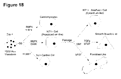
[0029] Figure 18. Differentiation scheme showing cardiomyocyte,
epicardium, and
EPDC development from hPSC-derived mesoderm.
Detailed description of the Disclosure
l. Definitions
8
CA 02921081 2016-02-11
WO 2015/035506 PCT/CA2014/000687
[0030] The term "activin component" as used herein means one or more
components, or
a composition comprising said component(s), optionally a culture medium
comprising a
molecule that activates nodal signal transduction, optionally Activin A
activity such as Activin A
and/or nodal.
[0031] The term "activin" or "ActA" as used herein refers to "Activin A",
(for example
Gene ID: 3624), for example human activinA, as well as active conjugates and
fragments
thereof, optionally including naturally occuring active conjugates and
fragments, that can for
example activate nodal signal transduction as well as active conjugates and
fragments thereof,
including naturally occuring active conjugates and fragments.
[0032] The term "activin/nodal inhibitor" and/or "activin/nodal/TGF-13R
inhibitor" as used herein
means any molecule that inhibits signal of the activin/nodal pathway and
particularly any
molecule that inhibits receptors ALK4, ALK7 and/or TGF-13RI, including but not
limited to
SB431542 (Sigma Aldrich) A83-01 (Tocris, 2929), D 4476, GW 788388, LY 364947,
RepSox,
SB 505124, SB 525334 (Sigma Aldrich), and SD 208.
[0033] The term "wnt inhibitor" as used herein means any agent, including
any
compound and/or protein that inhibits wnt signaling, including but not limited
to wnt antagonists
that bind either to the Wnt ligand itself, or to Wnt receptors, such as
Dickkopf (Dkk) proteins,
Wnt Inhibitory Factor-1 (WIF-1), and secreted Frizzled-Related Proteins
(sFRPs), as well as wnt
inverse agonists (e.g. an agent that binds to the same receptor as an agonist
but induces a
pharmacological response opposite to that of an agonist). Examples of Wnt
inhibitors include
XAV939, IWP 2, an inhibitor of wnt processing, and iCRT14, which is a potent
inhibitor of 13-
catenin-responsive transcription (CRT), both of which are available from
Tocris Bioscience, as
well as combinations thereof.
[0034] The term "wnt component" as used herein means any molecule that
activates
wnt/beta-catenin receptor signaling in a cardiovascular cell and incldues for
example Wnt3a and
as well as GSK3 selective inhibitors such as CHIR99021 (StemoleculeTM
CHIR99021
Stemgent), 6-Bromolndirubin-3'-Oxime (B10) (Cayman Chemical (cat:13123)), or
StemoleculeTM
BIO from Stemgent (cat:04003). CHIR99021 is a selective inhibitor of GSK3. The
GSK3
selective inhibitors contemplated are for example selective inhibitors for GSK-
3a/[3 in the Wnt
signaling pathway.
[0035] The term "FGF component" as used herein means a molecule such as a
cytokine,
including for example FGF, or a small molecule, that activates a FGF
signalling pathway, e.g.
9
CA 02921081 2016-02-11
WO 2015/035506 PCT/CA2014/000687
binds and activates a FGF receptor. The term "FGF" as used herein refers to
any fibroblast
growth factor, for example human FGF1 (Gene ID: 2246), FGF2 (also known as
bFGF; Gene
ID: 2247), FGF3 (Gene ID: 2248) , FGF4 (Gene ID: 2249), FGF5 (Gene ID: 2250),
FGF6 (Gene
ID: 2251), FGF7 (Gene ID: 2252), FGF8 (Gene ID: 2253), FGF9 (Gene ID: 2254)
and FGF10
(Gene ID: 2255) optionally including active conjugates and fragments thereof,
including naturally
occuring active conjugates and fragments. In certain embodiments, FGF is bFGF,
FGF10,
FGF4 and/or FGF2.
[0036] The term "BMP component" as used herein means any molecule
optionally any
BMP or growth and differentiation factor (GDF) that activates the receptor for
BMP4, including
for example BMP4 and BMP2,
[0037] The term "BMP inhibitor" as used herein means any inhibitor of BMP
signaling and
includes for example a type 1 BMP receptor inhibitor, BMP ligands and/or
soluble BMP
receptors. Optionally selected from dorsomorphin (DM), noggin, Chordin, LDN-
193189, soluble
BMPR1a, and/or soluble BMPR1b.
[0038] The term "BMP4" (for example Gene ID: 652) as used herein refers to
Bone
Morphogenetic Protein 4, for example human BMP4, as well as active conjugates
and
fragments thereof, optionally including naturally occuring active conjugates
and fragments, that
can for example activate BMP4 receptor signlaing.
[0039] The term "BMP component primed hPSCs" as used herein means hPSCs
that
have been contacted with a BMP component for at least 12 hours, preferably at
least 24 hours
or more preferably at least 48 hours. Typically these cells are in embryoid
bodies or monolayer
cultures.
[0040] The term "cardiovascular lineage cell" refers to a cell that
expresses a
cardiovascular mesoderm, cardiomyocyte or an epicardial gene expression
pattern, for example
expresses KDR, PDGFRa, NK2 homeobox 5 (NKX2-5), cardiac troponin T (cTnT),
signal-
regulatory protein alpha (SIRPA) or Wilms Tumour 1 (WTI) and is primed or has
the capacity to
differentiate into a cardiomyocyte lineage cell and/or an epicardial lineage
cell or an epicardial
derived cell (EPDC) such as a vascular smooth muscle like cell or a fibroblast
like cell as
described herein.
[0041] The term "cardiovascular mesoderm programming cocktail" as used
herein is a
combination comprising a BMP component and an activin component and optionally
a FGF
CA 02921081 2016-02-11
WO 2015/035506 PCT/CA2014/000687
component and the cardiovascular mesoderm programming cocktail is contacted
with the
hPSCs for about 3 to about 5 days.
[0042] The term "cardiovascular progenitor specification cocktail" as used
herein means
a one or more components, a composition comprising said component(s), for
specifying a
NKX2-5+ or WT1+ cardiovascular progenitor cell population for example a
cardiomyocyte
promoting component for specifying a NKX2-5+ cardiomyocyte lineage progenitor
cell
population or a epicardial promoting component for specifying a WT1+
epicardial lineage
progenitor cell population.
[0043] The term "cardiomyocyte promoting component" as used herein means
one or
more components or a composition comprising said component(s), said one or
more
components, comprising: 1) a combination of a Wnt inhibitor optionally
selected from, DKK1,
XAV939 and IWP2 and a BMP component, optionally wherein the BMP component is
BMP4 at
a concentration of at least 0.01 ng/mL, at least 0.05 ng/mL, at least 0.1
ng/mL, at least 0.5
ng/mL, at least 1.25 ng/mL, at least 2.5 ng/mL, at least 5 ng/mL, but less
than 1Ong/ml, or less
than 15 ng/mL or preferably about 0.5 ng/mL; or 2) a BMP inhibitor, such as
noggin or
dorsomorphin, for example noggin at a concentration of less than 200 ng/mL,
less than 150
ng/mL, less than 100 ng/mL, less than 50 ng/mL, or less than 25 ng/mL and/or
greater than 12.5
ng/mL; 3) a Wnt inhibitor, for example wherein there is sufficient endogenous
BMP4 produced
and/or 4) a cardiomyocyte lineage concentration of a BMP component, optionally
BMP4 for
example wherein the BMP4 is at a concentration of less than 0.63 ng/mL, less
than 0.5 ng/mL,
less than 0.4 ng/mL, or less than 0.3 ng/mL. The effective concentration
and/or combination can
be determined by monitoring and optimizing for NKX2-5 expression and/or
TNNT2/cTnT
expression.
[0044] The term "epicardial lineage promoting component" as used herein
means one or
more components or a composition comprising said component(s), the one or more
components comprising an epicardial lineage promoting concentration of a BMP
component,
optionally BMP4, and optionally a Wnt component. Optionally, the BMP4 is at a
concentration of
at least 1.25 ng/mL, at least 2.5 ng/mL, at least 5 ng/mL or at least 10 ng/mL
and/or the Wnt
component is CHIR99021. The effective concentration and/or combination can be
determined
by monitoring and optimizing for WT1 expression, basonuclin 1 (BNC1)
expression, annexin A8
(ANXA8) expression and/or T-box 18 (TBX18) expression.
11
CA 02921081 2016-02-11
WO 2015/035506 PCT/CA2014/000687
[0045] The term "a cardiomyocyte lineage cell" as used herein refers to a
cell that is
NKX2-5+ and which can differentiate to a cardiomyocyte, for example using a
method described
herein.
[0046] The term "an epicardial lineage cell" as used herein, refers to a
cell that is VVT1+
and which can differentiate to an epicardial cell, for example using a method
described herein
and/or an epicardial derived cells (EPDC).
[0047] The term "culturing" as used herein includes any in vitro method of
maintaining
and/or propagating a population of cells, including monolayer, bead, flask, or
3D cultures,
optionally where ambient conditions are controlled as in an incubator and
optionally involving
passaging of cells.
[0048] The term "epithelial-to-mesenchymal transition (EMT) cocktail" as
used herein
means one or more components or a composition comprising said component(s) for
inducing
EMT, the one or more components including a TGFI3 component such as TGFf3 or a
combination comprising a TGFf3 component and an FGF component such as bFGF.
[0049] The term "TGFO component" or as used herein a component or
compostion
comprising said component that promotes TGF13 signaling and includes for
example TGF131,
TG932 and/or TGF83.
[0050] A "KDR+ cell" as used herein means a cell exhibiting "kinase-insert
domain-
containing receptor" (KDR) cell surface expression and a "KDR+ cell
population" means a
population of cells, wherein at least 50%, at least 60%, at least 70%, at
least 80%, at least 90%
or at least 95% or more of the cells exhibit KDR cell surface expression.
[0051] The term "PDGFRa+ cell" as used herein means a cell exhibiting
"platelet derived
growth factor receptor a" cell surface expression and a PDGFRa+ cell
population means a
population of cells, wherein at least 50%, at least 60%, at least 70%, at
least 80%, at least 90%
or at least 95% or more of the cells exhibit PDGFRa cell surface expression.
[0052] The term "concentration" means diluted concentration in the cell
culture medium.
[0053] As used herein the term "purified population" with respect to a
population of cells
as used herein refers to a population of cells that has been removed and
separated (e.g.
isolated) from a mixed or heterogeneous population of cells and/or other
components such as
culture medium. In some embodiments, a purified population is a substantially
pure population
12
CA 02921081 2016-02-11
WO 2015/035506 PCT/CA2014/000687
of cells as compared to the heterogeneous population from which the cells were
isolated or
enriched from.
[0054] The term "substantially pure", with respect to a particular cell
population, refers to
a population of cells that is at least about 65%, preferably at least about
75%, at least about
85%, more preferably at least about 90%, and most preferably at least about
95% pure, with
respect to the cells making up a total cell population. Similarly, with regard
to a "substantially
pure" population of for example WT1+ cells, refers to a population of cells
that contain fewer
than about 30%, fewer than about 20%, more preferably fewer than about 15%,
10%, 8%, 7%,
most preferably fewer than about 5%, 4%, 3%, 2%, 1%, or less than 1%, of cells
that are not
VVT-1+.
[0055] The term "subject" as used herein includes all members of the
animal kingdom
including mammals, and suitably refers to humans.
[0056] The terms "treat", "treating", "treatment", etc., as applied to a
cell, include
subjecting the cell to any kind of process or condition or performing any kind
of manipulation or
procedure on the cell. As applied to a subject, the terms refer to providing
medical or surgical
attention, care, or management to an individual.
[0057] The term "treatment" as used herein as applied to a subject, refers
to an approach
aimed at obtaining beneficial or desired results, including clinical results
and includes medical
procedures and applications including for example pharmaceutical
interventions, surgery,
radiotherapy and naturopathic interventions as well as test treatments for
treating cancer.
Beneficial or desired clinical results can include, but are not limited to,
alleviation or amelioration
of one or more symptoms or conditions, diminishment of extent of disease,
stabilized (i.e. not
worsening) state of disease, preventing spread of disease, delay or slowing of
disease
progression, amelioration or palliation of the disease state, and remission
(whether partial or
total), whether detectable or undetectable. "Treatment" can also mean
prolonging survival as
compared to expected survival if not receiving treatment.
[0058] As used herein, the terms "administering," "introducing" and
"transplanting" are
used interchangeably in the context of delivering cells into a subject, by a
method or route which
results in at least partial localization of the introduced cells at a desired
site.
[0059] The term "contacting" is intended to include incubating the
component(s) and the
cell together in vitro (e.g., adding the compound to cells in culture) and the
step of contacting
can be conducted in any suitable manner. For example the cells may be treated
in adherent
13
CA 02921081 2016-02-11
WO 2015/035506 PCT/CA2014/000687
culture, or in suspension culture, 3D culture, or where the cells are cultured
on beads, the
cocktail components can be added temporally substantially simultaneously or
sequentially (e.g.
within 1 hour from an addition of a first component). The cells can also be
contacted with
another agent such as a growth factor or other differentiation agent or
environments to stabilize
the cells, or to differentiate the cells further and include culturing the
cells under conditions
known in the art for example for culturing the pluripotent (and/or
differentiated) population for
example as further described in the Examples.
[0060] The term "cell culture medium" (also referred to herein as a
"culture medium" or
"medium") as referred to herein is a medium for culturing cells containing
nutrients that maintain
cell viability and support proliferation and optionally differentiation. The
cell culture medium may
contain any of the following in an appropriate combination: salt(s),
buffer(s), amino acids,
glucose or other sugar(s), antibiotics, serum or serum replacement, and other
components such
as peptide growth factors, vitamins etc. Cell culture media ordinarily used
for particular cell
types are known to those skilled in the art.
[0061] The term "pluripotent stem cell" as used herein refers to a cell
with the capacity,
under different conditions, to differentiate to more than one differentiated
cell type, and for
example the capacity to differentiate to cell types characteristic of the
three germ cell layers,
and includes embryonic stem cells and induced pluripotent stem cells.
Pluripotent cells are
characterized by their ability to differentiate to more than one cell type
using, for example, a
nude mouse teratoma formation assay. Pluripotency is also evidenced by the
expression of
embryonic stem (ES) cell marker. As used herein, pluripotent stems can include
cell lines
including induced pluripotent stem cells (iPSC) and embryonic stem cells
(ESC). In an
embodiment, the pluripotent stem cells are not human embryonic stem cells.
[0062] As used herein, the terms "iPSC" and "induced pluripotent stem cell"
are used
interchangeably and refers to a pluripotent stem cell artificially derived
(e.g., induced or by
complete reversal) from a non-pluripotent cell, typically an adult somatic
cell, for example, by
inducing expression of one or more genes (including POU4F1/OCT4 (Gene ID;
5460) in
combination with, but not restricted to, SOX2 (Gene ID; 6657), KLF4 (Gene ID;
9314), cMYC
(Gene ID; 4609), NANOG (Gene ID; 79923), LIN28/ LIN28A (Gene ID; 79727)).
[0063] The term "embryonic stem cell" is used to refer to the pluripotent
stem cells of the
inner cell mass of the embryonic blastocyst (see, for example, U.S. Pat. Nos.
5,843,780,
6,200,806). Such cells can also be obtained from the inner cell mass of
blastocysts derived from
somatic cell nuclear transfer (see, for example, U.S. Pat. Nos. 5,945,577,
5,994,619,
14
CA 02921081 2016-02-11
WO 2015/035506 PCT/CA2014/000687
6,235,970). The distinguishing characteristics of an embryonic stem cell
define an embryonic
stem cell phenotype. Accordingly, a cell has the phenotype of an embryonic
stem cell if it
possesses one or more of the unique characteristics of an embryonic stem cell
such that that
cell can be distinguished from other cells. Exemplary distinguishing embryonic
stem cell
characteristics include, without limitation, gene expression profile,
proliferative capacity,
differentiation capacity, karyotype, responsiveness to particular culture
conditions, and the like.
[0064] The term "expression" refers to the cellular processes involved in
producing RNA
and proteins and as appropriate, secreting proteins, and cell surface
expression, including
where applicable, but not limited to, for example, transcription, translation,
folding, modification
and processing. "Expression products" include RNA transcribed from a gene and
polypeptides
obtained by translation of mRNA transcribed from a gene.
[0065] In understanding the scope of the present disclosure, the term
"comprising" and
its derivatives, as used herein, are intended to be open ended terms that
specify the presence
of the stated features, elements, components, groups, integers, and/or steps,
but do not exclude
the presence of other unstated features, elements, components, groups,
integers and/or steps.
The foregoing also applies to words having similar meanings such as the terms,
"including",
"having" and their derivatives. Finally, terms of degree such as
"substantially", "about" and
"approximately" as used herein mean a reasonable amount of deviation of the
modified term
such that the end result is not significantly changed. These terms of degree
should be construed
as including a deviation of at least 5% of the modified term if this
deviation would not negate
the meaning of the word it modifies.
[0066] In understanding the scope of the present disclosure, the term
"consisting" and its
derivatives, as used herein, are intended to be close ended terms that specify
the presence of
stated features, elements, components, groups, integers, and/or steps, and
also exclude the
presence of other unstated features, elements, components, groups, integers
and/or steps.
[0067] The recitation of numerical ranges by endpoints herein includes all
numbers and
fractions subsumed within that range (e.g. 1 to 5 includes 1, 1.5, 2, 2.75, 3,
3.90, 4, and 5). It is
also to be understood that all numbers and fractions thereof are presumed to
be modified by the
term "about." Further, it is to be understood that "a," "an," and "the"
include plural referents
unless the content clearly dictates otherwise. The term "about" means plus or
minus 0.1 to 50%,
5-50%, or 10-40%, preferably 10-20%, more preferably 10% or 15%, of the number
to which
reference is being made.
CA 02921081 2016-02-11
WO 2015/035506 PCT/CA2014/000687
[0068] Further, the definitions and embodiments described in particular
sections are
intended to be applicable to other embodiments herein described for which they
are suitable as
would be understood by a person skilled in the art. For example, in the
following passages,
different aspects of the invention are defined in more detail. Each aspect so
defined may be
combined with any other aspect or aspects unless clearly indicated to the
contrary. In particular,
any feature indicated as being preferred or advantageous may be combined with
any other
feature or features indicated as being preferred or advantageous.
11. Methods and Products
[0069] Described herein are methods for producing cardiovascular lineage
cells including
cardiomyocyte lineage cells, epicardial lineage cells and epicardial derived
cells. Components
and conditions for specifying these cell types as well as markers for
monitoring emergence of
these cell types are described.
[0070] Accordingly, an aspect includes a method of obtaining a
cardiovascular lineage
cell population, optionally cardiomyocyte lineage cell population or an
epicardial lineage cell
population from pluripotent stem cells (PSCs) optionally human PSCs (hPSCs)
comprising the
steps: (a) contacting BMP component primed hPSCs with a cardiovascular
mesoderm
programming cocktail suitable for inducing the hPSCs to differentiate to a
cardiovascular
mesoderm cell population under conditions suitable for the programming
cocktail to penetrate
the hPSCs and culturing the contacted hPSCs for a period of time to generate a
KDR+ and
PDGFRa+ cardiovascular mesoderm cell population; (b) contacting the
cardiovascular
mesoderm cell population with a cardiovascular progenitor specification
cocktail suitable to
specify a NKX2-5+ or VVT1+ cardiovascular progenitor cell population under
conditions suitable
for the specification cocktail to penetrate the cardiovascular mesoderm cell
population and
culturing the contacted cardiovascular mesoderm cell population for a period
of time to generate
a NKX2-5+ or WT-1+ cardiovascular progenitor cell population; and (c)
contacting the
cardiovascular progenitor cell population with a maturation cocktail under
conditions suitable for
the maturation cocktail to penetrate the cardiovascular progenitor cell
population and culturing
the contacted cardiovascular progenitor population for a period of time to
produce a
cardiovascular lineage population optionally cardiomyocyte lineage cells
expressing cardiac
troponin T (cTnT) and/or SIRPA and/or an epicardial lineage cell population
optionally
expressing VVT1 and/or comprising EPDCs.
[0071] KDR and PDGFRa can be used to monitor development of a
cardiovascular
mesoderm cell population. The expression of KDR can be monitored using an
antibody specific
16
CA 02921081 2016-02-11
WO 2015/035506 PCT/CA2014/000687
for KDR and/or the expression of PDGFRa can be monitoring using an antibody
specific for
PDGFRa. As both are cell surface expressed, KDR and PDGFRa expression can be
monitored
by measuring cell surface expression. For example, the expression of KDR and
PDGFRa can
be monitored using flow cytometry.
[0072] In an embodiment, the BMP component primed hPSCs are prepared by
contacting the hPSCs with BMP component for about 1 to about 2 days,
optionally wherein the
BMP component is BMP4 and/or BMP2.
[0073] In an embodiment, the cardiovascular mesoderm programming cocktail
comprises
a BMP component and an activin component and optionally a FGF component and
the
cardiovascular mesoderm programming cocktail is contacted with the hPSCs for
about 3 to
about 5 days.
[0074] In an embodiment, the FGF component comprises bFGF.
[0075] In an embodiment, the BMP component comprises BMP4 and/or BMP2.
[0076] In an embodiment, the activin component comprises Activin A.
[0077] Concentrations of activin component and BMP component can be
optimized as
descri bee
[0078] In an embodiment, the PSCs are comprised in embryoid bodies.
[0079] Using for example steps a) and b) above, it is demonstrated herein
that a NKX2-
5+ or VVT1+ cardiovascular progenitor cell population can be obtained.
[0080] Accordingly a further aspect includes a method for obtaining a NKX2-
5+ or WT1+
cardiovascular progenitor cell population from PSCs, optionally hPSCs,
comprising the steps:
(a) obtaining a KDR+ and PDGFRa+ cardiovascular mesoderm cell population from
hPSCs
optionally as described above; (b) contacting the KDR+ and PDGFRa+
cardiovascular
mesoderm cell population with a cardiovascular progenitor specification
cocktail under
conditions suitable for the specification cocktail to penetrate the
cardiovascular mesoderm cell
population and culturing the contacted cardiovascular mesoderm cell population
for a period of
time sufficient to generate a NKX2-5+ or VVT1+ cardiovascular progenitor cell
population.
[0081] In an embodiment, the KDR+ and PDGFRa+ cardiovascular mesoderm cell
population is dissociated prior to contacting with the cardiovascular
progenitor specification
cocktail.
17
CA 02921081 2016-02-11
WO 2015/035506 PCT/CA2014/000687
[0082] In some embodiments, the KDR+ PDGFRa+ expressing cells are purified
before
contacting with the cardiovascular progenitor specification cocktail.
[0083] In another embodiment, the cardiovascular mesoderm cell population
is contacted
with the cardiovascular progenitor specification cocktail for at least 12
hours to about 48 hours,
or any amount of time between 12 and 48 hours.
[0084] In an embodiment, the cardiovascular progenitor specification
cocktail comprises
a cardiomyocyte lineage promoting component, wherein the cardiomyocyte
promoting
component is in a suitable concentration for promoting cardiomyocyte
development and
specifies a NKX2-5+ cardiovascular progenitor population.
[0085] In an embodiment, the cardiomyocyte promoting component comprises a
BMP
inhibitor, for example for use with cardiovascular mesoderm cell population
endogenously
expressing a level of BMP that inhibits cardiomyocyte specification.
[0086] In an embodiment, the cardiomyocyte promoting component comprises
noggin at
a concentration of less than 200 ng/mL, less than 150 ng/mL, less than 100
ng/mL, less than 50
ng/mL, or less than 25 ng/mL and/or greater than 12.5 ng/mL.
[0087] In another embodiment, the cardiomyocyte promoting component is
dorsomorphin
at a concentration of less than 1 M, less than 0.5 M, less than 0.25 M, or
less than 0.1 M.
[0088] In another embodiment, the cardiomyocyte promoting component is
BMP4 at a
concentration of less than 0.63 ng/mL, less than 0.5 ng/mL, less than 0.4
ng/mL, or less than
0.3 ng/mL.
[0089] In another embodiment, the cardiovascular progenitor specification
cocktail
comprises: 1) a combination of a Wnt inhibitor optionally selected from, DKK1,
XAV939 and
IWP2 and a BMP component, optionally wherein the BMP component is BMP4 at a
concentration of at least 0.01 ng/mL, at least 0.05 ng/mL, at least 0.1 ng/mL,
at least 0.5 ng/mL,
at least 1.25 ng/mL, at least 2.5 ng/mL, at least 5 ng/mL, but less than
1Ong/ml, or less than 15
ng/mL or preferably about 0.5 ng/mL; or 2) a BMP inhibitor, such as noggin or
dorsomorphin, for
example noggin at a concentration of less than 200 ng/mL, less than 150 ng/mL,
less than 100
ng/mL, less than 50 ng/mL, less than 25 ng/mL or greater than 12.5 ng/mL; 3) a
Wnt inhibitor,
for example wherein there is sufficient endogenous BMP4 produced; and/or 4) a
cardiomyocyte
lineage concentration of a BMP component, optionally BMP4 for example wherein
the BMP4 is
at a concentration of less than 0.63 ng/mL, less than 0.5 ng/mL, less than 0.4
ng/mL, or less
18
CA 02921081 2016-02-11
WO 2015/035506 PCT/CA2014/000687
than 0.3 ng/mL. The effective concentration and/or combination can be
determined by
monitoring and optimizing for NKX2-5 expression and/or TNNT2/cTnT expression.
[0090] A Wnt inhibitor can be used without BMP to induce cardiomyocyte
specification for
example when the mesoderm population of cells produces sufficient endogenous
BMP
component.
[0091] A BMP component in a concentration that promotes cardiomyocyte
specification
can be used for example when the mesoderm population of cells produces
insufficient
endogenous BMP component.
[0092] The level of an endogenous component that is secreted, such as BMP4
or BMP2
can measured by ELISA or other quantitative immunoassays or quantitative RT-
PCR.
[0093] In another embodiment, the KDR+PDGFRa+ cardiovascular mesoderm
population and/or the NKX2-5+ cardiovascular progenitor population is
purified/isolated.
[0094] In another embodiment, the NKX2-5+ cardiovascular progenitor cell
population is
further contacted with a maturation cocktail optionally comprising a VEGF
component. The
maturation cocktail can be culture medium suitable for the cell type and/or
include additional
components.
[0095] A further aspect includes a method for producing cardiac troponin
T+ (cTnT)
cardiomyocyte lineage cell population comprising: (a) obtaining a NKX2-5+
cardiovascular
progenitor population according to the method of any one of claims 1 to 16;
(b) contacting the
cardiovascular progenitor cell population with a maturation cocktail
comprising a VEGF
component under conditions suitable for the maturation cocktail to penetrate
the cardiovascular
progenitor cell population; and (b) culturing the contacted cardiovascular
progenitor population
for a period of time sufficient to produce cardiomyocytes expressing cardiac
troponin T (cTnT).
[0096] In an embodiment, the NKX2-5+ cardiovascular progenitor population
is contacted
with the maturation cocktail for 4 or more days, for example at least about 4,
optionally about 5,
about 9, about 15 or about 20 days, optionally until mature contracting
cardiomyocytes are
produced. The cells can be kept in culture to mature until the desired cell
population is obtained.
[0097] A further aspect is a method of producing a WT1+ epicardial lineage
cell
population, comprising the steps: (a) obtaining a KDR+ and PDGFRa+
cardiovascular
mesoderm cell population from hPSCs optionally as defined above; (b)
contacting the
cardiovascular mesoderm cell population with a cardiovascular progenitor
specification cocktail
19
CA 02921081 2016-02-11
WO 2015/035506 PCT/CA2014/000687
comprising an epicardial lineage promoting component under conditions suitable
for the
specification cocktail to penetrate the cardiovascular mesoderm cell
population and culturing the
contacted cardiovascular mesoderm cell population for a period of time
sufficient to generate a
VVT1+ cardiovascular progenitor cell population.
[0098] In an embodiment, the cardiovascular progenitor specification
cocktail comprises
an epicardial cell promoting component, optionally wherein the epicardial cell
promoting
component comprises BMP4 in a suitable concentration for promoting epicardial
cell
development.
[0099] In another embodiment, the epicardial cell-promoting component
comprises BMP4
at a concentration of at least 1.25 ng/mL, at least 2.5 ng/mL, at least 5
ng/mL or at least 10
ng/mL.
[00100] In another embodiment, the epicardial cell promoting component
further
comprises a Wnt component, optionally CHIR99021.
[00101] In yet another embodiment, the epicardial cell promoting component
comprises
BMP4 and a Wnt component optionally CHIR 99021.
[00102] In another embodiment, the VVT1+ cardiovascular progenitor cell
population is
contacted with a maturation cocktail comprising a VEGF component.
[00103] In another embodiment, the VVT1+ cardiovascular progenitor
population is
contacted with the maturation cocktail for about 4 or more days, optionally
about 5, about 9,
about 15 or about 20 days to produce a maturation cocktail contacted VVT1+
epicardial lineage
cell population.
[00104] In another embodiment, the maturation cocktail contacted VVT1+
epicardial
lineage cell population is purified/isolated.
[00105] In another embodiment, the maturation cocktail contacted VVT1+
epicardial
lineage cell population is cultured to obtain a zona occludins 1 (Z01)+ VVT1+
epicardial lineage
cell population, optionally wherein the Z01+VVT1+ epicardial lineage cell
population is
purified/isolated.
[00106] In another embodiment, the maturation cocktail contacted VVT1+
epicardial
lineage cell population and/or the Z01+ WT1+ epicardial lineage cell
population is contacted
with an epithelial-to-mesenchymal transition (EMT) cocktail and cultured for a
period of time.
CA 02921081 2016-02-11
WO 2015/035506 PCT/CA2014/000687
[00107] In another embodiment, the EMT cocktail comprises: 1) a TGFp
component; 2) a
TGFp component and a FGF component, optionally wherein the TGFp component and
the FGF
component are sequentially administered; or 3) FGF component.
[00108] In an embodiment, the TGFb component is TGFb-1. In an embodiment,
the FGF
component is bFGF.
[00109] As shown in Example 2, treatment of VVT1+ cells EMT cocktail
comprising TGFb
gives rise to functional smooth muscle cells and treatment of VVT1+ cells with
EMT cocktail
comprising TGFb and bFGF gives rise to a higher percentage of smooth muscle
cells.
[00110] In an embodiment, the VVT1+ cells are contacted with EMT cocktail
from about 1
day to up to 3 weeks, for example about 1 day, 2 days, 3 days, 4 days, 5 days,
6 days, 7 days,
8 days, 9 days or about 10 days or about 1 week, 2 weeks or weeks. For example
where the
EMT cocktail comprises components that are sequentially administered, each
component can
be administered for about 1 day, 2 days, 3 days, 4 days, 5 days, 6 days, 7
days, 8 days, 9 days
or about 10 days, or about 1 week, about 2 weeks or about 3 weeks.
[00111] In an embodiment, the TGFb component is TGFp-1 and the
concentration is from
about 0.25 ng/ml to about 10 ng/ml or any one 0.1 ng/ml increment between 0.25
ng/ml and 10
ng/ml. Comparable concentrations of other TGFb components that produce similar
TGFb
signaling pathway activation can be used.
[00112] In an embodiment, the FGF component is bFGF and the concentration
is from
about 1 ng/ml to about 50 ng/ml or any 1 ng/ml increment between 1 ng/ml and
50 ng/ml.
Comparable concentrations of other FGF components that produce similar FGF
signaling
pathway activation can be used.
[00113] In yet another embodiment, the EMT cocktail is contacted with the
VVT1+
population of cells according to the following schedule: 1) TGFP-1 (for
example about 0.25 to
about 10 ng/ml) for about four days followed by four days with no additional
factor (TGF13), 2)
TGF0-1 (for example about 0.25 to about 10 ng/ml) for about four days followed
by about four
days with bFGF ( for example about 1 to about 50 ng/ml) (TGFp+bFGF), or 3)
bFGF (for
example about 1 to about 50 ng/ml) for about eight days (bFGF).
[00114] In another embodiment, the EMT cocktail comprises: 1) TGFp
component; or 2) a
TGFf3 component and a FGF component; and the cell population is cultured for a
period of time
to produce expression of an EMT marker such as SNAI1 and/or SNAI2 (detectable
for example
21
CA 02921081 2016-02-11
WO 2015/035506 PCT/CA2014/000687
by measuring SNAI1 and/or SNAI2 transcript expression levels), a mesenchymal
marker such
as vimentin and/or CD90 and/or a smooth muscle marker such as SMA, optionally
measured by
flow cytometry or expression of a smooth muscle gene optionally CNN1, MYH11,
TAGLN and
SMTN.
[00115] As demonstrated in Example 2, the smooth muscle-like cells
generated following
EMT exhibited NE and PE induced calcium transients. The proportion of cells
displaying calcium
transients was highest (70%) in the population induced by TGFp+bFGF indicating
that this
combination of signaling pathways efficiently promoted the development of
smooth muscle cells
capable of contraction (Fig. 16a). In an embodiment, the cell population is
cultured for a period
of time sufficient to produce a population of cells wherein at least 50%, at
least 60% or at least
70% of the cells of the population display a calcium transient upon NE or PE
stimulation.
[00116] In another embodiment, the population of cells is cultured until
the population of
cells expresses a smooth muscle marker or transcript to obtain a vascular
smooth muscle
lineage cell population, optionally until the population of cells expresses
increased levels of a
mesenchymal marker, optionally vimentin and/or CD90.
[00117] In another embodiment, the EMT cocktail comprises: an FGF component
and the
cell population is cultured to produce a fibroblast lineage cell population
expressing an
epicardial-derived fibroblast marker optionally TCF21, optionally measured by
qRT-PCR.
[00118] It is further demonstrated in Example 2, that EMT induced hPSC-
derived Epi cells
can acquire invasiveness. For example invasion was monitored eight days
following the
induction of EMT and cells induced with bFGF alone were the most migratory and
invaded the
matrigel to the greatest depth. bFGF treatment also led to an increase in
total cell number within
the regions of interest (ROI; e.g. a region where the recording took place).
Accordingly, in
another embodiment, the cell population is cultured for a period of time
sufficient to produce a
population of cells wherein a proportion of the cells of the population
acquire invasiveness.
Invasiveness includes for example a cell that can migrate at least 100 pm, 150
pm, 200 pm, 250
pm, 300 pm, 400 pm, 500 pm or 600 pm in a Matrigel assay as described in
Example 2.
[00119] In an embodiment, a cell population is purified/isolated optionally
using flow
cytometry, including fluorescence-activated cell sorting (FACS), magnetic
separation, affinity
chromatography, immunostaining and/or resistance to cytotoxic agent. In other
embodiment,
purification/ isolation is based on detecting non-surface expressed markers,
which can be
22
CA 02921081 2016-02-11
WO 2015/035506 PCT/CA2014/000687
achieved my monitoring an aliquot for example by quantitative RT or other PCR
and immuno-
based assays such as western blot.
[00120] In
another embodiment, the population of cells is cultured to express TCF21 to
obtain a fibroblast lineage cell population.
[00121] In
another embodiment, the maturation cocktail contacted WT1+ epicardial
lineage cell population and/or the WT1+ Z01+ epicardial lineage cell
population is cultured for a
period of time to obtain a retinol dehydrogenase expressing epicardial lineage
cell population,
optionally wherein the retinol dehydrogenase expressing epicardial lineage
cell population is
ALDH1A2 expressing or Aldefluor positive staining, optionally wherein the cell
population is at
least 50% Aldefluor positive.
[00122] In
another embodiment, the vascular smooth muscle lineage cell population, a
fibroblast lineage cell population and/or a retinol dehydrogenase expressing
epicardial lineage
cell population is purified/isolated.
[00123] In
another embodiment, the cardiovascular progenitor specification cocktail
further
comprises an activin/nodal inhibitor, optionally SB431542. For example,
SB431542 added
during the cardiovascular specification stage can promote both cardiomyocyte
and epicardial
specification.
[00124] In
another embodiment, the PSCs are a human PSC. In yet another embodiment,
the PSC is an induced pluripotent stem cell (iPSC) line, optionally a human
iPSC and/or an
embryonic stem cell (ESC) line, optionally a human ESC (hESC). In an
embodiment, the iPSC
is a fibroblast derived iPSC line.
[00125] A further
aspect includes a purified population of cardiovascular lineage cell or cell
population and/or a cell or cell population differentiated therefrom produced
according to the
method of described herein. For example it is demonstrated here that
cardiomyocyte or
epicardial lineage cells can be specified with contaminating cell types. In an
embodiment, the
purified population is comprised in a gel, optionally Matrigel. Accordingly,
the desired population
can be purified with minimal intervention.
[00126] In an
embodiment, the cells are adhered to a solid support such as a dish or flask.
[00127]
Another aspect includes a composition comprising a purified/isolated
cardiovascular lineage cell or cell population and/or a cell or cell
population differentiated
therefrom produced according to the method of described herein; and a suitable
diluent.
23
CA 02921081 2016-02-11
WO 2015/035506 PCT/CA2014/000687
[00128] A suitable diluent includes for example a suitable culture medium,
or freezing
medium containing for example serum, a serum substitute or serum supplement
and/or a
suitable cryoprotectant such as dimethyl sulphoxide (DMSO), glycerol
Methylcellulose or
polyvinyl pyrrolidone.
[00129] Another aspect includes a culture medium supplement comprising a
cardiovascular progenitor specification cocktail, optionally comprising a BMP
component in a
concentration for specifying cardiomyocytes or comprising a BMP component and
a Wnt
component for epicardial specification. The components can be in liquid or
powder form for
reconstitution.
[00130] The components can be comprised in a single supplement to be added
to base
media such as Life Technologies StemPro-34. The amount of the components in
the
supplement can for example be amounts that when diluted in a culture medium
(e.g. when
diluted in a 450 mL base medium) result in concentrations described herein.
[00131] Another aspect includes a culture medium comprising the
specification cocktail
optionally comprising a BMP component in a concentration for specifying
cardiomyocytes or
comprising a BMP component and a Wnt component for epicardial specification.
Typical culture
medium components such as can also be included
[00132] Also included in another aspect is a kit comprising: 1) an agent
for measuring
expression of a marker expressed on a cardiovascular lineage cell or cell
differentiated
therefrom the marker selected from KDR, PDGFRa, NKX2-5+, WTI, Z01, EMT marker
such as
SNAI1 and/or SNAI2, a mesenchymal marker such as vimentin and/or CD90 and/or a
smooth
muscle marker such as SMA, a smooth muscle gene optionally CNN1, MYH11, TAGLN
and
SMTN, TCF21, retinol dehydrogenase, and/or Aldefluor activity; and/or 2) a
component or
composition such as a culture medium comprising said component for inducing
differentiation of
a cardiovascular lineage cell population, the components selected from
cocktail optionally
comprising a BMP component in a concentration for specifying cardiomyocytes or
comprising a
BMP component and a Wnt component for epicardial specification:
[00133] The agent can for example be an antibody or fragment thereof for
immuno-assays
and flow based methods, primers for detecting a particular transcript and/or a
probe for
detecting expression by a probe based method such as RT-PCR, qRT-PCR, in situ
hybridization
and Millipore SmartFlare.
24
CA 02921081 2016-02-11
WO 2015/035506 PCT/CA2014/000687
001343 The composition and kit components can include any of the components
described elsewhere herein and optionally instructions for use. For example,
in an embodiment,
the kit comprises a supplement comprising components etc. to induce
differentiation of one or
more stages or lineages described herein (e.g. including components described
in the
Examples). In an embodiment, the kit comprises a base culture medium,
optionally a base
culture medium described herein and a culture medium supplement described
herein.
[00135] The cells produced according to a method described herein can be
used to
screen for agents that promote and/or inhibit cardiovascular lineage cell
differentiation.
[00136] Accordingly a further aspect includes a method for identifying a
cardiovascular cell
differentiation promotion agent comprising the steps: (a) contacting a test
cell population with a
test agent at a step in a method described herein; (b) monitoring for
expression of a marker
selected from KDR, PDGFRalpha, NKX2-5+, VVT1, Z01, a mesenchymal marker such
as
vimentin and/or CD90 and/or a smooth muscle marker such as SMA, a smooth
muscle gene
optionally CNN1, MYH11, TAGLN and SMTN, TCF21, retinol dehydrogenase, and/or
Aldefluor
activity levels in the test cell population and a control; and (c) identifying
the test agent as a
cardiovascular cell differentiating promotion agent when the test agent
induces and/or increases
expression of the cardiovascular marker and/or induces specification of
cardiomyocyte,
epicardial or EPDC .
[00137] For example, co-culture assays can be performed in which hPSC-
derived
epicardium and cardiomyocytes can be mixed and plated either in aggregate or
monolayer
format. After a pre-determined amount of time, cultures may be assayed by qRT-
PCR, flow
cytometry, or immuno-based methods for changes in gene and protein expression.
Cultures can
be assessed for example for sarcomere morphology by staining for alpha
actinin, atrial
natriuretic factor (ANF), and/or brain natriuretic peptide (BNP);
mitochondrial maturity which can
for example be assessed using flow cytometry and/or immunological methods;
myosin
regulatory light chain 7 (MYL7) which is predominantly expressed in adult
atrial muscle and/or
WTI downregulation (e.g. indicative of epicardial maturity). Examples of flow
cytometry and
immunological methods are provided in the Examples. .
[00138] In an embodiment, the method is used for drug screening of a
cardiovascular
drug, for example for promoting and/or interfering with cardiac and/or
vascular remodeling.
[00139] In an embodiment, HPSC derived cardiomyocyte and epicardial lineage
cells
and/or tissue produced using a method described herein are 1) co-cultured,
optionally in
CA 02921081 2016-02-11
WO 2015/035506 PCT/CA2014/000687
combination with endothelial cells; 2) contacted with a test agent; and 3)
assessed for i) cell
death or ii) increased proliferation, optionally in endothelial cell numbers;
and/or iii) altered
tissue organization, compared to a control, wherein a decrease in cell death,
an increase in
proliferation one or more of the cell lineages and/or l) decreased or II)
increased cellular
organization compared to the control is indicative that the test agent is a
putative cardiovascular
drug. Endothelial cell numbers can for example be assessed by staining for
CD31; cell death
and/or proliferation can be assessed for example by flow cytometry, cell
counting methods
and/or flow cytometry; and cellular organization be assessed visually.
[00140] In an embodiment, the method is for identifying putative agents for
promoting
epicardium differentiation, replacement of scar tissue, revascularization of
ischemic areas etc. In
an embodiment, HPSC derived cardiomyocyte and epicardial lineage cells and/or
tissue
produced using a method described herein are 1) co-cultured, optionally in
combination with
endothelial cells; 2) contacted with a test agent under hypoxic or other
cardiotoxic conditions;
and 3) assessed for i) cell death or ii) increased proliferation, optionally
in endothelial cell
numbers; and/or iii) altered tissue organization under hypoxic conditions,
compared to a control,
wherein a decrease in cell death, an increase in proliferation one or more of
the cell lineages
and/or 0 decreased or 11) increased cellular organization compared to the
control is indicative
that the test agent is a putative agent for promoting epicardium
differentiation, replacement of
scar tissue, revascularization of ischemic areas. Endothelial cell numbers can
for example be
assessed by staining for CD31; cell death and/or proliferation can be assessed
for example by
flow cytometry, cell counting methods and/or flow cytometry; and cellular
organization be
assessed visually.
[00141] Myocardial infarction can also be induced a variety of model
organisms and
hPSC-derived epicardial cells can be transplanted to the outer lay of the
heart. Heart function
recovery, myocyte proliferation/survival, and the contribution of EPDCs can be
assayed.
[00142] Accordingly a further aspect includes a method of introducing a
cardiovascular
population of cells into a subject in need thereof, comprising producing a
population of cells
according to a method described herein, purifying the cell population and
administering said
population of cells into the subject in need thereof.
[00143] The population of cells is optionally comprised is an isotonic
composition suitable
for administration to a subject.
1 26
CA 02921081 2016-02-11
WO 2015/035506 PCT/CA2014/000687
[00144] A further aspect includes a method of treating a subject in need
thereof,
comprising transplanting to the subject a population of cells produced
according to a method
described herein, optionally a purified cell population.
[00145] In an embodiment, the subject has suffered or is suffering a
transient ischemic
attack. In an embodiment, the subject has ischemic heart diseases.
[00146] In an embodiment the subject is administered a population produced
from hPSCs,
wherein the hPSCs are autologous iPSCs.
[00147] A number of genes and gene products are described herein. All
reference
accession numbers for genes and gene products referred to, including TNNT2 -
NM_001276345.1, NKX2-5 - NM_004387.3, WT1 - NM_024426.4, TBX18 -
NM_001080508.2,
GATA4 - NM_002052.3, GATA5 - NM_080473.4, ISL1 - NM_002202.2, TBX5 -
NM_000192.3,
BNC1 - NM_001717.3, ANXA8 - NM_001271702.1, SNAI1 - NM_005985.3, SNAI2 -
NM_003068.4, CCN1 - NM_001299.4, MYH11 - NM_001040113.1, TAGLN -
NM_001001522.1,
SMTN - NM_001207017.1, TCF21 - NM_198392.2, ALDH1A1 - NM_000689.4, ALDH1A2 -
NM_003888.3, and ALDH1A3 - NM_000693.2, the sequences associated therewith are
herein
incorporated by reference in their entirely.
[00148] The above disclosure generally describes the present application. A
more
complete understanding can be obtained by reference to the following specific
examples. These
examples are described solely for the purpose of illustration and are not
intended to limit the
scope of the application. Changes in form and substitution of equivalents are
contemplated as
circumstances might suggest or render expedient. Although specific terms have
been employed
herein, such terms are intended in a descriptive sense and not for purposes of
limitation.
[00149] The following non-limiting examples are illustrative of the present
disclosure:
Examples
Example 1
[00150] Media consisting of StemPro-34 (Life Technologies) supplemented
with 10 ng/ml
penicillin/streptomycin, 2 mM L-glutamine, 1 mM ascorbic acid, and 4 3 10_4 M
monothioglycerol (MTG) (Sigma). Human-BMP4, human-bFGF, human-Activin A, human-
DKK1,
and human-VEGF (R&D Systems) were added at the indicated time points and
concentrations.
The Activin/Nodal/TGF-b and BMP inhibition experiments were carried out with
SB-431542
(Tocris, Ellisville, MO) and dorsomorphin (Sigma), respectively. For
experiments involving Wnt
27
CA 02921081 2016-02-11
WO 2015/035506 PCT/CA2014/000687
signaling, CHIR-99021 (Stemgent), XAV-939 (R&D), or IWP2(R&D) were used at the
indicated
concentrations. Cultures were maintained in a 5% CO2, 5% 02, 90% N2
environment for the
first 10-12 days and were then transferred into a 5% CO2 air environment for
the remainder of
the culture period. At indicated time points, cells were harvested and
analyzed by flow cytometry
or cell sorted.
Results
[00151] Cardiomyocyte Specification. The development of the cardiovascular
lineages
from hPSCs progresses through at least three distinct steps, the induction of
KDR+PDGFR+
cardiovascular mesoderm and the specification of this mesoderm to a
cardiovascular fate
resulting in the development of NKX2-5+ cardiovascular progenitors and
subsequently the
maturation to contracting cardiomyocytes (Fig 1). While activin A and BMP4 are
the key
regulators of the first stage5 pathways controlling cardiac specification are
less well understood
and likely to differ from the induction step. To investigate the specification
step, a model was
established that enabled us to easily manipulate signaling pathways during
this stage of
development (Fig 1). With this approach, cardiovascular mesoderm is induced in
EBs with
optimal concentrations of activin and BMP4 as described5. At day 4 of mesoderm
induction, the
EBs are dissociated, the cells plated in monolayer in microtitre wells (1x10⁵
per well) and treated
with different pathway agonists and antagonists for 24-48 hours. Following
this specification
step, the cultures are maintained in the presence of VEGF and analyzed at day
15 for the
presence of contracting cells that express cardiac troponin T (cTnT) and/or
SIRPA. Initial
studies showed that cardiomyocytes routinely develop in the presence of the
Wnt inhibitor DKK1
and the activin/nodal/TGFp inhibitor SB431542 indicating that these pathways
are not required
for specification. BMP signaling, on the other hand, had a profound effect at
this stage, as
addition of high levels of BMP4 or the inhibitors noggin or dorsomorphin
completely blocked
cardiomyocyte development (Fig 2a). Neither manipulation dramatically impacted
mesoderm
development, although noggin did decrease the levels of KDR to some extent.
Inhibition of
cardiomyocyte development was not due to dramatic cell death as cell numbers
in each group
following the 48-hour treatment were not significantly different (Fig 2b).
[00152] C.2. Generation of epicardial cells. Titration of the agonist and
antagonist during
this specification step revealed that cardiomyocyte development requires low
levels of BMP
signaling, achieved by endogenous levels (produced by the differentiating
cells) or through the
addition of low levels of noggin (12.5-200ng/m1) or low levels of BMP4
(0.31ng/m1) (Fig 3). High
concentrations of noggin, as well as the addition of BMP4 at concentrations of
0.63ng/m1 or
28
CA 02921081 2016-02-11
WO 2015/035506 PCT/CA2014/000687
more, inhibited cardiomyocyte specification. As cell numbers in all groups
were comparable (Fig
2b), these observations suggest that other lineages are generated in the
absence of signaling or
in the presence of higher levels of signaling. As a first approach to identify
these cells, we
analyzed them for expression of a panel of myocardial and epicardial genes
over a 6-15 day
time course. As expected, genes indicative of cardiomyocyte development,
including TNNT2
and NKX2-5 were only expressed in the control cultures (Fig 4). Interestingly,
two epicardial
markers, WTI and TBX18 were expressed exclusively (WT1) or predominantly
(TBX18) in the
cells generated from the BMP4 treated mesoderm, raising the possibility that
they represent the
developing epicardial lineage. Recently identified epicardial markers BNC1 and
ANXA822 were
also highly expressed in the BMP4-treated cells. Expression of other cardiac
lineage markers
including GATA4, GATA5, ISL1, and TBX5 were expressed, to some degree, in both
the control
and BMP4 treated cultures. The Noggin treated cultures did not express
significant levels of any
of these genes. Immunostaining revealed that VVT1 (nuclear) was detected only
in cells induced
with BMP4, whereas cTnT was only present in cells of the non-treated group
(Fig 5), confirming
the RT-qPCR analyses (Fig 4). In vivo, the epicardial cells form an epithelial
layer that
surrounds the developing heart18. In addition to their distinct morphology,
epithelial cells in
culture are characterized by their ability to form tight junctions that can be
monitored by the
presence of the zona occludins 1 (Z01) protein. At D15 of culture the VVT1-
expressing cultures
did not show typical epithelial morphology and ZO1 expression was not observed
(Fig 6a).
However, following passage and culture in a larger format (from a 96-well to a
6-well plate) for 4
days, WT1+ cells expanded to generate a confluent monolayer with an epithelial
morphology
(Fig 6b). Collectively, these observations strongly suggest that the BMP4-
treated cells represent
hPSC-derived epicardial (Epi) cells. In Zebrafish the BMP pathway has been
observed to be
essential for development of the PE023.
[00153] To further characterize the VVT1+ Epi cells, both the day 15 and
the passaged
populations were analysed for expression of various surface antigens by flow
cytometry. The
expression patterns of these populations were compared to that of day15
cardiomyocytes (Fig
7). The cardiomyocytes and both WTI-expressing Epi populations stained
positive for
podoplanin (PDPN), a transmembrane glycoprotein found on developing mouse
cardiomyocytes
and on adult mouse epicardium22' 24 and thought to be associated with cell
migration.
Interestingly, both Epi populations were also positive for SIRPa, a receptor
previously shown to
be expressed on hPSC-derived and fetal cardiomyocytes8. The WT1+ Epi
populations did
express the mesenchymal/fibroblast marker CD90, although the levels were
downregulated with
passage. Consistent with our previous findings, cardiomyocytes did not express
CD908.
29
CA 02921081 2016-02-11
WO 2015/035506 PCT/CA2014/000687
PDGFR8, expressed on embryonic epicardium in the mouse, was detected at low
levels in both
Epi populations25. In contrast, the mesoderm progenitor marker PDGFRa was not
expressed on
any of the populations, indicating that it is downregulated with lineage
specification. The pan-
epithelial marker EPCAM was present on the cardiomyocytes but not on either of
the Epi
populations. EPCAM expression is not reported as being expressed in the
epicardium, most
likely due to its simple squamous morphology26, 27. None of the populations
expressed CD31,
VE-Cadherin or cKIT indicating the lack of contaminating endothelial and
hematopoietic cell
types. Collectively, the findings from these flow cytometric analyses suggest
that the Epi cells
generated from mesoderm treatment with BMP phenotypically resemble the
epicardium in the
mouse.
[00154] To demonstrate that the putative Epi cells are mesodermal origin,
the PDGFRa+
(mesoderm) and PDGFRa- (non-mesoderm) fractions from the day 4 populations
were isolated
and analyzed. As shown in Figure 8, the cardiomyocytes (Fig 7b) and the
VVT1+TBX18+ Epi cells
(Fig 7c,d) were generated only from the positive population indicating that
they are of
mesodermal origin. These observations represent the first demonstration that
it is possible to
generate epicardial cells from human pluripotent stem cells.
[00155] The findings that BMP4 can specify the cardiomyocyte and WT1+
epicardial-like
lineages in the presence of Wnt and activin/nodal inhibitors suggests that
these pathways do
not play a role at this stage of cardiovascular development. This
interpretation, is not however,
in line with the observations that the hearts of Dkk1-1-Dkk2-1- null mice have
an increased
thickness of the epicardium and a decrease in the size of the myocardium
compared to wild
type littermates, suggesting that Wnt signaling does, in fact, play some role
in the development
of this lineage28. To reconcile these differences, Wnt signaling was further
manipulated during
stage 2, specifically focusing on inhibition of the pathway by titration of
DKK1 or the small
molecule antagonists XAV939 or IWP2 in the presence of either BMP4, the small
molecule
inhibitor of BMP dorsomorphin29 (DM, in place of noggin) or no BMP pathway
regulators control
(no treatment). As shown in Figure 9a, increasing amounts of DKK1 did alter
the fate of the
BMP4-treated cultures and promoted the development of cardiomyocytes rather
than the VVT1+
Epi population. The addition of XAV939 or IWP2 had similar effects to that of
high
concentrations of DKK1 (Fig 9c and d). The cardiomyocyte potential of the DM-
treated and non-
BMP cultures were largely unaffected by these manipulations (Fig 9a,c and d).
As expected,
activation of the Wnt pathway by the addition of the small molecule Wnt
agonist CHIR99021
CA 02921081 2016-02-11
WO 2015/035506 PCT/CA2014/000687
(CHIR) inhibited cardiomyocyte development in the endogenous BMP control while
DM-treated
cultures were unaffected by CHIR addition (Fig 9a).
[00156] Expression analysis showed that addition of higher concentrations
of DKK1 or the
small molecule antagonists XAV939 and IWP2 decreased VVT1 expression in BMP4-
treated
cells indicating a loss of the epicardial population and instead specification
of the cardiomyocyte
lineage (Fig 9b-d). Activation of the Wnt pathway by CHIR did not impact VVT1
expression in
BMP4-treated cells but did result in increased levels in the endogenous BMP
population
whereas DM treated cultures showed no change in VVT1 expression (Fig 9b).
Taken together,
these observations demonstrate that Wnt signaling is required for the
specification of the
epicardial lineage.
[00157] For all remaining studies, cardiomyocytes were generated by the
addition of
E3MP4 and XAV939 to the D4 mesoderm whereas the combination of BMP4 and CHIR
was
used to induce the VVT1+ epicardial lineage. Cells treated with the BMP
inhibitor DM were used
as the non-cardiomyocyte, non-epicardium control population. Using this
protocol it was
possible to generate WT1+ epicardial-like cells from other hPSC lines
including a human
fibroblast-derived iPSC line (Sendai hiPSC) and the hESC line H7 (Fig 10a and
b).
[00158] To determine if the hESC-derived WT1+ epicardial-like cells can
undergo EMT, an
assay was designed in which D15 epicardial cells are passaged, allowed one day
to recover,
and then treated for a total of eight days with one of four treatment
regimens. The regimens
consisted of: 1) TGF13-1 for four days followed by four days with no
additional factor (TG93), 2)
TGFI3-1 for four days followed by four days with bFGF (TGF{3+bFGF), 3) bFGF
for eight days
(bFGF) or 4) no additional factors for eight days (Fig 11a). Following culture
under the different
conditions, the cells were harvested and analyzed by qRT-PCR and flow
cytometry.
[00159] Expression levels of WTI were downregulated immediately following
passage and
then gradually upregulated over the eight-day culture period (Fig 11c). Cells
treated with either
TGFI3 or TGF13+bFGF showed steady decreases in WTI expression over time,
indicating a loss
of epicardial identity (Fig 11c). In contrast, the levels of WTI expression
did not decline below
those of the control in the bFGF treated cells. Expression of the EMT markers
SNAll and
SNAI2 was also increased in the treated populations, although the levels
varied depending on
the cytokine combination. TGF(3, TGF13+bFGF and bFGF all led to increases in
SNAI2
expression while only bFGF induced the expression of SNAI1 (Fig 11c).
lmmunostaining
analyses illustrated that ZO1 expression was internalized or lost following
TGF13 or bFGF
31
CA 02921081 2016-02-11
WO 2015/035506 PCT/CA2014/000687
treatment (Fig 12). Expression of VVT1 by immunostaining was consistent with
transcript
expression determined by qRT-PCR. The most significant loss of VVT1 and ZO1
expression was
observed in TGFp-I-bFGF treated cells, although they were indistinguishable
morphologically
from those treated with only TGFp. Flow cytometric analyses showed that cells
in all treated
groups had upregulated the mesenchymal marker CD90 compared to the untreated
control (Fig
11b), supporting the interpretation that TGFO and bFGF had initiated EMT.
Taken together,
these findings indicate that the WT1+ cells can undergo EMT following
activation of the TGFf3
and bFGF pathways and as such provide further evidence that they represent the
in vitro
equivalent of the developing epicardium.
[00160] To identify the cell types being specified for during EMT
induction, the derivative
populations were analyzed for expression of the mesenchymal marker vimentin
(VIM) and the
smooth muscle marker a-smooth muscle actin (SMA) by immunostaining and for
transcripts of
the smooth muscle genes CNN1, MYH11, TAGLN and SMTN3 and the epicardial-
derived
fibroblast marker TCF2 /31 by qRT-PCR. While VIM was expressed to some degree
in all
populations, substantially brighter staining was observed in TGFI3 and
TGFp+bFGF treated cells
than in those treated with bFGF alone (Fig 13). SMA was also detected at
higher levels in the
TGFp and TGFp+bFGF treated cells compared to those cultured with bFGF or in
the absence of
factors. These patterns suggest that cells treated with either TGFp or
TGFp+bFGF are
progressing along the vascular smooth muscle lineage. In support of this is
the observation that
cells treated with either TGFp or TGFp+bFGF upregulated expression of CNN1,
MYH11,
TAGLN and SMTN but not TCF21 (Fig 14). In contrast, the bFGF induced
population expressed
TCF21 in addition to CNN1, TAGLN and SMTN (Fig 14). These cells, however, did
not express
MYH11. Collectively, these findings indicate that TGFp specifies hESC-derived
WT1+ epicardial
cells towards a smooth muscle-like fate whereas bFGF promotes the development
of fibroblast-
like cells. bFGF treatment following TGF13 appeared to enhance the smooth
muscle-like fate as
observed by the increased expression of CNN1, MYH11, TAGLN and SMTN in these
cells.
[00161] To test the contractile function of the smooth muscle-like cells
generated following
the EMT induced with TGFp and TGFp+bFGF, calcium transients were measured
following
stimulation with norepinephrine (NE) and phenylephrine (PE) using previously
described
methods32. The proportion of cells displaying calcium transients was highest
(70%) in the
population induced by TGFp+bFGF indicating that this combination of signaling=
pathways
efficiently promoted the development of smooth muscle cells capable of
contraction (Fig. 15a).
32
CA 02921081 2016-02-11
WO 2015/035506 PCT/CA2014/000687
Of the cells that exhibited calcium transients, similar rates of cycling were
observed in the TGF13
and TGF13+bFGF induced cells following PE stimulation. Cells from both
populations exhibited
faster calcium cycling rates than those in the non-induced control cultures
(Fig. 15b). The
amplitude of calcium response was greatest in PE-treated TGFI3 cells compared
to control
cultures (Fig. 15c). Finally, the duration of calcium transients following PE-
treatment were
significantly longer in the TGFB+bFGF induced cells than in those treated with
TGFf3 or those in
the control population (Fig. 15d). Taken together these findings demonstrate
that induction of
the Epi cells with the combination of TGFf3+bFGF (or TGFb to a lesser extent)
promotes the
development of smooth muscle cells capable of responding to agonists that
result in increased
calcium handling that may facilitate smooth muscle action potentials and
contractility.
[00162] It is well established that during heart development EPDCs, and in
particular
cardiac fibroblasts, invade the myocardial layer 33. To assess this potential
of the hPSC-derived
Epi cells, their ability to invade a 3D layer of Matrigel following induction
of EMT with the
different factors was measured. To enable us to easily track the migration of
the cells, the Epi
population was generated from GFP expressing hESCs48. Matrigel invasion was
monitored
eight days following the induction of EMT by confocal microscopy and evaluated
using 3D
image reconstruction (Fig. 16a). The cells induced with bFGF alone were the
most migratory
and invaded the matrigel to the greatest depth (Fig. 16b), supporting the
interpretation that they
are fibroblastic in nature. Along with invasion, bFGF treatment also led to an
increase in total
cell number within the regions of interest (ROI). None of the other groups
showed this
expansion (no treatment, 73.8 6.1 cells per ROI; TGFD, 56.8 9.5 cells per ROI;
bFGF,
379.7 40.5 cells per ROI, p=0.0017; TGFp+bFGF, 80.3 17.1 cells per ROI).
Notably, the
population induced with TGFI3 alone showed little capacity to invade the
Matrigel, even less
than the non-treated control that may contain some cells that have undergone
spontaneous
EMT to the fibroblast lineage (white arrow heads). Cells induced with the
combination of
TGF[3+bFGF behaved similarly to the control population and were considerably
less invasive
than those induced with BFGF alone. To further quantify the degree of
invasion, the proportion
of cells in each population that migrated to different depths was calculated.
Virtually all of the
cells in the non-induced, the TGFJ3-induced and TGF(3+bFGF -induced
populations were
detected within the first 200 [tm of the gel. In contrast, approximately half
of the cells in the
bFGF-induced population migrated beyond this depth, some as far as 6001..im.
Collectively,
these findings demonstrate that the bFGF-induced population displays migratory
behavior
consistent with that predicted for EPDC in vivo. The observation that the
cells with smooth
33
CA 02921081 2016-02-11
WO 2015/035506 PCT/CA2014/000687
muscle characteristics do not show this potential suggests that the maturation
of this lineage
likely occurs following migration into the tissue.
[00163] It
is well accepted that the epicardium produces retinoic acid during development
and following cardiac injury through the upregulation of the retinol
dehydrogenase ALDH1A2. At
D15 of differentiation the VVT1+ epicardial cells did not express ALDH1A2 (Fig
15a), nor did they
stain positive for Aldefluor by flow cytometry, a marker of aldehyde
dehydrogenase activity (Fig
15c). Following passage, however, the population showed steady increases in
ALDH1A2
expression (Fig 15a and b). The retinol dehydrogenases ALDH1A1 and ALDH1A3,
also
involved in the synthesis of retinoic acid but not associated with the
epicardium, were expressed
only at low levels (Fig 15a). Eight days following passage, the epicardial-
like population had
substantially upregulated aldehyde dehydrogenase activity as measured by
Aldefluor staining
where greater than 78% of the cells were positive (Fig 15d). Cultures in which
EMT had been
induced with TGFO, bFGF or TGFp+bFGF showed dramatically lower levels of
ALDH1A2
expression and Aldefluor staining, consistent with the interpretation that
they are no longer
epicardial cells (Fig 15b).
[00164]
Taken together, these studies show that passaged WT1+ epicardial cells have
the
ability to undergo EMT towards smooth muscle-like and fibroblast-like cells in
response to TGFp
and bFGF signaling. In the absence of an EMT-inducing signal, \Air cells
acquire an epithelial-
like morphology and aldehyde dehydrogenase activity through the upregulation
of ALDH1A2,
indicating their ability to synthesize RA (Fig 18).
[00165]
While the present application has been described with reference to what are
presently considered to be the preferred examples, it is to be understood that
the application is
not limited to the disclosed examples. To the contrary, the application is
intended to cover
various modifications and equivalent arrangements included within the spirit
and scope of the
appended claims.
[00166]
All publications, patents and patent applications are herein incorporated by
reference in their entirety to the same extent as if each individual
publication, patent or patent
application was specifically and individually indicated to be incorporated by
reference in its
entirety. Specifically, the sequences associated with each accession numbers
provided herein
including for example accession numbers and/or biomarker sequences (e.g.
protein and/or
nucleic acid) provided in the Tables or elsewhere, are incorporated by
reference in its entirely.
34
CA 02921081 2016-02-11
WO 2015/035506 PCT/CA2014/000687
CITATIONS FOR REFERENCES REFERRED TO IN THE SPECIFICATION
1. Murry, C.E. & Keller, G. Differentiation of embryonic stem cells to
clinically relevant
populations: lessons from embryonic development. Cell 132, 661-680 (2008).
2. Burridge, P.W., Keller, G., Gold, J.D. & Wu, J.C. Production of de novo
cardiomyocytes: human
pluripotent stem cell differentiation and direct reprogramming. Cell stem cell
10, 16-28 (2012).
3. Laflamme, M.A. et al. Cardiomyocytes derived from human embryonic stem
cells in pro-survival
factors enhance function of infarcted rat hearts. Nature biotechnology 25,
1015-1024 (2007).
4. Yang, L. et al. Human cardiovascular progenitor cells develop from a
KDR+ embryonic-stem-
cell-derived population. Nature 453, 524-528 (2008).
5. Kattman, S.J. et al. Stage-specific optimization of activin/nodal and
BMP signaling promotes
cardiac differentiation of mouse and human pluripotent stem cell lines. Cell
stem cell 8, 228-240
(2011).
6. Dubois, N.C. et al. SIRPA is a specific cell-surface marker for
isolating cardiomyocytes derived
from human pluripotent stem cells. Nature biotechnology 29, 1011-1018 (2012).
7. Limana, F., Capogrossi, M.C. & Germani, A. The epicardium in cardiac
repair: from the stem cell
view. Pharmacology & therapeutics 129, 82-96 (2011).
8. Cai, C.L. et al. A myocardial lineage derives from Tbx18 epicardial
cells. Nature 454, 104-108
(2008).
9. Zhou, B. et al. Epicardial progenitors contribute to the cardiomyocyte
lineage in the developing
heart. Nature 454, 109-113 (2008).
10. Christoffels, V.M. et al. Tbx18 and the fate of epicardial progenitors.
Nature 458, E8-9;
discussion E9-10 (2009).
11. Li, P. et al. IGF signaling directs ventricular cardiomyocyte
proliferation during embryonic heart
development. Development (Cambridge, England) 138,, 1795-1805 (2011).
12. Brade, T. et al. Retinoic acid stimulates myocardial expansion by
induction of hepatic
erythropoietin which activates epicardial Igf2. Development (Cambridge,
England) 138, 139-148
(2011).
13. Smart, N. et al. Thymosin beta-4 is essential for coronary vessel
development and promotes
neovascularization via adult epicardium. Annals of the New York Academy of
Sciences 1112, 171-
188 (2007).
14. Zhou, B. et al. Adult mouse epicardium modulates myocardial injury by
secreting paracrine
factors. The Journal of clinical investigation 121, 1894-1904 (2011).
15. Smart, N. et al. De novo cardiomyocytes from within the activated adult
heart after injury. Nature
474, 640-644 (2011).
16. Grieskamp, T., Rudat, C., Ludtke, T.H., Norden, J. & Kispert, A. Notch
signaling regulates
smooth muscle differentiation of epicardium-derived cells. Circulation
research 108, 813-823
(2011).
17. van Tuyn, J. et al. Epicardial cells of human adults can undergo an
epithelial-to-mesenchymal
transition and obtain characteristics of smooth muscle cells in vitro. Stern
cells 25, 271-278
(2007).
18. Austin, A.F., Compton, L.A., Love, J.D., Brown, C.B. & Barnett, J.V.
Primary and immortalized
mouse epicardial cells undergo differentiation in response to TGFbeta. Dev Dyn
237, 366-376
(2008).
19. Compton, L.A., Potash, D.A., Mundell, N.A. & Barnett, J.V. Transforming
growth factor-beta
induces loss of epithelial character and smooth muscle cell differentiation in
epicardial cells.
Developmental dynamics : an official publication of the American Association
of Anatomists 235,
82-93 (2006).
CA 02921081 2016-02-11
WO 2015/035506 PCT/CA2014/000687
20. Smith, C.L., Baek, S.T., Sung, C.Y. & Tallquist, M.D. Epicardial-
derived cell epithelial-to-
mesenchymal transition and fate specification require PDGF receptor signaling.
Circ Res 108,
e15-26 (2011).
21. Weeke-Klimp, A. et al. Epicardium-derived cells enhance proliferation,
cellular maturation and
alignment of cardiomyocytes. Journal of molecular and cellular cardiology 49,
606-616 (2010).
22. Bochmann, L. et al. Revealing new mouse epicardial cell markers through
transcriptomics. PloS
one 5, e11429 (2010),
23. Liu, J. & Stainier, D.Y. Tbx5 and Bmp signaling are essential for
proepicardium specification in
zebrafish. Circulation research 106, 1818-1828 (2010).
24. Mahtab, E.A. et al. Cardiac malformations and myocardial abnormalities
in podoplanin knockout
mouse embryos: Correlation with abnormal epicardial development. Dev Dyn 237,
847-857
(2008).
25. Mellgren, A.M. et al. Platelet-derived growth factor receptor beta
signaling is required for
efficient epicardial cell migration and development of two distinct coronary
vascular smooth
muscle cell populations. Circulation research 103, 1393-1401 (2008).
26. Momburg, F., Moldenhauer, G., Hammerling, G.J. & Moller, P.
Immunohistochemical study of
the expression of a Mr 34,000 human epithelium-specific surface glycoprotein
in normal and
malignant tissues. Cancer research 47, 2883-2891 (1987).
27. Bumol, T.F., Marder, P., DeHerdt, S.V., Borowitz, M.J. & Apelgren, L.D.
Characterization of the
human tumor and normal tissue reactivity of the KS1/4 monoclonal antibody.
Hybridoma 7, 407-
415 (1988).
28. Phillips, M.D., Mukhopadhyay, M., Poscablo, C. & Westphal, H. Dkkl and
Dkk2 regulate
epicardial specification during mouse heart development. International journal
of cardiology 150,
186-192 (2011).
29. Yu, P.B. et al. Dorsomorphin inhibits BMP signals required for
embryogenesis and iron
metabolism. Nature chemical biology 4, 33-41 (2008).
30. Cheung, C., Bernardo, A.S., Trotter, M.W., Pedersen, R.A. & Sinha, S.
Generation of human
vascular smooth muscle subtypes provides insight into embryological origin-
dependent disease
susceptibility. Nature biotechnology 30, 165-173 (2012).
31. Acharya, A. et al. The bHLH transcription factor Tcf21 is required for
lineage-specific EMT of
cardiac fibroblast progenitors. Development (Cambridge, England) 139, 2139-
2149 (2012).
32. El-Mounayri, O. et al. Serum-free differentiation of functional human
coronary-like vascular
smooth muscle cells from embryonic stem cells. Cardiovascular research 98, 125-
135 (2013).
33. Lie-Venema, H. et al. Origin, fate, and function of epicardium-derived
cells (EPDCs) in normal
and abnormal cardiac development. ScientificWorldJoumal 7, 1777-1798 (2007).
36