Note : Les descriptions sont présentées dans la langue officielle dans laquelle elles ont été soumises.
CA 02928390 2016-04-21
WO 2015/076937
PCT/US2014/058450
DEVICES, SYSTEMS, AND METHODS FOR NAVIGATING A BIOPSY TOOL
TO A TARGET LOCATION AND OBTAINING A TISSUE SAMPLE USING
THE SAME
BACKGROUND
Technical Field
[0001] The present disclosure relates to biopsy sampling and, more
particularly,
to devices, systems, and methods for navigating a biopsy tool to a target
location and
obtaining a tissue sample using the biopsy tool.
Description of Related Art
[0002] A bronchoscope is inserted into a patient's airways through the
patient's
nose or mouth. A typical bronchoscope includes an elongated flexible tube
having an
illumination assembly for illuminating the region distal to the bronchoscope's
tip, an
imaging assembly for providing a video image from the bronchoscope's tip, and
a
working channel through which instruments, e.g., diagnostic instruments such
as biopsy
tools and/or therapeutic instruments such as ablation probes, can be inserted.
[0003] Bronchoscopes are limited in how far they may be advanced through
the
airways due to their size. Where the bronchoscope is too large to reach a
target location
deep in the lungs, a locatable guide ("LG") enveloped by a sheath is often
utilized to
navigate from the end of the bronchoscope to the target location. That is, the
LG,
together with a navigation system, enables the position and orientation of the
LG to be
tracked as the LG is advanced through the airways.
[0004] In use, the LG/sheath combination is inserted through the working
channel of the bronchoscope and into the patient's airways. Once the LG has
been
navigated to the target location, aided by the position and orientation
tracking provided
by the navigation system, the LG is retracted through the sheath, leaving the
sheath in
position. With the LG retracted, the sheath is often referred to as an
extended working
1
CA 02928390 2016-04-21
WO 2015/076937
PCT/US2014/058450
channel ("EWC") because it effectively functions as an extension of the
working channel
of the bronchoscope.
[0005] Once the LG has been retracted from the EWC, the EWC may be used
as
an avenue for guiding working tools, e.g., biopsy tools, ablation probes,
etc., to the target
location. However, once the LG is removed from the EWC, tracking is no longer
provided and, thus, the operator is operating blind, relying on the EWC to
remain fixed
at the target location. Repositioning of the working tool at the target
location is likewise
required to be performed without guidance.
SUMMARY
[0006] As used herein, the term "distal" refers to the portion that is
being
described which is further from a user, while the term "proximal" refers to
the portion
that is being described which is closer to a user. Further, to the extent
consistent, any of
the aspects and features detailed herein may be used in conjunction with any
or all of the
other aspects and features detailed herein.
[0007] A biopsy tool provided in accordance with the present disclosure
includes
an elongated flexible body defining a distal end and a distal biopsy member
disposed at
the distal end of the elongated flexible body. The distal biopsy member
incorporates a
sensor assembly including at least one location sensor configured to enable
detection of a
location of the sensor assembly within a patient's airways. The distal biopsy
member
has a tissue-receiving portion defining a window and including first and
second
longitudinally-extending faces disposed on either side of the window. The
faces are
angled inwardly and towards one another to define an acute interior angle
therebetween.
Each face defines a sharpened cutting edge. The sharpened cutting edges are
disposed
on either side of the window. The faces are positioned such that the sharpened
cutting
edges increasingly approximate one another in the proximal-to-distal direction
and
culminate at an apex point.
2
CA 02928390 2016-04-21
WO 2015/076937
PCT/US2014/058450
[0008] In aspects, the tissue-receiving portion of the distal biopsy
member is
recessed relative to a body of the distal biopsy member to define proximal and
distal
shoulders at proximal and distal ends of the tissue-receiving portion.
[0009] In aspects, the distal biopsy member is configured to connect to
a vacuum
source for applying suction adjacent the window.
[0010] Another biopsy tool provided in accordance with the present
disclosure
includes, similarly as above, an elongated flexible body defining a distal end
and a distal
biopsy member disposed at the distal end of the elongated flexible body. The
distal
biopsy member incorporates a sensor assembly including at least one location
sensor
configured to enable detection of a location of the sensor assembly within a
patient's
airways. The distal biopsy member includes an outer member defining a hollow
configuration and an inner member including a shaft and a distal end cap. The
inner
member is slidable relative to the outer member between a retracted position,
wherein
the shaft is disposed within the outer member and the distal end cap is at
least partially
disposed within outer member, and an extended position, wherein the distal end
cap and
the shaft extend distally from the outer member such that the distal end cap
is distally-
spaced from the outer member. The distal end cap defines a sharpened distal
tip
configured to facilitate tissue penetration and a sharpened proximal rim
configured to
facilitate cutting tissue disposed between the distal end cap and the outer
member upon
return of the inner member towards the retracted position.
[0011] In aspects, the inner member is rotatable relative to the outer
member to
further facilitate cutting tissue disposed between the distal end cap and the
outer member
upon return of the inner member towards the retracted position.
[0012] In aspects, the distal end cap defines a hollow interior
configured to
receive a portion of a tissue sample therein.
3
CA 02928390 2016-04-21
WO 2015/076937
PCT/US2014/058450
[0013] Yet another biopsy tool provided in accordance with the present
disclosure includes, similarly as above, an elongated flexible body defining a
distal end
and a distal biopsy member disposed at the distal end of the elongated
flexible body.
The distal biopsy member incorporates a sensor assembly including at least one
location
sensor configured to enable detection of a location of the sensor assembly
within a
patient's airways. The distal biopsy member includes an outer member and an
inner
member. The outer member includes a head portion defining a distal end cap and
having
a mouth extending through a lateral wall of the head portion towards the
distal end cap.
The inner member is disposed within the outer member and defines an open
distal end
having a sharpened rim positioned adjacent the mouth of the outer member.
[0014] In aspects, the inner member is fixed relative to the outer
member.
Alternatively, the inner member may be rotatable relative to the outer member.
[0015] In aspects, the distal biopsy member is configured to connect to
a vacuum
source for applying suction adjacent the open distal end of the inner member.
[0016] Still yet another biopsy tool provided in accordance with the
present
disclosure includes, similarly as above, an elongated flexible body defining a
distal end
and a distal biopsy member disposed at the distal end of the elongated
flexible body.
The distal biopsy member incorporates a sensor assembly including at least one
location
sensor configured to enable detection of a location of the sensor assembly
within a
patient's airways. The distal biopsy member includes an outer member and an
inner
member. The outer member includes a head portion defining a distal end cap and
having
a first mouth extending through a lateral wall of the head portion towards the
distal end
cap. The inner member is disposed within the outer member. The inner member
defines
a second mouth extending through a lateral wall of the inner member and
positioned
adjacent the first mouth. The inner member further includes a sharpened rim
disposed
about the second mouth.
4
CA 02928390 2016-04-21
WO 2015/076937
PCT/US2014/058450
[0017] In aspects, the inner member is fixed relative to the outer
member.
Alternatively, the inner member may be rotatable relative to the outer member
to move
the first and second mouths at least between an aligned position, a partially
overlapping
position, and an occluded position.
[0018] In aspects, the distal biopsy member is configured to connect to
a vacuum
source for applying suction adjacent the second mouth of the inner member.
BRIEF DESCRIPTION OF THE DRAWINGS
[0019] Various aspects and features of the present disclosure are
described
hereinbelow with references to the drawings, wherein:
[0020] FIG. 1 is a perspective view of a system provided in accordance
with the
present disclosure configured for navigating a biopsy tool to a target
location and
obtaining a tissue sample using the biopsy tool;
[0021] FIG. 2 is a perspective view of the distal end of one embodiment
of a
biopsy tool provided in accordance with the present disclosure and configured
for use
with the system of FIG. 1;
[0022] FIG. 3 is a perspective view of the distal end of another
embodiment of a
biopsy tool provided in accordance with the present disclosure and configured
for use
with the system of FIG. 1;
[0023] FIG. 4A is a perspective view of the distal end of another
embodiment of
a biopsy tool provided in accordance with the present disclosure and
configured for use
with the system of FIG. 1;
[0024] FIG. 4B is a perspective view of the distal end of yet another
embodiment
of a biopsy tool provided in accordance with the present disclosure and
configured for
use with the system of FIG. 1;
CA 02928390 2016-04-21
WO 2015/076937
PCT/US2014/058450
[0025] FIG. 5A is a perspective view of the distal end of still another
embodiment of a biopsy tool provided in accordance with the present disclosure
and
configured for use with the system of FIG. 1;
[0026] FIG. 5B is a perspective view of the distal end of still yet
another
embodiment of a biopsy tool provided in accordance with the present disclosure
and
configured for use with the system of FIG. 1;
[0027] FIG. 6 is a perspective view of an embodiment of a sensor
configured for
use with any of the biopsy tools of the present disclosure;
[0028] FIG. 7 is a perspective view of another embodiment of a sensor
configured for use with any of the biopsy tools of the present disclosure;
[0029] FIG. 8 is a perspective view of yet another embodiment of a
sensor
configured for use with any of the biopsy tools of the present disclosure; and
[0030] FIG. 9 is an exploded, perspective view of a transmitter mat
configured
for use with the system of FIG. 1 for tracking a biopsy tool through a
patient's airways.
DETAILED DESCRIPTION
[0031] Devices, systems, and methods for navigating a biopsy tool to a
target
location and obtaining a tissue sample using the biopsy tool are provided in
accordance
with the present disclosure and described in detailed below. The various
biopsy tools of
the present disclosure, for example, each generally include a flexible body, a
biopsy
member disposed at the distal end of the flexible body, and a sensor assembly
integrated
into the biopsy tool and positioned adjacent the biopsy member. The biopsy
member is
configured to facilitate obtaining a tissue sample. The sensor assembly
enables
determination of the current location of the biopsy member, thus facilitating
navigation
of the biopsy member to target tissue and/or manipulation of the biopsy member
relative
to target tissue. Detailed embodiments of such devices, systems incorporating
such
devices, and methods using the same as described below. However, these
detailed
6
CA 02928390 2016-04-21
WO 2015/076937
PCT/US2014/058450
embodiments are merely examples of the present disclosure, which may be
embodied in
various forms.
[0032] With reference to FIG. 1, a system provided in accordance with
the
present disclosure and configured for planning a pathway to target tissue
(planning
phase), navigating a positioning assembly to the target tissue (navigation
phase), and
navigating a biopsy tool to the target tissue to obtain a tissue sample from
the target
tissue using the biopsy tool (biopsy phase) is shown generally identified by
reference
numeral 10. System 10 generally includes an operating table 40 configured to
support a
patient "P;" a bronchoscope 50 configured for insertion through the patient's
mouth into
the patient's airways; monitoring equipment 60 coupled to bronchoscope 50 for
displaying video images received from bronchoscope 50; a tracking system 70
including
a tracking module 72, a plurality of reference sensors 74, and a transmitter
mat 76; a
computer 80 including software and/or hardware used to facilitate pathway
planning,
identification of target tissue, and navigation to target tissue; a
positioning assembly 90
including an LG 92 and an EWC 96; and a biopsy tool 100 operable to obtain a
tissue
sample, e.g., for subsequent diagnostic testing. The planning and navigation
phases will
initially be detailed below, followed by a detailed description of biopsy
tools provided in
accordance with the present disclosure and use of such biopsy tools in
conjunction with
system 10 in performing the biopsy phase.
[0033] With respect to the planning phase, computer 80 utilizes computed
tomographic (CT) image data for generating and viewing a three-dimensional
model of
the patient's airways, enables the identification of target tissue on the
three-dimensional
model (automatically, semi-automatically or manually), and allows for the
selection of a
pathway through the patient's airways to the target tissue. More specifically,
the CT
scans are processed and assembled into a three-dimensional CT volume, which is
then
utilized to generate a three-dimensional model of the patient's airways. The
three-
7
CA 02928390 2016-04-21
WO 2015/076937
PCT/US2014/058450
dimensional model may be displayed on a display monitor associated with
computer 80,
or in any other suitable fashion. Using computer 80, various views of the
three-
dimensional model may be provided and/or the three-dimensional model may be
manipulated to facilitate identification of target tissue on the three-
dimensional model
and selection of a suitable pathway through the patient's airways to access
the target
tissue. Once selected, the pathway is saved for use during the navigation
phase(s).
[0034] Continuing with reference to FIG. 1, patient "P" is shown lying
on
operating table 40 with bronchoscope 50 inserted through the patient's mouth
and into
the patient's airways. Bronchoscope 50 includes a source of illumination and a
video
imaging system (not explicitly shown) and is coupled to monitoring equipment
60, e.g., a
video display, for displaying the video images received from the video imaging
system
of bronchoscope 50.
[0035] With respect to the navigation phase, a six degrees-of-freedom
electromagnetic tracking system 70, e.g., similar to those disclosed in U.S.
Patent No.
6,188,355 and published PCT Application Nos. WO 00/10456 and WO 01/67035, the
entire contents of each of which is incorporated herein by reference, or other
suitable
positioning measuring system, is utilized for performing registration and
navigation,
although other configurations are also contemplated. Tracking system 70
includes a
tracking module 72, a plurality of reference sensors 74, and a transmitter mat
76.
Tracking system 70 is configured for use with positioning assembly 90 and
biopsy tool
100, as detailed below. Positioning assembly 90 includes a LG 92 having a
steerable
distal tip 93 incorporating a sensor 94, an EWC 96, and a handle 98. LG 92 and
EWC
96 are configured for insertion through a working channel of bronchoscope 50
into the
patient's airways (although LG 92 and EWC 96 may alternatively be used without
bronchoscope 50) and are selectively lockable relative to one another via a
locking
mechanism 99. Steerable distal tip 93 of LG 92 may be configured for steering
in any
8
CA 02928390 2016-04-21
WO 2015/076937
PCT/US2014/058450
suitable fashion, e.g., using a plurality of steering wires (not shown)
coupled between
handle 98 and distal tip 93, to facilitate maneuvering distal tip 93 of LG 92
and EWC 96
through the patient's airways. Distal tip 93 of LG 92 may further define, at-
rest, a linear,
curved, or angled configuration, depending on a particular purpose. Sensor 94
is
integrated with distal tip 93 of LG 92 and allows monitoring of the position
and
orientation of distal tip 93, in six degrees of freedom, relative to the
reference coordinate
system. Sensor 94 of LG 92 may be configured similar to any of the sensors
detailed
below (see FIGS. 6-8).
[0036] As shown in FIG. 1, transmitter mat 76 is positioned beneath
patient "P."
The internal configuration of transmitter mat 76 will be detailed below with
reference to
FIG. 9. Transmitter mat 76 and the plurality of reference sensors 74 are
interconnected
with tracking module 72, which derives the location of each sensor 74 in six
degrees of
freedom. One or more of reference sensors 74 are attached to the chest of the
patient
"P." The six degrees of freedom coordinates of reference sensors 74 are sent
to
computer 80 (which includes the appropriate software) where they are used to
calculate a
patient coordinate frame of reference. Registration, as detailed below, is
generally
performed by identifying locations in both the three-dimensional model and the
patient's
airways and measuring the coordinates in both systems. Further details of such
a
registration technique can be found in U.S. Patent Application Pub. No.
2011/0085720,
the entire contents of which is incorporated herein by reference, although
other suitable
registration techniques are also contemplated. An exemplary embodiment of a
transmitter mat 76, and the use thereof for determining location data, is
detailed below.
[0037] In use, with respect to the navigation phase, LG 92 is inserted
into EWC
96 such that sensor 94 projects from the distal end of EWC 96. LG 92 and EWC
96 are
then locked together via locking mechanism 99. LG 92, together with EWC 96,
are then
inserted through bronchoscope 50 and into the airways of the patient "P," with
LG 92
9
CA 02928390 2016-04-21
WO 2015/076937
PCT/US2014/058450
and EWC 96 moving in concert with one another through bronchoscope 50 and into
the
airways of the patient "P." Automatic registration is performed by moving LG
92
through the airways of the patient "P." More specifically, data pertaining to
locations of
sensor 94 while LG 92 is moving through the airways is recorded using
transmitter mat
76, reference sensors 74, and tracking module 72. A shape resulting from this
location
data is compared to an interior geometry of passages of the three-dimensional
model
generated in the planning phase, and a location correlation between the shape
and the
three-dimensional model based on the comparison is determined, e.g., utilizing
the
software on computer 80. In addition, the software identifies non-tissue space
(e.g., air
filled cavities) in the three-dimensional model. The software aligns, or
registers, an
image representing a location of sensor 94 of LG 92 with an image of the three-
dimensional model based on the recorded location data and an assumption that
LG 92
remains located in non-tissue space in the patient's airways. This completes
the
registration portion of the navigation phase.
[0038] Referring still to FIG. 1, once the planning phase has been
completed,
e.g., the target tissue has been identified and the pathway thereto selected,
and
registration has been completed, system 10 may be utilized to navigate LG 92
through
the patient's airway to the target tissue. To facilitate such navigation,
computer 80,
monitoring equipment 60, and/or any other suitable display may be configured
to display
the three-dimensional model including the selected pathway from the current
location of
sensor 94 of LG 92 to the target tissue. Navigation of LG 92 to the target
tissue using
tracking system 70 is similar to that detailed below with respect to the
navigation of
biopsy tool 100 to the target tissue and, thus, is not detailed here for
purposes of brevity.
[0039] Once LG 92 has been successfully navigated to the target tissue,
completing the navigation phase, LG 92 may be unlocked from EWC 96 and
removed,
leaving EWC 96 in place as a guide channel for guiding biopsy tool 100 to the
target
CA 02928390 2016-04-21
WO 2015/076937
PCT/US2014/058450
tissue. Details of various embodiments of biopsy tools, along with the use of
the same in
the biopsy phase, are described below.
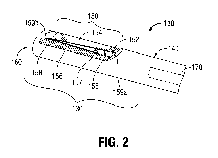
[0040] Referring now to FIG. 2, in conjunction with FIG. 1, one
embodiment of a
biopsy tool provided in accordance with the present disclosure for obtaining a
tissue
sample from the target tissue is shown generally identified by reference
numeral 100. As
detailed below, biopsy tool 100 is further configured for use in conjunction
with tracking
system 70 to facilitate navigation of biopsy tool 100 to the target tissue
and/or tracking of
biopsy tool 100 as it is manipulated relative to the target tissue to obtain
the tissue
sample. Although registration and navigation are detailed above with respect
to LG 92
of positioning assembly 90, it is also envisioned that LG 92 be eliminated and
biopsy
tool 100 itself be utilized for registration and navigation, similarly as
detailed above with
respect to LG 92.
[0041] Biopsy tool 100, as best shown in FIG. 1, generally includes an
elongated
flexible body 110 interconnecting a proximal handle portion 120 and a rigid
distal biopsy
member 130. Proximal handle portion 120 is configured to facilitate
manipulation of
biopsy member 130, e.g., through bronchoscope 50 and EWC 96, and relative to
tissue.
Flexible body 110 is configured to enable insertion of biopsy tool 100 into a
patient
airways, e.g., through bronchoscope 50 and EWC 96 to the target tissue. Biopsy
tool
100 is further configured to connect to a vacuum source "V" for applying
suction at
biopsy member 130, as will be detailed below.
[0042] With reference to FIG. 2, rigid distal biopsy member 130 includes
a throat
portion 140, a tissue-receiving portion 150, and a distal end cap 160. Throat
portion 140
defines a generally cylindrical configuration and houses a sensor 170. Sensor
170, in
conjunction with tracking system 70 (FIG. 1), enables tracking of biopsy
member 130 of
biopsy tool 100 as biopsy member 130 is advanced through the patient's
airways, as
detailed below. Thus, with additional reference to FIG. 1, computer 80,
monitoring
11
CA 02928390 2016-04-21
WO 2015/076937
PCT/US2014/058450
equipment 60, and/or any other suitable display may be configured to display
the three-
dimensional model and selected pathway, both of which were generated during
the
planning phase, along with the current location of sensor 170 of biopsy member
130 to
facilitate navigation of biopsy member 130 to the target tissue and/or
manipulation of
biopsy member 130 relative to the target tissue. Various sensors suitable for
use with
biopsy member 130 for this purpose are detailed below (see FIGS. 6-8).
Alternatively,
biopsy tool 100 may not include a sensor and, rather, only LG 92 may be
utilized for
navigation and positioning. Distal end cap 160 of biopsy member 130 defines a
generally blunt configuration, although distal end cap may alternatively be
configured to
facilitate tissue cutting.
[0043] Tissue-receiving portion 150 is configured to receive a tissue
sample
therethrough and into the generally hollow interior of biopsy member 130. More
specifically, tissue-receiving portion 150 includes a window 152 configured to
receive
tissue therethrough. Window 152 is defined by first and second longitudinally-
extending
faces 154, 156. Faces 154, 156 are angled into the interior of tissue-
receiving portion
150 and are oriented to define an acute interior angle therebetween, e.g., a
generally "V"-
shaped configuration. Faces 154, 156 each includes a sharpened cutting edge
155, 157,
respectively, disposed on one side of window 152. As a result of their
positioning and
orientation, faces 154, 156 are at least partially recessed relative to throat
portion 140
and distal end cap 160 of biopsy member 130. Thus, proximal and distal
shoulders 159a,
159b, respectively, are defined on either end of tissue-receiving portion 150.
Faces 154,
156 are further oriented relative to one another such that edges 155, 157
increasingly
approximate one another in the proximal-to-distal direction, ultimately
culminating at an
apex point 158 adjacent distal shoulder 159b. This feature facilitates dynamic
tissue
cutting, as detailed below.
12
CA 02928390 2016-04-21
WO 2015/076937
PCT/US2014/058450
[0044] Referring to FIGS. 1-2, in use, once the planning and navigation
phases
have been completed, and LG 92 removed from EWC 96, biopsy tool 100 may be
inserted through bronchoscope 50 and EWC 96 to the target tissue. Sensor 170
of biopsy
member 130, in conjunction with tracking system 70, as mentioned above,
enables
tracking of sensor 170 as it is advanced through the patient's airways. Thus,
even after
biopsy member 130 is extended distally from EWC 96, the position of biopsy
member
130 can be tracked, thus permitting navigation of biopsy member 130 to and/or
manipulation of biopsy member 130 relative to the target tissue to ensure
proper
positioning of biopsy member 130 relative to the target tissue and allowing
certain tissue
structures adjacent the target tissue to be avoided. Details of tracking and
navigating
using suitable sensors and tracking system 70 will be described in greater
detail below,
following the description of the various embodiments thereof.
[0045] Once biopsy member 130 of biopsy tool 100 is positioned as
desired,
vacuum source "V" may be activated to apply suction at window 152 of tissue-
receiving
portion 150 of biopsy member 130 to suction tissue into the interior of tissue-
receiving
portion 150. As a sample of tissue is suctioned through window 152, the sample
is cut
away from laterally surrounding tissue via the urging of tissue into contact
with edges
155, 157, e.g., as a result of the suction force applied to tissue. Once the
tissue sample
has been at least partially received within the interior of tissue-receiving
portion 150,
biopsy member 130 may be translated proximally relative to tissue, e.g., via
grasping and
translating proximal handle portion 120 proximally, such that the tissue
sample is
completely severed from surrounding tissue. This severing of the tissue sample
is aided
by the relative movement of approximating edges 155, 157 and apex point 158
relative to
and through tissue. Upon receiving and fully separating the tissue sample from
surrounding tissue, biopsy tool 100 may be withdrawn from the patient's
airways and the
tissue sample retrieved from biopsy tool 100 for testing. It is also
contemplated that
13
CA 02928390 2016-04-21
WO 2015/076937
PCT/US2014/058450
multiple sample be taken with biopsy tool 100, e.g., at the same location or
various
different locations, prior to withdrawal
[0046] Referring now to FIG. 3, another embodiment of a biopsy tool
provided in
accordance with the present disclosure for obtaining a tissue sample from the
target
tissue is shown generally identified by reference numeral 500. Similarly as
detailed
above with respect to the previous embodiment, biopsy tool 500 is configured
for use in
conjunction with tracking system 70 (FIG. 1) to facilitate navigation of
biopsy tool 500
to the target tissue and/or tracking of biopsy tool 500 as it is manipulated
relative to the
target tissue to obtain the tissue sample.
[0047] Biopsy tool 500 generally includes an elongated flexible body
(not
explicitly shown, similar to body 110 of biopsy tool 100 (FIG. 1))
interconnecting a
proximal handle portion (not explicitly shown, similar to handle portion 120
of biopsy
tool 100 (FIG. 1)) and a distal biopsy member 530. The handle portion (not
shown) is
manually operable to manipulate biopsy member 530. The flexible body (not
shown) is
configured to enable insertion of biopsy tool 500 into a patient airways,
e.g., through
bronchoscope 50 and EWC 96 to the target tissue (See FIG. 1).
[0048] Distal biopsy member 530 includes an outer member 540 and an
inner
member 550 that is both translatable and rotatable relative to outer member
540. Outer
member 540 defines a generally hollow configuration and includes an enlarged
body
portion 542. Body portion 542 is configured to at least partially receive
distal end cap
554 of inner member 550 when inner member 550 is disposed in the retracted
position,
as will be detailed below. Outer member 540 is further configured to house a
sensor 570
therein. Similarly as detailed above with respect to the previous embodiment,
sensor
570, in conjunction with tracking system 70 (FIG. 1), enables tracking of
biopsy member
530 of biopsy tool 500 as biopsy member 530 is advanced through the patient's
airways,
as detailed below. Various sensors suitable for use with biopsy member 530 for
this
14
CA 02928390 2016-04-21
WO 2015/076937
PCT/US2014/058450
purpose are detailed below (see FIGS. 6-8). Alternatively, biopsy tool 500 may
not
include a sensor and, rather, only LG 92 (FIG. 1) may be utilized for
navigation and
positioning.
[0049] Inner member 550 includes a shaft 552 and a distal end cap 554
mounted
at the distal end of shaft 552. Inner member 550 is translatable relative to
outer member
540 between a retracted position, wherein shaft 552 is disposed within outer
member 540
and wherein distal end cap 554 is at least partially disposed within enlarged
body portion
542 of outer member 540, and an extended position, wherein distal end cap 554
extends
and is distally-spaced from outer member 540 (as shown in FIG. 3). Distal end
cap 554
includes a sharpened tip 556 configured for facilitate puncturing and
penetrating tissue
upon advancement of distal end cap 554 into tissue, and a sharpened proximal
rim 558
configured to core tissue upon simultaneous rotation and proximal translation
of distal
end cap 554 relative to tissue. Distal end cap 554 may further define a
generally hollow
interior and an open proximal end configured to receive a tissue sample
therein, e.g.,
once the tissue sample has been cored from surrounding tissue.
[0050] With additional reference to FIG. 1, in use, once the planning
and
navigation phases have been completed, and LG 92 removed from EWC 96, biopsy
tool
500, with inner member 550 disposed in the retracted position, may be inserted
through
bronchoscope 50 and EWC 96 to the target tissue. Sensor 570 of biopsy member
530, in
conjunction with tracking system 70, as mentioned above, enable tracking of
sensor 570,
thus permitting navigation of biopsy member 530 to and/or manipulation of
biopsy
member 530 relative to the target tissue to ensure proper positioning of
biopsy member
530 relative to the target tissue and allowing certain tissue structures
adjacent the target
tissue to be avoided. Details of tracking and navigating using suitable
sensors and
tracking system 70 will be described in greater detail below, following the
description of
the various embodiments thereof.
CA 02928390 2016-04-21
WO 2015/076937
PCT/US2014/058450
[0051] Once biopsy member 530 of biopsy tool 500 is positioned as
desired, e.g.,
adjacent target tissue to be sampled, inner member 550, lead by sharpened tip
556 of
distal end cap 554, is translated distally from the retracted position to the
extended
position to penetrate the target tissue. Once advanced to a sufficient depth
within the
target tissue, inner member 550 may be returned to the retracted position
relative to outer
member 540 while being simultaneously rotated relative to outer member 540
such that
the tissue that was positioned between inner and outer members 550, 540,
respectively, is
cored or separated from surrounding tissue using sharpened proximal rim 558
and is
retained within the hollow interior of distal end cap 554 and/or outer member
540. In
some embodiments, biopsy tool 500 may further be configured to connect to the
vacuum
source "V" (FIG. 1) to facilitate obtaining a tissue sample. Upon receiving
and fully
separating the tissue sample(s) from surrounding tissue, biopsy tool 500 may
be
withdrawn from the patient's airways and the tissue sample retrieved from
biopsy tool
500 for testing.
[0052] Referring now to FIG. 4A, another embodiment of a biopsy tool
provided
in accordance with the present disclosure for obtaining a tissue sample from
the target
tissue is shown generally identified by reference numeral 600. Similarly as
detailed
above with respect to the previous embodiment, biopsy tool 600 is configured
for use in
conjunction with tracking system 70 (FIG. 1) to facilitate navigation of
biopsy tool 600
to the target tissue and/or tracking of biopsy tool 600 as it is manipulated
relative to the
target tissue to obtain the tissue sample.
[0053] Biopsy tool 600 generally includes an elongated flexible body
(not
explicitly shown, similar to body 110 of biopsy tool 100 (FIG. 1))
interconnecting a
proximal handle portion (not explicitly shown, similar to handle portion 120
of biopsy
tool 100 (FIG. 1)) and a distal biopsy member 630. The handle portion (not
shown) is
manually operable to manipulate biopsy member 630. The flexible body (not
shown) is
16
CA 02928390 2016-04-21
WO 2015/076937
PCT/US2014/058450
configured to enable insertion of biopsy tool 600 into a patient airways,
e.g., through
bronchoscope 50 and EWC 96 to the target tissue (See FIG. 1). Biopsy tool 600
is
further configured to connect to a vacuum source "V" (FIG. 1) for applying
suction at
biopsy member 630, as will be detailed below.
[0054] Distal biopsy member 630 includes an outer member 640 and an
inner
member 650 that is fixedly disposed within outer member 640. Outer member 640
defines a generally hollow configuration and includes a body portion 642 and a
head
portion 644. Body portion 642 is configured to house a sensor 670 therein.
Similarly as
detailed above with respect to the previous embodiments, sensor 670, in
conjunction
with tracking system 70 (FIG. 1), enables tracking of biopsy member 630 of
biopsy tool
600 as biopsy member 630 is advanced through the patient's airways, as
detailed below.
Various sensors suitable for use with biopsy member 630 for this purpose are
detailed
below (see FIGS. 6-8). Alternatively, biopsy tool 600 may not include a sensor
and,
rather, only LG 92 (FIG. 1) may be utilized for navigation and positioning.
[0055] Continuing with reference to FIG. 4A, head portion 644 of outer
member
640 includes a blunt distal cap 646 and a mouth 648 defined through a lateral
wall of
outer member 640 towards the distal end thereof. Mouth 648 provides access to
the
hollow interior of outer member 640 and inner member 650 which, as mentioned
above,
is fixedly disposed within outer member 640.
[0056] Inner member 650 defines a generally cylindrical configuration
and
includes a open distal end 652 defining a sharpened rim 654. Open distal end
652 of
inner member 650 terminates in the vicinity of mouth 648 of outer member 640
such that
sharpened rim 654 is exposed adjacent mouth 648. Further, inner member 650 is
coupled to the vacuum source "V" (FIG. 1) for applying suction at open distal
end 652 of
inner member 650 to suction a tissue sample through mouth 648 and into open
distal end
17
CA 02928390 2016-04-21
WO 2015/076937
PCT/US2014/058450
652 of inner member 650, while the tissue sample is severed from surrounding
tissue via
sharpened rim 654.
[0057] With additional reference to FIG. 1, in use, once the planning
and
navigation phases have been completed, and LG 92 removed from EWC 96, biopsy
tool
600 may be inserted through bronchoscope 50 and EWC 96 to the target tissue.
Sensor
670 of biopsy member 130, in conjunction with tracking system 70, as mentioned
above,
enables tracking of sensor 670, thus permitting navigation of biopsy member
630 to
and/or manipulation of biopsy member 630 relative to the target tissue to
ensure proper
positioning of biopsy member 630 relative to the target tissue and allowing
certain tissue
structures adjacent the target tissue to be avoided. Details of tracking and
navigating
using suitable sensors and tracking system 70 will be described in greater
detail below,
following the description of the various embodiments thereof.
[0058] Once biopsy member 630 of biopsy tool 600 is positioned as
desired,
mouth 648 is oriented towards target tissue and vacuum source "V" (FIG. 1) is
activated
to apply suction adjacent mouth 648 to suction a tissue sample through mouth
648 and
into open distal end 652 of inner member 650. As a sample of tissue is
suctioned
through mouth 648, the tissue sample is severed from surrounding tissue via
sharpened
rim 654. Upon receiving and fully separating the tissue sample(s) from
surrounding
tissue, biopsy tool 600 may be withdrawn from the patient's airways and the
tissue
sample retrieved from biopsy tool 600 for testing.
[0059] Turning to FIG. 4B, another embodiment of a biopsy tool provided
in
accordance with the present disclosure for obtaining a tissue sample from the
target
tissue is shown generally identified by reference numeral 700. Biopsy tool 700
is similar
to biopsy tool 600 (FIG. 4A) and, thus, only the differences therebetween will
be
described in detail below for purposes of brevity.
18
CA 02928390 2016-04-21
WO 2015/076937
PCT/US2014/058450
[0060] Biopsy tool 700 generally includes an elongated flexible body
(not
explicitly shown) interconnecting a proximal handle portion (not explicitly
shown) and a
distal biopsy member 730. Biopsy tool 700 is further configured to connect to
a vacuum
source "V" (FIG. 1) for applying suction at biopsy member 730, as will be
detailed
below.
[0061] Distal biopsy member 730 includes an outer member 740 and an
inner
member 750 that is disposed within and rotatably coupled to outer member 740,
thus
enabling rotation of inner member 750 relative to outer member 740. Outer
member 740
is configured to house a sensor 770 therein and includes a head portion 744
defining a
mouth 748. Inner member 750 defines a generally cylindrical configuration and
includes
a open distal end 752 defining a sharpened rim 754.
[0062] In use, once biopsy member 730 of biopsy tool 700 is positioned
as
desired, mouth 748 is oriented towards target tissue and vacuum source "V"
(FIG. 1) is
activated to apply suction adjacent mouth 748 to suction a tissue sample
through mouth
748 and into inner member 750. As a sample of tissue is suctioned through
mouth 748,
the tissue sample is severed from surrounding tissue via sharpened rim 754.
Severing the
tissue sample from surrounding tissue may be aided by selectively rotating
inner member
750 relative to outer member 740 while applying suction. Ultimately, biopsy
tool 700
may be withdrawn from the patient's airways and the tissue sample(s) retrieved
from
biopsy tool 700 for testing.
[0063] Referring now to FIG. 5A, another embodiment of a biopsy tool
provided
in accordance with the present disclosure for obtaining a tissue sample from
the target
tissue is shown generally identified by reference numeral 800. Similarly as
detailed
above with respect to the previous embodiments, biopsy tool 800 is configured
for use in
conjunction with tracking system 70 (FIG. 1) to facilitate navigation of
biopsy tool 800
19
CA 02928390 2016-04-21
WO 2015/076937
PCT/US2014/058450
to the target tissue and/or tracking of biopsy tool 800 as it is manipulated
relative to the
target tissue to obtain the tissue sample.
[0064] Biopsy tool 800 generally includes an elongated flexible body
(not
explicitly shown, similar to body 110 of biopsy tool 100 (FIG. 1))
interconnecting a
proximal handle portion (not explicitly shown, similar to handle portion 120
of biopsy
tool 100 (FIG. 1)) and a distal biopsy member 830. The handle portion (not
shown) is
manually operable to manipulate biopsy member 830. The flexible body (not
shown) is
configured to enable insertion of biopsy tool 800 into a patient airways,
e.g., through
bronchoscope 50 and EWC 96 to the target tissue (See FIG. 1). Biopsy tool 800
is
further configured to connect to a vacuum source "V" (FIG. 1) for applying
suction at
biopsy member 830, as will be detailed below.
[0065] Distal biopsy member 830 includes an outer member 840, an inner
member 850 that is fixedly disposed within outer member 840, and a sleeve 860
that is
disposed about outer member 840. Outer member 840 defines a generally hollow
configuration and includes a body portion 842 and a head portion 844. Body
portion 842
is configured to house a sensor 870, similarly as detailed above with respect
to the
previous embodiments.
[0066] Head portion 844 of outer member 840 includes a blunt distal cap
846 and
a mouth 848 defined through a lateral wall of outer member 840 towards the
distal end
thereof. Mouth 848 provides access to the hollow interior of outer member 840
and
inner member 850 which, as mentioned above, is fixedly disposed within outer
member
840.
[0067] Inner member 850 is fixedly disposed within outer member 840 and,
similar to outer member 840, includes a mouth 858 defined through a lateral
wall thereof
towards the distal end thereof. Mouth 858 defines a sharpened rim 854
configured to
facilitate tissue cutting and is positioned adjacent mouth 848 of outer member
840 such
CA 02928390 2016-04-21
WO 2015/076937
PCT/US2014/058450
that sharpened rim 854 is exposed adjacent mouth 848. Further, inner member
850 is
coupled to the vacuum source "V" (FIG. 1) for applying suction at mouth 858.
[0068] With
additional reference to FIG. 1, in use, once the planning and
navigation phases have been completed, and LG 92 removed from EWC 96, biopsy
tool
800 may be inserted through bronchoscope 50 and EWC 96 to the target tissue.
Sensor
870 of biopsy member 830, in conjunction with tracking system 70, as mentioned
above,
enables tracking of sensor 870, thus permitting navigation of biopsy member
830 to
and/or manipulation of biopsy member 830 relative to the target tissue to
ensure proper
positioning of biopsy member 830 relative to the target tissue and allowing
certain tissue
structures adjacent the target tissue to be avoided. Details of tracking and
navigating
using suitable sensors and tracking system 70 will be described in greater
detail below,
following the description of the various embodiments thereof.
[0069] Once
biopsy member 830 of biopsy tool 800 is positioned as desired,
mouth 848 is oriented towards target tissue and vacuum source "V" (FIG. 1) is
activated
to apply suction adjacent mouth 848 to suction a tissue sample through mouth
848 and
into mouth 858 of inner member 850. As a sample of tissue is suctioned through
mouth
848 and into mouth 858, the tissue sample is severed from surrounding tissue
via
sharpened rim 854. Severing the tissue sample from surrounding tissue may be
aided by
selectively translating biopsy member 830 proximally relative to tissue while
applying
suction. Upon
receiving and fully separating the tissue sample(s) from surrounding
tissue, biopsy tool 800 may be withdrawn from the patient's airways and the
tissue
sample retrieved from biopsy tool 800 for testing.
[0070] Turning
to FIG. 5B, another embodiment of a biopsy tool provided in
accordance with the present disclosure for obtaining a tissue sample from the
target
tissue is shown generally identified by reference numeral 900. Biopsy tool 900
is similar
21
CA 02928390 2016-04-21
WO 2015/076937
PCT/US2014/058450
to biopsy tool 800 (FIG. 5A) and, thus, only the differences therebetween will
be
described in detail below for purposes of brevity.
[0071] Biopsy tool 900 generally includes an elongated flexible body
(not
explicitly shown) interconnecting a proximal handle portion (not explicitly
shown) and a
distal biopsy member 930. Biopsy tool 900 is further configured to connect to
a vacuum
source "V" (FIG. 1) for applying suction at biopsy member 930, as will be
detailed
below.
[0072] Distal biopsy member 930 includes an outer member 940 and an
inner
member 950 that is disposed within and rotatably coupled to outer member 940.
Outer
member 940 is configured to house a sensor 970 and defines a mouth 948 through
a
lateral wall thereof towards the distal end thereof. Inner member 950, similar
to outer
member 940, includes a mouth 958 defined through a lateral wall thereof
towards the
distal end thereof Mouth 958 defines a sharpened rim 954 configured to
facilitate tissue
cutting and is positioned adjacent mouth 948 of outer member 940. Inner member
950 is
rotatable relative to outer member 940 to thereby vary the relative
positioning of mouths
948, 958, e.g., between an aligned position, a partially overlapping position,
and a fully
occluded position. Inner member 950 is coupled to the vacuum source "V" (FIG.
1) for
applying suction at mouth 958.
[0073] With additional reference to FIG. 1, in use, Once biopsy member
930 of
biopsy tool 900 is positioned as desired, inner member 950 is rotated such
that mouths
948, 958 are aligned with one another, and vacuum source "V" (FIG. 1) is
activated to
apply suction adjacent mouth 958 to suction a tissue sample through mouths
948, 958
and into inner member 950. Once a sample of tissue is suctioned through mouths
948,
958 and into inner member 950, inner member 950 is rotated relative to outer
member
940 such that mouths 948, 958 are moved towards an occluded position. As
mouths 948,
958 are moved towards the occluded position, tissue disposed therebetween is
cut via
22
CA 02928390 2016-04-21
WO 2015/076937
PCT/US2014/058450
sharpened rim 854, thereby severing the tissue sample from surrounding tissue.
Upon
receiving and fully separating the tissue sample(s) from surrounding tissue,
biopsy tool
900 may be withdrawn from the patient's airways and the tissue sample
retrieved from
biopsy tool 900 for testing.
[0074] Turning now to FIGS. 6-8, in conjunction with FIG. 1, various
different
sensors 248, 348, 448 (FIGS. 6-8, respectively) configured for use as the
sensor of any of
the biopsy tools detailed herein and/or sensor 94 of LG 92 are described.
Referring to
FIG. 6, sensor 248 is shown. Sensor 248 includes a plurality of field
component sensor
elements 251a, 251b, 1252a, 252b, 253. Each sensor element 251a, 251b, 252a,
252b,
253 is formed as a coil and arranged for sensing a different component of an
electromagnetic field generated by transmitter mat 76 (FIG. 9). More
specifically, first
and second pairs of sensor elements 251a, 251b and 252a, 252b are arranged
within
sensor housing 246 such that the respective elements 251a, 251b and 252a, 252b
of each
pair are equidistant from a common reference point 254, while sensor element
253 is
centered about reference point 254. Although shown in FIG. 6 as collinearly
disposed,
other configurations of sensor elements 251a, 251b, 1252a, 252b, 253 are also
contemplated. Further, as opposed to providing five sensor elements 251a, 25
lb, 1252a,
252b, 253 wherein sensor element 253 is centered about the reference point
254, six
sensors may be provide, e.g., wherein sensor element 253 is provided as a pair
of
elements disposed equidistant from reference point 254. The above-described
configuration of sensor 248 enables transmitter mat 76 and the plurality of
reference
sensors 74 (FIG. 1), together with tracking module 72 and computer 80 (FIG.
1), to
derive the location of sensor 248 in six degrees of freedom, as detailed
below, and as
further detailed in U.S. Patent No. 6,188,355 and published PCT Application
Nos. WO
00/10456 and WO 01/67035, previously incorporated herein by reference.
23
CA 02928390 2016-04-21
WO 2015/076937
PCT/US2014/058450
[0075] With reference to FIG. 7, sensor 348 is shown including two
sensor
components 351, 353 arranged within sensor housing 346, each component 351,
353
including three sensor elements 352a, 352b, 352c and 354a, 354b, 354c,
respectively.
Each sensor element 352a, 352b, 352c and 354a, 354b, 354c is configured as a
flat
rectangular coil, e.g., including a plurality of turns of conducting wire,
bent to define an
arcuate shape. As such, the elements 352a, 352b, 352c and 354a, 354b, 354c
combine to
define first and second generally cylindrical components 351, 353. Components
351,
353 are centered about reference axis 356 and positioned such that each of
elements
352a, 352b, 352c and 354a, 354b, 354c are equidistant from reference axis 356
and such
that each of elements 352a, 352b, 352c of component 351 are oriented 180
degrees offset
as compared to corresponding elements 354a, 354b, 354c, respectively, of
component
353. Thus, similarly as with sensor 248 (FIG. 6), sensor 348 enables
transmitter mat 76
and the plurality of reference sensors 74 (FIG. 1), together with tracking
module 72 and
computer 80 (FIG. 1), to derive the location of sensor 348 in six degrees of
freedom.
[0076] Turning to FIG. 8, sensor 448 includes three coils 451, 452, 453.
Coils
451 and 452, 453 are angled relative to housing 446, while coil 453 is
circumferentially
disposed within housing 446. Coils 451, 452, 453 are oriented to lie in
perpendicular
planes relative to one another and share a common center reference point 454.
By
sharing a common center reference point 454, each portion of each coil 451,
452, 453 is
equidistant from center reference point 454. Further, this configuration,
e.g., wherein
coils share a common center reference point 454 rather than being
longitudinally
displaced relative to one another, allows for the longitudinal dimension of
sensor 448 to
be minimized. Such a configuration still, however, enables transmitter mat 76
and the
plurality of reference sensors 74 (FIG. 1), together with tracking module 72
and
computer 80 (FIG. 1), to derive the location of sensor 448 in six degrees of
freedom.
24
CA 02928390 2016-04-21
WO 2015/076937
PCT/US2014/058450
[0077] Referring to FIG. 9, in conjunction with FIG. 1, an embodiment of
the
internal configuration of transmitter mat 76 of tracking system 70 (FIG. 1) is
shown,
although other suitable configurations are also contemplated. Transmitter mat
76 is a
transmitter of electromagnetic radiation and includes a stack of three
substantially planar
rectangular loop antennas 77a, 77b, 77c configured to connected to drive
circuitry (not
shown).
[0078] Antenna 77a is skewed in a first horizontal direction (when the
transmitter
mat 76 is horizontal) in that the loops on one side of the antenna 77a are
closer together
than the loops on the opposite side. As a result, antenna 77a creates a
magnetic field that
is stronger on the side where the loops are close together than on the
opposite side. By
measuring the strength of the current induced by antenna 77a in the sensor
assembly,
e.g., sensor assembly 145 of biopsy tool 100 (FIG. 3) or sensor 94 of LG 92
(FIG. 1), it
can be determined where the sensor assembly is located in the first direction
over
antenna 77a.
[0079] Antenna 77b is similar to antenna 77a except that antenna 77b is
skewed
in an second horizontal direction that is perpendicular to the first
direction. By
measuring the strength of the current induced by antenna 77b in the sensor
assembly, it
can be determined where the sensor assembly is located in the second direction
over
antenna 77b.
[0080] Antenna 77c defines a uniform, i.e., un-skewed, configuration.
Thus,
antenna 77c creates a uniform field that naturally diminishes in strength in a
vertical
direction when the transmitter mat 76 is horizontal. By measuring the strength
of the
field induced in the sensor assembly, it can be determined how far the sensor
assembly is
located above antenna 77c.
[0081] In order to distinguish one magnetic field from another, the
fields of
antennae 77a, 77b, 77c are generated using independent frequencies. For
example,
CA 02928390 2016-04-21
WO 2015/076937
PCT/US2014/058450
antenna 77a may be supplied with alternating current oscillating at 2.5 kHz,
antenna 77b
may be supplied with alternating current oscillating at 3.0 kHz, and antenna
77c may be
supplied with alternating current oscillating at 3.5 kHz, although other
configurations are
also contemplated. As a result of using independent frequencies, each of the
sensor
components of the sensor assembly (see FIGS. 6-8, for example) will have a
different
alternating current signal induced in its coils.
[0082] Referring additionally to FIG. 1, in use, signal generators and
amplifiers
of the driving circuitry (not shown) associated with tracking system 70 are
utilized to
drive each of antennas 77a, 77b, 77c of transmitter mat 76 at their
corresponding
frequencies. The electromagnetic waves generated by transmitter mat 76 are
received by
the various sensor elements of the sensor assembly e.g., the sensor elements
of sensors
248, 348, 448 (FIGS. 6-8, respectively) configured for use any of the biopsy
tools
provided herein or sensor 94 of LG 92, and are converted into electrical
signals that are
sensed via reference sensors 74. Tracking system 70 further includes reception
circuitry
(not shown) that has appropriate amplifiers and AID converters that are
utilized to
receive the electrical signals from reference sensors 74 and process these
signals to
determine and record location data of the sensor assembly. Computer 80 may be
configured to receive the location data from tracking system 70 and display
the current
location of the sensor assembly on the three-dimensional model and relative to
the
selected pathway generated during the planning phase, e.g., on computer 80,
monitoring
equipment 60, or other suitable display. Thus, navigation of the biopsy tool
and/or LG
92 to the target tissue and/or manipulation of the biopsy tool relative to the
target tissue,
as detailed above, can be readily achieved.
[0083] While several embodiments of the disclosure have been shown in
the
drawings, it is not intended that the disclosure be limited thereto, as it is
intended that the
disclosure be as broad in scope as the art will allow and that the
specification be read
26
CA 02928390 2016-04-21
WO 2015/076937
PCT/US2014/058450
likewise. Therefore, the above description should not be construed as
limiting, but
merely as exemplifications of particular embodiments. Those skilled in the art
will
envision other modifications within the scope and spirit of the claims
appended hereto.
27