Note : Les descriptions sont présentées dans la langue officielle dans laquelle elles ont été soumises.
CA 02931582 2016-05-25
WO 2015/078998
PCT/EP2014/075868
1
MICROFLUIDIC CARTRIDGE FOR MOLECULAR DIAGNOSIS,
DOCKING STATION USING SUCH A MICROFLUIDIC CARTRIDGE,
AND PROCESS FOR ANALYZING A BIOLOGICAL SAMPLE
The invention relates to the field of microfluidic devices used to make
molecular or
biological diagnostics.
The invention more particularly relates to a microfluidic cartridge for
analyzing at least
one nucleic acid contained in a biological sample.
The invention also relates to a docking station designed to use and operate
such a
microfluidic cartridge.
The invention finally relates to a method of analysis of a biological sample
implementing
such a microfluidic cartridge.
Microfluidic devices designed for the search and the analysis of at least one
nucleic acid
or one nucleotide sequence contained in a biological sample incorporate
various means in order
to: prepare the biological sample to extract nucleic acids from said sample;
amplify the at least
one target nucleic acid from the extracted nucleic acids using standard
amplification techniques
like, for example, Polymerase Chain Reaction (also known as PCR ); and
detect, e.g.
optically, and analyze the target nucleic acids using known molecular
recognition mechanisms
like, for example, hybridization.
Therefore, in order to perform the analysis of the at least one nucleic of a
sample, said
sample needs to be transferred sequentially in different functional areas of
the micro microfluidic
cartridge, each functional area being dedicated to a specific operation on the
sample.
The documents WO 2009/049268 and US 2012/115738 describe, for example, a
microfluidic device comprising a plurality of functional areas: an area of
sample preparation for
extraction of nucleic acids, a range of nucleic acid amplification, and a
surface analysis and
detection of amplified nucleic acids. Said detection area is likely to be a
biochip.
In those documents, the microfluidic cartridge features very complex structure
so as to
be modular and allow an easy and rapid reconfiguration in order to suit
various applications. In
particular, many pumps with shared-valve structure are implemented within each
of the
functional areas of the microfluidic cartridge to transfer fluids from one
functional area to the
other.
Therefore, the cartridge of documents WO 2009/049268 and US 2012/115738
presents
a large volume and the transfer of fluids cannot be operated in a simple
manner, with a limited
number of actuators.
The widespread use of these devices, especially in the context of molecular
diagnostics
in humans, for which the cartridge must be discarded after each use, is
limited by the
CA 02931582 2016-05-25
WO 2015/078998
PCT/EP2014/075868
2
complexity and high costs inherent in this technology. Furthermore, these
devices, such as the
one described in US 2012/0034705, often consisting of a variety of many
elements for achieving
the various stages of analysis, they are extremely fragile and difficult to
handle.
It is therefore desired that a microfluidic cartridge is mass producible,
inexpensive, and
most preferably disposable. However, because such microfluidic devices
integrate complex
steps of molecular analysis, it may be difficult to properly coordinate
various tasks of
conventional microfluidic devices. It is therefore also desired that the
microfluidic cartridge be
simple to operate and that many or substantially all of the fluid processing
steps be automated
directly on the microfluidic cartridge.
For that purpose, the present invention proposes a microfluidic cartridge
making it
possible, on the one hand, to integrate, within the latter, not only all the
fluids required for its
operation, but also the whole of microfluidic circuits, microchannels and
valves, the reaction
chamber and the biochip, and, on the other hand, to make transfers and
movements of fluids in
a simple manner, in a reduced volume and by means of a compact external
actuator.
More precisely, the present invention provides a microfluidic cartridge for
detecting at
least one nucleic acid of a sample, said microfluidic cartridge comprising:
-
a plurality of functional volumes split into functional areas such as at
least, a
sample preparation area, a nucleic acid amplification area, a nucleic acid
analysis area, a waste
area, and
20- a fluidic network of microchannels,
wherein:
-
at least three functional areas are fluidly connected to one central
distribution hub
of fluids distribution by one or more hub-connected microchannels, each of
said hubconnected
microchannels having a hub end and an area end, said central distribution hub
being capable of
pumping and injecting fluids from a first functional area to a second
functional area of said at
least three functional areas by passing through said central distribution hub,
said second
functional area being identical or different from said first functional area,
and
-
at least three valves, each located on a hub-connected microchannel, are
arranged in said microfluidic cartridge so that said at least three valves are
adapted to be
actuated mechanically by a single external cam-driven actuator.
The microfluidic cartridge according to the invention has thus the advantage,
thanks to
the use of the central distribution hub of fluids, to facilitate the transfers
of fluid from a first
functional area to a second functional area. This makes it possible to use
only one simple fluid
displacement system (typically a pumping system) for most of the fluid
movements of the
microfluidic cartridge, for inducing depressurization and pressurization in
order to displace the
fluid from a functional volume or area to another one and to reduce the volume
of the
microfluidic cartridge.
CA 02931582 2016-05-25
WO 2015/078998
PCT/EP2014/075868
3
The microfluidic cartridge comprises less moving elements and its cost is
therefore
reduced compared to prior-art cartridges.
Moreover, the system for actuation of the valves of the microchannels
connected to the
central hub may also be more compact and simpler than the system disclosed in
__ US 2012/0034705, thanks to the arrangement of these valves in the
microfluidic cartridge.
In one embodiment, the at least three functional areas that are connected to
the central
distribution hub are the sample preparation area and the waste area.
In one embodiment, the at least three functional areas comprise the nucleic
acid
analysis area, and/or the nucleic acid amplification area.
In another embodiment, the at least three functional areas comprise the sample
preparation area, the nucleic acid analysis area, and the waste area.
In another embodiment, the at least three functional areas comprise all
functional areas
of the microfluidic cartridge.
In one embodiment, the microfluidic cartridge further comprises at least two
valves that
__ are actuated by linear actuators and are independent of the cam-driven
actuator.
The microfluidic cartridge according to the present invention may be seen as a
"lab-on-a-chip" that can perform the complete nucleic acid analysis of a
sample, from sample
collection to the reading of the result, typically performed in the
diagnostics or microbiology
laboratory.
The detection of the presence in the sample, of a nucleic acid or molecular
marker
whose sequence is specific to a gene of interest, is understood as a molecular
diagnostics in
this application.
The microfluidic cartridge integrates usually over a few square centimeters
several
specialized functional areas and volumes performing complex analysis
conventionally made
using several laboratory apparatus. The advantages are that theses operations
can be
automated while consuming low reagents volumes.
Besides, other advantageous and non-limiting characteristics of the
microfluidic cartridge
according to the invention are described below. The said characteristics
correspond to various
embodiments of the invention that can be taken alone or in combination.
The at least three valves are spatially arranged in the microfluidic cartridge
so that they
are adapated to be actuated simultaneously by the single external cam-driven
actuator. In
particular embodiments of the invention, said at least three valves are
linearly or circularly
arranged in the microfluidic cartridge.
Typically, the said at least three valves are valves of hub-connected
microchannels,
connecting the sample preparation area, the nucleic acid analysis area, and
the waste area to
the central hub.
Preferentially, the said valves are located close to, or at the area end of
the said
CA 02931582 2016-05-25
WO 2015/078998
PCT/EP2014/075868
4
hub-connected microchannels, said area end being the one of the two ends of
the
hub-connected microchannel which is turned towards the corresponding
functional area. On the
opposite, the hub end is the one of the two ends of the hub-connected
microchannel which is
turned towards the central distribution hub of fluids.
In one embodiment of the invention, each hub microchannel comprises one valve
located, close to, or at their area end. Said valves are preferentially
spatially arranged in order
to be simultaneously actuated by the external cam-driven actuator as mentioned
above.
At least two functional areas of the plurality of functional areas can also be
directly fluidly
connected to each other by one or more area-connecting microchannels, each of
the said area-
connecting microchannels having at least a valve that is preferentially
actuated by a linear
actuator independent of the cam-driven actuator.
For example, said two functional areas are the nucleic acid amplification area
and the
nucleic acid analysis area.
In another example, the said two functional areas are the nucleic acid
analysis area and
the waste area.
Typically, a microfluidic cartridge according to the invention is disposable
and comprises:
- a cartridge plate comprising:
-
a substrate having a first face and a second face, a plurality of grooves
flush with the first or second surface and a plurality of through holes
connecting said first
and second surfaces, and
-
a first film bonded on first face of said substrate of the cartridge plate,
said
grooves flush with the first face being sealed by said first film to form the
hub-connected
microchannels, said first film being a first deformable membrane adapted to be
deformed
by an external actuator,
- a
cartridge body in contact with the cartridge plate on the second surface of
said
substrate, said cartridge body comprising:
-
a lateral wall which extends from the second surface of said substrate,
and
-
a plurality of internal walls which defines a plurality of functional
volumes
of said cartridge body, and
- a cartridge cover adapted to close the different
functional volumes.
In one embodiment, the cartridge plate comprises a second film bonded on the
second
face of the substrate of the cartridge plate, the plurality of grooves flush
with the second face
being sealed by said second film to form the area-connected microchannels.
Typically, the cartridge plate comprises at least one recessed cavity formed
in the
substrate and extending from the first face.
Typically also, the first film bonded on the first face of the substrate
closes said at least
CA 02931582 2016-05-25
WO 2015/078998
PCT/EP2014/075868
one recessed cavity to form at least one reactive chamber for nucleic acid
amplification.
In a preferred embodiment, a micro-array slide (or biochip) bonded on the
first face of
the substrate closes said at least one recessed cavity to form at least one
detection chamber for
nucleic acid analysis.
5 In a preferred embodiment, the microfluidic cartridge comprises a semi-
permeable
membrane between the cartridge body and the cartridge cover adapted to let air
pass through it
while preventing liquids to leak out of the functional volumes.
Typically, the functional volumes of the cartridge body encompass several
functional
areas (e.g.: at least a sample preparation area, a nucleic acid amplification
area, a nucleic acid
analysis area and a waste area). Said functional volumes are containers
adapted to receive
tubes, fluids such as sample, reagent products, or a purification column.
In one embodiment, the central hub of fluid distribution comprises a hub body
and a
plunger seal adapted to slide in and out of the hub body to pump from or
inject fluids in the
functional areas of said microfluidic cartridge through the hub-connected
microchannels.
In another embodiment, the central hub of fluid distribution comprises also a
syringe
having a plunger to which the plunger seal is attached.
The microfluidic cartridge is adapted to be inserted into a docking station,
within
equipment designed to perform at least the following functions: thermal
control, control of fluid
flow, valves actuation and optical detection.
The present invention also proposes a docking station intended to use and
operate a
microfluidic cartridge such as mentioned above.
Therefore, the present invention provides a docking station adapted to receive
a
microfluidic cartridge according to the invention, comprising:
- a cam-driven actuator adapted to simultaneously actuate the at
least three valves
of hub-connected microchannels,
- means for optical excitation of the micro-array slide of said
cartridge, and
- means for optical detection of an optical signal that is
representative of said
nucleic acid in the sample analyzed by the cartridge.
In one embodiment, the docking station also comprises actuation means adapted
to
actuate linear/independently actuated valves of said microfluidic cartridge.
In a particular embodiment, the cam-driven actuator is a rotational-motion
actuator.
In another particular embodiment, the cam-driven actuator is a linear-motion
actuator.
Preferentially, the cam-driven actuator of the docking station is designed to
open, among
the valves of the microchannels connected to the central hub, at most only one
of said valves.
In a preferred embodiment, the docking station according to the invention
comprises
sliding means adapted to slide the syringe of the central hub in and out of
the hub body to pump
from or inject fluids in the functional areas of said microfluidic cartridge
through the
CA 02931582 2016-05-25
WO 2015/078998
PCT/EP2014/075868
6
hub-connected microchannels.
It is also an object of the present invention to provide an apparatus for
analyzing a
biological sample comprising such docking station and microfluidic cartridge
according to the
present invention, for analyzing at least a nucleic acid of a sample.
Furthermore, the microfluidic cartridge according to the invention is
particularly adapted
to be used in a process for analyzing a biological sample.
Therefore, it is another object of the present invention to propose a process
for analyzing
a biological sample, comprising the steps of:
a) providing said biological sample into at least one functional volume of
a sample
preparation area of a microfluidic cartridge according to the invention,
b) allowing said biological sample to get into contact with at least one
reagent
and/or one purification column present in another functional volume of the
sample preparation
area by actuating at least one valve controlling the flow of fluids of
microchannels,
c) recovering the product resulting of step b to obtain an isolated DNA
sample,
d) transferring the isolated DNA sample to at least one functional volume
of the
nucleic acid amplification area,
e) allowing said isolated DNA sample to get into contact with at least a
reagent for
amplification and closing the valves of the functional volume of the
amplification area,
f) performing DNA amplification,
g) recovering amplified DNA obtained at step f) and transferring it to
another
functional volume of the hybridization area by actuating at least one valve
controlling the flow of
fluids of microchannels,
h) allowing said amplified DNA to get into contact with at least
one compound
capable of hybridizing with said DNA in the hybridization chamber, and
i) obtaining a microarray image and automatically analyzing it.
An embodiment of the invention will now be described in detail with reference
to the
drawings, in which:
- Figure 1 is a perspective view of a microfluidic cartridge in
a preferred
embodiment of the invention;
- Figure 2 is an exploded view of the microfluidic cartridge of Figure 1,
making
appear the cartridge plate, the cartridge body and the cartridge cover;
- Figure 3 is a schematic view of a preparation tube for a
biological sample to
analyze with the microfluidic cartridge of Figure 1;
- Figure 4 is an exploded schematic view of a syringe that can
be used in the
microfluidic cartridge of Figure 1 to pump and inject the fluids;
- Figure 5 is a schematic view of an amplification mix >> tube
to be inserted in the
microfluidic cartridge of Figure 1;
CA 02931582 2016-05-25
WO 2015/078998
PCT/EP2014/075868
7
- Figure 6 is a detailed view of the cartridge body of Figure 2;
- Figure 7 is a detailed view of the cartridge plate of Figure
2;
- Figure 8 is a bottom view of the cartridge plate of Figure 7;
- Figure 8A is a sectional view of Figure 8 according to the
section plane A-A;
- Figure 8B is a sectional view of Figure 8 according to the section plane
B-B;
- Figure 9 is a detailed view of the area referenced I in Figure
8;
- Figure 9A is a sectional view of Figure 9 according to the
section plane A-A;
- Figure 10 is a detailed view of the area referenced I in
Figure 8;
- Figure 10A is a sectional view of Figure 10 according to the
section line B-B;
- Figure 11 is a top view of the cartridge plate of Figure 7;
- Figure 12 is a schematic view of the mounting of the
microfluidic cartridge of
Figure 1 in a docking station in a preferred embodiment (mechanical part
only);
- Figure 13 is a schematic view of a rotational-motion cam-
driven actuator using
balls to actuate the valves of the microfluidic cartridge;
- Figure 14 is a schematic view of a perforated actuator plate allowing
the holding
in place of the balls of the cam-driven actuator of Figure 13;
- Figure 15 is a schematic view of a circular plate having a
detent operating in the
mechanism of the cam-driven actuator of Figure 13;
- Figure 16 is a detailed view of another example of cartridge
plate.
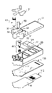
It has been shown in Figure 1 and Figure 2, respectively, an assembled view
and an
exploded view of a microfluidic cartridge 1 according to a preferred
embodiment of the
invention, in which the microfluidic cartridge 1 is herein a disposable
cartridge. It is meant by
this that the microfluidic cartridge 1 is intended to be disposed of and
placed in a container
intended for receiving biological wastes.
As shown in Figures 1 and 2, the microfluidic cartridge 1 comprises three main
elements,
i.e.: the cartridge plate 10, the cartridge body 20 and the cartridge cover
30.
The microfluidic cartridge 1 also comprises a sample tube 40 containing a
sample S, at
least an amplification-mix tube 50 and a syringe 60.
These different elements of the microfluidic cartridge 1 will be detailed
hereinafter.
The cartridge plate 10 of the microfluidic cartridge 1 first comprises a
substrate 100 such
as the one shown in detail in Figure 7.
This substrate 100 has substantially the shape of a thin blade and has a first
face 101
and a second face 102. The second face 102 is the face that is turned toward
the cartridge body
20 when the cartridge is assembled (see Figures 1 and 2).
The cartridge plate 10 may advantageously be made by injection molding of a
thermoplastic polymer material such as the cyclic olefin copolymers (COO) or
the cyclic olefin
polymers (COP). The cartridge plate 10 is here preferably made of
polypropylene (PP). The
CA 02931582 2016-05-25
WO 2015/078998
PCT/EP2014/075868
8
COO and COP are amorphous and transparent materials based on cyclic olefins,
whose
biocompatibility is excellent. These materials allow the making of a sealed
connection with a
membrane and/or adhesive patches. They may in particular by chosen in the
group comprising
polycarbonate, polyacrylamide, polyethylene, polymethyl-methacrylate (PMMA),
polydimetyl-
siloxane (PDMS), polyvinyl chloride (PVC).
Preferably, the dimensions of the substrate 100 of the cartridge plate 10 are
approximately, lengthwise and widthwise, comprised between 50 and 150 mm long,
preferentially between 85 and 125 mm and 25 and 75 mm wide, preferentially
between 40 and
60 mm. The thickness of the substrate 100 is preferentially comprised between
approximately 1
and 5 mm, preferentially between 1 and 2 mm.
Generally, the microfluidic cartridge 1 includes a fluidic network of
microchannels in
which various fluids circulate and which each comprise at least one valve for
controlling the
circulation of such fluids in the corresponding microchannels.
It will now be described, for the particular embodiment of the microfluidic
cartridge 1
shown in Figures 1 and 2, where and how are formed these microchannels and the
associated
valves with reference to Figures 7 to 11 showing various views of the
cartridge plate 10 and the
substrate 100 thereof.
Therefore, as shown in particular in Figures 7 and 8, the cartridge plate 10
first includes
a plurality of through holes, which are herein as a matter of reference in a
total of thirty-four and
which are referenced:
- H1 to H14,
- HOc to H5c, and H9c, H10c, and
- H7a, H7b, H1 1a, H12a, H15a to H15d, H16a to H16d.
All these through holes extend through the substrate 100, between the first
face 101 and
the second face 102, and preferably perpendicularly to these two faces 101,
102 (see for
example Figures 9A and 10A, respectively, for the through holes H4, H4C, and
the through
holes H8, H16a).
These through holes, opening on each of the first and second faces 101, 102,
fluidically
connect elements from either face to each other. It is meant by this that a
fluid can circulate in
these through holes, in one direction as in the other.
For a proper understanding, the distinction will be made, in the following
description,
between three different types of through holes (see Figures 7 to 10A):
- the through holes with recess: these are the through holes
referenced H1 to H14
(see Figures 8, 9 and 9A), flow through them is actuated by valves
- the central through holes: these are the through holes referenced HOc to
H5c,
H9c and H10c, and
- the simple through holes corresponding to the remaining
through holes and
CA 02931582 2016-05-25
WO 2015/078998
PCT/EP2014/075868
9
referenced H7a, H7b, H11a, H12a, H15a to H15d, H16a to H16d.
The through holes with recess, referenced H1 to H14 in Figures 7 and 8, each
have, at
their end turned toward the first face 101 of the substrate 100, a recess,
referenced R1 to R14,
respectively, in Figures 7 and 8, cylindrical in shape, made at the surface of
the first face 101 of
the substrate 100. This is, for example, illustrated in Figures 9A and 10A,
which are partial
sectional views of the substrate 100, where the through holes H4 (Figure 9A)
and H8 (Figure
10A) are shown with their respective recess R4 and R8.
The recesses R1 to R14 have:
-
a diameter comprised between 1 mm and 10 mm, preferentially between 2 mm
and 8 mm, preferably of about 4 mm, and
-
a depth comprised between 0.02 mm and 0.4 mm, preferentially between 0.05
mm and 0.15 mm, preferably of about 0.1 mm.
The central through holes, referenced H0c, H1c, H2c, H3c, H4c, H5c, H9c and
H10c
(see for example Figure 8), which are close to each other are arranged herein
in a circle. The
interest of such an arrangement will be seen hereinafter.
Besides, the cartridge plate 10 includes a first plurality of sixteen grooves,
referenced G1
to G16 in Figures 8 to 10A. These first grooves G1 to G16 are made in the
vicinity of the first
face 101 of the substrate 100, in such a manner to flush with this first face
101. This may be
observed, for example, in Figures 9A and 10A, in which the grooves G4 (Figure
9A) and G8,
G16 are shown.
Advantageously, these grooves G1 to G16 are parallel to the first face 101 of
the
substrate 100, having a depth generally comprised between 0.01 mm and 0.5 mm,
preferentially
between 0.2 mm and 0.4 mm, preferably of about 0.3 mm.
The width of these grooves G1 to G16 is herein equal to about 0.5 mm.
In the particular embodiment of the microfluidic cartridge 1 shown in Figure
1, it is
observed that these first grooves G1 to G16 extend (see Figure 8):
-
either between a central through hole H0c, H1c, H2c, H3c, H4c, H5c, H9c,
H10c
and a recess R1 to R10: this is the case of the grooves G1 to G10 (see for
example Figure 9A
for the groove G4 between the central hole H4c and the through hole G4 with
its recess R4);
- or
between the central through hole HOc and a simple through hole H11a, H12a:
this is the case of the grooves G11 and G12;
-
or between two simple through holes: this is the case of the grooves G15
and
G16 that extend between the through holes H15a and H15c, and between the
through holes
H16a and H16c, respectively. Regarding these particular grooves G15, G16, it
is also observed
that they respectively comprise on their way a simple through hole H1 5b, Hi
6b.
The grooves G6, G7, G8, G11 and G12 share a common part that connects each of
these grooves G6, G7, G8, G11, and G12 to the central hole H0c, and form this
way a branched
CA 02931582 2016-05-25
WO 2015/078998
PCT/EP2014/075868
structure.
As shown in Figures 7 and 11, the cartridge plate 10 finally comprises a
second plurality
of eight grooves Gila, G1 2a, G1 3a, G1 4a, G1 5a, G1 5b, G1 6a, G1 6b. These
second grooves
Gila, G1 2a, G1 3a, G1 4a, G1 5a, G1 5b, G1 6a, G1 6b, are made in the
vicinity of the second
5
face 102 of the substrate 100, in such a manner to flush with this second face
102. This may be
observed, for example, in Figure 10A, in which the groove G1 6a is shown.
As for the first grooves G1 to G16, these second grooves Gila, G1 2a, G1 3a,
G1 4a,
G1 5a, G1 5b, G1 6a, G1 6b, are advantageously parallel to the second face 102
of the substrate
100. They have the same dimensional characteristics as the first grooves G1 to
G16.
10
Generally, and as it can be understood by observing Figure 8 (bottom view of
the
substrate 100) and Figure 11 (top view of the substrate), the second grooves
Gila, G1 2a,
G1 3a, G1 4a, G1 5a, G1 5b, G1 6a, G1 6b, formed on the second face 102 of the
substrate 100
extend:
-
either between a through hole H6, H8, H11, H12, H13, H14, with recess, and
a
simple through hole H15a, H16a, H11 a, H12a, H15b, H16b, respectively: this is
the case, for
example, of the second grooves Gila, G1 2a, G1 3a, G1 4a, G1 5a and G1 6a (see
for example
Figure 10A);
-
or between two simple through holes H15c, H15d, H16c, H16d: this is the
case
for example of the groove G1 5b (between the simple through holes H15c, H15d)
and of the
groove G1 6b (between the simple through holes H16c, H16d).
The through holes, recesses and grooves made in the above-mentioned substrate
100
are intended to form, on the one hand, the fluidic network of microchannels,
and on the other
hand, the fluid control valves in these microchannels.
For that purpose, it is understood that it is necessary to close the through
holes,
recesses and grooves that are, as just described, open to the first surface
101 or the second
surface 102 of the substrate.
Therefore, the cartridge plate 10 first comprises a first film 11 (see Figure
2) that, when
the microfluidic cartridge 1 is assembled (see Figure 1), is located on the
first face 101 of the
substrate 100 of the cartridge plate 10.
Moreover, the form and dimensions of this first film 11 are adjusted so as
(see Figure 8):
- to follow the outer profile 103 of the substrate 100, and
-
to extend over a large half of the substrate 100 so as to cover the whole
of the
first grooves G1 to G16, the recesses R1 to R14, and the central through holes
HOc to H5c,
H9c, H10c, and the simple through holes H11 a, H12a, H15a to H15c, and H16a to
H16c.
The first film 11 is preferentially made in a material similar to the rigid
substrate 100 of
the cartridge plate 10. Generally, the first film 11 is here made of
polypropylene (PP).
Preferentially, the first film 11 is a thermoplastic film of about 0.1 mm
thick, bonded or
CA 02931582 2016-05-25
WO 2015/078998
PCT/EP2014/075868
11
welded to the surface of the first face 101 of the substrate, by thermo-
welding, e.g. by
laser-welding, bonding, adhering or chemical linking methods. This first film
11 closes the first
face 101 and provides the thickness of the microfluidic circuit.
Thus positioned and fixed on the first face 101 substrate 100, the first film
11 closes and
tightly seals the first grooves G1 to G16, the recesses R1 to R14, and the
central through holes
H0c, H1c a H5c, H9c, H10c, and the simple through holes H1 1a, H12a, H15a to
H15c,
and H16a to H16c.
In other words, the first film 11 cooperates with the first grooves, the
through holes and
the recesses to form a plurality of microfluidics channels, or microchannels,
and valves.
As shown in Figure 12, microchannels C1 to 012, 015, 016, are thus formed by
the
closing of the first grooves G1 to G12, G15, G16, flush with the first face
101 of the substrate
100 by means of the first film 11 deposited on this first face 101.
In the same manner, the valves V1 to V14 are formed by the deformable first
film 11
placed opposite a valve seat formed by the recessed R1 to R14 formed at the
surface of the
first face 101 of the substrate 100.
In a preferred manner, the surface of the deformable first film 11, placed
opposite the
recesses R1 to R14 is, at rest, approximately planar and parallel to the first
face 101 of the
substrate, and capable of being deformed by an external actuator (see infra).
The deformation
of the first film 11 at the level of the recesses R1 to R14 under the action
of this external
actuator allows opening or closing the valves Vito V14.
More precisely, the deflection of the first film 11 opposite each valve seat,
i.e. each
recess R1 to R14, allows the obturation of the corresponding through holes H1
to H14, whose
diameter is far lower than that of each recess R1 to R14. This allows the
making of a maximum
obturation of the cartridge plate 10 while using a first film 11 having
certain rigidity.
The cartridge plate 10 also comprises a second film 12 (see Figure 2), or a
plate, that,
when the microfluidic cartridge 1 is assembled (see Figure 1), is located on
the second face 102
of the substrate 100 of the cartridge plate 10.
The second film 12 is herein made of a material similar to the rigid substrate
100 of the
cartridge plate 10 and its thickness, of about 0.1 mm.
Alternatively, a plate may be used. This plate can have dimensions comprised
between
0.05 mm and 2 mm.
The second film 12 is bonded to the second face 102 of the substrate 100 by
bonding.
As a variant, the second film may be fixed on the second face by thermo-
welding,
adhering or chemical linking methods.
This second film 12 closes the second face 102 and allows the tightness of the
microfluidic circuit.
More precisely, the second plurality of grooves Gila, G12a, G13a, G14a, G15a,
G15b,
CA 02931582 2016-05-25
WO 2015/078998
PCT/EP2014/075868
12
G1 6a, G1 6b, is closed and sealed by the second film 12.
As for the first film 11, and as shown in Figure 12, microchannels Clla, Cl
2a, 013,
014, 015a, 015b, 016a, 016b, are thus formed by the closing of the second
grooves Gila,
G12a, G13a, G14a, G15a, G15b, G16a, G16b, flush with the second face 102 of
the substrate
100 by means of the second film 12 deposited on this second face 102.
The second film 12 has a rectangular opening 12A (see Figure 2) so as to allow
the
passage thereof through the cartridge body 20 during the assembly of the
microfluidic cartridge
1.
The first film 11 and the second film 12, thus applied on the substrate 100 of
the
cartridge plate 10, form with it the fluidic network of microchannels Cl to
015, C11a to 016a,
015b, 016b (see Figure 12).
It will be seen hereinafter how the microchannels and the valves formed in the
cartridge
plate 10 are used to transport and transfer the fluids required for the
analysis of the sample.
As shown in Figures 8 and 11, the cartridge plate 10 also includes at least
two recessed
cavities R1a, R2a separated from each other and formed in the substrate 100.
These two
recessed cavities R1 a, R2a are made in the first face 101 and extend from the
latter toward the
inside of the substrate 100 (see Figure 8A).
The two recessed cavities R1 a, R2a are tightly closed by the first film 11
deposited on
the first face 101 of the substrate 100, in such a manner to form two reaction
chambers for
nucleic acid amplification, called hereinafter amplification chambers and
referenced AMP1 and
AMP2 (see Figure 12).
The circulation of the fluids toward or out of these two amplification
chambers AMP1,
AMP2 is controlled by the valves V11, V13 and by the valves V12, V14,
respectively.
The valves that control the circulation of the fluids between the
amplification chambers
(typically V13 and V14) are actuated by linear actuators that are independent
of the cam-driven
actuator. Preferentially the valves that control the circulation of the fluids
toward the
amplification chambers (typically V11 and V12) are actuated by linear
actuators that are
independent of the cam-driven actuator.
In the same way, as shown in Figures 8 and 11, the cartridge plate 10 includes
two other
recessed cavities Rib, R2b and formed in the substrate 100.
These two recessed cavities Rib, R2b, substantially parallelepiped in shape,
are made
in the first face 101 and extend from the latter toward the inside of the
substrate 100. As shown
in Figure 8B, the two recessed cavities Rib, R2b have indented inclined flanks
105, 106,
respectively.
These two recessed cavities Rib, R2b are tightly closed by a biochip 110 (see
Figure 2)
bonded on the first face 101 of the substrate 100, so as to form two reaction
chambers for
nucleic acid analysis, called hereinafter hybridization chambers and
referenced HYB1 and
CA 02931582 2016-05-25
WO 2015/078998
PCT/EP2014/075868
13
HYB2 (see Figure 12).
The circulation of the fluids toward or out of these two analysis chambers
HYB1, HYB2,
is made through the through holes H7a, H15d (for the hybridization chamber
HYB1) and
through the through holes H7b, Hi 6d (for the hybridization chamber HYB2),
respectively.
In one embodiment, the cartridge may include, upstream from each amplification
chamber, a metering chamber, located between the central HUB and each
amplification
chamber. For example said metering chambers are connected to the said
amplification
chambers through valves V11 and V12 (see figure 12) or VV8 and VV14 (see
figure 16).
Typically said metering chambers are also connected to the central HUB, for
example via
microchannels C11 and 012. Alternatively, or additionally, said metering
chambers can also be
directly connected, through a microchannel, to a valve of a hub-connected
microchannel. Said
metering chambers are useful for calibrating the proper fluid level to be
injected to the
amplification chambers.
The hybridizations chambers comprise an affinity biosensor for detecting the
presence of
specific target molecules in the sample. The affinity biosensors interact with
the target molecule
by ligation. The cartridge according to the present invention is intended to
allow the detection in
parallel of the presence of several molecular hybridization markers within a
biological sample.
The capture of the amplification products, or amplicons, among a multiplicity
of candidates on a
surface is a technique that is well known of the one skilled in the art, to
perform a multiplexed
detection. The favorite mode of detection is the biochip. The biochip systems
are presently
widely used for the detection and the measurement of specific substances in
complex samples.
With such a biochip, the identity and quantity of a target DNA in a sample are
measured by
measuring the level of association of the target sequence with probes
specifically provided for
said sequence. In the DNA biochip technologies, a set of probe nucleic acids,
each having a
defined sequence, is immobilized on a solid support or substrate in such a way
that each probe
occupies a predetermined position.
According to the embodiment as exemplified in the present application, the
biochip 110
essentially includes a solid substrate 111, approximately planar, for example
a glass, silicon or
plastic plate, on the surface of which are immobilized probe molecules, whose
sequence is
specific for target nucleic acids. As a matter of example the size of a
biochip well suited for the
cartridge of the invention is approximately of 24 mm x 24 mm x 0.1 mm.
The cartridge body 20 of the microfluidic cartridge 1 will now be described
with reference
to Figures 1, 2 and 6.
Preferably, the cartridge body 20 is made separately from the cartridge plate
10. In this
case, the cartridge body 20 is made in three dimensions, advantageously by
injection molding
of a thermoplastic polymer material such as polypropylene (PP).
In a variant, the cartridge body may be made out of cyclic olefin copolymers
(COO) or
CA 02931582 2016-05-25
WO 2015/078998
PCT/EP2014/075868
14
cyclic olefin polymers (COP), in particular chosen in the group comprising
polycarbonate,
polyacrylamide, polyethylene, polymethyl-methacrylate (PMMA), polydimetyl-
siloxane (PDMS),
polyvinyl chloride (PVC).
In some embodiments, the cartridge body is made in three dimensions for
example by
stereolithography or by sintering.
According to another advantageous variant, the cartridge body and the
cartridge plate
may be fabricated together so as to form a single piece. In this case, said
piece is made for
example by injection molding using the same kind of materials used for the
cartridge plate 10
and for the cartridge body 20.
When the microfluidic cartridge 1 is assembled (see Figure 1), this cartridge
body 20 is
in contact with the cartridge plate 10 on the second face 102 of the substrate
100, at the level of
the first edge 22 of the cartridge body 20.
As shown in Figure 2, the cartridge body 20 includes a lateral wall 21
extending
perpendicular to the substrate 100, from the second face 102 of this substrate
100 to a second
edge 23 of the cartridge body 20.
The cartridge body 20 also includes a plurality of internal walls WO, W1, W2,
W3, W4,
W5, W6, W7, W8, W9, W10, which define a plurality of functional volumes CT,
Ti, T2, T3, T4,
T5, AMP, DET, T9, T10, respectively (see Figure 2).
These different functional volumes CT, Ti, T2, T3, T4, T5, AMP, DET, T9, T10,
of the
cartridge body 20 are containers intended to receive, during the use of the
microfluidic cartridge
1 for the analysis of the sample S, the sample S, which is treated or not,
different reagent
products, a purification column, as well as fluids or solids intended to the
preparation, the
amplification and the analysis of the sample S.
The functions of these different functional volumes will be described
hereinafter.
Besides, as shown in Figure 2, it is observed that the lateral wall 21 and the
six internal
walls WO, W5, W6, W7, W8, W9, also define the functional volume WST that, as
will be seen
hereinafter, is a volume for the wastes coming from the sample S and from the
different reagent
products.
When the microfluidic cartridge 1 is assembled (see Figure 1), the cartridge
body 20
being fixed to the cartridge plate 10 at the level of the second face 102,
each functional volume
Ti, T2, T3, T4, T5, T9, T10, CT, WST, AMP, DET, is closed at the level of the
first edge 22 of
the cartridge body 20 by the first face 102 of the substrate 100, such that:
-
the functional volumes Ti, T2, T3, T4, T5, T9, T10 comprise the through
holes
H1, H2, H3, H4, H5, H9, H10, respectively;
- the
functional volume CT surrounds and comprises the whole of the central
through holes H0c, H1c, H2c, H3c, H4c, H5c, H9c, H9c, H10c;
-
the functional volumes AMP et DET surround the two amplification chambers
CA 02931582 2016-05-25
WO 2015/078998
PCT/EP2014/075868
AMP1, AMP2, and the two detection chambers HYB1, HYB2, respectively;
- the
functional volume WST is in communication with the through holes H7, H7a,
and H7b.
Thus, it is understood (see in particular Figure 12) that the functional
volumes Ti, T2,
5
T3, T4, T5, T9, T10, are, each independently, in fluidic communication with
the functional
volume CT via the microchannels C1, C2, C3, C4, C5, C9, C10 controlled by the
valves V1, V2,
V3, V4, V5, V9, V10, and fluids can circulate, in one direction as in the
other, between these
different functional volumes.
For that purpose, the functional volume CT, also called central tube, forms a
hub body
10 into which, or out of which, a syringe 60 (see Figure 1 and 2) can
slide.
More precisely, as shown in Figure 4, the syringe 60 includes a plunger 62 and
a
plunger seal 61, in which the plunger 62 is fixed. For example, the plunger 62
may be attached
to the plunger seal 61 by inserting in force the plunger 62 into the plunger
seal 61 comprising
deformable attaching means.
15
The plunger 62 also comprises, on the opposite side with respect to the
plunger seal 61,
a flat 63 making it possible to push or pull on this plunger 62 to make the
syringe 60 slide in the
hub body CT.
The plunger seal 61 of the syringe 60 comprises two 0-rings 61A, 61B and has
an outer
diameter adjusted in such a manner that, once engaged in the central tube CT,
it can tightly
slide in the central tube CT.
That way, the syringe 60 can pump or inject fluids in the different functional
volumes Ti,
T2, T3, T4, T5, T9, T10, that are connected to the central tube CT through
microchannels C1,
C2, C3, C4, C5, C9, C10.
In a preferred embodiment, only the plunger seal 61 is part of the cartridge
body 20 of
the microfluidic cartridge 1. In this preferred embodiment, the plunger 62 of
the syringe 60 is
part of the docking station 1000. Therefore, the number of moving parts in the
microfluidic
cartridge 1 is reduced, like its cost of fabrication.
It will then be considered that the hub body CT and the syringe 60 are part of
a central
distribution hub of fluids, hereafter called central hub and referenced with
the reference
sign CH.
As can also been understood from Figure 12, this central hub CH is also
capable of
pumping or injecting fluids:
- from or
to the waste container WST via the microchannel C7, and thanks to the
valve V7;
- from or
to the amplification chambers AMP1, AMP2, via the microchannels C11,
C11a, C12, C12a and thanks to the valves V11, V12 ;
- from or
to the detection chambers HYB1, HYB2, via the microchannels C6, C8,
CA 02931582 2016-05-25
WO 2015/078998
PCT/EP2014/075868
16
C15a, C16a, C15, C16, C15b, C16b, thanks to the valves V6, V8.
In one embodiment of the microfluidic cartridge, the valves associated with
the waste
container or with the detection chambers are not located on hub-connected
microchannels, and
therefore are actuated by linear actuators that are independent of the cam-
driven actuator.
As shown in Figures and 2, the cartridge cover 30 of the microfluidic
cartridge 1 comes
and inserts into the cartridge body 20, resting on its second edge 23 so as to
close the different
functional volumes T2, T4, T5, T9, T10, WST, AMP, DET.
As the cartridge plate 10 and the cartridge body 20, the cartridge cover 30 is
made
advantageously by injection molding of a thermoplastic polymer material such
as
polypropylene (PP).
In a variant, the cartridge cover may be made by injection molding of a
thermoplastic
polymer material such as, for example, the cyclic olefin copolymers (COO) or
the cyclic olefin
polymers (COP), in particular chosen in the group comprising polycarbonate,
polyacrylamide,
polyethylene, polymethyl-methacrylate (PMMA), polydimetyl-siloxane (PDMS),
polyvinyl chloride
(PVC).
The cartridge cover 30 comprises venting holes 32 at the level of each
functional volume
T2, T4, T5, T9, T10, so as to permit the suction and the injection of fluids
in these volumes of
the central hub CH.
In an assembled configuration (Figure 1), before the use of the microfluidic
cartridge 1
for the analysis of the sample S, the cartridge cover 30 comprises a
protection film 31 tightly
covering the whole of the venting holes 32, so as to protect the content of
the functional
volumes T2, T4, T5, T9, T10, during transportation or storage of the
microfluidic cartridge 1.
This protection film 31 may be for example made of a plastic or metallic (e.g.
aluminum) thin
sheet.
In one embodiment, the microfluidic cartridge may further comprise a semi-
permeable
membrane between the cartridge body and the cartridge cover. This semi-
permeable
membrane comprises, on one side, a hydrophobic layer and, on the other side,
an adhesive
layer in order to seal the membrane to the second edge of the cartridge body.
The semi-permeable membrane acts as a GORETEXTm fabric, and is adapted to let
air
pass through it while preventing liquids to leak out of the functional
volumes. Therefore, this
semi-permeable membrane allows the venting of the various functional volumes
of the
microfluidic cartridge.
As shown in Figures 1 and 2, the microfluidic cartridge 1 also includes herein
two tubes
40, 50, which are assembled in the microfluidic cartridge 1 during the use
thereof, by plunging
into the tube Ti and the tube T3, respectively, of the cartridge body 20.
The first tube 40, that contains the sample S, is a sample tube that comprises
(see
Figure 3) a body 42 of cylindrical shape, a cap 41 closing the body 42 on one
side of the tube,
CA 02931582 2016-05-25
WO 2015/078998
PCT/EP2014/075868
17
and a terminal opening 43 located on the other side of the tube. The cap 41
may contain a semi
permeable membrane allowing air flow while retaining liquids.
According to the embodiment as exemplified in the present application, the
terminal
opening 43 is here closed by a plastic bead 44 according to a technology
similar to that of the
disposable ink cartridges.
In particular, the container Ti intended to receive the sample tube 40
comprises a
suction head designed to push the plastic bead 44 so as to eject the plastic
bead 44 from its
blocking position, where it prevents the flowing of the content of the sample
tube 40.
In a variant, the sample may be injected directly into the container, either
by using a tube
without suction head or by pipetting the sample with a micropipette or syringe
into the dedicated
container.
Besides, the sample tube 40 also comprises a filter 45 placed inside the body
42 of the
tube 40, so as to limit the quantity of large particles, coming from the
sample S or from by-
products of the sample S, entering into the microfluidic network.
The second tube 50 is a tube that comprises a mixture for the amplification
reaction,
referred to as amplification-mix tube, which is has a shape similar to that of
the first tube 40 with
a body 51, a cover 52, and a terminal part 53 also comprising a closing bead
(not shown).
The above-described microfluidic cartridge 1 is intended to be inserted in a
docking
station 1000, a partial sectional view of which is shown in Figure 13.
In the embodiment shown in Figure 13, the docking station 1000 includes a
rotational-
motion cam-driven actuator 1100.
More precisely, the cam-driven actuator 1100 includes a cam 1120 (see Figure
15),
which is herein an annular cylindrical part 1121 around an axis of revolution
Al, which has a
first surface 1121A and a second surface 1121B, substantially planar and
parallel to each other,
and a central opening 1123.
Advantageously, the cam 1120 comprises on its first surface 1121 a rectilinear
cam
recess 1124 extending along a radius of the cylindrical part 1121. The profile
of this cam recess
1124, considered along a perimeter of the annular part 1121, is herein curved
and has, on the
bottom of the cam recess 1124, a radius of curvature Rc.
The cam-driven actuator 1100 also comprises a planar guiding plate 1110 (see
Figures
13 and 14), herein perforated with ten cylindrical holes 1111 arranged
circularly and passing
perpendicularly through the guiding plate 1110. These guiding holes 1111 are
intended to guide
ten actuating balls 1102 of the cam-driven actuator 1100 (see Figure 13, where
only one
actuating ball 1102 is shown), such actuating balls having a ball diameter
adjusted so that they
can slide through said guiding holes 1111 without rubbing excessively on the
walls of these
latter.
As shown in Figure 13, the guiding plate 1110 of the rotational-motion cam-
driven
CA 02931582 2016-05-25
WO 2015/078998
PCT/EP2014/075868
18
actuator 1100 is located above the cam 1120 so that the actuating balls 1102
rest on the first
surface 1121A of the cam 1120.
Besides, the radius of the cylindrical holes 1111, and thus the diameter of
the actuating
balls, is adjusted with respect to the thickness of the guiding plate 1110 so
that:
- in the
case where an actuating ball 1102 rests on a planar part of the first surface
1121A of the cam 1120 (the case of Figure 13), the actuating ball 1102,
maintained in place by
its corresponding cylindrical hole 1111, projects upward from the guiding
plate 1110;
-
in the case where an actuating ball 1102 rests on the cam recess 1124 of
the
annular part 1121 of the cam 1120, the actuating ball 1102, guided by its
corresponding
cylindrical hole 1111, projects downward from the guiding plate 1110.
Therefore, upon a rotational motion of the cam 1120 around the axis of
revolution Al,
the actuating ball 1102 performs a translation motion parallel to said axis of
revolution Al, the
actuating ball 1102 being guided thanks to the corresponding cylindrical hole
1111 between an
engaged position where the actuating ball 1102 projects from the cylindrical
hole 1111, while
moving far from the cam 1120, and a disengaged position where the actuating
ball 1120 move
closer to the cam 1120.
In the preferred embodiment shown in Figure 15, where the cam 1120 comprises
only
one cam recess 1124, at most one actuating ball 1102 can be in a disengaged
position while
the other actuating balls 1102 are in an engaged position.
As shown in Figure 13, the cam actuator 1100 comprises a series of ten
plungers 1101
each located above an actuating ball 1102 and another guiding plate 1130,
similar to the first
guiding plate 1110, which is also perforated so as to guide the plungers 1101
in their translation
motion.
In the cam-driven actuator 1100, the cylindrical holes 1111, the actuating
balls 1102 and
the plungers 1101 are arranged circularly.
In the microfluidic cartridge 1 according to the invention, the ten valves V1
to V10 are
arranged so as to be mechanically actuated together by the external cam-driven
actuator 1100.
More precisely, the valve V1 to V10 of the microchannels Cl to 010 connected
to the
central hub CH are arranged circularly so that there is a plunger 1101
opposite each of the
valves V1 to V10.
So arranged, it is understood that:
-
when an actuating ball 1102 is in an engaged position (the case of Figure
13), it
places the corresponding plunger 1101 also in an engaged position where it
exerts a pressure
on the first film 11 of the cartridge plate 10, opposite the valve seat R1 of
the valve V1, so as to
deform the first film 11 and to seal the through hole H1, thus closing the
valve V1, and
-
on the contrary, when an actuating ball 1102 is in a disengaged position,
the
plunger 1101 also goes to a disengaged position where it does not exert any
more pressure to
CA 02931582 2016-05-25
WO 2015/078998
PCT/EP2014/075868
19
the first film 11 of the cartridge plate 10, so that the first film 11 is at
rest opposite the valve seat
R1 of the valve V1, so that the valve V1 is then open.
Therefore, as seen above, it is understood that the cam-driven actuator 1100
allows the
opening of at most one valve Vito V10 at the same time when the microfluidic
cartridge 1 is
inserted in the docking station 1000 and when the microfluidic station 1 is
actuated by the cam-
driven actuator 1100.
Although it is not shown, the docking station 1000 in the embodiment shown in
Figure 13
also includes sliding means to make the syringe 60 of the central hub CH slide
into and out of
the hub body CT.
These sliding means may comprise, for example, the plunger 62 of the syringe
60 and a
fork-shape lever that catches the plunger 62 of the syringe 60, below the flat
63, so as to lower
down or lift up the plunger 62.
Besides, as already known, the docking station 1000 also comprises:
-
optical excitation means for exciting the biochip 110 in contact with the
two
hybridization chambers HYB1, HYB2, and
-
optical detection means for detecting an optical signal emitted from the
hybridization chambers HYB1, HYB2 and that is representative of the at least
one nucleic acid
searched in the sample S analyzed by the microfluidic cartridge 1.
In this embodiment wherein the detection biochip 110 is used, the detection
and
quantification of the interaction between the target molecules and the probe
is made to a device
for optical detection: light radiation of a first wavelength excites
chromophores linked to the
target molecules. The light emitted by the chromophore at a second wavelength
in response to
their excitation light is then collected by a collection device.
It is also particularly advantageous that the present microfluidic cartridge
1, and thus the
reading of the biochip 110, be suitable for a system for collecting the light
emitted by the
chromophore in response to light excitation type contact imaging.
It can be considered that the microfluidic cartridge 1 is intended to be
placed in an
apparatus for reading optical contact imaging. Such contact imaging devices
have been notably
described in WO 2004042376, WO 2004068124, WO 2007045755, WO 2010007233 and WO
2012089987.
Advantageously, the substrate 100 is transparent.
In the case of detection of target nucleic acids by means of a biochip 110
fluorescence,
it may be advantageous that the substrate of the biochip 110 may comprise
fluorescent
substances immobilized on its surface which absorbs light at a first
excitation wavelength and
emit light at a second wavelength transmission, comprises means for increasing
the efficiency
of the amount of light emission based on the amount of excitation light.
A method intended to be implemented by an operator in order to analyze the
sample S
CA 02931582 2016-05-25
WO 2015/078998
PCT/EP2014/075868
contained in the sample tube 40, said tube being inserted in the functional
volume Ti of the
microfluidic cartridge 1 (see Figure 2), will now be described.
The sequence of operations performed by the diagnostics machine may comprise
the
following steps:
5 - DNA extraction & purification from the lysed sample
-
DNA amplification using any amplification method including but not limited
to
Polymerase Chain Reaction (PCR), Reverse transcriptase PCR and isothermal
amplification;
-
Hybridization on a microarray using highly-specific probes (such as the
HairLoopTM probes) or standard linear probes to discriminate markers up to SNP
discrimination
10 level.
-
Detection of hybridization by fluorescent labeling using a fluorescence
integrated
reader preferentially allowing contact-imaging devices integrated to the
docking station such as
described in WO 2004042376, WO 2004068124, W02007045755, W02010007233 and WO
2012089987.
15
In one embodiment, a pre-lysis step is performed prior injection of the sample
in the
cartridge.
Lysis buffer and/or reagents can be added to the sample prior injection in the
cartridge
and/or stored in one functional volume of the cartridge, as lyophilized
pellet.
The sample may be a solution or in suspension, in particular the sample may be
a bodily
20
fluid, such as feces, whole blood, plasma, serum, urine, sputum, saliva,
seminal fluid, mucus
and cerebrospinal fluid. The sample may also be a solid made soluble or
suspended in a liquid.
By nucleic acid it is intented according to the present invention, any
synthetic or naturally
occurring nucleic acid in any configuration (single-stranded or double-
stranded DNA).
It is noted that in some embodiments of the invention, the target nucleic acid
may be in
the form of RNA in the sample, typically when viral nucleic acid are searched
in the sample to
analyze. In such embodiment the nucleic acid may be subjected to RT PCR.
The main steps of this analysis method are performed in the functional volumes
of the
microfluidic cartridge 1 which comprises a plurality of functional areas
comprising at least:
-
a sample preparation area comprising the different functional volumes
designed
to extract a specific nucleic acid from the sample S to analyze;
-
a nucleic acid amplification area comprising the functional volumes adapted
to
perform the amplification of the nucleic acid contained in the sample S.
According to various
embodiments, the nucleic acid amplification area comprises one or more
amplification
chambers;
- a
nucleic acid analysis area, typically comprising the functional volume T9 and
T10. According to various embodiments, the nucleic acid amplification area
comprises one or
more detection chamber; and
CA 02931582 2016-05-25
WO 2015/078998
PCT/EP2014/075868
21
- a waste area comprising the functional volume WST designed for
the waste.
In the preferred embodiment described above, the different functional areas
are such
that:
- the sample preparation area comprises the sample tube 40, the
amplification-mix
tube 50, and the three functional volumes T2, T4, T5 comprising respectively a
purification
column, a first DNA wash buffer and a second DNA wash buffer;
- the nucleic acid amplification area comprises the two
amplification
chambers AMP1, AMP2;
- the nucleic acid detection area comprises the two functional
volumes T9 and T10
comprising, respectively, a buffer for hybridization and a hybridization wash
buffer.
Therefore, according to the embodiment of the microfluidic cartridge 1
described above
in reference to figures 1 and 2, the sample preparation area and the waste
area are directly
fluidly connected to the central hub CH of fluid distribution by one or more
hub-connected
microchannels and one or more cam-driven actuated valves. The nucleic acid
detection area
may optionally be directly fluidly connected to the central hub CH by one or
more hub-
connected microchannels (C15 and C16).
Still in the embodiment of the microfluidic cartridge 1 described in reference
to figure 1
and 2, the amplification area is not directly connected to the central hub CH
and fluidic
connection toward this area and notably to each amplification chamber is
controlled by at least
one valve actuated by a linear actuator.
As explained above, the central hub CH by means of the syringe 60 engaged in
the
central tub CT is able to transfer fluids from a first functional area to a
second functional area of
the plurality of functional areas by passing through it.
In the way the different functional areas are arranged in the microfluidic
cartridge 1, said
second functional area can be either identical or different from said first
functional area.
Moreover, one will understand with the following description of the analysis
method how
the plurality of functional areas cooperates with each other in order to
analyze the sample S.
Step a)
In a first step (step a), the operator provides the biological sample S into
at least one
functional volume of the sample preparation area of a microfluidic cartridge
1, namely here in
the sample tube 40 which is inserted into the microfluidic cartridge 1.
At start-up, the sample tube 40 may already contains a lysis buffer.
Disruption of most
cells may be done by chaotropic salts, detergents or alkaline denaturation.
The lysis of the
sample S is typically performed through a Lysis and Proteinase K Buffer
already present in the
sample tube 40 when injecting the sample S into this tube 40.
Once the microfluidic cartridge 1 is inserted in the docking station 1000, the
sample S is
incubated during a few minutes to completely break down cellular membranes by
the chemical
CA 02931582 2016-05-25
WO 2015/078998
PCT/EP2014/075868
22
lysis. The Proteinase K buffer finishes the digestion of protein cellular
components.
In another embodiment, lysis buffer and reagents (such as protein K buffer)
may also be
stored in a functional volume of the cartridge as lyophilized pellet.
In another embodiment, where the sample comprises hard-to-treat matrix or
microorganisms, the following steps might be necessary before insertion of the
sample
preparation tube into the microfluidic cartridge:
-
lysis of the sample using for example bashing beads together with a
specific cell
disruption buffer;
-
vortex and heat during a few minutes, for example 5 minutes, at a
temperature
up to 70 C;
-
addition of Binding Buffer and potentially of a reagent allowing
amplification
inhibitor absorption, such as InhibitEX Matrix, to the sample preparation
tube.
Step b)
The sample S is put into contact with a reagent typically present in the
purification
column T2.
For this, the cam-driven actuator 1100 is rotated by the docking station 1000
and put in a
position so as to actuate consecutively the valve V1 and the valve V2 in the
following way:
-
valve V1 open and valve V2 closed: the plunger 62 of the syringe 60 is slid
out of
the central tube CT by the fork-shape lever of the docking station 1000 in
order to pump the
lysed sample S from the sample tube 40 to the central tube CT;
-
valve V1 closed and valve V2 open: the plunger 62 of the syringe 60 is slid
in the
central tube CT by the fork-shape lever of the docking station 1000 in order
to inject the lysed
sample S from the central tube CT into the purification column T2.
The purification column T2 may contain a silica-like membrane for DNA binding.
According to various embodiments, the purification column may for example
contain a
gel, beads, or a paper filter for DNA binding and concentration. As a matter
of illustration,
agarose gel, silica beads and filter paper, such as cellulose, base
purification may also be used
according to the invention.
Once the binding is completed, the sample S is re-aspirated from the
purification column
(valve V2 open), and disposed to the waste area through the central hub CH
with valve V7
open, while the DNA is retained by the purification column T2.
Step c)
In this step, the product resulting of step b) is recovered by washing it in
order to remove
inhibitors and purify the DNA.
In the embodiment as exemplified in the present application, the binding
membrane of
the purification column T2 is washed successively by one or more DNA wash
buffers, typically
two, as contained in the functional volumes T4, T5.
CA 02931582 2016-05-25
WO 2015/078998
PCT/EP2014/075868
23
To this end, firstly, the valve V4 is open (all other valves being closed) by
the cam-driven
actuator 1100 and the first DNA wash buffer contained in the functional volume
T4 is pumped
out by the central hub CH and then the valve V2 is open (valve V4 being
therefore automatically
closed) and the central hub CH injects the first DNA wash buffer in the
purification column T2.
Secondly, the same operation is repeated with the second DNA wash buffer
contained in
the functional volume T5 (valve V2 closed / valve V5 open and then valve V2
open / valve V5
closed).
Thirdly, the DNA bound to the binding membrane is eluted with an elution
buffer. The
amplification-mix solution contained in the amplification-mix tube T3 can be
used as an elution
buffer. For that, valve V2 is closed, valve V3 is opened thanks to the
rotation of the cam-driven
actuator 1100 of the docking station 1000, and the central hub CH sucks out
the
amplification-mix solution into the central tube CT; then valve V3 is closed,
valve V2 is opened,
and the syringe 60 is slid into the central tube CT so that the central hub CH
injects the
PCR-mix solution into the purification column 20.
At the end of step c), one obtains an isolated DNA sample.
Step d)
After elution, the isolated DNA sample amplification-mix is transferred into
the two
amplification chambers AMP1, AMP2 for amplification.
For that, the isolated DNA sample is pumped from the purification column T2
(valve V2
still open) to the central tube CT.
Then, all valves V1, V2, V3, V4, V5, V6, V7, V8, V9, V10 are closed by
actuation of the
cam-driven actuator 1100.
The valve V11 and V12 of the microfluidic cartridge 1, which are independently
actuated
by two standard linear actuators, are opened, allowing of the isolated DNA
sample to go
through the micro-channels 011, 011 a, 012, C12a to the two amplification
chambers AMP1,
AMP2.
In some embodiments, the isolated DNA sample amplification-mix is transferred
to a
metering chamber prior to transfer to each amplification chamber (AMP1 and
AMP2), in order to
calibrate the proper volume to be injected in the said amplification chambers.
Step e)
During this step, after the valves V11, V12 of the nucleic acid amplification
area have
been closed, the isolated DNA sample is put into contact with a reagent for
amplification.
Step f)
The DNA amplification in the amplification chambers AMP1, AMP2 is performed by
standard amplification protocols of the prior art (typically any amplification
method including but
not limited to Polymerase Chain Reaction (PCR), Reverse transcriptase PCR and
isothermal
amplification) achieving a very good sensitivity and specificity up to 20
markers.
CA 02931582 2016-05-25
WO 2015/078998
PCT/EP2014/075868
24
In each amplification chamber AMP1, AMP2, a separate set of primers have
typically
been immobilized during the manufacturing process. These primers are re-
suspended when the
amplification chambers AMP1, AMP2 are filled by a ready-to-use solution
typically containing
polymerase, nucleotides and reaction buffers at optimal concentrations for
efficient amplification
of DNA templates.
At the end of this step, one obtains an amplified DNA sample.
Step q)
In this step, the hybridization buffer contained in the functional volume V10
is transferred
through the central hub CH to the two hybridization chambers (that can also be
named
detection chambers) HYB1, HYB2.
To this end, valve V10 is opened (all other valves Vito V9 being closed) by
the
rotational-motion cam-driven actuator 1100 of the docking station 1000 and
transferred to the
central tube CT.
Then, valves V6 and V8 may successively be opened in order to proceed to the
pre-filling of the hybridization chambers HYB1, HYB2 with the hybridization
buffer if necessary.
Amplification chamber valves are opened and amplification solution is pushed
to the
hybridization chambers HYB1, HYB2.
The amplified DNA sample is then put into contact with hybridization buffer
upon
opening of the valves V13, V14 (which are typically independently actuated)
into the
hybridization chamber HYB1, HYB2 through the area-connecting micro-channels
C13, C1 5B,
014, C16b connecting directly the two functional areas, namely the nucleic
acid amplification
area and the nucleic acid hybridization area.
Then, the valves V13, V14 are finally closed.
Step h)
In this step, the sample is placed into contact with the affinity sensor (e.g.
the biochip) in
such a way that the complementary sequences can be combined with an
immobilized probe, for
example by hybridization, association or linking to the probe. After the
elimination of the non-
associated material, the associated sequences are ready for detection and
measurement.
Typically, in this step the amplified DNA sample is hybridized, during several
minutes,
e.g. about 30 minutes, in the hybridization chambers HYB1, HYB2. Recovering of
the hybridized
DNA is made by transferring the hybridization wash buffer contained in the
functional volume T9
through the central hub CH to the hybridization chambers HYB1, HYB2. A
hybridized DNA
sample is therefore obtained.
In a variant of the analysis method, a DNA melting procedure at the end of
hybridization
may be added and would allow an increase in detection specificity.
Step i)
In this step, a microarray image is obtained and analyzed. It is noted that in
accordance
CA 02931582 2016-05-25
WO 2015/078998
PCT/EP2014/075868
with the paragraph below, the hybridization chambers can therefore also be
named detection
chambers.
The detection of the interaction between the target nucleic acids and the
probes are
performed by an optical detection device. The localized hybridization is
detected by the
5 emission of a chromogenic signal. Herein, "chromogenic signal" is to be
understood as any light
signal emitted directly, or indirectly, after excitation by a suitable light
source or after chemical or
enzymatic transformation. Hence, are included in the category of the
chromogenic signals, the
colorimetric, photoluminescent, fluorescent, chemoluminescent, bioluminescent
signals, or the
like. Such signals are either directly emitted by the molecules of interest,
or emitted by
10 detectable elements (tags), which are added and/or grafted thereto.
A fluorescence reader can therefore allow obtaining a fluorescent image of the
biochip
surface. For that purpose, the biochip is illuminated with a light source at
the wavelength of
excitation of the fluorophore marking the target molecules, and an adapted
optical system forms
an image of the fluorescence of the biochip at the wavelength of emission of
the fluorophores.
15 The light intensity of each point of this image is related to the
quantity of fluorophores present at
the corresponding point of the biochip, which is itself proportional to the
number of target
molecules that have been selectively attached at this place during the
hybridization phase,
which makes it possible to collect information (often quantitative) about the
nucleic acid content
of the sample. Detection of the signal is preferentially achieved by contact
imaging forming a
20 compact readout optical system as described for example in documents US
7,306,766,
FR2932885, US20050201899, PCT/FR2011/053208.
An automated analysis of the microarray image and a diagnostic report is then
generated about the analysis of the biological sample.
Many different configurations are possible within the scope of this invention,
including
25 variations on part geometries, materials, methods of assemblies and
configurations of parts
relative to each other. The description above is meant to illustrate and
represent one possible
embodiment of the invention, and should not be construed to limit the possible
scope of
variations.
For example, in the embodiment illustrated on figure 16, the microfluidic
cartridge
comprises a cartridge plate 2010.
The cartridge plate 2010 of the microfluidic cartridge comprises here a
plurality of twelve
valves VV1, VV2, VV3, VV4, VV4, VV5, VV6, VV7, VV8, VV9, VV10, VV1, VV12, each
valve VV1, VV2, VV3, VV4, VV4, VV5, VV6, VV7, VV8, VV9, VV10, VV1, VV12 being
located
on a microchannel 001, 002, 003, 004, 005, 006, 007, 008, 009, 0010, 0011,
0012
connected to the central distribution hub CH of fluids.
In this variant, all those twelve valves VV1, VV2, VV3, VV4, VV4, VV5, VV6,
VV7, VV8,
VV9, VV10, VV1, VV12 are arranged on a circle OR (see figure 16) on the
cartridge plate 2010
CA 02931582 2016-05-25
WO 2015/078998
PCT/EP2014/075868
26
in order to be actuated mechanically by an external cam-driven actuator.
The cartridge plate 2010 also comprises two couples of valves VV13, VV14,
VV15,
VV16 that may be actuated by independent linear actuators in order to transfer
fluids, for
example, from the sample preparation area to the nucleic acid amplification
area AMP1, AMP2
and the nucleic acid analysis area HYB1, HYB2.
Typically in this embodiment, the cartridge comprises metering chambers that
are
located between the central HUB and each amplification chamber. Said metering
chambers can
be connected to the said amplification chambers through valves VV8 and VV14.
Typically said
metering chambers are also connected to the central HUB. Additionally, said
metering
chambers can also be directly connected, through a microchannel, to a valve of
a
hub-connected microchannel (for example valves VV9 and VV7).
The person skilled in the art would adapt other elements of the microfluidic
cartridge,
e.g. the cartridge body and the cartridge cover, in order to adapt the
cartridge plate 2010 to the
different functional volumes of the microfluidic cartridge.