Note : Les descriptions sont présentées dans la langue officielle dans laquelle elles ont été soumises.
CA 02934504 2016-06-17
WO 2015/094076 1 PCT/SE2014/000152
MEDICAL DEVICE COMPRISING AN ELECTRODE AND A LIGHT SOURCE
FIELD OF THE INVENTION
The present invention relates to a first device comprising a medical micro
electrode
and a micro light source for disposition in soft tissue, to a second device
formed in tissue
from the first device, to a method of producing the first device, and to the
use of the
devices. Furthermore the present invention relates to bundles and arrays
comprising two or
more first devices of the invention and to corresponding bundles and arrays of
second
devices disposed in soft tissue.
BACKGROUND OF THE INVENTION
Devices for implantation into soft tissue comprising electrodes, light
sources, and
combinations thereof in tissue of the central nervous system (CNS), have a
wide field of
application. In principle, brain nuclei can be recorded from or stimulated by
such devices
and their functions monitored. Of particular interest are multichannel devices
for brain
nuclei stimulation. By multichannel devices, groups of nuclei or even
individual nuclei can
be addressed separately. This allows a user to select those nuclei whose
stimulation
produces a therapeutic effect. Selective stimulation should produce a result
superior to
non-selective stimulation. Stimulation of the brain or spinal cord can be of
particular value
in situations when brain nuclei are degenerated or injured. A multichannel
design may
provide for efficient measurement of the effects of systemic or local drug
administration or
gene transfer to neurons of the brain and spinal cord. Monitoring brain
activity through
implanted devices can be used to control drug delivery locally or systemically
or to control
electrical stimulation of brain nuclei. By infecting neurons with gene vectors
that cause the
neuron to express radiation sensitive, in particular visible light sensitive
ion channels it is
possible to stimulate or inhibit neurons by radiation, in particular visible
light. This is
referred to as an optogenetic technique. By combining electrode means,
radiation or visible
light emission means and radiation or visible light detection means it is
possible to record
neuron activity evoked by radiation, in particular visible light.
An implanted device of this kind should affect the adjacent tissue as little
as
possible. Since the brain, the spinal cord, and peripheral nerves exhibit
considerable
movements caused by body movements, heart beats, and respiration, it is
important that an
implanted device is capable of following the movements of the tissue with as
little as
possible displacement relative to target tissue.
US 2011-0046148 Al discloses a hybrid optical-electrical neural interface. The
interface can include an array comprising a plurality of micro optrodes
combining optical
stimulation and optional electric stimulation.
US 2013-0253261 Al discloses a method of sensing bioelectrical signals from a
patient of a particular neurological condition using an implanted electrode
combined with
CA 02934504 2016-06-17
WO 2015/094076 2 PCT/SE2014/000152
optical stimulation to cells transduced with a genetic agent of a viral vector
to treat the
condition.
US 2013-0237906 A discloses a liquid chrystal polymer-based electro-optrode
neural
interface comprising an integrated electrode and optrode.
OBJECTS OF THE INVENTION
A primary object of the invention is to provide device comprising a micro
electrode
and a micro light source for insertion into soft tissue, in particular one
capable of subtly
adapting to movements in surrounding tissue.
Another object of the invention is to provide a device of the aforementioned
kind
capable of stimulating single nerve cells or groups of nerve cells upon
insertion into soft
tissue;
A further object of the invention is to provide a device of the aforementioned
kind
capable of recording, upon insertion into soft tissue, optical and electrical
signals originating
from nerve cells;
An additional object of the invention is to provide bundles and array of the
device;
Still another object of the invention is to provide a method for producing the
insertable device of the invention;
Further objects of the invention will become apparent from the following
summary
of the invention, the description of preferred embodiments thereof illustrated
in a drawing,
and from the appended claims.
SUMMARY OF THE INVENTION
In this application "water insoluble" signifies insoluble in aqueous body
fluid, that is,
interstitial or extracellular fluid but also serum. "Flexible" signifies a
degree of flexibility that
allows displacement of a portion of the device by movement of tissue adjacent
to that
portion. Displacement of a portion of the device does not necessarily comprise
displacement of the entire device. "Electrically insulating" signifies
electrically insulating at
voltages/currents used in treating of human nervous tissue. "Oblong" signifies
a structure of
a length greater by a factor of five or more, in particular of ten or more,
than its diameter.
"Swellable" means capable of forming a transparent gel on contact with aqueous
body fluid
accompanied by expansion of volume, such as by a factor of 1.1 or 1.2.
"Porous" signifies
permeable for aqueous body fluid and biomolecules dissolved therein.
According to the present invention is disclosed a medical device for insertion
into
soft tissue having a front or distal end and a rear or proximal end,
comprising:
- a micro electrode;
- a micro light source capable of emitting light in a distal direction;
- a stiffening element comprising one of:
a) a material dissolvable or degradable in aqueous body fluid in an amount
CA 02934504 2016-06-17
WO 2015/094076 3 PCT/SE2014/000152
sufficient to make the stiffening element collapse in contact with aqueous
body fluid;
b) a material swellable in aqueous body fluid to form a transparent gel;
- a coat of a flexible non-conducting polymer material on the stiffening
element
preventing or at least delaying contact between the electrode and soft tissue
upon collapse
or swelling of the stiffening element, the coat having a distal opening
allowing light emitted
from the light source to leave the device upon said collapse or swelling;
- a base disposed at the proximal end of the device.
It is preferred for the base to be of an electrically non-conducting material
or to
consist to 80 % or 90 % or more of such a material. It is preferred for the
base to be of about
circular form, such as the form of a flat cylinder. The base is preferably
rigid.
It is preferred for the electrode, the light source and/or the coat of
flexible material
to be firmly attached to the base and to extend from the distal face of the
base in a distal
direction. It is preferred for the electrode and the light source to extend
from the distal face
for a smaller distance than the flexible coat.
Any miniature light source can be used but the use of an LED or a micro laser
is
preferred. In the present the invention "light source" comprises an optical
fiber which
receives, at its one end, light from a source which may or may not be
comprised by the
device and which fiber emits the received light at its other, distal end. The
light emitted
from the light source is preferably visible light, in particular monochrome
light, such as red
light, but may also be infrared light.
The micro electrode of the invention comprises or consists of a metal or a
metal
alloy or an electrically conducting polymer or carbon. Preferred metals
include aluminum,
silver, gold, iridium, platinum, and their alloys. The micro electrode can
have the form of a
straight or curved rod or a layer on an optical fiber or on the face of the
polymer coat facing
the stiffening element. The micro electrode is preferably electrically
insulated except for a
portion extending from its distal end in a proximal direction. Electrode
insulation is provided
by a layer of lacquer or polymer on the electrode.
It is preferred for the device for insertion into soft tissue to be of about
rotationally
symmetric form, in particular of about cylindrical form, in respect of a
central longitudinal
axis. The flexible, non-conducting polymer coat and the stiffening element are
also
preferred to be of about rotationally symmetric form, in particular of
cylindrical form. It is
preferred for the distal end of the electrode and/or of the optical fiber to
be withdrawn
from the distal opening in a proximal direction. It is also preferred for the
electrode to be
electrically insulated except for at its distal tip or end, or a portion
extending from its distal
tip or end in a proximal direction.
According to a first preferred aspect of the invention the electrode is
electrically
shielded by an electrically conducting layer kept at earth potential or animal
ground
potential integrated into the flexible polymer coat or attached to one face of
the flexible
polymer coat and covered by an electrically insulating layer.
CA 02934504 2016-06-17
WO 2015/094076 4 PCT/SE2014/000152
According to a second preferred aspect of the invention the stiffening element
comprises or consists of a carbohydrate and/or proteinaceous material and/or a
mixture
thereof. It is also possible to use other biocompatible gel forming polymers
such as
polyethylene glycol (PEG) and polypropylene glycol (PPG).
Upon insertion into soft tissue and dissolution, degradation or swelling of
its
stiffening element the device for insertion into soft tissue is extendable in
a longitudinal
(proximal-distal) direction, in particular by a portion of its polymer coat
being extendable.
To be extendable the flexible polymer coat need not be of a resiliently
flexible material. The
polymer coat, which is preferably non-resilient or only faintly resilient, is
made extendable
by providing it or at least a portion of it in a bellows shaped configuration.
Thus, according
to a third preferred aspect of the invention the flexible polymer coat of the
device for
insertion into soft tissue is bellows-shaped and the stiffening element does
reflect this
shape.
According to a fourth preferred aspect of the invention the device for
insertion into
soft tissue comprises a microprocessor control unit. The microprocessor can
control one or
more of electrode voltage; electrode potential including its variation over
time; emission of
light over time. The microprocessor unit may be capable of detecting voltage
phenomena
emanating from tissue structures, in particular neurons. In addition, the
microprocessor
unit can control a radiation sensor, in particular one for visible and/or near
infrared light.
The radiation sensor is preferably mounted at the base. It can detect light
reflected from
tissue structures, such as neurons, and/or fluorescent light emitted from such
structures.
According to a fifth preferred aspect of the invention the stiffening element
comprises two or more cylindrical sections of different composition disposed
adjacent to
each other in a longitudinal (distal-proximal) direction. At least one section
thereof can
comprise a pharmacologically active agent, in particular an agent affecting
neurons or glia
cells, such as dopamine, dopamine agonist, dopamine antagonist, serotonin,
serotonin
antagonist. In another preferred embodiment the pharmacologically active agent
is one
having anti-inflammatory properties. In still another preferred embodiment the
pharmacologically active agent is selected from neurotropic factor, in
particular BDNF and
NGF. The pharmacologically active agent also comprises genes.
According to a sixth preferred aspect of the invention the stiffening element
comprises two sections of different composition disposed adjacent to each
other in a radial
direction. It is preferred for at least one section thereof to comprise a
pharmacologically
active agent, in particular an agent affecting neurons, such as dopamine,
dopamine agonist,
dopamine antagonist, serotonin, serotonin antagonist, neurotropic factors such
as BDNF,
NGF, and genes.
According to a seventh preferred aspect of the invention the device for
insertion into
soft tissue comprises a reservoir filled with a solution of a
pharmacologically active agent, in
particular an aqueous solution. The reservoir is disposed in a proximal
section of the device,
in particular at or near its proximal end. Dissolution or degradation of the
stiffening element
puts the reservoir in communication with soft tissue into which the device has
been
CA 02934504 2016-06-17
WO 2015/094076 5 PCT/SE2014/000152
inserted. The communication is provided by the body fluid filled column
delimited by the
flexible polymer coat through which the solution of pharmacologically agent
can be forced
by applying pressure to the reservoir or through which the pharmacologically
agent can
diffuse so as to leave the column at its open distal end.
According to an eight preferred aspect the device for insertion into soft
tissue
comprises, at its rear end, a means for wireless communication with an
external control unit
and/or a non-wireless means for electrical and/or optical communication with
such unit,
such as one or more electrically insulated electrical conductors and/or one or
more optical
fibers.
According to another preferred embodiment, the device of the invention
comprises
a radiation sensor, in particular one sensitive to visible and/or near
infrared light. It is
preferred for the sensor to be mounted in the base.
According to still another preferred aspect of the invention the distal
opening is
selected from axial distal opening and radial distal opening. In a first
variety of the proto
device of the invention and a corresponding device of the invention a distal
opening is
covered by a sheet of translucent polymer material, which is preferably as
flexible or is
more flexible than the polymer coat. Illumination of soft tissue adjacent to
an radial distal
opening can occur directly by a beam of light emitted from the radiation
source or indirectly
by such beam being reflected one more times from an inner wall face of the
device before
leaving the inner void M through the radial opening. To enhance the intensity
of the portion
of the beam escaping through a radial distal opening section(s) of the inner
face of the wall
can be made more reflective by, for instance, using an appropriate polymer
material of high
reflectivity and/or by applying a high reflectivity polymer coat on an inner
face of the wall. A
high reflectivity polymer coat can comprise microscopic inorganic or organic
particles of
high reflectivity, such as TiO2 or platinum micro particles in the micrometer
range.
The device for therapeutic and/or diagnostic use of the invention is capable
of being
used for one or more of: a) emission of light into surrounding soft tissue; b)
detection of
light emitted from surrounding soft tissue; c) electrical stimulation of
surrounding tissue
structures; d) detection of electrical signals emitted from surrounding soft
tissue.
The device for therapeutic and/or diagnostic use of the invention disposed in
soft
tissue has a front (distal) end and a rear (proximal) end, and comprises:
- a micro electrode;
- a micro light source capable of emitting light in a distal direction;
- an about cylindrical coat of a flexible non-conducting polymer material
comprising a distal opening allowing light emitted from the light source to
leave the device, the coat delimiting an about cylindrical space filled with
aqueous body fluid and/or a transparent gel;
- a base disposed at the distal end of the device.
Upon insertion into soft tissue the device of the invention for insertion into
soft
tissue is transformed into a device for therapeutic and/or diagnostic use by
dissolution,
CA 02934504 2016-06-17
WO 2015/094076 6 PCT/SE2014/000152
degradation or swelling of its stiffening element. Except for substitution of
the stiffening
element by aqueous body fluid and/or a transparent gel, which renders the
device flexible
and capable of adapting to movements of adjacent tissue, and the optional cap
of body
fluid soluble material disposed on the distal face of the device for insertion
into soft tissue,
the device for therapeutic and/or diagnostic use of the invention shares most
or all features
of the former, its design and structure thus being identified.
According to the invention is also disclosed the use of the device for
therapeutic and/or diagnostic use for providing optical and/or electrical
stimulation to
structures of soft tissue such as neurons, for recording electrical signals
emanating from
such structures, for lesioning such structures, for combined drug delivery,
for recording of
nerve cell signals and for nerve cell stimulation.
According to the invention is furthermore disclosed a method of disposing the
device for therapeutic and/or diagnostic use of the invention in relation to a
selected
structure in the tissue, comprising:
- inserting a device of the invention for insertion into soft tissue with its
distal
end foremost to make it take up a first position;
- maintaining the device in the first position until the stiffening element
has
been dissolved, degraded or swelled to form a transparent gel;
- making the light source emit light in the direction of the selected
tissue
structure;
- monitoring the position of the selected tissue structure by detecting
light
reflected from the structure;
- displacing the device in respect of the selected tissue structure to make it
assume a second position.
The invention will now be explained in greater detail by reference to a number
of
preferred embodiments illustrated in a rough drawing, which is only intended
to show the
principles of the invention. The drawings are not to scale. Radial dimensions
are greatly
exaggerated.
DESCRIPTION OF THE FIGURES
All figures illustrate embodiments of the invention. In some of them the
combination of light source and electrode of the invention is only shown
schematically to
illustrate its disposition in the prestage device, the proto device or the
device of the
invention. It should be understood that each of the embodiments of combination
of
electrode and light source illustrated in Figs. 1h ¨ is' and Figs. 15, 16 is
comprised by all
embodiments of the prestage device, the proto device and the device of the
invention.
Figs. la through 1g illustrate, in a more general manner, distal terminal
portions of a
prestage, a proto device and a device of the invention. In particular, it is
shown in:
CA 02934504 2016-06-17
WO 2015/094076 7 PCT/SE2014/000152
Fig. la a prestage of the device of the invention, in a longitudinal
axial section
corresponding to axial section B-B in Fig. le;
Fig. lb a distal terminal portion of the prestage of Fig. la, in the
same view;
Fig. lc a distal terminal portion of a proto device of the invention
manufactured
from the prestage of Figs. la, lb, in the same view as in Fig. la;
Fig. 1d a distal terminal portion of the proto device of Fig. lc upon
insertion into soft
tissue and partial dissolution of its stiffening element, in the same view as
in
Fig. la;
Figs. le, if, a distal terminal portion of a first embodiment of the device
of the invention
(Fig. le) and a major portion of the device (Fig. if) formed from the proto
device of Figs. lc, ld by contact with aqueous body fluid, in the same view as
in Fig. la;
Fig. lg a radial section A-A (Fig. lb) of the proto device of Fig. 1c;
Fig. lh a distal terminal portion of the prestage of a second
embodiment of the
proto device of the invention, in a longitudinal axial section B*-B*;
Fig. li the prestage of Fig. lh, in a radial section A*- A*;
Fig. 11' a distal terminal portion of a first embodiment of the proto
device of the
invention, manufactured from the prestage of Figs. 1h, li, in an axial section
corresponding to section B*-B* in Fig. li;
Fig. 1m' the proto device of Fig. 11', in radial section A*-A*;
Fig. 11, lm a distal terminal portion of a first embodiment of the device
of the
invention, formed from the proto device of Figs. 11', lm' by contact with
aqueous body fluid, and in the same view;
Fig. 11* a variation of the proto device of Figs 11', lm', and in the
same view as in
Fig. 11';
Fig. lj a distal terminal portion of a prestage of a second embodiment
of the proto
device of the invention, in an axial section B*-B* (Fig. ii);
Fig. lk the prestage of Fig. 1j, in a radial section A*-A*;
CA 02934504 2016-06-17
WO 2015/094076 8 PCT/SE2014/000152
Fig. 1n' a distal terminal portion of a second embodiment of the proto
device of the
invention, manufactured from the prestage of Figs. 1j, 1k in a radial plane
A"-A" to remove its rounded tip section, in a longitudinal axial section B*-B*
(Fig. 1i);
Fig. 10' the proto device of Fig. in, in a radial section A**-A**;
Fig. in a distal terminal portion of a second embodiment of the device
of the
invention formed from the proto device of Figs. 1n', bo' upon insertion into
soft tissue, in an axial section;
Fig. lo the device of Fig. 1n', in a radial section A**-A**;
Fig. 1n* a variation of the proto device of Figs. 4n', 4o', in a the
same view as in
Fig. in;
Fig. 11:241 a distal terminal portion of a third embodiment of the proto
device of the
invention, in an axial section B**-B** (Fig. 1i);
Fig. 1ce the proto device of Fig. 1p', in a radial section A*-A*;
Fig. 1p a distal terminal portion of a third embodiment of the device
of the
invention, formed from the proto device of Figs. 1p1, 1q' upon contact with
aqueous body fluid, in an axial section corresponding to that of Fig. 1i);
Fig. 1q the embodiment of Fig. 1p, in a radial section A*-A*;
Fig. 1r1 a distal terminal portion of a fourth embodiment of the proto
device of the
invention, in an axial section;
Fig. 1s' the proto device of Fig. 1r1, in a radial section A**-A**;
Fig. 1r a distal terminal portion of a fourth embodiment of the device
of the
invention, formed from the proto device of Fig 1r1 upon contact with aqueous
body fluid, in an axial section;
Fig. is the device of Fig. 1r, in a radial section A**-A**;
Fig. 2 a fifth embodiment of the proto device of the invention, in an
axial section;
CA 02934504 2016-06-17
WO 2015/094076 9 PCT/SE2014/000152
Fig. 3 a sixth embodiment of the proto device of the invention, in an
axial
section;
Fig. 4 a distal terminal portion of an seventh embodiment of the
proto device of
the invention, in an axial section;
Fig. 5 a distal terminal portion of an eight embodiment of the proto
device of the
invention, in an axial section;
Fig. 6 a distal terminal portion of a ninth embodiment of the proto device
of the
invention, in an axial section;
Fig. 7 a distal terminal portion of a tenth embodiment of the proto
device of
the invention comprising a drug delivery compartment, in an axial section;
Fig. 8 a tenth embodiment of the device of the invention
corresponding to the
proto device of Fig. 7, in an axial section;
Figs. 9a-9c a bundle of four proto devices of the invention, in a
longitudinal section R-R
(9a) and two radial sections 0-0 and P-P (9b, 9c);
Figs. 10, 11 an array comprising six bundles, each bundle comprising two
proto devices of
the invention, in a longitudinal section (Fig. 10) and a corresponding bundle
in a perspective view (Fig. 11);
Fig. 12 an array comprising nine bundles, each bundle comprising five
proto devices
of the invention, in an angular side view;
Fig. 13 a distal portion of an eleventh embodiment of the proto device
of the
invention, in an axial section;
Fig. 14a an eleventh embodiment of a proto device of the invention, in
an axial
section;
Fig. 14b a twelfth embodiment of the device of the invention corresponding
to the
proto device of Fig. 14a, in the same view;
Fig. 15 a thirteenth embodiment of the proto device of the invention,
in an axial
section;
Fig. 16 a fourteenth embodiment of the proto device of the invention,
in an axial
CA 02934504 2016-06-17
WO 2015/094076 10 PCT/SE2014/000152
section comprising, in addition to the features of the thirteenth embodiment
radiation sensing means;
Fig. 17 a fifteenth embodiment of the proto device of the invention in
an axial
section A-A (Fig. 29), comprising an axial distal opening and three lateral
distal openings;
Fig. 18 a device of the invention formed from the proto device of Fig.
17 upon
implantation into soft tissue, in an axial section A-A (Fig. 30);
Fig. 19 a sixteenth embodiment of the proto device of the invention in
an axial
section corresponding to that of the embodiment of Fig. 17, comprising three
lateral distal openings;
Fig. 20 a device of the invention formed from the proto device of Fig. 19
upon
implantation into soft tissue, in an axial section corresponding to that of
the
embodiment of Fig. 18;
Fig. 21 a seventeenth embodiment of the proto device of the invention
in an axial
section corresponding to that of the embodiment of Fig. 17, comprising an
optical sensor;
Fig. 22 a device of the invention formed from the proto device of Fig.
21 upon
implantation into soft tissue, in an axial section corresponding to that of
the
embodiment of Fig. 18;
Fig. 23 an eighteenth embodiment of the proto device of the invention
in an axial
section corresponding to that of the embodiment of Fig. 17, comprising a
light reflecting inner wall section and a body fluid permeable wall section;
Fig. 24 a device of the invention formed from the proto device of Fig.
23 upon
implantation into soft tissue, in an axial section corresponding to that of
the
embodiment of Fig. 18;
Fig. 25 a nineteenth embodiment of the proto device of the invention in a
radial
section corresponding to that of the embodiment of Fig. 17 except for having
its lateral distal openings covered by a translucent flexible polymer coat;
Fig. 26 a device of the invention formed from the proto device of Fig.
25 upon
implantation into soft tissue, in a corresponding radial section;
CA 02934504 2016-06-17
WO 2015/094076 11 PCT/SE2014/000152
Fig. 27 a twentieth embodiment of the proto device of the invention in
a radial
section corresponding to that of the embodiment of Fig. 17 except for having
its lateral distal openings covered by flexible sheets of translucent polymer
material;
Fig. 28 a device of the invention formed from the proto device of Fig.
27 upon
implantation into soft tissue, in a corresponding radial section;
Fig. 29 the proto device of Fig. 17, in a radial section B-B;
Fig. 30 the device of Fig. 18, in a corresponding radial section;
Fig. 31 a bellows-type axial section of a flexible wall of a device of
the invention
consisting of the layer combination flexible coat/flexible electrode
layer/flexible insulation layer.
DESCRIPTION OF PREFERRED EMBODIMENTS
EXAMPLE 1. General disposition of a combination of micro electrode and optical
fiber in a
prestage device, a proto device and a device of the invention
Figs. la, lb show axial sections of a terminal portion and a major portion
including
the terminal portion of a prestage device 1" of the composition. The multi-S-
formed portion
extending from the terminal portion is extendable in a distal/proximal
direction.
The terminal portion comprises a blunt distal tip 9. A combination 2 of
optical fiber and
electrode is schematically rendered. The combination 2 is centered in the
distal and main
portions. The terminal portion is rotationally symmetric, cf central axis B-B
in Fig. lf. The
combination of electrode and optical fiber 2 is enclosed by a stiffening
element or layer 3,
which is also rotational symmetric at least in the straight distal terminal
portion. The
stiffening element 3 is of a material dissolvable in aqueous body fluid
including water or
degradable by the fluid or water, and is preferably of a biocompatible
carbohydrate and/or
proteinacious material such as glucose and albumin. Alternatively, the
stiffening element 3
is of a biocompatible material gelling by contact with aqueous body fluid,
such as gelatin or
hyaluronic acid or a mixture of gelatin or hyaluronic acid with carbohydrate
and/or
proteinacious material. In a gelled state the gelling material is translucent.
A thin layer 4 of a
flexible, electrically insulating material such as parylene C is disposed on
the stiffening
element so as to enclose it completely.
Fig. lc illustrates the distal terminal portion of a proto device 1' of the
invention
obtained by radially cutting the prestage device 1" in plane A-A. Reference
numbers 2, 3, 4
identify the same features as in Figs. la, lb. By cutting the prestage device
1" a circular, flat
terminal face 6 illustrated by Fig. lg is produced.
CA 02934504 2016-06-17
WO 2015/094076 12
PCT/SE2014/000152
Fig. 1d shows a state of the proto device 1' upon insertion into soft tissue
for a short
period of time. By contact with aqueous body fluid a terminal portion of the
stiffening
element 3 has been dissolved or degraded or transformed to a translucent gel,
the
transformed portion being identified by 8.
In Figs 1e and if the entire layer of stiffening element 3 has been
transformed.
Reference numbers 2-4 and 8 retain their meaning explained above.
EXAMPLE 2. Prestage device, proto device and device of the invention
comprising a first
combination of micro electrode and optical fiber
Figs. 1h and 1i illustrate axial B*-B* and radial A*-A* sections of the distal
terminal
portion of a prestage device 40" comprising a first combination of micro
electrode 22 and
optical fiber 21. The fiber 21 and the electrode 22 are disposed in parallel
and attached to
each other by permanent adhesive bridges 25. The combination of optical fiber
21 and
electrode 22 is enclosed by a layer or element 23 of a stiffening material.
The optical fiber
21 has polished flat distal face 31 disposed at about the same radial level as
the distal end
of the electrode 22.
By cutting the prestage device 40" radially in a plane A'-A' distally of the
face 31 the
proto device 40' illustrated in Figs. 11', 1rn' is formed, in which the
reference numbers of
Figs. 1h, 1i retain their meaning.
Upon insertion of the proto device 40' with its proximal end foremost into
soft
tissue, the stiffening element 23 is dissolved or degraded by contact with
aqueous body
fluid 8 and substituted by it or is transformed into a translucent gel 28,
Figs. 11, 1m. Cutting
the prestage device 40" distally of the end face 31 of the optical fiber 21
and the distal end
or tip of the electrode 22 the fiber 21 and the electrode 22 are disposed
withdrawn from
the distal face 26 of the stiffening element 23 and of the distal circular rim
26 (Fig. 11) of the
flexible polymer coat 24, respectively, thereby preventing or at least
delaying contact of the
electrode 22 and the optical fiber 21 of the device of the invention with
surrounding tissue.
In Fig. 11* a variety 401* of the proto device 40' is shown, of which the
distal face 26
is covered by a cap 27 of a water soluble material such as glucose or a
mixture of glucose
with lactose or gelatin. The function of the cap 27 is to facilitate insertion
of the proto
device into soft tissue and to delay contact of the electrode 22 with
surrounding tissue.
EXAMPLE 3. Prestage device, proto device and device of the invention
comprising a second
combination of micro electrode and optical fiber
Figs. 1j and 1k illustrate axial B**-B** and radial A'-A", A**-A** sections of
the distal
terminal portion of a prestage device 50" comprising a second combination of
micro
electrode 22 and optical fiber 21 enclosed by a layer or element 23 of
stiffening material.
The electrode 22 has polished flat distal face 31 and is enclosed by an
electrically
CA 02934504 2016-06-17
WO 2015/094076 13 PCT/SE2014/000152
conducting layer 22 forming an electrode. The distal end of the electrode
layer 22 and the
distal face 31 of the optical fiber 21 are disposed at the same radial level.
By cutting the prestage device 50" radially in a plane A**-A** distally of the
face 31
of the proto device 50' illustrated in Figs. 11', lm' is formed, in which the
reference numbers
of Figs. 1h, 1h retain their meaning.
Upon insertion of the proto device 50' with its proximal end foremost into
soft
tissue, the stiffening element 23 is dissolved or degraded by contact with
aqueous body
fluid 8 and substituted by it or is transformed into a translucent gel 28,
Figs. 11, 1m. Cutting
the prestage device distally of the end face 31 of the optical fiber and of
the electrode tip
disposes the end face 31 withdrawn from the distal face 26 of the stiffening
element 23 and
of the distal circular rim 26 (Fig. 11) of the flexible polymer coat 24,
thereby preventing or at
least delaying contact of the electrode 22 and the optical fiber 21 with
surrounding tissue.
In Fig. in"' a variety 501* of the proto device 50' is shown, the distal face
26 of which
is covered by a cap 27 of a water soluble material such as glucose. The
function of the cap
27 is to facilitate insertion into soft tissue.
EXAMPLE 4. Prestage device, proto device and device of the invention
comprising a third
combination of micro electrode and optical fiber
Figs. 1p1, 1cr illustrate axial B*-B* and radial A*-A* sections of the distal
terminal
portion of a proto device 60' of the invention, comprising a third combination
of micro
electrode 22 and optical fiber 21. The fiber 21 and the electrode 22 are
disposed in parallel
and attached to each other by permanent adhesive bridges 25. The combination
of optical
fiber 21 and electrode 22 is enclosed by a layer or element 23 of a stiffening
material, which
is in turn enclosed by a coat 24 of flexible polymer material such as Parylene
C. The optical
fiber 21 has polished flat distal face 31 disposed at about the same radial
level as the distal
end of the electrode 22. Except for a distal end portion the electrode 22 is
electrically
insulated by a lacquer coat 29. The proto device 60' has been produced from a
corresponding prestage device (not shown) in a manner described in Examples 2
and 3.
Upon insertion of the proto device 60' with its proximal end foremost
into soft tissue, the stiffening element 23 is dissolved or degraded by
contact with aqueous
body fluid 8 and substituted by it or is transformed into a translucent gel
28, to form a third
embodiment 60 of the device of the invention, Figs. 1p, 1q.
EXAMPLE 5. Prestage device, proto device and device of the invention
comprising a fourth
combination of micro electrode and optical fiber
Figs. 1r1, 1s' illustrate axial and radial A**-A** sections of the distal
terminal portion
of a proto device 70' of the invention, comprising a fourth combination of
micro electrode
22 and optical fiber 21. The combination of micro electrode 22 and optical
fiber 21 is
enclosed by a layer or element 23 of stiffening material. The optical fiber 21
has a polished
CA 02934504 2016-06-17
WO 2015/094076 14 PCT/SE2014/000152
flat distal face 31. It is enclosed by an electrically conducting layer 22
forming the electrode.
Except for a portion 33 extending proximally from its distal end the electrode
layer 22 is
covered by an insulating lacquer 32. The lacquer 32 is disposed between the
electrode layer
22 and the stiffening element 23. The distal end of the electrode layer 22 and
the distal face
24 of the optical fiber 21 are disposed at the same radial level.
Upon insertion of the proto device 70' with its proximal end foremost into
soft
tissue, the stiffening element 23 is dissolved or degraded by contact with
aqueous body
fluid 8 and substituted by it or is transformed into a translucent gel 28.
Thereby a
corresponding device 70 of the invention is formed, Figs. 1r, is.
EXAMPLE 6. Fifth embodiment of the proto device of the invention
The proto device 201' of Fig. 2 is about rotationally symmetric in respect of
a central
longitudinal axis D-D. The proto device 201' comprises, in addition to a
combination of
optical fiber and electrode 202, a stiffening element 203 of a water
dissolvable or
degradable material and a coat 204 of a flexible, water insoluble polymer
material on the
stiffening element 203. The proto device 201' is provided with a rounded cap
207 on its
front end. The purpose of the cap 207 is to minimize tissue damage caused by
inserting the
proto device 201' into soft tissue. The material of the cap 207 is one that is
readily
dissolvable in body fluid, that is, within a couple of minutes, but which is
different from
water soluble material of the stiffening element 203. The electrode and the
optical fiber are
electrically and optically, respectively, connected with a control unit 230
disposed at the
proximal end of the proto device 201'. The control unit is of the same kind as
that of the
following example.
EXAMPLE 7. Sixth embodiment of the embodiment of the proto device of the
invention
The proto device 301' of Fig. 3 is about rotationally symmetric in respect of
a central
longitudinal axis E-E. The proto device 301' comprises, in addition to a
combination of
optical fiber and electrode 302, a stiffening element 303 and a coat 304 of a
flexible, water
insoluble polymer material on the stiffening element 303. The proto device
301' is provided
with a rounded cap 307 on its front end. The purpose of the cap 307 is to
minimize tissue
damage caused by inserting the proto device 301' into soft tissue. The
material of the cap
307 is identical with the material of the stiffening element 303. The
electrode and the
optical fiber are electrically and optically, respectively, connected with a
control unit 330
disposed at the proximal end of the proto device 301'. The control unit 330
can be of
various kinds and for various purposes, such as for controlling the current
and voltage of
power fed to the electrode and/or for recording and/or transmitting electric
signals
received from the electrode and/or for emitting radiation into the optical
fiber or receiving
radiation emanating from the tissue through the optical fiber and detecting
it.
CA 02934504 2016-06-17
WO 2015/094076 15 PCT/SE2014/000152
EXAMPLE 8. Seventh embodiment of the proto device of the invention
Of the seventh embodiment 401' of the proto device of the invention
illustrated in Fig. 4 is
only shown a distal terminal portion. The proto device 401' is rotationally
symmetric about
a central longitudinal axis J-J and comprises an optical fiber 421, an
electrically conducting
coat 422 forming an electrode on the fiber 421, a stiffening layer or element
423 on the
electrode 422 and a second coat 424 of flexible, water insoluble polymer
material on the
stiffening element 423. A distal terminal section of the electrode layer 422
has the form of a
brush 422* of tiny metallic fibers extending in a radial direction from the
layer 422 so as to
provide for a large electrode tip surface. Except for the brush section 422*
the electrode
422 is insulated by a lacquer (not shown). The optical fiber has a distal
terminal flat face 431
disposed in the same radial plane as the distal rim of the flexible polymer
coat 424.
EXAMPLE 9. Eight embodiment of the proto device of the invention
Of the eight embodiment 501' of the proto device of the invention illustrated
in Fig. 5 is
only shown a distal terminal portion. The proto device 501' is rotationally
symmetric about
a central longitudinal axis K-K and comprises an optical fiber 521, an
electrically conducting
coat 522 forming an electrode on the fiber 521, a stiffening layer or element
523 on the
electrode 522 and a coat 524 of flexible, water insoluble polymer material on
the stiffening
element 523. An electrically conducting layer 533 is provided on the flexible
polymer coat
524 and is covered by a coat 524' of same material as the flexible polymer
coat 524, so as to
be fully enclosed by the insulating coats 524, 524'. The conducting layer 533
is kept on earth
potential for shielding the electrode 522. The optical fiber 521 has a distal
terminal flat face
531 disposed in the same radial plane as the distal rim of the flexible
polymer coat 524.
EXAMPLE 9. Ninth embodiment of the proto device of the invention
The proto device 601' of cylindrical form (central axis M-M) of the invention
of Fig. 6 is
similar to that of Fig. 1c except for the water soluble stiffening element
consisting of two
sections, a frontal (distal) section 603 and a proximal section 603' extending
rearwards from
the distal end of the frontal section 603. Elements 602, 604, 606 correspond
functionally to
elements 2, 4 and 6 of the embodiment of Fig. 1c. By providing two or more
water soluble
stiffening element sections joining each other in radial plane(s) it is
possible to vary its
dissolution profile more than what is possible with a one-section stiffening
element.
EXAMPLE 10. Tenth embodiment of the proto device of the invention
The tenth embodiment of the proto device of the invention 701' of Fig. 7
(axial
section N-N) comprises a front portion functionally corresponding to that of
the
embodiment of Fig. 1c, elements 702, 703, 704, corresponding to elements 2, 3,
and 4,
CA 02934504 2016-06-17
WO 2015/094076 16 PCT/SE2014/000152
respectively. The water soluble material of the stiffening element 703 does
not extend along
the entire proto device 701' but only over a portion thereof extending
rearwards from its
distal end. At the rear end of the stiffening element 703 a bulged container
715 of polymer
material through which the combination of optical fiber and electrode 702
extends centrally
is joined. The rear end of the container 715 of a polymer material such as
parylene or
silicone rubber is joined to a stiff polymer tube 717 through which the
combination of
optical fiber and electrode 702 further extends. The stiff tube 717 is so
dimensioned that a
tubular void 718 is formed between it and the container 715. The container 715
is filled
with a porous, water insoluble material 716, for instance silica. A
pharmacologically active
agent, such as dopamine, is adsorbed on the porous material 716. By
dissolution of the
water soluble stiffening agent 703 by aqueous body fluid entering through the
distal
terminal opening 719 the void between the combination of optical fiber and
electrode 702
and the flexible coat 704 of water insoluble polymer material becomes filled
with body
fluid. By this process the proto device of Fig. 7 is transformed to the device
701 of Fig. 8). By
provision of a controlled forward flow F of saline in the void 718 of tube 717
dopamine
adsorbed on the porous material 716 is dissolved and diffuses into the void
708 and, from
there, through the distal terminal opening 719 into adjoining tissue to exert
its effect on
biological structures, such as neurons, the electrical activity of which can
be monitored by
the electrode and which can be irradiated by radiation conducted by the
optical fiber of the
combination of optical fiber and electrode 702.
EXAMPLE 11. Bundle of proto devices of the invention
In the bundle 800' of four proto devices 801a' through 801d' of Fig. 9a
(section R-R),
9b (section 0-0) and 9c (section P-P) the proto devices are disposed in
parallel and
mounted in through bores of a cylindrical base 820. Each of the proto devices
801a', 801b',
801c', 801d' comprises a central combination of optical fiber and electrode
802a, 802b,
802c, 802d, a water soluble stiffening element or layer 803a, 803b, 803c, 803d
on each of
the combinations of optical fiber and electrode 802a, 802b, 802c, 802d and a
flexible
water-insoluble polymer coat 804a, 804b, 804c, 804d on the corresponding
stiffening
element 803a, 803b, 803c, 803d. The proto devices 801a', 801b', 801c', 801d'
are arranged
symmetrically in respect of a central bundle axis Q-Q. Proximal sections 810a,
810c of the
optical fibers and electrical conductors of the bundle are connected with a
control unit (not
shown).
Each of the various proto devices of the invention described in the preceding
embodiments can be bundled to form a bundle of proto devices of the invention.
A bundle
of proto devices of the invention can comprise two or more different proto
devices of the
invention. By insertion of a bundle of proto devices of the invention into
soft tissue a
corresponding bundle of devices of the invention is formed by dissolution or
degradation of
the water soluble or degradable stiffening elements.
CA 02934504 2016-06-17
WO 2015/094076 17 PCT/SE2014/000152
To facilitate insertion into soft tissue, the bundle of proto devices of the
invention
can be incorporated into a shell of a water soluble material (not shown). The
shell has a
sharp of blunt front end and is preferably rotationally symmetric about the
bundle axis Q-Q
and extends to the base 820.
EXAMPLE 12. First embodiment of an array of bundles of proto devices of the
invention
The array 950 of the invention shown in Fig. 10 (section V-V) comprises six
bundles
901'a, 902'a, 903'a, 904'a, 905'a, 906'a of proto devices of the invention.
Each bundle
comprises a pair of proto devices. Each of the bundles 900a', 900b', 900c',
900d', 900e',
9001' is mounted at its rear end in a bundling holder (Fig. 11). Only the
holder 911a for
bundle 900a' is specifically identified in Fig. 10. The bundling holders 911
are mounted by
gluing on an oblong, about rectangular flat base 910 with a pointed front end
909. The base
910 is preferably of a biocompatible polymer material like polypropylene,
polyacrylate or
polycarbonate. The holders 911a are mounted symmetrically in respect of the
long base axis
U-U so that three of the bundles 900a', 900b', 900c' of proto devices are
mounted at the
left hand long edge 970 of the base 910 and the other three 900d', 900e',
900f' at the right
hand long edge 971 in a manner so as to have front end portions of the bundles
900a',
900b', 900c', 900d', 900e', 900f' of proto devices extend over the respective
edge in oblique
forward directions. Near the rear end of the base 910 electrical and,
optionally, optical
conductors connecting the electrodes and optical fibers of the left hand
900a', 900b', 900c',
and right hand 900d', 900e', 900f' bundles are combined in flexible polymer
tubes 907, 908.
To facilitate insertion into soft tissue the array of proto bundles can be
incorporated in a
shell of a water soluble material (not shown).
After insertion into soft tissue, the array 950 of bundles 900a', 900b',
900c', 900d',
900e', 9001' of proto devices of the invention is transformed to a
corresponding array of
bundle of devices of the invention (not shown) by dissolution, degradation or
swelling of
their stiffening elements.
EXAMPLE 13. Second embodiment of an array of bundles of proto devices of the
invention
The array 1001 of Fig. 12 comprises a thin circular flat support of
polyurethane 1002
from one (top) face of which nine bundles of proto devices of the invention
1003, 1004,
1005, 1006, 1007, etc. of the invention extend perpendicularly so as to be
disposed in
parallel in respect of each other. Each bundle comprises five proto devices of
the invention.
The proto devices of the bundles 1003, 1004, 1005, 1006, 1007, etc. penetrate
the support
1002 and extend for a short distance from its other (bottom) face. They are
bundled in a
flexible tube 1008 and optically and electrically connected with a control
unit 1009. The
control unit 1009 allows a person to activate optical fibers and electrodes of
selected
bundle(s) and even selected optical fibers and electrodes of one bundle, as
well as to
receive optical and electrical signals emitted from soft tissue for
transmission to the control
CA 02934504 2016-06-17
WO 2015/094076 18 PCT/SE2014/000152
unit. The control unit 1009 also allows a person transmit radiation of
different kind through
selected optical fibers of the bundles. Various energizing and radiation
patterns can thus be
realized as well as electrical signal and radiation patterns emanating from
soft tissue
received and detected.
EXAMPLE 14. Eleventh embodiment of the proto device of the invention
In Fig. 13 is shown an axial section F'-F' of a distal terminal portion of a
tenth embodiment
1201' of the proto device of the invention. Reference number 1202 identifies a
combination
of optical fiber and electrode, which is withdrawn in a proximal direction by
a distance h
from the distal face 1206 a bellows-shaped stiffening element 1203 of
corresponding
geometry on which a correspondingly shaped flexible polymer coat 1204 is
disposed. On
dissolution of the water soluble stiffening element 1203 by tissue fluid
contacting the
stiffening element 1203 at its flat distal face 1206 a corresponding device of
the invention is
formed. The coat 1204 of device of the invention thus formed is extendible in
a proximal/
distal direction, thereby is designed to adapt to movements of different
portion of the
tissue into which the device is inserted, and to be anchored in the tissue.
EXAMPLE 15. Twelfth embodiment of the proto device of the invention
The rotationally symmetric (central axis F-F) eleventh embodiment of the proto
device 1301' of the invention illustrated in Fig. 14a comprises an LED 1309 as
a light source
and a cylindrical layer 1302 of gold or platinum on the inner face of a
cylindrical flexible
polymer coat 1302. A cap 1307 of a water soluble material is attached to the
distal face of
the coat 1304, the proximal end of which is attached to a circular base 1330.
The coat
1304/gold layer 1308, the cap 1307 and the base 1330 define a cylindrical
space occupied by
a stiffening element 1303 of a water soluble mixture of glucose and albumin or
gelatin
selected from natural gelatin and gelatin cross linked by heat or chemically.
The LED 1309
and the electrode layer 1302 are electrically connected with a control unit
(not shown) via a
multiple lead 1331.
Upon insertion of the proto device 1301' into soft tissue ST the stiffening
element is
contacted by aqueous soft tissue fluid STF at its distal face and dissolved. A
device of the
invention 1301 is thereby formed, Fig. 14b. Over time the solution of glucose
and albumin
in the void formerly occupied by the stiffening element 1303 is substituted by
pure soft
tissue fluid STF or, if the stiffening element is swellable like gelatin the
void becomes filled
with a translucent gel. By energizing the LED a neuron 1320 disposed distally
of the device
1301 is irradiated. By detecting light fluorescent light emitted from the
neuron 1320 is
position relative to the device 1301 can be determined, allowing the device to
be displaced
in a desired direction in respect of the neuron to dispose it optimally for
optical and/or
electric interaction with the neuron 1320.
CA 02934504 2016-06-17
WO 2015/094076 19 PCT/SE2014/000152
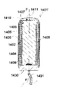
EXAMPLE 16. Thirteenth embodiment of the proto device of the invention
The twelfth embodiment 1401' of the proto device of the invention shown in
Fig. 15
corresponds to the eleventh embodiment 1301' of Fig. 14a except for the
electrode being
insulated except at its distal terminal portion and by a shielding metallic
layer 1405 being
disposed on the outer face of the flexible polymer coat 1404. On its outer
face the shielding
layer 1405 is covered by a coat 1406 of same material as the coat 1404 so as
to be fully
insulated. The layer 1404 shielding the electrode 1402 is kept on earth
potential to protect
the electrode 1402 from being disturbed by external electrical fields. The
electrode 1302 is
insulated by a lacquer 1408 at its inner face except for a small portion at
1410 extending
from its distal end. To avoid or at least delay contact with soft tissue the
electrode 1402 is
withdrawn in a distal direction by a distance h from the distal faces 1411 of
the stiffening
element 1403 and the flexible polymer coat 1404. The electrode layer 1402 and
the
shielding layer 1405 as well as the flexible polymer layers 1404, 1406 are
attached to the
base 1430 and electrically connected with the multiple lead 1431 via the base
1430.
The elements identified by reference numbers 1407 and 1409 correspond to
elements 1307
and 1309, respectively, of the embodiment of Fig. 14a.
EXAMPLE 17. Coating an metallic or polymer element with water soluble material
From the combination of optical fiber and electrical conductor or light source
grease and oil are removed by dipping the combination into diethyl ether for
10 seconds,
removing it and drying. A sugar coating of about 30 tm thickness is applied to
the
combination in the following manner. Sucrose (100 g) is dissolved in 50 ml
water. The
solution is boiled for about 5 min until it appears clear. The solution is
allowed to cool to 80
C. The combination held at its rear end by a pair of stainless steel pincers
is dipped fully
into the solution. It is removed from the solution by withdrawing it
vertically with a speed
of 6 mm/s. The sucrose coated combination is dried overnight so as to form a
dry sucrose
coat on the body of about 40 tm thickness. The thickness of the coat can be
selected by
varying the speed of withdrawal and or by multiple dipping. Lowering the speed
renders a
thinner coat.
EXAMPLE 18. Manufacture of a prestage of device the invention by coating the
dry sucrose
element of Example 14 with Parylene C
A coat of Parylene C of about 4 pm thickness is applied by a state-of-the-art
vacuum
coating process (http://www.scscookson.com/parvIene/properties.cfm) in which
di-para-
xylylene is vaporized and then pyrolized to paraxylylene, which is adduced
under high
vacuum to a deposition chamber kept at about room temperature and there
deposited on
the sucrose coated element of Example 17. The twice coated device thus
obtained
corresponds to a prestage device of the invention.
CA 02934504 2016-06-17
WO 2015/094076 20 PCT/SE2014/000152
EXAMPLE 19. Manufacture of a proto device of the invention from the prestage
device of
Example 18
The prestage device of Example 18 is dipped with its front end foremost into
molten
high melting paraffin (m.p. of about 40 C) in a short 3 mm diameter
polypropylene cylinder.
After cooling to room temperature, the paraffin block containing the prestage
device is put
on a polypropylene support and cut radially with a razor blade so as to sever
its tip. After
removing most of the paraffin by melting the block and withdrawing the proto
device thus
formed the latter is rinsed several times with pentane and dried. The recorded
impedance
of the insulated electrode body prior to cutting is >10 megohm, measured with
the
electrode body immersed into saline. The recorded impedance after cutting the
tip and
immersion of the proto device into saline for 2-3 h is <50 kohm.
Alternatively, the prestage
device of Example 15 is fixed under a microscope and portions of the Parylene
C coat near
the front end are removed by scraping the coat with a micro file made by
coating a thin
steel wire (0.1 mm diameter) with titanium oxide powder (grain of about 10 m)
by means
of cyanoacrylate pre-polymer dissolved in diethyl ether, into which the wire
is dipped
immediately prior to the application of the powder.
Dimensions of the proto device can vary within a broad range: diameters of up
to
100 lim or more are useful. A preferred diameter is from 5 rn - 30 .trn. The
length of the
proto device can be adapted to its desired location after insertion.
EXAMPLE 20. Fourteenth embodiment of the proto device of the invention
The fourteenth embodiment 1501' of the proto device of the invention shown in
Fig.
16 differs from the thirteenth embodiment 1401' by comprising, in addition to
a light
source 1509 mounted in basis 1530, a light sensor 1532, in particular one for
fluorescent
light, also mounted in basis 1530. The radiation sensor 1532 is electrically
connected by a
flexible, electrically conducting wire 1533 with a recording unit (not shown)
comprising a
microprocessor, a memory and a data output means such as a printer. The other
features
15XX of the proto device 1501' correspond to respective features 14XX of the
proto device
1401' of the thirteenth embodiment.
EXAMPLE 21. Fifteenth embodiment of the proto device of the invention and of a
corresponding device of the invention formed from the proto device upon
implantation into
soft tissue
The fifteenth embodiment 1601' of the proto device of the invention shown in
Figs.
17, 29 comprises a stiffening element 1603, which is degradable or soluble in
aqueous body
fluid. The stiffening element 1603 is mounted on a rigid cylindrical base 1613
of polymer
material such as highly cross-linked polyurethane. An LED light source 1609 is
mounted on
the distal face of the base 1613 and is energized by means of an insulated
flexible conductor
CA 02934504 2016-06-17
WO 2015/094076 21 PCT/SE2014/000152
1614 connected to a power source. The stiffening element 1603 is of
substantially
cylindrical form a rotationally symmetric in respect of its longitudinal axis
F-F. The stiffening
element 1613 and the base 1603 have about the same diameter. The stiffening
element
1603 is covered by consecutive layers of electrically insulating flexible
polymer 1608, an
electrically conducting flexible electrode layer 1604, and a flexible coat
layer 1602. The
flexible electrode layer 1604 has been attached to the insulating polymer
layer 1608 and, to
a narrow zone distal zone of the stiffening element 1603 not covered by the
insolating
polymer layer 1608 by a suitable method such as metal ion sputtering. Metals
of high
conductivity like gold and copper, are preferred for this purpose. The polymer
layers 1608
and 1602 have been attached by dipping the proto device under formation in
solutions of
the respective polymer in an organic solvent of low polarity in which the
stiffening element
1603 material is not soluble. The distal face 1611 of the stiffening element
is then covered
with a rounded cap 1610 of a material, which is readily soluble in aqueous
body fluid. The
cap 1610 is provided to facilitate insertion of the device into soft tissue.
To avoid or at least
make contact upon implantation of the electrode with surrounding soft tissue
more difficult
the electrode layer 1604 is slightly withdrawn from the distal rim of the
flexible polymer
coat as indicated by "h" in Fig. 17. A distal terminal portion of the
electrode layer 1604 is
not covered by the insulating inner flexible polymer layer 1608 to provide for
electrical
contact with body fluid. In addition to the distal axial opening 1615 are
provided three distal
radial openings 1605, 1606, 1607 of circular form with their centers disposed
in the same
radial plane B-B. The radial openings are arranged to allow light to emanate
in a radial
direction to affect or visualize neighboring soft tissue structures. To
enhance radial escape
of light the inner face of the electrically insulating polymer layer 1608 can
be provided with
a reflective coat, such as a thin coat of silver or platinum, or by using a
polymer with good
visible light reflectance properties for layer 1608. The wide beam of visible
light emitted by
the light source 1609 is directed in a distal direction; a portion of it hits
the inner face of the
insulting polymer layer or of a reflective coat on that layer. From there it
is reflected, in part
in the direction of a distal lateral opening 1605, 1606, 1607 through which it
escapes. Non-
insulated annular portions of the electrode layer 1604 are disposed in the
lateral openings,
only one 1604* of them being indicated in Figs. 17 and 18. These two kinds of
blank
electrode faces can be used in combination. Alternatively, if only one of them
is desired to
be used, the other can be made inactive by applying a layer of electrically
insulating
material on it (not shown in the Figures).
Upon implantation into soft tissue the proto device 1601' is transformed into
a
device 1601 of the invention shown in Figs. 18, 30 by dissolution or
degradation of its
stiffening element. "M" designates the inner space of the device 1601 filled
with body fluid
upon complete dissolution of the stiffening element 1603.
Fig. 31 illustrates a section 1601* of a physically modified wall of the
device 1601 of
the invention. The modification consists in providing the wall with the form
of a meander or
bellows form. The wall section 1601* comprises a flexible polymer coat 1604*,
an
electrode layer 1602*, and an inner insulating polymer layer 1608*. By such
modification a
CA 02934504 2016-06-17
WO 2015/094076 22 PCT/SE2014/000152
device of the invention comprising or consisting of non-resilient wall
materials can be made
extendible in an axial direction.
EXAMPLE 21. Sixteenth embodiment of the proto device of the invention and of a
corresponding device of the invention formed from the proto device upon
implantation into
soft tissue
The proto device 1701' of the invention illustrated in Fig. 19 is shown in an
axial view
corresponding to the proto device of Fig. 17, from which it differs by
substitution of cap
1610 by a portion of its flexible polymer coat 1704. Upon implantation into
soft tissue the
stiffening element 1703 is dissolved or degraded and substituted by aqueous
body fluid.
Thereby a corresponding device 1701 of the invention illustrated in Fig. 20 is
formed.
Reference numbers 17XX in Figs. 19 and 20 not specifically addressed refer to
elements of
corresponding kind 16XX illustrated in Figs. 17 and 18.
EXAMPLE 22. Seventeenth embodiment of the proto device of the invention and of
a
corresponding device of the invention formed from the proto device upon
implantation into
soft tissue
The proto device 1801' of the invention illustrated in Fig. 21 is shown in an
axial view
corresponding to the proto device of Fig. 17, from which it differs by
provision of an optical
sensor 1815 mounted on the distal face of the base 1813. The sensor 1815 is
sensitive to
visible light. It is particularly suited for monitoring fluorescent radiation
of a certain
wavelength, and is so selected from a number of commercially available light
sensors. It is
electrically coupled with a recording unit (not shown) by insulated flexible
lead 1816. The
recording unit can transform electrical signals from the sensor to numerical
data and store
these data in a memory. The recording unit is also capable of coordinating
tissue irradiation
by light source 1809, recording of sensor 1815 data, and electrode 1802
control. Reference
numbers 18XX in Fig. 21 not specifically addressed refer to elements of
corresponding kind
16XX illustrated in Figs. 17 and 18. Upon implantation into soft tissue the
proto device 1801'
is transformed into a device 1801 of the invention by dissolution or
degradation of its
stiffening element 1803, as shown in Fig. 22.
EXAMPLE 23. Eighteenth embodiment of the proto device of the invention and of
a
corresponding device of the invention formed from the proto device upon
implantation into
soft tissue
The proto device 1901' of the invention illustrated in Fig. 23 is shown in an
axial view
corresponding to the proto device of Fig. 17, from which it differs by a
reflective inner wall
portion 1919 and a distal wall portion 1918 provided with micro openings. The
micro
openings are provided by laser technique; their function is to provide access
of body fluid to
CA 02934504 2016-06-17
WO 2015/094076 23 PCT/SE2014/000152
the stiffening element 1903 to allow or facilitate its dissolution and the
transport of its
constituents out of the interior M of the device. The diameter of the micro
openings are in
the order of a 50 i.im or less, more preferred from 5 Inn to 30 wn. Reference
numbers 19XX
in Fig. 21 not specifically addressed refer to elements of corresponding kind
16XX illustrated
in Figs. 17 and 18. Upon implantation into soft tissue the proto device 1901'
is transformed
into a device 1901 of the invention by dissolution or degradation of its
stiffening element
1903, as shown in Fig. 24.
EXAMPLE 24. First variety of the proto device of the invention illustrated in
Fig. 17 and of a
corresponding device of the invention illustrated in Fig. 18 formed from the
proto device
upon implantation into soft tissue.
The proto device 2001' of the invention illustrated in Fig. 25 is shown in a
sectional
radial view only, which correspond to the radial view of Fig. 29 of the proto
device of Fig. 17
(section B-B). The section B-B dissects the centers of the circular windows
2005, 2006, 2007,
which are covered by portions of the flexible polymer coat 2004. The coat 2004
is of a
translucent polymer material.
Upon implantation into soft tissue the proto device 2001' is transformed into
a
device 2001 of the invention by dissolution or degradation of its stiffening
element 2003, as
shown in Fig. 26. The void filled with body fluid is designated M. Reference
numbers 20XX in
Fig. 24 not specifically addressed refer to elements of corresponding kind
16XX illustrated in
Fig. 17.
EXAMPLE 25. Second variety of the proto device of the invention illustrated in
Fig. 17 and of
a corresponding device of the invention illustrated in Fig. 18 formed from the
proto device
upon implantation into soft tissue.
The proto device 2101' of the invention illustrated in Fig. 27 is shown in a
sectional
radial view only, which correspond to the radial view of Fig. 29 of the proto
device of Fig. 17
(section B-B). The section B-B dissects the centers of the circular windows
2105, 2106, 2107,
which are covered sheets of a translucent flexible polymer material 2115,
2116, 2117.
Upon implantation into soft tissue the proto device 2101' is transformed into
a device 2001
of the invention by dissolution or degradation of its stiffening element 2103,
as shown in
Fig. 28. The void filled with body fluid is designated M. Reference numbers
20XX in Figs. 27,
28 not specifically addressed refer to elements of corresponding kind 20XX
illustrated in Fig.
17.
Materials
Electrode. The electrode is preferably of a noble metal or an alloy of noble
metals or
comprising noble metals such as gold, silver, platinum, iridium, but other
biologically
CA 02934504 2016-06-17
WO 2015/094076 24 PCT/SE2014/000152
acceptable metals such as stainless steel and tantalum can also be used as
well as gold
plated copper. Aluminum is a preferred metal for coating an optical glass
fiber. Instead of a
metal or metal alloy the electrical conductor may consist of or comprise an
electrically
conducting polymer such as PEDOT. Electrically conducting states of carbon may
also be
used. Portions of the electrical conductor that are not electrically insulated
from tissue fluid
upon removal of the first coat may be advantageously provided with surface
enlarging
elements or structures such as a roughened surface, forests of conducting
nanowires, for
instance carbon nanowires, or be porous. Surface enlarging structures of this
kind will
reduce the impedance of the electrical conductor. The electrical connection of
the
conductor with a control unit can be provided by a metal wire or similar
coupled between
the rear end of the electrical conductor and the control unit or by the
conductor itself, a
rear section thereof functioning as an electrical coupling means. In such case
the rear
section has to be electrically insulated.
Stiffening element coat. The combination of electrode and light source of the
invention is
embedded in/coated with one or more biocompatible first coat materials, which
may be
water dissolvable, swellable and/or degradable. If embedded in two or more of
such
materials they differ in their dissolution rate. Preferred first coat
materials are water
soluble carbohydrates and proteins as well as mixtures thereof. However, it is
also possible
to use water insoluble polymer materials swellable in water and/or degradable
in body
fluid. A suitable stiffening element coat material of which the dissolution
time can be
controlled is obtained by repeatedly boiling and cooling an aqueous solution
of a sugar or a
mixture of sugars selected from sucrose, lactose, mannose, maltose and an
organic acid
selected from citric acid, malic acid, phosphoric acid, tartaric acid. By
selecting particular
combinations of sugar(s) and organic acid(s) it is possible to obtain
materials with different
dissolution times. Gelatin may also be used as a first coat material. It is
well known that
different types of gelatin or gelatin based materials have different
dissolution rates. If the
first coat of water soluble or swellable material comprises two or more
sections disposed
along oblong combination of optical fiber/light source and electrode. The
selection of a
proper combination of gelatins provides a distal first coat section of shorter
dissolution time
and a proximal first coat section of longer dissolution time. The use of a
sugar-based first
coat material for the distal first coat section and of a gelatin-based first
coat material for the
proximal first coat section or vice versa is also possible, as well as the use
of gelatin for a
distal first coat section and of gum arabic for a first coat proximal section.
The selection of
further useful combinations of first coat materials, such as various types of
natural gums, is
within the easy reach of a person skilled in the art. Optionally, first coat
materials with
substantially longer dissolution times, such as modified collagen, cellulose
derivatives,
modified starch or other biocompatible materials, such as poly-glycolic acid
can also be
used.
Optionally a polymer insulating coat of the prestage device, the proto device,
the
bundle of proto devices and the array or proto devices and bundles of the
invention or a
CA 02934504 2016-06-17
WO 2015/094076 25 PCT/SE2014/000152
further coat of water dissolvable material on the first coat can be covered,
completely or in
part, by a biocompatible gliding agent to reduce friction during insertion
into tissue. Useful
gliding agents include glycerol monopalmitate, glycerol dipalmitate, glycerol
monostearate,
glycerol distearate, palmityl alcohol, stearyl alcohol. A thin coat of gliding
agent can be
applied by, for instance, spraying with a solution of the agent in ethanol or
ethyl acetate.
Flexible polymer coat. In principle, polymer materials of all kinds suitable
for electrical
insulation can be used. However, the tiny structure of the prestage device of
the invention
to be produced by polymer coating restricts the number of application methods
and useful
polymers. While deposition of monomer from the gas phase is preferred, such as
for
providing a parylene coat, dipping of a prestage device coated with water
soluble/swellable
/degradable stiffening element material into a polymer or prepolymer solution,
withdrawing it from the solution, and evaporating the solvent, optionally
allowing a
prepolymer to settle, is also useful. The dipping method should take recourse
to a polymer
solvent that does not interact with the water soluble/swellable/degradable
material, in
particular a non-polar solvent such as an alkane or alkene or cycloalkane or a
non-polar
aromatic solvent or a mixture thereof, in particular pentane or hexane but
also diethyl ether
or dichloromethane. Suitable polymers comprise biocompatible types of
polyurethane,
polyurethane urea and polyimide. Other useful polymers include silicones of
various kind.
Further useful polymers include polyethylene terephthalate (PET). The flexible
polymer
coat of the invention moves with surrounding tissue and does not restrict
tissue movement.
The thickness of the flexible coat is from a few 1..tm and up to 20 1.1m or 50
p.m or more.
Bundles of proto electrodes. Proto devices of the invention can be bundled in
different ways,
such as by incorporation of their rear end portions in a base of polymer or
other material or
by joining their rear end portions with glue. The bundling can be temporary,
such as for
keepings the devices in a fixed relationship prior to and during insertion
into soft tissue, or
permanent. A bundle of proto devices comprises a bundling means disposed in a
proximal
direction from the distal end of the two or more devices comprised by the
bundle and
aligned in parallel or about in parallel. The bundling means is preferably
permanent, that is,
is not dissolved or degraded by body fluid but may also be temporary, that is,
be dissolved
or degraded upon disposition of the bundle in soft tissue. A preferred
permanent bundling
means is an adhesive, in particular a cold setting polymer adhesive, such as a
polyurethane
or polyacrylate adhesive. The polymer adhesive is one not dissolvable or
degradable by
body fluid, except for over very long periods of more than a year or five
years the adhesive
is applied to the aligned proto devices at proximal portions thereof.
A water dissolvable or degradable adhesive of corresponding properties allows
the
proto devices to dissociate quickly or slowly upon insertion. A swellable but
not water
soluble adhesive allows the proto devices inserted into soft tissue and the
devices of the
invention formed from them to be displaced in a restricted manner while an
insoluble and
CA 02934504 2016-06-17
WO 2015/094076 26 PCT/SE2014/000152
non-swellable adhesive will restrain their movement to bending and, if
designed
extendable, to changes in length.
Individual proto devices of a bundle may differ in length. For instance, a
central
proto device of a bundle may be longer than peripheral devices thereof to
provide a central
bundle point.
Upon insertion into soft tissue, the proto devices of a bundle are transformed
to
devices of the invention and the bundle of proto devices is thereby
transformed to a bundle
of devices of the invention.
In this application an array of proto devices or bundles of proto devices
forms a
proto device pattern comprising numerous proto devices and/or bundles of proto
devices
bundles of the invention disposed on and attached to at least one face of an
electrically non
conducting support. Thin supports of a suitable polymer like polypropylene,
polyacrylate,
polycarbonate and parylene C comprising substantially only two faces are
preferred. The
supports can be flat but may also be curved. The proto devices and/or bundles
of proto
devices can be mounted on one or both surfaces of the support. The proto
devices and the
bundles of proto devices attached to the support can protrude from the support
at an
angle, in particular an angle of from about 15' to about 75 and even up to
about 900, the
angle being one included by the device or bundle of devices and its projection
onto the
mounting face of the support and/or at an angle of from about 15' to about 75'
included by
the proto device or proto device bundle long axis and a central long axis of
the support. The
support may contain pores or be semi-permeable to body fluids, that is,
permeable to at
least water and inorganic salts.
Upon insertion into soft tissue and contact with aqueous body fluid in the
tissue, the
proto device, the bundle of proto devices and the array of proto devices or
bundles of proto
devices are transformed to a corresponding device, a bundle of devices and an
array of
devices of the invention.
The support of an array of the invention can also be of a material that is
soluble or
degradable in soft tissue. Useful materials comprise those identified above as
useful water
soluble/swellable/degradable first coat materials.
If desired an array support can be equipped with a control unit, such as one
comprising or consisting of an electronic chip in electric contact with the
the electrical
conductor(s) of individual devices. The control unit can comprise or be in
electrical contact
with a unit for electric tissue stimulation and/or signal amplifier(s) for
recording electrical
nerve signals. The array support can also be equipped with a radiation control
unit, which
comprises radiation emitting means such as one or more LEDs optically coupled
with optical
fibers of the array. Furthermore the array support can also be equipped with
light sensor(s).