Note : Les descriptions sont présentées dans la langue officielle dans laquelle elles ont été soumises.
CA 02935839 2016-07-04
WO 2014/110406
PCT/US2014/011095
1
APPARATUS, METHOD, SYSTEM FOR THE
DETERMINATION OF THE AGGREGATION RATE OF RED
BLOOD CELLS
CROSS REFERENCE TO RELATED APPLICATION
[01] This application is related to and claims priority from earlier
filed U.S. Provisional Application for Patent Serial No. 61/586,502
filed January 13, 2012, the entire contents of which are incorporated
herein by reference.
BACKGROUND OF THE INVENTION
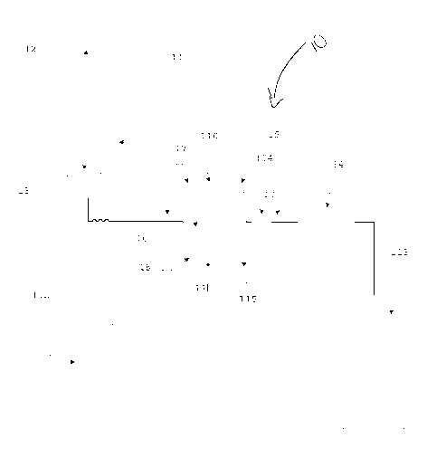
[02] The present invention generally relates to an apparatus, method,
system for the determination of the aggregation rate of red blood cells.
More specifically, the invention concerns a method, system, and the
relative apparatus used to determine the aggregation rate of red blood
cells, and other parameters related to these, such as viscosity,
deformability, elasticity, density, in the field of in vitro medical
analyses, using optical systems after or during inducted forces for red
blood cell disruption and redistribution generated by ultrasound
waves.
CA 02935839 2016-07-04
WO 2014/110406
PCT/US2014/011095
2
[03] The state of the art for the determination of a test value
corresponding to blood subsidence from a aggregogram or
syllectogram of a blood sample is ascertained by reference to the
article "Syllectometry, a new method for studying rouleaux formation
of red blood cells" by Zijlstra published in 1963.
[04] Aggregation is the first of three phases describing the
sedimentation rate that is composed by: 1) Aggregation 2)
Precipitation and 3) Packing. Erythrocyte Sedimentation Rate, which
Westergren method is considered the gold standard method, is
extensively used as a screening test for the determination of
inflammatory status of a patient.
[05] In the sedimentation phenomenon, aggregation is the first and
the fastest among the three phases, which lasts less than two minutes,
where red blood cells (RBC) forming chains (face to face aggregates)
termed "Ruloux". This phase is reversible by mixing action, due, for
example, with the repeated inversion of the test tube containing the
sample. Rulouxformation causes are still not completely clear; the
most important causes are related to proteins dispersed in plasma,
such as fibrinogen. However, it is known that aggregation between
CA 02935839 2016-07-04
WO 2014/110406
PCT/US2014/011095
3
RBC is strictly related to infections, inflammatory and connective
tissue disorders.
[06] A second stage aggregation phase, after Ruloux formation,
spherical aggregates are formed between Ruloux with uniform
increased mass, that sediment, after an initial acceleration, at constant
speed conforming Stokes law. This second phase is called
precipitation, and is the phase evaluated during the Westergren (WG)
standard method.
[07] As Stokes law describes that the constant speed is a balance
between gravity force, viscosity and hydrostatic stress. The viscosity
in a fluid as plasma is heavy affected by thermal effects and can
modify sedimentation rate independently the encountered Ruloux
level. Also
lipids dispersed in plasma, in conjunction with
lipoproteins, can increase viscosity and reduce the precipitation phase
and the resulting sedimentation rate measure.
[08] Syllectometry is a measuring method that is commonly used to
determine the red blood cell aggregability, which can be related to
consequent sedimentation rate. As reference, in syllectometry light is
incident to a layer where the sample is exposed to shear stress.
CA 02935839 2016-07-04
WO 2014/110406
PCT/US2014/011095
4
Luminous flux attenuation/increase or backscatter ultrasound wave
are used for determination of variations in sample density after the
abrupt stop of driving mechanism. The subsequent time-dependent
plot is called syllectogram.
[09] Therefore, there remains a need in the prior art for an apparatus,
method, system for the determination of the aggregation rate of red
blood cells which does not require a stopped flow technique for
aggregation kinetic detection.
BRIEF SUMMARY OF THE INVENTION
[10] The invention preserves the advantages of prior apparatus,
methods, and systems for the determination of the aggregation rate of
red blood cells. In addition, it provides new advantages not found in
currently available apparatus, methods, and systems for the
determination of the aggregation rate of red blood cells and
overcomes many disadvantages of such currently available systems.
[11] The present invention generally relates to an apparatus, method,
system for the determination of the aggregation rate of red blood cells.
CA 02935839 2016-07-04
WO 2014/110406
PCT/US2014/011095
More specifically, the invention concerns a method, system, and the
relative apparatus used to determine the aggregation rate of red blood
cells, and other parameters related to these, such as viscosity,
deformability, elasticity, density, in the field of in vitro medical
analyses, using optical systems after or during inducted forces for red
blood cell disruption and redistribution generated by ultrasound
waves.
[12] The invention provides a method and the relative reusable
apparatus for the determination of aggregation rate index, and
subsequent erythrocytes sedimentation rate for whole blood samples.
The invention reduces the complexity of the pumping systems
removing the need of the stopped flow condition. The invention
provides other rheological parameters such as viscosity,
deformability, elasticity, density. The invention provides a method
and the relative apparatus for reduce the sample mixing time needed
for the disruption of the aggregates RBC chains, using an alternative
method prior and during the rheological behavior detection. The
invention reduces the amount of sample volume needed for avoid
CA 02935839 2016-07-04
WO 2014/110406
PCT/US2014/011095
6
contamination by residuals of previous sample applying an enhanced
washing system.
BRIEF DESCRIPTION OF THE DRAWINGS
[13] The novel features which are characteristic of the present
invention are set forth in the appended claims. However, the
invention's preferred embodiments, together with further objects and
attendant advantages, will be best understood by reference to the
following detailed description taken in connection with the
accompanying drawing in which:
[14] Fig. 1 is a schematic view of an embodiment of the apparatus,
method, and system for the determination of the aggregation rate of
red blood cells.
DETAILED DESCRIPTION OF THE PREFERRED
EMBODIMENTS
[15] In accordance with the invention of Fig. 1, the present invention
generally relates to an apparatus, method, system for the
determination of the aggregation rate of red blood cells. More
specifically, the invention 10 concerns a method, system, and the
CA 02935839 2016-07-04
WO 2014/110406
PCT/US2014/011095
7
relative apparatus used to determine the aggregation rate of red blood
cells, and other parameters related to these, such as viscosity,
deformability, elasticity, density, in the field of in vitro medical
analyses, using optical systems after or during inducted forces for red
blood cell disruption and redistribution generated by ultrasound
waves.
[16] The invention provides a method and the relative reusable
apparatus for the determination of aggregation rate index, and
subsequent erythrocytes sedimentation rate for whole blood samples.
The invention reduces the complexity of the pumping systems
removing the need of the stopped flow condition. The invention
provides other rheological parameters such as viscosity,
deformability, elasticity, density. The invention provides a method
and the relative apparatus for reduce the sample mixing time needed
for the disruption of the aggregates RBC chains, using an alternative
method prior and during the rheological behavior detection. The
invention reduces the amount of sample volume needed for avoid
contamination by residuals of previous sample applying an enhanced
washing system.
CA 02935839 2016-07-04
WO 2014/110406
PCT/US2014/011095
8
[17] In one embodiment, the apparatus 10 for the determination of
RBC aggregation, and their subsequent sedimentation rate, according
to the invention comprises a reading cell container 16 where the
sample is introduced. The apparatus 10 provides this reading cell
container 16 equipped with two parallel optical windows for allow to
a light radiation to pass through the sample herein introduced or
reading the backscatter of the incident light. The apparatus 10
comprises a collimated light source composed in such way that light
passes through the windows of the container mentioned above, and
can be reflected. On the opposite side of the light source 17 is present
an optical detector 18 for the evaluation of the light attenuated by the
sample. The optical detector 18 could be positioned on the same side
of the light source 17 for the detection of the light scattering. The
reading cell container 16 is equipped with electromechanical actuator
110,111 able to vibrate the sample herein introduced, disrupting the
aggregates naturally present in the blood sample, and evenly distribute
the erythrocytes within the entire volume of sample. The apparatus
has a temperature control system 114, 115 for the sample container for
standardize the reaction environment.
CA 02935839 2016-07-04
WO 2014/110406
PCT/US2014/011095
9
[18] The apparatus 10 comprises further an electronic control device
112 able to acquire the optical variance detected by the optical
detector, drive the electromechanical actuators 110,111 and acquire
the container temperature values. This electronic control device 112 is
also able to convert the detected time dependent light variation into an
aggregation index and his subsequent erythrocyte sedimentation rate,
providing the result of the evaluated phenomenon in the way of
numerical result comparable to the common used parameters used in a
clinical laboratory.
[19] According with another embodiment of the invention, an
apparatus 10 or system has been developed. The apparatus or system
is comprised of a mixer device 11 for a low homogenization of the
sample inside a collection tube 12. The homogenization can be
achieved by a Vortex like mixer or by the radial or axial rotation of
the sample tube.
[20] After the homogenization the sample is then withdrawn by a
needle 13 and aspirate by a pump device 14 through hydraulic circuit
15. The hydraulic circuit 15 connects the aspiration needle 13 to the
reading cell container 16 allow their filling by the sample, guaranteed
CA 02935839 2016-07-04
WO 2014/110406
PCT/US2014/011095
by the optical sensor composed by the emitter 17 and an optical
receiver 18 and a secondary optical flow sensor 19 controlled by an
electronic control device 112.
[21] The light emitter source 17 is composed, in one embodiment
but not in limitative manner, by a Light Emitter Diode (LED), and can
be substituted, for example, by a laser source or an incandescent lamp.
The optical receiver 18, in this embodiment but not in limitative
manner, is composed by CCD sensor for two dimensional
characterization of the reaction. This sensor can be substitute by a
single receiver element such as photodiode, photomultiplier etc.
[22] After the complete or desired filling of the reading cell 16 the
pump device 14 is stopped by the electronic control device 112, and
the sample is processed by the electromechanical devices 110, 111,
for example composed by piezoceramics, activated to a predetermined
power by the control device 112, to disrupt aggregates and evenly re-
suspend the RBC on the sample volume. A prerequisite for an
aggregation kinetic detection is a complete disruption of the
aggregates, normally formed in a steady state of the sample. This
CA 02935839 2016-07-04
WO 2014/110406
PCT/US2014/011095
11
disruption can be achieved by an intensive mixing phase before and
during the transportation of the sample in the reading cell or detection.
[23] As an alternative to a predetermined power, the piezoceramic
power is initially ramped up to a level where cell emulsification is
detected through the optical reading. This process is stopped and a
duplicate sample is introduced. The power applied can be optimized at
fraction of the emulsification power level which results in maximum
dispersion, without cell damage.
[24] During this phase the control device 112 acquires the signal
detected by the optical receiver 18 and stops the electromechanical
devices 110, 111 or actuators when the light variation detected by the
receiver 18 stops decreasing, indicating the complete disruption of the
aggregate present into the sample. This recorded plot expresses the
disruption rate of the RBC and is post evaluated by the system.
[25] In one embodiment, the shape of the reading cell container 16
walls comprises sound lenses for focusing the wave pressure shear to
emphasize the shear inducted to the sample.
CA 02935839 2016-07-04
WO 2014/110406
PCT/US2014/011095
12
[26] After the electromechanical devices 110,111 stops, the signal
detected by the receiver 18 is still recorded by the control device 112
for a predetermined amount of time as a plot of kinetic aggregation.
[27] After the end of the acquisition the sample is evacuated from
the reading cell 16 by the pump device 14 to a waste reservoir 113.
During the evacuation, the electromechanical devices 110, 111 are
activated with a high power for remove proteins eventually bonded to
the reading cell container 16 walls. An evacuation of the reading
chamber avoids the pollution of the sample currently under measure
by the residual of the previous measured sample with washing and
does not require a large flow amount of sample currently under
measure for removal the residuals of the previous measured sample.
After the evacuation, the system is ready for a new sample
withdrawing and analysis.
[28] The reading cell container 16 is also maintained to a controlled
temperature by the thermoelectric device 114 and the temperature is
acquired by the control device 112 through the temperature sensor 115
for providing standardized conditions of reaction.
CA 02935839 2016-07-04
WO 2014/110406
PCT/US2014/011095
13
[29] During the dispersion phase induced by the electromechanical
devices 110, 111, the resultant signal is evaluate to extract the mean
viscosity value of the sample plasma by considering the time need by
the sample to completely re-suspend. After a complete re-suspension
of the sample a burst of ultrasound waves is inducted to the sample for
evaluating the red blood cell deformability. This deformability is
considered as the time needed by the media to absorb the wave shear
impressed, also decay after the wave share absorption is evaluated in
function of time as index of the mean shape recovery ability.
[30] It should be appreciated that the system, method, and apparatus
may include one or more components or steps listed above in a variety
of configurations depending upon desired performance or
requirements.
[31] It would be appreciated by those skilled in the art that various
changes and modifications can be made to the illustrated
embodiments without departing from the spirit of the present
CA 02935839 2016-07-04
WO 2014/110406
PCT/US2014/011095
14
invention. All such modifications and changes are intended to be
within the scope of the present invention.
