Note : Les descriptions sont présentées dans la langue officielle dans laquelle elles ont été soumises.
CA 02936023 2016-07-06
WO 2015/103712 PCT/CH2014/000003
Method for generating a 3D reference computer model of at least one anatomical
structure
BACKGROUND OF THE INVENTION
1. Field of the Invention
The invention relates to a method for generating a 3D reference computer model
of at
least one anatomical structure according to the preamble of claim 1, to a
method for
generating a status related 3D computer model of a patient's anatomical
structure in a
pre-, intra- or post-operative status according to the preamble of claim 10 or
11 and to a
method for monitoring a surgical treatment according to the preamble of claim
26.
During surgical treatments of fractures and the correction of osseous
deformities bone
fragments are anatomically repositioned and stably fixed at a correct position
by using
suitable fixation techniques. Problems may arise by an unrecognized
malposition of
bone fragments and implants during surgery, or through their secondary
dislocation in
the postoperative course. A faulty osteosynthesis due to anatomically
incorrect
repositioning of bone fragments, improper surgical technique, unsuitable
selection of an
implant and/or its positioning is to be avoided.
Bone fractures and osseous deformities are routinely assessed using different
radiological imaging techniques before, during, and after surgery. Usually
conventional
x-rays are used, i.e. planar projection images. Particularly complex
interventions are
assessed for diagnostic purposes by using a tomographic layer imaging,
preferably by
using computer tomography (CT). This is done by analyzing these layer images
or their
three-dimensional computer models preferably preoperatively, in the case of
special
issues also intra - or post-operatively.
However, so far in clinical routine the bone fragments and the osteosynthesis
cannot be
assessed spatially coherent over the entire course of therapy. Three-
dimensional
medical imaging as CT's in all stages of therapy would be needed. As mentioned
this is
technically possible, but so far costs, radiation-hygienic reasons, generation
of artifacts,
personal, organizational and technical effort clearly oppose a routine spatial
assessment of osteosynthesis in all stages of therapy.
2. Description of the Related Art
CA 02936023 2016-07-06
WO 2015/103712 PCT/CH2014/000003
2
A process for the reduction of fragments of a fractured bone is known from US-
A
2011/0082367 REGAZZONI. This known process includes steps of generating 3D
representations of bones and bone fragments on the basis of a digital data set
obtained
by means of CT's of a fractured bone, as well as of the contralateral healthy
bone of a
patient. The 3D representation of the mirrored contralateral healthy bone is
used as a
reference model for the relative position of the 3D representations of
repositioned bone
fragments. Subsequently, the 3D representations of the proximal and distal
bone
fragments are matched with the 3D representation of the reference model using
three-
dimensional image registration. Furthermore, the configurations of markers
and/or
anatomical landmarks on the proximal and the distal bone fragment are
extracted and
transferred to the reference model, The relative positions of the markers
and/or
anatomical markers transferred to the reference model of the proximal and
distal bone
fragments then allow to establish a digital reference data set suitable for
the real
reduction of the bone fragments during the operation. A disadvantage of this
known
method can be that each a CT of the fractured bone and of the contralateral
healthy
bone is needed.
BRIEF SUMMARY OF THE INVENTION
It is therefore an object of the invention to provide a method for generating
a 3D
reference computer model of at least one anatomical structure which requires
an
acquisition of standard 2D medical images only.
The invention solves the posed problem with a method for generating a 3D
reference
computer model of at least one anatomical structure comprising the features of
claim 1,
with a method for generating a status related 3D computer model of a patient's
anatomical structure in a pre-, intra- or post-operative status comprising the
features of
claim 10 or 11 and to a method for monitoring a surgical treatment comprising
the
features of claim 26.
The advantages of the method according to the invention can essentially be
seen in:
- a full 3D computer model of a patient affected by a bone fracture or bone
deformity can be established from conventional 2D medical images only. This
permits the patient to be assessed and its treatment to be guided and tracked
in
3D; and
CA 02936023 2016-07-06
WO 2015/103712 PCT/CH2014/000003
3
- less image information is needed to get comprehensive information to assess
the
surgical treatment of the patient at any stage.
Certain terms as used herein are understood as follows:
3D reference computer model:
A full body 3D atlas model 30 with standard deviation information is
superposed on the
first and second medical images (preferably medical 2D images) of a given
clinical case
and they are referenced on the 3D atlas model, by the specific values gathered
in
predefined well detectable zones and/or artificial additional markers outside
and/or
anatomical landmarks inside the body. The first and second medical images
might be
taken, with different existing and new technologies/modalities (e.g. known X-
ray
techniques or CT-scans) to allow differentiating several independent, but
known values,
respectively value maps, which are an integrated part of the full body 3D
atlas model.
Differences of the first and second medical images ( the individual 2D images)
to the 3D
atlas model are first used in an analysis using only healthy information like
landmarks of
unfractured bone to adapt the 3D atlas model to the first and second medical
images of
the individual case and fill up the gaps of information, such that the
"normal" 3D atlas
model is converted to the individual measures and by this transformed in a 3D
reference
computer model, i.e. in a full 3D redesign of the individual healthy body.
Status related 3D computer model:
By superposing the 3D reference computer model on the first and second medical
images additional variations especially from the pathological area are
detected either as
deformities or as fragments in dislocation. By using this technology the 3D
reference
computer model as a full 3D model of the healthy situation can be transformed
to a
corresponding status related 3D computer model, e.g. a pathological 3D model.
Alternatively or additionally, by superposing the 3D reference computer model
on
subsequent pre-, intra- or post-operative sets of medical images the 3D
reference
computer model as a full 3D model of the healthy situation can be transformed
to a
corresponding status related 3D computer model in a pre-, intra- or
postoperative status
i.e. to any pathological or surgically treated 3D redesign at any stage of
healing.
Graphical 3D computer model:
CA 02936023 2016-07-06
WO 2015/103712 PCT/CH2014/000003
4
The graphical 3D computer model includes computer-aided planning and
performing a
virtual surgical treatment of anatomical structures to be treated by using the
3D
reference computer model and/or the pre-operative status related 3D computer
model.
Implant:
The term implant as used herein is understood as including all solid means
artificially
implanted or to be implanted in the human or animal body completely or
partially which
can be detected by conventional x-rays, CT or magnetic resonance imaging (MRI)
and
which have a limited variability in their form, such as orthopedic implants,
dental
implants, pacemakers or stents.
Registration:
Image registration is understood as the process of mapping one or more target
images
of an object to a reference image, thereby establishing point-by-point
correspondence
between the reference image and the target image. The step õregistering"
preferably
comprises the following sub-steps (B. Zitova, J. Flusser, Image registration
methods: a
survey, Image and Vision Computing 21, 2003, 977- 1000):
1) Feature detection: salient and distinctive objects (closed boundary
regions,
edges, contours, line intersections, corners etc.) are manually or, preferably
automatically detected. For further processing these features can be
represented
by their point representatives (centers of gravity, line endings, distinctive
points);
2) Feature matching: in this step the correspondence between the features
detected
in the sensed image (3D atlas model 30) and those detected in the reference
image (first and second medical images 10, 11) is established. Various feature
descriptors and similarity measures along with spatial relationships among the
features are used for that purpose;
3) Transform model estimation: the type and parameters of the so-called
mapping
functions, aligning the sensed image with the reference image, are estimated.
The parameters of the mapping functions are computed by means of the
established feature correspondence; and
4) Image resampling and transformation: the sensed image (3D atlas model 30)
is
transformed by means of the mapping functions. By means of the mapping
function the sensed image (3D atlas model 30) is transformed to overlay it
over
the reference image (first and second medical images 10, 11).
CA 02936023 2016-07-06
WO 2015/103712 PCT/CH2014/000003
The above sub-steps are used herein for the case of a "scene to model
registration"
where the images of a scene (anatomy of the patient) and a model of the scene
(3D
atlas model) are registered.
Further advantageous embodiments of the invention can be commented as follows:
In a special embodiment the first and second medical images are taken from
different
perspectives that are minimum 600 angularly offset with respect to each other.
In a further embodiment the at least one anatomical structure is a bone and
the
registering step includes before performing the image registration the sub-
steps of:
extracting a first section of the first medical image, wherein the first
section of the first
medical image comprises a section of a proximal bone fragment spaced apart
from a
fracture site or from a deformed portion of a bone; extracting a second
section of the
first medical image, wherein the second section of the first medical image
comprises a
section of a distal bone fragment spaced apart from a fracture site or from a
deformed
portion of a bone; and repeating the above steps for the second medical image.
In a further embodiment the first and second medical images include a
plurality of
anatomical structures and the 3D reference computer model comprises each a
graphical 3D sub-model for each anatomical structure. An advantage achieved by
this
means is, that individually trackable graphical 3D sub-models for the
anatomical
structures to be treated like bones or bone fragments can be integrated in the
3D
reference computer model allowing an individual analysis of certain anatomical
structures.
In another embodiment the method further comprises the additional step of:
introducing
at least one digital graphical 3D sub-model in the 3D reference computer
model. A
graphical 3D sub-model of an implant and/or of a surgical instrument can be
copied
from a database in the 3D reference computer model, such as for example a CAD
database.
In a further embodiment the digital graphical 3D sub-model represents an
implant.
CA 02936023 2016-07-06
WO 2015/103712 PCT/CH2014/000003
6
In again another embodiment the digital graphical 3D sub-model represents a
surgical
instrument.
In a further embodiment the generation of the 3D reference computer model
comprises
an automatic or manual identification and localization of anatomical
landmarks, lines
and/or regions of the anatomical structures to be treated.
In a further embodiment the generation of the 3D reference computer model
comprises
an automatic or manual identification and localization of distinctive points,
lines and/or
regions of each implant and preferably of each surgical instrument.
The method for generating a status related 3D computer model of a patient's
anatomical
structure in the pre-operative status by using the 3D reference computer model
comprises the step of: registering each of the first and second medical images
to the 3D
reference computer model. By subsequently superposing the 3D reference
computer
model on the first and second medical images additional variations especially
from the
pathological area are detected either as deformities or as fragments in
dislocation.
For subsequent status related 3D computer models of a patient's anatomical
structure
in a pre-, intra- or post-operative status the following steps are performed:
a) acquiring a
pre-, intra- or post-operative set of medical images including at least two
medical
images of at least one anatomical structure in a pre-, intra- or post-
operative status and
from different perspectives by using a computer assisted medical imaging
device,
wherein the at least two medical images are each represented by a respective
set of
digital 2D image data; b) generating a graphical 2D or 3D computer model of at
least
one anatomical structure in the form of a set of digital data by using the pre-
, intra- or
post-operative medical images; and c) registering the graphical 2D or 3D
computer
model to the 3D reference computer model. The advantages achieved are that due
to
the registration of conventional preoperative x-rays, intraoperative 2D planar
or spatial
3D C-arm images, or postoperative X-ray images to the initially generated 3D
reference
computer model of anatomical structures (e.g. a bone or bone fragment) these
pre-,
intra- or postoperatively acquired sets of medical images can now always be
represented as status related 3D computer models over the entire course of
therapy.
CA 02936023 2016-07-06
WO 2015/103712 PCT/CH2014/000003
7
A spatial representation preoperatively generated once and preferably by using
a CT is
beneficial for several reasons: it generates a spatial representation of the
region to be
treated at the beginning of the therapy. This spatial information can be used
for
diagnostics and therapy planning. In addition, preoperatively there is more
time
available for their processing and analysis as for example during the
operation. Further,
other imaging techniques, generated by using intraoperative 2D or 3D C-arm
images
are less or even inappropriate for temporal or technical reasons, to generate
3D
computer models of anatomical structures such as bone. The same applies to
conventional preoperative and postoperative x-rays, where the scaled
representation of
3D computer models of anatomical structures such as bone fragments is not
possible;
at least not without considerable additional effort. These X-ray images
represent planar
images, generated from one direction of projection only. But their high image
resolution
is beneficial.
In a special embodiment the status related 3D computer model additionally
comprises a
representation of at least one implant.
In a further embodiment the status related 3D computer model additionally
comprises a
representation of at least one surgical instrument.
In another embodiment the pre-, intra- or post-operative set of medical images
includes
a plurality of anatomical structures and the status related the 3D computer
model
comprises each a graphical 2D or 3D sub-model for each anatomical structure
and
preferably for each implant and/or surgical instrument.
In another embodiment the status related 3D computer model forms the reference
model to which the 3D reference computer model is adapted during the
registration
step. The status related 3D computer model is used as a target model, to which
the 3D
reference computer model (object model or source model) is modified. The
acquisition
of the pre-, intra- or postoperative sets of medical images can include two or
more
digital medical images, which are obtained each at a predefined angle of the
image
plane of the C-arm with respect to the gravity vector so that the positions of
anatomical
structures to be treated and hence the position of the 3D reference computer
model are
defined in a system of coordinates which is fixed with respect to the
operation room.
CA 02936023 2016-07-06
WO 2015/103712 PCT/CH2014/000003
8
In again another embodiment the acquisition of the set of medical images - in
a pre -,
intra - or postoperative status - includes an acquisition of one or more
digitized medical
images by means of a computer-aided medical imaging technique. The acquisition
of
two or more digitized medical images is performed at an angle relative to each
other
permits to generate a 3D computer model. On the other hand, different
fragments/sections of a long bone can be mapped in each of the digitized
medical
images so that intra-operatively used C-arm equipment with a relatively small
image
frame can be used to acquire the pre-, intra- or postoperative sets of medical
images.
The procedure distinguishes itself by the fact that only one X-ray can be
sufficient and
standard image acquisitions "in two planes" as known to the skilled person can
be
avoided. Additional advantages of the method are thus a reduced radiation
exposure
and expenditure. In the case of corrective osteotomies and fracture treatments
the
entire osteosynthesis construct consisting of bone fragments, any residual
bone defect
and the implants used can be spatially assessed over the entire course of
therapy. On
the computer display a graphical representation of the status related 3D
computer
model of the anatomical structure such as the fracture or osteotomy is
visible,
representing spatially the bone fragments depending on the stage of therapy
before,
during or after surgery. Thus, a 3D imaging procedure is not necessary. As
soon as
implant material is radiologically visible, its position can also be spatially
determined and
represented by referencing its 3D computer model to the 3D computer models of
anatomical structures, such as for example the bone fragments.
In a further embodiment the generation of the status related 3D computer model
includes an automatic or manual re-identification and re-localization of the
anatomical
landmarks, lines and/or regions of the anatomical structures to be treated as
identified
and localized in the 3D reference computer model. In the simplest case, the
status
related 3D computer model is based on a single digital medical image with the
re-
identified and re-localized anatomical landmarks. The registration can
therefore be
effected with a feature-based registration process. In the case of feature-
based
registration processes, a certain, usually relatively small number of
features, e.g.
anatomical landmarks are extracted from the images. This is done either
manually or
automatically. The selected anatomical features are preferably spread over the
whole
image and do not only focus on a single region. The registration is then
effected by
CA 02936023 2016-07-06
WO 2015/103712 PCT/CH2014/000003
9
matching the selected features, e.g. the selected anatomical landmarks on the
source
model, i.e. the 3D reference computer model with the identical anatomical
landmarks on
the reference or target model, i.e. on the status related 3D computer model.
In addition
to anatomical landmarks regions in the image that clearly distinguish from
adjacent
regions, can be used as region features or lines or edges, which are present
as lines or
contours of regions can be used as features. Lines can be represented and
extracted by
their endpoints as well.
In a further embodiment the generation of the status related 3D computer model
includes an automatic or manual re-identification and re-localization of the
distinctive
points, lines and/or regions of each implant and each surgical instrument as
identified
and localized in the 3D reference computer model. The registration of the 3D
sub-
models of implants or surgical instruments can be effected in two ways:
(1) first, graphical 3D sub-models of anatomical structures of the 3D
reference computer
model are registered to the graphical 3D sub-models of the anatomical
structures of the
status related 3D computer model and subsequently the graphical 3D sub-models
of
implants or surgical instruments of the 3D reference computer model are
registered with
one or more graphical 3D sub-models of anatomical structures of the previously
registered graphical 3D sub-models of anatomical structures of the 3D
reference
computer model by thereby taking into consideration the relative positions
between the
graphical 3D sub-models of implants or surgical instruments and the graphical
3D sub-
models of the anatomical structures in the status related 3D computer; or
(2) first, graphical 3D sub-models of anatomical structures of the 3D
reference computer
model are registered to the graphical 3D sub-models of the anatomical
structures of the
status related 3D computer model and subsequently the graphical 3D sub-models
of
implants and/or surgical instruments of the 3D reference computer model are
registered
to the graphical 3D sub-models of implants and/or surgical instruments of the
status
related 3D computer model.
The generation of a graphical 3D computer model by using the 3D reference
computer
model and/or the pre-operative status related 3D computer model preferably
comprises
the step of: computer-aided planning and performing a virtual surgical
treatment of
anatomical structures to be treated.
CA 02936023 2016-07-06
WO 2015/103712 PCT/CH2014/000003
In another embodiment the graphical 3D computer model comprises a graphical 3D
sub-model of the anatomical structures to be treated in the form of a digital
data set by
using the first and second medical images.
In another embodiment the computer-aided planning comprises an integration of
at
least a further graphic 3D sub-model of an implant in the graphical 3D
computer model.
In a further embodiment the computer-aided planning comprises an integration
of at
least a further graphic 3D sub-model of a temporary auxiliary means,
preferably of a
surgical instrument in the graphical 3D computer model. By this means the
position of
implants or temporary equipment, such as guide wires, surgical tools and
instruments
can be spatially determined and represented in each treatment step up to the
end of the
therapy. This is achieved by matching the positions of corresponding 3D
computer
models of implants or temporary auxiliary means which are archived in the
computer
and can be retrieved, with firstly the correctly positioned 3D computer models
of
anatomical structures (as described above) and secondly with the positions of
the
implants and/or temporary auxiliary means visible on the X-ray images. The 3D
computer models of implants or temporary auxiliary means are thus represented
spatially over the complete course of therapy by repeated registrations on the
different
imaging modalities such as conventional preoperative x-rays, intraoperative
planar 2D
or spatial 3D C-arm images, or postoperative X-ray images.
In a further embodiment the computer-aided planning comprises an assessment of
the
bio-mechanical stability of the virtually surgically treated anatomical
structures using a
computer simulation, preferably using a finite element computer analysis. By
means of
computer-aided analysis and planning of the surgical operation, i.e. the re-
positioning of
the anatomical structures, the type and position of temporary and permanent
implants
can be spatially represented, virtually planned on the computer and the
biomechanical
stability e.g. of an osteosynthesis can be assessed by means of computer
simulation
and re-evaluated in each treatment step. The treatment plan can then be
continued or
modified if necessary.
CA 02936023 2016-07-06
WO 2015/103712 PCT/CH2014/000003
11
In again a further embodiment the graphical 3D computer model comprises at
least a
graphical 3D sub-model of at least an intermediate result of anatomical
structures
virtually treated according to the computer-aided planning.
In another embodiment the graphical 3D computer model comprises as a sub-model
a
treatment plan, which preferably defines the exact sequence of surgery and
includes
appropriate control requirements.
In a special embodiment of the method for monitoring a surgical treatment a
status
related 3D computer model is generated in a preoperative status allowing a
monitoring
of at least an object before surgical treatment.
In a further embodiment a status related 3D computer model is generated in at
least
one intraoperative status allowing a monitoring of the at least an object
during surgical
treatment.
In a further embodiment a status related 3D computer model is generated in at
least
one postoperative status allowing a monitoring of the at least an object after
surgical
treatment.
In another embodiment the method further comprises assessing and/or analysing
differences between the 3D reference computer model and a status related 3D
computer model.
In another embodiment the method further comprises assessing and/or analysing
differences between the graphical 3D computer model and a status related 3D
computer model.
In again another embodiment the method further comprises assessing and/or
analysing
differences between a status related 3D computer model and a subsequent status
related 3D computer model.
In a further embodiment the differences are automatically assessed and/or
analysed.
CA 02936023 2016-07-06
WO 2015/103712 PCT/CH2014/000003
12
A preferred use of the method is for the quality assurance of surgical
treatments. A
further component and advantage of this method is that all data that is
generated over
the entire course of therapy can be integrated into a quality management
system and
can thus be analyzed. This can positively affect in turn the style, selection
and
implementation of the therapy; for example standardizing the therapy
procedures with
respect to relevant parameters.
Furthermore the method for generating a 3D reference model and/or the method
for
generating a status related 3D computer model and/or the method for generating
a
graphical 3D computer model and/or the method for monitoring a surgical
treatment can
be used for:
- a treatment of bone fractures.
- a treatment of osseous deformities.
- for dental implantology.
A BRIEF DESCRIPTION OF THE DRAWINGS
Several embodiments of the invention will be described in the following by way
of
example and with reference to the accompanying drawings in which:
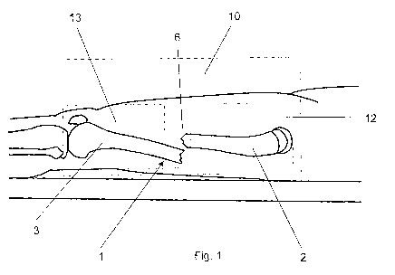
Fig. 1 illustrates a lateral view of a patient's fractured bone; and
Fig. 2 illustrates a schematic view of a registration step according to an
embodiment of
the method according to the invention;
Fig. 3 illustrates a perspective view of a 3D reference computer model of the
patient's
bone in an unfractured state according to an embodiment of the method
according to
the invention;
Fig. 4 illustrates a flow chart of an embodiment of the method for generating
a status
related 3D computer model in a pre-operative status according to the
invention;
Fig. 5 illustrates a flow chart of an embodiment of the method for generating
a status
related 3D computer model of a patient's anatomical structure in a pre-, intra-
or post-
operative status according to the invention; and
CA 02936023 2016-07-06
WO 2015/103712 PCT/CH2014/000003
13
Fig. 6 illustrates a flow chart of an embodiment of the method for generating
a 3D
computer aided planned model according to the invention.
DETAILED DESCRIPTION OF THE INVENTION
Image based assessment of a bone fracture/deformity is always based on the
experience of the assessor and therefore restricted by his subjective
interpretation.
Standard two-dimensional (2D) imaging is commonly used for patient assessment
providing only restricted information. The aim of the invention is an
objective 3D
assessment of the individual situation by providing a full 3D model of the
individual
clinical case based on standard images taken to assess the clinical situation.
Basically the method according to the invention can be applied for all
anatomical
structures, which can be acquired by means of a computer-aided medical imaging
technique. Further, all implants and instruments that can be used
intraoperatively and
which are geometrically clearly detectable at least in part by a computer-
aided medical
imaging procedure can also be used.
An exemplary embodiment of the method according to the invention for
generating a 3D
reference computer model 20 of a bone, and in particular of a femur is
elucidated with
reference to figs. 1 to 3. A full status related 3D computer model 25 of the
individual
clinical case based on at least a first and a second standard medical image
10, 11 is
provided to assess the clinical situation.
Exemplarily, the method for generating this 3D reference computer model 20 of
at least
one anatomical structure comprises the steps of: A) acquiring at least a first
and a
second medical image 10, 11 of at least one anatomical structure in a
preoperative
status and from different perspectives by using a computer assisted medical
imaging
device, wherein the first and second medical images 10, 11 are represented by
a
respective first and second set of digital 2D image data; and B) generating a
3D
reference computer model 20 of an anatomical structure by: i) selecting and
extracting a
3D atlas model 30 of an anatomical structure to be treated from a generic
anatomical
atlas provided in the form of a digital data source; and ii) registering at
least a section
12, 13 of each of the first and second medical images 10, 11 to the selected
3D atlas
CA 02936023 2016-07-06
WO 2015/103712 PCT/CH2014/000003
14
model 30. Preferably, the first and second medical images 10, 11 are taken
from
different perspectives that are minimum 600 angularly offset with respect to
each other.
The registering step can include before performing the image registration the
sub-steps
of: 1) extracting a first section 12 of the first medical image 10, wherein
the first section
12 of the first medical image 10 comprises a section 4 of a proximal bone
fragment 2
spaced apart from a fracture site 6 or from a deformed portion of a bone 1; 2)
extracting
a second section 13 of the first medical image 10, wherein the second section
13 of the
first medical image 10 comprises a section 5 of a distal bone fragment 3
spaced apart
from a fracture site (6) or from a deformed portion of a bone 1; and 3)
repeating the
above steps for the second medical image 11.
As illustrated in fig. 4, this 3D reference computer model 20 of at least one
anatomical
structure can be used for comparison with the pre-operatively acquired first
and second
medical images 10, 11 of at least one anatomical structure or with a graphical
2D or D3
computer model 15 thereof.
By subsequently superposing this 3D reference computer model 20 on the first
and
second medical images 10, 11 additional variations especially from the
pathological
area are detected either as deformities or as fragments in dislocation. By
using this
technology this 3D reference computer model 20 as a full 3D model of the
healthy
situation can be compared with a corresponding status related 3D computer
model 25,
e.g. a pathological 3D model to assess the current situation at any stage of
treatment.
By this means a first pre-operative status related 3D computer model 25 of a
patient's
anatomical structure in the pre-operative status can be obtained by performing
the step
of registering each of the first and second medical images 10, 11 to the 3D
reference
computer model 20.
By comparing the 3D reference computer model 20 with the first pre-operative
status
related 3D computer model 25, which has been obtained by using the first and
second
medical image 10, 11, the actual situation of the at least one anatomical
structure of a
patient can be assessed and/or differences between the 3D reference model 20
and the
first pre-operative status related 3D computer model 25 can be automatically
and/or
manually analyzed in order to characterize the clinical picture of the at
least one
anatomical structure of a patient.
CA 02936023 2016-07-06
WO 2015/103712 PCT/CH2014/000003
As illustrated in fig. 5, the 3D reference computer model 20 of at least one
anatomical
structure can be used for comparison with a selectable pre-, intra- or
postoperative set
of medical images 40, 50, 60 of at least one anatomical structure or with a
graphical 2D
or D3 computer model 15 thereof. Status related 3D computer models 25 in a pre-
,
intra- or post-operative status can be obtained by performing the steps of: i)
acquiring a
pre-, intra- or post-operative set of medical images 40, 50, 60 including at
least two
medical images of at least one anatomical structure in a pre-, intra- or post-
operative
status and from different perspectives by using a computer assisted medical
imaging
device, wherein the at least two medical images are each represented by a
respective
set of digital 2D image data; ii) generating a graphical 2D or 3D computer
model 15 of at
least one anatomical structure in the form of a set of digital data by using
the pre-, intra-
or post-operative medical images 40, 50, 60; and iii) registering the
graphical 2D or 3D
computer model 15 to the 3D reference computer model 20.
By using these pre-, intra- or post-operative sets of medical images 40, 50,
60 the full
tracking of treatment can be made. Alternatively, any kind of three-
dimensional (3D)
image information of a patient may be used to be compared either with the 3D
atlas
models 30 or with the 3D reference computer model 20, i.e. the 3D redesign of
the
individual healthy body as well as with any subsequent status related 3D
computer
model 25, i.e. with any pathological 3D redesign as captured with the method
according
to the invention at any stage of healing.
The 3D reference model 20 or any further post-operative status related 3D
computer
model 25 of a healthy situation (healthy 3D redesign) can be used to enhance
the full
body 3D atlas by adding its specific deviation values to the atlas or even by
adding new
specific values in the measured areas to the value maps when carefully
validated. Using
this loop the 3D atlas model 30 is automatically "learning" from any new
information. If
the 3D atlas model 30 would be available on the worldwide web to any system
using
this technology, all the systems would profit from a fast growing 3D atlas
model allowing
more and more precise assessments and the community of systems would learn to
distinguish between "normal" as being in a certain range of variation in a
certain set of
patients as well as "pathological" being outside these variations in the
healthy regions of
the assessed patients.
CA 02936023 2016-07-06
WO 2015/103712 PCT/CH2014/000003
16
Example 1:
Hereinafter, the method for generating a 3D reference computer model 20
according to
the invention, the method for generating a status related 3D computer model 25
according to the invention and the method for generating a graphical 3D
computer
model 21 are described at an example of a surgical treatment of bone fractures
and a
correction of osseous deformities.
First, preoperative first and second medical images 10, 11 of the anatomical
structures
of a patient to be treated are acquired by means of a computer-aided medical
imaging
procedure. The method includes obtaining adequate image information of the
operation
area prior to surgery. The method provides acquiring a preoperative first
medical image
data set of an anatomical structure of a patient to be treated, preferably
using a CT, for
example the region with a bone fracture or osseous deformity. Alternatively,
or in
addition other 3D layer imaging techniques such as cone beam computed
tomography
can (called digital volume tomography), magnetic resonance tomography or 3D
laser
scanning can be used. As an output the first preoperative medical image data
set will be
obtained in the form of a digital image data set, for example, a data set in
the DICOM
format (digital imaging and communication in medicine).
Second, a 3D reference computer model 20 of the anatomical structures to be
treated is
generated as a digital data set by using the first and second medical images
10, 11. In
particular, identification, localization and representation of the anatomical
structures
before the operation is effected in this step.
Using the preoperative first and second medical images 10, 11, the anatomical
structures to be treated, e.g. bone fragments in the case of bone fractures or
bone
segments in the case of osseous deformities are identified, located and stored
in the
form of the 3D reference computer model 20 using appropriate computer
software, so
that the anatomical structures can represented e.g. as 3D bone fragments on a
computer screen. This can be effected by methods of identification, i.e. the
detection of
anatomical geometric patterns of the anatomical structures such as bone
fragments;
their localization, i.e. the definition of their spatial location; and their
representation, i.e.
their adequate spatial representation as a 3D computer model. This includes
also
techniques of image segmentation. For example, in the case of corrective
osteotomies
CA 02936023 2016-07-06
WO 2015/103712 PCT/CH2014/000003
17
two or more virtual bone fragments according to the osteotomy planning are
identified
and localized in this step, wherein a prospective cutting line is used to
separate the
bone fragments. This step is effected automatically or manually on a computer
before
the operation, wherein as input the preoperative first and second medical
images 10, 11
and computer software and methods for the processing of this image data set
are used,
i.e. for the identification, localization and spatial representation of the 3D
anatomical
structures like e.g. bone fragments in the case of bone fractures. A processed
digital set
of data will be obtained as output, which permits a graphical representation
of the
anatomical structures, e.g. the individual bone fragments.
The 3D reference computer model 20 obtained as described above can be matched
with respect to its spatial position by means of image registration with a
status related
3D computer model 25 which can be generated from a pre-, intra- or
postoperative set
of digital medical images 40, 50, 60. Therewith, a status related 3D computer
model 25
can represented on a computer screen in the actual pre -, intra - or
postoperative
position of the anatomical structures to be treated over the entire course of
therapy.
Within a monitoring of surgical treatment therefore the 3D reference computer
model 20
can be used for a position-oriented representation of the anatomical
structures to be
treated, preoperatively in the operating room immediately before surgery,
intraoperatively, after completing the surgery and/or post-operatively after
surgery for
the quality assurance of the surgical treatment as described below.
Before the registration step of the 3D reference computer model 20 with a
status related
3D computer model 25 is effected, the desired pre-, intra - or postoperative
set of
medical images 40, 50, 60 of the anatomical structures to be treated and/or
the implants
is obtained by means of a computer-aided medical imaging.
This is followed by generating a status related 3D computer model 25 of the
anatomical
structures to be treated as a digital data set. After the digitized pre-,
intra- or
postoperative set of medical images 40, 50, 60 have been obtained, e.g. by
using pre-
intra- or postoperative X-ray imaging of the anatomical structures, the same
anatomical
landmarks of the anatomical structures, e.g. from bone fragments and bone
contours of
the fracture zone and the healthy bone surface including the articular
surface, bone grey
values and/or geometric bone patterns are re-identified and re-localized on
one or more
CA 02936023 2016-07-06
WO 2015/103712 PCT/CH2014/000003
18
of the digitized medical images or directly on the status related 3D computer
model 25,
to subsequently register the 3D reference computer model 20 of the anatomical
structures to be treated, e.g. the bone fragments to the status related 3D
computer
model 25 in the pre-, intra- or postoperative situation. Conventional planar X-
rays, X-
rays in two planes, or X-rays obtained in the operating room immediately prior
to
surgery, which have been preferably obtained by means of a 2D or 3D imaging
process
using a C-arm, are used as pre-, intra - or postoperative imaging techniques.
Subsequently, the registration of the 3D reference computer model 20 with the
status
related 3D computer model 25 is performed. A new representation is therefore
achieved, wherein the 3D reference computer model 20 of the anatomical
structures to
be treated, e.g. the bone fragments position are visible in their correct
position
according to the actual pre-, intra- or postoperative medical imaging. Any
shifts in the
position of the anatomical structures, e.g. the bone fragments after the time
of
acquisition of the first and second medical images 10, 11 or a computed
tomography
(CT) are therefore actualized and thus compensated.
Alternatively, instead of using the 3D reference computer model 20 for
registration with
any status related 3D computer model 25 a graphical 3D computer model 21 that
has
been obtained by computer-aided planning can be used for registration with any
status
related 3D computer model 25. This graphical 3D computer model 21 can be
generated
by using the 3D reference computer model 20 and/or the pre-operative status
related
3D computer model 25 as a basis and by further performing the step of computer-
aided
planning and/or performing a virtual surgical treatment of anatomical
structures to be
treated. Analogously to the generation of the 3D reference computer model 20
the
generation of this graphical 3D computer model 21 comprises an identification,
localization and representation of the anatomical structures prior to surgery.
The 3D preoperative planning on the computer is represented in detail in fig.
6, wherein
the 3D preoperative planning on the computer may include all or only a part of
the steps
2011 to 2021 represented in fig. 6. In addition to the clinical examination of
the patient,
studies of the clinical documentation including assessment of the medical
imaging now
CA 02936023 2016-07-06
WO 2015/103712 PCT/CH2014/000003
19
a preoperative planning of the surgical treatment is effected on the computer
using
appropriate software: herein, for example, the correct virtual reduction of
the 3D bone
fragments in the case of bone fractures is a central task (step 2012). The
anatomical
reduction of 3D bone fragments allows the representation and analysis of bone
rest
defects, if any. In the case of osseous deformities, however, the osteotomy is
spatially
set (step 2011) virtually on the computer and then the 3D bone fragments are
moved in
the planned position (step 2012). Thereto, the above defined 3D bone fragments
are
constantly newly represented, respectively registered according to the planned
position
of the osteotomy.
As a further feature of this 3D preoperative planning on the computer the
fracture or the
osteotomy can be virtually analyzed (step 2013). By this means shape, size and
the
degree of dislocation of bone fragments and the residual defect or the created
defect,
as well as resulting overlapping of bone fragments (important in the case of
osteotomies
or bone grafting) can be calculated. Furthermore, well-known fracture
classifications 8,
e.g. the classification of AO COIAC, or Muller AO classification, which are
stored and
available on databases, can be used.
Then, the virtual osteosynthesis (step 2016) for bone fractures as well as for
osseous
deformities is planned by selecting archived 3D computer models 9 temporary
equipment, e.g. surgical instruments, and definitive implants like plates,
intramedullary
nails, screws, guide wires, in appropriate size and positioning the same in
the graphical
3D computer model 21 as a graphical 3D sub models. In the case of bone defects
the
planning of autologous or alloplastic material (e.g. bone graft or cement)
together with
the quantity can be additionally included, wherein the defect is virtually
restored with
corresponding virtual packings, which correspond to the volume and the
mechanical
properties of bone. Furthermore, an execution plan (step 2017) is determined
and
integrated as a sub-model in the graphical 3D computer model 21, which defines
the
exact sequence of surgery and includes appropriate control requirements. By
this
means the sequence of the reduction of the bone fragments or osteotomies is
determined, as well as the sequence and use the temporary tools and the
definitive
implants. A virtual graphic 3D computer model of the interim results that can
be
compared with the real intermediate result during the operation is part of the
control
requirements.
As a further feature of this 3D preoperative planning on the computer the
virtual
osteosynthesis consisting of bone fragments and implant which has been
obtained
CA 02936023 2016-07-06
WO 2015/103712 PCT/CH2014/000003
during the operation planning can be virtually bio-mechanically tested (step
2018), e.g.
by means of a finite element analysis.
As input the preoperative status related 3D computer model 25 is used. On this
basis
graphical 3D sub-models of bone fragments, respectively of the whole region
with
osseous deformities can be established prior to planning. The following
software tools
can be used for the planning and execution of a virtual surgical treatment:
1. Software tool for generating virtual osteotomies, particularly in the case
of
osseous deformities;
2. Software tool for virtual re-positioning of the 3D bone fragments;
3. Archived 3D computer templates of temporary auxiliary means and definitive
implants like plates, screws, intramedullary nails, Kirschner wires;
4. Software tool for the analysis of the components (such as number, size,
geometry of bone fragments and implants) and the planning processes (e.g.
degree of dislocation, osteotomy angle) during the planning;
5. Software tool for establishing a primary execution plan and alternatives;
and
6. Software tool for the analysis of the biomechanical properties of the
osteosynthesis.
A graphical 3D computer model 21 is generated as output, which can include the
anatomical structures virtually surgically treated in accordance with computer-
based
planning including the implants and/or surgical instruments, one or more
graphical 3D
sub-models of one or more intermediate results of the anatomical structures
virtually
treated according to the computer-based planning and a computer-based planning
of
osteosynthesis for treating fractures, respectively for the correction of
osseous
deformities.
The 3D monitoring of the surgical treatment can comprise one or more of the
subsequently described steps:
1) Monitoring before the operation; and/or
2) Monitoring during the operation; and/or
3) Monitoring in the case of postoperative treatment control.
CA 02936023 2016-07-06
WO 2015/103712 PCT/CH2014/000003
21
1. Monitoring prior to surgery:
At the beginning, a pre-operative set of medical images including a first and
second
medical image 10, 11 of anatomical structures to be treated is acquired.
Anatomical
landmarks of bone fragments and bone contours of the fracture zone and healthy
bone
surface including an articular surface, bone grey values as well as geometric
patterns of
bone are re-identified and re-located on the preoperative X-ray images to
register the
3D reference computer model 20 to the pre-operative status related 3D computer
model
25. Conventional planar X-ray or X-rays in two planes are used as preoperative
imaging
techniques, or X-rays acquired in the operating room immediately before the
operation,
preferably acquired by using a 2D or 3D C-arm.
As a result a new representation is achieved on which the pre-operative 3D
computer
model 25 of the anatomical structures, e.g. the bone fragments are visible in
their
correct position according to the actual imaging. Any location shifts of bone
fragments
from the time after the image acquisition of the first and second medical
image 10, 11
can be updated accordingly and thus compensated.
Now, the 3D surgical planning can be included, i.e. the entire planned
osteosynthesis
construct can be visualized including the positions of implants and their
insertion
direction and end position. Thus, a prospective spatial positioning of
implants is carried
out pre-operatively as well. After registration of all the described
components, the
various components can demand shown on the computer or hidden.
2. Monitoring during the operation:
A further X-ray control is effected, but now intraoperatively during surgery,
preferably a
2D or 3D C-arm image control. As well a further image registration as
described above
is performed: so, anatomical landmarks of bone fragments and bone contours of
the
fracture zone and healthy bone surface including an articular surface, bone
grey values
as well as geometric patterns of bone are re-identified and re-located on the
intraoperative X-ray to register the pre-operative status related 3D computer
model 25
of bone fragments. The actual position of the 3D bone fragments can thus be
spatially
determined or monitored intraoperatively. If an implant is fixed to bone at
the beginning
of the operation the registration process can be improved or facilitated. This
can be
useful especially for corrective osteotomies, since less anatomical landmarks
are at the
disposal, which are identifiable analogously in the preoperative 3D imaging.
CA 02936023 2016-07-06
WO 2015/103712 PCT/CH2014/000003
22
In the case of corrective osteotomies it can be useful to firstly effect only
a partial shift to
evaluate the spatial position the bone fragments by means of re-identification
and re-
localization. Further measures can be initiated to improve the result of the
osteotomy.
Only after control of the spatially correct position of the bone fragments the
definitive
fixation is performed.
Once implants and/or surgical instruments are visible on another
intraoperative X-ray
control in the course of the operation their spatial position can be
determined by a
registration with the previously spatially defined pre-operative status
related 3D
computer model 25 of the bone fragments and a corresponding positioning of
graphical
3D sub-models of implants or surgical instruments.
Again a 3D surgical planning can be included, i.e. the planned and current
osteosynthesis can be visualized, analysed and tested virtual bio-mechanically
including
positions of implants and/or surgical instruments and their direction of
insertion and final
position.
Further X-ray controls with repeated re-identification and re-localization
during operation
including information of the preoperative planning and simulation may assist
the
surgeon to continue the operation successfully and three-dimensionally
documented, to
modify and finally terminating with a control of the spatial location of the
osteosynthesis.
3. Monitoring during postoperative controls of progression
Routine post-operative controls of progression by means of X-ray controls are
carried
out. On these X-rays the status related 3D computer model 25 of the bone
fragments as
well as the graphical 3D sub-models of implants can be selectively re-
identified and re-
localized after the osteosynthesis. By means of the postoperative X-ray
controls it can
be determined whether or when a spatial position shift of bone fragments or
the
implants has occurred, in particular whether a shift has occurred
postoperatively. Again
the position of the pre-operative status related 3D computer model 25, i.e. of
bone
fragments and the implants can be compared with subsequent pre- or intra-
operatively
generated status related 3D computer models 25. The computerized preoperative
planning can be visualized and the current situation can be simulated e.g. by
means of
finite element analysis in order to test the biomechanical stability of
current
osteosynthesis. In further controls of progression a re-evaluation can be
performed, i.e.
based on the results represented it can be decided whether the therapy can be
terminated or whether new diagnostic or therapeutic measures should be
initiated.
CA 02936023 2016-07-06
WO 2015/103712 PCT/CH2014/000003
23
If a precise registration on a planar X-ray only can be achieved, the standard
X-ray
documentation "in two levels" is not necessary. Thus, the radiation exposure
and
expenditure can be reduced.
One or more of the findings and results obtained during steps effected during
the
monitoring procedure can be transferred into a quality management system for
surgical
treatments.
The method for monitoring a surgical treatment can be effected by comparing
any
status related 3D computer model 25 with the 3D reference model 20. Any status
related 3D computer model 25 may be compared either with the 3D atlas models
30 or
with the 3D reference computer model 20, i.e. the 3D redesign of the
individual healthy
body as well as with any previous status related 3D computer model 25, i.e.
with any
pathological 3D redesign as captured with the method according to the
invention at any
stage of healing.
Alternatively, instead of using the 3D reference computer model 20 for
comparison with
any status related 3D computer model 25 a graphical 3D computer model 21 that
has
been obtained by computer-aided planning can be used for comparison with any
status
related 3D computer model 25. This graphical 3D computer model 21 can be
generated
by using the 3D reference computer model 20 and/or the pre-operative status
related
3D computer model 25 as a basis and by further performing the step of computer-
aided
planning and/or performing a virtual surgical treatment of anatomical
structures to be
treated. Analogously to the generation of the 3D reference computer model 20
the
generation of this graphical 3D computer model 21 comprises an identification,
localization and representation of the anatomical structures prior to surgery.
Example 2:
The method for generating a 3D reference computer model 20 according to the
invention, the method for generating a status related 3D computer model 25
according
to the invention and the method for generating a graphical 3D computer model
21 are
described below at another example for applications in the dental
implantology. The
course of therapy in the case of implantation of one or more dental implants
can be
monitored over the course of the therapy as follows: preoperatively at least a
first and
second medical image 10, 11 of the operation area and the neighbouring region,
e.g.
CA 02936023 2016-07-06
WO 2015/103712 PCT/CH2014/000003
24
around the adjacent teeth and/or of the alveolar ridge are acquired, i.e. a
preoperative
medical 3D image data set is obtained 10 and a 3D reference computer model 25
and/or a pre-operative status related 3D computer model 25 and/or a sub model
thereof
is generated. Preferably, the 3D imaging is performed using an optical 3D
scanning
procedure, e.g. laser scanning. This 3D imaging can be effected solely or in
addition to
a preoperative CT or digital volume tomography. The monitoring of the
individual
therapy steps is now effected by acquiring the surgical field before, and then
during the
surgery including the surgical instruments like pilot drills and the dental
implants, as well
as immediately after surgery or after introduction of the dental prosthetic
work (i.e. a
crown or bridge) by means of the optical laser scanning together with the
neighbouring
region, and by registering these 3D images obtained at various stages of
therapy. The
3D images described form additional status related 3D computer models 25,
which were
generated on the basis of one or more pre-, intra- or postoperative sets of
medical
images and which have been registered with the 3D reference computer model 20.
This
registration should be preferably performed at non-operated structures, e.g.
on
anatomical structures such as teeth or the alveolar ridge. The registration
allows the
determination of the spatial position of the implants and surgical
instruments. Steps
including a 3D preoperative planning can be included in the therapy as
described. The
result of the therapy, e.g. the entire dental prosthetic treatment, can be
compared with
the virtual planning, respectively re-evaluated in any phase.
An advantage of this embodiment of the invention is that laser scanning is a
3D imaging
modality without generating radiation. It can be used as soon as surfaces of
the
operation region as well as implants, surgical instruments, but also fracture
segments
and osteotomies are sufficiently visible and thus detectable. Advantageously,
no
additional exposure of the patient to radiation is required. A further
advantage is the
very detailed reproduction of surfaces like those of the teeth or implants.
Alternatively, conventional dental X-rays for monitoring over the course of
the therapy
can be used in the field of dental implantology, as described. Here, an X-ray
exposure is
present, but, however, minimal. If the implants or surgical instruments are
not directly
sufficiently visible, because they are located in the bone and/or under the
mucous
membrane, and thus cannot or insufficiently be acquired by means of laser
scanning,
temporary bodies with known geometry, e.g. an in-growing cap, can be screwed
on the
CA 02936023 2016-07-06
WO 2015/103712 PCT/CH2014/000003
implants or surgical instruments. If the operated region with a well visible
in-growing cap
per inserted implant is now scanned, the corresponding computer template of
the in-
growing cap including the computer template of the inserted implant or
surgical
instrument can be included in the registration procedure so that their
positions can be
unambiguously determined.
Although the invention has been described in conjunction with specific
embodiments
thereof, it is evident that many alternatives, modifications and variations
will be apparent
to those skilled in the art. Accordingly, it is intended to embrace all such
alternatives,
modifications and variations that fall within the scope of the appended
claims.
It is appreciated that certain features of the invention, which are, for
clarity, described in
the context of separate embodiments, may also be provided in combination in a
single
embodiment. Conversely, various features of the invention, which are, for
brevity,
described in the context of a single embodiment, may also be provided
separately or in
any suitable subcombination or as suitable in any other described embodiment
of the
invention. Certain features described in the context of various embodiments
are not to
be considered essential features of those embodiments, unless the embodiment
is
inoperative without those elements.