Note : Les descriptions sont présentées dans la langue officielle dans laquelle elles ont été soumises.
ELECTROMAGNETIC TOMOGRAPHY SOLUTIONS FOR SCANNING
HEAD
CROSS-REFERENCE TO RELATED APPLICATION
[0001] The
present application claims priority to U.S. provisional patent application
serial number 61/729,319 to Serguei Y. Semenov, filed November 21, 2012 and
entitled
"ELECTROMAGNETIC TOMOGRAPHY SOLUTIONS FOR SCANNING HEAD."
Additionally, each of the following patents, patent applications and patent
application
publications may be referred to:
(a) U.S. Patent No. 7,239,731 to Semenov et al., issued July 3, 2007 and
entitled "SYSTEM AND METHOD FOR NON-DESTRUCTIVE
FUNCTIONAL IMAGING AND MAPPING OF ELECTRICAL
EXCITATION OF BIOLOGICAL TISSUES USING
ELECTROMAGNETIC FIELD TOMOGRAPHY AND
SPECTROSCOPY," which is intended, at least, to provide background
and technical information with regard to the systems and environments of
the inventions of the current patent application;
(b) U.S. Patent Application Publication No. 2012/0010493 Al, which was
published January 12, 2012 based on U.S. Patent Application Serial No.
13/173,078 to Semenov, filed June 30, 2011 and entitled "SYSTEMS
AND METHODS OF 4D ELECTROMAGNETIC TOMOGRAPHIC
(EMT) DIFFERENTIAL (DYNAMIC) FUSED IMAGING," which is
intended, at least, to provide explanation of the use of "4D" technology in
EMT systems, including with regard to inventions of the current patent
application; and
(c) U.S. Patent Application Publication No. 2014/0276012 Al, which was
published September 18, 2014 based on U.S. Patent Application Serial No.
13/894,395 to Semenov, filed May 14, 2013 and entitled
"WEARABLE/MAN-PORTABLE
ELECTROMAGNETIC
TOMOGRAPH IC IMAGING," which is intended, at least, to explain
wearable and/or man-portable components of an electromagnetic
tomographic imaging system.
1
CA 2936145 2019-04-05
COPYRIGHT STATEMENT
[0002] Al! of the material in this patent document is subject to
copyright protection
under the copyright laws of the United States and other countries. The
copyright owner has no
objection to the facsimile reproduction by anyone of the patent document or
the patent
disclosure, as it appears in official governmental records but, otherwise, all
other copyright rights
whatsoever are reserved.
BACKGROUND OF THE PRESENT INVENTION
Field of the Present Invention
[0003] The present invention relates generally to electromagnetic
tomography, and,
in particular but not exclusively, to electromagnetic tomography solutions for
use with the heads
of humans and other animals.
Background
[0004] Stroke is the 2nd leading cause of death after ischeamic heart
diseases, and is
responsible for 4.4 million deaths (9 percent of all deaths) each year.
According to American
Heart Asociation/Stroke Association, every 40 seconds someone in America has a
stroke. Every
3 minutes, someone dies of one. Stroke kills more than 137,000 Americans a
year. About
795,000 Americans each year suffer a new or recurrent stroke. In Europe there
are
approximately 1.1 million deaths each year; in the EU there are approximately
460,000 deaths
each year caused by stroke disease.
[0005] Stroke is a leading cause of serious, long-term disabilities
worldwide, causing
significant economic impact. The Potential Years of Life Lost (PYLL)
calculated by OECD
shows a significant number, which should be preventable.
[0006] Acute ischemic strokes account for about 85% of all strokes; each
begins with
a blood clot (thrombus) forming in the circulatory system at a site distant
from the brain. The
clot breaks away from this distant site forming an embolus which then travels
through the
circulatory system; on reaching the brain, the embolus lodges in the small
vessels, interrupting
blood flow to a portion of brain tissue. With this reduction in blood flow,
tissue damage quickly
ensues. Clinical management of stroke has been enhanced by the use of
thrombolytics (clot
busters) combined with the application of brain imaging techniques that reveal
the
2
CA 2936145 2019-04-05
pathophysiological changes in brain tissue that result from the stroke. In
particular, the clinical
decision to use a thrombolytic must be made within 3 hours of the onset of
symptoms and
requires a firm diagnosis of an ischemic stroke. This clinical decision
currently relies on imaging
methods such as computed tomography (CT) and magnetic resonance imaging (MRI)
to reliably
determine ischemic perfusion changes. Subsequent management of the stroke is
enhanced by
imaging the extent of the area of brain tissue with compromised blood flow.
Current clinical
imaging methods, including CT, positron emission tomography (PET) and MRI each
offer useful
information on tissue properties related to perfusion, ischemia and
infarction.
[0007] While each of these methods has its own advantages, none
currently offers a
rapid or cost effective imaging solution that can be made widely available at
the "bedside" in the
emergency department or to first response paramedical services.
Electromagnetic tomography
(EMT), on the other hand, is a relatively recent imaging modality with great
potential for
biomedical applications, including a non-invasive assessment of functional and
pathological
conditions of biological tissues. Using EMT, biological tissues are
differentiated and,
consequentially, can be imaged based on the differences in tissue dielectric
properties. The
dependence of tissue dielectric properties from its various functional and
pathological conditions,
such as blood and oxygen contents, ischemia and infarction malignancies has
been demonstrated.
[0008] Two-dimensional (2D), three-dimensional (3D) and even "four-
dimensional"
(4D) EMT systems and methods of image reconstruction have been developed over
the last
decade or more. Feasibility of the technology for various biomedical
applications has been
demonstrated, for example, for cardiac imaging and extremities imaging.
[0009] As in any biomedical imaging, the classical EMT imaging scenario
consists of
cycles of measurements of complex signals, as scattered by a biologic object
under study,
obtained from a plurality of transmitters located at various points around the
object and measured
on a plurality of receivers located at various points around the object. This
is illustrated in FIG.
1. As recounted elsewhere herein, the measured matrix of scattered EM signals
may then be
used in image reconstruction methods in order to reconstruct 3D distribution
of dielectric
properties of the object, i.e., to construct a 3D image of the object.
[0010] Generally, it is very important for image reconstruction to
precisely describe a
distribution of EM field with an imaging domain 21. The distribution of EM
field with an
imaging chamber is a very complex phenomenon, even when there is no object of
interest inside.
3
CA 2936145 2019-04-05
[0011] FIG. 2 is a schematic view of a prior art EM field tomographic
spectroscopic
system 10. Such a system 10 could carry out functional imaging of biological
tissues and could
also be used for a non-invasive mapping of electrical excitation of biological
tissues 19 using a
sensitive (contrast) material (solution or nanoparticles) injected into the
biological tissue 19 or
carried in the circulation system, characterized by having dielectric
properties that are a function
of electrical field, generated by biological excited tissue 19. As illustrated
in FIG. 2, the system
included a working or imaging chamber 12, a plurality of "EM field source-
detector" clusters
26, an equal number of intermediate frequency ("IF") detector clusters 28, and
a control system
(not shown). Although only two EM field source-detector clusters 26 and two IF
detector
clusters 28 are shown, a much larger number of each are actually used.
[0012] The imaging chamber 12 is a watertight vessel of sufficient size
to
accommodate a human body or one or more parts of a human body together with a
matching
liquid. The imaging chamber 12 and its EM field clusters 26, as well as the IF
detector clusters
28, have sometimes been mounted on carts in order to permit the respective
components to be
moved if necessary, and the carts may then be locked in place to provide
stability.
[0013] Oversimplified, the system 10 operates as follows. An object of
interest (e.g.,
biological tissue) is placed in the imaging domain 21. The transmitting
hardware generates
electromagnetic (EM) radiation and directs it to one of the antennas. This
antenna transmits
electromagnetic waves into imaging domain 21, and all of the other antennas
receive
electromagnetic waves that have passed through some portion of the imaging
domain 21. The
receiving hardware detects the resulting signal(s), and then the same cycle is
repeated for the
next antenna and the next one until all antennas have served as a transmitter.
The end result is a
matrix of complex data which is transmitted to one or more computers in the
control system that
process the data to produce an image of the object 19 in the imaging domain
21. An algorithm
called an "inversion" algorithm is utilized in this process.
[0014] Electromagnetic tomography uses non-ionizing electromagnetic
radiation to
differentiate between human tissues. Using a compact antenna design, it
creates a low power
EM field (less than used in cellular phones), which interacts with the
biological object and is
then measured by sensors. Special imaging algorithms are then used to inverse
a "data tensor"
and reconstruct a 3D distribution of dielectric properties within a biological
subject inside the
EM field ¨ i.e. to obtain a so-called "image tensor" or, simply, an image of
the object. These
4
CA 2936145 2019-04-05
imaging algorithms are in very general terms similar to the ones used in
classical imaging
methods (such as back-projection method used in Computed Tomography (CT)).
However, the
wave nature of propagation of EM waves needs to be accounted for in imaging
algorithms,
siginificantly complicating them. In addition, EMT imaging of the brain
presents a significant
challenge, as the brain is an object of interest that is located inside a high
dielectric contrast
shield, comprising the skull (with low dielectric contrast (E ¨10-15) and
cerebral spinal fluid
(with high E ¨ 55-60)).
[0015] The images are possible due to the contrast in dielectric
properties of various
tissues. The contrasts in dielectric properties can also be mapped between
normal tissues and
tissues under different functional or pathological conditions (functional
contrasts). Examples
include: malignancies in breast, liver and lung; tissue blood content/flow;
hypoxia; ischemia;
infarction; compartmental injury; stroke; and brain trauma.
[0016] Unfortunately, existing EMT solutions are not well-suited for
certain
applications. In this regard, FIGS. 4 and 5 are schematic illustrations of two
three-dimensional
settings for the system of FIG. 2. As evident therefrom, conventional EMT
imaging chambers
are oriented vertically so as to hold the matching liquid. Such an arrangement
makes it very
difficult to use the technology to image a human head because of the
inconvenience of
positioning a patient's head in the imaging chamber. This is particularly
problematic in the
emergency setting, where a patient may not be capable of positioning himself
in an arrangement
that allows him to insert his head into the imaging chamber. As a result,
current implementations
of EMT technology are not very suitable for use in diagnosing or treating
stroke. Thus, a need
exists for a safe, portable and cost-effective supplement to current imaging
modalities for acute
and chronic assessment of cerebral vascular diseases, including stroke. In
particular, a need
exists for the use of EMTensor technology in a mobile setting, such as in an
ambulance or
helicopter, and continual, safe and cost effective monitoring of an efficacy
of treatment in ICUs
and other medical facilities.
SUMMARY OF THE PRESENT INVENTION
[0017] Broadly defined, the present invention according to one aspect is
an
electromagnetic tomography (EMT) system for imaging a human head, as shown and
described.
CA 2936145 2019-04-05
[0018] Broadly defined, the present invention according to another
aspect is an
electromagnetic tomography (EMT) system for imaging a human head, including:
an integrated
scanning apparatus; and a hub computer system.
[0019] In a feature of this aspect, the integrated scanning apparatus
includes an
imaging chamber. In a further feature, the imaging chamber is vertically
oriented such that a
human head may be inserted horizontally into the imaging chamber.
[0020] In another feature of this aspect, the integrated scanning
apparatus houses a
plurality of rings of antennas. In further features, each ring of the
plurality of rings is vertically
oriented; the rings of the plurality of rings are concentric with each other;
and/or the rings
include a first set of rings of antennas that are transmitting and receiving
antennas, and a second
set of rings of antennas that are receiving antennas only.
[0021] In further features pertaining to the first and second sets of
rings, the second
set of rings is divided into two subsets, and the first set of rings of
antennas is located between
the two subsets; the first subset of rings includes one ring; and/or the
second subset of rings
includes four rings.
[0022] In a further feature pertaining to the rings, each ring includes
32 antennas.
[0023] In another feature of this aspect, the integrated scanning
apparatus is man-
portable.
[0024] In another feature of this aspect, the integrated scanning
apparatus and hub
computer system are transportable. In a further feature, the integrated
scanning apparatus and
hub computer system are mobile.
[0025] Broadly defined, the present invention according to another
aspect is an
integrated scanning apparatus for imaging a human head in an electromagnetic
tomography
(EMT) system, as shown and described.
[0026] Broadly defined, the present invention according to another
aspect is an
integrated scanning apparatus for imaging a human head in an electromagnetic
tomography
(EMT) system, including: a housing defining a vertically oriented imaging
chamber in which a
human head may be inserted horizontally; and an array of antennas.
[0027] In a feature of this aspect, the integrated scanning apparatus is
transportable.
In a further feature, the integrated scanning apparatus is mobile. In a still
further feature, the
integrated scanning apparatus is man-portable.
6
CA 2936145 2019-04-05
[0028] In another feature of this aspect, the array of antennas is
arranged in a
plurality of rings of antennas. In further features, the rings of the
plurality of rings are concentric
with each other; the rings include a first set of rings of antennas that are
transmitting and
receiving antennas, and a second set of rings of antennas that are receiving
antennas only; and/or
each ring includes 32 antennas.
[0029] In further features pertaining to the first and second sets of
rings, the second
set of rings is divided into two subsets, and the first set of rings of
antennas is located between
the two subsets; the first subset of rings includes one ring; and/or the
second subset of rings
includes four rings.
[0030] Broadly defined, the present invention according to another
aspect is a
wearable scanning apparatus for imaging a human head in an electromagnetic
tomography
(EMT) system, as shown and described.
[0031] Broadly defined, the present invention according to another
aspect is a method
of treating a stroke patient using an electromagnetic tomography (EMT) system,
as shown and
described.
[0032] Broadly defined, the present invention according to another
aspect is a method
of treating a stroke patient using an electromagnetic tomography (EMT) system,
including: in
response to an emergency report and request from or on behalf of stroke
patient, providing an
ambulance equipped with a scanning apparatus for imaging a human head in an
electromagnetic
tomography (EMT) system; placing the scanning apparatus on or around the
stroke patient's
head; carrying out an EMT scanning process; providing data from the EMT
scanning process to
a hub computer system; producing EMT image results based on the provided data;
and providing
the EMT image results to a medical practitioner at a treatment center for use
in diagnosing or
treating the stroke patient upon the patient's arrival at the treatment
center.
[0033] Broadly defined, the present invention according to another
aspect is an image
chamber unit for gathering measurement data pertaining to a human head in an
electromagnetic
tomography (EMT) system, including: an antenna assembly at least partially
defining a
horizontally-oriented imaging chamber and including an array of antennas
arranged around the
imaging chamber, the array of antennas including at least some transmitting
antennas and at least
some receiving antennas, wherein the transmitting antennas transmit a low
power
electromagnetic field, wherein the receiving antennas receive the low power
electromagnetic
7
CA 2936145 2019-04-05
field after passing through a human head in the imaging chamber and provide
corresponding
signals to a control system so as to produce a data tensor that may be
inversed to reconstruct a
3D distribution of dielectric properties within the human head and thereby to
create an image of
the object; and a housing, at least partially containing the antenna assembly,
having a front entry
opening into the imaging chamber. The head of a human patient may be inserted
horizontally
through the front entry opening and into the imaging chamber.
[0034] In a feature of this aspect the antenna assembly includes a
plurality of antenna
disks, each antenna disk including an array of antennas. Each antenna disk
includes a center
opening, wherein the imaging chamber is at least partially defined by the
plurality of center
openings. The antenna disk center openings are circular and collectively
define a cylindrical
portion of the imaging chamber. The antenna assembly further includes a back
disk attached to a
rear of the antenna disks, wherein the back disk closes and defines a rear of
the horizontally-
oriented imaging chamber.
[0035] In a further feature, the array of antennas on each antenna disk
is arranged in a
ring whose center axis is oriented horizontally. The rings include a first set
of rings of antennas
that are transmitting and receiving antennas, and a second set of rings of
antennas that are
receiving antennas only. The second set of rings is divided into two subsets,
and wherein the
first set of rings of antennas is located between the two subsets. The first
subset of rings includes
one ring. The second subset of rings includes four rings. Each ring includes
32 antennas.
[0036] In another feature of this aspect, the image chamber unit further
includes a
flexible membrane separating a front portion of the imaging chamber from a
rear portion of the
imaging chamber. The flexible membrane conforms to a portion of the shape of a
human head
when the human head is inserted through the front entry opening and into the
front portion of the
imaging chamber. The rear portion of the imaging chamber is filled with a
liquid. The liquid is
a matching liquid for an electromagnetic tomography operation. The matching
liquid is a
mixture of glycerol, water and brine. The antenna assembly further includes a
back disk attached
to a rear of a plurality of antenna disks, and wherein the back disk includes
at least one inlet for
pumping the matching liquid into the rear portion of the imaging chamber. In a
further feature of
this aspect the image chamber unit of, further includes a catch basin disposed
adjacent the entry
opening so as to receive liquid leaking from the front of the imaging chamber.
The catch basin
includes a drain tube. In a further feature of this aspect the image chamber
further includes a
8
CA 2936145 2019-04-05
sanitary protective cap disposed in front of and against the flexible membrane
to provide sanitary
protection for a human head when the human head is inserted into the front
entry opening and
against the membrane. In yet a further feature of this aspect the image
chamber further includes
a protective ring around the entry opening to protect the human head from
injury when inserting
the head through the entry opening.
[0037] Broadly defined, the present invention according to another
aspect is an
electromagnetic tomography (EMT) system for gathering measurement data
pertaining to a
human head, including: an image chamber unit including an antenna assembly at
least partially
defining a horizontally-oriented imaging chamber and including an array of
antennas arranged
around the imaging chamber, the array of antennas including at least some
transmitting antennas
and at least some receiving antennas, a control system that causes the
transmitting antennas to
transmit a low power electromagnetic field that is received by the receiving
antennas after
passing through a human head in the imaging chamber and produces a data tensor
from resulting
signals that may be inversed to reconstruct a 3D distribution of dielectric
properties within the
human head and thereby to create an image of the object; and a housing, at
least partially
containing the antenna assembly, having a front entry opening into the imaging
chamber. The
head of a human patient may be inserted horizontally through the front entry
opening and into
the imaging chamber.
[0038] In a feature of this aspect the antenna assembly includes a
plurality of antenna
disks, each antenna disk including an array of antennas. Each antenna disk
includes a center
opening, wherein the imaging chamber is at least partially defined by the
plurality of center
openings. The antenna disk center openings are circular and collectively
define a cylindrical
portion of the imaging chamber. The antenna assembly further includes a back
disk attached to a
rear of the antenna disks, wherein the back disk closes and defines a rear of
the horizontally-
oriented imaging chamber. In a feature of this aspect, the array of antennas
on each antenna disk
is arranged in a ring whose center axis is oriented horizontally. The rings
include a first set of
rings of antennas that are transmitting and receiving antennas, and a second
set of rings of
antennas that are receiving antennas only. The second set of rings is divided
into two subsets,
and wherein the first set of rings of antennas is located between the two
subsets. The first subset
of rings includes one ring. The second subset of rings includes four rings.
Each ring includes 32
antennas.
9
CA 2936145 2019-04-05
[0039] In another feature, the image chamber unit further includes a
flexible
membrane separating a front portion of the imaging chamber from a rear portion
of the imaging
chamber. The flexible membrane conforms to a portion of the shape of a human
head when the
human head is inserted through the front entry opening and into the front
portion of the imaging
chamber. The rear portion of the imaging chamber is filled with a liquid. The
liquid is a
matching liquid for an electromagnetic tomography operation. The matching
liquid is a mixture
of glycerol, water and brine. The antenna assembly further includes a back
disk attached to a
rear of a plurality of antenna disks, and wherein the back disk includes at
least one inlet for
pumping the matching liquid into the rear portion of the imaging chamber. In a
further feature of
this aspect the image chamber unit of, further includes a catch basin disposed
adjacent the entry
opening so as to receive liquid leaking from the front of the imaging chamber.
The catch basin
includes a drain tube. The catch basin is attached to the image chamber unit.
The catch basin is
separate from, but positioned next to, the image chamber unit.
[0040] In a further feature of this aspect the image chamber further
includes a
sanitary protective cap disposed in front of and against the flexible membrane
to provide sanitary
protection for a human head when the human head is inserted into the front
entry opening and
against the membrane. In yet a further feature of this aspect the image
chamber further includes
a protective ring around the entry opening to protect the human head from
injury when inserting
the head through the entry opening.
[0041] In another feature, the electromagnetic tomography (EMT) system
further
included a patient support. The patient support includes a headrest extending
therefrom so as to
position and/or orient a patient's head within the imaging chamber. The image
chamber unit is
disposed on top of the patient support, on one end thereof, and wherein the
control system is
carried beneath the patient support.
[0042] In another feature, the electromagnetic tomography (EMT) system
further
included a hydraulic system supplying liquid to the imaging chamber. The
hydraulic system
includes a holding tank for the liquid and a pump. The holding tank is a first
tank, wherein the
hydraulic system further includes a second internal tank, and wherein the
liquid flows from the
first tank to the imaging chamber and from the imaging chamber to the second
tank. In a further
feature of this aspect an inline valve is disposed between the first tank and
the imaging chamber.
In a further feature of this aspect a backflow valve is disposed between the
imaging chamber and
CA 2936145 2019-04-05
the second tank. In a further feature of this aspect a check valve is disposed
between the imaging
chamber and the second tank in parallel with the backflow valve. In a further
feature of this
aspect a temperature sensor is disposed at an inlet to the imaging chamber. A
heater to raise the
temperature of the liquid based on the status of the temperature sensor. A
liquid sensor that
prevents heating if liquid is not present in the second tank. In a further
feature of this aspect, the
electromagnetic tomography (EMT) system includes an overflow path from the
second tank. The
overflow path connects the second tank back to the first tank. The pump
includes a remote
control. The pump is a bi-directional pump.
[0043] Broadly defined, the present invention according to another
aspect is an image
chamber unit for gathering measurement data pertaining to a human head in an
electromagnetic
tomography (EMT) system, including: an antenna assembly at least partially
defining a imaging
chamber and including an array of antennas arranged around the imaging
chamber, the array of
antennas including at least some transmitting antennas and at least some
receiving antennas,
wherein the transmitting antennas transmit a low power electromagnetic field,
wherein the
receiving antennas receive the low power electromagnetic field after passing
through a human
head in the imaging chamber and provide corresponding signals to a control
system so as to
produce a data tensor that may be inversed to reconstruct a 3D distribution of
dielectric
properties within the human head and thereby to create an image of the object;
a housing, at least
partially containing the antenna assembly, having an entry opening into the
imaging chamber; a
flexible membrane separating a first portion of the imaging chamber from a
second portion of the
imaging chamber. The the head of a human patient may be inserted through the
front entry
opening and into the imaging chamber.
[0044] In a feature of this aspect the imaging chamber is horizontally-
oriented,
wherein the entry opening is a front entry opening, wherein the first portion
of the imaging
chamber is at a front of the imaging chamber near the front entry opening, and
wherein the
second portion of the imaging chamber is at a rear of the imaging chamber such
that the flexible
membrane separates the front portion of the imaging chamber from the rear
portion of the
imaging chamber. The flexible membrane conforms to a portion of the shape of a
human head
when the human head is inserted through the front entry opening and into the
front portion of the
imaging chamber. the rear portion of the imaging chamber is filled with a
liquid. The liquid is a
11
CA 2936145 2019-04-05
matching liquid for an electromagnetic tomography operation. The matching
liquid is a mixture
of glycerol, water and brine.
[0045] In a further feature the antenna assembly further includes a back
disk attached
to a rear of a plurality of antenna disks, and wherein the back disk includes
at least one inlet for
pumping the matching liquid into the rear portion of the imaging chamber.
[0046] In a further feature the image chamber unit further includes a
catch basin
disposed adjacent the entry opening so as to receive liquid leaking from the
front of the imaging
chamber. The catch basin includes a drain tube. In a further feature of this
aspect the image
chamber further includes a sanitary protective cap disposed in front of and
against the flexible
membrane to provide sanitary protection for a human head when the human head
is inserted into
the front entry opening and against the membrane.
[0047] In a further feature the antenna assembly includes a plurality of
antenna disks,
each antenna disk including an array of antennas. Each antenna disk includes a
center opening,
wherein the imaging chamber is at least partially defined by the plurality of
center openings.
The antenna disk center openings are circular and collectively define a
cylindrical portion of the
imaging chamber. The antenna assembly further includes a back disk attached to
a rear of the
antenna disks, wherein the back disk closes and defines a rear of the
horizontally-oriented
imaging chamber. The array of antennas on each antenna disk is arranged in a
ring whose center
axis is oriented horizontally The rings include a first set of rings of
antennas that are
transmitting and receiving antennas, and a second set of rings of antennas
that are receiving
antennas only. The second set of rings is divided into two subsets, and
wherein the first set of
rings of antennas is located between the two subsets. The first subset of
rings includes one ring.
The second subset of rings includes four rings. Each ring includes 32
antennas.
[0048] In a further feature the image chamber further includes a
protective ring
around the entry opening to protect the human head from injury when inserting
the head through
the entry opening.
[0049] Broadly defined, the present invention according to another
aspect is a method
of using an electromagnetic tomography (EMT) system to generate a data tensor
for imaging a
human head, including: positioning a patient on his back on a patient support;
inserting the head
of the patient horizontally through a front entry opening of an image chamber
unit, the image
chamber unit including an antenna assembly at least partially defining a
horizontally-oriented
12
CA 2936145 2019-04-05
imaging chamber and including an array of antennas arranged around the imaging
chamber, the
array of antennas including at least some transmitting antennas and at least
some receiving
antennas; and using a control system, causing the transmitting antennas to
transmit a low power
electromagnetic field that is received by the receiving antennas after passing
through the
patient's head in the imaging chamber and producing a data tensor from
resulting signals that
may be inversed to reconstruct a 3D distribution of dielectric properties
within the human head
and thereby to create an image of the patient's head. The image chamber unit
includes a housing
that at least partially contains the antenna assembly, wherein the front entry
opening is in the
housing, and wherein the method further includes providing a membrane, within
the imaging
chamber, that separates a front portion of the imaging chamber from a rear
portion.
[0050] In a feature of this aspect, the method includes a step of
conforming the
flexible membrane to a portion of the shape of the patient's head when the
head is inserted
through the front entry opening and into the front portion of the imaging
chamber.
[0051] In a feature of this aspect, the method further includes a step
of filling the rear
portion of the imaging chamber with a liquid. The liquid is a matching liquid
for an
electromagnetic tomography operation. The matching liquid is a mixture of
glycerol, water and
brine. The antenna assembly further includes a back disk attached to a rear of
a plurality of
antenna disks, and wherein the method further includes pumping the matching
liquid into the
rear portion of the imaging chamber through at least one inlet in the back
disk. In a further
feature of this aspect the method further includes a step of positioning a
catch basin adjacent the
entry opening so as to receive liquid leaking from the front of the imaging
chamber. The catch
basin includes a drain tube.
[0052] In a further feature the method includes a step of placing a
sanitary protective
cap over the patient's head so that the protective cap is disposed between the
patient's head and
the flexible membrane to provide sanitary protection for a human head when the
human head is
inserted into the front entry opening and against the membrane.
[0053] Broadly defined, the present invention according to another
aspect is a method
of using an electromagnetic tomography (EMT) system to generate a data tensor
for imaging a
human head, including: in response to an emergency report and request from or
on behalf of
stroke patient, providing an ambulance equipped with an image chamber unit for
gathering
measurement data pertaining to a human head in an electromagnetic tomography
(EMT) system,
13
CA 2936145 2019-04-05
the image chamber unit including: an antenna assembly at least partially
defining a horizontally-
oriented imaging chamber and including an array of antennas arranged around
the imaging
chamber, the array of antennas including at least some transmitting antennas
and at least some
receiving antennas, wherein the transmitting antennas transmit a low power
electromagnetic
field, wherein the receiving antennas receive the low power electromagnetic
field after passing
through a human head in the imaging chamber and provide corresponding signals
to a control
system so as to produce a data tensor that may be inversed to reconstruct a 3D
distribution of
dielectric properties within the human head and thereby to create an image of
the object, and a
housing, at least partially containing the antenna assembly, having a front
entry opening into the
imaging chamber; positioning the stroke patient on his back on a patient
support; inserting the
head of the patient horizontally through the front entry opening of the image
chamber unit and
into the imaging chamber; using a control system, causing the transmitting
antennas to transmit a
low power electromagnetic field that is received by the receiving antennas
after passing through
the patient's head in the imaging chamber and producing a data tensor from
resulting signals that
may be inversed to reconstruct a 3D distribution of dielectric properties
within the human head
and thereby to create an image of the patient's head; providing the data
tensor to a hub computer
system; producing EMT image results based on the provided data; and providing
the EMT image
results to a medical practitioner at a treatment center for use in diagnosing
or treating the stroke
patient upon the patient's arrival at the treatment center.
[0054] In a
feature of this aspect, the method further includes providing a membrane,
within the imaging chamber, that separates a front portion of the imaging
chamber from a rear
portion. In a further feature of this aspect, the method further includes a
step of conforming the
flexible membrane to a portion of the shape of the patient's head when the
head is inserted
through the front entry opening and into the front portion of the imaging
chamber. In a further
feature of this aspect, the method further includes a step of filling the rear
portion of the imaging
chamber with a liquid. The liquid is a matching liquid for an electromagnetic
tomography
operation. The matching liquid is a mixture of glycerol, water and brine. The
the antenna
assembly further includes a back disk attached to a rear of a plurality of
antenna disks, and
wherein the method further includes pumping the matching liquid into the rear
portion of the
imaging chamber through at least one inlet in the back disk.
14
CA 2936145 2019-04-05
[0055] In a further feature the method includes the step of positioning
a catch basin
adjacent the entry opening so as to receive liquid leaking from the front of
the imaging chamber.
The catch basin includes a drain tube.
[0056] In yet a a further feature the method includes the step of
placing a sanitary
protective cap over the patient's head so that the protective cap is disposed
between the patient's
head and the flexible membrane to provide sanitary protection for a human head
when the human
head is inserted into the front entry opening and against the membrane
[0057] Further areas of applicability of the present invention will
become apparent
from the detailed description provided hereinafter. It should be understood
that the detailed
description and specific examples, while indicating the preferred embodiment
of the invention,
are intended for purposes of illustration only and are not intended to limit
the scope of the
invention.
BRIEF DESCRIPTION OF THE DRAWINGS
[0058] Further features, embodiments, and advantages of the present
invention will
become apparent from the following detailed description with reference to the
drawings,
wherein:
FIG. 1 is a graphical illustration of the principle of electromagnetic
tomography
(EMT);
FIG. 2 is a schematic view of a prior art EM field tomographic spectroscopic
system;
FIG. 3 is a schematic diagram illustrating the operation of the system of FIG.
1 in
a two-dimensional context;
FIGS. 4 and 5 are schematic illustrations of two three-dimensional settings
for the
system of FIG. 2;
FIG. 6 is a front isometric view of an EMT system for imaging a human head in
accordance with one or more preferred embodiments of the present invention;
FIG. 7 is a front plan view of the EMT system of FIG. 6;
FIG. 8 is a rear perspective view of the EMT system of FIG. 6;
FIG. 9 is a cross-sectional, partially schematic, right side view of the image
chamber unit of FIG. 7, taken along line 9-9;
CA 2936145 2019-04-05
FIG. 10 is a view of the image chamber unit similar to that of FIG. 9, but
shown
with a patient support and a catch basin in place adjacent the unit;
FIG. 11 is a view of the image chamber unit similar to that of FIG. 10, but
shown
with an upper portion of a patient's head inserted into the entry opening;
FIGS. 12 and 13 are a rear isometric view and a rear plan view, respectively,
of
the membrane of the image chamber unit of FIG. 6;
FIG. 14 is a side cross-sectional view of the membrane of FIG. 13, taken along
line 14-14;
FIG. 15 is a view of the image chamber unit similar to that of FIG. 11, but
shown
with a fluid disposed within the working chamber on the opposite side of the
membrane
from the patient's head;
FIG. 16 is a schematic diagram of the hydraulic system of FIG. 8;
FIG. 17 is a left front isometric view of portions of the disk assembly of
FIG. 9;
FIG. 18 is a schematic representation of concentric rings of antennas;
FIG. 19 is a top cross-sectional view of the disk assembly of FIG. 17, taken
along
line 19-19;
FIG. 20 is a front view of one of the antenna disks of FIG. 19;
FIG. 21 is a top cross-sectional view of the antenna disk of FIG. 20;
FIG. 22 is a schematic diagram of the EMT system of FIG. 6;
FIG. 23 is a schematic representation of the operation of the rings of
antennas
around the imaging domain;
FIGS. 24A and 24B are a more detailed schematic diagram of the control system
of FIG. 22;
FIG. 25 is a schematic diagram of one of the transmitting/receiving switch
units
of FIG. 22;
FIG. 26 is a schematic diagram of one of the receiving switch units of FIG.
22;
FIG. 27 is a schematic diagram of the power unit of FIG. 22;
FIG. 28 is a schematic block diagram of additional or alternative details of a
control system for the EMT system;
16
CA 2936145 2019-04-05
FIGS. 29 and 30 are a top front perspective view and a bottom rear perspective
view, respectively, of another EMT system for imaging a human head in
accordance with
one or more preferred embodiments of the present invention;
FIG. 31 is a top plan view of the system in use in an ambulance;
FIG. 32 is a side perspective view of a cap serving as a wearable image
chamber
unit in accordance with one or more preferred embodiments of the present
invention; and
FIG. 33 is a pictorial illustration of a timeline for use of an EMT system,
including the cap of FIG. 32, for imaging a human head in response to the
onset of stroke
symptoms in a patient.
DETAILED DESCRIPTION
[0059] As a preliminary matter, it will readily be understood by one
having ordinary
skill in the relevant art ("Ordinary Artisan") that the present invention has
broad utility and
application. Furthermore, any embodiment discussed and identified as being
"preferred" is
considered to be part of a best mode contemplated for carrying out the present
invention. Other
embodiments also may be discussed for additional illustrative purposes in
providing a full and
enabling disclosure of the present invention. As should be understood, any
embodiment may
incorporate only one or a plurality of the above-disclosed aspects of the
invention and may
further incorporate only one or a plurality of the above-disclosed features.
Moreover, many
embodiments, such as adaptations, variations, modifications, and equivalent
arrangements, will
be implicitly disclosed by the embodiments described herein and fall within
the scope of the
present invention.
[0060] Accordingly, while the present invention is described herein in
detail in
relation to one or more embodiments, it is to be understood that this
disclosure is illustrative and
exemplary of the present invention, and is made merely for the purposes of
providing a full and
enabling disclosure of the present invention. The detailed disclosure herein
of one or more
embodiments is not intended, nor is to be construed, to limit the scope of
patent protection
afforded the present invention, which scope is to be defined by the claims and
the equivalents
thereof It is not intended that the scope of patent protection afforded the
present invention be
defined by reading into any claim a limitation found herein that does not
explicitly appear in the
claim itself.
17
CA 2936145 2019-04-05
[0061] Thus,
for example, any sequence(s) and/or temporal order of steps of various
processes or methods that are described herein are illustrative and not
restrictive. Accordingly, it
should be understood that, although steps of various processes or methods may
be shown and
described as being in a sequence or temporal order, the steps of any such
processes or methods
are not limited to being carried out in any particular sequence or order,
absent an indication
otherwise. Indeed, the steps in such processes or methods generally may be
carried out in
various different sequences and orders while still falling within the scope of
the present
invention. Accordingly, it is intended that the scope of patent protection
afforded the present
invention is to be defined by the appended claims rather than the description
set forth herein.
[0062]
Additionally, it is important to note that each term used herein refers to
that
which the Ordinary Artisan would understand such term to mean based on the
contextual use of
such term herein. To the extent that the meaning of a term used herein¨as
understood by the
Ordinary Artisan based on the contextual use of such term ______________
differs in any way from any
particular dictionary definition of such term, it is intended that the meaning
of the term as
understood by the Ordinary Artisan should prevail.
[0063]
Furthermore, it is important to note that, as used herein, "a" and "an" each
generally denotes "at least one," but does not exclude a plurality unless the
contextual use
dictates otherwise. Thus, reference to "a picnic basket having an apple"
describes "a picnic
basket having at least one apple" as well as "a picnic basket having apples."
In contrast,
reference to "a picnic basket having a single apple" describes "a picnic
basket having only one
apple."
[0064] When
used herein to join a list of items, "or" denotes "at least one of the
items," but does not exclude a plurality of items of the list. Thus, reference
to "a picnic basket
having cheese or crackers" describes "a picnic basket having cheese without
crackers," "a picnic
basket having crackers without cheese," and "a picnic basket having both
cheese and crackers."
Finally, when used herein to join a list of items, "and" denotes "all of the
items of the list."
Thus, reference to "a picnic basket having cheese and crackers" describes "a
picnic basket
having cheese, wherein the picnic basket further has crackers," as well as
describes "a picnic
basket having crackers, wherein the picnic basket further has cheese."
[0065]
Referring now to the drawings, in which like numerals represent like
components throughout the several views, one or more preferred embodiments of
the present
18
CA 2936145 2019-04-05
invention are next described. The following description of one or more
preferred embodiment(s)
is merely exemplary in nature and is in no way intended to limit the
invention, its application, or
uses.
[0066] FIG. 6 is a front isometric view of an EMT system 110 for imaging
a human
head 19 in accordance with one or more preferred embodiments of the present
invention, FIG. 7
is a front plan view of the EMT system 110 of FIG. 6, and FIG. 8 is a rear
perspective view of
the EMT system 110 of FIG. 6. As shown therein, the system 110 includes an
image chamber
unit 131, a control cabinet 135, a hydraulic system 140 for supplying,
circulating, and otherwise
managing a matching fluid to the image chamber unit 131, and a rolling
carriage 132. In at least
some embodiments, the image chamber unit 131 and the control cabinet 135 are
housed together
in a single enclosure 134 and are supported on a rolling carriage 132.
Furthermore, in at least
some embodiments, some or all of the hydraulic system 140 is supported on the
rolling carriage
132 as well. However, in some embodiments, the image chamber unit 131 and
control cabinet
135 are separate from each other and each may or may not be carried on its own
rolling carriage.
In some of these embodiments, the image chamber unit 131 and control cabinet
135 are not
located in the same room. Although not illustrated in FIGS. 6-8, the system
110 also includes a
user interface computer 208, described elsewhere herein, which may be
connected to the rest of
the system 110 via Ethernet or other port 136 located on the side of the
control cabinet 131.
[0067] FIG. 9 is a cross-sectional, partially schematic, right side view
of the image
chamber unit 131 of FIG. 7, taken along line 9-9. As shown therein, the image
chamber unit 131
includes a disk assembly 126, a membrane 133, and fluid inlets 167,168. The
disk assembly 126
includes a plurality of antenna disks 170 and a back disk 183, wherein at
least the antenna disks
170 are open in their centers. The center openings of the antenna disks 170
together with the
back disk 183 at least partially define a "working" chamber or "imaging"
chamber 122. In at
least some embodiments, the antenna disk center openings are circular, and the
circular openings
thus define a cylindrical portion of the working chamber 122 (perhaps best
seen in FIG. 17),
which simplifies the operation of the tomography somewhat, but in other
embodiments the center
openings and working chamber 122 may take on other shapes. In at least some
embodiments,
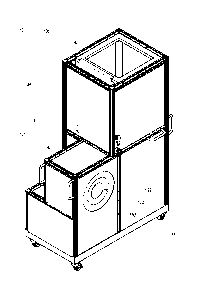
the volume of the working chamber 122 is approximately 12 liters.
[0068] The center opening of the frontmost antenna disk 170 defines an
entry
opening 169 for receiving a patient. The entry opening 169 is preferably
surrounded by a
19
CA 2936145 2019-04-05
protective ring 182 (shown in FIGS. 6 and 7) covering the surfaces of the
antenna disk 170 and
other portions of the working chamber 122. FIG. 10 is a view of the image
chamber unit 131
similar to that of FIG. 9, but with a patient support 120 and a catch basin
165 in place adjacent
the unit 131, and FIG. 11 is a view of the image chamber unit 131 similar to
that of FIG. 10 but
shown with an upper portion of a patient's head 19 inserted into the entry
opening. For comfort
and convenience, the patient may be positioned on the patient support 120,
which may be a
gurney, cart, table, stretcher, or the like. In at least some embodiments of
the present invention,
a headrest 118 extends from the end of the patient support 120. The headrest
118 is preferably
padded and adjustable. Adjustability of the headrest 118 may be provided in
one or more of the
longitudinal direction (toward or away from the end of the patient support
120), the vertical
direction (up or down relative to the patient support 120), and rotationally
(for example, about an
axis that is parallel with the end of the patient support 120). In the
illustrated embodiment, the
entry opening and the working chamber 122 are sized to correspond specifically
to a human
head, but it will be appreciated that other dimensions may be utilized for
other body parts or to
accommodate the entirety of a human body. The entry opening is substantially
liquid-sealed by
the membrane 133 such that the front of the working chamber 122 is separated
by the membrane
133 from the rear of the chamber 122. Fluid leaks through the front of the
working chamber 122,
such as around or through the membrane 133, may be captured in the catch basin
165 disposed in
front of the unit 131. It is contemplated that the catch basin 165 can be
integral with or
otherwise part of the image chamber unit 131.
[0069] FIGS. 12 and 13 are a rear isometric view and a rear plan view,
respectively,
of the membrane 133 of the image chamber unit 131 of FIG. 6, and FIG. 14 is a
side cross-
sectional view of the membrane 133 of FIG. 13, taken along line 14-14. The
membrane 133 is
preferably somewhat hat-shaped, with a center crown portion 127 extending
"upward" or
"inward" from an outer brim portion 128. The brim portion 128 is shaped to be
fastened to the
antenna disks 170 and may include apertures 129 for this purpose. As shown in
FIG. 14, the
crown portion 127 may be thinner than the brim portion 128 and is preferably
flexible enough to
wrap snugly around the patient's head 19, as shown in FIG. 11. In at least
some embodiments,
the membrane 133 is made of latex or similar material.
[0070] FIG. 15 is a view of the image chamber unit 131 similar to that
of FIG. 11 but
shown with a fluid disposed within the working chamber 122 on the opposite
side of the
CA 2936145 2019-04-05
membrane 133 from the patient's head 19. The fluid may be supplied to or from
the working
chamber 122 via the inlets 167,168, which may be arranged in or on the back
disk 183. The fluid
itself is a "matching" fluid that is chosen for its properties so as to
enhance the tomographic
process. Flow and other movement of the fluid is controlled by the hydraulic
system 140.
[0071] FIG. 16 is a schematic diagram of the hydraulic system 140 of
FIG. 8. As
shown therein, the hydraulic system 140 includes an external tank 141, a bi-
directional pump
142, a valve 159, backflow valve 160, a check (directional) valve 161, an
inner upper tank 146,
one or more liquid sensors 147, a lighter 148, one or more temperature sensors
149,150, and a
variety of hoses, tubes, fittings, and the like, some of which are described
herein. The external
tank 141 holds a quantity of a matching fluid. A hose 151 connects the
external tank 141 to the
pump 142, and another hose 152 connects the pump 142 to a fitting 153 on the
enclosure 134. In
at least some embodiments, the pump hoses 151,152 are 3/4" flexible tube
hoses, and the hose
fitting 153 is a quick release fitting.
[0072] The pump 142 is used to supply matching fluid from the external
tank 141 to
the working (image) chamber of the image chamber unit 131. The matching fluid
is a solution or
gel that is needed or useful inside the imaging chamber when the object 19 is
being measured
inside it to address electromagnetic body-matching problems. In at least some
embodiments, the
matching liquid is a mixture of glycerol (Ph. Eur.), water and brine. In at
least some
embodiments, the pump 142 is connected by cable 154 to a standard power
supply, such as a
220V electrical source, which may be provided from the control cabinet 135 via
an outlet 137,
preferably located on the outer surface of the enclosure 134, and a
corresponding water proof
socket 155. Direction, speed, and other control of the pump 142 may be
provided by remote
control 156. One pump 142 suitable for use in at least some preferred
embodiments is a Watson
Marlow 620 RE IP66 pump.
[0073] Inside the image chamber unit 131, another hose 157 is connected
between the
external fitting 153 and a first inlet 167 to the working chamber, and still
another hose 158 is
connected between a second inlet 168 to the working chamber and the inner
upper tank 146. In
at least some embodiments, the hose 157 is a 3/4" flexible tube hose. An
inline valve 159 may
optionally be provided in the hose 157 from the pump 134, while a backflow
valve 160 and
check (directional) valve 161 may be provided in the hose 158 to the inner
upper tank 146. The
backflow valve 160 provides at least two functions. First, when it is closed,
the pump 142 may
21
CA 2936145 2019-04-05
be used to generate an under-pressure, thereby denting in the membrane 133 (as
seen from
outside the image chamber unit 131) and readying the unit 131 for a patient's
head to be inserted
therein. Second, when the patient's head is positioned inside the membrane
133, opening the
backflow valve 160 allows the matching fluid to flow from the reservoir 146
back to the imaging
chamber, which in turn causes the patient's head to be slowly enclosed by the
membrane 133 and
the liquid. The check valve 161, on the other hand, performs a safety function
by avoiding the
buildup of an overpressure if the backflow valve 160 is closed. The check
valve 161 includes a
manual control lever 181, as shown in FIG. 6.
[0074] The temperature sensors 149,150 may be used to determine the
temperature of
the matching fluid inside the working chamber, or in close proximity thereto.
If the temperature
becomes uncomfortably cool, the lamp or lighter 148 may be utilized to trigger
heating of the
inner upper tank 146. Unintentional heating of an empty tank 146 may be
avoided by using the
liquid sensors 147 to verify that sufficient liquid is present in the tank.
[0075] An overfill path may be provided between the inner upper tank 146
and the
external tank 141 so as to return any excess matching liquid to the external
tank 141. The
overfill path may include an internal hose 162, an external hose 163, and a
fitting 164 on the
exterior of the enclosure 134, wherein the internal hose 162 is connected
between the inner upper
tank 146 and the fitting 164 and the external hose is connected between the
fitting 164 and the
external tank 141. Generally, the overfill path is only utilized if the
reservoir 146 is accidentally
overfilled, in which case the overfill path allows the excess liquid to return
to the external tank
141. In at least some embodiments, the overfill path hoses 162,163 are 3/4"
flexible tube hoses,
and the hose fitting 164 is a quick release fitting.
[0076] A leakage path may also be provided. The leakage path may include
a catch
basin 165 and a drain hose or tube 166. The catch basin 165 may be disposed
adjacent the
working chamber so as to receive fluid escaping therefrom, such as during
dismantling of the
system 110. In some embodiments, the drain hose 166 connects the catch basin
165 to the
external tank, such as by the overflow path, while in others the drain hose
166 is routed to a
waste tank (not shown) and/or is left open or unconnected.
[0077] FIG. 17 is a left front isometric view of portions of the disk
assembly 126 of
FIG. 9. As shown therein, the disk assembly 126 includes a plurality of
antenna disks 170
arranged concentrically such that their center openings define the interior of
the working
22
CA 2936145 2019-04-05
chamber 122, as described previously. Notably, whereas traditional EMT systems
have used
rings of transmitters/receivers/sensors that have been oriented in a
horizontal plane to define a
vertical working chamber, the rings of transmitter/receivers and receivers of
the present
invention are each oriented vertically so as to define a horizontal working
chamber. Each
antenna disk 170 includes a multitude of antennas 173 arranged in a ring
around the working
chamber 122. FIG. 18 is a schematic representation of these concentric rings
180 of antennas
173. Although other numbers of disks 170 and rings 180 may be utilized, five
antenna disks 170
and thus five antenna rings 180 are present in the embodiment shown in FIGS.
17 and 18.
Furthermore, although other numbers of antennas 173 may be utilized, 32
antennas 173 are
present in the embodiment shown in FIGS. 17 and 18, and thus a total of 160
antennas 173 are
utilized. In one embodiment, preferred for its simplicity, the antennas 173 in
the middle ring 180
are both transmitting and receiving antennas, while the antennas 173 on the
other four rings 180
are receiving antennas only. In one contemplated embodiment, the rings 180
(i.e., the center
openings of the antenna disks 170) are 285 mm in diameter. In FIG. 17,
transmitting/receiving
antenna "9" on ring "C" is shown as transmitting an electromagnetic field or
signal, all or some
of which is received at each of various transmitting/receiving antennas on
ring "C" and at each of
various receiving antennas on rings "A", "B", "D", and "E". It will be
appreciated, however,
that any or all of the transmitting/receiving antennas on ring "C" and/or any
or all of the
receiving antennas on any or all of the other rings may receive the
transmitted field or signal and
thus may be incorporated into the tomographic process.
[0078] FIG. 19 is a top cross-sectional view of the disk assembly 126 of
FIG. 17,
taken along line 19-19; FIG. 20 is a front view of one of the antenna disks
170 of FIG. 19, and
FIG. 21 is a top cross-sectional view of the antenna disk 170 of FIG. 20.
Notably, some visual
detail regarding the electrical connections for the antennas has been omitted
in FIG. 17; however,
much of the omitted visual detail is shown in FIG. 20. Each antenna disk 170
includes two
mating rings 171,172, the antennas 173 themselves, a corner element 174 for
each antenna 173, a
cable plate 175, and a cable assembly 176 for each antenna 173. Each cable
assembly 176
includes a cable and/or conduit with an appropriate terminator 177,178 on each
end. Screws or
other cable positioners 179 are provided to hold the cable assemblies 176 in
place.
[0079] FIG. 22 is a schematic diagram of the EMT system 110 of FIG. 6.
As shown
therein, the EMT system 110 includes the image chamber unit 131 (including the
working
23
CA 2936145 2019-04-05
chamber 122), the hydraulic system 140, the patient support 120, and a control
system 200. The
control system 200 includes two 16-channel transmitting/receiving switch units
201 for the
transmitting/receiving antenna disk 170, two 16-channel receiving switch units
202 for each of
the receiving antenna disks 170, a control unit 203, a network analyzer 204, a
power unit 205,
one or more fan units 206, a hub 207, and a user interface computer 208. In at
least some
embodiments, the switch units 201,202, control unit 203, network analyzer 204,
power unit 205,
fan units 206, and hub 207 are supported on a rack 209 in the control cabinet
135. The user
interface computer 208 may be supported on or in the enclosure 134 or may be
supported
elsewhere, such as on a nearby desk, a user's lap, or in some cases even
outside the room.
[0080] FIG. 23 is a schematic representation of the operation of the
rings 180 of
antennas 173 around the imaging domain, which is defined by the imaging
chamber. The
general task is to make complex Si,j,k parameters matrix measurement, where i
is the
transmitting antenna (i = 1...32), j is the receiving antenna (j = 1...31),
and k is the ring of the
receiving antenna (k = 1...5). The more practical case for the number of
receiving antennas that
are measured for each transmitting antenna may be between 12 and 20 (i.e.,
only receivers
generally opposite the transmitting antenna), and the most practical case may
be for 17 receiving
antennas to be measured for each transmitting antenna, but other numbers are
also viable.
Typical attenuations may be ¨90dB to ¨130dB. In at least some embodiments,
frequencies may
be 0.8-1.5 GHz, step 50 MHz. In at least some embodiments, channel-to-channel
isolation may
be ¨80dB to ¨100 dB. In at least some embodiments, maximum power output may be
+20dBm
(100 mW). In at least some embodiments, single frame data acquisition time may
be less than 60
mSec ("frame" being defined as the full cycle of S matrix measurements). In at
least some
embodiments, the number of acquired frames may be from 1 to 1000. In at least
some
embodiments, the dielectric properties of the matching media between antennas
and object may
be ¨(30-to-60)+j(15-to-25).
[0081] FIGS. 24A and 24B are a more detailed schematic diagram of the
control
system 200 of FIG. 22. As shown therein, the hub 207, which may provide both
wireless and
wired connections, communicatively connects the control unit 203, the network
analyzer 204,
and the user interface computer 208. The control unit 203 includes a host
controller that
interfaces with the hub 207 as well as provides a trigger input to the network
analyzer 204 and
receives "ready for trigger" and/or "busy" signals from the network analyzer
204. The host
24
CA 2936145 2019-04-05
controller also receives an ECG input and controls drivers for MW switches.
The control unit
203 also includes various circuitry, including amplifiers, multiplexers, and
the like, to generate
input signals for the ports of the network analyzer 204, which may be a ZVA 4
port vector
network analyzer available from Rohde & Schwarz. The network analyzer 204 is
also
communicatively connected to the hub 207, preferably via a LAN, and operations
of the control
unit 203 and network analyzer 204 are under the control of the user interface
computer 208.
Power is supplied by a power converter which may receive 24V power from the
power unit 205
as described elsewhere herein.
[0082] FIG. 25 is a schematic diagram of one of the
transmitting/receiving switch
units 201 of FIG. 22, and FIG. 26 is a schematic diagram of one of the
receiving switch units 202
of FIG. 22. FIG. 27 is a schematic diagram of the power unit 205 of FIG. 22.
As shown therein,
the AC line input is converted into power for the hub 207, the network
analyzer (VNA) 204, and
for 24V AC/DC converters used to power the control unit 203 and
transmitter/receiver and
receiver switch units 201,202. FIG. 28 is a schematic block diagram of
additional or alternative
details of a control system for the EMT system 110.
[0083] In operation, a patient 15 is placed on his back on a patient
support 120 and
transported to the image chamber unit 131, shown in FIG. 9, or the image
chamber unit 131 is
transported to the location of the patient 15. For sanitary purposes, a single-
use protective cap
(not shown) may be placed over the patient's head 19. Such a protective cap
may be made of
plastic, latex, or the like. The patient's head 19 is then inserted into the
entry opening 169 in the
working chamber 122 as shown in FIG. 11. The headrest 118 may be adjusted as
necessary or
desired to arrange the patient's head in the desired position and orientation
within the working
chamber 122. The patient's head 19 bears against the membrane 133, which then
conforms to
the shape of the patient's head 19. With the patient's head 19 properly
arranged, a technician
fills the working chamber with a quantity of the prepared matching liquid.
Filling may be
carried out using the remote control of the pump, which in at least some
embodiments has toggle
switches to start and stop the pump, control the direction of flow (in or
out), and flow rate.
Filling is preferably initiated at a low flow rate to avoid splashing of
matching liquid. Matching
liquid is pumped into the working chamber until it is full, as shown in FIG.
15.
[0084] In addition to filling the working chamber with the matching
liquid, the
technician may also power on the various electronic components, including the
control unit, the
CA 2936145 2019-04-05
network analyzer, transmitter and receiver units, and the like. Using the user
interface computer,
software may then be utilized to calibrate and operate the system.
Functionally, much of the
operation of the EMT system 110 may be similar to that described in the
aforementioned U.S.
Patent No. 7,239,731, U.S. Patent Application Publication No. 2012/0010493 Al
(U.S. Patent
Application Serial No. 13/173,078), and/or U.S. Patent Application Publication
No.
2014/0276012 Al (U.S. Patent Application Serial No. 13/894,395), but various
particular
embodiments and features thereof may be described herein. Measurements are
taken, a matrix of
complex data is generated, and various algorithms are used to transform such
data into
tomographic images of the interior of the patient's head 19.
[0085] Other embodiments of the present invention are likewise possible.
In
particular, EMT systems having components that are more easily transported
than those of the
system 110 described hereinabove are possible without departing from the scope
of the present
invention. In this regard, FIGS. 29 and 30 are a top front perspective view
and a bottom rear
perspective view, respectively, of another EMT system 210 for imaging a human
head 19 in
accordance with one or more preferred embodiments of the present invention.
The system 210
includes an image chamber unit 231, a control cabinet 235, and a hydraulic
system 240 for
supplying, circulating, and otherwise managing a matching fluid to the image
chamber unit 231.
The entire system 210 may be carried on a patient support 220, which again may
be a gurney,
cart, table, stretcher, or the like. In particular, the image chamber unit
231, which includes a
built-in headrest 218, is carried on a top surface of the patient support 220,
near one end, and the
control cabinet 235 is carried beneath the patient support 220. Such a system
210 may be more
conveniently transported, and in particular, the system 210 may be rolled with
the patient support
220 onto and off of an ambulance and into a medical facility. In this regard,
FIG. 31 is a top plan
view of the system 210 in use in an ambulance 211.
[0086] In at least some embodiments, an image chamber unit of a type
described
herein is man-portable. As used herein, "man-portable" means cable of being
carried or borne
by one human. In particular, an image chamber unit of a type described herein
may take the
form of a wearable hat, helmet, cap, or the like. FIG. 32 is a side
perspective view of a cap
serving as a wearable image chamber unit in accordance with one or more
preferred
embodiments of the present invention. Aspects of such wearable apparatuses may
be described,
for example, in U.S. Patent Application Serial No. 13/894,395.
26
CA 2936145 2019-04-05
[0087] At least some embodiments of the EMT systems presented herein,
including
without limitation the mobile embodiments such as the one presented in FIGS.
29-31 and the
wearable cap of FIG. 32, may be utilized advantageously outside of the
clinical setting. FIG. 33
is a pictorial illustration of a timeline for use of an EMT system, including
the cap of FIG. 32, for
imaging a human head in response to the onset of stroke symptoms in a patient.
As shown
therein, at 8:00 pm, a patient may be resting at home when he experiences the
onset of stroke-
like symptoms, such as disorientation and weakness in the face and arms. In
response, he or a
family member or friend contacts a medical provider, and an ambulance is
dispatched.
Meanwhile, a doctor or other medical practitioner is contacted and updated on
the situation. The
patient's head is placed in a mobile imaging unit, and scanning begins as
shown around 8:25 pm.
(In FIG. 33, the mobile image chamber unit is the cap of FIG. 32, but it will
be appreciated that
the unit of FIGS. 29-31 may be used instead.) Resulting data may be provided
to the doctor,
ambulance staff, imaging specialists, and other personnel. Some of the data
may be used directly
for diagnosis, treatment, or the like, while complex image-related data may be
processed
according to the systems and methods of the present invention to reconstruct
images from which
further diagnosis, treatment, or the like may be triggered. In at least some
embodiments, such
processing may generate an automatic alert that the data indicates that a
potential stroke is likely.
Notably, in at least some embodiments, such processing is carried out by a
third party service
provider who specializes in reconstruction of images according to the systems
and methods of
the present invention. During transport, from approximately 8:45 pm to 9:00
pm, the cap 331
continues to provide data regarding the patient's condition, and the local
hospital staff is further
updated and arranges and prepares for further treatment. Once the patient
arrives at the hospital
or other treatment center, the images and data may be used in providing
timely, accurate
information about the status of the stroke injury, and appropriate treatment
and follow-up may be
administered. Such a system could be utilized to provide the desired "under 3
hour" treatment
that can make a major difference in the final outcome of the stroke injury and
its affect on the
patient.
[0088] It will be appreciated that in at least some embodiments, the
systems,
apparatuses and methods presented hereinabove may be incorporated into a 4D
EMT differential
(dynamic) fused imaging system. 4D EMT differential (dynamic) fused imaging
system suitable
27
CA 2936145 2019-04-05
for use with one or more preferred embodiments of the present invention are
described in
Appendix B.
[0089] Based on the foregoing information, it will be readily understood
by those
persons skilled in the art that the present invention is susceptible of broad
utility and application.
Many embodiments and adaptations of the present invention other than those
specifically
described herein, as well as many variations, modifications, and equivalent
arrangements, will be
apparent from or reasonably suggested by the present invention and the
foregoing descriptions
thereof, without departing from the substance or scope of the present
invention.
[0090] Accordingly, while the present invention has been described
herein in detail in
relation to one or more preferred embodiments, it is to be understood that
this disclosure is only
illustrative and exemplary of the present invention and is made merely for the
purpose of
providing a full and enabling disclosure of the invention. The foregoing
disclosure is not
intended to be construed to limit the present invention or otherwise exclude
any such other
embodiments, adaptations, variations, modifications or equivalent
arrangements; the present
invention being limited only by the claims appended hereto and the equivalents
thereof.
28
CA 2936145 2019-04-05