Note : Les descriptions sont présentées dans la langue officielle dans laquelle elles ont été soumises.
ak 02938755 2016-08-12
IDENTIFYING AND PRESENTING SUSPECTED MAP SHIFTS
CROSS-REFERENCE TO RELATED APPLICATION
[0001] This application claims the benefit of U.S. Provisional
Patent Application 62/214,262, filed September 4, 2015 and U.S.
Patent Application 15/228,588, filed August 4, 2016, which are
incorporated herein by reference.
FIELD OF THE INVENTION
[0002] The present invention relates generally to medical imaging,
and specifically to a method for color-coding ablation locations
by their respective registrations.
BACKGROUND OF THE INVENTION
[0003] A wide range of medical procedures involves placing
objects, such as sensors, tubes, catheters, dispensing devices,
and implants, within the body. Real-time imaging methods are
often used to assist doctors in visualizing the object and its
surroundings during these procedures. In most situations,
however, real-time three-dimensional imaging is not possible or
desirable. Instead, systems for obtaining real-time spatial
coordinates of the internal object are often utilized.
[0004] U.S. Patent Application 2007/0016007, to Govari et al.,
whose disclosure is incorporated herein by reference, describes
a hybrid field-based and impedance-based location sensing
system. The system includes a probe adapted to be introduced
into a body cavity of a subject.
[0005] U.S. Pat. No. 6,574,498, to Gilboa, whose disclosure is
incorporated herein by reference, describes a system for
determining the position of a work piece within a cavity of an
opaque body. The system claims to use a transducer that
1
ak 02938755 2016-08-12
interacts with a primary field, and several transducers that
interact with a secondary field.
[0006] U.S. Pat. No. 5,899,860, to Pfeiffer, et al., whose
disclosure is incorporated herein by reference, describes a
system for determining the position of a catheter inside the
body of a patient. A correction function is determined from the
difference between calibration positions derived from received
location signals and known, true calibration positions,
whereupon catheter positions, derived from received position
signals, are corrected in subsequent measurement stages
according to the correction function.
[0007] Documents incorporated by reference in the present patent
application are to be considered an integral part of the
application except that to the extent any terms are defined in
these incorporated documents in a manner that conflicts with the
definitions made explicitly or implicitly in the present
specification, only the definitions in the present specification
should be considered.
[0008] The description above is presented as a general overview of
related art in this field and should not be construed as an
admission that any of the information it contains constitutes
prior art against the present patent application.
SUMMARY OF THE INVENTION
[0009] There is provided, in accordance with an embodiment of the
present invention a method, including performing a first
registration of a tracking system, which is configured to track
a location of a probe within an organ of a human body, with a
baseline coordinate system, measuring first locations of the
probe within the organ following the first registration,
presenting, on an image of the organ, first indicators marking
2
ak 02938755 2016-08-12
the first locations with a first visual effect, at positions on
the image that are determined in accordance with the first
registration, after measuring the first locations, performing a
second registration of the tracking system with the baseline
coordinate system, measuring second locations of the probe
within the organ following the second registration, and
presenting, on the image of the organ, second indicators marking
the second locations with a second visual effect, which is
visually distinct from the first visual effect, at positions on
the image that are determined in accordance with the second
registration.
[0010]
In embodiments of the present invention, the tracking
system and the baseline system are each selected from a group
consisting of a field-based location tracking system, an
impedance-based location tracking system and a medical imaging
system.
In some embodiments, the image includes a simulated
surface of the organ.
In additional embodiments, the first
visual effect includes a first color and the second visual
effect includes a second color different from the first color.
[0011]
In further embodiments, performing the first registration
includes identifying a relationship between the tracking system
and the baseline coordinate system, and the second registration
is performed upon detecting a change in the relationship.
In
supplementary embodiments, the probe includes a catheter, and
the organ includes a heart. In some embodiments, the locations
include ablation locations.
[0012]
There is also provided, in accordance with an embodiment of
the present invention an apparatus, including a tracking system
configured to track a location of a probe within an organ of a
human body, a baseline coordinate system, a display, and a
processor configured to perform a first registration of the
3
ak 02938755 2016-08-12
tracking system with the baseline coordinate system, to measure
first locations of the probe within the organ following the
first registration, to present, on the display, first indicators
on the organ marking the first locations with a first visual
effect, at positions on the image that are determined in
accordance with the first registration, after measuring the
first locations, to perform a second registration of the
tracking system with the baseline coordinate system, to measure
second locations of the probe within the organ following the
second registration, and to present, on the image of the organ,
second indicators marking the second locations with a second
visual effect, which is visually distinct from the first visual
effect, at positions on the image that are determined in
accordance with the second registration.
[0013]
There is further provided, in accordance with an embodiment
of the present invention, a computer software product for
sensing, using a baseline coordinate system and a tracking
system configured to track a location of a probe within an organ
of a human body, the product including a non-transitory
computer-readable medium, in which program instructions are
stored, which instructions, when read by a computer, cause the
computer to perform a first registration of the tracking system,
with the baseline coordinate system, to measure first locations
of the probe within the organ following the first registration,
to present, on an image of the organ, first indicators marking
the first locations with a first visual effect, at positions on
the image that are determined in accordance with the first
registration, after measuring the first locations, to perform a
second registration of the tracking system with the baseline
coordinate system, to measure second locations of the probe
within the organ following the second registration, and to
4
ak 02938755 2016-08-12
present, on the image of the organ, second indicators marking
the second locations with a second visual effect, which is
visually distinct from the first visual effect, at positions on
the image that are determined in accordance with the second
registration.
BRIEF DESCRIPTION OF THE DRAWINGS
[0014] The disclosure is herein described, by way of example only,
with reference to the accompanying drawings, wherein:
[0015] Figure 1 is a schematic pictorial illustration of a medical
system comprising multiple systems configured to track a
location of a catheter in a heart while performing an ablation
procedure, in accordance with an embodiment of the present
invention;
[0016] Figure 2 is a schematic pictorial illustration of the
catheter in the heart, in accordance with an embodiment of the
present invention;
[0017] Figure 3 is a flow diagram that illustrates a method of
presenting ablation locations in the heart, in accordance with
an embodiment of the present invention; and
[0018] Figure 4 is a schematic pictorial illustration of the
ablation locations presented on an electroanatomical map, in
accordance with an embodiment of the present invention.
ak 02938755 2016-08-12
DETAILED DESCRIPTION OF EMBODIMENTS
OVERVIEW
[0019] Various diagnostic and therapeutic procedures involve
mapping of the electrical potential on the inner surface of a
cardiac chamber. Electrical mapping can be performed, for
example, by inserting a medical probe (e.g., a catheter), whose
distal end is fitted with a position sensor and a mapping
electrode (also referred to herein as a probe electrode), into
the cardiac chamber. The cardiac chamber is mapped by
positioning the probe at multiple points on the inner chamber
surface. At each point, the electrical potential is measured
using the mapping electrode, and the distal end position is
measured using the position sensor. The measurements are
typically presented as a map of the electrical potential
distribution over the cardiac chamber surface.
[0020]
While positioning the medical probe within the cardiac
chamber, impedance-based and/or magnetic field-based (also
referred to herein as field-based) position sensing systems can
be used to determine a location of the probe within the cardiac
chamber.
In impedance-based location sensing systems, such as
those described in U.S. Patent 8,456,182, whose disclosure is
incorporated herein by reference, a set of adhesive skin patches
is affixed to a subject's body, and a distal end of a medical
probe (e.g., a catheter) is inserted into a body cavity of the
subject.
[0021]
The patches include respective electrodes in contact with a
surface of the subject. Typically the set of patches comprises
three or more patches. A control console delivers a current to
an electrode (also referred to herein as an impedance-based
location sensor) positioned at the distal end of the probe.
6
ak 02938755 2016-08-12
Each of the patches receives a portion of the current, and
conveys its respective received current back to the control
console.
From the received currents the control console can
determine a respective impedance between each of the patches and
the mapping electrode, and compute, based on the impedances,
impedance-based location coordinates for the distal end.
The
impedance-based location coordinates are three-dimensional
coordinates measured with respect to a frame of reference
defined by the patches, herein assumed to have impedance-based
coordinates, and enable the distal end to be tracked in this
frame of reference in the body cavity.
[0022]
In field-based position sensing systems, multiple magnetic
field generators may be positioned in the vicinity of the
subject. A field-based position sensor, also herein termed a
magnetic tracking sensor, is positioned at the distal end of the
probe, and the sensor conveys a probe signal to the control
console in response to the magnetic fields received from the
field generators.
Upon receiving the probe signal from the
tracking sensor, the control console can compute, based on the
probe signal, field-based probe location coordinates for the
distal end.
The field-based probe location coordinates are
three-dimensional coordinates with respect to a frame of
reference defined by field-based location coordinates of the
magnetic field generators, and also enable the distal end to be
tracked in the field-based frame of reference.
[0023]
Field-based position sensing systems are typically more
accurate than impedance-based location sensing systems.
For
example, field-based position sensing systems may be accurate to
within one millimeter while impedance-based position systems may
be accurate to within three millimeters.
However, field-based
7
ak 02938755 2016-08-12
systems are typically more costly than the impedance-based
systems.
[0024]
Medical systems, typically those using multiple probes
during a medical procedure, may incorporate into at least one of
the probes an electrode and a field-based position sensor. Such
a probe, herein termed a reference probe, may be used to map the
volume of the body cavity in both systems, and a correlation
between the two mappings may then be applied to other probes
having only mapping electrodes, the mapping electrodes being
used for tracking the probes in an impedance-based system. In
order that the impedance-based location coordinates of the
reference probe correspond to its field-based location
coordinates, the frames of reference of the two systems are
registered, so generating a relation between the two frames of
reference. Using the relation, inter alia, typically increases
the accuracy of the impedance-based system, as well as allowing
the electrode-only probes to be tracked in the field-based
system.
[0025]
During a medical procedure such as an ablation of tissue in
a heart, the heart may move due to the patient breathing even
though the patient is immobile. While breathing is overall a
cyclic process, the amplitude and period of the breathing
typically vary during a procedure. Such motions, including
motions of the "complete" patient, may be allowed for, for
example by positioning the reference probe in a fixed position
within the heart, and tracking location changes of the heart
with this probe.
[0026] Upon detecting motion of the patient during the procedure,
the multiple coordinate systems may need to be re-registered.
However, each re-registration may introduce errors into
locations that were measured prior to the re-registration.
In
8
ak 02938755 2016-08-12
instances where there are multiple re-registrations, errors in
values of earlier measured positions that were registered with
earlier registrations may be more significant than errors of
recently measured positions that were registered with more
recent registrations.
[0027]
Embodiments of the present invention provide methods and
systems for identifying one or more locations that may be more
liable to error, by presenting each of the locations using a
respective visual effect that is associated with its respective
registration.
In embodiments of the present invention, the
locations are measured by a control console comprising a
tracking system that is configured to track a location of a
probe within an organ of a human body.
[0028]
As explained hereinbelow, a first registration is performed
between the tracking system and a baseline coordinate system.
Examples of tracking and baseline coordinate systems include,
but are not limited to, field-based location tracking systems,
impedance-based location tracking systems and medical imaging
systems.
[0029]
Subsequent to the first registration, the control console
measures first locations of the probe within the organ, and
presents, on an image of the organ, first indicators marking the
first locations with a first visual effect, at positions on the
image that are determined in accordance with the first
registration.
After measuring the first locations, a second
registration is performed between the tracking system and the
baseline coordinate system, and upon measuring second locations
of the probe within the organ, the control console presents, on
the image of the organ, second indicators marking the second
locations with a second visual effect, which is visually
9
ak 02938755 2016-08-12
distinct from the first visual effect, at positions on the image
that are determined in accordance with the second registration.
[0030]
In embodiments where the visual effects comprise different
colors, the locations can be "color-coded" by presenting the
first locations measured using a first color, and presenting the
second locations using a second color.
By color-coding the
locations, systems implementing embodiments of the present
invention enable a physician to decide whether or not to re-
measure any particular location.
SYSTEM DESCRIPTION
[0031]
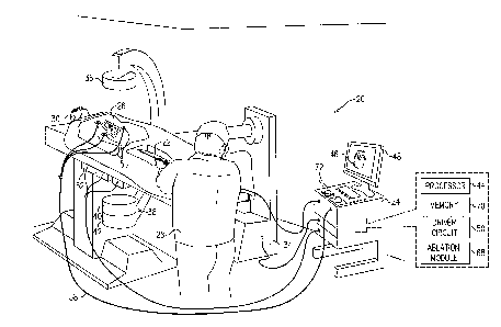
Figure 1 is a schematic pictorial illustration of a medical
system 20 comprising a medical probe 22 and a control console
24, and Figure 2 is a schematic illustration of the medical
probe inside a chamber of a heart 26, in accordance with an
embodiment of the present invention.
System 20 may be based,
for example, on the CARTO system, produced by Biosense Webster
Inc. (Diamond Bar, California).
In embodiments described
hereinbelow, it is assumed that probe 22 is used for diagnostic
or therapeutic treatment, such as performing ablation of heart
tissue in heart 26.
Alternatively, probe 22 may be used,
mutatis mutandis, for other therapeutic and/or diagnostic
purposes in the heart or in other body organs.
[0032]
An operator 28 inserts probe 22 through the vascular system
of a patient 30 so that a distal end 32 (Figure 2) of probe 22
enters a chamber of heart 26.
In the configuration shown in
Figure 1, operator 28 uses a fluoroscopy unit 34 to visualize
distal end 32 inside heart 26. Fluoroscopy unit 34 comprises an
X-ray source 36, positioned above patient 30, which transmits X-
rays through the patient. A flat panel detector 38, positioned
below patient 30, comprises a scintillator layer 40 which
ak 02938755 2016-08-12
converts the X-rays which pass through patient 30 into light,
and a sensor layer 42 which converts the light into electrical
signals. Sensor layer 42 typically comprises a two dimensional
array of photodiodes, where each photodiode generates an
electrical signal in proportion to the light detected by the
photodiode.
[0033]
Control console 24 comprises a processor 44 that converts
the signals from fluoroscopy unit 34 and probe 22 into an
electroanatomical map 46 (also referred to herein as image 46),
which comprises information regarding the procedure that the
processor presents on a display 48. Display 48 is assumed, by
way of example, to comprise a cathode ray tube (CRT) display or
a flat panel display such as a liquid crystal display (LCD),
light emitting diode (LED) display or a plasma display. However
other display devices can also be employed to implement
embodiments of the present invention.
In some embodiments,
display 48 may comprise a touchscreen configured to accept
inputs from operator 28, in addition to presenting
electroanatomical map 46.
[0034]
System 20 can use field-based position sensing to determine
position coordinates of distal end 32 inside heart 26.
In
configurations where system 20 uses field-based based position
sensing, console 24 comprises a driver circuit 50 which drives
field generators 52 to generate magnetic fields within the body
of patient 30. Typically, field generators 52 comprise coils,
which are placed below the patient at known positions external
to patient 30.
These coils generate magnetic fields in a
predefined working volume that contains heart 26. A magnetic
field sensor 54 (also referred to herein as position sensor 54)
within distal end 32 of probe 22 generates electrical signals in
response to the magnetic fields from the coils, thereby enabling
11
ak 02938755 2016-08-12
processor 44 to determine the position of distal end 32 within
the cardiac chamber.
Magnetic field-based position tracking
techniques are described, for example, in U.S. Patents
5,391,199, 6,690,963, 5,443,489, 6,788,967, 5,558,091, 6,172,499
and 6,177,792, whose disclosures are incorporated herein by
reference.
[0035]
In an alternative embodiment, the roles of location sensor
54 and magnetic field generators 52 may be reversed. In other
words, driver circuit 50 may drive a magnetic field generator in
distal end 32 to generate one or more magnetic fields.
The
coils in ach field generator 52 may be configured to sense the
fields and generate signals indicative of the amplitudes of the
components of these magnetic fields. Processor 44 can receive
and process these signals in order to determine the position
coordinates of distal end 32 within heart 26.
[0036]
During a medical procedure, an array of adhesive skin
patches 56 are affixed to patient 30. In other words, each of
the skin patches in the array is affixed to a surface of the
body of patient 30. At least one of the patches comprises one
or more magnetic field sensors (e.g., coils) that can measure
the magnetic fields produced by field generators 52, and
responsively convey the magnetic field measurements to console
24.
Based on the magnetic measurements received from the
magnetic field sensors (also referred to herein as patch
sensors) in a given patch 56, processor 44 can determine a
current field-based position, relative to the field generators,
of the given skin patch.
[0037]
Each patch 56 also comprises a body surface electrode in
contact with the surface of the body, and console 24 is
connected by a cable 58 to the body surface electrodes. In the
configuration shown in Figure 2, distal end 32 is enveloped by
12
ak 02938755 2016-08-12
an insulating exterior surface 60, and comprises a probe
electrode 62 that typically comprises one or more thin metal
layers formed over the insulating exterior surface at a distal
tip 64 of probe 22.
[0038]
In operation, processor 44 can determine impedance-based
location coordinates of distal end 32 inside heart 26 based on
the impedances measured between patches 56 and probe electrode
62. Impedance-based position tracking techniques are described,
for example, in U.S. Patents 5,983,126, 6,456,864 and 5,944,022,
whose disclosures are incorporated herein by reference.
In
embodiments of the present invention, processor 44 can use a
distance 66 between position sensor 54 and electrode 62 when
performing a registration between the position-dependent
magnetic signals received from the magnetic field sensor and
position-dependent electrical signals (i.e.,
impedance
measurements) received from patches 56.
[0039]
In some embodiments, system 20 can use electrode 62 for
both impedance-based location sensing and for other activities
such as potential acquisition for electrical mapping of the
heart, or ablation. Console 24 also comprises a radio frequency
(RF) ablation module 68 that delivers electrical power to
electrode 62. Processor 44 uses ablation module 68 to monitor
and control ablation parameters such as the level of ablation
power applied via electrode 62.
Ablation module 68 may also
monitor and control the duration of the ablation that is
provided.
[0040]
Processor 44 receives and processes the signals generated
by position sensor 54 in order to determine field-based position
coordinates of distal end 32, typically including both field-
based location and field-based orientation coordinates.
In
addition to determining the field-based coordinates of distal
13
ak 02938755 2016-08-12
end 32, processor 44 can also receive and process impedances
from patches 56 in order to determine impedance-based location
coordinates of the distal end. The method of position sensing
described hereinabove is implemented in the above-mentioned
CARTO system and is described in detail in the patents and
patent application cited above.
[0041]
Based on the signals received from probe 22 and other
components of system 20, processor 44 drives display 48 to
update map 46, so as to present a current position of distal end
32 in the patient's body, as well as to present status
information and guidance regarding the procedure that is in
progress.
Processor 44 stores data representing map 46 in a
memory 70. In some embodiments, operator 28 can manipulate map
46 using one or more input devices 72.
In embodiments, where
display 48 comprises a touchscreen display, operator 28 can
manipulate map 46 via the touchscreen display.
[0042]
In the configuration shown in Figure 2, probe 22 also
comprises a force sensor 74 contained within distal end 32.
Force sensor 74 measures a force F applied by distal tip 64 on
endocardial tissue 76 of heart 26 by generating a signal to the
console that is indicative of the force exerted by the distal
tip on the endocardial tissue.
While performing an ablation,
processor 44 can measure force F to verify contact between
distal tip 64 and endocardial tissue 76.
[0043]
In one embodiment, force sensor 74 may comprise a magnetic
field transmitter and receiver connected by a spring in distal
tip 64, and may generate an indication of the force based on
measuring the deflection of the spring. Further details of this
sort of probe and force sensor are described in U.S. Patent
Application Publications 2009/0093806 and 2009/0138007, whose
disclosures are incorporated herein by
reference.
14
ak 02938755 2016-08-12
Alternatively, distal end 32 may comprise another type of force
sensor.
[0044] Processor 44 typically comprises a general-purpose
computer, with suitable front end and interface circuits for
receiving signals from probe 22 and controlling the other
components of console 24.
Processor 44 may be programmed in
software to carry out the functions that are described herein.
The software may be downloaded to console 24 in electronic form,
over a network, for example, or it may be provided on non-
transitory tangible media, such as optical, magnetic or
electronic memory media.
Alternatively, some or all of the
functions of processor 44 may be carried out by dedicated or
programmable digital hardware components.
PRESENTING RE-REGISTERED MAP POINTS
[0045]
The following description assumes, by way of example, that
an ablation procedure is performed on patient 30. Those having
ordinary skill in the art will be able to adapt the description,
mutatis mutandis, for other procedures such as a cardiac chamber
mapping procedure. In the ablation procedure, processor 44 can
generate electroanatomical map 46 comprising map points
comprising ablation locations collected from probe 22. Each map
point may comprise a respective coordinate within a body cavity,
and possibly a physiological property collected by probe 22 at
the respective coordinate.
While the description referencing
Figures 3 and 4 hereinbelow assumes map points 110 represent
ablation locations in heart 26, map points representing any
other types of locations mapped by probe 22, such as a location
on a surface, in any organ of patient 30 are considered to be
within the spirit and scope of the present invention.
ak 02938755 2016-08-12
[0046]
Figure 3 is a flow diagram that illustrates a method of
identifying and presenting shifts in map 46, and Figure 4 is a
schematic pictorial illustration of the electroanatomical map
comprising map points 110 (also referred to herein as
indicators), in accordance with an embodiment of the present
invention.
In Figure 4, map points 110 are differentiated by
appending a letter to the identifying numeral, so that the map
points comprise map points 110A-11OG.
[0047]
In a construction step 80, processor 44 constructs a
simulated surface of the cardiac chamber. In some embodiments,
processor 44 constructs the simulated surface based on
additional map points (not shown), typically three-dimensional
mapping points of the surface, previously collected from probe
22.
In a presentation step 82, the processor presents, on
display 48, electroanatomical map 46 comprising the simulated
surface.
[0048]
In an insertion step 84, operator 28 inserts distal end 32
of probe 22 into the cardiac chamber. As operator 28 maneuvers
probe 22, processor 44 determines current impedance based
location coordinates for distal end 32 based on an impedance
between patches 56 and electrode 62, and determines field-based
location coordinates for the distal end 32 based on signals
received from position sensor 54.
In an initial registration
step 86, processor identifies a relationship between the
impedance-based location coordinates and the field-based
location coordinates, and uses the relationship to register the
impedance-based location coordinates to the field-based location
coordinates.
A method for determining a relationship such as
that referred to above is provided in U.S. Patent 8,456,182, to
Bar-Tal et al., which is incorporated herein by reference.
16
cik 02938755 2016-08-12
[0049]
During the medical procedure, as processor 44 collects map
points 110, the processor may detect a change in the current
relationship between the impedance-based location coordinates
and the field-based location coordinates of distal end 32. Such
a change may be caused, for example, by movement of patient 30.
Upon detecting the change in the relationship, processor 44 can
re-register the impedance-based location coordinates to the
field-based location coordinates. In embodiments of the present
invention, upon re-registering the impedance-based location
coordinates to the field-based location coordinates, processor
44 changes the color used to present ablation locations
collected subsequent to the re-registration.
Therefore, when
initializing system 20, processor 44 can define a set of colors
that can be used to present the ablation locations on display
48.
[0050]
In a first assignment step 88, processor 44 assigns, from
the set of colors, an initial color to a display color.
As
operator 28 uses probe 22 to perform an ablation on intracardiac
tissue 76, in a receive step 90, processor 44 receives, from
probe 22, measurements indicating a given ablation location
marked by a given map point 110.
In some embodiments, the
measurements comprise impedances between patches 56 and
electrode 62, and processor 44 can use the registration to
determine or measure (i.e., find coordinates for) the given map
point.
[0051]
In a presentation step 92, processor 44 presents, on
display 48, electroanatomical map 46 comprising a fusion of the
simulated surface and the given ablation location.
In
embodiments of the present invention, processor 44 calculates
the given ablation location using the (current) registration,
and presents the given ablation location in heart 26 using the
17
ak 02938755 2016-08-12
assigned display color. In a first comparison step 94, if the
ablation procedure is not complete, then in a second comparison
step 96, processor 44 checks to see if the current registration
is still valid.
[0052]
If processor 44 detects a change in the relationship
between the impedance-based location coordinates and the field-
based location coordinates, then the current registration is not
valid, and in a re-registration step 98, the processor uses the
changed relationship to re-register the impedance-based location
coordinates to the field-based location coordinates.
In a
second assignment step 100, processor 44 processor 44 assigns,
from the set of colors, a previously unassigned color to the
display color, and the method continues with step 92.
[0053]
Returning to step 96, if the current registration is still
valid then the method continues with step 90. Returning to step
94, if the ablation procedure is complete, then the method ends.
[0054]
In embodiments of the present invention, as described in
the description referencing Figure 3 hereinabove, processor 44
assigns a unique visual effect (e.g., a given color from the set
of colors) to the ablation locations collected with each given
registration.
In the example shown in Figure 4, processor 44
collects ablation locations 110A-110C using a first registration
and presents locations 110A-110C in map 46 using a first visual
effect, collects ablation locations 110D and 110E using a second
registration and presents locations 110D and 110E in the
electroanatomical map using a second visual effect, and collects
ablation locations 110E-110H using a third registration and
presents locations 110E-110H in the electroanatomical map using
a third visual effect.
In the example shown in Figure 4, the
visual effects comprise "fill" (also known as "hatch") patterns.
In additional embodiments, processor 44 can use other types of
18
ak 02938755 2016-08-12
visual effects such as color, intensity, size of indicators 110,
and blinking attributes to differentiate between ablation
locations measured with different registrations.
[0055] The example in the description referencing Figure 3
hereinabove describes presenting ablation locations received
from a tracking system comprising an impedance-based location
tracking system that is registered to a baseline coordinate
system comprising a field-based location tracking system.
Presenting map points collected using other types of tracking
systems that are registered to other types of baseline
coordinate systems is considered to be within the spirit and
scope of the present invention.
For example, each of the
tracking and baseline coordinate systems may comprise an
impedance-based tracking system, a field-based tracking system,
or a medical imaging system such as fluoroscopy unit 34.
Therefore in the configuration shown in Figures 1 and 2, the
impedance-base location coordinates may be registered to field-
based location coordinates (or vice-versa), the impedance-based
location coordinates may be registered to image-based location
coordinates in electroanatomical map 46 (or vice-versa) that
processor 44 generates using image data received from
fluoroscopy unit 34, or the field-based location coordinates can
be registered to the image-based location coordinates (or vice-
versa). Examples of additional medical imaging systems that may
be configured as tracking systems and/or baseline coordinate
systems include, but are not limited to ultrasonic imaging
systems or a magnetic resonance imaging (MRI) systems and
computed tomography (CT) imaging systems.
[0056]
It will be appreciated that the embodiments described above
are cited by way of example, and that the present invention is
not limited to what has been particularly shown and described
19
CA 02938755 2016-08-12
hereinabove.
Rather, the scope of the present invention
includes both combinations and subcombinations of the various
features described hereinabove, as well as variations and
modifications thereof which would occur to persons skilled in
the art upon reading the foregoing description and which are not
disclosed in the prior art.