Note : Les descriptions sont présentées dans la langue officielle dans laquelle elles ont été soumises.
CA 02940602 2016-08-30
Adding a Tracking Sensor to a Rigid Tool
COPYRIGHT NOTICE
[0001] A portion of the disclosure of this patent document contains mate-
rial that is subject to copyright protection. The copyright owner has no
objection
to the facsimile reproduction by anyone of the patent document or the patent
disclosure, as it appears in the Patent and Trademark Office patent file or
rec-
ords, but otherwise reserves all copyright rights whatsoever.
CROSS-REFERENCE TO RELATED APPLICATIONS
[0002] This Application claims the benefit of U.S. Application No.
15/212,539 filed July 18, 2016, and U.S. Provisional Application No.
62/221,367
filed September 21, 2015, which are herein incorporated by reference. The sec-
ond noted Application shares disclosure with commonly assigned copending
Application Ser. No. 14/986,179, entitled Adjustable Tracking Sensor Suitable
For
Different Rigid Tools.
BACKGROUND OF THE INVENTION
1. Field of the Invention.
[0003] This invention relates to instruments for performing medical ex-
aminations of the interior of cavities of the body. More particularly, this
inven-
tion relates to mounting sensors on a probe for determining the position of
the
probe in the body.
2. Description of the Related Art.
[0004] In a surgical procedure the distal end of a rigid tool used in the
procedure, such as an endoscope, may be tracked by fixedly incorporating a
location sensor in the tool. A typical surgical procedure where such a tool is
used comprises an investigative ENT (Ear, Nose and Throat) procedure. The
sensor may be located at the distal end of the tool; alternatively, the sensor
may
be located away from the distal end, towards the proximal end of the tool. In
the
latter case, since the sensor is in a fixed position, compensation for the dis-
1 of 14
CA 02940602 2016-08-30
placement of the sensor from the distal end may be applied to the sensor's
measured location.
[0005] However, a number of rigid tools used in surgery do not have lo-
cation sensors fixedly incorporated into the tool, and while such tools may be
tracked by other means, such as fluoroscopy, this type of tracking is
typically
more inconvenient than tracking using a location sensor on the tool.
SUMMARY OF THE INVENTION
[0006] Embodiments of the present invention overcome the problem as-
sociated with tools that do not have sensors incorporated in the tools by
provid-
ing a sensor that is attached to the tool at any convenient position on the
tool.
Once attached, the displacement of the attached sensor from the distal end of
the tool is automatically measured, and is incorporated into the readings of
the
attached sensor.
[0007] There is provided according to embodiments of the invention a
method, which is carried out by receiving a computerized tomography scan of a
patient, registering an image of the patient derived from the scan with a
magnet-
ic tracking system configured to track a sensor in proximity to the patient,
mounting the sensor on a probe, positioning the distal end of the probe in con-
tact with the patient. The method is further carried out by while the distal
end is
in contact with the patient using data of the registered image to determine a
vec-
tor representing a translation from the sensor to the distal end, and while
track-
ing the sensor with the magnetic tracking system, adding the vector to a
location
of the sensor to determine a position of the distal end.
[0008] According to a further aspect of the method, mounting the sensor
comprises providing a carriage that conforms to a diameter of the probe, plac-
ing the sensor on the carriage, and attaching the carriage to the probe.
[0009] According to an additional aspect of the method, mounting the
sensor is performed by sliding the carriage onto the probe.
[0010] According to a further aspect of the method, the carriage has a
hole formed therein that is dimensioned to an outer diameter of the probe, and
sliding the probe through the hole.
2 of 14
CA 02940602 2016-08-30
[0011] According to yet another aspect of the method, the carriage is fix-
edly mounted on the probe.
[0012] In still another aspect of the method, a calibration includes deter-
mining a first vector corresponding to a first translation between a location
of
the sensor and a point on the longitudinal symmetry axis of the probe, and de-
termining a second vector corresponding to a second translation between the
point and the distal end of the probe, and adding the first vector and the
second
vector to establish a displacement vector.
[0013] Another aspect of the method includes determining a length of the
second translation by measuring a displacement on the image between the
point and another point wherein the other point is determined by detecting a
predetermined change in the data of the registered image.
[0014] According to still another aspect of the method, the data of the
registered image comprise voxel values that indicate radiodensity.
[0015] According to yet another aspect of the method, the data of the
registered image comprise Hounsfield units and determining a length of the
second translation includes measuring the predetermined change in the Houns-
field units.
[0016] According to one aspect of the method, registering an image in-
cludes disposing a plurality of magnetic field radiators in contact with the
pa-
tient, radiating alternating magnetic fields at respective frequencies from
the
magnetic field radiators, detecting the magnetic fields in the sensor, and
analyz-
ing the detected magnetic fields to derive the location and an orientation of
the
sensor with respect to the magnetic field radiators.
[0017] In another aspect of the method the magnetic field radiators are
mounted on a frame and the frame is placed in contact with the patient.
[0018] An additional aspect of the method includes deriving the location
and the orientation of the sensor with respect to anatomic features of the
patient
based on the registered image.
[0019] According to a further aspect of the method, registering an image
includes receiving a computerized tomographic image of the patient.
[0020] There is further provided according to embodiments of the inven-
tion an apparatus, including a carriage dimensioned to a diameter of a
cylindri-
3 of 14
CA 02940602 2016-08-30
cal probe and configured to rigidly fasten onto the shaft of the probe, a
location
sensor disposed in the carriage and responsive to a plurality of magnetic
fields
at respective frequencies, and a processor operative to compute a position of
the distal end of the probe responsively to signals from the location sensor.
[0021] Another aspect of the apparatus includes a frame having a plurali-
ty of magnetic field radiators mounted thereon for producing the plurality of
magnetic fields, wherein the processor is operative to register an image of a
pa-
tient with the magnetic field radiators.
[0022] According to one aspect of the apparatus, the processor is opera-
tive for computing the position of the distal end of the probe with respect to
co-
ordinates on the registered image.
BRIEF DESCRIPTION OF THE SEVERAL VIEWS OF THE DRAWINGS
[0023] For a better understanding of the present invention, reference is
made to the detailed description of the invention, by way of example, which is
to
be read in conjunction with the following drawings, wherein like elements are
given like reference numerals, and wherein:
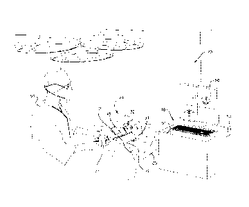
[0024] Fig. 1 is a schematic illustration of a nasal sinus surgery system,
according to an embodiment of the present invention;
[0025] Fig. 2 is a schematic illustration of a magnetic field radiation as-
sembly used in the surgery system, according to an embodiment of the present
invention;
[0026] Fig. 3A is a schematic cross-sectional side view of a probe; Fig. 3B
is a schematic cross-sectional front view of the probe; Fig. 3C is a schematic
di-
agram illustrating vectors related to the probe, according to an embodiment of
the present invention;
[0027] Fig. 4 is a flowchart of steps that are implemented in the operation
of the surgery system, according to an embodiment of the present invention;
and
[0028] Fig. 5 is a schematic illustration of a screen used during imple-
mentation of the flowchart, according to an embodiment of the present inven-
tion.
4 of 14
CA 02940602 2016-08-30
DETAILED DESCRIPTION OF THE INVENTION
[0029] In the following description, numerous specific details are set
forth in order to provide a thorough understanding of the various principles
of
the present invention. It will be apparent to one skilled in the art, however,
that
not all these details are necessarily needed for practicing the present
invention.
In this instance, well-known circuits, control logic, and the details of
computer
program instructions for conventional algorithms and processes have not been
shown in detail in order not to obscure the general concepts unnecessarily.
[0030] Documents incorporated by reference herein are to be consid-
ered an integral part of the application except that, to the extent that any
terms
are defined in these incorporated documents in a manner that conflicts with
def-
initions made explicitly or implicitly in the present specification, only the
defini-
tions in the present specification should be considered.
[0031] Turning now to the drawings, reference is now made to Fig. 1,
which is a schematic illustration of a nasal sinus surgery system 20, and to
Fig. 2,
which is a schematic illustration of a magnetic field radiation assembly 24
used
in the system, according to an embodiment of the present invention. System 20
is typically used during an invasive and/or investigative procedure on a nasal
sinus of a patient 22.
[0032] For the procedure, assembly 24 may be positioned beneath the
head of the patient, for example by fixing the assembly to a bed 25 upon which
the patient is lying. Assembly 24 comprises five magnetic field radiators 26,
which by way of example are fixed in a horseshoe shaped frame, the frame be-
ing positioned beneath the patient so that the radiators surround the head of
pa-
tient 22. Radiators 26 are configured to radiate alternating magnetic fields
at re-
spective frequencies into a region 30, in proximity to assembly 24 and which
in-
cludes the head of patient 22. The alternating magnetic fields induce signals
in a
sensor 32, typically a set of three orthogonal coils, and the signals may be
ana-
lyzed to derive the location and orientation of the sensor with respect to
assem-
bly 24. It will be understood that the location and orientation of sensor 32
may
be determined for substantially any positioning of the sensor within region
30.
5 of 14
CA 02940602 2016-08-30
[0033] As is described in more detail below, sensor 32 is affixed to a rig-
id probe 28, and determination of the location and orientation of the sensor
ena-
bles the location and orientation of a distal end 34 of the probe, that may be
in-
serted into the nasal sinus of the patient, to be tracked. A system using
magnetic
field radiators, such as radiators 26, for tracking an entity inserted into a
patient
is described in US Patent Application 14/792,823, to Govari et al., which is
in-
corporated herein by reference. In addition, the Carto system produced by
Biosense Webster, of Diamond Bar, CA, uses a tracking system similar to that
described herein for finding the location and orientation of a coil in a
region ir-
radiated by magnetic fields.
[0034] Elements of system 20, including radiators 26, may be controlled
by a system processor 40, comprising a processing unit communicating with
one or more memories. Typically the elements may be connected by cables to
the processor, for example, radiators 26 may be connected by a cable 58 to
processor 40. Alternatively or additionally, the elements may be coupled wire-
lessly to the processor. Processor 40 may be mounted in a console 50, which
comprises operating controls 51 that typically include a keypad and/or a point-
ing device such as a mouse or trackball. Console 50 also connects to other ele-
ments of system 20, such as a proximal end 52 of probe 28. A physician 54 uses
the operating controls to interact with the processor while performing the pro-
cedure, and the processor may present results produced by system 20 on a
screen 56.
[0035] Processor 40 uses software stored in a memory of the processor to
operate system 20. The software may be downloaded to processor 40 in elec-
tronic form, over a network, for example, or it may, alternatively or
additionally,
be provided and/or stored on non-transitory tangible media, such as magnetic,
optical, or electronic memory.
[0036] Fig. 3A is a schematic cross-sectional side view of probe 28, Fig.
3B is a schematic cross-sectional front view of the probe, and Fig. 3C is a
sche-
matic diagram illustrating vectors related to the probe, according to an embod-
iment of the present invention. In the following description of probe 28, the
probe is assumed to comprise a rigid cylinder 60, having a longitudinal sym-
6 of 14
CA 02940602 2016-08-30
metry axis 62. In Figs. 3A and 3B the probe has been drawn on a set of xyz or-
thogonal axes, with longitudinal symmetry axis 62 defining the z-axis. For
clari-
ty, in Figs. 3A and 3B the xyz axes of the probe are drawn displaced from
cylin-
der 60.
[0037] Sensor 32 is fixed to cylinder 60 by a sensor holder 64, which is
typically formed from a plastic so as to completely encapsulate the sensor. As
explained herein, signals from the sensor, generated in response to the magnet-
ic fields interacting with the sensor, are used to determine a location and an
ori-
entation of the sensor. Conducting wires that convey the signals from the
sensor
may be connected to proximal end 52 of probe 28, and from there to console 50.
The conducting wires are not shown in Figs. 3A and 3B.
[0038] The sensor is assumed to have a sensor direction 70, typically, but
not necessarily, the direction of an internal axis of symmetry of the sensor,
and
the orientation referred to herein measures the orientation of the sensor
direc-
tion with respect to a frame of reference defined by radiators 26. Sensor
direc-
tion 70 of sensor 32 is shown schematically in Figs. 3A and 3C as an arrow.
[0039] Sensor holder 64 is produced to have a hole 68, which is formed
to have a diameter substantially the same as that of cylinder 60, but
sufficiently
different so that there is a sliding fit between the holder and cylinder 60.
When
holder 64 is produced, a center of hole 68 is made to be a known distance A
from sensor 32. A typical value of A is 0.5 cm, but A may be smaller or larger
than this value. A series of sensor holders may be constructed, having holes
68
that are dimensioned to tools having different diameters. In addition, by
virtue
of being comprised in holder 64, the center of hole 68 has a known orientation
0
with respect to sensor direction 70. There is thus a known displacement vector
(A, 0), herein also termed vector V, from sensor 32 to the center of hole 68,
as
shown in Fig. 3C.
[0040] Hole 68 has an axis of symmetry 69 that is typically orthogonal to
vector V, and which by virtue of being formed when holder 64 is produced, has
a known direction 4) with respect to vector V (Fig. 3C).
[0041] As is also described below, in operating system 20 hole 68 of sen-
sor holder 64 is slid onto cylinder 60, and the holder is fixed to the
cylinder
when the holder is close to proximal end 52. It will be understood that in
sliding
7 of 14
CA 02940602 2016-08-30
cylinder 60 within hole 68 axes 69 and 62 are coincident, and also coincide
with
direction 4). Holder 64 comprises a setscrew 72, having a head, which may be
grasped by physician 54. Using the head the professional is able to hand-
tighten
the setscrew to fix holder 64 at a desired position along cylinder 60. The dis-
tance from the center of sensor 32 to distal end 34 is assumed to be a
distance B.
Unlike distance A, distance B is not known when sensor holder 64 is fixed to
cyl-
inder 60, but as is described below, in operation of system 20 processor 40 is
able to calculate distance B.
[0042] Fig. 4 is a flowchart of steps that are implemented in the operation
of system 20, and Fig. 5 is a schematic illustration of screen 56 during
implemen-
tation of the flowchart, according to an embodiment of the present invention.
The steps of the flowchart are also illustrated by Figs. 3A, 3B, and 3C.
[0043] In an initial step 100, the head of patient 22 is scanned by comput-
erized tomography (CT), herein by way of example assumed to be fluoroscopic
CT, and the CT data from the scan is acquired by processor 40. The CT scan of
patient 22 may be performed independently of the implementation of the re-
maining steps of the flowchart, which correspond to the sinus surgery proce-
dure. Typically, step 100 may be performed a number of days before the follow-
ing surgery steps of the procedure.
[0044] In a first procedure step 102, radiation assembly 24 is mounted
beneath the head of patient 22. Radiators 26 are then operated, and in a regis-
tration step 104 a frame of reference of the radiators is registered with the
frame
of reference of the subject's head. The registration is typically by any means
known in the art, e.g., by placing a magnetic field sensor coil, or a grouping
of
such coils, in one or more known locations and orientations with respect to
the
external features of the patient as well as with the frame holding the
radiators.
[0045] In an initial display step 106, processor 40 generates a represen-
tation 150, also referred to herein as image 150, of external features of the
pa-
tient, using the CT data received in step 100. The CT data is in the form of
voxels
with Hounsfield units (HU), and it will be appreciated that image 150 of the
ex-
ternal features of patient 22 can be generated from voxel values and their HU
values. Processor 40 displays image 150 on screen 56, and Fig. 5 schematically
illustrates the image as displayed on the screen.
8 of 14
CA 02940602 2016-08-30
[0046] In an operation step 108, the physician slides hole 68 of sensor
holder 64 onto rigid cylinder 60 of probe 28, and the physician then uses set-
screw 72 to lock the sensor holder in place, near proximal end 52 of the
probe.
Once the holder is locked in place, the physician brings distal end 34 of the
probe into contact with a selected region of the external features of the
patient,
for example a region at the side of the patient's nose.
[0047] The positioning of the distal end of necessity brings sensor holder
64 and its encapsulated sensor 32 into region 30 (Figs. 1 and 2), so that
proces-
sor 40 is able to calculate the location and orientation of the sensor. Once
the
processor has performed this calculation, it typically introduces an icon 152,
representative of sensor direction 70, onto screen 56, in proximity to image
150.
Icon 152 is located and orientated on screen 56 in accordance with the
location
and orientation of sensor 32, determined from the sensor signals, within the
common frame of reference of image 150 and radiators 26.
[0048] By virtue of the fact that the physician is holding probe 28, the
physician is aware of the actual location and orientation of sensor 32.
Compari-
son of the location and orientation of icon 152 with the actual location and
orien-
tation of sensor 32 provides confirmation to the physician of the correct
opera-
tion of system 20.
[0049] In a calibration step 110 the physician notifies processor 40 that
the distal end of the probe is in contact with an external feature of the
patient,
typically by using controls 51. On receipt of the notification, the processor
per-
forms two translations on the known location of sensor 32. A first translation
cor-
responds to vector V (A, 0), (Fig. 3C) so that the processor translates the
location
of the sensor by a value A along a direction defined by 0 to a point P on axis
62
(Fig. 3A). A point P', corresponding to point P, is drawn in Fig. 5, to
illustrate the
termination of the first translation. Typically, point P is not drawn on
screen 56.
[0050] From point P the processor performs a second translation, in a di-
rection corresponding to direction (I). Since axes 69 and 62 are coincident,
the
second translation is in a direction corresponding to translating along axis
62.
The processor uses the data for image 150 to determine the actual length of
the
second translation, by determining from the image data where point P, moving
in direction 4) along axis 69, meets an external surface of patient 22. The
meeting
9 of 14
CA 02940602 2016-08-30
with the external surface occurs when there is at least a predetermined change
in radiodensity as measured in the image, e.g., a change in the value of the
Hounsfield units of the image data. Suitable values for the change are 200 -
500
Hounsfield units. The meeting is assumed to be at a point Q on axis 62. Point
0 is
at a distance B, now known, from point P, and the second translation thus
corre-
sponds to a vector (B, (I)), herein also termed vector W, and illustrated in
Fig. 3C.
[0051] It will be understood that even though the calculation of the posi-
tion of point Q uses CT image data, since the image is registered with the
actual
external features of patient 22, point Q corresponds with an actual external
point
__ of the patient.
[0052] At the conclusion of the calibration step the processor deletes
icon 152 from screen 56, and positions an icon 154 at a position of image 150
corresponding to point Q. Comparison of the location and orientation of icon
154
with the actual location and orientation of distal end 34 provides
confirmation to
__ the physician of the correct completion of the calibration step.
[0053] The sum of the two translations, V + W, of the calibration step is a
vector that is stored by processor 40.
[0054] In a continuing tracking step 112, the processor adds the vector
stored in step 110 to the location of the sensor in order to determine the
location
__ of distal end 34. The orientation of the distal end corresponds to
direction 4,
which is also determined by the processor in tracking the sensor. Thus the pro-
cessor is able to calculate the location and orientation of distal end 34 by
deter-
mining the location and orientation of sensor 32. The processor may position
an
icon corresponding to the location and orientation of the distal end on screen
56.
__ In some embodiments, if the distal end is within patient 22, the external
features
of image 150 that may obscure the icon are rendered at least partially
transpar-
ent. The position of the distal end with respect to anatomic features of the
patient
may be derived based on the calculated position of the distal end with respect
to coordinates on the registered image.
[0055] It will be appreciated by persons skilled in the art that the present
invention is not limited to what has been particularly shown and described
hereinabove. Rather, the scope of the present invention includes both
10 of 14
CA 02940602 2016-08-30
combinations and sub-combinations of the various features described
hereinabove, as well as variations and modifications thereof that are not in
the
prior art, which would occur to persons skilled in the art upon reading the
foregoing description.
11 of 14