Note : Les descriptions sont présentées dans la langue officielle dans laquelle elles ont été soumises.
HIGH FREQUENCY ULTRASOUND TRANSDUCER HAVING AN
ULTRASONIC LENS WITH INTEGRAL CENTRAL MATCHING LAYER
TECHNICAL FIELD
[0001] The
disclosed technology generally relates to the fields of ultrasonic transducers
and medical diagnostic imaging. More specifically, the disclosed technology
relates to high
frequency ultrasonic transducers and acoustic lenses configured for use
therewith.
PATENTS AND PATENT APPLICATIONS
[0002] The
following patents are referred to herein: U.S. Patent No. 7,052,460, titled
"SYSTEM FOR PRODUCING AN ULTRASOUND IMAGE USING LINE-BASED IMAGE
RECONSTRUCTION," and filed December 15, 2003; U.S. Patent No. 7,255,648,
titled
"HIGH FREQUENCY, HIGH FRAME-RATE ULTRASOUND IMAGING SYSTEM," and filed
October 10, 2003;
U.S. Patent No. 7,230,368, titled "ARRAYED ULTRASOUND
TRANSDUCER," and filed April 20, 2005; U.S. Patent No. 7,808,156, titled
"ULTRASONIC
MATCHING LAYER AND TRANSDUCER," and filed March 2, 2006; U.S. Patent
No. 7,901,358, titled "HIGH FREQUENCY ARRAY ULTRASOUND SYSTEM," and filed
November 2, 2006; and
U.S. Patent No. 8,316,518, titled "METHODS FOR
MANUFACTURING ULTRASOUND TRANSDUCERS AND OTHER COMPONENTS," and
filed September 18, 2009.
BRIEF DESCRIPTION OF THE DRAWINGS
[0003] The
invention may be more completely understood in consideration of the
accompanying drawings, which are incorporated in and constitute a part of this
specification,
and together with the description, serve to illustrate the disclosed
technology.
[0004]
FIG. 1 is a schematic view of an ultrasound imaging system configured in
accordance with one or more embodiments of the disclosed technology.
CA 2942379 2942379 2019-06-17
CA 02942379 2016-09-09
WO 2015/138796 PCT/US2015/020279
[0005] FIG. 2A is a cross-sectional schematic view of an ultrasound
transducer
stack configured in accordance with one or more embodiments of the disclosed
technology.
[0006] FIG. 2B is an enlarged view of a portion P of FIG. 2A.
[0007] FIG. 3 is a schematic side view of a prior art transducer.
[0008] FIG. 4 is a schematic side view of a transducer configured in
accordance
with one or more embodiments of the disclosed technology.
DETAILED DESCRIPTION
[0009] Ultrasonic transducers provide a means for converting electrical
energy into
acoustic energy and vice versa. When the electrical energy is in the form of a
radio
frequency (RF) signal, a transducer can produce ultrasonic signals with the
same
frequency characteristics as the driving electrical RF signal. Conventional
clinical
ultrasound transducers are typically operated at center frequencies ranging
from less
than 1 Megahertz (MHz) to about 10 MHz. Ultrasound in the frequency spectrum
of 1-
MHz generally provides a means of imaging biological tissue with a resolution
ranging from several millimeters to generally greater than 150 microns and at
depths
from a few millimeters to greater than 10 centimeters.
[0010] In contrast, high frequency ultrasonic (HFUS) transducers are
generally
ultrasonic transducers with center frequencies above 15 MHz and ranging to
over 60
MHz (e.g., 15 MHz, 20 MHz, 25 MHz, 30 MHz, 40 MHz, 50 MHz, 60 MHz). HFUS
transducers provide higher resolution than transducers that operate at lower
frequencies (e.g., less than 15 MHz.) while limiting a maximum depth of
penetration.
As a result, HFUS transducers can image biological tissue at depths ranging
from, for
example, a fraction of a millimeter (e.g., 0.25mm, 0.5mm, 0.75mm) to 3 cm or
greater
(e.g., 4cm) with resolutions ranging, for example, from about 20 microns to
about 200
microns.
[0011] For transducers operating at frequencies less than 10 MHz, for
example, a
wide variety of lens materials are available to produce convex lenses that are
substantially acoustically impedance-matched to a medium (e.g., tissue in a
subject) to
be imaged. Acoustic energy received at these transducers is typically
almost
-2-
CA 02942379 2016-09-09
WO 2015/138796 PCT/US2015/020279
completely transmitted through the lens material to be received by the
transducer, with
almost no energy reflected back into the medium, and thus no multipath
artifacts are
created. In
addition, one skilled in the art will understand that a well-designed
transducer, having a well matched lens material will not exhibit multiple
reflections
within the lens itself. In the case of HFUS transducers, however, very few
materials are
suitable for constructing acoustic lenses due to significantly higher acoustic
attenuation.
As those of ordinary skill in the art will appreciate, acoustic attenuation in
polymers
tends to increase exponentially with frequency. Accordingly, an acoustic
attenuation of
ultrasound energy at 20 MHz in a polymer can be an order of magnitude (e.g.,
10 times
greater, 20 times greater, 100 times greater) than an acoustic attenuation of
ultrasound
energy of 10 MHz and below in the same polymer.
[0012] There can
be many challenges associated with fabricating HFUS
transducers that do not arise when working with traditional clinical
ultrasonic
transducers that operate at frequencies below about 10 MHz. Those of ordinary
skill in
the art will appreciate that structures (e.g., transducer layers, matching
layers, lenses)
associated with an ultrasound transducer generally scale in a manner that is
inversely
proportional to an operating frequency of the transducer. For example, a 50
MHz
transducer will have structures about 10 times smaller than a 5 MHz
transducer. In
many cases, materials or techniques used with lower frequency transducers
(e.g., less
than about 10 MHz.) cannot be scaled down to sizes and/or shapes suitable for
use in
HFUS transducers. Accordingly, new technologies may need to be developed or
adapted in the fabrication of HFUS transducers. In other
cases, entirely new
requirements exist when dealing with the higher radio frequency electronic and
acoustic
signals associated with HFUS transducers.
[0013]
Conventional HFUS transducers typically include hard plastic acoustic
lenses shaped and/or formed into concave lenses in order to focus an elevation
dimension of the transducer. Suitable HFUS lens materials may include, for
example,
polymethylpentene (e.g., TPX6), cross-linked polystyrene (e.g., Rexolite ),
and
polybenzimidazole (e.g., Celazolee), all of which have relatively low
attenuation at
frequencies greater than about 15 MHz. Acoustic lenses made from materials
suited
for HFUS use, however, may also have acoustic impedances significantly or
substantially different (e.g., 10% different, 25% different, 50% different)
from an
-3-
CA 02942379 2016-09-09
WO 2015/138796 PCT/US2015/020279
acoustic impedance of a subject to be imaged. The resulting acoustic impedance
mismatch (e.g., a difference of 0.1 MRayl, 0.3 MRayl, 0.5 MRayl, 1 MRayl, 2
MRayls)
between the lens and the subject can cause multipath imaging artifacts when
ultrasound energy is transmitted from the transducer and received at the
transducer to
form an ultrasound image. An acoustic impedance mismatch at the front of the
lens
with respect to the coupling medium or the subject can also result in intra-
lens
reflections and/or lens reverberation artifacts that can degrade the axial
resolution of
the ultrasound transducer.
[0014] The
multipath or multi-bounce artifacts can cause a ghost image of bright
specular reflectors appearing an equal depth below the true image of the
specular
reflector. A skin line of a subject, for example, may be imaged at a depth of
4mm in the
image and cause a multipath artifact at a depth of 8mm. Those of ordinary
skill in the
art will appreciate that such an artifact may be produced when ultrasonic
energy
emitted from the transducer strikes a strong specular reflector (e.g., a skin
line of a
subject) roughly normal to the path of the ultrasound. A portion (e.g., 5%,
10%) of the
emitted ultrasonic energy may be reflected back from the specular reflector
toward the
transducer lens, whereupon a second reflection may occur if the lens is not
substantially acoustically matched to the transmission medium (e.g., gel,
water). The
second reflection may then propagate back to the specular reflector a second
time,
where again, a specular reflection occurs and acoustic energy is once again
received
by the transducer. A cascade of such reflections can cause a series of
multipath
artifacts to appear in an ultrasound image. Such
partial reflections can occur
repeatedly until no significant energy remains in the reflections. One
approach to
mitigating imaging artifacts may include positioning an acoustic matching
layer on an
outer surface of an acoustic lens. Lenses having matching layers on their
outer
surfaces, however, can be very difficult to fabricate and, in many cases, are
impractical
for use with ultrasound transducers that operate at higher frequencies (e.g.,
greater
than about 15 MHz.).
[0015] Lens
reverberation artifacts caused by, for example, intra-lens multiple
reflections can be similar to the multipath artifacts described above.
Intradens
reflections, however, occur entirely within the lens material and may be
caused by an
acoustic mismatch between the outer surface of the lens and the acoustic
coupling
-4-
CA 02942379 2016-09-09
WO 2015/138796 PCT/US2015/020279
medium or the subject being imaged. A partial echo is produced at the front
face of the
lens as the acoustic pulse exits the transducer and enters the subject. This
echo can
then reverberate between any internal acoustic mismatch in the transducer
acoustic
stack, such as the back surface of the lens for example. As those of ordinary
skill in the
art will appreciate, every effort will be made to acoustically match the back
surface of
the lens to the acoustic stack of the transducer, typically through the use of
some form
of acoustic matching layer. However, due to the low attenuation of HFUS lens
materials, even a small reflection from the back surface/stack interface can
give rise to
a lens reverb artifact. The effect of the lens reverb artifact is to
effectively lengthen the
pulse of the transducer as each reverb echo become part of the main transducer
pulse
and thus any echoes received by the transducer.
[0016] FIG. 3 is
a schematic view of a prior art transducer 380 that illustrates one
example of the intra-lens reflections and reverberation artifacts described
above. The
transducer 380 includes a transducer layer 382, a matching layer 384 and an
acoustic
lens 386 having a lower surface 388 and a thickness T. The transducer 380
transmits
and receives ultrasound energy (e.g., high frequency ultrasound of 15 MHz or
greater)
through a skin line 392 of a subject 390 (e.g., a human patient, an animal).
The
transducer layer 382 is configured to transmit a primary ultrasound signal S
into the
subject 390 and receives ultrasound echoes S', which are used to form an
ultrasound
image.
[0017] First,
second and third reflections R1, R1' and R1" illustrate one example of
the multipath artifacts described above. The skin line 392 reflects a portion
(e.g., 5%,
10%, 20%) of the signal S thereby forming the first reflection R1. The first
reflection R1
propagates back toward the transducer layer 382, which reflects a portion
(e.g., 5%,
10%, 20%) of the first reflection R1 back toward the subject thereby forming
the second
reflection R1'. The skin line 392 reflects a portion of the second reflection
R1' back
toward the transducer layer 382 thereby forming the third reflection R1".
The
transducer layer 382 receives the echoes S along with portions of the first
reflection R1
and third reflection R1", all of which are combined by an image processor (not
shown)
to form an ultrasound image. As those of ordinary skill in the art will
appreciate, the
reflections R1 and R1" can cause undesirable artifacts in the ultrasound
image.
-5-
CA 02942379 2016-09-09
WO 2015/138796 PCT/US2015/020279
[0018] First, second and third reflections R2, R2' and R2" illustrate one
example of
the intra-lens reverberation artifacts described above. The lower surface 388
of the lens
386 reflects a portion (e.g., 5%, 10%, 20%) of the signal S thereby forming
the first
reflection R2. The first reflection R2 propagates back toward the transducer
layer 382,
which reflects a portion (e.g., 5%, 10%, 20%) of the first reflection R2 back
toward the
subject thereby forming the second reflection R2'. The lower surface 388 of
the lens
386 reflects a portion of the second reflection R2' back toward the transducer
layer 382
thereby forming the third reflection R2". The transducer layer 382 receives a
combination of the echoes S along with portions of the first reflection R2 and
the third
reflection R2" to form an ultrasound image. The reflections R2 and R2" can
cause
undesirable artifacts in the ultrasound image. In many instances, reflections
similar to
R1, R1", R2 and R2" can cause artifacts in the same ultrasound image, which
can
significantly reduce image quality.
[0019] FIG. 4 is a schematic side view of a transducer 480 configured in
accordance with one or more embodiments of the disclosed technology. The
transducer 480 includes an lens 486 having a curved surface 422 and a center
portion
426. The center portion 426 has an average thickness approximately equal to an
odd
multiple of a quarter wavelength (e.g., 1/4-wavelength, 3/4-wavelength, 5/4-
wavelength,
7/4-wavelength) of the center frequency of the transducer 480. A signal S2 is
transmitted into the subject 390. The skin line 392 reflects a portion of the
signal S2 to
form a first reflection R3, and the curved portion 422 reflects a portion of
the signal S2
to form second reflections R4. In contrast to reflections R1 and R2 discussed
above,
the first reflection R3 and the second reflections R4 are not specular
reflections and
thus do not travel back to the transducer 382. Accordingly the lens 486 can
significantly reduce artifacts in a HFUS image, such as the intra-lens and
multipath
reflections discussed above with reference to FIG. 3.
[0020] The disclosed technology can provide a reduction of multipath
artifacts
(e.g., intra-lens reverberation artifacts, external multi-bounce artifacts) in
HFUS
transducers described above. In one aspect of the present disclosure, an
ultrasound
transducer includes an acoustical lens in which a center portion of the lens
(e.g., the
thinnest part of the concave shape of the lens between two end portions of the
lens)
has a thickness of about a fractional portion of a wavelength of the
transducer center
-6-
CA 02942379 2016-09-09
WO 2015/138796 PCT/US2015/020279
frequency. In some embodiments, for example, the lens center portion can have
an
average thickness approximately equal to an odd multiple of a quarter
wavelength (e.g.,
1/4-wavelength, 3/4-wavelength, 5/4-wavelength, 7/4-wavelength) of the
transducer
center frequency (e.g., 15 MHz. 20 MHz., 25 MHz., 30 MHz.). Incorporating the
lens
described above onto an ultrasound transducer results in the central portion
of the lens
effectively adding an additional matching layer (e.g., a quarter wavelength
matching
layer) to the front of the transducer. The disclosed technology therefore
provides a lens
having reduced acoustic reflectivity to normal incident plane waves, thus
mitigating
multipath acoustic artifacts in the image, and reducing intra lens reverb
artifacts as well.
In some embodiments, for example, the disclosed technology can increase the
transmission coefficient of an HFUS transducer lens from 85% to about 95%.
Stated
differently, the disclosed technology can reduce the reflection coefficient of
an HFUS
transducer lens from 15% to less than between 5% and 10% or less, thereby
significantly increasing sensitivity of the HFUS transducer (e.g., an increase
between
1dB and 2.5dB).
[0021] In another aspect of the disclosed technology, an ultrasound
transducer
stack includes a transducer layer and a lens layer. The transducer layer is
configured
to transmit ultrasound energy at a center frequency (e.g., 15 MHz. or higher).
The lens
layer has an upper surface underlying the transducer layer. At least a portion
of the
lens layer has a concave curvature in a direction normal to an axial direction
of the
transducer. A center portion of the lens layer has an average thickness that
is
substantially equal to an odd multiple (e.g., 1, 3, 5) of a 1/4 wavelength of
the center
frequency of the transducer layer. In some embodiments, a matching layer is
disposed
between the lens layer and the transducer layer. In one embodiment, for
example, the
matching layer is attached to the lens layer by another matching layer that
comprises
cyanoacrylate. In some embodiments, the lens layer has an acoustic impedance
substantially different (e.g., 10% different, 25% different, 50% different)
than an
acoustic impedance of water.
[0022] In yet another aspect of the disclosed technology, an ultrasound
system
includes an ultrasound imaging system coupled to an ultrasound transducer
probe.
The ultrasound transducer probe is configured to transmit ultrasound toward a
subject
and receive ultrasound energy from the subject. The transducer probe includes
a lens
-7-
CA 02942379 2016-09-09
WO 2015/138796 PCT/US2015/020279
layer and one or more transducer elements configured to operate at a center
frequency
(e.g., between about 15 MHz and about 60 MHz). A portion of the lens layer has
a
concave curvature in a direction normal to an axial direction of the
transducer. A center
portion of the concave curvature has an average thickness substantially equal
to (e.g.,
within about 1%, within about 2%, within about 5%) an odd multiple (e.g., 1,
3, 5, 7, 9)
of a 1/4 wavelength of the center frequency of the one or more transducer
elements. In
some embodiments, a reflection coefficient of the lens layer is less than
about 5%. In
some embodiments, the reflection coefficient is between, for example, about 1%
and
15%.
[0023] In still another aspect of the disclosed technology, a method of
constructing
an ultrasound transducer includes fabricating an acoustic lens layer and
attaching or
bonding the lens layer to a first matching layer operationally coupled to a
transducer
layer. The lens layer is fabricated to have a center curved section and two
flat side
sections. Fabricating the curved section includes fabricating a center portion
having a
midpoint and two endpoints such that the center portion has a first thickness
at the
midpoint and a second thickness at each of the two endpoints. An average of
the first
thickness and the second thickness is substantially equal to (e.g., within
about 1%,
within about 2%, within about 5%) an odd multiple of a 1/4 wavelength (e.g.,
1/4-
wavelength, 3/4 wavelength, 5/4-wavelength) of the center frequency (e.g.,
between
about 15 MHz and about 60 MHz) of the ultrasound transducer. In some
embodiments,
the method further includes bonding or attaching a second matching layer to
the lens
layer with the first matching layer such that the second matching layer is
positioned
between the first matching layer and the transducer layer. In some
embodiments, the
lens layer has a speed of sound significantly different (e.g., 100% different,
200%
different) than a speed of sound in water.
[0024] In another aspect of the disclosed technology, an ultrasound
transducer
stack includes a transducer layer comprising one or more ultrasound transducer
elements configured to operate at a center frequency of 15 MHz or greater
(e.g.,
between about 15 MHz and about 60 MHz). The transducer stack further includes
an
acoustic lens having a rear surface attached to a matching layer operationally
coupled
to the transducer layer. A front surface of the acoustic lens includes two
flat side
sections and a center curved section extending therebetween in an elevation
direction
-8-
CA 02942379 2016-09-09
WO 2015/138796 PCT/US2015/020279
relative to the transducer stack. A first thickness of the center curved
section in an axial
direction relative to the transducer stack is less than an odd multiple of 1/4-
wavelength
of the center frequency. The thickness of the center curved section increases
outwardly a first distance in the elevation direction to an endpoint having a
second
thickness in the axial direction that is greater than an odd multiple of 1/4-
wavelength of
the center frequency such that the average thickness in the axial direction of
the center
curved section between the midpoint and the endpoint is substantially an odd
multiple
of 1/4-wavelength of the center frequency. In some embodiments, a length of
the
center curved section is twice the first distance. In some embodiments, the
length of
the center curved section is about 10% or less of a total length of the
transducer stack
in the elevation direction. In some embodiments, the first thickness is
between about
95% and 99.5% of the odd multiple of the 1/4-wavelength of the center
frequency, and
the second thickness is between about 100.5% and 105% of the odd multiple of
the
1/4-wavelength of the center frequency.
Suitable Systems
[0025] FIG. 1 is a schematic view of an ultrasound system 100 configured in
accordance with an embodiment of the disclosed technology. The ultrasound
system
100 includes an ultrasound probe 104 coupled to an image processing system 102
via
a link 106 (e.g., a wire, a wireless connection). The probe 104 includes a
transducer
110 (e.g., an HFUS stack). The transducer 110 can transmit ultrasound energy
(e.g.,
HFUS energy) into a subject and receive at least a portion of the reflected
ultrasound
energy from the subject. The received ultrasound energy can be converted into
a
corresponding electrical signal and transmitted electrically to the image
processing
system 102, which can form one or more ultrasound images based on the received
ultrasound energy.
[0026] FIG. 2A is a cross section schematic view of an ultrasound
transducer
stack 210 (e.g., the transducer 110 of FIG. 1) configured in accordance with
one or
more embodiments of the disclosed technology. The transducer stack 210
includes an
acoustic lens 220, a first matching layer 240, a second matching layer 250, a
third
matching layer 255 and a transducer layer 260 (e.g., a piezoelectric
transducer layer, a
PMUT layer, a CMUT layer). In some embodiments, the first matching layer 240
can
-9-
include a bonding material (e.g., cyanoacrylate, a polymer, an epoxy) having a
1/4-wavelength
thickness and can be configured to bond or otherwise attach a front surface of
the second
matching layer 250 to a rear surface 228 of the lens 220. A rear surface of
the matching layer
250 is bonded or otherwise attached to a front surface of the third matching
layer 255. A rear
surface of the third matching layer 255 is attached to a front surface of the
transducer layer 260.
A centerline 221 extends along an axial direction (i.e., along the y-axis
shown in FIG. 2A) of the
transducer stack 210. In the illustrated embodiment, the transducer stack 210
includes a three
matching layers¨the first matching layer 240, the second matching layer 250
and the third
matching layer 255. In some embodiments, however, the transducer stack 210 may
include one
or more additional matching layers as disclosed, for example, in U.S. Patent
No. 7,808,156.
Other embodiments of the transducer stack 210 may not include one or more of
the first
matching layer 240, the second matching layer 250 and the third matching layer
255.
[0027] The
lens 220 includes a curved section 222 that has a concave curvature (e.g.,
cylindrical, parabolic or hyperbolic curvature) in an elevation direction
(i.e., along the x-axis
shown in FIG. 2) of the transducer stack 210. The curved section 222 is
bounded by side section
224 (identified individually as a first side section 224a and a second side
section 224b). The
curved section 222 has a curved outer surface 227 and the flat side portions
224 have outer
surfaces 229 (identified individually as a first outer surface 229a and a
second outer surface
229b). The curved section 222 includes a center portion 226 centered at the
centerline 221. As
discussed in more detail with reference to FIG. 2A, the center portion 226 has
a first thickness
Ti at a midpoint and a second thickness T2 at two endpoints. The center
portion 222 has a
length L (e.g., less than 0.5mm, 0.5mm, 0.7mm, 1mm, greater than 1mm) in the
elevation
direction of the transducer. In some embodiments, the length L can extend
between about 1%
and 10% of the length of the transducer in the elevation direction. In some
embodiments, the
length L and a radius of curvature of the center portion 226 can be determined
by the focal
number (e.g., F2, F5, F8, F10) of the lens and the focal depth of the
transducer. As those of
ordinary skill in the art will appreciate, the focal number of the lens is
proportional to a ratio of
the focal depth of the transducer and a length of the curved section 222 of
the lens.
CA 2942379 2942379 2019-06-17
CA 02942379 2016-09-09
WO 2015/138796 PCT/US2015/020279
[0028] The lens 220 can comprise, for example, polymethylpentene, cross-
linked
polystyrene and/or polybenzimidazole. In other embodiments, however, the lens
220
can comprise any suitable material (e.g., metals, such as aluminum or
stainless steel,
or ceramic materials, such as PZT or alumina) having a speed of sound higher
than a
speed of sound of a medium being imaged (e.g., water, tissue in a subject).
Moreover,
in some embodiments, the first thickness T1 of the center portion 226 may be
slightly
less than an odd multiple of 1/4 of the wavelength (e.g., between
approximately 95%
and 99.5% of an odd multiple of the1/4 wavelength thickness) of a center
frequency
(e.g., 15 MHz or greater) of the transducer layer 260. Correspondingly, the
second
thickness T2 may be slightly more than an odd multiple of 1/4 of the
wavelength (e.g.,
between approximately 100.5% and 105% of an odd multiple of the 1/4 wavelength
thickness) of the center frequency. The center portion 226 of the curved
section 222
therefore has a substantially average thickness of approximately an odd
multiple of 1/4
of the wavelength (within a +/- 5% of an odd multiple of 1/4 wavelength).
Fabricating
the center portion 226 to have an average thickness substantially equal to a
fractional
wavelength of the center frequency of the transducer layer 260 can provide an
improved acoustic match to a subject being imaged and therefore can
significantly
reduce multipath reflections compared to an acoustic lens having an arbitrary
thickness.
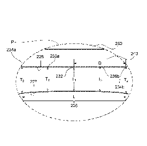
[0029] FIG. 2B is an enlarged view of a portion P of FIG. 2A showing the
center
portion 226 in more detail. The center portion 222 has a midpoint 232 and
extends
between a first endpoint 234a and second endpoint 234b in the elevation
direction. The
midpoint 232 is spaced apart from each of the first and second midpoints 234a
and
234b a distance D in the elevation direction (i.e., one-half the length L).
The thickness
of the center portion 222 in the axial direction increases outwardly from T1
at the
midpoint 232 to the thickness T2 at each of the first and second midpoints
234a and
234b. The average thickness of the center portion 222 substantially equal to
an odd
multiple (e.g., 1, 3, 5, 7) of a 1/4-wavelength of the center frequency of the
transducer
layer 260 (FIG. 2A) Further, at intermediate points 236a and 236h, the center
portion
222 has a thickness T3 generally corresponds to the average thickness of the
center
portion 222 between midpoint 232 and each of the first and second midpoints
234a and
234b.
-11-
CA 02942379 2016-09-09
WO 2015/138796 PCT/US2015/020279
[0030] In some
embodiments, however, the center portion 226 can be configured
to have an average 3/4-wavelength thickness to provide adequate dielectric
strength to
meet desired medical electrical safety standards. In other embodiments, the
center
portion 226 may have an average thickness less than 3/4 wavelength. In some
embodiments, for example, the center portion 226 can be fabricated to have an
average thickness of the 1/4 of the wavelength of an operational center
frequency (e.g.,
20 MHz, 25 MHz, 30 MHz) of the transducer layer 260. In some embodiments, the
average thickness of the center portion 226 can be any odd multiple (e.g., 1,
3, 5, 7, 9)
of 1/4 of the wavelength of the operational center frequency of the transducer
layer 260
(FIG. 2A). In other embodiments, however, the average thickness can be any
suitable
fraction of the wavelength of the operational center frequency of the
transducer layer
260 (FIG. 2A). Those of ordinary skill in the art will appreciate, for
example, that for
broadband ultrasound transducers, a 1/4 wavelength lens thickness will
generally
perform better than a 3/4 wavelength lens thickness, and increasing odd
multiples of
1/4-wavelength generally perform progressively worse. In
contrast, narrowband
transducers (e.g., OW Doppler transducers) can have acoustic lenses with
increasing
odd multiples of the 1/4-wavelength without a significant reduction in
performance.
[0031]
Fabricating the center portion 226 to have of an average thickness
corresponding generally to a fractional portion (e.g., 1/4, 3/4) of the
wavelength can, in
addition to minimizing multi-path artifacts, acoustically enhance a central
part of the
elevation dimension (i.e., along the x-axis of FIG. 2A) of the transducer
layer 260 (FIG.
2A), thereby providing a desirable boost to a normal component of the
elevation beam.
This can be viewed as achieving the equivalent of mild apodization of the
elevation
beam by enhancing the central part of the beam relative to the edges, as
opposed to
attenuating the edges relative to the center of the beam. The apodization of
the
elevation beam can lead to a reduction in sidelobes in the elevation beam.
[0032] From the
foregoing, it will be appreciated that specific embodiments of the
invention have been described herein for purposes of illustration, but that
various
modifications may be made without deviating from the scope of the invention.
Accordingly, the invention is not limited except as by the appended claims.
-12-