Note : Les descriptions sont présentées dans la langue officielle dans laquelle elles ont été soumises.
CA 02943921 2016-09-26
WO 2015/145389
PCT/1B2015/052248
EFFECT OF LIPOPHILIC NUTRIENTS ON DIABETIC EYE DISEASES
Field
The present invention relates to a method of delaying the development and
maturation of eye
related complications of diabetes by administering a composition containing
lipophilic nutrients.
More particularly, the present invention relates to a method of delaying the
development and
maturation of eye related complications of diabetes by administering a
composition containing
lutein and its isomers, lutein ester, zeaxanthin isomers, turmeric extract,
curcumin or
curcuminoids, either alone or in combination thereof, derived from plant
extract/oleoresin
containing xanthophylls/ xanthophylls esters which are safe for human
consumption and are
particularly useful as dietary supplements for nutrition and health promoting
benefits.
Background of the invention
Eye is the most important and complex organ in the human body that is
protected by the bony
orbit of the eye. It is divided into anterior segment which consists of the
cornea, iris, lens, ciliary
body, and the anterior portion of the sclera. The posterior segment is bounded
anteriorly by the
lens and extends to the back of the eye. The retina optic disc is also
included in the posterior
segment. The light passes through anterior portion called cornea, aqueous
humor, crystalline
lens, pupil, vitreous humor to reach the retina of the eye, this pathway is
called visual axis of the
eye. The lens refracts light rays and helps focus the image of an object on
the retina (fovea)
through accommodation.
Diabetes and diabetic complications: Diabetes is one of the most occurring non-
communicable,
heterogeneous, metabolic disorder characterized by hyperglycemia resulting
from defective
insulin production, resistance to insulin action or both, there are two forms
of diabetes, type 1
and type 2. Type 1 diabetes mellitus is the consequence of an autoimmune-
mediated destruction
of pancreatic 0-cells, leading to insulin deficiency.
Type 2 diabetes mellitus is characterized by insulin resistance and relative,
rather than absolute,
insulin deficiency. According to the latest World Health Organization (WHO)
estimation
currently there are about 366 million diabetic people in the world, it is
expected to increase to
1
CA 02943921 2016-09-26
WO 2015/145389
PCT/1B2015/052248
552 million by 2030 and India has about 62 million diabetics. Prolonged
exposure to chronic
hyperglycemia can lead to various complications including vascular and non-
vascular
complications. Vascular complications are further divided into macrovascular
and microvascular
complications. Tissues like retina, kidney, peripheral nerves and lens are
most affected by long
term complications of the diabetes, which results in the development of
diabetic retinopathy,
nephropathy, neuropathy and cataract respectively.
Diabetic cataract: Cataract is characterized by opacity of the eye lens, and
the leading cause of
blindness worldwide. The development of cataract in diabetics is 2-5 times
more when compared
with the non-diabetic counterparts. Furthermore, patients with diabetes
mellitus have higher
complication rates from cataract surgery. Both diabetes and cataract pose an
enormous health
and economic burden, particularly in developing countries, where diabetes
treatment is
insufficient and cataract surgery often inaccessible. Many clinical
interventions have been
reported to countering cataract including diabetic cataract but are not
completely successful at
clinical practice.
Diabetic Retinopathy: Diabetic retinopathy (DR) is one of the most common
micro vascular
complications of diabetes. DR occurs in 70% of all persons having diabetes for
more than 15
years and is the most common cause of blindness. DR is a disease of retina,
resulting in loss of
vision, macular edema, recurrent vitreous hemorrhages, tractional or secondary
rhegmatogenous
retinal detachment, and so forth. Since the last two decades there have been
significant
developments in the emerging field of pharmacotherapy of DR. The advent of
laser
photocoagulation three decades back, was really useful in limiting vision loss
in most of the
cases and is still considered the gold standard therapy for the treatment of
DR. However,
corticosteroids and anti-VEGF agents have shown promising results with regard
to prevention of
neo-vascularisation, but remained limited in use due to their short-duration
effects. Therefore,
pharmacotherapy of DR is still an adjunct to pan retinal photocoagulation.
In recent years, a great deal of attention has been focused on biological
activities of carotenoids.
Carotenoids are naturally occurring xanthophylls in plants that are involved
in light harvesting
reactions and protection of plant organelles against singlet oxygen induced
damage. Dietary
2
CA 02943921 2016-09-26
WO 2015/145389
PCT/1B2015/052248
carotenoids serve as antioxidants in the tissues (Thurnham DL. Carotenoids:
function and
fallacies. Proc Nutr Soc 1994; 53: 77-87) and protect the body from oxidative
damage.
Mammalian species do not synthesize carotenoids and therefore these have to be
obtained from
dietary sources such as fruits and vegetables and/or dietary supplements.
Numerous
epidemiological studies support a strong inverse relationship between
consumption of carotenoid
rich fruits and vegetables and incidence of degenerative diseases (Coleman H,
Chew E.
Nutritional supplementation in age-related macular degeneration. Curr Opin
Ophthalmol 2007;
18(3): 220-223)
Lutein is one of the major xanthophylls present in green leafy vegetables and
egg yolk. Lutein
and zeaxanthin are known to selectively accumulate in the macula of the human
retina. They
have been thought to work as antioxidants and as blue light filters to protect
the eyes from such
oxidative stresses as cigarette smoking and sunlight exposure, which can lead
to age-related
macular degeneration and cataracts.
Xanthophylls can show both optical (R-and S-stereo isomers) and geometrical
isomers (trans, E-
and cis, Z-). The conformation of R- and S-stereo isomers is based on circular
dichroism (CD)
spectral and chiral column high-performance liquid chromatography (HPLC)
studies while the
conformation of cis- and trans-isomers is based on electronic, infrared,
nuclear magnetic
resonance (NMR), high-performance liquid chromatography-mass spectrometry
(HPLC-MS) and
high-performance liquid chromatography-nuclear magnetic resonance (HPLC-NMR)
on-line
spectroscopy studies. It is well known that when an organic molecule has a
carbon atom with
four different types of atoms or groups attached to it, that carbon atom is
designated as chiral
carbon atom. The chiral carbon atom is responsible for two different spatial
arrangements
leading to the formation of optical isomers while the number of double bonds
of the polyene
chain and the presence of a methyl group and the absence of steric hindrance
decide the number
of trans- and cis-isomers. In the case of trans-zeaxanthin, the carbon atoms
at 3 and 3' positions
in the two end rings are both chiral atoms.
3
CA 02943921 2016-09-26
WO 2015/145389
PCT/1B2015/052248
Thus, trans-zeaxanthin has two chiral centers at the carbon atoms C3 and C3',
based on the
positions of the secondary hydroxy groups attached to them. Therefore, there
are four possible
stereo isomers of trans-zeaxanthin namely, (3R-3'R)-isomer, (3S-3'S)-isomer
and (3R-3'S)- or
(3S-3'R)-isomer. In these isomers (3R-3'S)- & (3S-3'R)- are identical. Thus,
there are three chiral
isomers of trans-zeaxanthin. The isomer causing rotation of polarized light in
a right handed
manner is called R-stereo isomer, the isomer causing left handed rotation is
called S-stereo
isomer, and the third isomer possessing twofold opposite effects (R,S;
optically inactive) which
is called meso-form of zeaxanthin.
The conjugated double bonds of lutein and zeaxanthin contribute to the
distinctive colors of each
pigment, and also influence the ability of these to quench singlet oxygen. Due
to the extra
conjugated double bond, zeaxanthin is believed to be a stronger anti-oxidant
compared to lutein.
Regarding the location of xanthophylls at a cellular level, they are reported
to be bound to
specific proteins referred to as xanthophylls binding protein (XBP). The XBP
is suggested to be
involved in the uptake of lutein and zeaxanthin from the blood stream and
stabilization of the
same in the retina. The study of xanthophylls and XBP by femto-second
transient absorption
spectroscopy showed better stability for (3R,3'S)-zeaxanthin enriched XBP
compared to
(3R,3'R)-zeaxanthin while the photo physical properties of the xanthophylls:
(3R,3'R)-zeaxanthin
and (3R, 3'S,meso)-zeaxanthin are generally identical. It is likely that the
meso-zeaxanthin is
better accommodated with XBP wherein the protein protects the xanthophylls
from degradation
by free radicals. Thus, the complex may be a better antioxidant than the free
xanthophylls,
facilitating improved protection of ocular tissue from oxidative damage.
(Billsten et al.,
Photophysical Properties of Xanthophylls in Caroteno proteins from Human
Retina,
Photochemistry and Photobiology, 78, 138-145, 2003)
Epidemiological studies suggested that higher dietary intake of lutein and
zeaxanthin reduces the
risk of cataracts and age-related macular degeneration. Previous studies
showed that rats treated
with combination of insulin and lutein exhibited delayed development and
maturation of cataract
than treated with lutein or insulin alone. Serum lutein and zeaxanthin
concentrations in DR
patients were found to be significantly lower than those in normal subjects,
and their intake was
4
CA 02943921 2016-09-26
WO 2015/145389
PCT/1B2015/052248
proved to improve the visual acuity, contrast sensitivity, and macular edema,
suggesting that
lutein and zeaxanthin supplementation might be targeted as potential
therapeutic agents in
treating DR.
Curcumin has been identified as the active principle of turmeric and has been
shown to exhibit
antioxidant, anti-inflammatory, antimicrobial, and anticarcinogenic
activities. Curcumin is a
natural extract from the spice Turmeric. Turmeric is derived from the plant
Curcuma Longa, a
member of the ginger family. Curcumin is a known antioxidant which inherently
has many
health benefits. Curcumin was shown to induce apoptosis in human retinal
endothelial cells and
decrease VEGF release into media in vitro and it also inhibits diabetes-
induced elevation of
serum VEGF levels in rat. Vascular endothelial growth factor (VEGF)
expression, induced by
high glucose levels and hypoxia, is a main feature in retinopathy. Several
studies have also
shown that VEGF may also play a role in the development of the earliest stages
of retinopathy.
Though dietary supplements such as curcumin, lutein, zeaxanthin, etc have
offered some benefits
in preclinical studies, the translation has been very poor and the doses used
in clinical trials are
unfeasible to practice in reality. One of the major reasons for the lack of
clinical success with
curcumin is linked to its extensive intestinal and hepatic metabolic
biotransformation resulting in
poor bioavailability. Recently, the focus is to address bioavailability
concerns of the supplements
with a view to improve the therapeutic efficacy.
The lipophilic nutrients such as curcumin, lutein, zeaxanthin, ginger, etc are
poorly absorbed if
administered either as oil suspensions or as beadlets, which are the currently
used forms. The
main reason for poor absorption is their poor solubility in water. Due to
their insolubility their
bioavailability is very poor. Lipophilic nutrients have limited absorption in
the body due to
limited solubility in the gastrointestinal tract. Generally, the
bioavailability of such nutrients is
below 40%. The bioavailability can be enhanced by reducing the particle size,
which in turn will
enhance their efficiency of micellization. Dispersion of nutritional products
at molecular level is
generally regarded as a technique of reducing the particle size. Such
molecular dispersions
provide higher efficiency for micellization of nutrients in water and thereby
increase the
bioavailability.
5
CA 02943921 2016-09-26
WO 2015/145389
PCT/1B2015/052248
Hence, it is interesting to search the effects of a composition containing
soluble lipophilic
nutrients in diabetic rats with respect to its beneficial effect on retina by
a nutrigenomics
approach and the effect was compared with regular lipophilic nutrients.
Nutrigenomics is the science of interaction between nutrients and genes. It is
the study of
how genetic expression affect your need for certain nutrients and help
maintain optimal health
throughout your life. Nutrigenomics promotes an increased understanding of how
nutrition
influences metabolic pathways and homeostatic control, how this regulation is
disturbed in the
early phases of diet-related diseases, and the extent to which individual
sensitizing genotypes
contribute to such diseases. Our goal is to better understand how
phytonutrients affect gene
expression.
Numerous references are available that provide compositions containing
carotenoids used for the
prevention/treatment of diabetic eye diseases.
In Brown et al. (Am J Clin Nutr. 1999), dietary antioxidants, including
carotenoids, are
hypothesized to decrease the risk of age-related cataracts by preventing
oxidation of proteins or
lipids within the lens. However, prospective epidemiologic data concerning
this phenomenon are
limited. The authors examined prospectively the association between carotenoid
and vitamin A
intake and cataract extraction in men. US male health professionals (n =
36644) who were 45-75
y of age in 1986 were included in this prospective cohort study. Others were
subsequently
included as they became 45 y of age. During 8 y of follow-up, 840 cases of
senile cataract
extraction were documented. They observed a modestly lower risk of cataract
extraction in men
with higher intakes of lutein and zeaxanthin but not of other carotenoids
(alpha-carotene, beta-
carotene, lycopene, and beta-cryptoxanthin) or vitamin A after other potential
risk factors,
including age and smoking, were controlled for. Men in the highest fifth of
lutein and zeaxanthin
intake had a 19% lower risk of cataract relative to men in the lowest fifth
(relative risk: 0.81;
95% CI: 0.65, 1.01; P for trend = 0.03). Among specific foods high in
carotenoids, broccoli and
spinach were most consistently associated with a lower risk of cataract.
Lutein and zeaxanthin
may decrease the risk of cataracts severe enough to require extraction,
although this relation
6
CA 02943921 2016-09-26
WO 2015/145389
PCT/1B2015/052248
appears modest in magnitude. This study is a cohort study done with the US
male population.
This study establishes a relation between nutrition deficiency and the
occurrence of cataract.
EP 2618832 A2 relates to a composition comprising an enzyme selected from the
group
comprising superoxide dismutase (SOD) and SOD mimics and the like, in
association
with lutein and at least one stereoisomer of zeaxanthin; and also includes a
kit of parts
comprising such composition, wherein the kit comprises a first part comprising
the
enzyme, and a second part comprising lutein and at least one zeaxanthin
isomer. The
composition or the kit of parts may be included in a functional food, a
nutraceutical
composition or a food or dietary supplement, a medicament or a pharmaceutical
composition, or a veterinarian product. The reference also relates to a
composition for use in
treating, preventing, and/or stabilizing a disease, condition and/or disorder
of the eye associated
with oxidative stress, by administering to a subject in need thereof a
medicament or a
pharmaceutical composition. However, the reference does not make use of
zeaxanthin isomers.
W02010032267A2 relates to an herbal formulation for prevention and treatment
of diabetes and associated complications comprising extracts from selected
Indian medicinal
herbs. The reference relates to associated formulations for different diabetes
related
complications, which are individually useful in clinical requirements, such as
improving renal
health, preventing renal diseases, preventing diabetic retinopathy, and/or in
the prevention and treatment of oxidative damage to the heart and/or blood
vessels. The
formulations are versatile and can be processed into extracts/concentrates and
further
pharmacologically modified into tablets or capsules or granules or syrups or
herbal health drinks
or inhalable herbal medicinal preparations or ocular preparations or
transdermal absorbable
preparations such as ointments/gels or injectable medicine. This is a poly
herbal formulation and
there is no synergistic data to support the claim.
CN 102178925A relates to a lutein ophthalmic preparation for protecting
eyesight, which is
prepared from the following raw materials: 5 to 13 parts of water-soluble
lutein (based on
C4oH5602), 50 to 80 parts of taurine, 0.1 to 0.5 parts of selenium (based on
Se), 10 to 25 parts
of zinc (based on Zn), 0.5 to 1.0 part of water-soluble vitamin A, and 0.8 to
2.0 parts of
7
CA 02943921 2016-09-26
WO 2015/145389
PCT/1B2015/052248
glutathione; an auxiliary material consists of a diluent, a wetting agent, an
isoosmotic adjusting
agent, a preservative, an antioxidant and water for injection; formulations
comprise eye drops,
eye lotion and the like; and the lutein ophthalmic preparation is suitable for
eye diseases such as
myopia, long sightedness, cataract, glaucoma, retinal pigment degeneration,
macular
degeneration and the like. Various nutrition factors are reasonably
compatible, a blank of
a lutein external preparation is filled, and the bioavailability and health-
care effect are obviously
improved; and through actual application by 300,000 people, the total
effective rate for the
myopia, cataract and diabetic eye disease is over 90 percent and the lutein
ophthalmic
preparation has positive promotion value. This ophthalmic preparation contains
only one macular
carotenoid i.e. lutein and does not mention use of zeaxanthin isomers.
In Sasaki et al. (TOYS, March 2009, Vol. 50, No. 3), the aim of this study was
to investigate,
with the use of a mouse endotoxin-induced uveitis (EIU) model, the
neuroprotective effects of
lutein against retinal neural damage caused by inflammation. EIU was induced
by intraperitoneal
injection of lipopolysaccharide (LPS). Each animal was given a subcutaneous
injection of lutein
or vehicle three times: concurrently with and 3 hours before and after the LPS
injection. Analysis
was carried out 24 hours after EIU induction. Levels of rhodopsin protein and
signal transducer
and activator of transcription 3 (STAT3) activation were analysed by
immunoblotting. Lengths
of the outer segments of the photoreceptor cells were measured. Dark-adapted
full-field
electroretinograms were recorded. Oxidative stress in the retina was analyzed
by
dihydroethidium and fluorescent probe. Expression of glial fibrillary acidic
protein (GFAP) was
shown immunohistochemically. The EIU-induced decrease in rhodopsin expression
followed by
shortening of the outer segments and reduction in a-wave amplitude were
prevented by lutein
treatment. Levels of STAT3 activation, downstream of inflammatory cytokine
signals, and
reactive oxygen species (ROS), which are both upregulated during EIU, were
reduced by lutein.
Pathologic change of Muller glial cells, represented by GFAP expression, was
also prevented by
lutein. The present data revealed that the antioxidant lutein was
neuroprotective during EIU,
suggesting a potential approach for suppressing retinal neural damage during
inflammation.
Lutein is a nutritional supplement and subjects have to take daily dose for
prevention or
treatment of any disease. In this study, Lutein was administered through
injection. Daily
supplementation of lutein via injection is painful causing discomfort to the
subject.
8
CA 02943921 2016-09-26
WO 2015/145389
PCT/1B2015/052248
CA 2760932 Al relates to ophthalmic formulations that deliver a variety of
therapeutic agents,
including but not limited to rapamycin (sirolimus), analogs thereof (rapalogs)
or other
mammalian target of rapamycin (mTOR) inhibitors, to a subject for an extended
period of time.
The ophthalmic formulations may be placed in an aqueous medium of a subject,
including but
not limited to intraocular or periocular administration, or placement
proximate to a site of a
disease or condition to be treated in a subject. A method may be used to
administer a therapeutic
agent to treat or prevent age-related macular degeneration, macular edema,
diabetic retinopathy,
uveitis, dry eye, or a hyperpermeability disease in a subject.
Summary
Overcoming the difficulty of delivering therapeutic/ preventive agents to
specific regions of the
eye presents a major challenge to treatment of most eye disorders. Due to poor
bioavailability of
lipophilic nutrients the delivery of many potentially important therapeutic/
preventive agents to
the eye is hindered.
From above it is clear that there is a need to provide a technology which can
overcome the
difficulty of delivering the therapeutic/ preventive agents for diabetic eye
complications even at
reduced dose levels.
Molecular dispersions of lipophilic nutrients are provided, which are useful
for delaying the
development and maturation of eye related complications of diabetes and which
are safe for
human consumption and are particularly useful as dietary supplements for
nutrition and health
promoting benefits.
In one embodiment, molecular dispersions of lipophilic nutrients such as
curcumin or trans-
lutein and zeaxanthin isomers namely (R,R)-zeaxanthin and (R,S)-zeaxanthin or
trans-lutein and
(R,R)-zeaxanthin in a solid or liquid hydrophilic carrier, derived from plant
extract/oleoresin
containing xanthophylls/ xanthophylls esters are provided, and which are
useful for delaying the
development and maturation of eye related complications of diabetes.
9
CA 02943921 2016-09-26
WO 2015/145389
PCT/1B2015/052248
In one embodiment, molecular dispersions of a composition are provided, which
contain at least
80% by weight of total xanthophylls, out of which the trans-lutein content is
80-95% w/w;
(R,R)-zeaxanthin is 14-20% w/w; (R,S)-zeaxanthin is 0.01-1% w/w or trans-
lutein content is 80-
95% w/w; (R,R)-zeaxanthin is 14-20% w/w, and traces of other carotenoids
derived from the
plant extracts/oleoresin containing xanthophylls/xanthophylls esters or
curcumin which contains
5-95% of curcuminoids.
In one embodiment, molecular dispersions of a xanthophyll composition
containing trans-lutein
and zeaxanthin isomers namely (R,R)-zeaxanthin and (R,S)-zeaxanthin, or trans-
lutein and
(R,R)-zeaxanthin, in a solid or liquid hydrophilic carrier are provided,
wherein the composition
has higher antioxidant potential than the free xanthophylls and which are
useful for delaying the
development and maturation of eye related complications of diabetes.
In one embodiment, molecular dispersions of a curcumin composition containing
curcuminoids
in a solid or liquid hydrophilic carrier are provided, and which are useful
for delaying the
development and maturation of eye related complications of diabetes.
In one embodiment, molecular dispersions of lipophilic nutrients are provided,
which have
higher efficiency for micellization which enhances the bioavailability
resulting in increased
levels of lipophilic nutrients in tissues, in which these molecular
dispersions are effective at
relatively lower concentrations and are useful for delaying the development
and maturation of
eye related complications of diabetes.
In one embodiment, molecular dispersions of lipophilic nutrients in solid or
liquid hydrophilic
carriers are provided, which have higher bioavailability.
In some embodiments, the molecular dispersions of lipophilic nutrients which
are prepared by
using safe solvents (GRAS) and are suitable for human consumption, with
minimum solvent
residues.
10
CA 02943921 2016-09-26
WO 2015/145389
PCT/1B2015/052248
Further advantages of the compositions and/or methods herein will become
apparent from a
consideration of the ensuing description.
The usefulness of the products, compositions, and/or methods described herein
below, which are
illustrated in the examples, should not be construed to limit the scope of the
present innovations
in any manner whatsoever.
Methods herein are related to delaying the development and maturation of eye
related
complications of diabetes by administering a composition containing lipophilic
nutrients. More
particularly, methods herein are related to delaying the development and
maturation of eye
related complications of diabetes by administering a composition containing
lutein and its
isomers, lutein ester, zeaxanthin isomers, turmeric extract, and/or curcumin
or curcuminoids,
derived from plant extract/oleoresin containing xanthophylls/ xanthophylls
esters, and which are
safe for human consumption and are particularly useful as dietary supplements
for nutrition and
health promoting benefits.
The molecular dispersions herein are in the form of powders, tablets,
capsules, sachets, beadlets,
microencapsulated powders, oil suspensions, liquid dispersions, pellets, soft
gel capsules,
chewable tablets or liquid preparations.
Molecular dispersions herein of trans-lutein and zeaxanthin isomers namely
(R,R)-zeaxanthin
and (R,S)-zeaxanthin, or of trans-lutein and (R,R)-zeaxanthin, and/or of
curcumin containing
curcuminoids in a solid or liquid hydrophilic carrier have enhanced water
solubility and
bioavailability, which helps in effectively delivering the molecules and shows
potential in
delaying the development and maturation of eye related complications of
diabetes. The use of
carotenoids namely, trans-lutein and zeaxanthin isomers having higher
antioxidative potential in
highly water soluble form with enhanced bioavailability for delaying the
development and
maturation of eye related complications of diabetes has not yet been reported
in earlier literature.
11
CA 02943921 2016-09-26
WO 2015/145389
PCT/1B2015/052248
In some embodiments, compositions herein are molecular dispersions of
hydrophilic liquid and
solid carriers. In some embodiments, a process for making compositions herein
includes making
them into molecular dispersions of hydrophilic liquid and solid carriers,
which enhances water
solubility of the nutrients which characteristics are beneficial for
formulating further as beverage
or soft gelatin capsule or as licaps.
Compositions herein in some embodiments include a water soluble, molecular
dispersion of
lipophilic nutrients comprising:
(a) a stabilizer,
(b) a water soluble hydrophilic carrier, and
(c) and optionally a surfactant,
and which are useful for converting oily nutrients into powders, tablets,
capsules, ointments,
pastes, lotions, liniments, mouthwashes, sachets, gargles and which are
suitable for incorporation
into beverages.
In some embodiments, compositions herein are free flowing water soluble
molecular dispersions
of lipophilic nutrients such as lutein, zeaxanthin, beta carotene and lycopene
in water soluble
liquid or solid hydrophilic carriers which can be formulated further as
beverage or soft gelatin
capsule or as licaps.
In some embodiments, a process for the preparation of free flowing water
soluble molecular
dispersions of lipophilic nutrients such as lutein, zeaxanthin, beta carotene
and lycopene in water
soluble liquid or solid hydrophilic carriers which can be formulated further
as beverage or soft
gelatin capsule or as licaps, is provided.
In some embodiments, a solution of lipophilic nutrient in a polar or non polar
organic solvent can
be dispersed in certain water soluble hydrophilic liquid or solid carrier
systems. Upon removal of
solvent under vacuum, the resultant dispersion remains as a homogenous liquid
or solid
dispersion which is suitable for filling in to soft gel capsules or in to
licaps. Such liquid or solid
dispersions are suitable for filling in to capsules or for making granules,
tablets, filling in to
sachets or for making beverages.
12
CA 02943921 2016-09-26
WO 2015/145389
PCT/1B2015/052248
In some embodiments, compositions herein are free flowing water soluble
molecular dispersions
of lipophilic nutrients such as lutein, zeaxanthin, beta carotene and lycopene
in water soluble
hydrophilic liquid or solid carriers, which are useful for converting into
soft gelatin capsules,
licaps, ointments, pastes, lotions, liniments, mouthwashes, gargles etc. which
are also suitable
for incorporation in to beverages.
In some embodiments, a process is provided for the preparation of free flowing
water soluble
molecular dispersions of lipophilic nutrients in water soluble hydrophilic
liquid or solid carriers,
which are useful for converting into soft gelatin capsules, licaps, ointments,
pastes, lotions,
liniments, mouthwashes, gargles etc. which are also suitable for incorporation
in to beverages
which comprises,
(i) dissolving a lipophilic nutrient in a non polar / polar solvent or a
mixture thereof to
form a solution;
(ii) filtering the resulting solution to remove insoluble impurities;
(iii) separately, dissolving the water soluble hydrophilic liquid or solid
carrier, stabilizer and
optionally, a surfactant in a polar solvent to form a clear solution;
(iv) blending the solution obtained in step (i) with the solution obtained
in step (iii);
(v) heating the resulting mixture to remove the solvent at a temperature in
the range of 20 -
45 deg C. and at a pressure ranging between 500-760 mm of mercury;
(vi) cooling the resultant molecular dispersion to ambient temperature; and
(vii) passing the cooled molecular dispersion obtained in step (vi) through a
sieve of
appropriate mesh size to remove any agglomerate or lumps to produce a free
flowing water
soluble or solid dispersion of lipophilic nutrients.
The term lipophilic' though refers to lipid-like, it generally covers all
compounds that are poorly
water soluble. Thus, the scope of the term includes poorly water soluble amino
acids, proteins,
minerals, herbal extracts such as curcumin, carbohydrates, alkaloids,
flavonoids and glycosides.
13
CA 02943921 2016-09-26
WO 2015/145389
PCT/1B2015/052248
The lipophilic nutrient which can be used includes, but is not limited to
lutein, lutein ester,
zeaxanthin isomers, lycopene, beta carotene, tocopherols, astaxanthin, omega-3
fatty acids,
ubiquinone, phytosterols, lecithins and the mixtures thereof.
The water soluble hydrophilic liquid or solid carrier used for forming the
dispersing solution
includes polyethylene glycol 200, polyethylene glycol 400, ethylene glycol,
propylene glycol,
glycerol, sorbitol, glucose syrup, corn steep liquor mannitol, polyethylene
glycol 6000,
polyethylene glycol 10000, Polyethylene glycol 20000, polyvinyl pyrrolidone,
hydroxyl propyl
methyl cellulose, sucrose, glucose, sodium chloride, hydroxyl propyl
cellulose, polyvinyl
alcohol, soluble starch, hydrolyzed starch and mixtures thereof.
In some embodiments the solvent employed for preparing the solution of the
lipophilic nutrient
product may be selected from Acetone, Hexane, Ethyl Acetate, Isopropyl
Alcohol, Ethanol,
Dichloromethane, Methanol, etc., more preferably form Acetone, Ethanol,
Dichloromethane,
Isopropyl alcohol, and more preferably Dichloro Methane and Isopropyl Alcohol.
The stabilizer which may be used in the process may be selected from ascorbic
acid, BHA, BHT,
ascorbyl palmitate, rosemary extract, mixed natural tocopherols, alpha
tocopheryl acetate,
sodium ascorbate, castor oil derivatives, sodium lauryl sulfate and mixtures
thereof.
The surfactant which may be used in the process may be selected from
polysorbate 20,
polysorbate 60, polysorbate 80, sodium lauryl sulfate and mixtures thereof.
The heating step in vacuum for evaporating the solvent present in the nutrient
dispersion may be
carried at a temperature preferably in the range of 35 to 45 deg C.
In some embodiments, the lipophilic nutrient is dispersed in a water soluble
hydrophilic liquid or
solid carrier, at a molecular level, so that its solubility and consequently,
its bioavailability is
enhanced several fold. In dispersing hydrophilic nutrients in hydrophilic
liquid or solid carriers
the resulting dispersions of the nutrients have significantly higher
solubility and bioavailability.
Further, dispersing the lipophilic nutrients in a hydrophilic liquid or solid
carrier helps in the
14
CA 02943921 2016-09-26
WO 2015/145389
PCT/1B2015/052248
formulation of lipophilic nutrient in the forms such as hard gelatin capsule
and soft gelatin
capsule.
For achieving a molecular dispersion, the lipophilic nutrient needs to be
dissolved in a polar or
non polar solvent. Depending on the chemical nature of the lipophilic
nutrient, a polar or a non-
polar or a mixture of polar and non-polar solvent can be used. If required,
the mixture of the
lipophilic nutrient and the solvent may be warmed to enhance the rate of
dissolution. Quite often,
the lipophilic nutrient requires a stirring for additional period of time to
complete the dissolution.
To ensure the complete dissolution, it may be necessary to pass the resultant
solution through a
filter medium and use only the filtrate for molecular dispersion. If the
viscosity of the solution is
high, it may be further diluted with the solvent used for dissolution, so that
filtration step can be
performed faster.
The hydrophilic (water soluble) carrier is dissolved in a suitable polar
solvent such as ethanol,
isopropyl alcohol, acetone, methanol, propylene glycol and/or water to form
clear solution. The
hydrophilic water soluble carrier used for dispersion may also be mixed with
stabilizers and
optionally with a surfactant. If required, the stabilizers may be required to
be dissolved in
solvent before mixing with the hydrophilic carrier. If the resultant mixture
is not a clear solution,
the solution may be filtered discarding the residue.
The dispersion of the lipophilic nutrient is then mixed with the hydrophilic
liquid or solid carrier
to obtain a homogenous mass. For this purpose, a simple magnetic stirrer or an
electrically
operated agitator may be used. Mixing can also be effected using a liquid-
liquid homogenizer or
emulsifier. Depending on the viscosity of the resultant mixture, the time
required to achieve a
homogenous mass may range from 15 minutes to 1 hour. For those nutrients which
are sensitive
to atmospheric oxidation, the mixing step can be performed under an inert
atmosphere or in the
presence of an antioxidant stabilizer.
For those nutrients wherein a faster dissolution is required in the
gastrointestinal tract, one can
optionally incorporate a food grade surfactant to enhance the solubility of
the lipophilic nutrient
and its bioavailability.
CA 02943921 2016-09-26
WO 2015/145389
PCT/1B2015/052248
The homogenous mass so obtained is then subjected to a step of heating under a
reduced
pressure. Since vast majority of the lipophilic nutrients are sensitive to the
heat, light and
oxygen, it may be necessary to carry out such heating at low temperatures,
preferably not
exceeding 45 deg C. It would also be preferable to heat under an inert
atmosphere using inert
gases such as nitrogen or argon. The process of heating is continued until the
resultant dispersion
has less than 25 parts per million of the solvent.
After ensuring the reduction of solvent residues at less than 25 parts per
million, the resultant
molecular dispersion is allowed to cool to ambient temperature and then it is
passed through 100
mesh sieve to remove any agglomerate or lumps The homogenous mass is then
filled in to
appropriate containers.
Brief Description of the Figures
Figure 1 shows a graph of the effects of treatment on fasting plasma glucose
in STZ-induced
diabetic rats.
Figure 2 shows a graph on the delay of diabetic cataract in rats by treatment.
Figure 3 shows the SDS-PAGE pattern of soluble fraction of lens proteins.
Figure 4 shows a graph on size exclusion chromatography of lens protein
soluble fraction.
Figure 5 shows a graph on spectrofluoremetric measurement of lens sorbitol.
Figure 6 shows a graph on EIPLC measurement of plasma lutein levels.
Figure 7 shows representative wave forms of oscillatory potentials (OPs) from
different groups
and total amplitudes.
Figure 8 shows representative histology of retina.
Figure 9 shows expression of Rhodopsin by real-time PCR (A) and
immunohistochemistry (B).
Figure 10: Expression of NGF by real-time PCR (A) and immunohistochemistry
(B).
Figure 11 shows expression of VEGF by immunoblotting.
Figure 12 shows expression of PDGF by immunohistochemistry.
Figure 13 shows serum lutein levels measured by RP-I-IPLC.
Figure 14 shows representative wave forms of OPs from individual animals of
different groups
and total amplitudes.
16
CA 02943921 2016-09-26
WO 2015/145389
PCT/1B2015/052248
Figure 15 shows representative histology of retina.
Figure 16 shows expression of rhodopsin by real-time PCR (A) and
immunohistochemistry (B).
Figure 17 shows expression of NGF by real-time PCR (A) and
Immunohistochemistry (B).
Figure 18 shows expression of VEGF by immunoblotting.
Figure 19 shows expression of PDGF by immune histochemistochemistry
Detailed Description
Diabetes mellitus can cause a variety of eye problems, the most common being
diabetic
retinopathy (DR) and diabetic cataract which are the most common causes of
blindness.
Antioxidant compounds are considered to have high antioxidant potential in the
prevention of
many human ailments such as age related macular degeneration, cataract,
diabetic eye
complications and various other diseases.
Lutein is a naturally occurring antioxidant found in green leafy vegetables
like spinach. Lutein is
also found in eye mainly present in macula lutea. It is well known that lutein
is a carotenoid and
powerful antioxidant. It has been used to treat cataracts and macular
degeneration which is an
age related degenerative disorder. Lutein has also shown protective
antioxidant activity in human
HepG2 cell lines.
Zeaxanthin is one of the most common carotenoid alcohols found in nature.
Lutein and
zeaxanthin have identical chemical formulas and are isomers, but they are not
stereoisomers. The
only difference between them is in the location of the double bond in one of
the end rings. This
difference gives lutein three chiral centers whereas zeaxanthin has two.
Because of symmetry,
the (3R,3'S) and (3S,3'R) stereoisomers of zeaxanthin are identical.
Therefore, zeaxanthin has
only three stereoisomeric forms. The (3R,3'S) stereoisomer is called meso-
zeaxanthin.
The conjugated double bonds of lutein and zeaxanthin contribute to the
distinctive colors of each
pigment, and also influence the ability of these to quench singlet oxygen. Due
to the extra
conjugated double bond, zeaxanthin is believed to be a stronger anti-oxidant
compared to lutein.
It has been demonstrated that the complex of lutein and zeaxanthin isomers act
as a better
antioxidant than the free xanthophylls, facilitating improved protection from
oxidative damages.
17
CA 02943921 2016-09-26
WO 2015/145389
PCT/1B2015/052248
Curcumin, a yellow pigment from Curcuma longa, is a major component of
turmeric and is
commonly used as a spice and food-coloring agent. It is also used as a
cosmetic and in some
medical preparations. The desirable preventive or putative therapeutic
properties of curcumin
have also been considered to be associated with its antioxidant and anti-
inflammatory properties.
Curcumin is thought to play a vital role against a variety of chronic
pathological complications
such as cancer, atherosclerosis, and neurodegenerative diseases.
The lipophilic nutrients are poorly absorbed if administered either as oil
suspensions or as
beadlets, which are the currently used forms. The main reason for poor
absorption is their poor
solubility in water. Due to their insolubility their bioavailability is very
poor. Dispersion of
nutritional products at molecular level provides higher efficiency for
micellization of nutrients in
water and thereby increases the bioavailability.
Compositions herein of lipophilic nutrients contain at least 80% by weight of
total xanthophylls,
out of which the trans-lutein content is 80-95% w/w; (R,R)-zeaxanthin is 14-
20% w/w; (R,S)-
zeaxanthin is 0.01-1% w/w, or trans-lutein content is 80-95% w/w; (R,R)-
zeaxanthin is 14-20%
w/w, and traces of other carotenoids derived from the plant extracts/oleoresin
containing
xanthophylls/xanthophylls esters, or curcumin which contains 5-95% of
curcuminoids in highly
water soluble form, and have enhanced bioavailability in delaying the
development and
maturation of eye related complications of diabetes.
Compositions herein comprise lipophilic nutrients; stabilizer; water soluble
hydrophilic carrier;
and optionally a surfactant.
Compositions herein contain at least 80% by weight of total xanthophylls, out
of which the trans-
lutein content is 80-95% w/w; (R,R)-zeaxanthin is 14-20% w/w; (R,S)-zeaxanthin
is 0.01-1%
w/w, or trans-lutein content is 80-95% w/w; (R,R)-zeaxanthin is 14-20% w/w,
and traces of
other carotenoids derived from the plant extracts/oleoresin containing
xanthophylls/xanthophylls
esters, or curcumin which contains 5-95% of curcuminoids.
18
CA 02943921 2016-09-26
WO 2015/145389
PCT/1B2015/052248
The stabilizer used is selected from ascorbic acid, butylated hydroxyanisole
(BHA), butylated
hydroxytoluene (BHT), ascorbyl palmitate, rosemary extract, mixed natural
tocopherols, alpha
tocopheryl acetate, sodium ascorbate, castor oil derivatives, sodium lauryl
sulfate and mixtures
thereof.
The carrier used is selected from polyethylene glycol 200, polyethylene glycol
400, ethylene
glycol, propylene glycol, glycerol, sorbitol, glucose syrup, corn steep
liquor, mannitol,
polyethylene glycol 6000, polyethylene glycol 10000, Polyethylene glycol
20000, polyvinyl
pyrrolidone, hydroxyl propyl methyl cellulose, sucrose, glucose, sodium
chloride, hydroxyl
propyl cellulose, polyvinyl alcohol, soluble starch, hydrolyzed starch, and
mixtures thereof.
The surfactant is selected from polysorbate 20, polysorbate 60, polysorbate
80, lecithin, sucrose
fatty acid esters, glyceryl fatty acid esters, sodium lauryl sulfate, and
mixtures thereof.
Studies with rats were carried out to test the activity of lipophilic
nutrients in diabetic eye
complications with four samples viz water soluble compositions of trans-lutein
and zeaxanthin
isomers (sold under the brand name UltraSol Lutemax2020Tm); concentrates
containing trans-
lutein and zeaxanthin isomers (sold under the brand name Lutemax2o2o ; and
water soluble
compositions containing curcumin (sold under the brand name UltraSol
CurcuWinTM) and
curcumin powder.
In some embodiments, the compositions herein include xanthophyll compositions
containing
macular pigments of trans-lutein and zeaxanthin isomers, including (R,R)-
zeaxanthin and (R,S)-
zeaxanthin derived from the plant extract/oleoresin containing
xanthophylls/xanthophylls esters
which is safe for human consumption and useful for nutrition and health care.
In some embodiments, a xanthophyll composition contains at least 80% by weight
of total
xanthophylls of which the trans-lutein content is at least 80% by weight and
the remaining
being zeaxanthin isomers, including (R,R)-zeaxanthin and (R,S) ¨zeaxanthin
derived from the
plant extract/ oleoresin containing xanthophylls /xanthophylls esters which is
safe for human
consumption and useful for nutrition and health care.
19
CA 02943921 2016-09-26
WO 2015/145389
PCT/1B2015/052248
In some embodiments, a xanthophyll composition contains at least 85% by weight
of total
xanthophylls, out of which the trans-lutein content is at least 85% by weight
and the remaining
being zeaxanthin isomers, including (R,R)-zeaxanthin and (R,S) ¨zeaxanthin
derived from the
plant extract/ oleoresin containing xanthophylls/xanthophylls esters which is
safe for human
consumption and useful for nutrition and health care.
In some embodiments, a xanthophyll composition contains at least 85% by weight
of total
xanthophylls, out of which at least 80% by weight being trans-lutein, at least
6 % by weight
being (R,R)-zeaxanthin and at least 6% by weight being (R,S)-zeaxanthin
derived from the plant
extract/ oleoresin containing xanthophylls/xanthophylls esters which is safe
for human
consumption and useful for nutrition and health care.
In some embodiments, a xanthophyll composition contains at least 85% by weight
trans-lutein
and at least 4% by weight (R,R)¨zeaxanthin and at least 5% by weight
(R,S)¨zeaxanthin derived
from the plant extract/oleoresin containing xanthophylls/xanthophylls esters
which is safe for
human consumption and useful for nutrition and health care.
In some embodiments, a xanthophyll composition contains at least 85% by weight
of total
xanthophylls, out of which at least 80% by weight being trans-lutein, the
remaining 15% by
weight being zeaxanthin isomers, including (R,R)-zeaxanthin and
(R,S)¨zeaxanthin derived from
the plant extract/oleoresin containing xanthophylls/xanthophylls esters which
is safe for human
consumption and useful for nutrition and health care.
In some embodiments, a process for the preparation of the xanthophyll
composition containing
macular pigments consisting of trans-lutein, zeaxanthin isomers, namely (R,R)-
zeaxanthin and
(R, S)- zeaxanthin derived from the plant extract/oleoresin containing
xanthophylls/xanthophylls
esters which is safe for human consumption and useful for nutrition and health
care, in which:
a) the saponification step to convert xanthophyll esters present in plant
extract / oleoresin into the
de-esterified form can be combined with limited isomerization of lutein to
produce xanthophyll
CA 02943921 2016-09-26
WO 2015/145389
PCT/1B2015/052248
composition containing higher amount of trans-lutein, the remaining being
zeaxanthin isomers,
including (R,R)-zeaxanthin and (R,S) ¨zeaxanthin and traces of other
carotenoids derived from
the plant extract/ oleoresin containing xanthophyll/xanthophylls esters which
is safe for human
consumption and useful for nutrition and health care;
b) in the saponification step, potassium hydroxide or sodium hydroxide can be
dissolved in 1-
propanol without the addition of water;
c) the temperature of the saponification / isomerization can be between 70 to
100 Deg C
preferably around at 95 degree and the period of saponification can be 1-2
hr.; and
d) the ethyl acetate employed in the process can be recovered and used if
required, thereby
making the process economical.
In some embodiments, a xanthophyll composition contains macular pigments of
trans-lutein,
zeaxanthin isomers, including (R,R)- zeaxanthin and (R,S)- zeaxanthin, derived
from the plant
extract/oleoresin containing xanthophylls/xanthophylls esters which is safe
for human
consumption and useful for nutrition and health care which comprises of at
least 80% by weight
is total xanthophylls, of which the ratio of trans- lutein and zeaxanthin
isomers being in the
range of 4:1 to 6:1 and the ratio of the isomers of zeaxanthin being in the
range of 80:20 to
20:80. In some embodiments, the ratio of trans-lutein and zeaxanthin isomers
is at a ratio of
about 5:1.
In some embodiments, a xanthophyll composition contains at least 85% by weight
of total
xanthophylls, of which the trans-lutein content is at least 85% and the ratio
of trans- lutein and
zeaxanthin isomers being in the range of 4:1 to 6:1 and the ratio of the
isomers of zeaxanthin
being in the range of 80 to 20: 20 to 80
In some embodiments, a process for the preparation of a xanthophyll
composition containing
macular pigments of trans-lutein and zeaxanthin isomers, including (R, R)-
zeaxanthin and
(R, S)- zeaxanthin derived from the plant extract/oleoresin containing
xanthophylls/xanthophylls
21
CA 02943921 2016-09-26
WO 2015/145389
PCT/1B2015/052248
esters which is safe for human consumption and useful for nutrition and health
care, which
comprises:
(a) saponifying and partially isomerising simultaneously the xanthophyll
esters present in the
plant extract / oleoresin containing xanthophyll esters by admixing the
extract /oleoresin with
alkaline solution of 1-propanol , the ratio of alkali and 1- propanol being in
the range of 1:0.5 to
1:1 by weight / volume, heating the resultant mass at a temperature in the
range 70-100 degree C,
preferably 95 Deg C for a period in the range of 1 to 5 hrs to obtain a
saponified / isomerised
crude concentrate;
(b) admixing the resultant saponified / isomerised crude concentrate obtained
in step (a) with
water, the ratio of the concentrate and water used being in the range from 1:2
to 1:3
volume/volume, to form a diluted oily mixture;
(c) extracting the diluted oily mixture obtained in step (b) with ethyl
acetate , the ratio of diluted
oily mixture and ethyl acetate used being in the range of 1:1.5 to 1:2
volume/volume to get an
extract containing the xanthophyll composition;
(d) evaporating the composition obtained in step (c) to remove ethyl acetate;
(e) purifying the composition resulting from step (d) by washing first with
non polar and later
with polar solvents and filtering;
(f) drying the resulting composition under vacuum at a temperature in the
range of 40 to 45 Deg
C for a period ranging from 48-72 hours;
(g) if desired recovering the ethyl acetate used in step (c) by conventional
methods and if
required reused; and
(h) if desired storing the resulting composition in an inert atmosphere at -
20 Deg.
22
CA 02943921 2016-09-26
WO 2015/145389
PCT/1B2015/052248
By adjusting the temperature, period and the amount of alkali in the step (a)
the ratios in steps (b)
and (c), the desired composition can be obtained
It is to be noted that leafy and green vegetables, corn, fruits and/or
marigolds may be used as the
source for the xanthophylls oleoresin. But considering that lutein is present
along with
zeaxanthin in free form associated with large amounts of chlorophyll and other
undesirable
carotenoids in most of the fruits, though according to the present invention
the use of leafy
and green vegetables corn, fruits is possible and considering the low
concentration of lutein
and zeaxanthin in the above materials and further the elaborate steps of
purification which is
required being not economical, marigold is the preferred choice as the
starting material for the
preparation of the composition of the present invention
Specifically, commercially available food grade marigold oleoresin produced by
hexane
extraction can be used as starting material ( Kumar et.al Process for the
Preparation of
Xanthophylls Crystals , US Patent No. 6,743,953,2004 ;Kumar US Patent No
.6,737,535, 2004)
for the preparation of the xanthophyll composition comprising of trans-lutein
,and zeaxanthin
isomers.
Marigold flower (Tagetes erecta) is considered to be the best possible
commercial source for
trans-lutein as it contains lutein mono ¨and diesters as the major carotenoid
constituents.
The alkali used in step (a) may be selected from sodium hydroxide or potassium
hydroxide.
The non-polar solvent used in step (d) may be a hydrocarbon solvent which may
be selected
from pentane, hexane and heptane, and the like, preferably hexane. The polar
solvent used in
step (e) may be selected from a lower aliphatic alcohol.
The inert atmosphere used for storing the resulting composition may be
maintained inert gas like
nitrogen.
In some embodiments, the extract containing xanthophyll ester is mixed with 1-
propanol in
which alkali is already dissolved. The ratio of alkali to 1-propanol and the
plant extract is 0.5-
23
CA 02943921 2016-09-26
WO 2015/145389
PCT/1B2015/052248
1:0.5-1.0 and 1.0 respectively. The mixture is heated to a temperature of 90
degree C and
maintained for 1-5 hours, under agitation. The total xanthophylls in the
reaction mixture are
determined by Spectrophotometric analysis (AOAC-16th Edition Method 970.64)
while the
HPLC analysis of the same provides the percentage of trans- lutein and
zeaxanthin. (Hadden et
al., J.Agric.Food.Chem, 47, 4189-494, 1999).
The saponification of the extract/oleoresin results in the liberation of
xanthophylls in free form
along with alkali salts of fatty acids. The isomerization reaction converts
part of the lutein from
marigold into (R,S)-zeaxanthin. The isomerization of lutein to zeaxanthin
isomers can be varied
by changing process parameters such as alkali: solvent ratio, temperature and
duration. The
composition of the xanthophylls in the reaction mixture is analyzed by
extracting into
hexane:acetone:ethanol:toluene (10:7:6:7 v/v) followed by addition of hexane
and 10% sodium
sulphate solution and analyzing the upper layer by HPLC.
After obtaining the desired degree of isomerization and the xanthophylls
composition with trans-
lutein content typically around 85%, the reaction mixture is diluted with
water and stirred well at
room temperature to obtain a yellow oily layer containing xanthophylls in free
form associated
with fatty acid, soaps and impurities.
After transferring this oily layer into a separatory funnel, ethyl acetate is
added and the
xanthophylls extracted. The ethyl acetate layer is washed twice with an equal
volume of de-
ionized water. Thus, the fatty acids and soapy materials are removed into
water which is then
discarded. The ethyl acetate extract is concentrated by distilling off the
solvent under reduced
pressure to recover ethyl acetate and the crude xanthophyll concentrate.
The xanthophyll concentrate composition is subjected to purification by
agitating with hexane at
room temperature for one hour, followed by filtration. The xanthophyll mass is
further washed
with ethanol and the resulting orange crystals is dried under vacuum at
ambient temperature for
72 hours.
24
CA 02943921 2016-09-26
WO 2015/145389
PCT/1B2015/052248
in some embodiments, the composition is a water-soluble composition having
enhanced
bioavaila.bility comprises a synergistic combination of curcumin, at least an
antioxidant, a
hydrophilic carrier and a fat.
In some embodiments, a process for the preparation of the curcumin composition
which
comprises the steps of dissolving curcumin, at least one antioxidant, a
hydrophilic carrier and a
fat in a solvent to form a homogenous mass; warming the resultant mass at a
temperature ranging
from 25 C. to 60 C. for a period of 4 to 8 hours to obtain a dry wet mass;
removing the solvent
by evaporation to form dry mass and pulverizing the dry mass to form a fine
powder.
In some embodiments, a water-soluble composition has enhanced bioavailability
containing
curcumin which is available in an orally administrable form.
in some embodiments, a water-soluble composition has enhanced bioavailability
containing
curcumin, which is safer for human consumption without any significant side
effects.
In some embodiments, a process is described for the preparation of a water-
soluble composition
containing curcumin having enhanced bioavailability.
In some embodiments, a water-soluble composition has enhanced bioavailability
which
comprises a synergistic combination of curcumin, at least an antioxidant, a
hydrophilic carrier
and a fat.
in some embodiments, a process for the preparation of a novel water-soluble
composition having
enhanced bioavailability comprises:
(i) dissolving curcumin, at least one antioxidant, a hydrophilic carrier and a
fat in a solvent to
form a homogenous mass;
(ii) warming the resultant mass at a temperature ranging from 25 C. to 60 C.
for a period of 4
to 8 hours to obtain a dry wet mass;
(iii) removing the solvent by evaporation to form dry mass and
(iv) pulverizing the dry mass to form a fine powder.
CA 02943921 2016-09-26
WO 2015/145389
PCT/1B2015/052248
Curcumin used in the step (i) can be commercially available one with an assay
ranging between
85-96%. It can also be an extract of turmeric rich in curcumin. The amount of
curcumin added
may be sufficient to produce a water soluble curcumin with an assay of 1-55%
curcumin.
The antioxidants used in step (i) can be selected from natural tocopherols,
ascorbyl palmitate,
rosemary extract, apigallocatechin gallate, catechins, ascorbic acid and
mixture thereof. The
amount of antioxidant used may range between 1-10%.
The hydrophilic carrier used in the step (i) can be selected from soluble
starch, hydroxy propyl
methyl cellulose, sodium carboxy methyl cellulose, polyvinyl pyrrolidone,
polyethylene glycols
200-20000, glycerol, sorbitol., mannitol, glucose, sugar and mixture thereof
The quantity of
hydrophilic carrier added may range between 10-90%.
The fat used in the step (i) may be selected from milk fat, medium chain
triglycerides, long chain
triglycerides, hydrogenated vegetable oils, and mixtures thereof. The quantity
of fat used may
range from 1-25%.
The solvent used for dissolving in the step (i) may be selected from isopropyl
alcohol, acetone,
methanol, alcohol, and mixtures thereof The temperature maintained for
obtaining an
homogenous mass may range from ambient to 70 deg C.; preferably 25 C. to 60
C.
The removal of solvent in step (ii) can be performed in vacuum distillation or
evaporation
technique, or by spray drying technique. The resultant dry mass is pulverized
by using mortar
and pestle, mixer-grinder, multi-mill, ball mill, jet mill and the like.
The beneficial effects of curcumin have been well known. However, there are
many problems
associated with the bioavailability of curcumin when delivered in the oral
form. Major portion of
ingested curcumin is excreted through the feces unmetabolized and the small
portion that gets
absorbs is converted into other metabolites and excreted. Curcumin does not
easily penetrate the
gastrointestinal tract and is subject to liver and other intestinal enzymes.
Owing to these
26
CA 02943921 2016-09-26
WO 2015/145389
PCT/1B2015/052248
enzymes, the curcumin within the body is rapidly metabolised thus reducing its
bioavailability- in
the body. The small amount of curcumin that enters the bloodstream is rapidly
metabolized by
the liver and kidney. Therefore, although curcumin is highly lipophilic (and
so easily crosses the
blood brain harrier), only -very small amounts of orally administered c-
urcumin are registered in
the serum and in the brain tissue.
Cytochrome P450 is a phase I metabolizing isoenzyme which is required for
metabolizing toxic
chemicals such as heterocyclic amines to induce DNA adduct formation leading
to
carcinogenesis. Curcurnin when ingested in the body enters the
gastrointestinal tract and is found
to inhibit Cytochrome P450. As mentioned hereinabove, there have been studies
carried out to
increase the bioavailability of curcumin when used along with piperine.
Piperine is a bioenhancer
which inhibits Cytochrome P450 and thereby prevents metabolism of curcumin in
the body.
Compositions herein are seen to enhance the bioavailability without the
presence of any
additional bioenhancer.
The water soluble compositions of curcumin comprises of an antioxidant, a
hydrophilic carrier
and a fat. The antioxidant along with curcurnin inhibits the Cytochrome P450.
On the other hand,
the presence of fat coating on the composition prevents the composition from
attack by liver
microsom.al or other intestinal enzymes as these enzymes attack only aqueous
compounds. Thus,
the antioxidant and the fat play a vital role in enhancing the bioavailability
of curcumin.
Earlier studies demonstrated that Lutemax2020 delayed diabetic cataract in
rats at 1% in the
diet but not at (0.1%). Further Lutemax2020 (1%) only delayed but did not
completely prevent
diabetic cataract and hence the water soluble composition of trans-lutein and
zeaxanthin isomers
(UltraSol Lutemax2O2OTM) was used to test further the effects of the
compositions in the
prevention/treatment of diabetic eye complications, such as diabetic cataract
and diabetic
retinopathy.
The following examples are given by the way of illustration of the present
innovations and
therefore should not be construed to limit their scope.
27
CA 02943921 2016-09-26
WO 2015/145389
PCT/1B2015/052248
To determine the effect of water soluble composition containing trans-lutein
and zeaxanthin
isomers and water soluble composition containing curcumin in comparison to
regular
compositions containing trans-lutein and zeaxanthin isomers and regular
compositions
containing curcumin in preventing or delaying diabetic cataract and diabetic
retinopathy the rats
were induced diabetes by using streptozotocin (STZ).
Example
Effect of UltraSol Lutemax2O2OTM and UltraSol CurcuWinTM in diabetic cataract
Experimental design
Male Wistar strain (WNIN) rats (2 months old; Average BW of 213 14 g) obtained
from the
National Center for Laboratory Animal Sciences, National Institute of
Nutrition, Hyderabad,
India (NCLAS, MN). Animals were maintained at NCLAS, MN and kept for
acclimatization in
an experimental room for two weeks. Diabetes was induced in overnight fasted
animals by a
single intraperitoneal injection of STZ (30 mg/ kg) in 0.1 M citrate buffer,
pH 4.5. Another set of
rats, which received only a vehicle, served as the control (Group I; n=12).
Fasting blood glucose
levels were measured 72 h after STZ injection. Animals having blood glucose
levels >150 mg/dL
were considered diabetic and those were only divided into five groups (Group
II- VI). A group of
control rats (n=6) were fed with 0.01% soluble curcumin (Group VII) and
soluble 0.5% lutein
alone (Group VIII).
All the animals were housed in individual cages maintained on their respective
diets for 12
weeks and drinking water was provided ad libitum throughout the study period.
Table 1: Experimental groups and diets
Number of
Group Diet
Control 12 American Institute of Nutrition (AIN) 93
II Diabetic 14 AIN 93
III Diabetic -i-SC 12 AIN 93 with soluble curcumin (SC) 0.01 %
28
CA 02943921 2016-09-26
WO 2015/145389
PCT/1B2015/052248
IV Diabetic +RC 12 AIN 93 with regular curcurnin (RC) 0.01
c'/')
V Diabetic +SL 12 AIN 93 with soluble lutein (SL) 0.5 "/)
VI Diabetic --HRL 12 AIN 93 with regular lutein (RL) 0.5 %
VII Control +SC 6 MN 93 with soluble curcumin (SC) 0.01 %
VIII Control -I- SL 6 AIN 93 with soluble lutein (SL) 0.5 %
Animal Care: Institutional and national guidelines for the care and use of
animals were followed
and all experimental procedures involving animals were approved by the IAEC
(institutional
animal ethical committee) of National Institute of Nutrition.
Animals were housed in individual cages in a temperature (22 C) and humidity
controlled room
with a 12-h light/dark cycle. All the animals had free access to water.
Food intake (daily) and body weights (weekly) were monitored.
Slit lamp examination and Cataract grading: Eyes were examined every week
using a slit
lamp bio microscope on dilated pupils. Initiation and progression of lens
opacity was graded into
five categories (0-4).
Mortality: During the course of the study, 3 animals in Group II and 2 animals
each from Groups
III-VI have died to hyperglycemia as expected.
Blood/ Lens collection and processing: Blood was drawn once in every week from
retro orbital
plexus for glucose and insulin estimation. At the end of 12 weeks, animals
were sacrificed by
CO2 asphyxiation and lenses were dissected by posterior approach and stored at
-70 C until
further analysis. A 10% homogenate was prepared from 3-5 pooled lenses in 50
mM phosphate
buffer, pH 7.4. All the biochemical parameters were analyzed in the soluble
fraction of the lens
homogenate (15,000x g at 4 C) except for lens malondialdehyde (MDA), which was
determined
in the total homogenate.
Biochemical estimations: Lens MDA, as thiobarbituric acid reacting substances
(TBARS)
protein carbonyl content were determined according to the methods for
estimating the protein
carbonyl content described by Suryanarayana P et al. in the research paper
"Curcumin and
29
CA 02943921 2016-09-26
WO 2015/145389
PCT/1B2015/052248
turmeric delay streptozotocin-induced diabetic cataract in rats," Invest
Ophthalmol Vis Sci
2005;46(6):2092-9. Total, soluble and insoluble protein was assayed by Lowry
method using
bovine serum albumin (BSA) as standard.
Plasma lutein levels: Plasma lutein levels were measured by HPLC using 4.6
x150 mm, 5 pm,
spherisorb waters C18 column connected to Dionex UltiMate 3000 Rapid
Separation Liquid
Chromatography (RSLC). The column was equilibrated with mobile phase,
isocratic solvent
mixture of acetonitrile: dichloroethane: methanol in a ratio of 70:20:10 (v/v)
at flow rate of 0.5
ml/min at 25 C. 2 p1 of plasma samples (extracted with hexane) were loaded
onto the column
and lutein was detected at 300-600 nm.
Sodium dodecyl sulfate polyacrylamide gel electrophoresis (SDS-PAGE) and size
exclusion
chromatography of lens proteins: Subunit profile and cross-linking of soluble
proteins were
analyzed on 10% polyacrylamide in the presence of sodium dodecyl sulfate (SDS)
under
reducing conditions. Crystalline distribution in the soluble protein fraction
was performed by
size exclusion chromatography on a 600x7.5 mm TSK-G4000 SW column (TOSOH Co.,
Japan)
using a HPLC system. The column was equilibrated with 0.1 M sodium phosphate
buffer pH 6.7
containing 0.1 M sodium chloride at a flow rate of 1 ml/min.
Statistical analysis: One-way analysis of variance (ANOVA) was used for
testing statistical
significance between groups of data and individual pair difference was tested
by means of
Duncan's multiple-range test. Heterogeneity of variance was tested by the
nonparametric Mann
Whitney test where p < 0.05 was considered as significant.
Results
Fasting blood glucose: Figure 1 summarizes the results of fasting plasma
glucose in different
groups of animals throughout the treatment period. The plasma glucose
concentrations of the
diabetic control rats were significantly higher than those of the non-diabetic
control rats
throughout the experiment. Though there was lower mean fasting plasma glucose
levels
observed in groups treated with SC and SL, but no significant effect of
treatment on plasma
glucose in diabetic rats was observed.
CA 02943921 2016-09-26
WO 2015/145389
PCT/1B2015/052248
Figure 1: Effects of treatment on fasting plasma glucose in STZ-induced
diabetic rats. (Figure 1
is shown in the drawings accompanied with the specification). The data is
expressed as mean
standard error of measurement (SEM). Control (Non-diabetic control); D
(Diabetic control);
D+RL (diabetic + regular lutein); D+SL (Diabetic + soluble lutein); D+RC
(Diabetic + regular curcumin); D+SC (Diabetic + soluble curcumin) ***=p<0.001.
Cataract development and progression: Onset and progression of cataract is
monitored by slit lamp biomicroscope examination as described below: Eyes were
examined
every week using a slit lamp biomicroscope (Kowa SL15, Portable, Japan) on
dilated pupils.
Initiation and progression of lenticular opacity was graded into five
categories as follows:
"clear", clear lenses and no vacuoles present; "stage 1", vacuoles cover
approximately one
half of the surface of the anterior pole, forming a subcapsular cataract;
"stage 2", some
vacuoles have disappeared and the cortex exhibits a hazy opacity; "stage 3", a
hazy cortex
remained and dense nuclear opacity is present; and "stage 4", a mature
cataract is observed as a
dense opacity in both cortex and nucleus (Figure 2).
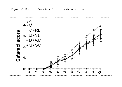
Figure 2: Delay of diabetic cataract in rats by treatment. (Figure 2 is shown
in the drawings
accompanied with the specification). The data is expressed as mean SEM.
Control (Non-
diabetic control); D (Diabetic control); D-f-RL (diabetic + regular lutein);
D+SL (Diabetic +
soluble lutein); D+RC (Diabetic + regular curcurnin); D+SC (Diabetic + soluble
curcumin)
The onset of cataract due to hyperglycemia was observed in diabetic animals
after three weeks of
STZ injection. The average incidence of cataract was calculated and presented
in Figure 2.
Though there was no delay in the onset there is a clear delay in the
progression and maturation
of cataract in all the treatment groups when compared to Group-D. Group-D
animals showed
lens opacification (stage-IV) by the end of the 10th week, while the treatment
groups
showed around stage-2.5 to 3. The data clearly indicates there is a
significant delay in the
progression and maturation of cataract intervention groups from the sixth week
onwards, when
compared to group-D. At the end of ten weeks, the severity of cataracts was
significantly lower
in groups D+RL (stage 3.1), D+SL (stage 2.7), D-f-RC (stage 3.0) and D+SC
(stage 3.2) than in
31
CA 02943921 2016-09-26
WO 2015/145389
PCT/1B2015/052248
Group-D (Stage 4), indicating that intervention with any agent delayed the
maturation of
diabetic cataract due to slow progression, Further SL seems to be more
effective than RL, but
SC did not show superiority in efficacy over RC in progression of cataract.
All the lenses in
Group-C during the entire experimental period appeared to be normal, clear and
free of
opacities.
LENS BIOCHEMICAL ANALYSIS:
Individual lenses were weighed and pooled into 4 lenses for a pool, and such 4-
5 pools were
formed per group. A 10 % homogenate was prepared in 50 mM sodium phosphate
buffer pH 7.4,
with tissue homogenizer at intermittent time gaps to avoid excess heat
generation. Separate
aliquots of total homogenate (TH) were made 250 IA for TBARS assay, 150 IA for
sorbitol
estimation, and 20 IA for protein estimation. Remaining homogenate was
centrifuged at 10,000
RPM for 30 min at 4 C. Supernatant was separated into labelled vials, as
total soluble protein
(TSP).
Determining soluble percentage of protein in lens homogenate: Protein
estimation was
done in lens homogenate and soluble fraction by Lowry's method. Amount of
protein present
per gram weight of lens was calculated. Percentage of soluble protein was
calculated by
multiplying the fraction of soluble protein by 100.
We analyzed the total and soluble protein content in the lenses of all the
experimental groups.
There was a significant decrease in both total and soluble protein in Ciroup-D
compared with
the control group. This could be due to a partial leakage of proteins into the
aqueous humor, or
aggregation of proteins and insolubilization. Among the treatment groups, SL
and RC
significantly prevented loss of soluble protein compared to group- D, whereas,
SL alone had
shown significant difference against group D in percentage of soluble protein.
SC and RL had
shown partial beneficial effect in preventing insolubilization of lens
proteins but were relatively
less significant statistically.
32
CA 02943921 2016-09-26
WO 2015/145389
PCT/1B2015/052248
Table 2: Protein content in total and soluble fraction of lens homogenate. The
data is expressed
as mean SEM. n=6; Control (Non-diabetic control); D (Diabetic control);
D+RIL (diabetic +
regular lutein); D+SL (Diabetic + soluble lutein); D+RC (Diabetic + regular
curcumin); D+SC
(Diabetic + soluble curcumin); ***--p<0.001, **=P<0.01 and
*=P<0.05 Vs C; ##= P<0.01 and fi= P<0.05 Vs D
Table: 2
Total protein Soluble protein Percentage soluble
Group
(mg/gm lens) (mg/gm lens) protein
Control (C)
516.54 9.3 388.77 8.2 75.19 2.2
281.34 31.0*** 128.87 11.0*** 46.87 2.5***
D+RL
311. 79 7. 8 156.55 4.7 50.35 1.9
D+SL ##
218.78 14.3 58.04 1.8
376.53 20.9
D+RC
202.63 21.6
368.50 19.2 54.43 3.8
D+SC
351.83 41.5 178.99 25.9 50.16 1.3
SDS-PAGE protein profiling: Differences in protein distribution pattern were
observed by
running the lens protein samples on 12% polyacrylamide gel. 30 lig of protein
was loaded per
well along with molecular weight marker for SDS-PAGE (Broad range SDS-PAGE
marker,
BioRad). The SDS-electrophoretic pattern of the soluble protein fraction
showed a band
corresponding to aggregated proteins at-50 kDa in group-D in relation to the
group-C and
with reduced band intensity in treatment groups RL, SL, RC and SC. Figure 3
represents the
SDS-PAGE pattern of soluble fraction of lens proteins, as shown in the
drawings accompanied
with the specification.
SIZE EXCLUSION HIGH PERFORMANCE LIQUID CHROMATOGRAPHY:
33
CA 02943921 2016-09-26
WO 2015/145389
PCT/1B2015/052248
Size exclusion chromatography on TSK-3000 HPLC column resulted in resolution
of crystalline
lens proteins. HPLC profile demonstrated the reduced peak area in low
molecular weight region,
and increased peak area in the high molecular weight protein region in Group-D
(red line) TSP,
as compared to group-C (black line). This suggests there was a phenomenon of
protein
aggregation in diabetic conditions. Intervention with all except RL, i.e. SL,
RC and SC
normalized the profile of TSP. Except RL, all other compositions SL, RC and SC
intervened
with TSP and normalized the profile, while RL did not show any effect on TSP
levels and they
remained abnormal like diabetic condition.
Figure 4 represents the size exclusion chromatography of lens protein soluble
fraction, as shown
in the drawings accompanied with the specification.
SORBITOL LEVELS: We assessed accumulation of sorbitol in the lens of all
experimental
animals and the data were presented in the Figure 8. Group-D showed
significantly elevated
levels of sorbitol (5.877 0.27) when compared with group-C (0.301 0.04 u
moles/gm lens of
sorbitol). Among the intervention groups, except SC, remaining treatments did
not lower sorbitol
accumulation compared to group-D. Group-SC showed significantly lower sorbitol
levels when
compared to group-D and significantly higher sorbitol levels when compared
with
group-C. This might be attributed to the additional pharmacological action of
curcumin as an
aldose reductase inhibitor.
Figure 5: Spectrofluoremetric measurement of lens sorbitol (Figure 5 is shown
in the drawings
accompanied with the specification). The data is expressed as mean SEM. n=6;
Control (Non-
diabetic control); D (Diabetic control); D+RL (diabetic + regular lutein);
D+SL (Diabetic +
soluble lutein); D+RC (Diabetic + regular curcumin); D+SC (Diabetic + soluble
curcumin);
*=P<0.05 Vs C; # = P<0.05 Vs D.
Plasma lutein levels: Plasma lutein levels were measured by HPLC. Since rodent
chow diet
does not contain carotenoids, lutein was not detected in control and diabetic
rats. However, lutein
could be detected in lutein supplemented groups. Feeding of diabetic rats with
regular lutein
results in 0.01 micromoles/L of plasma lutein (Figure 6). Feeding of soluble
lutein led to seven
34
CA 02943921 2016-09-26
WO 2015/145389
PCT/1B2015/052248
fold increase in plasma lutein 0.07 micromoles (Figure 6), suggesting that
soluble lutein
increases bioavailability of lutein significantly and may be the reason for
improved beneficial
effects with soluble lutein compared to regular lutein.
Figure 6: HPLC measurement of plasma lutein levels (Figure 6 is shown in the
drawings
accompanied with the specification). The data was expressed as mean SEM. n=6;
Control
(Non-diabetic control); D (Diabetic control); D+RL (diabetic + regular
lutein); D+SL (Diabetic +
soluble lutein).
Conclusion
Supplementation of curcumin rescued photoreceptor degeneration in transgenic
rats with P23H
rhodopsin mutation. Feeding of dietary antioxidant curcumin was effective in
delaying
streptozotocin (STZ)-induced diabetic cataract in rats mainly through its
antioxidant property.
In addition, curcumin inhibited diabetes-induced expression of vascular
endothelial growth
factor (VEGF) in rat retina and lens aldose reductase (AR).
Soluble lutein is more effective in delaying diabetic cataract compared to
regular lutein at dose
of 0.5% in the diet which is reflected in molecular analysis related to
cataract genesis. Increased
bioavailability of soluble lutein might explain the observed biological
effects of soluble lutein
compared to regular lutein. The efficacy of soluble curcumin was almost
comparable with
regular curcumin.
Example 2-
Effect of UltraSol Lutemax2O2OTM in diabetic retinopathy by Nutrigenomics
approach
The effect of soluble lutein (UltraSol Lutemax2O2OTM) was investigated in
diabetic rats with
respect to its beneficial effect on retina by a nutrigenomics approach and the
effect was
compared with regular lutein (Lutemax2020 ).
Methodology
Animal model: The streptozotocin (STZ) rat model of diabetes has been one of
the most
commonly used models of human disease with respect to diabetes. It is known to
mimic many of
the acute and some of the chronic complications observed in human diabetes.
This model has the
CA 02943921 2016-09-26
WO 2015/145389
PCT/1B2015/052248
advantage of being highly reproducible and the timelines for various
complications to develop
are well recognized and reproducible. Given the established similarities of
some of the structural,
functional and biochemical abnormalities of human disease, it is considered an
appropriate
model to assess mechanisms of diabetes and evaluate potential therapies.
Experimental design: Male Wistar-NIN rats with an average body weight of 120
anis are
obtained from the National center for laboratory animal sciences, National
Institute of Nutrition,
Hyderabad (NCLAS, MN). Animals were maintained at NCLAS. MN and kept for
acclimatization in an experimental room for two weeks. Diabetes was induced in
overnight
fasted animals by a single intraperitoneal injection of STZ (30 mg/ kg) in 0.1
M citrate buffer,
pH 4.5. Another set of rats, which received only a vehicle, served as the
control (Group-C; n=6).
Fasting blood glucose levels were nieasured 72 h after STZ injection. Animals
having blood
glucose levels >150 ing/dt, were considered diabetic and all the animals are
divided into four
groups as shown in the Table 3 below.
Table 3
Group No. of Diet
animals
I Control 6 AIN 93
II Diabetic 9 AIN 93
HI Diabetic + Soluble lutein (SL) 8 AIN 93 with soluble lutein
0.5 %
IV Diabetic + Regular lutein (R1_,) 6 AIN 93 with regular
lutein 0.5 %
All the animals were housed in individual cages maintained on their respective
diets for 12
weeks and drinking water was provided ad libitum throughout the study period.
Daily food
intake and weekly body weights, fasting glucose levels were noted. Before
sacrifice
electroretinogram was performed and glycosylated hemoglobin (HbAlC) was
estimated. At the
end of 12 weeks, rats were euthanized and retinas harvested for histological
and molecular
analysis (gene and protein expression).
36
CA 02943921 2016-09-26
WO 2015/145389
PCT/1B2015/052248
Electroretino gram (ERG) Analysis: Diabetic retinoapthy is characterized by
disturbances in
retinal function. The retinal function can be assessed by electroretinogram.
Diabetes results in
ischemia and apoptosis in different retinal cell layers, which results in
changes in the functions of
the retina. It is well reported that oscillatory potentials (OPs, where OP
represents waves, called
oscillatory waves, which are a major component of ERG) are more affected in
diabetes than a- or
b- waves. OPs represent the functional aspects of inner retinal layers,
ganglion cell layer and
inner plexiform layer.
Animals were dark-adapted for overnight and prepared for the ERG procedure
under dim red
illumination. The pupils of the rats were dilated with atropine eye drops. The
ground electrode
was a subcutaneous needle in the tail, and the reference electrode was an ear
clip electrode. The
active contact lens electrodes were placed on the cornea. The recordings were
performed with a
UTAS Visual Diagnostic System. The responses were differentially amplified
with a gain of
1,000 using alternating current-coupled UBA-4204 Amplifier. A flash stimuli of
-2 to 8 dB were
delivered via a with BigShotTM Ganzfeld System (LKC Technologies;
Gaithersburg, MD, USA).
The oscillatory potentials were extracted from the wave form and the sum of
all OPs was
calculated.
Quantitative real-time PCR: Total RNA was extracted from the retina of rats
using Tri reagent.
Isolated RNA was further purified by RNeasy Mini Kit (Qiagen) and quantified
by measuring
the absorbance at 260 and 280 nm on ND1000 spectrophotometer (NanoDrop
technologies,
Delaware, USA). The quality of RNA preparation was assessed by electrophoresis
on a
denaturing agarose gel. Two lig of total RNA was reverse transcribed using
High Capacity
cDNA Reverse Transcription kit. Reverse transcription reaction was carried out
with a
thermocycler (ABI 9700). Real-time polymerase chain reaction (PCR) (ABI-7500)
was
performed in triplicates with 25ng cDNA templates using SYBR green master mix
with gene
specific primers. Normalization and validation of data were carried using 0-
actin as an internal
control and data were compared between samples according to comparative
threshold cycle (2-
AAct) method.
SDS-PAGE and Immunoblotting:Retina were homogenized in a buffer containing 20
mMTris,
100 mM NaC1, 1 mM ethylenediamine tetraacetic acid (EDTA) (TNE buffer; pH7.5)
containing
37
CA 02943921 2016-09-26
WO 2015/145389
PCT/1B2015/052248
1 mM dithiothreitol (DTT), 1 mM phenylmethyl sulfonyl fluoride (PMSF), 1 pg/m1
of each
aprotinin, leupeptin, and pepstatin. The homogenate was centrifuged at
12,000xg for 20 min. The
supernatant was collected and used for immunoblot analysis. Equal amount of
protein from the
supernatant was subjected to 12% SDS-PAGE and proteins were transferred onto
polyvinylidene
fluoride (PVDF) membrane. Nonspecific binding was blocked with 5% BLOT-
QuickBlocker
reagent (WB57, Calbiochem) in phosphate buffered saline with Tween (PBST) and
incubated
overnight at 4 C with primary antibodies diluted in phosphate buffered saline
(PBS). After
washing with PBST, membranes were then incubated with anti-rabbit IgG (1:3500)
secondary
antibodies conjugated to horseradish peroxidase (HRP). The immunoblots were
developed with
enhanced chemiluminescence detection reagents (RPN2232, GE Health Care,
Buckinghamshire,
UK) and digital images were recorded by Image analyzer (Syngene, G-box).
Quantification of
band intensity was performed with Image J software.
Histopathology: The eye balls from selected animals were collected in fixative
4 %
paraformaldehyde solution in separately labeled vials. The tissues were given
some nicks so as to
facilitate penetration of fixative into deep tissue. They were kept at room
temperature for 24-48
hrs followed by replacing the fixative with 20 mM sodium phosphate buffer.
Buffer was
scheduled to be replaced with fresh buffer one each week until
histopathological processing.
Tissues were embedded in paraffin and sections were taken in microtome. Coated
slides are used
for immunohistochemistry and immunofluorescence, whereas uncoated slides for
hemotoxylin
and eosin (H & E) staining.
Statistical Analysis: The data was expressed as mean SEM. n=6; C (Non-
diabetic control); D
(Diabetic control); D+RL (diabetic + regular lutein); D+SL (Diabetic + soluble
lutein); ***
=p<0.001, ** =P<0.01 and *=P<0.05 Vs C; ## = P<0.01 and # = P<0.05 Vs D.
Results were analyzed for statistical significance by one way ANOVA followed
by Dunnett's
multiple comparison test for comparing all the groups with control group.
Between group
significance was checked by two tailed unpaired t-test.
38
CA 02943921 2016-09-26
WO 2015/145389
PCT/1B2015/052248
Results
Electroretinograph: In diabetic rats (D) amplitude of OPs were reduced (334.2
V) compared to
normal control (C) animals (498.4 V). It is also noted that implicit time for
OPs is increased in
group-D. Ingestion of antioxidant lutein resulted in lowering the reduction in
OP amplitudes,
which is suggested by the sum of OPs; RL (442.6) and SL (561.9). Group-SL had
shown
significant difference in sum of OPs compared to group-D, in fact it is better
than normal control
rats.
Figure 7: Representative wave forms of OPs are shown from different groups and
total
amplitudes (Figure 7 is shown in the drawings accompanied with the
specification). C, Non
diabetic control; D, Diabetic control; D+RL, Diabetes+Regular Lutein; D+SL,
Diabetes+Soluble
Lutein.
Morphology of retina: In control rats C, all the layers of the retina are
intact and with maximum
thickness of retina, and also noted with dense INL, and distinguishable
separation (OPL)
between INL and ONL. In contrast, diabetic retina (D) showed significantly
reduced total retinal
thickness and were also marked by less dense ONL and almost merged INL and
ONLs.
Treatment with lutein prevented gross morphological changes to a significant
extent in diabetic
retina. Soluble formulation of lutein was shown to be better than the regular
lutein, indicated by
dense ONLs (Fig 8). Figure 8 represents histology of retina, as shown in the
drawings
accompanied with the specification.
Table 4: Thickness of layers of retina (p) of different groups.
Retinal Total retinal
GCL+IPL INL OPL ONL PRL
kers thickness
121.74 46.813 10.767 5.024 25.697 19.426
58.848 12.508 9257 0.791 23.051 12.184
RI., 89.409 31.518 12.117 1.41 23.181
12.600
SL 112.714 40.877 16.074 4.718 31.196
23.07/
A representative table of retinal layers thickness (in pm) measured by the use
of Lieca
application suit, Leica, Switzerland. C, Non diabetic control; D, Diabetic
control; D+RL,
39
CA 02943921 2016-09-26
WO 2015/145389
PCT/1B2015/052248
Diabetes+Regular Lutein; D+SL, Diabetes+Soluble Lutein; GCL, ganglion cell
layer; IPL,
inner plexiform layer; INL, inner nuclear layer; OPL, outer plexiform layer;
ONL, outer
nuclear layer; PRL, photo receptor layer.
Expression of retinal markers (genes) by real-time PCR, immunohistochemistry
and
immunoblotting:
Rhodopsin (Rho): Rhodopsin (Rho) is a biological pigment in photoreceptor
cells of
the retina that is responsible for the first events in the perception of
light. The mRNA levels of
Rho gene as quantified by real time PCR showed decreased levels in the retina
of diabetic
animals. Treatment with RL and SL prevented its decline and however feeding of
SL had
significant effect when compared to RC and even better than normal rats (Fig.
9A, as shown in
the drawings accompanied with the specification). Further, we quantified
protein levels of Rho
by immunofluorescence, which is in coincidence with mRNA levels.
Immunofluorescence
imaging of Rho protein showed decreased expression in diabetic rat retina in
comparison to
normal control rat retina. Treatment with RL and SL prevented loss of Rho
protein expression in
diabetic retina indicated by intense Rho positive fluorescence. Furthermore,
SL is found to be
distinctly more effective than RL in preventing loss of Rho protein expression
in rat retina in
diabetic animals respectively (Fig. 9B, as shown in the drawings accompanied
with the
specification).
Nerve Growth Factor (NGF): NGF is the best-characterized neurotrophin, known
to play a key
role in the survival and differentiation of select neurons in the peripheral
and central nervous
system. Since its discovery in the 1950s, NGF has shown promise in the
treatment of progressive
neurodegenerative disorders. In animals, NGF is known to promote nerve
terminal outgrowth
and neuron recovery after ischemic, traumatic, and toxic injuries. We checked
the status of NGF
gene expression by real time PCR and found down regulation in the retinas of
diabetic animals.
Treatment with RL could not prevent its decline whereas SL prevented its down
regulation
(Fig.10A, as shown in the drawings accompanied with the specification). The
protein levels of
NGF as estimated by immunofluorescence yielded similar results.
Immunofluorescence imaging
of NGF protein showed its decreased expression in diabetic rat retina in
comparison to normal
control rat retina (Fig. 10B, as shown in the drawings accompanied with the
specification).
Treatment with SL prevented loss of NGF protein in diabetic retina indicated
by intense NGF
CA 02943921 2016-09-26
WO 2015/145389
PCT/1B2015/052248
positive fluorescence. RL was unable to prevent the loss of NGF protein
effectively in diabetic
rat retina.
Vascular endothelial growth factor (VEGF): Vascular endothelial growth factor
(VEGF) is a
signal protein produced by cells that stimulates vasculogenesis and
angiogenesis. It is part of the
system that restores the oxygen supply to tissues when blood circulation is
inadequate. When
VEGF is over expressed, it can contribute to disease. VEGF causes widespread
retinal vascular
dilation, produces breakdown of the blood-retinal barrier, and is implicated
in ocular
neovascularization. Western blotting for VEGF indicated the up regulation of
VEGF expression
in diabetic retina (Fig.11, as shown in the drawings accompanied with the
specification).
Treatment with both RL and SL inhibited diabetes induced VEGF over expression.
Platelet-derived growth factor (PDGF): PDGF is a growth factor that plays a
significant role
in blood vessel formation (angiogenesis), the growth of blood vessels from
already-existing
blood vessel tissue. Several studies have shown elevated PDGF concentrations
in vitreous
samples from patients with diabetic retinopathy. Like VEGF, PDGF is a
proangiogenic growth
factor that may promote aberrant neovascularization in diabetic retinopathy.
Furthermore, PDGF
may stimulate the formation and traction of epiretinal membranes in patients
with diabetic
retinopathy, leading to tractional retinal detachment. Indeed, the development
of inhibitors that
antagonize PDGF signaling in pathologic retinal neovascularization remains an
active area of
ophthalmic drug development. Immunohistochemistry for PDGF indicated the up
regulation of
protein in diabetic retina (Fig.12, as shown in the drawings accompanied with
the specification).
Treatment with both RL and SL inhibited diabetes induced PDGF over expression.
Plasma lutein: To understand the effect of soluble lutein administration on
its bioavailability
plasma levels of carotenoids were measured by HPLC method. The data showed
undetectable
lutein levels in plasma of rats fed with normal diet (AIN-93). The plasma
lutein levels were in
detectable range in rats fed with AIN-93 diets mixed with regular lutein and
soluble lutein. More
interestingly, rats fed with SL diet were found to contain seven folds higher
lutein levels
compared with rats fed with RL diet. This in particular confirmed the success
of this formulation
in improving the bioavailability of lutein.
41
CA 02943921 2016-09-26
WO 2015/145389
PCT/1B2015/052248
Figure 13 represents serum lutein levels measured by reverse phase (RP)-HPLC,
as shown in the
drawings accompanied with the specification. Values are expressed as mean
SEM (n=6). ,
$$$<0.001 D+SL Vs. D+RL.
Conclusion
Lutein administration to rats prevented diabetes induced abnormalities in the
retina. Lutein
retained the functionality of retina of rats which is lost in diabetic rats as
checked by ERG. It is
also evident by the morphological study of retina as done by H & E staining.
Lutein prevented
decline in the expression of rhodopsin and nerve growth factor (NGF) which
have vital roles in
maintaining health of the retina. Lutein prevented over expression of VEGF and
PDGF that are
involved in stress and angiogenesis. Interestingly rats treated with soluble
lutein showed
profound benefit when compared with regular lutein, and the increased
bioavailability is shown
by the increased plasma levels. Further, the antioxidant and anti-inflammatory
potential of lutein
may contribute for its beneficial effect. Hence soluble lutein can be used to
treat and or prevent
diabetic retinopathy.
Example 3
Effect of UltraSol CurcuWinTM in diabetic retinopathy by Nutrigenomics
approach
The effect of soluble curcumin (UltraSol CurcuWinlm) was investigated in
diabetic rats with
respect to its beneficial effect on retina by nutrigenomics approach and the
effect was compared
with regular curcumin.
Methodology
Same as mentioned in example 2. All the animals were divided into four groups
as shown below
Table 5
Group No. of Diet
animals
I Control 6 AIN 93
H Diabetic 9 AIN 93
42
CA 02943921 2016-09-26
WO 2015/145389
PCT/1B2015/052248
HI Diabetic + Soluble lutein (SC) 8 AIN 93 with soluble curcumin
0.01 %
IV Diabetic + Regular lutein (RC) 6
AIN 93 with regular curcumin 001 %
Electroretinograph: In diabetic rats (D) amplitude of OPs were reduced (334.2
[IV) compared to
normal control (C) animals. It is also noted that implicit time for OPs is
increased in group-D.
Ingestion of antioxidant curcumin resulted in lowering the reduction in OP
amplitudes suggested
by the sum of OPs; RC (445.7), SC (455.3). SC not only lowered the reduction
in sum of OPs,
but also normalized the implicit times.
Figure 14 represents wave forms of OPs from individual animals of different
groups and total
amplitudes, as shown in the drawings accompanied with the specification. C,
Non diabetic
control; D, Diabetic control; D+RC, Diabetes+Regular Curcumin; D+SC,
Diabetes+Soluble
Curcumin.
Morphology of Retina: In control rats C, all the layers of the retina are
intact and with maximum
thickness of retina and also noted with dense INL, and distinguishable
separation (OPL) between
INL and ONL. In contrast, diabetic rats retina (D) showed significantly
reduced total retinal
thickness and were also marked by less dense ONL and almost merged INL and
ONLs.
Treatment with curcumin prevented gross morphological changes to a significant
extent in
diabetic retina. Soluble formulation of this active principle was shown to be
better than the
regular active principle, indicated by dense ONLs.
Figure 15, Representative histology of retina, as shown in the drawings
accompanied with the
specification.
Table 6: Thickness of layers of retina (pt) of different groups.
Retinal Total retinal
GCL+EPL ESL OPL ONL PRL
layers thickness
121.74 46.813 10.767 5.024 25.697 19.426
58.848 12.508 9.257 0.791 23.052 12.14
= =
RC 106.014 35.692 13.648 5.685 30.108
19.520
SC 124.430 54.004 14.926 6.597 29.320
21.857
A representative table of retinal layers thickness (in jam) measured by the
use of Lieca
application suit, Leica, Switzerland. C, Non diabetic control; D, Diabetic
control; D+RC,
43
CA 02943921 2016-09-26
WO 2015/145389
PCT/1B2015/052248
Diabetes+Regular Curcumin; D+SC, Diabetes+Soluble Curcumin. GCL, ganglion cell
layer; IPL, inner plexiform layer; INL, inner nuclear layer; OPL, outer
plexiform layer;
ONL, outer nuclear layer; PRL, photo receptor layer.
Expression of retinal markers (genes) by real-time PCR, immunohistochemistry
and
immunoblotting:
Rhodopsin (Rho): Rhodopsin, is a biological pigment in photoreceptor cells of
the retina that is
responsible for the first events in the perception of light. The mRNA levels
of Rho gene as
quantified by real time PCR showed decreased levels in the retina of diabetic
animals. Treatment
with RC and SC prevented its decline and however SC treatment has more
beneficiary effect
when compared to RC (Fig.16.A, as shown in the drawings accompanied with the
specification).
Protein levels of Rho were quantified by immunofluorescence, which is in
coincidence with
mRNA levels. Immunofluorescence imaging of Rho protein showed its decreased
expression in
diabetic rat retina in comparison to normal control rat retina. Treatment with
RC and SC
prevented loss of Rho protein expression in diabetic retina indicated by
intense Rho positive
fluorescence. Furthermore, SC is found to be more effective than RC in
preventing loss of Rho
protein expression in rat retina in diabetic animals respectively (Fig.16.B,
as shown in the
drawings accompanied with the specification).
Nerve Growth Factor (NGF): NGF is the best-characterized neurotrophin, known
to play a key
role in the survival and differentiation of select neurons in the peripheral
and central nervous
system. Since its discovery in the 1950s, NGF has shown promise in the
treatment of progressive
neurodegenerative disorders. In animals, NGF is known to promote nerve
terminal outgrowth
and neuron recovery after ischemic, traumatic, and toxic injuries. We checked
the status of NGF
gene expression by real time PCR and found down regulation in the retinas of
diabetic animals.
Treatment with both regular as well as soluble curcumin showed equal
beneficiary effect in
preventing down regulation of NGF gene under diabetic conditions (Fig. 17.A,
as shown in the
drawings accompanied with the specification). The protein levels of NGF as
estimated by
immunofluorescence yielded similar results. Immunofluorescence imaging of NGF
protein
showed its decreased expression in diabetic rat retina in comparison to normal
control rat retina
(Fig.17.B, as shown in the drawings accompanied with the specification).
Treatment with RC
and SC prevented loss of NGF protein in diabetic retina indicated by intense
NGF positive
fluorescence.
44
CA 02943921 2016-09-26
WO 2015/145389
PCT/1B2015/052248
Vascular endothelial growth factor (VEGF):
Vascular endothelial growth factor (VEGF) is a signal protein produced by
cells that stimulates
vasculogenesis and angiogenesis. It is part of the system that restores the
oxygen supply to
tissues when blood circulation is inadequate. When VEGF is over expressed, it
can contribute to
disease. VEGF causes widespread retinal vascular dilation, produces breakdown
of the blood-
retinal barrier, and is implicated in ocular neovescularization.
Western blotting for VEGF indicated the up regulation of VEGF expression in
diabetic retina
(Fig.18, as shown in the drawings accompanied with the specification).
Treatment with both RL
and SL inhibited diabetes induced VEGF over expression.
Platelet-derived growth factor (PDGF): PDGF is a growth factor that regulates
cell growth
and division. In particular, it plays a significant role in blood vessel
formation (angiogenesis),
the growth of blood vessels from already-existing blood vessel tissue. Several
studies have
shown elevated PDGF concentrations in vitreous samples from patients with
diabetic
retinopathy. Like VEGF, PDGF is a proangiogenic growth factor that may promote
aberrant
neovascularization in diabetic retinopathy. Furthermore, PDGF may stimulate
the formation and
traction of epiretinal membranes in patients with diabetic retinopathy,
leading to tractional retinal
detachment. Indeed, the development of inhibitors that antagonize PDGF
signaling in pathologic
retinal neovascularization remains an active area of ophthalmic drug
development.
Immunohistochemistry for PDGF indicated the up regulation of protein in
diabetic retina (Fig.19,
as shown in the drawings accompanied with the specification). Treatment with
both RC and SC
inhibited diabetes induced PDGF over expression. Soluble curcumin was more
effective than
regular curcumin (Fig. 19, as shown in the drawings accompanied with the
specification).
Conclusion
Curcumin administration to rats prevented diabetes induced abnormalities in
the retina.
Curcumin retained the functionality of retina of rats which is lost in
diabetic rats as checked by
ERG. It is also evident by the morphological study of retina as evaluated by H
& E staining
where thickness of various layers of retina was measured. Curcumin prevented
decline in the
expression of rhodopsin and nerve growth factor which have vital role.
Curcumin prevented
over expression of VEGF and PDGF that are involved in stress and angiogenesis.
Interestingly
rats treated with soluble curcumin showed profound benefit when compared with
regular
CA 02943921 2016-09-26
WO 2015/145389
PCT/1B2015/052248
curcumin and this might be due to increased bioavailability. Hence soluble
curcumin can be
used to treat and or prevent diabetic retinopathy.
Advantages
1. Provides dry free flowing, water soluble/miscible form of lipophilic
nutrient.
2. Provides a suitable method of delivering poorly water soluble, oily,
lipophilic nutrient in the
form of granule, powder, tablets, ointment, paste, mouth wash, gargle, sachet,
capsules or in to
beverages.
3. Provides a dosage form of poorly water soluble, oily, lipophilic nutrient
with high
bioavailability.
4. The compositions herein are effective to regulate blood glucose levels.
5. The compositions and methods herein containing lipophilic nutrients are
effective to regulate
amacrine cells dysfunction in diabetic retinopathy.
6. The compositions herein are effective to prevent loss of retinal layers,
Rhodopsin levels and
NGF protein levels in diabetic retinopathy.
7. The compositions herein are effective to inhibit diabetes induced PDGF over
expression in
diabetic retinopathy.
8. The compositions herein are effective to prevent accumulation of in-soluble
lens proteins in
diabetic cataract.
9. The compositions herein are effective to prevent accumulation of sorbitol
levels in lens in
diabetic cataract.
10. The compositions herein are effective to reduce protein aggregation and
normalize the profile
of total soluble protein in diabetic cataract.
46