Note : Les descriptions sont présentées dans la langue officielle dans laquelle elles ont été soumises.
CA 02945588 2016-10-12
WO 2015/166399 PCT/1B2015/053052
GLYCAN SAMPLE PREPARATION
RELATED APPLICATIONS
[0001] This application claims the benefit of priority from US Provisional
Applications Nos.
61/986736 filed on April 30, 2014 and 62/150722, filed on April 21, 2015, the
contents of both
of which are hereby incorporated by reference in their entirety.
FIELD
[0002] Methods, systems, and kits for the analysis of glycans and/or
glycoconjugates are
disclosed herein. In some aspects, the present teachings can enable automated
glycoanalysis
protocols that exhibit a substantially reduced sample preparation time
relative to known methods.
INTRODUCTION
[0003] Protein glycosylation typically refers to a post-translational
modification in which an
oligosaccharide or glycan is attached to a protein. Given the importance of
glycosylation on
protein folding, transport, and cell-cell interactions, for example, many
research tools have been
developed to characterize and/or analyze glycoconjugates and the glycans
associated therewith.
Such tools have become critical in the biomedical sciences, biopharmaceutical
industry (e.g.,
biomarker discovery), and in efficacy/safety assessment of protein
therapeutics for regulatory
agencies.
[0004] Though the most common glycoanalytical methods of capillary
electrophoresis and
hydrophilic interaction liquid chromatography can be effective, these methods
can necessitate
extensive sample preparation, including glycoprotein capture, N-glycan
release, fluorescent
derivatization, purification, and pre-concentration steps. Currently used
protocols to fulfill these
tasks, however, are time consuming and require multiple centrifugation and/or
vacuum-
centrifugation steps, for example, thereby reducing throughput and making
automation difficult
and/or expensive.
[0005] Accordingly, there remains a need for efficient and effective
methods for the
purification of glycans and/or the analysis of glycoconjugates or the glycans
associated
therewith.
SUMMARY
1
CA 02945588 2016-10-12
WO 2015/166399 PCT/1B2015/053052
[0006] The present teachings relate to methods, systems, and kits for the
purification and/or
analysis of a glycoconjugate or glycan, and specifically, to magnetic bead-
based sample
preparation methods that can enable full automation and/or reduced sample
preparation time
relative to current protocols. In some aspects, the methods described herein
can provide for one
or more of glycoprotein digestion, N-glycan release, fluorescent labeling, and
glycan purification
for subsequent capillary electrophoresis with laser induced fluorescence
detection (CE-LIF),
liquid chromatography (LC), or other analytical methods (e.g., mass
spectrometry (MS), nuclear
magnetic resonance (NMR)), without requiring time-consuming sample preparation
steps such as
centrifugation or vacuum-centrifugation.
[0007] In accordance with various aspects, certain embodiments of the
applicants' teachings
relate to a method of purifying glycans that comprises reacting a sample
containing one or more
glycoconjugates (e.g., glycoproteins, glycopeptides, antibodies, proteoglycan,
glycosphingolipid,
chondroitin sulfate, heparin sulfate, hyaluronan, glycolipid,
glycoseaminoglycan, fusion
glycoprotein, antibody-drug conjugate) with a deglycosylation reagent (e.g.,
an endoglycosidase
for N-linked glycans, beta elimination for 0-linked glycans) so as to release
glycans from the
glycoconjugates, associating the released glycans with a plurality of magnetic
particles (e.g.,
carboxyl-coated magnetic beads), applying a magnetic field to draw down the
plurality of
magnetic particles having the released glycans associated therewith, removing
a supernatant
from the drawn-down magnetic particles so as to remove the deglycosylation
reagent (e.g.,
enzyme) and the deglycosylated sample, and dissociating or eluting the glycans
from the
magnetic particles. In some aspects, the method can prepare the glycans for
analysis via one of
CE (e.g., with LIF, with UV labeling), LC (e.g., with fluorescent or UV
detection), MS, and
NMR, and combinations thereof. In some aspects, the glycans are purified
without a
centrifugation or vacuum centrifugation step.
[0008] In accordance with various aspects, certain embodiments of the
applicants' teachings
relate to a method of analyzing one or more glycoconjugates (e.g.,
glycoproteins, glycopeptides,
antibodies, proteoglycan, glycosphingolipid, chondroitin sulfate, heparin
sulfate, hyaluronan,
glycolipid, glycoseaminoglycan, fusion glycoprotein, antibody-drug conjugate)
that comprises
reacting a sample containing one or more glycoconjugates with a
deglycosylation reagent (e.g.,
an endoglycosidase) to release glycans from the glycoconjugates and
associating the glycans
with a plurality of magnetic particles (e.g., carboxyl-coated magnetic beads).
A magnetic field
2
CA 02945588 2016-10-12
WO 2015/166399 PCT/1B2015/053052
can be applied to draw down the plurality of magnetic particles having the
glycans associated
therewith, and the supernatant can be removed so as to remove the
deglycosylation enzyme and
deglycosylated conjugates from the drawn-down magnetic particles. The glycans
can be reacted
with a labeling reagent so as to form labeled glycans that can then be
analyzed (e.g., via capillary
electrophoresis with laser induced fluorescent or UV detection). In accordance
with various
aspects of the present teachings, the labeled glycans can be prepared for
analysis without
centrifugation or vacuum centrifugation.
[0009] The method can include eluting the glycans from the magnetic
particles before or
after labeling the glycans. In some aspects, for example, the method can
include eluting the
glycans from the magnetic particles prior to reacting the glycans with the
labeling reagent. For
example, the glycans can be eluted from the magnetic particles by adding a
mixture comprising
the labeling reagent and an acid catalyst (e.g., acetic acid). In related
aspects, the glycans can be
reacted with the labeling reagent so as to form labeled glycans by adding a
reducing agent (e.g.,
NaBH3CN or pic-BH3) to initiate the reaction of the glycans with the labeling
reagent.
[0010] In some aspects, the method can also comprise associating the
labeled glycans with
the plurality of magnetic particles (e.g., magnetic microparticles, beads),
applying a magnetic
field to draw down the plurality of magnetic particles having the labeled
glycans associated
therewith, removing a supernatant from the drawn-down plurality of magnetic
particles having
the labeled glycans associated therewith so to remove excess labeling reagent,
and eluting the
labeled glycans from the plurality of magnetic particles. For example, the
labeled glycans can be
associated with the plurality of magnetic particles by adding acetonitrile and
the labeled glycans
can be eluted from the plurality of magnetic beads by adding an aqueous media
(e.g., water).
[0011] In accordance with various aspects of the present teachings, the
deglycolsylation
reagent can comprise PNGase F enzyme, glycans can be associated with the
plurality of
magnetic particles by adding acetonitrile, and the labeling reagent can
comprise one of 1-
aminopyrene-3,6,8-trisulfonic acid (APTS), 8-aminonaphthalene-1,3,6-
trisulfonic acid (ANTS),
2-anthranilic acid (2-AA), 2-aminobenzoic acid (2-AB) (that can be reacted
with the glycans, for
example, by adding a reducing agent such as NaBH3CN or pic-BH3).
[0012] In accordance with various aspects, certain embodiments of the
applicants' teachings
relate to a kit for purifying glycans that can comprise a plurality of
carboxyl-coated magnetic
3
CA 02945588 2016-10-12
WO 2015/166399 PCT/1B2015/053052
particles, deglycosylation reagents for releasing glycans from glycoconjugates
contained within a
sample (e.g., one or more endoglycosidases, PNGase F, hydrazine), and reagents
for associating
the glycans with the plurality of carboxyl-coated magnetic particles.
[0013] In some aspects, the kit can further comprise reagents for labeling
of the released
glycans, particularly for fluorescent labeling of the released glycans. For
example, the kits can
include one or more of APTS, ANTS, 2-AA, 2-AB, an acid catalyst (e.g., acetic
acid), and a
reducing agent such as NaBH3CN or pic-BH3.
[0014] In some aspects, reagents for associating the released glycans with
the plurality of
carboxyl-coated magnetic particles comprises acetonitrile.
[0015] In some aspects, the method can further comprise maintaining a
temperature equal to
or greater than about 37 C (e.g., equal to or greater than about 50 C) when
reacting the sample
with the deglycosylation enzyme.
[0016] In various aspects, the kit can further comprise reagents for
analyzing the labeled
glycans via capillary electrophoresis (e.g., with laser induced fluorescent
detection), liquid
chromatography, MS, or NMR. For example, the kit can include a fluorescently-
labeled internal
standard for the CE-LIF analysis of the labeled glycans (e.g., APTS-, ANTS-, 2-
AA-, or 2-AB-
labeled maltose).
[0017] In accordance with various aspects, a composition for separating
glycans using
capillary electrophoresis is provided comprising lithium acetate buffer,
polyethylene oxide,
ethylene glycol and linear polyacrylamide.
[0018] In various embodiments, the lithium acetate is at a concentration in
the composition
of between 10mM and 50mM at a pH of between 4 and 5.5, the polyethylene oxide
has a
molecular weight of between 100 and 1000 kDa and is at a concentration in the
composition of
between 0.5% and 5%, the ethylene glycol is at a concentration in the
composition of less than
60% and/or the linear polyacrylamide has a molecular weight of about 10 kDa
and is at a
concentration in the composition of between 0.5% and 5%.
[0019] In various embodiments, the composition can comprise lithium acetate
buffer at a
concentration in the composition of between 25 and 30 mM at a pH of about
4.75; polyethylene
oxide having a molecular weight of about 900 kDa at a concentration in the
composition of about
4
CA 02945588 2016-10-12
WO 2015/166399 PCT/1B2015/053052
1%; ethylene glycol at a concentration in the composition of about 20%; linear
polyacrylamide
having a molecular weight of about 10 kDa at a concentration in the
composition of about 3%.
[0020] These and other features of the applicants' teachings are set forth
herein.
BRIEF DESCRIPTION OF THE DRAWINGS
[0021] The skilled person in the art will understand that the drawings,
described below, are
for illustration purposes only. The drawings are not intended to limit the
scope of the applicants'
teachings in any way.
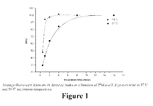
[0022] Figure 1 illustrates the effect of incubation duration and
temperatures during PNGase
F digestion in accordance with various aspects of the applicants' teachings.
[0023] Figures 2A-B demonstrate the effect on desialyation of labeling
incubation
temperature and time in accordance with various aspects of the applicants'
teachings.
[0024] Figures 3A-C illustrate an exemplary peak area distribution of
various glycan
structures using an exemplary magnetic bead based cleanup protocol in
accordance with various
aspects of the present teachings relative to conventional sample cleanup
methods.
[0025] Figure 4 schematically depicts an exemplary magnetic bead based
sample preparation
workflow for N-glycosylation analysis in accordance with various aspects of
the present
teachings.
[0026] Figure 5 demonstrates the effect of temperature and incubation time
on APTS
labeling efficiency in accordance with various aspects of the applicants'
teachings.
[0027] Figure 6 demonstrates the effect of APTS concentrations on labeling
efficiency at
various incubation temperatures in accordance with various aspects of the
applicants' teachings.
[0028] Figure 7 demonstrates the reproducibility as the function of the
amount of magnetic
bead suspension used in accordance with various aspects of the applicants'
teachings.
[0029] Figure 8 schematically depicts an exemplary magnetic bead based
sample preparation
workflow for N-glycosylation analysis in accordance with various aspects of
the present
teachings.
CA 02945588 2016-10-12
WO 2015/166399 PCT/1B2015/053052
[0030] Figure 9 shows the results obtained from CE-LIF analysis of multiple
APTS labeled
IgG glycans utilizing an automated liquid handling device in accordance with
various aspects of
the present teachings.
[0031] Figure 10 shows results from a CE-LIF analysis of individual glycans
and a mixture
of glycans in accordance with various aspects of the present teachings.
[0032]
DETAILED DESCRIPTION
[0033] It will be appreciated that for clarity, the following discussion
will explicate various
aspects of embodiments of the applicants' teachings, while omitting certain
specific details
wherever convenient or appropriate to do so. For example, discussion of like
or analogous
features in alternative embodiments may be somewhat abbreviated. Well-known
ideas or
concepts may also for brevity not be discussed in any great detail. The
skilled person will
recognize that some embodiments of the applicants' teachings may not require
certain of the
specifically described details in every implementation, which are set forth
herein only to provide
a thorough understanding of the embodiments. Similarly it will be apparent
that the described
embodiments may be susceptible to alteration or variation according to common
general
knowledge without departing from the scope of the disclosure. The following
detailed
description of embodiments is not to be regarded as limiting the scope of the
applicants'
teachings in any manner.
[0034] In accordance with various aspects of the present teachings, the
methods, systems,
and kits described herein can be used in the purification and/or analysis of a
glycoconjugate or
glycan that can enable full automation and/or reduce sample preparation time
relative to current
protocols. In some aspects, the methods described herein can provide for one
or more of
glycoconjugate digestion and/or glycan release, fluorescent labeling, and
glycan purification for
subsequent analysis, without requiring time-consuming sample preparation steps
such as
centrifugation or vacuum-centrifugation. Full automation to enable high
throughput
glycosylation profiling and sequencing, for example, may be vital to fulfill
contemporary needs
of the biopharmaceutical industry (e.g., development of biotherapeutic agents,
biomarker
discovery), and in regulatory agencies' efficacy/safety assessments of protein
therapeutics, which
require high-throughput and highly reproducible glycosylation screening
methods. Despite this
6
CA 02945588 2016-10-12
WO 2015/166399 PCT/1B2015/053052
need, one of the major handicaps of currently used sample preparation
protocols for
glycosylation analysis is the lack of easy automation, which currently require
high end
(expensive) robotic systems with centrifugation capabilities.
[0035] In accordance with various aspects, the exemplary magnetic bead-
based sample
preparation described herein can be performed in several hours, without
requiring any
centrifugation and/or vacuum centrifugation steps, thus enabling rapid, fully-
automatable
analysis that can utilize, for example, shorter incubation times during glycan
release and
labeling, the use of liquid handling robots for sample preparation, and/or
multicapillary methods.
As described below, the exemplary methods can thus improve processing time,
efficiency,
reproducibility, and ease of automation relative to conventional
centrifugation-based sample
preparation protocols.
[0036] In one embodiment, for example, an exemplary method generally
comprises the
following five individual steps, though it will be appreciated that methods
that include more or
fewer steps are within the scope of the present teachings: 1) deglycosylation
of the gluconjugate;
2) glycan capture; 3) glycan labeling; 4) clean up; and 5) glycan analysis
(e.g., by CE, LC,
MS, NMR).
[0037] It will further be appreciated that any sample containing or
suspected of containing a
glycan or glycoconjugate can be used in accordance with the present teachings,
including a
sample of blood, plasma, serum, urine or saliva. Further, the sample can
contain free glycans
(e.g., a previously deglycosylated sample) and/or glycoconjugates.
[0038] Exemplary glycoconjugates that can be analyzed according to the
present teachings
include glycoproteins such as fetuin, RNase B, and antibodies (e.g., IgG), all
by way of non-
limiting example. Other exemplary glycoconjugates that can be utilized include
proteoglycan,
glycosphingolipid, chondroitin sulfate, heparin sulfate, hyaluronan,
glycolipid,
glycoseaminoglycan, fusion glycoprotein, and antibody-drug conjugates. The
glycans that are
associated with the glycoconjugates generally comprise one or more sugar units
(e.g., glucose,
fucose, mannose, xylose, sialic acids N-Acetylglucosamine (G1cNAc), N-
acetylgalactosamine
(GalNAc) and oligosaccharides) that are covalently bonded to the base molecule
via a glycosidic
bond, for example. As will be appreciated by a person skilled in the art, the
glycans can
comprise a variety of carbohydrate units, branched or unbranched chains with
various linkages
7
CA 02945588 2016-10-12
WO 2015/166399 PCT/1B2015/053052
and positions, and/or oligosaccharides of various lengths that can be attached
to the base
molecule via N-linked glycosylation (e.g., a glycan linked to an amide
nitrogen of an asparagine
(Asn) residue of a protein), 0-linked glycosylation (e.g., a glycan linked to
an oxygen atom of
amino acid residue in a protein such as 0-Ga1NAc or 0-G1cNAc), C-linked
glycosylation (e.g.,
mannose added to a tryptophan residue in an amino acid sequence), and phospho-
serine
glycosylation (e.g., a glycan linked through the phosphate in a phospho-
serine), all by way of
non-limiting example.
Deglycosylation
[0039] In some aspects, methods of analyzing a glycoconjugate or glycan
associated
therewith can include a deglycosylation step that breaks the glycosidic bond
so as to remove the
glycan from the glycoconjugate. It will be appreciated that any
deglycosylation reagent known
in the art and modified in accordance with the present teachings can be
utilized. By way of non-
limiting example, methods and systems in accordance with the present teachings
can utilize
PNGase F (e.g., to remove N-linked oligosaccharides from the glycoproteins),
PNGase A, other
endoglycosidases such as O-Glycosidase, Endoglycosidase H and the
Endoglycosidase F), and
exoglycosidases (e.g., Neuraminidase), chemical agents such as hydrazine and
mixtures thereof.
[0040] As discussed in detail below, though PNGase F is conventionally used
in enzymatic
deglycosylations of N-glycans at 37 C with overnight incubation due to its
stability, specificity
and simple sample preparation conditions, applicants have achieved
significantly reduced
deglycosylation times by performing the deglycosylation at elevated
temperatures (e.g., greater
than 37 C, about 50 C). Indeed, maximum peak intensities of the released
glycans were found
to occur after one hour of incubation time at 50 C, whereas the N-
deglycosylation process
proceeded significantly slower at the conventional 37 C.
[0041] It should be appreciated in light of the present teachings that the
methods of analyzing
glycans can also be performed on a sample that has previously been
deglycosylated (e.g., a
sample containing glycans already dissociated from a protein or other
biopolymer) or a sample
containing only glycans (e.g., a glycan standard) such that a deglycosylation
or digestion step is
not required.
8
CA 02945588 2016-10-12
WO 2015/166399 PCT/1B2015/053052
Glycan Capture
[0042] After the glycoconjugate has been digested such that at least a
portion of the glycans
are released therefrom, the released glycans can be separated from the
deglycosylation reagent
(e.g., enzyme) and any disassociated polypeptide, for example. In accordance
with various
aspects of the present teachings, for example, a suspension of magnetic
particles can be mixed
with the sample such that the glycans can become associated with the magnetic
beads. In some
aspects, the suspension of magnetic particles can comprise acetonitrile, which
can promote the
capture of the released glycans. Following the association, a magnetic field
can be applied to the
mixture so as to attract (e.g., draw down, separate) the magnetic particles
having the glycans
associated therewith such that the supernatant containing the non-associated
deglycosylation
reagent, such as an enzyme and remaining polypeptide can be removed, for
example, by pouring
off or aspirating the supernatant.
[0043] Applicants have surprisingly discovered that the carboxylate-
modified surface of a
polymeric-coated magnetic particle (e.g., a carboxyl-coated magnetic bead) can
be particularly
effective in capturing the released glycans or glycoconjugates. Though it was
commonly
believed that the negatively-charged carboxyl groups extending from the
surface of a magnetic
bead and negatively-charged glycans (e.g., syalilated) would repel one another
and prevent
glycan capture, the applicants' have found that the carboxyl-coated magnetic
beads can
effectively and efficiently lead to partitioning of charged and/or uncharged
glycan. Such a result
is counterintuitive, especially in light of the relatively small molecular
weight of most glycans
(i.e., such that the glycans should be more susceptible to repulsive forces).
Without being bound
by a particular theory, it is believed that the binding buffer (e.g.,
acetonitrile) acts as a crowding
reagent around the carboxylated bead so as to form an environment favorable
for glycan capture.
Though to the applicants' knowledge the application of magnetic bead
technology to the
separation of glycans in accordance with the present teachings has not been
performed, a
commercial example of such a carboxyl-coated magnetic bead for use in
accordance with the
present teachings comprise Agencourt Cleanseq magnetic beads from Beckman
Coulter, Inc.
(Brea, CA, USA).
9
CA 02945588 2016-10-12
WO 2015/166399 PCT/1B2015/053052
Glycan Labeling
[0044] Some glycoanalytical methods require that the glycan(s) be labeled
to enable further
analysis and/or detection. By way of example, analysis of the glycans using CE-
LIF can utilize
chemical derivatization of the sugars in order to provide them with adequate
charge and UV
active or fluorescent characteristics. Accordingly, in some aspects, a glycan
labeling step can be
performed. In some aspects, the glycan can be labeled via a reductive
amination based reaction,
e.g., using one of 1-aminopyrene-3,6,8-trisulfonic acid (APTS), 8-
aminonaphthalene-1,3,6-
trisulfonic acid (ANTS), 2-anthranilic acid (2-AA), 2-aminobenzoic acid (2-
AB), using one or
more reducing agents, catalysts, and amounts of dye, all by way of non-
limiting example.
Exemplary reducing agents for initiating the reaction with the labeling
reagent include sodium-
cyanoborohydride (NaBH3CN) and 2-picoline-borane (pic-BH3) or other reducing
agents. In
some aspects, the reducing agents or the use of the same can be optimized to
reduce sialic acid
loss. It will also be appreciated that various dye concentrations can be used
so as to increase
labeling efficiency, though more efficient clean-up steps may also be required
to ensure excess
dye removal.
[0045] Applicants have found that the labeling reaction with the glycans
can occur while the
glycans are associated with the magnetic particles or, for example, after
being dissociated (e.g.,
eluted) from the magnetic particles. For example, in some aspects, the
labeling reagent can be
added to the magnetic particles with acetic acid, which can be effective to
elute the glycans from
their association with the magnetic particles. Following elution, the reducing
agent can then be
added to initiate the reaction of the labeling reagent (e.g., dye) with the
glycans.
Clean-Up and Glycan Analysis
[0046] In some aspects, after the glycans have been labeled, excess
labeling reagents (e.g.,
unconjugated APTS) can be removed via one or more clean-up steps. As discussed
above, for
example, free labeled glycans can be associated with the magnetic beads via
the addition of
acetonitrile. Following the association, a magnetic field can be applied to
the mixture so as to
attract the magnetic particles having the labeled glycans associated therewith
such that the
supernatant containing the excess labeling reagents can be removed, for
example, by pouring off
or aspirating the supernatant. In some aspects, acetonitrile can be added one
or more times to
wash the magnetic beads and labeled glycans associated therewith.
CA 02945588 2016-10-12
WO 2015/166399 PCT/1B2015/053052
[0047] With the excess labeling agent removed, the labeled glycans can be
released from the
magnetic beads (e.g., through the addition of an eluent such as water). Though
various volumes
of the reagents for associating and releasing the glycans from the magnetic
particles can be
utilized to fully purify the labeled glycans, it will be appreciated that the
degree of purification
and sample loss may be inversely related such that the amount of the eluent
necessary for
obtaining adequate purification can be optimized to minimize possible sample
loss.
[0048] Following addition of the eluent to release the labeled glycans from
the magnetic
particles, a magnetic field can again be applied to separate the magnetic
particles from the eluate
containing the free labeled glycans. The eluate (e.g., the supernatant
relative to the drawn-down
magnetic particles) can then be removed for further analysis, for example, via
one of CE-LIF,
LC, MS, and NMR.
[0049] When analyzing using Capillary Electrophoresis based analysis (e.g.
CE-LIF),
separation in CE of the glycans can be performed using gel compositions
suitable for separation
of the glycans. A suitable gel composition can include, for example, the use
of lithium acetate
buffer at concentrations in of approximately 10 mM-50 mM where the buffer is
at a pH of
between approximately 4 and 5.5. The composition further comprises
polyethylene oxide at My
of between approximately 100kDa and 1000 kDa at concentrations of between
approximately
0.5% and 5%. The composition may also include ethylene glycol at a
concentration of less than
approximately 60%. The composition may also contain linear polyacrylamide
(LPA) with
molecular weight of about 10 kDa and at concentration of between 0.5% and 5%.
[0050] Preferably, the composition can comprise lithium acetate buffer with
concentration
between 25 and 30 mM, at pH of about 4.75, polyethylene oxide (My of about 900
kDa) at
concentration of 1%, ethylene glycol at concentration of about 20% and linear
polyacrylamide
(MVV of about 10 kDa) at concentration of about 3%. Particularly preferred,
the lithium acetate
concentration in the composition is about 30 mM.
EXAMPLES
[0051] The above teachings will now be demonstrated using the following
examples,
provided to demonstrate but not limit the present teachings. As described
below, an exemplary
rapid and high-throughput magnetic bead based sample preparation workflow for
CE-LIF based
N-glycosylation analysis is provided in which all preparation steps can be
easily automated using
11
CA 02945588 2016-10-12
WO 2015/166399 PCT/1B2015/053052
simple liquid handling robots. It is noted that in the exemplary workflows
described below,
centrifugation steps and overnight incubations, which are otherwise part of
conventional glycan
preparation methods, are avoided.
[0052] The exemplary sample preparation protocols have been demonstrated
using
representative glycoprotein standards with complex, sialyated and high mannose
type
glycosylations. As discussed below, all individual preparation steps, such as
glycan release,
fluorescent labeling and APTS-clean-up were optimized to decrease processing
time and
efficiency for the magnetic bead based method. It should be appreciated by
those skilled in the
art that adjustments can be made to the volumes, concentrations, and times
described below, for
example, to obtain optimum results in accordance with the present teachings.
Chemicals
[0053] Water and acetonitrile were Chromasolv HPLC grade. IgG, fetuin,
RNase B, human
serum, acetic acid, sodium-cyanoborohydride (NaBH3CN), 2-picoline-borane (pic-
BH3) were
obtained from Sigma Aldrich (St. Louis, MO). 1-aminopyrene-3,6,8-trisulfonate
(APTS),
carbohydrate separation gel (NCHO), maltooligosaccharide ladder, and Agencourt
Cleanseq
magnetic beads were from Beckman Coulter, Inc. (Brea, CA, USA). The
deglycosylation kit (10
glycoprotein solution, 1 L 10x denaturation buffer, 8 pL water, 2.5 pL 10x G7
buffer, 2.5
p.L 10% NP40, 1 pL PNGase F) was purchased from New England Biolabs (Ipswich,
MA).
Capillary Electrophoresis
[0054] Capillary electrophoresis profiling of APTS labeled N-glycans was
performed in a
PA800+ automated CE instrument (Beckman Coulter sold through Sciex), equipped
with a solid
state laser induced fluorescent detector (excitation 488 nm, emission 520 nm).
All separations
were accomplished in 50 cm effective length (60 cm total) neutral coated, 50
p.m i.d. capillary
columns filled with N-CHO Carbohydrate Separation Gel Buffer (both from
Sciex). The applied
electric field strength was 500 V/cm, with the cathode at the injection side
and the anode at the
detection side (reversed polarity). Samples were injected by pressure at 1 psi
(6.89 kPa) for 5
seconds. For migration time correction and quantification purposes, APTS
labeled maltose (G2)
was co-injected with each sample as an internal standard. The Karat 32 version
9.1 software
package (Sciex) was used for data acquisition and analysis.
12
CA 02945588 2016-10-12
WO 2015/166399 PCT/1B2015/053052
Example 1
Optimization of the incubation time for glycan release
[0055] Utilizing a liquid handling robot-friendly open 96 well-plate
format, the effect of
temperature on the deglycosylation of a glycoconjugate was analyzed. As
evaporation at
temperatures greater than 60 C could cause protein precipitation or buffer
evaporation
(especially in small volumes (e.g., 10-50 L)), digestion efficiency was
compared at 50 C and
37 C for the deglycosylation of IgG and fetuin glycoprotein standards using
0.5, 1, 2, 4, 8 and 16
hours of incubation. Each digestion reaction time-point mixture contained 7.7
mU PNGase F
and was prepared following the manufacturer's protocol. Three releases were
made with each
digestion strategy and three repetitions were made with each release,
generating nine data points
per digestion time and temperature. The released glycans were APTS labeled and
analyzed by
CE-LIF.
[0056] Though no significant differences in peak distribution (as measured
by peak area
percentages) were observed between the two incubation strategies, the RFU
values demonstrated
changes in the amount of the released glycans. With reference now to Figure 1,
peak intensities
at 37 C increased significantly more slowly relative to 50 C, where the
maximum level was
reached after about one hour of incubation time. The similarity in area
percentages compared to
the overnight digestion suggest that the same glycosylation pattern can be
released using shorter
incubations (no digestion bias), with the main difference in the amount of
released sugars. The
higher temperature glycan release accelerated the reaction, and thus, PNGase F
digestion was
performed for one hour at 50 C in the following steps.
APTS labeling optimization
[0057] Conditions for labeling so as to achieve the labeling efficiency of
conventional
centrifugation-based methods with respect to peak intensity and area
distribution, while
nonetheless accommodating simple liquid handling robots and magnetic bead
based automation,
were analyzed. In accordance with various aspects of the present teachings,
high labeling
efficiency was achieved without overnight incubation and vacuum-centrifugation
based sample
concentration.
13
CA 02945588 2016-10-12
WO 2015/166399 PCT/1B2015/053052
[0058] First, mono- and bi-sialo glycan standards of A2G2S2, A2G2S1,
FA2G2S2 and
FA2G2S1 were labeled in duplicates with 20 mNI APTS in 15% acetic acid for 2
hours at 37, 50,
65, and 80 C. Non-sialylated counterparts of these glycans (A2G2 and FA2G2)
were also
labeled and used for spiking the higher temperature reaction mixtures to
identify possible
temperature induced desialylation. As shown in Figure 2A, the increase in the
reaction
temperature significantly elevated the desialylation process for all
sialylated glycan standards.
Bi-sialylated standards exhibited greater sialic acid loss. On average, 2%
sialic acid loss was
observed at 50 C, 11% at 65 C, and 33% at 80 C, suggesting that carefully
chosen derivatization
temperature can be important during glycan labeling when sialylated structures
are expected in
the sample.
[0059] The effect of incubation time was also examined at 37 C, with some
differences
observed between the overnight incubation and the two hour incubation, as
shown in Figure 2B.
The mono-sialylated structures exhibited 3% acid loss and the bi-sialylated
structures exhibited
6% sialic acid loss with the overnight incubation.
[0060] Combined, these results demonstrate that labeling temperatures and
incubation times
can be important in reductive amination. For example, though shorter
incubation times for
APTS labeling may lower signal intensity, Figure 2B demonstrates that
overnight labeling may
also generate sialic acid loss. Based on the above findings, the remainder of
this exemplary
process utilized a labeling incubation time of two hours at 37 C.
[0061] To compensate for the lower signal intensity of the shorter
incubation, the effect of
catalyst concentration (acetic acid) and APTS concentration on the reductive
amination reaction
was analyzed, as shown in Table 1 (below). Using 20 mM APTS in 15, 20 and 25%
acetic acid,
mono- and bi-sialylated glycan standards (A2G252, A2G251, FA2G252 and
FA2G2S1), were
labeled, while trying to avoid any sialylation loss. Examination of the peak
area percentages
demonstrated that there was no detectable sialic acid loss, while
significantly higher peak
intensities were obtained, with increased (20%) acetic acid concentration, as
shown in Table 1
(Section A).
[0062] The effect of various APTS concentrations, in combination with the
results of Figure
2 and Table 1 (Section A), is depicted in Table 1 (Section 2).
Maltooligosaccharide ladders were
labeled in triplicates using 20, 40 and 80 mM APTS in 20% acetic acid at 37 C
for two hours.
14
CA 02945588 2016-10-12
WO 2015/166399
PCT/1B2015/053052
As shown in Table 1 (Section B), increasing the APTS concentration increased
the labeling
efficiency. However, because the exemplary magnetic bead based method
described below
utilized at least 20 [IL of labeling reagents, a higher volume of 40 mM APTS
was utilized despite
the higher labeling efficiencies demonstrated by the 80 mM APTS.
[0063]
Utilizing the higher dye and catalyst concentrations, released glycans from
100 jig
IgG, fetuin, and RNase B glycoprotein standards were labeled in duplicates
using 40 mM APTS
in 20% acetic acid at 37 C for two hours and compared to the original labeling
strategy for two
hours and overnight (20 mM APTS, 15% acetic acid, Figure 2B). As shown in
Table 1 (Section
C), the combination of higher dye and catalyst concentration resulted in ¨20%
higher labeling
efficiency compared to the original two hours labeling without any sialic acid
loss, though less
than the efficiency of the overnight labeling (in which sialic acid loss was
detected). In sum,
these results demonstrated that labeling at 37 C for two hours with 40 mM APTS
in 20% acetic
acid can effectively increase labeling efficiency, while generating less
sialic acid loss.
Section A
Acetic acid cc. 15% 20% 25%
A2G2S1 1.90 10.04 10.95
A2G2S2 1.14 5.25 5.88
FA2G2S1 6.61 14.94 15.52
FA2G2S2 3.20 13.04 13.52
Section B
APTS cc 20 mM 40 mM 80 mM
3 Ladders average 14.64 39.33 46.03
SectionC
Labeling strategy 211 original 211 new OV original
IgG 9.47 24.22 52.91
fetuin 18.93 28.17 66.31
RNase B 13.16 23.50 63.34
Table 1. Optimization of labeling conditions to increase the labeling
efficiency
Magnetic bead based sample preparation
CA 02945588 2016-10-12
WO 2015/166399 PCT/1B2015/053052
[0064] As noted above, the exemplary protocol utilizes magnetic beads for
sample
preparation to accommodate automation, while avoiding centrifugation steps
that make
automation difficult.
[0065] In this exemplary protocol, carboxyl coated magnetic beads were used
to capture
complex carbohydrates following their release from the glycoconjugates (i.e.,
purification after
glycan release) and when fluorophore-labeled (i.e., purification after APTS
labeling).
[0066] In order to clean the APTS reaction mixture (i.e., to remove excess,
unconjugated
APTS), it was attempted to determine the minimum amount of magnetic bead
suspension
necessary for obtaining adequate purification, while minimizing any possible
sample loss. APTS
labeled hIgG (complex type), fetuin (highly sialylated) and RNase B (high
mannose type)
glycans were purified in triplicates using 200 pi, magnetic bead suspension.
Binding and
washing steps were accomplished by using 150 pL 87.5% acetonitrile, while the
elution step was
accomplished with the use of 25 pL of water. The more than 150 L of magnetic
bead
suspension and binding/elution solutions were readily handled by automatic
pipettors, and could
likewise be accommodated by simple liquid handling robots using regular
pipette tips or
syringes. The eluate was directly analyzed by CE-LIF without any further
processing. Second
and third elution fractions were also analyzed to assess the efficiency of the
first elution. It was
found that when the clean-up mixture was suspended properly and 25 pL water
was used for
elution, no detectable sample remained on the beads (i.e., the second and
third elution gave
negative results). On the other hand, when only 15 pL water was used in the
first elution, traces
of remaining APTS labeled glycans were detected in the subsequent elutions.
[0067] Importantly, no differences were observed in peak area distribution
using this
magnetic bead based cleanup protocol in comparison to conventional sample
cleanup methods
reported in the literature, suggesting no apparent bias for the different
glycan structures (neutral,
sialylated, high mannose) towards the beads while most of the free APTS was
removed during
the clean-up, as shown in Figures 3A-C.
[0068] A similar approach was utilized to capture the released glycans
after PNGase F
digestion. Magnetic beads in 87.5% acetonitrile solution were added to the
PNGase F reaction
mixture after the incubation step to bind the released glycans. In this case,
however, the free
glycans were eluted by an aqueous APTS solution (40 mM in 20% acetic acid)
followed by the
16
CA 02945588 2016-10-12
WO 2015/166399 PCT/1B2015/053052
addition of the reducing agent (such as 1 M pic-BH3 in MeCN, or NaB3CN in THF)
to
immediately initiate the labeling reaction without any interim steps. Again,
this approach while
very effective did not require any vacuum centrifugation based sample pre
concentration or any
other purification steps to remove the remaining polypeptide chain and PNGase
F enzyme in the
digestion reaction mixture.
Magnetic bead based sample preparation protocol
[0069] In accordance with the above optimizations and as schematically
depicted in Figure 4,
the following exemplary magnetic bead based glycan sample preparation protocol
was
performed.
[0070] With reference now to Figure 4, the exemplary magnetic bead based
glycan sample
preparation protocol began with a one hour PNGase F digestion at 50 C (Step
A). Then, as
shown in Step B, the exemplary method utilized a magnetic bead based
partitioning of the
released glycans from the remaining polypeptide chains and digestion enzyme
using 200 pL
magnetic bead suspension in 87.5% final acetonitrile concentration. The tube
was then placed on
the magnet. After removing the supernatant, the captured glycans were eluted
from the beads in
the same tube by the addition of 21 pL 40 mM APTS in 20 % acetic acid, and the
reductive
amination reaction was started with the addition of 7 pL reducing agent (such
as of 1 M pic-BH3
in MeCN or NaB3CN in THF) (Step C). Following a two hour incubation at 37 C,
the excess
labeling dye was removed in Step D by using the same magnetic beads and
approach as in Step
B. After pouring off the supernatant, the captured APTS-labeled glycans were
eluted from the
beads by the additon of 25 pL of HPLC water and partitioned by placing the
tube on the magnet
(Step E). The eluate/supernatant was removed and analyzed by CE-LIF (Step F).
[0071] The reliability and reproducibility of the method was demonstrated
by preparing six
IgG, fetuin and RNase B samples utilizing the exemplary magnetic bead based
protocol and
compared to a conventional overnight incubations and centrifugation-based
protocol, similar to
as described in Varadi, C., et al., Analysis of haptoglobin N-glycome
alterations in inflammatory
and malignant lung diseases by capillary electrophoresis, Electrophoresis.,
2013. 34(16): p.
2287-94. Three repetitions were used with each release generating 54
dataset/preparation
platforms. All samples were analyzed for representing of neutral and slightly
sialylated (Panel
17
CA 02945588 2016-10-12
WO 2015/166399
PCT/1B2015/053052
A), high mannose (Panel B) and highly sialylated (Panel C) glycans. Mann-
Whitney pairwise
comparison was used to explore the differences in peak area percentages.
[0072] Table 2 demonstrates the efficiency of the optimized processes
described above in
accordance with various aspects of the present teachings, in that there were
no significant
differences in the area percentages between the protocols except the higher
sialylation level of
fetuin using the shorter incubation. Excellent reproducibility was observed by
using the full
magnetic bead based protocol. Mann-Whitney pairwise comparison was applied to
explore the
differences in peak area percentages. Integrating 28 peaks, the significance
(p) level was
examined between the two methods where only 4 peaks showed significant
differences (p<0.05).
All of the different peaks were highly sialylated fetuin glycans and similarly
to the previous
discussion regarding labeling optimization, the overnight method produced
lower sialylation
levels suggesting the importance of shorter incubation time during reductive
amination. The
significantly higher area percentage of peaks 1, 2 generated by the magnetic
bead based protocol
correlates with the lower values of peaks 5, 7, suggesting that the
desialylation of species with
high sialylation rate (tetra- and tri-sialylated) increased the amount low
sialylated species (bi-
and mono-sialylated).
Panel A Magbead protocol Overnight protocol
IgG Average Area % STDEV RSD% Average Area %
STDEV RSD% Mann-Whitney significance level
FA2G2S2 1.19 0.04 3.72 1.21 0.09 7.01
0.937
FA2BG2S2 1.23 0.03 2.24 1.20 0.03 2.55
0.132
FA2(3)G1S1 1.71 0.06 3.78 1.73 0.10 5.77
0.818
FA2G2S1 7.45 0.25 3.34 7.48 0.31 4.12
0.937
FA2BG2S1 1.74 0.16 8.98 1.65 0.17 10.41
0.485
FA2 22.12 0.47 2.11 22.23 0.15 0.68
0.699
FA2B 3.97 0.11 2.83 3.97 0.04 1.12
0.589
FA2(6)G1 22.93 0.40 1.76 23.01 0.60 2.61
1.000
FA2(3)G1 11.59 0.06 0.51 11.57 0.17 1.49
0.589
FA2B(6)G1 4.86 0.41 8.52 4.69 0.14 3.08
0.818
FA2B(3)G1 1.02 0.09 9.12 1.07 0.06 5.39
0.132
FA2G2 18.13 0.47 2.61 18.30 0.12 0.64
0.589
FA2BG2 1.46 0.04 3.01 1.40 0.04 2.56
0.065
Panel B Magbead protocol Overnight protocol
RNaseB Average Area % STDEV RSD% Average Area %
STDEV RSD% Mann-Whitney significance level
Man5 43.45 0.62 1.43 43.90 0.37 0.85
0.132
18
CA 02945588 2016-10-12
WO 2015/166399
PCT/1B2015/053052
Man6 33.39 0.36 1.09 33.50 0.25 0.74
0.699
Man7* 3.56 0.13 3.59 3.51 0.04 1.00
0.394
Man7** 2.58 0.08 3.17 2.52 0.07 2.69
0.240
Man7*** 2.18 0.03 1.17 2.22 0.11 5.10
0.699
Man8 8.60 0.30 3.51 8.22 0.13 1.63
0.065
Man9 6.00 0.35 5.81 6.04 0.23 3.80
0.699
Panel C Magbead protocol Overnight protocol
fetuin Average Area % STDEV RSD% Average Area %
STDEV RSD% Mann-Whitney significance level
Peak 1 6.95 0.28 3.97 5.08 0.25 4.92
0.002
Peak 2 15.46 0.58 3.72 11.43 0.47 4.09
0.002
Peak 3 3.19 0.07 2.13 3.24 0.10 3.13
0.485
Peak 4 3.97 0.11 2.82 4.11 0.18 4.34
0.065
Peak 5 25.93 0.59 2.27 27.90 0.32 1.16
0.002
Peak 6 32.57 0.30 0.92 32.67 0.21 0.65
0.589
Peak 7 8.45 0.14 1.61 12.73 0.67 5.27
0.002
Peak 8 3.32 0.12 3.49 3.17 0.14 4.50
0.093
Table 2. Measured differences between two methods examining the peak area
percentage of 28
N-glycans
Example 2
APTS labeling optimization
[0073] Conditions for labeling so as to achieve the labeling efficiency
of conventional
centrifugation-based methods with respect to peak intensity and area
distribution, while
nonetheless accommodating simple liquid handling robots and magnetic bead
based automation,
were analyzed. In accordance with various aspects of the present teachings,
high labeling
efficiency was achieved without overnight incubation and vacuum-centrifugation
based sample
concentration.
[0074] Incubation conditions for APTS labeling were tested at 37 C and
50 C using 0.5, 1,
2, 4, 8 and 16 hour incubation times for glycans released from three IgG
samples (100
pg/sample) and three fetuin samples (100 pg/sample). The labeling conditions
applied were the
same that in previously the published protocols, i.e., 6 pL of 20 mM APTS and
2 pL of 1 M
NaBH3CN in THF. As above, three replicates were made with each release
generating nine data
points per labeling condition. As shown in Figure 5, an approximately 2%
decrease in the peak
area of sialyated fetuin glycans was observed at 50 C, though an increase in
RFU values was
19
CA 02945588 2016-10-12
WO 2015/166399 PCT/1B2015/053052
also found, as during digestion optimization. Increased intensity was detected
with longer
incubation times, with significantly higher intensities at 50 C. At this
temperature, only 4 hours
of incubation time gave similar RFU values that of the overnight reaction at
37 C.
[0075] To further decrease derivatization time, the minimum APTS
concentration required to
still obtain high RFU values in a two hour reaction was determined.
Maltooligosaccharide ladder
samples were labeled with 20, 40 and 80 mIVI APTS at 37 C, 50 C, and 65 C
followed by CE-
LIF. With reference now to Figure 6, the effect of labeling temperature is
clearly shown as the
37 C labeling methods revealed a slight increase with elevated APTS
concentration, while at
50 C and 65 C significantly higher values were detected.
[0076] These results demonstrate that labeling temperatures can be crucial
in analysis of
pharmaindustrially important glycoproteins, such as monoclonal antibodies. For
example,
though lower incubation temperatures during APTS labeling may lower RFU
values, the
reduction in desialylation can help preserve the sialylated glycans. Based on
the above findings,
the remainder of this exemplary process utilized a labeling incubation
temperature of 37 C.
Magnetic bead based sample preparation for liquid handling robots
[0077] In order to optimize the glycan sample preparation for easy
automation,
centrifugation steps, including vacuum centrifugation, were avoided. Rather,
as discussed
otherwise herein, applicants discovered that carboxyl-coated magnetic beads
could be effective
in capturing both the released and labeled glycans. The exemplary APTS clean-
up protocol was
optimized to provide for sufficient magnetic beads, while minimizing sample
loss. APTS-
labeled fetuin and IgG glycans were purified using 5, 10, 20, 40, 80, 160, and
200 pL magnetic
beads in triplicates. Besides the easy handling of magnetic bead based sample
preparation, one of
the other advantages of the use of magnetic beads is their applicability in
small volumes, e.g.,
20 pL. Binding and washing steps were made with 150 pL 87.5% acetonitrile,
while the elution
step only utilized 25 pL of water, which was then directly analyzed by CE-LIF.
Second and
third elution fractions were also evaluated to determine the efficiency of the
first elution. It was
determined that when the first elution was made in 25 pL and suspended
properly, no sample
remained on the beads, i.e., the second and third elutions were negative.
However, when the first
elution was made in only 15 pL some remaining sample was detected.
CA 02945588 2016-10-12
WO 2015/166399 PCT/1B2015/053052
[0078] No differences were found in peak area distribution using the
magnetic bead based
cleanup protocol, proving no particular bias for the different glycans towards
the beads.
However, as shown in Figure 7, significant differences were observed in the
reproducibility of
the cleaned-up samples based on the amount of magnetic beads. RSD % of RFU
values were
calculated from triplicate magnetic bead based clean up steps, revealing the
highest
reproducibility with the use of 200 pL of magnetic beads.
[0079] The same magnetic bead based approach was likewise used for glycan
capture
following the PNGase F digestion step. After the one hour PNGase F digestion
step, 113 pL
acetonitrile was added to the reaction mixture (final concentration 87.5%).
After one washing
step with 87.5% acetonitrile, the glycans were eluted by 21 pL 40 mM APTS in
20 % aqueous
solution of acetic acid. The addition of the reducing agent initiated the
labeling reaction without
the need for centrifugation based sample concentration.
Magnetic bead based sample preparation protocol
[0080] As schematically depicted in Figure 8, the following exemplary
magnetic bead based
glycan sample preparation protocol was performed. The exemplary magnetic bead
based glycan
sample preparation protocol began with a one hour PNGase F digestion at 50 C,
and the released
glycans were captured by adding acetonitrile to the reaction mixture (final
concentration 87.5%)
so as to associate the released glycans with the magnetic beads. The beads
were drawn down by
a magnet and the supernatant removed. The glycans were eluted by 21 pL 40 mIVI
APTS in a
20% aqueous solution of acetic acid. The APTS labeling reaction was initiated
with the addition
of 7 L of the reducing agent (1 M NaBH3CN in THF) and incubated at 37 C for
two hours. 150
pL acetonitrile (final concentration of 87.5%) was again added to capture with
the magentic
beads the labeled glycans. The supernatant was removed and the beads and
glycans were twice
washed with the acetonitrile (150 L, final concentration of 87.5%). 25 pL of
HPLC water was
then added to elute the labeled glycans and the eluate containing the labeled
glycans was
subjected to CE-LIF.
[0081] The above data demonstrates that when compared to conventional
glycan sample
preparation protocols utilizing overnight, centrifugation-based
digestion/labeling, processes in
accordance with the present teachings can provide comparable results in
approximately four
hours. Moreover, without the need of any centrifugation and or vacuum
centrifugation steps as
21
CA 02945588 2016-10-12
WO 2015/166399 PCT/1B2015/053052
in conventional methods, full automation can be enabled with simple liquid
handling robots for
high throughput sample processing, for example, in a 96-well plate with
excellent yield, and high
reproducibility.
Automated Magnetic bead based sample preparation
[0082] The sample preparation method in the within teachings was utilized
in a fully
automated protocol which included endoglycosidase digestion, rapid fluorophore
labeling and
clean-up in a high throughput sample processing system. A liquid handling
robot (Biomek FXP
Laboratory Automation Workstation, Beckman Coulter, Brea, CA) was utilized
together with a
Capillary Electropheresis ¨ Lased Induced Fluorescence (CE-LIF) device (PA 800
plus,
Beckman Coulter, sold through Sciex, Brea, CA).
[0083] The automation workstation was setup with 96 well plate holders, a
magnetic stand,
1000 [IL and 25 [IL pipette tips, a quarter reservoir, and sample and reagent
vials. The quarter
reservoir contained acetonitrile (Sigma Aldrich, MO) and the Agencourt
CleanSeq magnetic
beads (Beckman Coulter, Brea, CA). The reagent vials contained reagents for
the PNGase F
digestion (Prozyme, CA), 8-aminopyrene-1,3,6-trisulfonate (ATPS) (Beckman
Coulter, sold
through Sciex, Brea, CA) in 20% acetic acid and 1 M sodium-cyanoborohydrate
(in THF). To
reduce evaporation induced volume loss, a pipette box lid was used to cover
the quarter
reservoirs. The glycoprotein samples were incubated in a Biomek vortex heater
block. For better
re-suspension, an extra plate was applied under the sample plate, in which
case the magnets were
positioned under the sample plate, rather than off to the side. In this way
the magnet could pull
down the magnetic beads to the bottom of the vials and with fast
aspiration/dispensing, the beads
were easily re-suspendable.
[0084] The enzymatic digestion using PNGase F was performed at 50 C for 1
hour followed
by glycan capture on the magnetic beads in 87.5% acetonitrile medium. APTS
labeling of the
bound carbohydrates was initiated in situ on the beads by the addition of
sodium
cyanoborohydride and incubated at 37 C for 2 hours. After the enzymatic
digestion, the glycans
are recaptured by the addition of 100% acetonitrile resulting in a 87.5%
concentration regarding
the acetonitrile. The APTS labeling reaction is started in situ on the beads
at 37 C for 2 hours by
the addition of 21 ill of 40 mIVI APTS in 20% acetic acid and 7 ill of 1 M
sodium
cyanoborohydride in THF. Next, the labeled glycans are recaptured again on the
beads by the
22
CA 02945588 2016-10-12
WO 2015/166399 PCT/1B2015/053052
addition of 100% acetonitrile to reach the final concentration of 87.5%. Then,
the beads are
washed repeatedly with 87.5% acetonitrile media for high efficiency dye
removal. The
fluorophore labeled glycans were eluted from the beads by the addition of 25
[IL of water and
were ready for CE-LIF (488 nm excitation, 520 nm emission). For the
separation, 20 cm
effective length NCHO capillaries (Beckman Coulter sold through Sciex) were
used (30 cm total
length, 50 [tm ID) with 25 mM lithium acetate (pH 4.75) background electrolyte
containing 1 %
polyethylene oxide (Mv-900,000, Sigma-Aldrich). The applied voltage was 30 kV
and the
separation temperature was 20 C. The samples were pressure injected by 3 psi
for 6 seconds.
This composition was found to allow rapid separation of glycans in a short
period of time. The
entire liquid handling protocol was programmed using the Biomek Software
version 4Ø The
CE-LIF data were acquired and analyzed by the Karat 32 software package
(Beckman Coulter,
sold through Sciex, Brea, CA).
[0085] The laboratory automation workstation offered fast sample
preparation option,
reduced flow-induced shear strain on native biological sample matrices and
minimized
contamination risks. For higher accuracy liquid handling or unknown source and
amount of
samples conductive pipette tips can be used that are capable of high precision
liquid handling.
Due to the large amount of deck space available in the liquid handling system,
buffer preparation
for the CE-LIF analysis was also done automatically including the
solubilization step of the
separation sieving matrix.
[0086] The resulting fluorophore labeled glycans were subject to CE-LIF
analysis that was
optimized for rapid separation to accommodate the high throughput of the fully
automated
preparation process. The electropherograms of the APTS labeled IgG glycans,
injected
consecutively from the 96 well plate coming out of the liquid handling robot,
are shown in
Figure 9. The baseline separation of the major IgG glycans were obtained in
less than 3 minutes.
This separation method can be readily applied for large scale processes where
rapid analysis of
hundreds of samples is crucial, such as in clone selection.
[0087] More particular procedures concerning the automated method are
discussed herein.
[0088] 200 ill of Agencourt Cleanseq magnetic beads are added from the
quarter reservoir to
the sample plate after quick aspiration and dispensing for bead re-suspension.
In general, the
addition of the magnetic particles is at the dye (label) removal step at the
second part of the
23
CA 02945588 2016-10-12
WO 2015/166399 PCT/1B2015/053052
process, but, adding the beads at the first step enables the usage of all 96
well of the plates for
samples ¨ there is no need for separate wells for sample and bead
preparations. From this step to
the dye (label) cleaning step, the beads are constantly kept on the wall by
the magnetic stand. It
has been found that using this can lead to improved results due the reduction
in bead loss.
[0089] After 30 sec of waiting, the supernatant (storage solution) is
removed. The removal of
the liquids is always performed from the bottom of the wells with low
aspiration speed. In this
case, those magnetic beads that the magnet could not pull down during initial
draw down can be
caught, while the liquid level is lowering.
[0090] 20 [IL of glycoprotein sample per well is added from the sample rack
or reservoir to
the sample plate (for a 96 well plate, approximately 960 [IL total of sample).
Using this amount
of sample is enough to cover the beads with liquid preventing the beads from
drying out.
[0091] 1 [IL of denaturation buffer (NEB 10X Denaturating buffer) is added
per sample from
the regent rack to the sample plate (for a 96 well plate, approximately, 96
[IL total of buffer).
Each sample is incubated at 65 C for 10 minutes on the vortex shaker (1000
rpm).
[0092] In parallel with the 10 minute incubation step, 2.5 [IL of NP-40
(NEB 10% NP-40),
2.5 [IL of reaction buffer (NEB 10X G7 Reaction buffer), 3 [IL of water and 1
[IL of PNgase F
enzyme (NEB) are mixed per sample on the reagent plate or reservoir (for a 96
well plate,
approximately 240 [IL total of NP-40 and reaction buffer, approx. 96 [IL total
of PNgase F
enzyme and approx. 288 [IL of water). This is now the digestion mixture.
Mixing reagents for
enzymatic digestion in parallel to the incubation step allows for a decrease
in the time between
sample preparation steps. The temperature is lowered on the thermostat to 50
C.
[0093] 19 [IL of digestion mixture is added to each well (for a 96 well
plate, approx. 1824
[IL total of digestion mixture) ¨ total sample volume is 30 [IL. This solution
addition is
performed on the vortex ¨ no labware movement is needed.
[0094] Sample is incubated at 50 C for 60 minutes on the vortex shaker
(1000 rpm).
[0095] 210 [IL of 100% acetonitrile per sample is added to obtain the 87.5%
acetonitrile final
concentration (for a 96 well plate, approximately 20.16 mL total of
acetonitrile). This solution
addition is performed on the vortex unit to prevent the beads from strongly
sticking to the wall of
the wells due to the magnet. During the addition, a different pipetting
technique is used. Due to
24
CA 02945588 2016-10-12
WO 2015/166399 PCT/1B2015/053052
acetonitrile spilling out from the pipette tips, additional air is aspirated
after the solution. When
dispensing, the air is pushed out above the sample level, then the
acetonitrile is dispensed while
the tips are moved up. Using this pipetting technique is enough to mix the
organic solution in the
aqueous sample without making separate layers.
[0096] The sample is vortexed for 20 seconds at 1600 rpm at room
temperature (25 C).
[0097] Material is incubated at room temperature (25 C) for 2 minutes and
40 seconds to
allow glycan re-capture (total glycan capture time is 3 minutes). In parallel
with the 3 minutes
glycan capture step, 7 [IL of 1 M sodium-cyanoborohydrate in THIF and 21 ill
of 20 mM APTS
mixed on the reagent plate or reservoir (for a 96 well plate, approximately
672 [IL total of 1 M
sodium-cyanoborohydrate and 2.016 mL of 20 mM APTS). This is now the
dying/labeling
solution.
[0098] The sample plate is put back on to the magnetic stand. After 30
seconds pause, while
the beads are being captured with the magnets, the supernatant is removed.
After supernatant
removal, 28 [IL of labeling dye solution is added per well (for a 96 well
plate, approximately
2.668 mL total of dying solution). Material is incubation at 37 C for 2 hours
on the vortex
shaker (1000 rpm).
[0099] 196 [IL of 100% acetonitrile is added per sample (for a 96 well
plate, approximately
18.816 mL of acetonitrile) on the vortex unit to re-bond the glycans to the
beads. The sample is
vortexed for 20 seconds at 1600 rpm at room temperature (25 C) and sample is
then allowed to
incubate at room temperature (25 C) for 2 minutes and 40 seconds for glycan
capture (total
glycan capture time is 3 minutes).
[00100] The sample plate is put back on to the magnetic stand. After 30
seconds pause to
allow the beads to be captured with the magnets, the supernatant is removed.
[00101] While the glycan capture can be performed by keeping the magnetic
beads constantly
on the wall, using a similar principle as in the dye cleaning step, the
efficiency can be low.
Therefore, the re-suspension of the beads is preferred. In organic media, when
acetonitrile is
used the beads exhibit strong attraction to the wall of the wells and it is
difficult to remove these
beads even with hard vortexing. Also, the beads are washed with 87.5%
acetonitrile repeatedly,
but due to rapid acetonitrile evaporation, this results in a concentration
that changes with time.
CA 02945588 2016-10-12
WO 2015/166399 PCT/1B2015/053052
[00102] To obtain good re-suspension, and avoid concentration changes the
following
procedure can be utilized, repeated 3 times.
a. Addition of 20 ill of water. In aqueous solutions, the CleanSeq beads
are less
likely bond to the wall, and can be re-suspended easily by a simple vortexing
step.
b. Vortexing the sample for 10 seconds at 1600 rpm at room temperature (25
C).
c. Addition of 140 ill of 100% acetonitrile. In this case only 100%
acetonitrile is
used during the protocol, so, even if it is evaporating, the concentration
remains
the same.
d. Vortexing the sample for 20 seconds at 1600 rpm at room temperature (25
C).
e. Incubation at room temperature (25 C) for 2 minutes and 40 seconds for
glycan
re-capture (total glycan capture time is 2 minutes).
f. The sample plate is put back on to the magnetic stand.
g. After 30 seconds pause, while the beads are being captured by the magnets,
the
supernatant is removed.
[00103] Elution of the samples with 20 ill of water by vortexing it for 30
seconds at 1600 rpm,
then the sample plate is put back on the magnetic stand, and after 30 sounds,
the samples are
transferred to a universal vial, ready to be analyzed using capillary
electrophoresis.
[00104] An analysis of individual glycans (FA2G2, A2G2, FA2G1, A2G1, FA2 and
A2) as
well as mixture of the individual glycans was accomplished using CE-LIF using
the within
teachings. Separation using CE was performed with a gel composition containing
lithium acetate
buffer with concentration of 30 mM, at pH of 4.75, polyethylene oxide (My of
about 900 kDa) at
concentration of 1%, ethylene glycol at concentration of about 20% and linear
polyacrylamide
(MVV of about 10 kDa) at concentration of about 3%. Results of the analysis
are shown in
Figure 10.
References
[00105] All references listed herein are incorporated by reference in their
entirety.
[00106] 1. Bogyo, M. and P.M. Rudd, New technologies and their impact on
'omics' research.
Curr Opin Chem Biol., 2013. 17(1): p. 1-3. doi: 10.1016/j.cbpa.2013.01.005.
26
CA 02945588 2016-10-12
WO 2015/166399 PCT/1B2015/053052
[00107] 2. Maeda, E., et al., Analysis of Nonhuman N-Glycans as the Minor
Constituents in
Recombinant Monoclonal Antibody Pharmaceuticals. Analytical Chemistry
(Washington, DC,
United States), 2012. 84(5): p. 2373-2379.
[00108] 3. Ma, S. and W. Nashabeh, Carbohydrate Analysis of a Chimeric
Recombinant
Monoclonal Antibody by Capillary Electrophoresis with Laser-Induced
Fluorescence Detection.
Analytical Chemistry (Washington, DC, United States), 1999. 71(22): p. 5185-
5192.
[00109] 4. Royle, L., et al., HPLC-based analysis of serum N-glycans on a 96-
well plate
platform with dedicated database software. Analytical Biochemistry, 2008.
376(1): p. 1-12.
[00110] 5. Evangelista, R.A., A. Guttman, and F.-T.A. Chen, Acid-catalyzed
reductive
amination of aldoses with 8-aminopyrene-1,3,6-trisulfonate. Electrophoresis,
1996. 17(2): p.
347-51.
[00111] 6. Olajos, M., et al., Sample Preparation for the Analysis of Complex
Carbohydrates
by Multicapillary Gel Electrophoresis with Light-Emitting Diode Induced
Fluorescence
Detection. Analytical Chemistry (Washington, DC, United States), 2008. 80(11):
p. 4241-4246.
[00112] 7. Tarentino, A.L., C.M. Gomez, and T.H. Plummer, Deglycosylation of
asparagine-
linked glycans by peptide:N-glycosidase F. Biochemistry, 1985. 24(17): p. 4665-
4671.
[00113] 8. Zhou, H., et al., PNGase F catalyzes de-N-glycosylation in a
domestic microwave.
Analytical Biochemistry, 2012. 427(1): p. 33-35.
[00114] 9. Szabo, Z., A. Guttman, and B.L. Karger, Rapid Release of N-Linked
Glycans from
Glycoproteins by Pressure-Cycling Technology. Analytical Chemistry
(Washington, DC, United
States), 2010. 82(6): p. 2588-2593.
[00115] 10. Palm, A.K. and M.V. Novotny, A monolithic PNGase F enzyme
microreactor
enabling glycan mass mapping of glycoproteins by mass spectrometry. Rapid
Commun Mass
Spectrom., 2005. 19(12): p. 1730-8.
[00116] 11. Sandoval, W.N., et al., Rapid removal of N-linked
oligosaccharides using
microwave assisted enzyme catalyzed deglycosylation. International Journal of
Mass
Spectrometry, 2007. 259(1-3): p. 117-123.
27
CA 02945588 2016-10-12
WO 2015/166399 PCT/1B2015/053052
[00117] 12. Guttman, A., et al., High-resolution capillary gel
electrophoresis of reducing
oligosaccharides labeled with 1-aminopyrene-3,6,8-trisulfonate. Anal Biochem.,
1996. 233(2): p.
234-42.
[00118] 13. Guttman, A., F. -T.A. Chen, and R.A. Evangelista, Separation of
1-aminopyrene-
3,6,8-trisulfonate-labeled asparagine-linked fetuin glycans by capillary gel
electrophoresis.
Electrophoresis, 1996. 17(2): p. 412-17.
[00119] 14. Guttman, A. and T. Pritchett, Capillary gel electrophoresis
separation of high-
mannose type oligosaccharides derivatized by 1-aminopyrene-3,6,8-trisulfonic
acid.
Electrophoresis, 1995. 16(10): p. 1906-11.
[00120] 15. Ruhaak, L.R., et al., Optimized Workflow for Preparation of APTS-
Labeled N-
Glycans Allowing High-Throughput Analysis of Human Plasma Glycomes using 48-
Channel
Multiplexed CGE-LIF. Journal of Proteome Research, 2010. 9(12): p. 6655-6664.
[00121] 16. Ruhaak, L.R., et al., 2-picoline-borane: a non-toxic reducing
agent for
oligosaccharide labeling by reductive amination. Proteomics., 2010. 10(12): p.
2330-6.
[00122] 17. Szabo, Z., et al., Improved sample preparation method for
glycan analysis of
glycoproteins by CE-LIF and CE-MS. Electrophoresis, 2010. 31(8): p. 1389-1395.
[00123] 18. Su, Y.H., et al., Removal of high-molecular-weight DNA by
carboxylated
magnetic beads enhances the detection of mutated K-ras DNA in urine. Ann N Y
Acad Sci.,
2008. 1137:82-91.
[00124] 19. Vila, A.M., et al., Development of a new magnetic beads-based
immunoprecipitation strategy for proteomics analysis. J Proteomics., 2010.
73(8): p. 1491-501.
[00125] 20. Xu, Y., et al., Solid-Phase Reversible Immobilization in
Microfluidic Chips for
the Purification of Dye-Labeled DNA Sequencing Fragments. Analytical
Chemistry, 2003.
75(13): p. 2975-2984.
[00126] 21. Bergemann, C., et al., Magnetic ion-exchange nano- and
microparticles for
medical, biochemical and molecular biological applications. Journal of
Magnetism and Magnetic
Materials, 1999. 194(1-3): p. 45-52.
28
CA 02945588 2016-10-12
WO 2015/166399 PCT/1B2015/053052
[00127] 22. Yeh, C.H., et al., Magnetic bead-based hydrophilic interaction
liquid
chromatography for glycopeptide enrichments. J Chromatogr A., 2012. 1224:70-8.
[00128] 23. Loo, D., A. Jones, and M.M. Hill, Lectin magnetic bead array for
biomarker
discovery. J Proteome Res., 2010. 9(10): p. 5496-500.
[00129] 24. Varadi, C., et al., Analysis of haptoglobin N-glycome alterations
in inflammatory
and malignant lung diseases by capillary electrophoresis. Electrophoresis.,
2013. 34(16): p.
2287-94.
[00130] The section headings used herein are for organizational purposes only
and are not to
be construed as limiting. While the applicants' teachings are described in
conjunction with
various embodiments, it is not intended that the applicants' teachings be
limited to such
embodiments. On the contrary, the applicants' teachings encompass various
alternatives,
modifications, and equivalents, as will be appreciated by those of skill in
the art.
[00131] What is claimed is:
29