Note : Les descriptions sont présentées dans la langue officielle dans laquelle elles ont été soumises.
CA 02949004 2016-11-14
WO 2015/173788
PCT/1B2015/053644
NOVEL ANTI-INFECTIVE STRATEGY AGAINST INFLUENZA VIRUS AND S. AUREUS
COINFECTIONS
FIELD OF THE INVENTION
The present invention relates to MEK inhibitor, p38 inhibitor and/or NFKB
inhibitor for use in a
method for the prophylaxis and/or treatment of a co-infection comprising a
bacterial infection and
an influenza virus infection or a bacterial infection alone. Also provided are
compositions comprising
such inhibitors for use in the prophylaxis and/or treatment of a co-infection
comprising a bacterial
infection and an influenza virus infection or a bacterial infection alone. In
addition an in vitro test
system, wherein the test system comprises cultured cells infected with an
influenza virus and a
bacterium or with a bacterium alone is provided.
BACKGROUND OF THE INVENTION
Influenza A viruses are the causative agents of severe respiratory diseases
resulting in significant
morbidity and mortality. Most of the fatal cases in the course of an influenza
virus (IV) infection are
actually a result of secondary pneumonia caused by different bacteria, such as
Staphylococcus
aureus (S. aureus), Streptococcus pneumoniae and Haemophilus influenzae
(Morens et al., 2008,
Chertow et al., 2013). The most striking problems of bacterial co-infection
are the suddenly
increased pathogenicity (lwao et al., 2012, Paddock et al., 2012, Parker et
al., 2012) and a limited
arsenal of potent anti-infectives against the different pathogens. The high
variability of influenza
viruses and the continous emergence of new strains (Neumann et al.,
2009,Taubenberger et al.,
2010, Parry, 2013), specific characteristics of the bacterial strains
(Grundmann et al., 2006, Moran et
al., 2006, Gillet et al., 2007, Shilo et al., 2011), as well as the rapid
resistance development of both,
influenza viruses (Hayden et al., 1992, Bright et al., 2006, Pinto et al.,
2006, De Clercq et al., 2007,
Pinto et alõ 2007) and bacteria (Grundmann et al., 2006, Moran et al., 2006,
Shilo et al., 2011)
against the available drugs/antibiotics are the major reasons for the poor
treatment options.
Moreover, it is incidental that treatment of coinfections with influenza
viruses and bacteria is not
possible with a single compound, so far. The current invention solves this
problem in that it proposes
a novel anti-infective strategy against IV and S. aureus co-infections by
using single drugs.
Furthermore, the present invention solves the problem of rapid resistence
development of bacteria
by providing drug that targets cellular factors rather than the bacterium
itself.
CA 02949004 2016-11-14
WO 2015/173788
PCT/1B2015/053644
2
SUMMARY OF THE INVENTION
The technical problem is solved by the embodiments reflected in the claims,
described in the
description, and illustrated in the Examples and Figures.
The above being said, the present invention relates to a MEK inhibitor, p38
inhibitor and/or NFKB
inhibitor for use in a method for the prophylaxis and/or treatment of a co-
infection comprising a
bacterial infection and an influenza virus infection.
In addition the present invention relates to a MEK inhibitor, p38 inhibitor
and/or NFKB inhibitor for
use in a method for the prophylaxis and/or treatment of a bacterial infection.
Despite intensive research in the last century, IV still represent a severe
threat to mankind. Seasonal
outbreaks that are especially dangerous for the elderly and immunocompromised
individuals are due
to infections with influenza A or B viruses.
Within the last decades, there is an increasing incidence of methicillin-
resistant S. aureus strains,
causing problems especially in infants and children who were concomitantly
infected with IV (Iverson
et al., 2011, Thorburn et al., 2012). One major problem occurring upon
bacterial co-infections is the
sudden and highly increased pathogenicity, which is probably caused by
accelerated cytokine
expression, also resulting in tissue damage. Particularly, upon co-infection
with Panton-Valentine
leukocidin (PVL)-expressing S. aureus severe lung epithelium damage is
observed, due to
uncontrolled release of proteases after PVL-mediated neutrophil killing
(Gillet et al., 2007, Niemann
et al., 2012). Bacterial co-infections usually occur within the first six days
of an IV infection, resulting
in even more fulminant illness, pneumonia and higher mortality (Iverson et
al., 2011, Chertow et al.,
2013). However in some cases bacterial co-infection comes up, when virus-
infection already seems
to be cleared. For treatment of viral/bacterial co-infections only limited
possibilities exist.
One promising antiviral strategy to fight influenza is based on the fact that
IV, as intracellular
pathogens, strongly depend on the cellular signaling machinery (Gong et al.,
2009, Ludwig, 2009). IV
acquired the ability to highjack cellular factors for its own purpose (Ludwig
et al.,2003). Furthermore,
IV are able to suppress the innate immune response of their hosts. Given these
dependencies,
cellular virus-supportive functions are most promising candidates for novel
antiviral intervention
(Ludwig et al., 2003, Ludwig, 2011, Planz, 2013). During the last years we and
others identified the
Raf/MEK/ERK mitogenic kinase cascade (Pleschka et al., 2001, Ludwig et al.,
2004, Olschlager et al.,
2004, Marjuki et al., 2006, Ludwig, 2009, Droebner et al., 2011), the IKK/NFKB
module (Pleschka et
CA 02949004 2016-11-14
WO 2015/173788
PCT/1B2015/053644
3
al., 2001, Wurzer et al., 2004, Marjuki et al., 2006, Mazur et al., 2007,
Ludwig et al., 2008, Dudek et
al., 2010, Droebner et al., 2011, Ehrhardt et al., 2013, Haasbach et al.,
2013), the p38-(Borgeling et
al., 2014) and also the PI3K-signaling (Ehrhardt et al., 2006, Ehrhardt et
al., 2007a, Ehrhardt et al.,
2007b, Ehrhardt et al., 2009, Eierhoff et al., 2010) pathways as suitable
targets for an anti-viral
approach.
Targeting cellular rather than viral factors prevents the problem of
resistance because the pathogen
cannot replace the missing cellular function. For several cellular factors
chemical compounds are
available and although in an early stage, some of them have entered clinical
testing or are even
already licensed.
In contrast to IV replication, S. aureus division is host-cell independent.
Novel antibacterial
alternatives do not target essential gene products elaborated by the pathogen,
but inhibit virulence
factors during S. aureus infection without killing the bacterium or boosting
host immunity (Park et al.,
2012). Other strategies prevent colonization of S. aureus in the human host
(Park et al., 2012). These
compounds also exhibit a lower potential to induce resistance. Recently, there
is accumulating
evidence that S. aureus also uses cellular signaling for its own benefits
during infection (Oviedo-
Boyso et al., 2011), but such bacterial-supportive cellular factors have not
yet been characterized as
targets for antibacterial therapy in detail.
The present inventors surprisingly observed, that drugs against intracellular
signaling factors, such as
NFKB, MEK or p38 MAP kinase, that were previously shown to possess anti
influenza activity, also
exhibit anti S. aureus activity and reduces both viral- and bacterial titers
in a coinfection scenario.
Thus, these signaling inhibitors are most promising candidates for the
treatment of IV or S. aureus
infections alone, but, most importantly also against severe influenza
accompanied with bacterial
coinfection.
In one embodiment, the MEK inhibitor, p38 inhibitor and/or NFKB inhibitor
is/are for use in the
methods for the prophylaxis and/or treatment of a co-infection or bacterial
infection of the present
invention, wherein the the bacterial infection is mediated by a bacterium
selected from the group
consisting of Staphylococcaceae, Streptococcaceae, Legionellaceae,
Pseudomonadaceae,
Chlamydiaceae, Mycoplasmataceae, Enterobacteriaceae, Pseudomonadales and/or
Pasteurellaceae.
In another embodiment the MEK inhibitor, p38 inhibitor and/or NFKB inhibitor
is/are for use in the
methods for the prophylaxis and/or treatment of a co-infection of the present
invention, wherein the
CA 02949004 2016-11-14
WO 2015/173788
PCT/1B2015/053644
4
influenza virus infection is mediated by influenza A virus or influenza B
virus, preferably the influenza
A virus is H1N1, H2N2, H3N2, H6N1, H7N7, H7N9, H9N2 H1ON7, H1ON8 or H5N1. In
one
embodiment, the influenza A virus is H1N1. In other embodiments, the influenza
A virus is H3N2,
H5N1 and H7N9. In additional embodiments, the influenza A virus is H3N2, H5N1,
H1N1 and H7N9.
In a further embodiment the MEK inhibitor, p38 inhibitor and/or NFKB inhibitor
is/are for use in the
methods for the prophylaxis and/or treatment of a co-infection or bacterial
infection of the present
invention, wherein the MEK inhibitor is selected from the group consisting of
U0126, PLX-4032,
AZD6244, AZD8330, AS-703026, GSK-1120212, RDEA-119, RO-5126766, RO-4987655, CI-
1040, PD-
0325901, GDC-0973, TAK-733, PD98059, ARRY-438162, PF-3644022 and PD184352,
preferably
AZD8330, GSK-1120212, U0126, GDC-0973, CI-1040, PD0325901, ARRY-438162, PF-
3644022 and
AZD6244, most preferably U0126, GDC-0973, CI-1040, AZD8330 and GSK-1120212.
In another embodiment the MEK inhibitor, p38 inhibitor and/or NFKB inhibitor
is/are for use in the
methods for the prophylaxis and/or treatment of a co-infection or bacterial
infection of the present
invention, wherein the p38 inhibitor is selected from the group consisting of
SB202190, LY2228820,
CAY10571, SB 203580, Tie2 Kinase Inhibitor, 2-(4-Chloropheny1)-4-
(fluoropheny1)-5-pyridin-4-y1-1,2-
dihydropyrazol-3-one, CGH 2466, 5B220025, Antibiotic LL Z1640-2, TAK 715,
5B202190
hydrochloride, SKF 86002, AMG548, CM PD-1, E0 1428, JX 401, ML 3403, RWJ
67657, SB 202190, SB
203580, SB 203580 hydrochloride, SB 239063, SCIO 469, SX 011, TAK 715,
Parrapimod., Losmapimad
(GW856554 Diirnapirrod (S8681323), VX 702, VX 745, Doramapimod (BIRB 796), BMS-
582949;
ARRY-797, PH797804 preferably VX-702, 513202190, Parnapirrod, lasrnapimod
(GW856553),
Dilmapirnod (S8681323), Dorarrapirrad (B1RB 796), BMS-582949., ARRY-797,
PH797804 and SCIO-
469.
In another embodiment the MEK inhibitor, p38 inhibitor and/or NFKB inhibitor
is/are for use in the
methods for the prophylaxis and/or treatment of a co-infection or bacterial
infection of the present
invention, wherein the NFKB inhibitor is selected from the group consisting of
LASAG (also called LG-
ASA), 5C75741, MG 132, TPCA-1, PCTC, IMD 0354, Luteolin, Caffeic acid
phenethyl ester,
Cardamonin, PF 184, IKK 16, SC 514, Withaferin A, Arctigenin, Bay 11-7085,
PSI, PR 39, Ro 106-9920,
Bay 11-7821, ML-130, Celastrol, Tanshinone IIA, HU 211, Gliotoxin, CID
2858522, Honokiol,
Andrographolide, 10Z-Hymenialdisine, ACHP, Pristimerin, Sulfasalazine, ML
12013 dihydrochloride,
Amlexanox, 9-Methylstreptimidone, N-Stearoyl phytosphingosine, 2-(1,8-
naphthyridin-2-y1)-Phenol,
5-Aminosalicylic acid, BAY 11-7085, Ethyl 3,4-Dihydroxycinnamate, Helanalin,
NE-KB Activation
CA 02949004 2016-11-14
WO 2015/173788
PCT/1B2015/053644
Inhibitor II, JSH-23, Glucocorticoid Receptor Modulator, CpdA, PPM-18, aspirin
(ASA),
Pyrrolidinedithiocarbamic acid ammonium salt, (R)-MG132, SC75741 Rocaglamide,
Sodium salicylate,
QNZ, PS-1145, CAY10512, bortezomib, salsalate, resveratrol, deoxyspergualin,
sulindac, thalidomide,
AGRO-100, CHS 828 and/or Curcumin preferably, bortezomib, curcumin, aspirin
(ASA), salsalate,
5 resveratrol, sodium salicylate, LASAG (also called LG-ASA),
deoxyspergualin, sulindac, thalidomide,
AGRO-100, CHS 828 even more preferably SC75741, ASA and LASAG (also called LG-
ASA) and most
preferably LASAG (also called LG-ASA).
In additional embodiments, the MEK inhibitor, p38 inhibitor and/or NFKB
inhibitor is/are for use in
the methods for the prophylaxis and/or treatment of a co-infection or
bacterial infection of the
present invention, wherein the MEK inhibitor is combined with another MEK
inhibitor, the p38
inhibitor and/or the NFKB inhibitor; the p38 inhibitor is combined with
another p38 inhibitor, the
MEK inhibitor and/or the NFKB inhibitor or the NFKB inhibitor is combined with
another NFKB
inhibitor, the p38 inhibitor and/or the MEK inhibitor.
In further embodiments, the MEK inhibitor, p38 inhibitor and/or NFKB inhibitor
is/are for use in the
methods for the prophylaxis and/or treatment of a co-infection of the present
invention, wherein the
MEK inhibitor, the p38 inhibitor and/or the NFKB inhibitor are combined with
one or more inhibitors
targeting the influenza virus and/or the bacterium. In one embodiment, the MEK
inhibitor, the p38
inhibitor and/or the NFKB inhibitor is/are administered contemporaneously,
previously or
subsequently to the one or more inhibitors targeting the influenza virus
and/or the bacterium. As
such the MEK inhibitor, p38 inhibitor and/or NFKB inhibitor can be combined
with 1, 2, 3, 4, 5, 6, 7,
or 8 inhibitors targeting the influenza virus. Similraly, the MEK inhibitor,
p38 inhibitor and/or NFKB
inhibitor can be combined with 1, 2, 3, 4, 5, 6, 7, or 8 inhibitors targeting
the bacterium.
In one embodiment, the one or more inhibitors targeting the influenza virus is
a neuraminidase
inhibitor, preferably oseltamivir phosphate, zanamivir, oseltamivir or
peramivir.
In another embodiment, the one or more inhibitors targeting the influenza
virus is a compound
targeting an ion channel protein (M2), preferably amantadine and/or
rimantadine. In further
embodiments, the one or more inhibitors targeting the influenza virus is a
compound targeting
polymerase or endonuclease activity via interfering with a component of the
viral polymerase
complex, PB1, PB2, PA or NP, preferably NP blocker Nucleozin or polymerase
inhibitor T-705.
In further embodiments, the MEK inhibitor, p38 inhibitor and/or NFKB inhibitor
is/are for use in the
methods for the prophylaxis and/or treatment of a bacterial infection of the
present invention,
CA 02949004 2016-11-14
WO 2015/173788
PCT/1B2015/053644
6
wherein the MEK inhibitor, the p38 inhibitor and/or the NFKB inhibitor are
combined with one or
more inhibitors targeting the bacterium.
In another embodiment, the MEK inhibitor, p38 inhibitor and/or NFKB inhibitor
is/are for use in the
methods for the prophylaxis and/or treatment of a co-infection or bacterial
infection of the present
invention, the one or more inhibitor targeting the bacterium is an antibiotic,
preferably Gentamicin,
Rifampicin, Lysosthaphin, Erythromycin, Levofloxacin Vancomycin, Teicoplanin,
Penicillin and
Oxacillin.
In additional embodiments, the MEK inhibitor, p38 inhibitor and/or NFKB
inhibitor is/are for use in
the methods for the prophylaxis and/or treatment of a co-infection or
bacterial infection of the
present invention is in a subject, preferably a vertebrate.
Also provided for by the present invention is a composition, comprising a MEK
inhibitor, a p38
inhibitor and/or a NFKB inhibitor for use in a method for the prophylaxis
and/or treatment of a co-
infection comprising a bacterial infection and an influenza virus infection.
Preferably, the
composition further comprises a carrier.
The present invention also relates to a composition, comprising a MEK
inhibitor, a p38 inhibitor
and/or a NFKB inhibitor for use in a method for the prophylaxis and/or
treatment of a bacterial
infection. Preferably, the composition further comprises a carrier.
Also provided for by the present invention is a composition, comprising a MEK
inhibitor, a p38
inhibitor and/or a NFKB inhibitor and one or more inhibitors targeting the
influenza virus and/or the
bacterium for use in a method for the prophylaxis and/or treatment of a co-
infection comprising a
bacterial infection and an influenza virus infection. Preferably, the
composition further comprises a
carrier.
The present invention also relates to a composition, comprising a MEK
inhibitor, a p38 inhibitor
and/or a NFKB inhibitor and one or more inhibitors targeting the bacterium for
use in a method for
the prophylaxis and/or treatment of a a bacterial infection. Preferably, the
composition further
comprises a carrier.
In further embodiments, the MEK inhibitor, p38 inhibitor and/or NFKB inhibitor
is/are for use in the
methods for the prophylaxis and/or treatment of a co-infection of the present
invention, wherein the
MEK inhibitor, the p38 inhibitor and/or the NFKB inhibitor reduces both the
viral and bacterial
infection, when contacting it/them with an in vitro test system, wherein the
test system comprises
cultured cells infected with
a) an influenza virus and
b) a bacterium
CA 02949004 2016-11-14
WO 2015/173788
PCT/1B2015/053644
7
when compared to the in vitro test system before the contacting.
In one embodiment, the reduction of the viral infection is a reduction in
plaque forming units
(pfu)/m1 and the reduction in the bacterial infection is a reduction in colony
forming units (CFU)/ml.
In another embodiment, the MEK inhibitor, p38 inhibitor and/or NFKB inhibitor
is/are for use in the
methods for the prophylaxis and/or treatment of a bacterial infection of the
present invention,
wherein the MEK inhibitor, the p38 inhibitor and/or the NFKB inhibitor reduces
the bacterial
infection, when contacting it/them with an in vitro test system, wherein the
test system comprises
cultured cells infected with a bacterium, when compared to the in vitro test
system before the
contacting.
The present invention also relates to an in vitro test system, wherein the
test system comprises
cultured cells infected with
a) an influenza virus and
b) a bacterium.
The invention also provides for the use of the in vitro test system of of the
present invention for the
determination of inhibitors effective in reducing a coinfection comprising a
bacterial infection and an
influenza virus infection. In one embodiment, the reduction of the viral
infection is a reduction in
plaque forming units (pfu)/m1 and the reduction in the bacterial infection is
a reduction in colony
forming units (CFU)/ml.
In addition the present invention relates to a method for detecting molecules
effective in the
prophylaxis and/or treatment of a co-infection comprising a bacterial
infection and an influenza virus
infection comprising contacting the in vitro test system of the present
invention with a compound of
interest, wherein the compound of interest reduces both the viral and
bacterial infection, compared
to the in vitro test system before the contacting.
The present invention also provides for an in vitro test system, wherein the
in vitro test system
comprises cultured cells infected with a bacterium.
The present invention, in addition, relates to a use of the in vitro test
system of the present invention
for the determination of inhibitors effective in reducing a bacterial
infection.
Furthermore, the present invention relates to the use of the in vitro test
systems of the present
invention for the examination of innate host cell responses, which optionally
includes examination of
CA 02949004 2016-11-14
WO 2015/173788
PCT/1B2015/053644
8
the level of signal transduction, resulting cytokine and chemokine expression,
induction of apoptosis
and necrosis and/or redox hemostasis regulating health and disease.
Also provided for by the present invention is a method for detecting molecules
effective in the
prophylaxis and/or treatment a bacterial infection comprising contacting the
in vitro test system of
the present invention with a compound of interest, wherein the compound of
interest reduces the
bacterial infection, compared to the in vitro test system before the
contacting.
The present invention furthermore relates to a cultured cell infected with an
influenza virus and a
bacterium.
Also provided for is a cultured cell infected with a bacterium.
BRIEF DESCRIPTION OF THE DRAWINGS
The figures show:
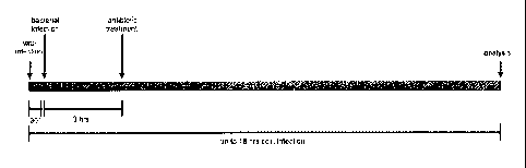
Fig. 1: Time scale of co-infection procedure. Cells were infected with IV for
30 min. Co-infection with
S. aureus 6850 was conducted or cells were mock-treated. Extracellular
bacteria were lysed and
removed by antibiotic treatment 3 h post bacterial infection. After a PBS
wash, cells were
supplemented with fresh medium (DMEM/INV) and incubated up to 18 hrs of viral
infection
Fig. 2: The MEK inhibitor U0126 reduces IV titers (A/Puerto Rico/8/34) and S.
aureus load, even in a
co-infection situation. Human lung epithelial cells were seeded in 6-well
plates (8x10⁵ cells/well) in 2
ml DMEM [10% FCS]. 16 ¨ 20 hrs after seeding, cells were rinsed and incubated
with PBS/BA [0.2%
bovine serum albumin (BSA), 1 mM MgC12, 0.9 mM CaC12, 100 Wm! penicillin, 0.1
mg/ml
streptomycin] (500 ul per 6 well) or PBS/BA containing the influenza virus
A/Puerto Rico/8/34 at a
multiplicity of infection (M01=0.1) at 37 C. After 30 min incubation, the
virus dilution was aspirated,
cells were rinsed with PBS and supplemented with Invasion medium DMEM/INV [1%
human serum
albumin, 25 nmo1/1 HEPES] (2 ml per 6 well) with or without S. aureus 6850
(M01=0.5) in presence of
50 M U0126 or DMSO (solvent control). 3 hrs post bacterial infection cells
were treated with
antibiotics to remove extracellular bacteria. Therefore cells were rinsed with
PBS and subsequently
incubated with DM EM/INVantibiotics [2 ug/mIlysostaphin (Sigma)] (1 ml per 6
well) for 20 min at 37
C. After an additional wash with PBS cells were supplemented with DMEM/INV
containing 50 M
U0126 or DMSO and 0.333 ug/m1Trypsin (Invitrogen). After an incubation period
of further 14 hrs at
37 C IV titers and intracellular bacteria were determined as described in
(Hrincius et al., 2010,
Tuchscherr et al., 2011).
CA 02949004 2016-11-14
WO 2015/173788
PCT/1B2015/053644
9
IV titers are depicted as plaque forming units (pfu)/m1 (A, C) and S. aureus
titers are depicted as
colonie forming units (CFU)/m1 (B,D). Data represent the means SD of three
independent
experiments with two biological samples. Statistical significance was
evaluated by a two-tailed two
sample t-test (*p < 0.05; ** p < 0.01; *** p <0.001). (E) S. aureus in
presence of 50p.M U0126 results
in reduced bacterial titers 18 hrs upon incubation in comparison to DMSO
treated bacteria. A defined
amount of S. aureus 6850 suspension culture was diluted in DMEM/INV
supplemented with 50p.M
U0126 or DMSO and incubated at 37 C for 18 hrs. Bacteria were diluted and
determined by serial
dilution on agar plates. Data represent the means SD of three independent
experiments with two
biological samples. Statistical significance was evaluated by a two-tailed two
sample t-test (*p < 0.05;
** p <0.01; *** p < 0.001).
Fig. 3: The MEK inhibitor U0126 reduces IV titers (A/FPV/Bratislava/79) and S.
aureus load, even in a
co-infection situation. Human lung epithelial cells were seeded in 6-well
plates (8x10⁵ cells/well) in 2
ml DMEM [10% FCS]. 16 ¨ 20 hrs after seeding, cells were rinsed and incubated
with PBS/BA [0.2%
bovine serum albumin (BSA), 1 mM MgC12, 0.9 mM CaCl2, 100 Wm! penicillin, 0.1
mg/ml
streptomycin] (500 p.I per 6 well) or PBS/BA containing the Influenza virus
A/FPV/Bratislava/79 at a
multiplicity of infection (MOI=0.001) at 37 C. After 30 min incubation, the
virus dilution was
aspirated, cells were rinsed with PBS and supplemented with Invasion medium
DMEM/INV [1%
human serum albumin, 25 nmo1/1 HEPES] (2 ml per 6 well) with or without S.
aureus 6850 (M01=0.5)
in presence of 50p.M U0126 or DMSO (solvent control). 3 hrs post bacterial
infection cells were
treated with antibiotics to remove extracellular bacteria. Therefore cells
were rinsed with PBS and
subsequently incubated with DMEM/INVantibiotics [2 p.g/m1 lysostaphin (Sigma)]
(1 ml per 6 well)
for 20 min at 37 C. After an additional wash with PBS cells were supplemented
with DMEM/INV
containing 50p.M U0126 or DMSO. After an incubation period of further 14 hrs
at 37 C IV titers and
intracellular bacteria were determined as described in (Hrincius et al., 2010;
Tuchscherr et al., 2011).
IV titers are depicted as plaque forming units (pfu)/m1 (A, C) and S. aureus
titers are depicted as
colonie forming units (CFU)/m1 (B, D). Data represent the means SD of two
independent
experiments with two biological samples. Statistical significance was
evaluated by a two-tailed two
sample t-test (*p < 0.05; ** p <0.01; *** p < 0.001).
Fig. 4: The p38 inhibitor 513202190 reduces IV titers and S. aureus load, even
in a co-infection
situation. Human lung epithelial cells were seeded in 6-well plates (8x10⁵
cells/well) in 2 ml DMEM
[10% FCS]. 16 ¨ 20 hrs after seeding, cells were rinsed and incubated with
PBS/BA [0.2% bovine
serum albumin (BSA), 1 mM MgC12, 0.9 mM CaCl2, 100 Wm! penicillin, 0.1 mg/ml
streptomycin] (500
CA 02949004 2016-11-14
WO 2015/173788
PCT/1B2015/053644
p.I per 6 well) or PBS/BA containing the Influenza virus A/Puerto Rico/8/34 at
a multiplicity of
infection (M01=0.1) at 37 C. After 30 min incubation, the virus dilution was
aspirated, cells were
rinsed with PBS and supplemented with Invasion medium DMEM/INV [1% human serum
albumin, 25
nmo1/1 HEPES] (2 ml per 6 well) with or without S. aureus 6850 (M01=0.5) in
presence of 10 M
5 513202190 or DMSO (solvent control). 3 hrs post bacterial infection cells
were treated wich antibiotics
to remove extracellular bacteria. Therefore cells were rinsed with PBS and
subsequently incubated
with DMEM/INVantibiotics [2 ug/m1 lysostaphin (Sigma)] (1 ml per 6 well) for
20 min at 37 C. After
an additional wash with PBS cells were supplemented with DMEM/INV containing
10 M 513202190
or DMSO and 0.333 ug/mITrypsin (Invitrogen). After an incubation period of
further 14 hrs at 37 C IV
10 titers and intracellular bacteria were determined as described in
(Hrincius et al., 2010; Tuchscherr et
al., 2011). IV titers are depicted as plaque forming units (pfu)/m1 (A, C) and
S. aureus titers are
depicted as colonie forming units (CFU)/m1 (B, D). Data represent the means
SD of three
independent experiments with two biological samples. Statistical significance
was evaluated by a
two-tailed two sample t-test (* p <0.05; ** p < 0.01; *** p < 0.001).
Fig. 5: The NF-kappaB (NFKB) inhibitor LG-ASA reduces IV titers and S. aureus
load, even in a co-
infection situation. Human lung epithelial cells were seeded in 6-well plates
(8x10⁵ cells/well) in 2 ml
DMEM [10% FCS]. 16 ¨ 20 hrs after seeding, cells were rinsed and incubated
with PBS/BA [0.2%
bovine serum albumin (BSA), 1 mM MgC12, 0.9 mM CaCl2, 100 Wm! penicillin, 0.1
mg/ml
streptomycin] (500 ul per 6 well) or PBS/BA containing the influenza virus
A/Puerto Rico/8/34 at a
muitiplicity of infection (M01=0.1) at 37 C. After 30 min incubation, the
virus dilution was aspirated,
cells were rinsed with PBS and supplemented with invasion medium DMEM/INV [1%
human serum
albumin, 25 nmo1/1 HEPES] (2 ml per 6 well) with or without S. aureus 6850
(M01=0.5) in presence of
5mM LG-ASA. Water was used as solvent control. 3 hrs post bacterial infection
cells were treated
with antibiotics to remove extracellular bacteria. Therefore cells were rinsed
with PBS and
subsequently incubated with DM EM/INVantibiotics [10% FBS, 2 ug/m1 lysostaphin
(Sigma)] (1 ml per
6 well) for 20 min at 37 C. After an additional wash with PBS cells were
supplemented with
DMEM/INV containing 5mM LG-ASA or water and 0.333 ug/m1 Trypsin (Invitrogen).
After an
incubation period of further 14 hrs at 37 C IV titers and intracellular
bacteria were determined as
described in (Hrincius et al., 2010, Tuchscherr et al., 2011). IV titers are
depicted as plaque forming
units (pfu)/m1 (A, C) and S. aureus titers are depicted as colonie forming
untits (CFU)/m1 (B, D). Data
represent the means SD of two independent experiments (virus titer) and
three independent
experiments (bacterial titer) with two biological samples. Statistical
significance was evaluated by a
two-tailed two sample t-test (* p <0.05; ** p < 0.01; *** p < 0.001).
CA 02949004 2016-11-14
WO 2015/173788
PCT/1B2015/053644
11
Fig. 6: The viral neuraminidase inhibitor tamiflu reduces IV replication but
enhances S. aureus load.
Human lung epithelial cells were seeded in 6-well plates (8x10⁵ cells/well) in
2 ml DMEM [10% FCS].
16 ¨ 20 hrs after seeding, cells were rinsed and incubated with PBS/BA [0.2%
bovine serum albumin
(BSA), 1 mM MgC12, 0.9 mM CaCl2, 100 Wm! penicillin, 0.1 mg/ml streptomycin]
(500 p.I per 6 well) or
PBS/BA containing the Influenza virus A/FPV/Bratislava/79 at a multiplicity of
infection (MOI=0.001)
at 37 C. After 30 min incubation, the virus dilution was aspirated, cells
were rinsed with PBS and
supplemented with invasion medium DMEM/INV [1% human serum albumin, 25 nmo1/1
HEPES] (2 ml
per 6 well) with or without S. aureus 6850 (M01=0.5) in presence of 2p.M
tamiflu or Hepes (solvent
control). 3 hrs post bacterial infection cells were treated with antibiotics
to remove extracellular
bacteria. Therefore cells were rinsed with PBS and subsequently incubated with
DMEM/INVantibiotics (2 p.g/m1 lysostaphin (Sigma)] (1 ml per 6 well) for 20
min at 37 C. After an
additional wash with PBS cells were supplemented wich DMEM/INV containing 2p.M
tamiflu or
Hepes. After an incubation period of further 14 hrs at 37 C IV titers and
intracellular bacteria were
determined as described in (Hrincius et al., 2010; Tuchscherr et al., 2011).
Fig. 7: Titers of intracellular S. aureus 6850 are reduced upon LG-ASA
treatment. Human lung
epithelial cells (A549) were infected with 0.5M01 S. aureus 6850 DMEM/INV [1%
human serum
albumin, 25 nmo1/1 HEPES] for 3h in presence (A, C) and absence (B, D) of the
indicated amounts of
LG-ASA. Three hours post infection an antibiotic wash was included using
DMEM/INVantibiotics [2
p.g/m1 lysostaphin (Sigma)] to remove non-internalized bacteria and
subsequently cells were
supplemented with DMEM/INV containing the indicated amounts of LG-ASA. Cell
morphology was
monitored by light microscopy (A, B) and amounts of internalized bacteria were
determined by serial
dilution on agar plates 18 hours post infection (C, D).
Fig. 8: Table 2: p38 inhibitors.
Fig. 9: Table 3: NFKB inhibitors.
Fig. 10: Table 4: NFKB inhibitors.
Fig. 11: Table 5: antibiotics.
CA 02949004 2016-11-14
WO 2015/173788
PCT/1B2015/053644
12
Fig. 12: Time scale of the co-infection procedure in vitro. Human lung
epithelial cells (A549) were
infected with influenza A virus (IAV) for 30min at a multiplicity of infection
(M01) indicated, dissolved
in PBS/BA [0.2% bovine serum albumin, 1mM MgC12, 0.9mM CaCl2, 100U/m1
penicillin, 0.1mg/m1
streptomycin] at 37 C. After 30min incubation, the virus dilution was
aspirated, cells were rinsed
with PBS. Afterwards bacterial infection with Staphylococcus aureus 6850 (S.
aureus) was performed
or cells were mock-treated. Therefore cells were supplemented with invasion
medium DMEM/INV
[1% human serum albumin, 25nmo1/1 HEPES] with or without S. aureus in addition
to the indicated
amounts of inhibitor (U0126 or CI-1040) or solvent control. 3hrs post
bacterial infection an antibiotic
wash [DMEM, 10%F6S, 2u.g/m1 Lysostaphin or 100 g/m1 Gentamicin, 20min] was
introduced to
remove non-internalized bacteria. After an additional PBS wash, cells were
supplemented with
infection medium DMEM (0.2% BA, 1mM MgC12, 0.9mM CaCl2, 100U/m1 penicillin,
0.1mg/m1
streptomycin) in presence or absence of the inhibitor and were incubated up to
18hrs post viral
infection at 37 C.
Fig. 13: Inhibition of the MEK/ERK signaling results in enhanced cell survival
after singular and co-
infection.
Human lung epithelial cells (A549) were infected with the avian influenza
virus strain
A/FPV/Bratislava/79 (H7N7) (FPV) or the human influenza virus strain
A/Wisconsin/67/2005 (H3N2)
at a multiplicity of infection (M01=0.01) (H7N7) or (M01=0.5) (H3N2) at 37 C.
After 30min the virus
dilution was removed, cells were rinsed with PBS and supplemented with
invasion medium
DMEM/INV (containing 1% human serum albumin, 25nM HEPES) with or without S.
aureus 6850
(M01=0.1) in presence of 50 M U0126 or solvent control. 3hrs post bacterial
infection cells were
treated with DMEM/FBS containing 10% FBS, 2u.g/m1 lysostaphin for 20min to
remove non-
internalized bacteria. After an additional wash with PBS cells were
supplemented with infection
medium DMEM/BA (0.2% BA, 1mM MgC12, 0.9mM CaCl2, 100U/m1 penicillin,
0.1mg/mIstreptomycin)
containing 50 M U0126 or solvent. After an incubation period of 18hrs at 37 C
cell morphology was
monitored by light microscopy.
Fig. 14: Inhibition of MEK/ERK signaling results in reduced viral titers
during singular viral and co-
infection.
Human lung epithelial cells (A549) were infected with the avian influenza
virus strain
A/FPV/Bratislava/79 (H7N7) (A, C) or the human influenza virus strain
A/Wisconsin/67/2005 (H3N2)
(B, D) at a multiplicity of infection (M01=0.01) (A, C), (M01=0.5) (B, D) at
37 C. After 30min the virus
dilution was removed, cells were rinsed with PBS and supplemented with
invasion medium
CA 02949004 2016-11-14
WO 2015/173788
PCT/1B2015/053644
13
DMEM/INV (containing 1% human serum albumin, 25nM HEPES) with or without S.
aureus 6850
(M01=0.1) in presence of 50 M U0126 or solvent control. 3hrs post bacterial
infection cells were
treated with DMEM/FBS containing 10% FBS, 2u.g/m1 lysostaphin for 20min to
remove extracellular
bacteria. After an additional wash with PBS cells were supplemented with
infection medium
DMEM/BA (0.2% BA, 1mM MgC12, 0.9mM CaCl2, 100U/m1 penicillin, 0.1mg/m1
streptomycin)
containing 50 M U0126 or solvent. After an incubation period of 18hrs at 37 C
viral titers were
determined by standard plaque assay. Viral titers are depicted as plaque
forming units/ml (PFU/ml)
with a linear (A, B) or logarithmic scale (C, D). Data represent the means
SD of four independent
experiments with two biological samples. Statistical significance was
evaluated by a two-tailed one
sample t-test (* p <0.05; ** p <0.01).
Fig. 15: MEK inhibition by administration of U0126 results in reduced
bacterial growth.
Human lung epithelial cells (A549) were infected with the avian influenza
virus strain
A/FPV/Bratislava/79 (H7N7) or the human influenza virus strain
A/Wisconsin/67/2005 (H3N2) (A, C)
at a multiplicity of infection (M01=0.01) (H7N7) or (M01=0.5) (H3N2) at 37 C.
After 30min the virus
dilution was removed, cells were rinsed with PBS and supplemented with
invasion medium
DMEM/INV (containing 1% human serum albumin, 25nM HEPES) with or without S.
aureus 6850
(M01=0.1) (A) in presence of 50 M U0126 or solvent control. 3hrs post
bacterial infection cells were
treated with DMEM/FBS containing 10% FBS, 2u.g/m1 lysostaphin for 20min (A, C)
to remove non-
internalized bacteria. After an additional wash with PBS cells were
supplemented with infection
medium DMEM/BA (0.2% BA, 1mM MgC12, 0.9mM CaCl2, 100U/m1 penicillin,
0.1mg/mIstreptomycin)
containing 50 M U0126 or solvent. Amounts of internalized bacteria were
determined by serial
dilution of cell lysates on agar plates 18hrs post infection (A, C). The
impact of U0126 on bacterial
growth was analyzed by administration of U0126 as indicated to an over-night
culture of S. aureus
6850 (100 CFU/ml). After 16hrs serial dilutions were plated on BHI agar (B,
D). Bacterial titers are
depicted as colony forming units/ml (CFU/ml) with a linear (A, B) or
logarithmic scale (C, D). Data
represent the means SD of four (A, C) or three (B, D) independent
experiments with two biological
samples. Statistical significance was evaluated by a two-tailed two sample t-
test (*** p < 0.001).
Fig. 16: Inhibition of the MEK/ERK signaling leads to reduction of viral
proteins and pro-inflammatory
chemokines.
Human lung epithelial cells (A549) were infected with the human influenza
virus strain
A/Wisconsin/67/2005 (H3N2) at a multiplicity of infection (M01=5) at 37 C.
After 30min the virus
dilution was removed, cells were rinsed with PBS and supplemented with
invasion medium
CA 02949004 2016-11-14
WO 2015/173788
PCT/1B2015/053644
14
DMEM/INV (containing 1% human serum albumin, 25nM HEPES) with or without S.
aureus 6850
(M01=50) in presence of 50 M U0126 or solvent control. 3hrs post bacterial
infection cells were
treated with DMEM/FBS containing 10% FBS, 100 g/m1 Gentamicin for 30min to
remove
extracellular bacteria. After an additional wash with PBS cells were
supplemented with infection
medium DM EM/BA (0.2% BA, 1mM MgC12, 0.9mM CaCl2, 100U/m1 penicillin, 0.1mg/m1
streptomycin)
containing 50 M U0126 or solvent. After an incubation period of 8hrs at 37 C
mRNA levels of CCL3
and CCL5 were analyzed by qRT-PCR with specific primers (A, B). Viral protein
expression (PB-1) and
the level of ERK-1/2 phosphorylation, as well as bacterial cell wall
components (PGN) were
determined by western blot analysis (C). Data represent preliminary results of
three biological
samples and two technical replicates within one experiment.
Fig. 17: Administration of U0126 leads to reduced bacterial titers in vivo
independent of viral titers.
12 weeks old Balb/C mice were infected with 50 PFU of the influenza virus
strain A/Puerto Rico/8/34
(PR8, H1N1) on day 0 (anesthesized with Isoflurane). Starting on day 1 mice
were treated daily with
i.p. injection of 30mg/kg/day U0126 or solvent control (10% DMSO, 30%
Cremophor EL, 60% PBS).
On day 3 mice were infected with 5*107 CFU of Staphylococcus aureus 6850 under
anesthesia with
Isoflurane and directly treated with U0126 or solvent control. On day 4 lungs
were removed and
homogenized in PBS (0.1g per 1000111 PBS). For calculation of bacterial titers
serial dilutions of the
homogenate were plated on BHI agar. For determination of viral titers a
standard plaque assay was
performed. Statistical analysis was done using Mann-Whitney U Test (* p
<0,05).
Fig. 18: The specific MEK inhibitor CI-1040 reduces viral titers in singular
and co-infection.
Human lung epithelial cells (A549) were infected with the avian influenza
virus strain
A/FPV/Bratislava/79 (H7N7) (A, C) or the human influenza virus strain A/Puerto
Rico/8/34 (H1N1) (B,
D) at a multiplicity of infection (M01=0.01) at 37 C. After 30min the virus
dilution was removed, cells
were rinsed with PBS and supplemented with invasion medium DMEM/INV
(containing 1% human
serum albumin, 25nM HEPES) with or without S. aureus 6850 (M01=0.1) in
presence of 10 M CI-1040
or solvent control. 3hrs post bacterial infection cells were treated with
DMEM/FBS containing 10%
FBS, 2u.g/m1 lysostaphin for 20min to remove extracellular bacteria. After an
additional wash with
PBS cells were supplemented with infection medium DMEM/BA (0.2% BA, 1mM MgC12,
0.9mM CaCl2,
100U/m1 penicillin, 0.1mg/m1 streptomycin) containing 10 M CI-1040 or solvent.
After an incubation
period of 18hrs at 37 C viral titers were determined by standard plaque assay.
Viral titers are
depicted as plaque forming units/ml (PFU/ml) with a linear (A, B) or
logarithmic scale (C, D). Data
represent the means of two independent experiments with two biological
samples.
CA 02949004 2016-11-14
WO 2015/173788
PCT/1B2015/053644
Fig.19: Administration of CI-1040 has an inhibitory effect on bacterial growth
in vitro.
The impact of C1-1040 on bacterial growth was analyzed by administration of CI-
1040 in different
concentrations (as indicated) to an over-night culture of S. aureus 6850 (100
CFU/ml). After 16hrs
serial dilutions were plated on BHI agar (A, B). Bacterial titers are depicted
as colony forming units/ml
5 (CFU/ml) with a linear (A) or logarithmic scale (B). Data represent
preliminary data with two
biological samples.
Fig. 20: Treatment with the specific MEK inhibitor Cobimetinib (GDC-0973)
reduces pathogen load in
vivo.
1 0 8 weeks old Balb/C mice (5 per group) were infected with 50 PFU of
influenza virus strain A/Puerto
Rico/8/34 (PR8, H1N1) on day 0 (anesthesized with Isoflurane). 6hrs post viral
infection mice were
treated with oral administration of 10mg/kg/day Cobimetinib or solvent control
(10% DMSO, 5%
Tween 20, 85% PBS). Treatment was then performed daily. On day 3 mice were
infected with 5*107
CFU of Staphylococcus aureus 6850 under anesthesia with Isoflurane and 6hrs
later treated with
15 Cobimetinib or solvent control. On day 4 lungs were removed and
homogenized in PBS (0.1g per
1000111 PBS). For calculation of bacterial titers serial dilutions of the
homogenate were plated on BHI
agar. For determination of viral titers a standard plaque assay was performed.
Statistical analysis was
done using Mann-Whitney U test.
Fig. 21: LG-ASA improves cell morphology upon infection with influenza A virus
(IAV) and/or
Staphylococcus aureus (S. aureus)
Human lung epithelial cells (A549) were infected with the influenza virus
strain A/Puerto Rico/8/34
(H1N1) at a multiplicity of infection (M01=0.1) dissolved in PBS/BA [0.2%
bovine serum albumin,
1mM MgC12, 0.9mM CaC12, 100U/m1 penicillin, 0.1mg/m1 streptomycin] at 37 C.
After 30min
incubation, the virus dilution was aspirated, cells were rinsed with PBS and
supplemented with
invasion medium DMEM/INV [1% human serum albumin, 25nmo1/1 HEPES] with or
without S. aureus
SH1000 (M01=0.1) in presence of 5mM LG-ASA or solvent control. 3hrs post
bacterial infection cells
were treated with antibiotics [DMEM, 10%F6S, 2u.g/m1 lysostaphin, 20min] to
remove extracellular
bacteria. After an additional wash with PBS cells were supplemented with
DMEM/INV containing
5mM LG-ASA or solvent. After an incubation period of 18hrs at 37 C cell
morphology was monitored
by light microscopy.
Fig. 22: The NFKI3 inhibitor LG-ASA reduces influenza virus titers and S.
aureus load
CA 02949004 2016-11-14
WO 2015/173788
PCT/1B2015/053644
16
Human lung epithelial cells (A549) were infected with the influenza virus
strain A/Puerto Rico/8/34
(H1N1) at a multiplicity of infection (M01=0.1) dissolved in PBS/BA [0.2%
bovine serum albumin,
1mM MgC12, 0.9mM CaC12, 100U/m1 penicillin, 0.1mg/m1 streptomycin] at 37 C (A-
H). After 30min
incubation, the virus dilution was aspirated, cells were rinsed with PBS and
supplemented with
invasion medium DMEM/INV [1% human serum albumin, 25nmo1/1 HEPES] with or
without S. aureus
SH1000 (M01=50) (A, B), (M01=0.01) (C-D), S. aureus 6850 (M01=50) (E, F),
(M01=0.1) (G, H) in
presence of 5mM LG-ASA or solvent control. 3hrs post bacterial infection cells
were treated with
antibiotics [DMEM, 10%F6S, 2u.g/m1 lysostaphin, 20min] to remove extracellular
bacteria. After an
additional wash with PBS cells were supplemented with DMEM/INV containing 5mM
LG-ASA or
solvent. After an incubation period of 8hrs (A, B, E, F) or 18hrs (C, D, G, H)
at 37 C IAV titers (A, C, E,
G) were determined by standard plaque assays. Cells were lysed by a hypotonic
shock and amounts
of internalized bacteria (B, D, F, H) were determined by serial dilution on
agar plates. Data represent
the means SD of three (A ¨ H) with two biological samples. Statistical
significance was evaluated by
a two-tailed one sample t-test (* p <0.05; ** p < 0.01).
Fig. 23: The NFKI3 inhibitor LG-ASA reduces influenza virus titers and S.
aureus load
This figure presents data obtained as in Figure 22 in a different way. In
particular, the untreated
controls of each experiment were arbitrarily set as 100% and then the means
were calculated.
Statistical significance was evaluated by a two-tailed one sample t-test (* p
< 0.05; ** p <0.01).
Fig. 24: Inhibition of NFKI3 signaling results in reduced bacterial
internalisation
Human lung epithelial cells (A549) were preincubated with 5mM (A - D) and 10mM
(C, D) LG-ASA for
4h and then infected with S. aureus 6850 (M01=50) (A, B) for 30 ¨ 120min and
USA 300 (M01=5) (C,
D) for 120min in presence and absence of the indicated amounts of LG-ASA
dissolved in DMEM/INV
[1% human serum albumin, 25nmo1/1 HEPES]. 30 ¨ 120min post infection an
antibiotic wash [DMEM,
10%F6S, 2u.g/m1 lysostaphin, 20min] was included to remove non-internalized
bacteria. Cells were
washed with PBS three times and lysed by hypotonic shock. Amounts of
internalized bacteria were
determined by serial dilution on agar plates (A ¨ D). Data (A, C) represent
the means SD of three
independent experiments with two biological samples. In Figure 24 B, D the
untreated controls of
each experiment were arbitrarily set as 100% and then the means were
calculated. Statistical
significance was evaluated by a two-tailed one sample t-test (* p <0.05; ** p
<0.01; *** p < 0.001).
Fig. 25: Stimulation of NFKI3 signaling results in enhanced bacterial
internalization
CA 02949004 2016-11-14
WO 2015/173788
PCT/1B2015/053644
17
Human lung epithelial cells (A549) were preincubated with 5mM LG-ASA and 2.5
ng/ml TNF-alpha for
4h and then infected with (A) S. aureus 6850 or (B) S. aureus U5A300 (M01=5)
for 120min in presence
and absence of the indicated amounts of LG-ASA and TNF-alpha dissolved in DM
EM/INV [1% human
serum albumin, 25nmo1/1 HEPES] (A). 120min post infection an antibiotic wash
[DMEM, 10%F6S,
2u.g/m1 lysostaphin, 20min] was included to remove non-internalized bacteria.
Cells were washed
with PBS three times and lysed by hypotonic shock. Amounts of internalized
bacteria were
determined by serial dilution on agar plates (A) 120min post infection. (A)
Data represent the means
SD of four independent experiments with two biological samples. Statistical
significance was
evaluated by an one-way ANOVA followed by a multiple comparison test (* p <
0.05; ** p < 0.01; ***
p < 0.001). (B) Data represent the means SD of three independent experiments
with three
biological samples whereby the untreated controls of each experiment were
arbitrarily set as 100%
and then the means were calculated.
Fig. 26: Treatment of IAV/S. aureus co-infected mice with LG-ASA results in
enhanced survival and
reduced body weight loss
(A) BALB/c mice (4 mice per group) were infected with 50 PFU of the
influenza virus A/Puerto
Rico/8/34 at day 0. On day 6 after influenza virus infection mice were
additionally infected with 108
CFU S. aureus 6850. On day 7 after influenza virus infection co-infected mice
were treated once a day
with LG-ASA (1M, 10min) via inhalation. Survival was monitored for 14 days.
While (4/4) (black line) untreated co-infected mice died 1 day after co-
infection, (2/4) LG-ASA treated
co-infected mice survived (grey line).
(B) Two independent experiments are depicted. 9 weeks old Balb/C mice (4
mice per group)
were infected with 50 PFU of influenza virus A/Puerto Rico/8/34 on day 0
(anesthesized with
Isoflurane) in the morning. 6hrs post viral infection mice were weighed and
treated with aerosolic
H20 or 1M LG-ASA in an inhalation chamber for 10min. This treatment was also
performed on day 1,
2 and 3 at the same time as on day 0. On day 3 in the morning mice were
infected with 5*107 CFU of
Staphylococcus aureus 6850 under anesthesia with Isoflurane. On day 4 mice
were weighed the last
time. Statistical analysis was done using Mann-Whitney U Test (* p < 0.05).
DETAILED DESCRIPTION OF THE INVENTION/DETAILED DESCRIPTION OF A PREFERENTIAL
EMBODIMENT
The following description includes information that may be useful in
understanding the present
invention. It is not an admission that any of the information provided herein
is prior art or relevant
CA 02949004 2016-11-14
WO 2015/173788
PCT/1B2015/053644
18
to the presently claimed inventions, or that any publication specifically or
implicitly referenced is
prior art.
The above being said, the present invention relates to a MEK inhibitor, p38
inhibitor and/or NFKB
inhibitor for use in a method for the prophylaxis and/or treatment of a co-
infection comprising a
bacterial infection and an influenza virus infection.
In addition the present invention relates to a MEK inhibitor, p38 inhibitor
and/or NFKB inhibitor for
use in a method for the prophylaxis and/or treatment of a bacterial infection.
When used herein, a "MEK inhibitor" may also be designated as a Mitogen
Activated Proteinkinase
(MAPK) kinase inhibitor. It is known that in a MAPK pathway, a MAPK kinase
kinase (MAPKKK)
activates a MAPK kinase (MAPKK) which in turn activates a MAPK which
transduces a signal to, for
example, a transcription factor or other kinases or effector/signal
transducing protein; see, for
example, Figure 1 of Fremin and Meloche (Fremin and Meloche (2010), J.
Hematol. Oncol. 11;3:8).
MEK inhibitors of the invention preferably inhibit MEK1/2 of a subject, such
as a mammal or bird as
described herein. However, it may be that a MEK inhibitor of the invention
does not only inhibit a
MEK, preferably MEK1/2, but also its upstream kinase (i.e. MAPKKK), thereby
exerting a dual
inhibition. Without being bound by theory, PLX-4032 may be such a dual
inhibitor. Hence, a MEK
inhibitor of the invention may in a preferred aspect by a dual inhibitor,
thereby inhibiting a MEK,
preferably MEK1/2 and the corresponding upstream MAPKKK. MEK1/2 is the MAPKK
in the Ras/Raf
pathway, whereby Ras/Raf acts as MAPKKK and ERK1/2 acts as MAPK.
A MEK inhibitor can be a small molecule, large molecule, peptide,
oligonucleotide, and the like. The
MEK inhibitor may be a protein or fragment thereof or a nucleic acid molecule.
Also included by the
term MEK inhibitor is a pharmaceutically acceptable salt of the MEK inhibitor.
The determination of whether or not a compound is a MEK inhibitor is within
the skill of one of
ordinary skill in the art. In one embodiment, the MEK inhibitors are selected
from the group
consisting of the compounds/inhibitors listed in table 1.
The MEK inhibitors of the invention are selected preferably from U0126, PLX-
4032, AZD6244,
AZD8330, AS-703026, GSK-1120212, RDEA-119, RO-5126766, RO-4987655, CI-1040, PD-
0325901,
GDC-0973, TAK-733, PD98059, PD184352 ARRY-438162 and PF-3644022, preferably
AZD8330, GSK-
1120212, U0126, GDC-0973, CI-1040, PD0325901, ARRY-438162, PF-3644022 and
AZD6244 and most
preferably U0126, CI-1040, GDC-0973 (Cobimetinib), AZD8330, GSK-1120212, most
preferably
U0126, GDC-0973, CI-1040, AZD8330 and GSK-1120212.
CA 02949004 2016-11-14
WO 2015/173788 PCT/1B2015/053644
19
Some of these inhibitors are further described in table 1 below.
Table 1: MEK inhibitors.
ci Structural formula I
,\
---------,---4- N -C)-----/ \ C1-1040
I _ H
F-----"--f;"---NH 2-(2-chloro-4-iodophenylamino)-N-
F_.õ.}...,c 1 (cyclopropylmethoxy)-3,4-
difluorobenzamide
1
i
H Structural formula 11
F
,- H 1 PD0325901
_õ--ik,1
(R)-N-(2,3-dihydroxypropoxy)-
=--..õ-?--" , .--:-<=--si
1 '
F 3,4-difluoro-2-(2-fluoro-4-iodo-
phenylamino)benzamide
Br Structural formula III
1.
i --1 AZD6244
-...,õ.1
I '
6-(4-bromo-2-chlorophenylamino)-
7-fluoro-N-(2-hydroxyethoxy)-3-methyl-
i 6
3H-benzo[d]imidazole-5-carboxamide
F Structural formula IV
HO 0
41' H N 4. I GDC-0973
NH [3,4-diluoro-2-[(2-fluoro-4-
11 F
iadophenyljamina]phenyrj
[3-hydroxy.-34(2S)-2-pipericlinyll-1-
azeticlinylimethanone
Structural formula V
1
HO'Th
RDEA-119
i ...1, ....if ....t
II
...,...f., (S)-N-(3,4-difluoro-2-(2-fluoro-4-
iodophenyl-
amino)-6-methoxypheny1)-1-(2,3-
F dihydroxypropyl) cyclopropane-1-
sulfonamide
Structural formula VI
o litr'''' Is
GSK-1120212
N
.11, ....õ1,, ....._
N-(3-(3-cyclopropy1-5-(2-fluoro-4-
iodophenylamino)-
1 I 6,8-dimethy1-2,4,7-trioxo-3,4,6,7-
tetrahydropyrido
I: 11 jil.: [4,3-d]pyrimidin-1(2H)-
yl)phenyl)acetamide
14
CA 02949004 2016-11-14
WO 2015/173788 PCT/1B2015/053644
H Structural formula VII
0-, , N õ----õõOH
Ft H I 0 AZD8330
---',-; -N -,-,-.--=----
i o 2-(2-fluoro-4-iodophenylamino)-N-
....---..,...,-. ..., N õ.õ..-4,...., (2-
hydroxyethoxy)- 1,5-dimethy1-6-oxo-
I H
0 1,6-dihydropyridine-3-carboxamide
Structural formula VIII
R05126766
,N ,O., õ;,,,,,,, ,..0, ...a õ...õ,-.,
li I N 0 0 C20H16FN505S
N......?..-- ,,......-- ,i,JI,N-.
4- H NII2
Structural formula IX
H
..p
yH R04987655
.,...----,0 es.,:,_ ....,,,....0N::õ..,
L 4õL, It L, !I, C20H19F3IN305
6 P
Structural formula X
.--- i
TAK-733
0 HN)1,,,,õõi
(R)-3-(2,3-dihydroxypropyI)-6-fluoro-5-
it 1 '
HO,=.,r.kc, , 0,-.7.;.,,l- (2-fluoro-4-iodophenylamino)-8-methylpyrido
7 1
L1, ,....., [2,3-d]pyrimidine-4,7(3H,8H)-dione
HO"'" N* N 0
I
0 r,) Structural formula XI
='.,..:,
\ PLX-4032
tk. K '\.; j NO-[[5-(4-Chloropheny1)-1H-pyrrolo
....- ---"--:x..---.N..
1 )= [2,3-bjpyridin-3-ylicarbonA-2,4-
difluorophenylj- 1-
',... .-- N
N k propanesulfonarnicie
7..e$torat mtra.trafwM
OH Structural formula XII
tici1/4,A,,,1 A5703026
Nis.l. :0 (S)-N-(2,3-dihydroxypropyI)-3-
clia
i r....... ill.: (2-fluoro-4-iodophenylamino)
NI' 1,7 i isonicotinamide
0 Structural formula XIII
a
PD98059
---- 0
2-(2-amino-3-methoxyphenyI)-4H-chromen-4-one
CA 02949004 2016-11-14
WO 2015/173788
PCT/1B2015/053644
21
Structural formula XIV
HNI
PD184352
o
2-(2-chloro-4-iodophenylamino)-
F
N-(cyclopropylmethoxy)- 3,4-difluorobenzamide
Also preferred is a selection from PLX-4032, AZD6244, AZD8330, GDC-0973,
RDEA119, GSK1120212,
R051267766, R04987655, TAK-733, and AS703026. Even more preferably, they are
selected from
AZD6244, AZD8330, GSK1120212 and PLX-4032 or from PD-0325901, AZD-6244, AZD-
8330 and
RDEA-119. These MEK inhibitors are known in the art and, for example,
described in Table 1 of
Fremin and Meloche (2010), J. Hematol. Oncol. 11;3:8.
More information on some of these inhibitors can also be obtained from Arthur
and Ley (2013)
Mitogen-activated protein kinases in innate immunity; Nature Reviews
Immunology 13,679-
692(2013).
Indeed, as demonstrated in the appended Examples, the MEK inhibitor U0126 and
CI-1040 disclosed herein show an effect in co-infection scenarios as well as
on bacterial infection
alone.
Also a p38 inhibitor is provided for use in the methods for the prophylaxis
and/or treatment of a co-
infection or bacterial infection of the present invention. A "p38 MAP kinase
inhibitor" is well known
in the art. The terms "p38 inhibitor," "p38 kinase inhibitor," and "p38 MAP
kinase inhibitor" are used
interchangeably herein. In the context of the present invention a p38 MAP
kinase inhibitor inhibits
p38 MAP kinase. Preferably, the p38 MAP kinase inhibitor inhibits one of the
isoforms of p38 MAP
kinase, preferably one of the four isoforms (a, 13, y or 5) of p38 MAP kinase
with the a-isoform being
preferred, more preferably it inhibits any combination of two isoforms of p38
MAP kinase, even
more preferably it inhibits any combination of three isoforms of p38 MAP
kinase and most
preferably, it inhibits all isoforms or the a, 13, y and 5 isoform of p38 MAP
kinase. In some
embodiments, the p38 MAP kinase inhibitor inhibits the isoform of p38 that is
involved in
inflammatory diseases, autoimmune diseases, destructive bone disorders,
proliferative disorders,
infectious diseases, viral diseases or neurodegenerative diseases. It is
reported that the a-isoform of
p38 MAP kinase is involved in inflammation, proliferation, differentiation and
apoptosis, whereas the
biological functions of p38 13, p38 5 and p38 y are not yet understood
completely. Accordingly, it is
preferred herein that the p38 MAP kinase inhibitor inhibits the a-isoform.
A p38 MAP kinase inhibitor can be a small molecule, large molecule, peptide,
oligonucleotide, and
the like. The p38 MAP kinase inhibitor may be a protein or fragment thereof or
a nucleic acid
CA 02949004 2016-11-14
WO 2015/173788
PCT/1B2015/053644
22
molecule. Also included by the term p38 inhibitor is a pharmaceutically
acceptable salt of the 38
inhibitor.
The determination of whether or not a compound is a p38 kinase inhibitor is
within the skill of one of
ordinary skill in the art.
There are many examples of p38 inhibitors in the art. U.S. Pat. Nos.
5,965,583, 6,040,320, 6,147,096,
6,214,830, 6,469,174, 6,521,655 disclose compounds that are p38 inhibitors.
U.S. Pat. Nos.
6,410,540, 6,476,031 and 6,448,257 also disclose compounds that are p38
inhibitors. Similarly, U.S.
Pat. Nos. 6,410,540, 6,479,507 and 6,509,361 disclose compounds that are
asserted to be p38
inhibitors. U.S. Published Application Nos. 20020198214 and 20020132843
disclose compounds that
are said to be p38 inhibitors. Another p38 MAP kinase inhibitor is BIRB 796 BS
(1-(5-tert-butyl-2-p-
toly1-2H-pyrazol-3-y1)-344-(2-morpholin-4-yl-ethoxy)-naphthalen-1-y1]-urea);
see Branger (2002), J.
Immunol. 168:4070-4077 or US 6,319,921 for further p39 MAP kinase inhibitors.
Other p38 MAP kinase inhibitors are AMG 548 (Amgen), BIRB 796 (Boehringer
Inge!helm), VX 702
(Vertex/Kissei), SCIO 469, SCIO 323 (Scios Inc.), SB 681323 (GlaxoSmithKline),
PH-797804 (Pfizer) and
Org-48762-0 (Organon NV); see, for example, Lee and Dominguez in Curr Med
Chem.
2005;12(25):2979-2994 and Dominguez in Curr Opin Drug Discov Devel. 2005
Jul;8(4):421-430.
According to the present invention, the inhibitor may exhibit its regulatory
effect upstream or
downstream of p38 MAP kinase or on p38 MAP kinase directly, with the latter
mode of action being
preferred. Examples of inhibitor regulated p38 MAP kinase activity include
those where the inhibitor
may decrease transcription and/or translation of p38 MAP kinase, may decrease
or inhibit post-
translational modification and/or cellular trafficking of p38 MAP kinase, or
may shorten the half-life
of p38 MAP kinase. The inhibitor may also reversibly or irreversibly bind p38
MAP kinase, inhibit its
activation, inactivate its enzymatic activity, or otherwise interfere with its
interaction with
downstream substrates.
The four isoforms of the p38 MAP kinase share a high level of sequence
homology. The alpha and
beta isoforms of the p38 MAP kinase are closely related while the gamma and
delta isoforms are
more divergent. Given the high degree of structural similarity, it is not
surprising that certain
compounds with the ability to inhibit one p38 MAP kinase isoform can often
inhibit other isoforms of
the MAP kinase. Accordingly, in some embodiments, an inhibitor of p38 MAP
kinase that is specific
CA 02949004 2016-11-14
WO 2015/173788
PCT/1B2015/053644
23
for the a-isoform of the kinase possesses at least three categories of
structural features that are
theorized to permit isoform specific inhibition.
Selective binding of a candidate p38 MAP kinase inhibitor can be determined by
a variety of
methods. The genes for the various isoforms of p38 MAP kinase are known in the
art. One of
ordinary skill in the art could readily clone and express the various isoforms
of the kinase, purify
them, and then perform binding studies with candidate compounds to determine
isoform binding
characteristics. This series of experiments was performed for the a-isoform of
p38 MAP kinase and
provided in U.S. Pat. No. 6,617,324 B1.
Another kinase selectivity assay is described in Mihara (2008), Br. J.
Pharmacol. 154(1):153-164.
In some embodiments herein, a p38 MAP kinase inhibitor inhibits one of the
four isoforms of p38
MAP kinase, more preferably it inhibits any combination of two isoforms of p38
MAP kinase, even
more preferably it inhibits any combination of three isoforms of p38 MAP
kinase, e.g., p38-
a(MAPK14), -13(MAPK11), -y (MAPK12 or ERK6). Alternatively, but also
preferred, it inhibits all four
isoforms of p38 MAP kinase.
In one embodiment, the p38 inhibitor is selected from the group consisting of
the inhibitors listed in
table 2 (Fig. 8). In another embodiment, the p38 inhibitor is selected from
the group consisting of
513202190, LY2228820, CAY10571, SB 203580, Tie2 Kinase Inhibitor, 2-(4-
Chloropheny1)-4-
(fluoropheny1)-5-pyridin-4-y1-1,2-dihydropyrazol-3-one, CGH 2466, 5B220025,
Antibiotic LL 21640-2,
TAK 715, 513202190 hydrochloride, SKF 86002, AMG548, CMPD-1, E0 1428, JX 401,
ML 3403, RWJ
67657, SB 202190, SB 203580, SB 203580 hydrochloride, SB 239063, SCIO 469, SX
011, TAK 715,
Pamapimod, Losmapimod (GW856553), Dilmapimod (513681323), VX 702, VX 745,
Doramapimod
(BIRB 796), BMS-582949, ARRY-797, PH797804, SC10-469, preferably VX-702,
513202190,
Pamapimod, Losmapimod (GW856553), Dilmapimod (513681323), Doramapimod (BIRB
796), BMS-
582949, ARRY-797, PH797804 and SC10-469.
More information on some of these inhibitors can also be obtained from Arthur
and Ley (2013)
Mitogen-activated protein kinases in innate immunity; Nature Reviews
Immunology 13,679-
692(2013).
In addition to the MEK inhibitor and the p38 inhibitor, the present invention
is also directed to a
NFKB (NFkB/NFkappaB) inhibitor for use in the methods for the prophylaxis
and/or treatment of a
co-infection or bacterial infection of the present invention. The
determination of whether or not a
compound is a NFKB inhibitor is within the skill of one of ordinary skill in
the art.
CA 02949004 2016-11-14
WO 2015/173788
PCT/1B2015/053644
24
NE-KB (nuclear factor kappa-light-chain-enhancer of activated B cells) is a
protein complex that
controls transcription of DNA. NE-KB is found in almost all animal cell types
and is involved in cellular
responses to stimuli such as stress, cytokines, free radicals, ultraviolet
irradiation, oxidized LDL, and
bacterial or viral antigens. Vertebrate NFKB transcription complexes can be
any of a variety of homo-
and heterodimers formed by the subunits p50 (NEKB1), p52 (NEKB2), c-Rel, RelA
(p65) and RelB
(Gilmore TD. (2006) Oncogene 25: 6680-6684). These complexes bind to DNA
regulatory sites called
KB sites, generally to activate specific target gene expression. In most cell
types, NE-KB dimers are
located in the cytoplasm in an inactive form through association with any of
several IKB inhibitor
proteins (IKB a, 13,E,y, p105 and p100). In response to a wide array of
stimuli IKB is rapidly
phosphorylated, ubiquitinated and degraded by the proteasome. The freed NE-KB
dimer then
translocates to the nucleus where it can modulate specific gene expression.
The phosphorylation and degradation of IKB is important for the regulation of
NFKB complexes,
which is mediated by the IKB kinase (IKK) complex containing two kinase
subunits, IKKa and IKKI3,
and an associated scaffold-like regulatory protein called NEMO (aka IKKy)
(Gilmore and Herscovich
(2006) Inhibitors of NE-KB signaling: 785 and counting Oncogene. 25, 6887-
6899). Notably, as for
example shown in Example 3 of the present invention, NE-KB siganling is also
important for bacterial
(such as S. aureus) internalisation into cells.
According to the present invention, the inhibitor may exhibit its regulatory
effect upstream or
downstream of NFKB or directly on NFKB, with the latter mode of action being
preferred. Examples
of inhibitors regulating NFKB activity include those where the inhibitor may
decrease transcription
and/or translation of NFKB, or may shorten the half-life of NFKB. The
inhibitor may also reversibly or
irreversibly bind NFKB, inhibit its activation, inactivate its activity, or
otherwise interfere with its
interaction with downstream targets, such as trgets on genes. Also, an NFKB
inhibitor can inhibit
protein kinases such as molceules which inhibit IkBa phosphorylation by e.g.
IKK inhibition.
Compounds that have such an activity are SC-893, BMS-345541, which may serve
as reference
compounds. Also a NFKB inhibitor may inhibit protein phosphatases, or inhibit
the proteasome, or
ubiquitination. Examples of such NFKB inhibitors, which may also serve as
reference compounds
include protein tyrosine phosphatase inhibitors, boronate, bortezomib, NPI-
0052. Alternatively, a
NFKB inhibitor may block the nuclear translocation of NFKB, or its binding to
DNA. Examples of such
inhibitors include, which may also serve as reference compounds, SN50,
dehydroxymethylepoxyquinomicin and NFKB decoy ODNs. Further information on
inhibitor of NFKB
CA 02949004 2016-11-14
WO 2015/173788
PCT/1B2015/053644
can be obtained from Gupta et al. (2010) (Gupta et al. (2010) Inhibiting NFKB
activation by small
molecules as a therapeutic strategy. Biochim Biophys Acta. 1799(10-12): 775-
787).
A NFKB inhibitor can be a small molecule, large molecule, peptide,
oligonucleotide, and the like. The
NFKB inhibitor may be a protein or fragment thereof or a nucleic acid
molecule. Also included by the
5 term NFKB inhibitor is a pharmaceutically acceptable salt of the NFKB
inhibitor. In one embodiment,
the NFKB inhibitor is selected from the group consisting of the
inhibitors/molceules as listed in tables
3 and 4 in Figures 9 and 10. In another embodiment, the NFKB inhibitor is
selected from the group
consisting of the inhibitors/molceules as listed in table 3 in Figure 9. In
another embodiment, the
NFKB inhibitor is selected from the group consisting of the
inhibitors/molceules as listed in table 4 in
10 Figure 10.
In another embodiment, the NFKB inhibitor is selected from the group
consisting of LASAG, SC75741
(and derivatives), MG 132, TPCA-1, PCTC, IMD 0354, Luteolin, Caffeic acid
phenethyl ester,
Cardamonin, PF 184, IKK 16, SC 514, Withaferin A, Arctigenin, Bay 11-7085,
PSI, PR 39, Ro 106-9920,
Bay 11-7821, ML-130, Celastrol, Tanshinone IIA, HU 211, Gliotoxin, CID
2858522, Honokiol,
15 Andrographolide, 10Z-Hymenialdisine, ACHP, Pristimerin, Sulfasalazine,
ML 12013 dihydrochloride,
Amlexanox, 9-Methylstreptimidone, N-Stearoyl phytosphingosine, 2-(1,8-
naphthyridin-2-yI)-Phenol,
5-Aminosalicylic acid, BAY 11-7085, Ethyl 3,4-Dihydroxycinnamate, Helanalin,
NF-k13 Activation
Inhibitor II, JSH-23, Glucocorticoid Receptor Modulator, CpdA, PPM-18, aspirin
(ASA),
Pyrrolidinedithiocarbamic acid ammonium salt, (R)-MG132, 5C75741 (and
derivatives), Rocaglamide,
20 Sodium salicylate, QNZ, PS-1145, CAY10512, bortezomib, salsalate,
resveratrol, LASAG,
deoxyspergualin, sulindac, thalidomide, AGRO-100, CHS 828 and/or Curcumin,
preferably
bortezomib, curcumin, salsalate, resveratrol, sodium salicylate, LASAG, ASA,
deoxyspergualin,
sulindac, thalidomide, AGRO-100, CHS 828 even more preferably 5C75741 (and
derivatives) ASA and
LASAG and most preferably LASAG.
25 With the term "5C75741" or "5C75741 (and derivates)" in addition to
5C75741 also derivates of
5C75741 are envisaged by the present invention.
In general a person skilled in the art knows how to find out if a coumpound is
an MEK inhibitor, p38
inhibitor and/or NFKB inhibitor. A further example of how one could determine
if a compound is a
MEK inhibitor and/or p38 inhibitor would be to isolate the MEK and/or p38 NFKB
protein. The
protein can be isolated from cells where the MEK and/or p38 protein is
naturally expressed or where
it has been overexpressed by means of transfection of an oligonucleotide or
infection with a virus
that directs the expression of the MEK and/or p38 protein. Additionally, MEK
and/or p38 protein can
also be expressed recombinantly. Upon isolating the protein a person of
ordinary skill in the art can
CA 02949004 2016-11-14
WO 2015/173788
PCT/1B2015/053644
26
measure the activity of the kinase in the presence or absence of a potential
MEK and/or p38
inhibitor. If the kinase activity is less in the presence than in the absence
of an alleged inhibitor, that
inhibitor is a MEK and/or p38, respectively.
If acting on MEK and/or p38 directly, the inhibitor should exhibit an 1050
value of about 5 u.M or less,
preferably 500 nm or less, more preferably 100 nm or less. In a related
embodiment, the inhibitor
should exhibit an 1050 value relative to the p38-a isoform that is preferably
at least ten fold less than
that observed when the same inhibitor is tested against other p38 MAP kinase
isoforms in the same
or comparable assay. It should be noted that 1050 values are assay dependent
and may change from
determination to determination. It is more important to look at relative
relationships of compounds'
1050 values rather than the exact values themselves.
An 1050 is the concentration of compound which inhibits the enzyme to 50% of
the activity as
measured in the absence of an inhibitor.
1050 values are calculated using the concentration of inhibitor that causes a
50% decrease as
compared to a control. 1050 values are assay dependent and will vary from
measurement to
measurement. As such, 1050 values are relative values. The values assigned to
a particular inhibitor
are to be compared generally rather than on an absolute basis.
Samples or assays comprising MAP and/or MAPK kinase that are treated with a
potential activator,
inhibitor, or modulator are compared to control samples without the inhibitor,
activator, or
modulator to examine the extent of inhibition. Control samples (untreated with
inhibitors) can be
assigned a relative MAP and/or MAPK kinase activity value of 100%. Inhibition
of MAP and/or MAPK
kinase is achieved when the MAP and/or MAPK kinase activity value relative to
the control is about
80%, optionally 50% or 25-0%. Activation of MAP and/or MAPK kinase is achieved
when the MAP
kinase activity value relative to the control is 110%, optionally 150%,
optionally 200-500%, or 1000-
3000% higher. Exemplary MAP kinase binding activity assays of the present
invention are: a MAP
and/or MAPK kinase ligand blot assay (Aymerich et al., Invest Opthalmol Vis
Sci. 42:3287-93, 2001); a
MAP and/or MAPK kinase affinity column chromatography assay (Alberdi et al., J
Biol Chem.
274:31605-12, 1999) and a MAP and/or MAPK kinase ligand binding assay (Alberdi
et al., J Biol Chem.
274:31605-12, 1999). Each incorporated by reference in their entirety.
Also the selectivity of the inhibitors may be measured by a kinase selectivity
assay is described in
Mihara (2008), Br. J. Pharmacol. 154(1):153-164.
CA 02949004 2016-11-14
WO 2015/173788
PCT/1B2015/053644
27
In the case of the NFKB inhibitor one can measure for example the gene
products (proteins) of target
genes of NFKB in a non-treated control cell and compare the expression of
these target gene
products to a cell, which has been treated with a NFKB Inhibitor. Some target
genes are described in
Oeckinghaus and Ghosh (2009) The NE-KB Family of Transcription Factors and Its
Regulation. Cold
Spring Harb Perspect Biol. Oct 2009; 1(4): a000034.
The expression level is reduced, when the cell treated with the inhibitor.
Other strategies may be to
detect lkBa degradation together with p-p65 accumulation and nuclear
translocation of NFKB by
Westernblot. Also NFKB interactions with DNA of cells not treated with an
inhibitor compared to cells
that have been treated with an inhibitor may be analysed by using an
electrophoretic mobility shift
assay (EMSA).
The inhibitory properties of a molecule can also be analysed by comparing its
action to a reference
compound. A "reference compound" as referred to herein means a compound, which
may be used
as a positive control for the determination if a molecule has MEK inhibitor,
p38 inhibitor and/or NFKB
inhibitor properties. As such also any of the inhibitors listed herein may be
used as such a reference
compound. A possible test may be one in which cells, which are e.g. stimulated
to activate the MEK,
p38 and or NFKB pathway are treated with a reference compound and in parallel
e.g. in a different
well with a compound of interest.
The inhibitors of the present invention can be used in a method for treating
and/or prophylaxis. As
such the term "treating" or "treatment" includes administration of a MEK
inhibitor, p38 Inhibitor,
and/or NFKB inhibitor preferably in the form of a medicament, to a subject
suffering from a
coinfection comprising a bacterial infection and an influenza virus infection
for the purpose of
ameliorating or improving symptoms. Similarly included is the administration
of a MEK inhibitor, p38
Inhibitor, and/or NFKB inhibitor preferably in the form of a medicament, to a
subject suffering from a
bacterial infection for the purpose of ameliorating or improving symptoms.
Furthermore, the terms "prophylaxis" as used herein, refers to any medical or
public health
procedure whose purpose is to prevent a disease. As used herein, the terms
"prevent", "prevention"
and "preventing" refer to the reduction in the risk of acquiring or developing
a given condition,
namely a coninfection comprising an influenza virus infection and a bacterial
infection or a bacterial
infection alone. Also meant by "prophylaxis" is the reduction or inhibition of
the recurrence of a
CA 02949004 2016-11-14
WO 2015/173788
PCT/1B2015/053644
28
coninfection comprising an influenza virus infection and a bacterial infection
or a bacterial infection
alone in a subject.
The inhibitors of the present invention are effective in treating a
coinfection. A "co-infection" as used
herein comprises an influenza virus infection and a bacterial infection. Such
a coinfection can take
place by simultaneous infection of a host e.g. a subject and/or single cell
with a bacterium and an
influenza virus. It can also be that a host e.g. a subject and/or cell is
simultaneously infected with one
or more viral particles and one or more bacteria. However, such a coinfection
can also take place
sequentially. In such a case is firstly infected with one or more viral
particles and later in time the
same host and/or cell becomes infected with one or more bacteria or vice
versa. The time period
between the two infections can be a time period of at most 14 days, 13 days,
12 days, 11 days, 10
days, 9 days, 8 days, 7 days, 6 days, 5 days, 4 days, 3 days, 2 days, 1 day,
12 hours, 6 hours, 3 hours,
1.5 hours or at minimum 30 minutes.
Such a situation may also be a superinfection, in which a second infection is
superimposed on an
earlier one especially by a different microbial agent of exogenous or
endogenous origin that is
resistant to the treatment used against the first infection.
Within the co-infection the influenza virus infection can be mediated by
influenza A virus or influenza
B virus, preferably the influenza A virus is H1N1, H2N2, H3N2, H6N1, H7N7,
H7N9, H9N2 H1ON7,
H1ON8 or H5N1. In one embodiment, the influenza A virus is H1N1. In other
embodiments, the
influenza A virus is H3N2, H5N1 and H7N9. In additional embodiments, the
influenza A virus is H3N2,
H5N1, H1N1 and H7N9.
The present invention also relates to a "bacterial infection" which can take
place in the setting of a
co-infection described above or can occur as the only infection present in a
host e.g. a subject and/or
cell. The bacterial infection can be mediated by any bacterium, preferably it
is mediated by a
bacterium selected from the group consisting of Staphylococcaceae,
Streptococcaceae,
Legionellaceae, Pseudomonadaceae, Chlamydiaceae, Mycoplasmataceae,
Enterobacteriaceae,
Pseudomonadales and/or Pasteurellaceae.
In other embodiments the bacterial infection is mediated by a bacterium
selected from the group
consisting of of Staphylococcus, preferably Staphylococcus aureus, methicillin
sensitive and
methicillin resistant Staphylococcus aureus, Panton-Valentine leukocidin (PVL)-
expressing
Staphylococcus aureus and/or Streptococcaceae, preferably Streptococcus mitis,
Streptococcus
pyogenes or Streptococcus pneumonia, Legionella, preferably
Legionella pneumophila,
Pseudomonas, preferably Pseudomonas aeruginosa, Chlamydophila, preferably
Chlamydophila
CA 02949004 2016-11-14
WO 2015/173788
PCT/1B2015/053644
29
pneumonia, Mycoplasma, preferably Mycoplasma pneumonia, Klebsiella, preferably
Klebsiella
pneumonia, Moraxella, preferably Moraxella catarrhalis and/or Haemophil us,
preferably
Haemophilius influenza. Preferably the bacterium is selected from the group
consisting of
Staphylococcus aureus, Streptococcus pneumonia or Haemophilius influenza. Most
preferably the
bacterium is Staphylococcus aureus.
It is also envisaged by the present invention that the inhibitors can be
combined with each other. As
such in one embodiment the MEK inhibitor is combined with another MEK
inhibitor, the p38 inhibitor
and/or the NFKB inhibitor. In further embodiments, the p38 inhibitor is
combined with another p38
inhibitor, the MEK inhibitor and/or the NFKB inhibitor.ln another embodiment
the NFKB inhibitor is
combined with another NFKB inhibitor, the p38 inhibitor and/or the MEK
inhibitor. With repspect to
the above, the term "another inhibitor" is used to clarify that e.g. one MEK
inhibitor can also be
combined with another MEK inhibitor, while these two MEK inhibitors are not
the same. E.g. the MEK
inhiboitor CI-1040 can be combined with the MEK inhibitor GDC-0973. This
equally relates to the p38
and NFKB inhibitors.
In one embodiment, the MEK inhibitor, the p38 inhibitor and/or the NFKB
inhibitor is/are
administered contemporaneously, previously or subsequently to the one or more
additional
inhibitors targeting the influenza virus and the bacterium.
In further embodiments, the MEK inhibitor, p38 inhibitor and/or NFKB inhibitor
is/are for use in the
methods for the prophylaxis and/or treatment of a co-infection of the present
invention, wherein the
MEK inhibitor, the p38 inhibitor and/or the NFKB inhibitor are combined with
one or more inhibitors
targeting the influenza virus and/or the bacterium. In one embodiment, the MEK
inhibitor, the p38
inhibitor and/or the NFKB inhibitor is/are administered contemporaneously,
previously or
subsequently to the one or more inhibitors targeting the influenza virus
and/or the bacterium.
In general, an inhibitor targeting the influenza virus is any inhibitor or
medicament effective in
influenza therapy. Different substances are known to be effective in reducing
an influenza infection.
Among them are for example neuraminidase inhibitors, compounds targeting an
ion channel protein
(M2) and compounds targeting polymerase or endonuclease activity via
interfering with a
component of the viral polymerase complex, PB1, PB2, PA or NP. By the
invention also
pharmaceutically acceptable salts of these inhibitors are envisoned.
CA 02949004 2016-11-14
WO 2015/173788
PCT/1B2015/053644
A "neuraminidase inhibitor" is an antiviral drug targeted at influenza virus,
which works by blocking
the function of the viral neuraminidase protein, thus preventing the virus
from binding to a cell it
aims to infect and/or preventing the virus from reproducing by budding from
the host cell, since the
newly produced viruses cannot bud off from the cell in which they have
replicated. Also comprised
5 are pharmaceutically acceptable salts of a neuraminidase inhibitor.
Preferred neuraminidase
inhibitors are oseltamivir, zanamivir, peramivir, or a pharmaceutically
acceptable salt of any of these
substances, such as oseltamivir phosphate, oseltamivir carboxylate, etc. Most
preferred
neuraminidase inhibitors are oseltamivir phosphate, zanamivir, oseltamivir or
peramivir.
10 Compounds targeting an ion channel protein (M2) are for example
amantadine and/or rimantadine,
while compounds targeting polymerase or endonuclease activity via interfering
with a component of
the viral polymerase complex, PB1, PB2, PA or NP are for example the NP
blocker Nucleozin or the
polymerase inhibitor T-705.
15 Alternatively or additionally, the MEK inhibitor, p38 inhibitor and/or
NFKB inhibitor can be combined
with one or more inhibitors targeting the bacterium. An inhibitor targeting
the bacterium can be any
inhibitor effective in reducing bacterial infection. A preferred inhibitor,
well known to the skilled
artesian is an antibiotic. Preferred antibiotics can be obtained from table 5
(Fig. 11). Thus, in one
embodiment, the antibiotic is selected from the group consisting of the
antibiotics as listed in table 5
20 (Figure 11). In a further embodiment, the antibiotic is selected from
the group consisting of the class
of antibiotics as listed in table 5 (Figure 11). In another embodiment, the
antibiotic is selected from
the group consisting of the generic name of the antibiotics as listed in table
5 (Figure 11). More
preferred are antibioics selected from Gentamicin, Rifampicin, Lysosthaphin,
Erythromycin,
Levofloxacin, Vancomycin, Teicoplanin, Penicillin and Oxacillin.
The "subject", which may be treated by the inhibitors or combinations of
inhibitors of the present
invention preferably, is a vertebrate. In the context of the present invention
the term "subject"
means an individual in need of a treatment of a co-infection or a bacterial
infection alone. Preferably,
the subject is a patient suffering from a co-infection or a bacterial
infection alone or being at a risk
thereof. Preferably, the patient is a vertebrate, more preferably a mammal.
Mammals include, but
are not limited to, farm animals, sport animals, pets, primates, mice and
rats. Preferably, a mammal
is as a human, dog, cat, cow, pig, mouse, rat etc., particularly preferred, it
is a human. In some
embodiments, the subject is a human subject, which optionally is more than 1
year old and less than
14 years old; between the ages of 50 and 65, or older than 65 years of age. In
other embodiments
CA 02949004 2016-11-14
WO 2015/173788
PCT/1B2015/053644
31
the the subject is a human subject, which is selected from the group
consisting of subjects who are at
least 50 years old, subjects who reside in chronic care facilities, subjects
who have chronic disorders
of the pulmonary or cardiovascular system, subjects who required regular
medical follow-up or
hospitalization during the preceding year because of chronic metabolic
diseases, renal dysfunction,
hemoglobinopathies, or immunosuppression, subjects with less than 14 years of
age, subjects
between 6 months and 18 years of age who are receiving long-term aspirin
therapy, and women who
will be in the second or third trimester of pregnancy during the influenza
season.
In the method of the invention, the MEK inhibitor, p38 inhibitor or NFKB
inhibitor as well as the
inhibitor targeting the influenza virus and the inhibitor targeting the
bacterium may be administered
orally, intravenously, intrapleurally, intramuscularly, topically or via
inhalation. Preferably, the MEK
inhibitor is administered via nasal inhalation or orally.
The present invention also envisages different compositions. The present
invention relates to a
composition comprising a MEK inhibitor, a p38 inhibitor and/or a NFKB
inhibitor for use in a method
for the prophylaxis and/or treatment of a co-infection comprising a bacterial
infection and an
influenza virus infection. The present invention similarly relates composition
comprising a MEK
inhibitor, a p38 inhibitor and/or a NFKB inhibitor for use in a method for the
prophylaxis and/or
treatment of a bacterial infection. Also provided for by the present invention
is a composition
comprising a MEK inhibitor, a p38 inhibitor and/or a NFKB inhibitor and one or
more inhibitors
targeting the influenza virus and/or the bacterium for use in a method for the
prophylaxis and/or
treatment of a co-infection comprising a bacterial infection and an influenza
virus infection. In
addition, the present invention relates to a composition comprising a MEK
inhibitor, a p38 inhibitor
and/or a NFKB inhibitor and one or more inhibitors targeting the the bacterium
for use in a method
for the prophylaxis and/or treatment of a bacterial infection.
The composition comprising the MEK inhibitor, the p38 inhibitor and/or the
NFKB inhibitor and
additionally eventually one or more inhibitors targeting the the bacterium
and/or one or more
inhibitors targeting the influenza virus may be a pharmaceutical composition.
Preferably, such
compositions further comprise a carrier, preferably a pharmaceutically
acceptable carrier. The
composition can be in the form of orally administrable suspensions or tablets;
nasal sprays, sterile
injectable preparations (intravenously, intrapleurally, intramuscularly), for
example, as sterile
injectable aqueous or oleaginous suspensions or suppositories. When
administered orally as a
suspension, these compositions are prepared according to techniques available
in the art of
pharmaceutical formulation and may contain microcrystalline cellulose for
imparting bulk, alginic
CA 02949004 2016-11-14
WO 2015/173788
PCT/1B2015/053644
32
acid or sodium alginate as a suspending agent, methylcellulose as a viscosity
enhancer, and
sweeteners/flavoring agents known in the art. As immediate release tablets,
these compositions may
contain microcrystalline cellulose, dicalcium phosphate, starch, magnesium
stearate and lactose
and/or other excipients, binders, extenders, disintegrants, diluents, and
lubricants known in the art.
The injectable solutions or suspensions may be formulated according to known
art, using suitable
non-toxic, parenterally acceptable diluents or solvents, such as mannitol, 1,3-
butanediol, water,
Ringer's solution or isotonic sodium chloride solution, or suitable dispersing
or wetting and
suspending agents, such as sterile, bland, fixed oils, including synthetic
mono- or diglycerides, and
fatty acids, including oleic acid.
The inhibitor or inhibitors are preferably administered in a therapeutically
effective amount.
The pharmaceutical composition for the use of the invention and comprising a
MEK inhibitor, a p38
inhibitor and/or a NFKB inhibitor and optionally one or more inhibitors
targeting an influenza virus
and/or one or more inhibitors targeting a bacterium is administered to a
patient which is a mammal
or a bird. Examples of suitable mammals include, but are not limited to, a
mouse, a rat, a cow, a goat,
a sheep, a pig, a dog, a cat, a horse, a guinea pig, a canine, a hamster, a
mink, a seal, a whale, a
camel, a chimpanzee, a rhesus monkey and a human, with human being preferred.
Examples of
suitable birds include, but are not limited to, a turkey, a chicken, a goose,
a duck, a teal, a mallard, a
starling, a Northern pintail, a gull, a swan, a Guinea fowl or water fowl to
name a few. Human
patients are a particular embodiment of the present invention.
The "therapeutically effective amount" for each active compound/inhibitor can
vary with factors
including but not limited to the activity of the compound used, stability of
the active compound in
the patient's body, the severity of the conditions to be alleviated, the total
weight of the patient
treated, the route of administration, the ease of absorption, distribution,
and excretion of the active
compound by the body, the age and sensitivity of the patient to be treated,
adverse events, and the
like, as will be apparent to a skilled artisan. The amount of administration
can be adjusted as the
various factors change over time.
The inhibitors, methods and uses described herein are applicable to both human
therapy and
veterinary applications. The compounds described herein, in particular, MEK
inhibitor, a p38 inhibitor
and/or a NFKB inhibitor and optionally one or more inhibitors targeting an
influenza virus and/or one
or more inhibitors targeting a bacterium having the desired therapeutic
activity may be administered
in a physiologically acceptable carrier to a subject, as described herein.
Depending upon the manner
of introduction, the compounds may be formulated in a variety of ways as
discussed below. The
CA 02949004 2016-11-14
WO 2015/173788
PCT/1B2015/053644
33
concentration of therapeutically active compound in the formulation may vary
from about 0.1-100
wt %. The agents maybe administered alone or in combination with other
treatments.
The pharmaceutical compounds in the method of present invention can be
administered in any
suitable unit dosage forms. Suitable oral formulations can be in the form of
tablets, capsules,
suspension, syrup, chewing gum, wafer, elixir, and the like. Pharmaceutically
acceptable carriers such
as binders, excipients, lubricants, and sweetening or flavoring agents can be
included in the oral
pharmaceutical compositions. If desired, conventional agents for modifying
tastes, colors, and shapes
of the special forms can also be included.
For injectable formulations, the pharmaceutical compositions can be in
lyophilized powder in
admixture with suitable excipients in a suitable vial or tube. Before use in
the clinic, the drugs may be
reconstituted by dissolving the lyophilized powder in a suitable solvent
system to form a composition
suitable for intravenous or intramuscular injection.
In accordance with another embodiment of the present invention, a
pharmaceutical composition is
provided, comprising a therapeutically effective amount of a a MEK inhibitor,
a p38 inhibitor and/or a
NFKB inhibitor as well as a therapeutically effective amount of a
neuraminidase inhibitor chosen
from the group of oseltamivir, oseltamivir phosphate, zenamivir and peramivir.
In one embodiment, the composition can be in an orally administrable form
(e.g., tablet or capsule or
syrup etc.) with a therapeutically effective amount (e.g., from 0.1 mg to 2000
mg, 0.1 mg to 1000mg,
0.1 to 500mg, 0.1 to 200mg, 30 to 300mg, 0.1 to 75mg, 0.1 to 30 mg) of a MEK
inhibitor, a p38
inhibitor and/or a NFKB inhibitor and a therapeutically effective amount
(e.g., from 0.1 mg to 2000
mg, 0.1 mg to 1000mg, 0.1 to 500mg, 0.1 to 200mg, 30 to 300mg, 0.1 to 75mg,
0.1 to 30 mg) of
neuraminidase inhibitor as described above.
In further embodiments, the MEK inhibitor, p38 inhibitor and/or NFKB inhibitor
is/are for use in the
methods for the prophylaxis and/or treatment of a co-infection of the present
invention, wherein the
MEK inhibitor, the p38 inhibitor and/or the NFKB inhibitor reduces both the
viral and bacterial
infection, when contacting it/them with an in vitro test system, wherein the
test system comprises
cultured cells infected with
a) an influenza virus and
b) a bacterium
CA 02949004 2016-11-14
WO 2015/173788
PCT/1B2015/053644
34
when compared to the in vitro test system before the contacting. In another
embodiment, the MEK
inhibitor, p38 inhibitor and/or NFKB inhibitor is/are for use in the methods
for the prophylaxis
and/or treatment of a bacterial infection of the present invention, wherein
the MEK inhibitor, the
p38 inhibitor and/or the NFKB inhibitor reduces the bacterial infection, when
contacting it/them with
an in vitro test system, wherein the test system comprises cultured cells
infected with a bacterium,
when compared to the in vitro test system before the contacting.
As such the present invention also relates to an in vitro test system, wherein
the test system
comprises cultured cells infected with
a) an influenza virus and
b) a bacterium.
Along this line, the present invention also provides for an in vitro test
system, wherein the in vitro
test system comprises cultured cells infected with a bacterium.
In the cases where the in vitro test system includes a viral and bacterial
infection, again, these
infections can be taking place sequentially or simultaneously.
A "cultured cell" or "cultured cells" is/are cells, which are not present in
their natural environment
e.g. within a plant or animal. Rather a cultured cell may be a primary cell
culture, which comprises
cells isolated from their natural environment, or a cell line. Preferably the
cultured cells are human
lung epithelial cells. Preferably, the cultured cells are seeded at a density
of about 1 x10⁵, 2 x10⁵, 3
x105, 4 x10⁵, 5 x10⁵, 6 x10⁵, 7 x10⁵, 8x10⁵, 9x10⁵, 10x105' 11 x105
mostpreferably 8x10⁵ cellsin 0.5 ml,
1 ml, 1.5 ml, 2 ml, 2.5 ml, 3 ml, 3.5 ml, 4 ml medium such as DMEM. Most
preferred is a density of
8x10⁵ cells per in 2 ml DMEM.
Such cultured cells are infected with a virus and a bacterium or in other
embodiments with a
bacterium alone. As already described above, a co-infection can take place in
a sequential or
simultaneous manner. For example the cultured cells may be infected first with
the influenza virus
and 30 minutes later with bacterium/bacteria. It is also possible to
additionally add an antibiotic to
the culture after 3 hours, to remove extracellular bacteria. In such a
scenario the antibioticum would
then become washed off again. In other embodiments, the cells are only
infected with a bacterium.
The term "contacting" as used herein refers to the bringing of a cell
comprising an influenza virus and
a bacterium spatially into close proximity to a MEK inhibitor, a p38 inhibitor
and/or a NFKB inhibitor.
CA 02949004 2016-11-14
WO 2015/173788
PCT/1B2015/053644
This can for example mean that an inhibitor is applied to the medium in which
the cultured cells are
located via a syringe.
Upon contacting then, if the inhibitor is active, the viral infection as well
as the bacterial infection
becomes reduced. In some embodiments, again, the inhibitor of the present
invention is used to
5 reduce only a bacterial infection in the absence of an influence virus
infection.
In one embodiment, the reduction of the viral infection is a reduction in
plaque forming units
(pfu)/m1 and the reduction in the bacterial infection is a reduction in colony
forming units (CFU)/ml.
The "plaque forming units (pfu)/m1" is a measure of the number of particles
capable of forming
10 plaques per unit volume, such as virus particles. It is a functional
measurement rather than a
measurement of the absolute quantity of particles: viral particles that are
defective or which fail to
infect their target cell will not produce a plaque and thus will not be
counted. For example, a solution
of influenza virus with a concentration of 1,000 PFU/ 1 indicates that 1 ul of
the solution contains
enough virus particles to produce 1000 infectious plaques in a cell monolayer.
In the case of the
15 present invention, a cell culture treated with an inhibitor shows a
reduced number of plaque forming
units in a culture after the treatment, when compared to a culture before the
treatment with an
inhibor of the present invention.
A possible "reduction in plaque forming units (pfu)/m1" is analysed in the
following way. First the
cultured cells, which are co-infected with an influenza virus and a bacterium
are analysed for their
20 ability to generate plaque forming units (pfu)/m1 by e.g. sucking of
some cells from the petridish and
plating them for counting the bacterial plaques that will form. This result is
then compared to the
number of plaque forming units (pfu)/m1 generated by cells of the same culture
after the inhibitor
was applied. If the number of the plaque forming units (pfu)/m1 is reduced
after the treatment with
an inhibitor compared to the number generated before the application of the
inhibitor, there is a
25 reduction in the plaque forming units.
The "colony forming units (CFU)/m1" estimates the number of viable bacteria in
a sample. Different
methods exist. For example to generate colony forming units a sample (e.g.
cultured cells in a small
volume) are spread across the surface of a nutrient agar plate and allowed to
dry before incubation
30 for counting. A viable bacterium is defined as the ability to multiply
via binary fission under the
controlled conditions. The visual appearance of a colony in a cell culture
requires significant growth -
when counting colonies it is uncertain if the colony arose from one cell or
1,000 cells. Therefore
results are reported as CFU/ml (colony-forming units per milliliter) for
liquids, and CFU/g (colony-
forming units per gram) for solids to reflect this uncertainty (rather than
cells/mlor cells/g).
CA 02949004 2016-11-14
WO 2015/173788
PCT/1B2015/053644
36
"Colony forming units (CFU)/ml" can be analysed in the following way. First
the cultured cells, which
are co-infected with an influenza virus and a bacterium or with a bacterium
alone are analysed for
their ability to generate colony forming units (CFU)/ml by e.g. sucking of
some cells from the
petridish and plating them for counting. This result is then compared to the
number of colony
forming units (CFU)/ml generated by cells of the same culture after the
inhibitor was applied. If the
number of the colony forming units (CFU)/ml is reduced to the number generated
before the
application of the inhibitor, there is a reduction.
In general the person skilled in the art knows these well known techniques of
analyzing bacterial and
viral infections. How one can measure the plaque forming units (pfu)/m1 and
the colony forming
units (CFU)/ml is further described in the literature (Tuchscherr, L. et al.
(2011). Staphylococcus
aureus phenotype switching: an effective bacterial strategy to escape host
immune response and
establish a chronic infection (EMBO molecular medicine 3, 129-141 and
Hrincius, E.R et al. (2010)
CRK adaptor protein expression is required for efficient replication of avian
influenza A viruses and
controls JNK mediated apoptotic responses. Cellular microbiology 12, 831-843).
In addition the present invention relates to the following items:
Item 1. The invention also provides for the use of the in vitro test
system of of the present
invention for the determination of inhibitors effective in reducing a
coinfection comprising a
bacterial infection and an influenza virus infection. In one embodiment, the
reduction of the viral
infection is a reduction in plaque forming units (pfu)/m1 and the reduction in
the bacterial infection is
a reduction in colony forming units (CFU)/ml.
Item 2. In addition the present invention relates to a method for
detecting molecules
effective in the prophylaxis and/or treatment of a co-infection comprising a
bacterial infection and an
influenza virus infection comprising contacting the in vitro test system of
the present invention with
a compound of interest, wherein the compound of interest reduces both the
viral and bacterial
infection, compared to the in vitro test system before the contacting. In one
embodiment, the
reduction of the viral infection is a reduction in plaque forming units
(pfu)/m1 and the reduction in
the bacterial infection is a reduction in colony forming units (CFU)/ml.
Item 3. The present invention, in addition, relates to a use of the in
vitro test system of the
present invention for the determination of inhibitors effective in reducing a
bacterial infection.
CA 02949004 2016-11-14
WO 2015/173788
PCT/1B2015/053644
37
Item 4. Furthermore, the present invention relates to the use of the
in vitro test systems of
the present invention for the examination of innate host cell responses, which
optionally includes
examination of the level of signal transduction, resulting cytokine and
chemokine expression,
induction of apoptosis and necrosis and/or redox hemostasis regulating health
and disease.
Item 5. Also provided for by the present invention is a method for
detecting molecules
effective in the prophylaxis and/or treatment a bacterial infection comprising
contacting the in vitro
test system of the present invention with a compound of interest, wherein the
compound of interest
reduces the bacterial infection, compared to the in vitro test system before
the contacting.
Item 6. The present invention furthermore relates to a cultured cell
infected with an
influenza virus and a bacterium.
Item 7. Also provided for is a cultured cell infected with a
bacterium.
Item 8. The present invention also relates to a method for the
prophylaxis and/or treatment
of a co-infection comprising a bacterial infection and an influenza virus
infection in a subject,
comprising administering a therapeutically effective amount of a MEK
inhibitor, a p38 inhibitor
and/or a NFKB inhibitor of the present invention or a pharmaceutical
composition of the present
invention to said subject.
Item 9. Also the present invention provides for a use of a MEK
inhibitor, a p38 inhibitor
and/or a NFKB inhibitor of the present invention or a composition of the
present invention for the
preparation of a medicament.
Item 10. In addition the present invention relates to a use of a MEK
inhibitor, a p38 inhibitor
and/or a NFKB inhibitor of the present invention or a composition of the
present invention for the
prophylaxis and/or treatment of a co-infection comprising a bacterial
infection and an influenza virus
infection.
Item 11. Similarly, the present invention also provides for a method
for the prophylaxis and/or
treatment of a bacterial infection in a subject, comprising administering a
therapeutically effective
amount of a MEK inhibitor, a p38 inhibitor and/or a NFKB inhibitor of the
present invention or a
pharmaceutical composition of the present invention to said subject.
CA 02949004 2016-11-14
WO 2015/173788
PCT/1B2015/053644
38
Item 12. In addition the present invention relates to a use of a MEK
inhibitor, a p38 inhibitor
and/or a NFKB inhibitor of the present invention or a composition of the
present invention for the
prophylaxis and/or treatment of a bacterial infection.
***
It must be noted that as used herein, the singular forms "a", "an", and "the",
include plural
references unless the context clearly indicates otherwise. Thus, for example,
reference to "a reagent"
includes one or more of such different reagents and reference to "the method"
includes reference to
equivalent steps and methods known to those of ordinary skill in the art that
could be modified or
substituted for the methods described herein.
All publications and patents cited in this disclosure are incorporated by
reference in their entirety. To
the extent the material incorporated by reference contradicts or is
inconsistent with this
specification, the specification will supersede any such material.
Unless otherwise indicated, the term at least" preceding a series of elements
is to be understood to
refer to every element in the series. Those skilled in the art will recognize,
or be able to ascertain
using no more than routine experimentation, many equivalents to the specific
embodiments of the
invention described herein. Such equivalents are intended to be encompassed by
the present
invention.
Throughout this specification and the claims which follow, unless the context
requires otherwise, the
word "comprise", and variations such as "comprises" and "comprising", will be
understood to imply
the inclusion of a stated integer or step or group of integers or steps but
not the exclusion of any
other integer or step or group of integer or step. When used herein the term
"comprising" can be
substituted with the term "containing" or sometimes when used herein with the
term "having".
When used herein "consisting of excludes any element, step, or ingredient not
specified in the claim
element. When used herein, "consisting essentially of does not exclude
materials or steps that do
not materially affect the basic and novel characteristics of the claim.
In each instance herein any of the terms "comprising", "consisting essentially
or and "consisting of
may be replaced with either of the other two terms.
CA 02949004 2016-11-14
WO 2015/173788
PCT/1B2015/053644
39
Several documents are cited throughout the text of this specification. Each of
the documents cited
herein (including all patents, patent applications, scientific publications,
manufacturer's
specifications, instructions, etc.), whether supra or infra, are hereby
incorporated by reference in
their entirety. Nothing herein is to be construed as an admission that the
invention is not entitled to
antedate such disclosure by virtue of prior invention.
EXAMPLES
The following examples illustrate the invention. These examples should not be
construed as to limit
the scope of this invention. The examples are included for purposes of
illustration and the present
invention is limited only by the claims.
Example 1
Within the last years the need for additional and alternative therapeutic
strategies apart from
vaccination or treatment with conventional antivirals against IV
(neuraminidase and M2 blockers)
and conventional antibiotics against S. aureus steadily increased. In meantime
for antiviral
intervention several cellular factors have been identified as potential
targets. In quite contrast, the
knowledge about the rote of cellular factors during bacterial infection and in
particular as targets for
antibacterial treatment either by reduction of bacterial amount and/or onset
of accelerating cytokine
expression is less understood, even more in presence of IV co-infection.
We established an infection protocol that allows determination of (1) progeny
virus titers and (2)
titers of intracellular bacteria as well as (3) changes in host defense
mechanisms in presence or
absence of potential antiinfectives upon IV and S. aureus co-infection. In an
initial approach we
investigated the effect of the MEK inhibitor (U0126 = 50 M), the p38 inhibitor
(513202190 = 10u.M)
and the NFKB inhibitor (LASAG = 5mM) in comparison to a solvent control
against IV and S. aureus
infection in a singular or co-infection situation. As a control the viral
neuraminidase inhibitor
Oseltamivir (Tamiflu) (2 m) was used in comparison to Hepes. For virus
infection we used the human
influenza virus A/Puerto Rico/8134 (H1N1) or the avian influenza virus
A/FPV/Bratislava/79 (H7N7)
and for bacterial infection we used the S. aureus strain 6850. Procedure of
infection (Figure 1):
Human lung epithelial cells were seeded in 6-well plates (8x10⁵ cells/well) in
2 ml DMEM [10%FCS].
16 ¨ 20 hrs after seeding, cells were rinsed and incubated with PBS/BA [0.2%
bovine serum albumin
(BSA), 1 mM MgC12, 0.9 mM CaCl2, 100 Wm! penicillin, 0.1 mg/ml streptomycin]
(500 ul per 6 well) or
PBS/BA containing the virus at the indicated multiplicity of infection (M01)
at 37 C. After 30 min
incubation, the virus dilution was aspirated, cells were rinsed with PBS and
supplemented with
CA 02949004 2016-11-14
WO 2015/173788
PCT/1B2015/053644
invasion medium DMEM/INV [1% human serum albumin, 25 nmo1/1 HEPES] (2 ml per 6
well) with or
without bacteria at the MOI indicated in presence or absence of the test
compound. 3 hrs post
bacterial infection cells were treated with antibiotics to remove
extracellular bacteria. Therefore cells
were rinsed with PBS and subsequently incubated with DMEM/INVantibiotics [2
ug/m1 lysostaphin
5 (Sigma)] (1 ml per 6 well) for 20 min at 37 C. After an additional wash
with PBS cells were
supplemented with DMEM/INV containing the testsubstance and were incubated for
the times
indicated at 37 C. Upon A/Puerto Rico/8/34 DMEM/INV was additionally
supplemented with 0.333
ug/m1 Trypsin (Invitrogen). Determination of IV titers and intracellular
bacteria were performed as
described in (Hrincius et al., 2010, Tuchscherr et al., 2011).
10 IV titers are depicted as plaque forming units (pfu)/m1 and S. aureus
titers are depicted as colonie
forming untits (CFU)/ml. Data represent the means SD of two to three
independent experiments
with two biological samples. Statistical significance was evaluated by a two-
tailed two sample t-test
(* p <0.05; ** p <0.01; *** p <0.001).
In a co-infection situation the presence of S. aureus affected IV replication
and the presence of IV
15 affected the intracellular amount of S. aureus levels, respectively, due
to changes in innate immune
responses as well as autophagic and apoptotic mechanisms. Nonetheless, as
expected we observed
inhibitory effects of U0126 (Fig. 2, 3), 513202190 (Fig. 4) and LG-ASA (Fig.
5) on IV replication.
Interestingly, viral titers were also reduced upon treatment with these
inhibitors in presence of S.
aureus. Furthermore, we were agreeably surprised when intracellular amounts of
S. aureus were
20 reduced in presence of U0126, 513202190 or LG-ASA independent from the
absence or presence of IV.
Another, amazing observation concerned bacterial replication in presence of
U0126 (50 M). When S.
aureus was cultivated over night at 37 C in DMEM/INV without living cells,
bacterial titers were
already reduced, but to the same degree than during infection of cells,
indicating the dependence of
bacteria from a cellular factor (Fig. 2E).
25 As control we investigated viral titers and intracellular bacterial
amounts upon application of the viral
neuraminidase inhibitor Oseltamivir (Tamiflu) (Fig. 6). While IV titers were
significantly reduced in
absence or presence of S. aureus, intracellular bacterial amounts were rather
increased.
The results demonstrate the great potential of substances targeting cellular
factors as antiinfectives
against IV and S. aureus co-infection, rather than substances against the
pathogen itself.
Example 2
In further experiments the effect of the MEK inhibitor U0126, CI-1040 and
Cobimetinib (GDC-0973) in
comparison to a solvent control against influenza A virus (IAV) and S. aureus
6850 infection in a
singular or co-infection situation were investigated.
CA 02949004 2016-11-14
WO 2015/173788
PCT/1B2015/053644
41
The co-infection procedure is depicted in Figure 12. To examine the viability
of human lung epithelial
cells in our experimental setting, cell morphology was monitored 18hrs upon
infection by light
microscopy (Figure 13), the time at which the pathogen load was determined
(Figure 14).
As visible in Figure 13 singular infection with S. aureus 6850 (6850), the
influenza virus strains
A/FPV/Bratislava/79 (H7N7) (FPV) or A/Wisconsin/67/2005 (H3N2), as well as the
co-infection
resulted in slight but clearly detectable cell damage. In presence of U0126
(50 M) the cell layer
appeared much less damaged.
The effect of the MEK inhibitor U0126 (50 M) against influenza virus
replication A/FPV/Bratislava/79
(H7N7) (FPV) or A/Wisconsin/67/2005 (H3N2) in comparison to a solvent control
was determined in
human lung epithelial cells in a singular or co-infection situation (Figure
14).
Inhibition of MEK/ERK signalling resulted in a significant reduction of virus
titers upon infection with
the IAV subtypes H7N7 and H3N2 in a singular infection situation (Figure 14).
Virus titers were
reduced in a co-infection situation in the presence of U0126, too, up to
significant levels in H7N7/S.
aureus co-infected cells (Figure 14).
Furthermore, the effect of the MEK inhibitor U0126 (50 M) against internalized
S. aureus 6850 was
analysed (Figure 15A, C). In this experimental setting bacterial titers were
only slightly decreased in
presence of the inhibitor.
To further investigate the effect of U0126 on bacterial growth in general, a
cell-free over-night
culture of S. aureus 6850 was supplemented with different amounts of U0126 (10
M and 50u.M) or
solvent (Figure 15B, D). Bacterial growth was inhibited in presence of U0126
in a concentration
dependent manner, in comparison to the solvent control (Figure 15B, D).
Since pro-inflammatory cytokine- and chemokine expression contributes to
severe inflammation and
tissue damage, the mRNA synthesis of respective chemokines, such as CCL3, also
known as
macrophage inflammatory protein la (MIP1a) and CCL5, also known as RANTES were
analysed by
qRT-PCR in an infection experiment in presence or absence of U0126 (50 M)
(Figure 16A, B). The
IAV-induced CCL3 mRNA synthesis, which was increased in presence of S. aureus
6850, was reduced
in presence of U0126. Similarly, the IAV-induced CCL5 mRNA synthesis, which
was reduced in
presence of S. aureus, was further reduced in presence of U0126 (50 M).
In western-blot analysis the inhibitory effect of U0126 on MEK/ERK signalling
was verified by use of a
phosphospecific ERK1/2 antibody (Figure 16C). Furthermore, a reduction of
viral protein synthesis
(PB1) was observed in presence of upon inhibition of MEK/ERK signalling.
To verify the anti-pathogen potential of U0126 in an in vivo mouse model,
influenza virus-infected
mice were left untreated or treated with U0126 daily and super-infected with
S. aureus 6850 (Figure
17). The administration of U0126 led to a reduction in bacterial titers in
vivo independent of viral
CA 02949004 2016-11-14
WO 2015/173788
PCT/1B2015/053644
42
titers. The fail of reduced virus titers might be explained by the late
administration of U0126 at a
time point when virus titers are already decreasing in the infection course.
Former experiments have
indicated that the inhibitor has a higher inhibitory effect, when it is given
before influenza virus
infection. Nonetheless, the bacterial titers were significantly reduced upon
application of U0126.
Within other approaches the effect of the MEK inhibitor CI-1040 (10 M) against
influenza virus
replication A/FPV/Bratislava/79 (H7N7) (FPV) or A/Puerto Rico/8/34 (H1N1) in
comparison to a
solvent control was determined in human lung epithelial cells in a singular or
co-infection situation
(Figure 18).
Inhibition of MEK/ERK signalling by CI-1040 resulted in a reduction of virus
titers upon infection with
the IAV subtypes H7N7 and H1N1 in a singular and co-infection situation
(Figure 18).
To further investigate the effect of CI-1040 on bacteria growth in general, a
cell-free over-night
culture of S. aureus 6850 was supplemented with different amounts of CI-1040
(1 M and 10u.M) or
solvent (Figure 19A, B). Bacterial growth was slightly inhibited in presence
of CI-1040 in a
concentration dependent manner, in comparison to the solvent control (Figure
19A, B).
To verify the anti-pathogen potential of another MEK inhibitor, Cobimetinib
was tested in an in vivo
mouse model, influenza virus-infected mice were left untreated or treated
daily with Cobimetinib
and were super-infected with S. aureus 6850 (Figure 20). The administration of
Cobimetinib led to a
slight but clearly detectable reduction in viral and bacterial titers in vivo.
Since it has been shown
recently that the maximal tolerated dose of Cobimetinib is 30mg/kg/day. Thus,
the inhibitory effect
might be improved by higher dosages than used in the present experiment
(10mg/kg/day), which
was far less from the maximal tolerated dosage.
In conclusion, the results show different MEK inhibitors as potential anti-
IAV/S. aureus substances.
Example 3
In further experiments the effect of the NFKB-inhibitor LG-ASA (LASAG) against
influenza A virus (IAV)
and S. aureus 6850 infection in a singular or co-infection situation were
investigated.
The co-infection procedure is depicted in Figure 21 (upper part). To examine
the viability of human
lung epithelial cells upon infection in absence and presence of LG-ASA (5mM)
cell morphology was
monitored 18hrs upon infection by light microscopy (Figure 21). While
infection with IAV and/or S.
aureus in absence of LG-ASA (left panel) results in cell destruction, cell
morphology was improved in
presence of LG-ASA (right panel).
CA 02949004 2016-11-14
WO 2015/173788
PCT/1B2015/053644
43
The effect of the NFKB-inhibitor LG-ASA (5mM) against influenza virus
replication A/Puerto Rico/8/34
(H1N1) was determined in human lung epithelial cells in a singular or co-
infection situation (Figure
22/23) 8h (Figure 22/23 A, B, E, F) and 18h (Figure 22/23 C, D, G, H) post
infection. Two different S.
aureus strains were used for infection (a) S. aureusSH1000 (Figure 22/23 A-D)
and (b) S. aureus6850
(Figure 22/23 E-H).
While IAV replication was reduced in presence of LG-ASA 8h and 18h upon
infection, a reduction of
bacterial titers was only visible 18h p.i.. In Figure 22 the results are
depicted in a linear scale. To
better visualize the pathogen inhibitory effect of LG-ASA, the untreated
controls of the three
independent experiments were arbitrarily set as 100% and the mean is presented
(Figure 23).
Since LG-ASA was added directly during bacterial infection, these results
indicate a very early effect
on bacterial internalization, which potentiates during ongoing release and new
bacterial
internalization.
Very recently it has been shown that NFKB is required for phagocytosis of S.
aureus by monocytes
(Zhu et al., 2014; Exp.Cell Res. 1;327(2):256-63). Based on these findings and
our own observations
we wanted to know if LG-ASA prevents bacterial uptake. We pre-treated human
lung epithelial cells 4
hours before bacterial infection and determined internalized bacteria up to
two hours post infection.
To ensure that only internalized bacteria were detected, non-internalized
bacteria were removed by
an antibiotic wash prior to cell lysis. The data indicate a time dependent
uptake of S. aureus6850,
which is blocked in presence of LG-ASA (Figure 24A, 6). A concentration
dependent inhibitory effect
of LG-ASA could be further demonstrated on the bacterial strain S.
aureusUSA300 (Figure 24C, D). To
better visualize the pathogen inhibitory effect of LG-ASA, which is visible in
Figure 24A, C the
untreated controls of the three independent experiments were arbitrarily set
as 100% and the mean
is depicted (Figure 246, D).
To verify the importance of NFKB-mediated signalling onto S. aureus
internalisation, NFKB was
induced by TNF-a stimulation 4 hours prior bacterial infection. The activation
of NFKB resulted in the
enhanced uptake of S. aureus6850 and S. aureusUSA300. As a control TNF-a-
induced activation was
simultaneously blocked by LG-ASA, which resulted in the inhibition of TNF-a-
promoted bacterial
uptake, as expected (Figure 25).
To verify the anti-pathogen potential of LG-ASA in an in vivo mouse model
different co-infection
settings in presence and absence of LG-ASA were tested.
As visible in Figure 26 treatment of IAV/S. aureus co-infected mice with LG-
ASA results in enhanced
survival (Figure 26A) and reduced body weight loss (Figure 266).
CA 02949004 2016-11-14
WO 2015/173788
PCT/1B2015/053644
44
In conclusion, our results show that NFKB inhibitors such as LG-ASA act as
anti-lAWS. aureus
substances in vitro and in vivo.
10
20
30
CA 02949004 2016-11-14
WO 2015/173788
PCT/1B2015/053644
REFERENCES
Borgeling, Y., Schmolke, M., Viemann, D., Nordhoff, C., Roth, J. and Ludwig,
S. (2014). Inhibition of 38
mitogen-activated protein kinase impairs influenza virus-induced primary and
secondary host gene
responses and protects mice from lethal H5N1 infection. The Journal of
biological chemistry 289, 13-
5 27.
Chertow, D.S. and Memoli, M.J. (2013). Bacterial coinfection in influenza: a =
rand rounds review.
JAMA : the 'ournal of the American Medical Association 309, 275-282.
Droebner, K., Pleschka, S., Ludwig, S. and Planz, 0. (2011). Antiviral
activity of the MEK-inhibitor
U0126 against pandemic H1Nly and highly pathogenic avian influenza virus in
vitro and in vivo.
10 Antiviral research 92, 195-203.
Dudek, S.E., Luig, C., Pauli, E.K., Schubert, U. and Ludwig, S. (2010). The
clinically approved
proteasome inhibitor PS-341 efficiently blocks influenza A virus and vesicular
stomatitis virus
propagation by establishing an antiviral state. Journal of virology 84, 9439-
9451,
Ehrhardt, C. and Ludwig, S. (2009). A new player in a deadly game: influenza
viruses and the P13K/Akt
15 signalling pathway. Cellular microbiology 11, 863-871.
Ehrhardt, C., Marjuki, H., Wolff, T., Nurnberg, B., Planz, 0., Pleschka, S.
and Ludwig, S. (2006). Bivalent
role of the phosphatidylinosito1-3-kinase (P13K) during influenza virus
infection and host cell defence.
Cellular microbiology 8, 1336-1348.
Ehrhardt, C., Buckle, A., Hrincius, E.R., Haasbach, E., Anhlan, D., Ahmann,
K., et al. (2013). The NF-
20 kappaB inhibitor 5C75741 efficiently blocks influenza virus propagation
and confers a high barrier for
development of viral resistance. Cellular microbiology 15, 1198-1211.
Ehrhardt, C., Wolff, T. and Ludwig, S. (2007a). Activation of
phosphatidylinositol 3-kinase signaling by
the nonstructural NS1 protein is not conserved among type A and B influenza
viruses. Journal of
virology 81, 12097-12100.
25 Ehrhardt, C., Wolff, T., Pleschka, S., Planz, 0., Beermann, W., Bode,
J.G., et al. (2007b). Influenza A
virus NS1 protein activates the P13K/Akt pathway to mediate antiapoptotic
signaling responses.
Journal of virology 81, 3058-3067.
Eierhoff, T., Hrincius, ER., Rescher, U., Ludwig, S. and Ehrhardt, C. (2010).
The epidermal growth
factor receptor (EGFR) promotes uptake of influenza A viruses (1AV) into host
cells. PLoS pathogens 6,
30 e1001099.
Gillet, Y., Vanhems, P., Lina, G., Bes, M., Vandenesch, F., Floret, D. and
Etienne, J. (2007). Factors
predicting mortality in necrotizing communityacquired pneumonia caused by
Staphylococcus aureus
containing Panton-Valentine leukocidin. Clinical infectious diseases: an
official publication of the
Infectious Diseases Society of America 45, 315-321.
CA 02949004 2016-11-14
WO 2015/173788
PCT/1B2015/053644
46
Gong, J., Fang, H., Li, M., Liu, Y., Yang, K., Liu, Y. and Xu, W. (2009).
Potential targets and their
relevant inhibitors in anti-influenza fields. Current medicinal chemistry 16,
3716-3739.
Grundmann, H., Aires-de-Sousa, M., Boyce, J. and Tiemersma, E. (2006).
Emergence and resurgence
of meticillin-resistant Staphylococcus aureus as a public-health threat.
Lancet 368, 874-885.
Haasbach, E., Railing, S.J., Ehrhardt, C., Droebner, K., Ruckle, A., Hrincius,
E.R., et al. (2013). The NF-
kappaB inhibitor 5C75741 protects mice against highly pathogenic avian
influenza A virus. Antiviral
research 99, 336-344.
Hayden, F. (2009). Developing new antiviral agents for influenza treatment:
what does the future
hold? Clinical infectious diseases : an official publication of the Infectious
Diseases Society of America
48 Suppl 1, S3-13.
Hayden, F.G. and Hay, A.J. (1992). Emergence and transmission of influenza A
viruses resistant to
amantadine and rimantadine. Current topics in microbiology and immunology 176,
119-130.
Iverson, A.R., Boyd, K.L., McAuley, J.L., Piano, L.R., Hart, M.E. and
McCullers, J.A. (2011). Influenza
virus primes mice for pneumonia from Staphylococcus aureus. The Journal of
infectious diseases 203,
880-888.'
lwao, Y., Ishii, R., Tomita, Y., Shibuya, Y., Takano, T., Hung, W.C., et al.
(2012). The emerging 5T8
methicillin-resistant Staphylococcus aureus clone in the community in Japan:
associated infections,
genetic diversity, and comparative genomics. J Infect Chemother 18, 228-240.
Lee, LT., Lee, C.W., Tung, W.1-1., Wang, S.W., Lin, C.C., Shu, J.C. and Yang,
C.M. (2010). Cooperation
of TLR2 with MyD88, PI3K, and Rad in lipoteichoic acid-Induced cPLA2/C0X-2-
dependent airway
inflammatory responses. The American journal of pathology 176, 1671-1684.
Ludwig, S. (2009). Targeting cell signalling pathways to fight the flu:
towards a paradigm change in
anti-influenza therapy. The Journal of antimicrobial chemotherapy 64, 1-4.
Ludwig, S. (2011). Disruption of virus-host cell interactions and cell
signaling pathways as an anti-viral
approach against influenza virus infections. Biological chemistry 392, 837-
847.
Ludwig, S. and Planz, 0. (2008). Influenza viruses and the NF-kappaB signaling
pathway - towards a
novel concept of antiviral therapy. Biological chemistry 389, 1307-1312.
Ludwig, S., Planz, 0., Pleschka, S. and Wolff, T. (2003). Influenza-virus
induced signaling cascades:
targets for antiviral therapy? Trends in molecular medicine 9, 46-52.
Ludwig, S., Wolff, T., Ehrhardt, C., Wurzer, W.J., Reinhardt, J., Planz, 0.
and Pleschka, S. (2004). MEK
inhibition impairs influenza B virus propagation without emergence of
resistant variants. FEBS letters
561,37-43.
CA 02949004 2016-11-14
WO 2015/173788
PCT/1B2015/053644
47
Marjuki, H., Alam, M.I., Ehrhardt, C., Wagner, R., Planz, 0., Klenk, H.D., et
al. (2006). Membrane
accumulation of influenza A virus hemagglutinin triggers nuclear export of the
viral genome via
protein kinase Calphamediated activation of ERK signaling. The Journal of
biological chemistry
281, 16707-16715.
Mazur, 1., Wurzer, W.J., Ehrhardt, C., Pleschka, S., Puthavathana, P.,
Silberzahn, T., et al. (2007).
Acetylsalicylic acid (ASA) blocks influenza virus propagation via its NF-
kappaB-inhibiting activity.
Cellular microbiology 9, 1683-1694.
Moran, G.J., Krishnadasan, A., Gorwitz, R.J., Fosheim, G.E., McDougal, L.K.,
Carey, R.B., et al. (2006).
Methicillin-resistant S. aureus infections among patients in the emergency
department. The New
England Journal of medicine 355, 666-674.
Morens, D.M., Taubenberger, J.K. and Fauci, A.S. (2008). Predominantrote of
bacterial pneumonia as
a cause of death in pandemic influenza: implications for pandemic influenza
preparedness. The
Journal of infectious diseases 198, 962-970.
Neumann, G., Noda, T. and Kawaoka, Y. (2009). Emergence and pandemic potential
of swine-origin
H1N1 influenza virus. Nature 459, 931-939.
Niemann, S., Ehrhardt, C., Medina, E., Warnking, K., Tuchscherr, L., Heitmann,
V., et al. (2012).
Combined action of influenza virus and Staphylococcus aureus panton-valentine
leukocidin provokes
severe lung epithelium damage. The Journal of infectious diseases 206, 1138-
1148.
Olschlager, V., Pleschka, S., Fischer, T., Rziha, H.J., Wurzer, W., Stitz, L.,
et al. (2004). Lung-specific
expression of active Raf kinase resuits in increased mortality of influenza A
virus-infected mice.
Oncogene 23, 6639-6646.
Oviedo-Boyso, J., Cortes-Vieyra, R., Huante-Mendoza, A., Yu, H.B., Valdez-
Alarcon, J.J., Bravo-Patino,
A., et al. (2011). The phosphoinositide-3-kinase-Akt signaling pathway is
important for
Staphylococcus aureus internalization by endothelial cells. Infection and
immunity 79, 4569-4577.
Paddock, C.D., Liu, L., Denison, A.M., Bartlett, J.H., Holman, R.C., Deleon-
Carnes, M., et al. (2012).
Myocardial injury and bacterial pneumonia contribute to the pathogenesis of
fatal Influenza B Virus
infection. The Journal of infectious diseases 205, 895-905.
Park, B. and Liu, G.Y. (2012). Targeting the host-pathogen interface for
treatment of Staphylococcus
aureus infection. Seminars in immunopathology 34, 299-315.
Parker, D. and Prince, A. (2012). Immunopathogenesis of Staphylococcus aureus
pulmonary infection.
Seminars in immunopathology 34, 281-297.
Parry, J. (2013). H7N9 avian flu infects humans for the first time. Bmj 346,
f2151.
Pinto, L.H. and Lamb, R.A. (2006). The M2 proton channels of influenza A and B
viruses. The Journal
of biological chemistry 281, 8997-9000.
CA 02949004 2016-11-14
WO 2015/173788
PCT/1B2015/053644
48
Pinto, L.H. and Lamb, R.A. (2007). Controlling influenza virus replication by
inhibiting its proton
channel. Molecular bioSystems 3, 18-23.
Planz, 0. (2013). Development of cellular signaling pathway inhibitors as new
antivirals against
influenza. Antiviral research 98, 457-468.
Pleschka, S., Wolff, T., Ehrhardt, C., Hobom, G., Planz, 0., Rapp, U.R. and
Ludwig, S. (2001). Influenza
virus propagation is impaired by inhibition of the Raf/MEK/ERK signalling
cascade. Nature cell biology
3, 301-305.
Shilo, N. and Quach, C. (2011). Pulmonary infections and community associated
methicillin resistant
Staphylococcus aureus: a dangerous mix? Paediatric respiratory reviews 12, 182-
189.
Taubenberger, J.K, and Kash, J.C. (2010). Influenza virus evolution, host
adaptation, and pandemic
formation. Cell host & microbe 7, 440-451.
Thorburn, K. and Riordan, A. (2012). Pulmonary bacterial coinfection in
infants and children with viral
respiratory infection. Expert review of antiinfective therapy 10, 909-916.
Wurzer, W.J., Ehrhardt, C., Pleschka, S., Berberich-Siebelt, F., Wolff, T.,
Walczak, H., et al. (2004). NF-
kappaB-dependent induction of tumor necrosis factor-related apoptosis-inducing
ligand (TRAIL) and
Fas/FasL is crucial for efficient influenza virus propagation. The Journal of
biological chemistry 279,
30931-30937.
Zhu F, Yue W, Wang Y. The nuclear factor kappa B (NE-KB) activation is
required for phagocytosis of
staphylococcus aureus by RAW 264.7 cells. Exp Cell Res. 2014 Oct 1;327(2):256-
63.