Note : Les descriptions sont présentées dans la langue officielle dans laquelle elles ont été soumises.
MULTI-FORCE SENSING INSTRUMENT AND METHOD OF USE FOR
ROBOTIC SURGICAL SYSTEMS
FEDERAL FUNDING
[0001] This invention was made with Government support of Grant No. RO1
EB 000526
and BRP Grant 1 RO1 EB 007969, awarded by the Depattutent of Health and Human
Services,
The National Institutes of Health (NIH). The U.S. Government has certain
rights in this
invention.
BACKGROUND
1. Field of Invention
[0002] The field of the currently claimed embodiments of this invention
relates to multi-
force sensing instruments, robotic systems that incorporate the instruments,
and methods of use.
2. Discussion of Related Art
[0003] Retinal microsurgery refers to intraocular surgical treatment of
disorders related
to the retina, vitreous, and macula of the eye. Typical diseases include
retina detachment,
macular degeneration, and diabetic retinopathy. Retinal microsurgery demands
advanced
surgical skills that are near or beyond natural human capabilities. During
retinal microsurgery, a
surgical microscope is placed above the patient to provide magnified
visualization of the interior
of the eye. The surgeon inserts small instruments (e.g. 25 Ga) through trocars
on the sclera, the
white part of the eye, to perform delicate tissue manipulation in the
posterior of the eye.
[0004] An example of a common surgical task is epiretinal membrane
(ERM) peeling to
restore the patient's vision from ERM distortion. The surgeon carefully peels
the thin, semi-
transparent scar tissue (the ERM) off the retina using a micro-forceps, as
shown in Figs. 1A and
1B. Steady and precise motion is desired, because the thickness of the ERM 1
can be an order
1
Date Recue/Date Received 2021-09-30
of magnitude smaller than human hand tremor 2. Additionally the force applied
on the ERM has
to stay below the strength of the retina tissue. However, the forces exerted
between the
instrument tip and the retina are well below the human sensory threshold 1.
The absence of force
sensing raises the risk of applying excessive force on the retina, which can
potentially cause
retina hemorrhage and tearing. During the ERM peeling, the eye should be
stable to minimize
the motion of the target membrane. This requires the tool motion to comply at
the sclerotomy
site. Only three rotational degrees of freedom (DOF) about the sclera entry
point and one
translational DOF along the instrument axis are allowed, while lateral
translations are prohibited
by the sclera constraint. This corresponds to the concept of remote center-of-
motion (RCM) in
robotics, devised by Taylor et al 4. A fixed RCM is often considered to be a
fundamental
requirement in minimally invasive Surgery (MIS).
[0005] Unlike MIS, the imaging component of retinal microsurgery, the
microscope, is
located outside the patient and is rarely moved, as shown in Fig. 1A. Instead,
the retinal surgeon
needs to reposition the patient's eye while the tools are inserted, in order
to adjust the view and
gain tool access to the region of interest. As a result, the location of the
RCM point (the sclera
entry point) is not necessarily fixed, and can move up to 12 mm during retinal
microsurgery 5.
The repositioning of the eye requires all of the instruments inserted in the
eye (e.g. a micro-
forceps and a light pipe) to move in coordination. Unsynchronized instrument
motion can cause
cornea striae, which distorts the view of the retina in the microscope.
Suboptimal ergonomics
and fatigue impose further limitations on surgical performance.
[0006] Many robotic systems have been developed and investigated to
explore the
potential to enhance and expand the capabilities of retinal surgery and
microsurgery in general.
Master-slave teleoperated robotic systems 6-10 have the advantage of motion
scaling to achieve
high precision. Building both master and slave robots results in complex
systems and high cost.
Furthermore, the surgeon's perception of the interaction between the slave
robot and the patient
is inadequate. Another approach is handheld robotic devices that provide
active tremor
cancellation 1112. Despite increased size and weight attributed to additional
actuators, these
devices provide an intuitive interface. However, the workspace is constrained
by the tracking
system and scaled feedback of the human-imperceptible forces cannot be
implemented. The
third approach is untethered micro-robots moved by controlled nonuniform
magnetic fields 13.
2
Date Recue/Date Received 2021-09-30
The untethered control enables a large workspace and complex maneuvers. The
drawbacks
include the large footprint and limited surgical application.
[0007] Some embodiments of the current invention can use the Steady-
Hand Eye Robot
with hands-on cooperative control 14-17, where the user and the robot both
hold the surgical
instrument. The user input force applied on the instrument handle controls the
velocity with
which the robot follows the user motion. This control approach is also termed
admittance
velocity control. The human hand tremor is damped by the stiff robot
structure. The
cooperatively controlled robot provides not only the precision and sensitivity
of a machine, but
also the manipulative transparency and immediacy of hand-held instruments.
This robotic
system can further be augmented with virtual fixtures' , as well as
incorporated with smart
instruments with various sensing modalities.
[0008] Virtual fixtures are algorithms that provide assistive motion
guidance with
anisotropic robot behavior. The robot motion constraints assist the user to
avoid forbidden
regions 18-19, as well as to guide along desired paths 20-21. Virtual fixtures
can be prescribed 18-19
generated from patient anatomy 22 or from real-time computer vision 2 . The
implementation
includes impedance 19 and admittance methods 2021, as well as optimization
algorithms with
desired geometric constraints 2223. With the aid of virtual fixtures, the
mental and physical
demands on the user to accomplish a desired maneuver are reduced, while the
task performance
is notably increased. The surgeon can concentrate on the critical surgical
tasks (e.g. ERM
peeling) if virtual fixtures can manage the inherent surgical motion
constraints, such as RCM
and tool coordination, by providing an intuitive, guided robot behavior.
[0009] Smart instruments with force sensing capability are essential
for safe interaction
between the robot and the patient. Various force sensors have been developed
for microsurgery,
micromanipulation, and MIS 24-28. Handle mounted force sensors 29 cannot
distinguish forces
exerted at the tool tip from those at the trocar. Therefore, a family of force
sensing instruments
3033 has been developed with fiber optic sensors integrated into the distal
portion of the
instrument that is typically located inside the eye. Auditory 34 and haptic 35
force feedbacks
have demonstrated the efficacy of regulating the tool-to-tissue interaction
force. During a
freehand manipulation, the surgeon can often sense the contact force at the
sclera entry point,
and utilizes it as an important indicator to guide the desired motion, e.g.
RCM and tool
3
Date Recue/Date Received 2021-09-30
coordination. However, the stiffness of the Steady-Hand Eye Robot attenuates
the user
perceptible level of the sclera force, inducing undesired large sclera forces.
We devised a dual
force sensing instrument 36 to provide force feedback from both tool tip force
and sclera force.
The drawback is that the force sensor cannot provide the sclera force value
and the location
where the sclera force is applied on the tool shaft. Instead, it measures the
moment attributed to
the sclera force. Therefore, there remains a need for multi-force sensing
instruments, robotic
systems that incorporate the instruments, and methods of use.
SUMMARY
[0010] A multi-force sensing instrument according to some embodiments
of the current
invention includes a tool that has a tool shaft having a distal end and a
proximal end, a strain
sensor arranged at a first position along the tool shaft, at least one of a
second strain sensor or a
torque-force sensor arranged at a second position along the tool shaft, the
second position being
more towards the proximal end of the tool shaft than the first position, and a
signal processor
configured to communicate with the strain sensor and the at least one of the
second strain sensor
or the torque-force sensor to receive detection signals therefrom. The signal
processor is
configured to process the signals to determine a magnitude and position of a
lateral component
of a force applied to the tool shaft when the position of the applied force is
between the first and
second positions. The lateral component of the force is a component of the
force that lies in a
plane that is orthogonal to the tool shaft at the position at which the force
is applied.
[0011] A robotic system according to some embodiments of the current
invention
includes a robot having a tool connector, a multi-force sensing instrument
attached to the tool
connector of the robot, and a feedback system configured to communicate with
the multi-force
sensing instrument to provide at least one of feedback control of the robot or
feedback
information to a user of the robotic system. The multi-force sensing
instrument incudes a tool
that has a tool shaft having a distal end and a proximal end, a strain sensor
arranged at a first
position along the tool shaft, at least one of a second strain sensor or a
torque-force sensor
arranged at a second position along the tool shaft, the second position being
more towards the
proximal end of the tool shaft than the first position, and a signal processor
configured to
4
Date Recue/Date Received 2021-09-30
CA 02950139 2016-11-23
WO 2015/184351 PCT/US2015/033322
communicate with the strain sensor and the at least one of the second strain
sensor or the torque-
force sensor to receive detection signals therefrom. The signal processor is
configured to
process the signals to determine a magnitude and position of a lateral
component of a force
applied to the tool shaft when the position of the applied force is between
the first and second
positions. The lateral component of the force is a component of the force that
lies in a plane that
is orthogonal to the tool shaft at the position at which the force is applied.
[0012] A method
of controlling a robotic system according to some embodiments of the
current invention includes performing
an action with a multi-force sensing instrument. The
multi-force sensing instrument includes a tool that has a tool shaft having a
distal end and a
proximal end, a strain sensor arranged at a first position along the tool
shaft, at least one of a
second strain sensor or a torque-force sensor arranged at a second position
along the tool shaft,
the second position being more towards the proximal end of the tool shaft than
the first position,
and a signal processor configured to communicate with the strain sensor and
the at least one of
the second strain sensor or the torque-force sensor to receive detection
signals therefrom. The
signal processor is configured to process the signals to determine a magnitude
and position of a
lateral component of a force applied to the tool shaft when the position of
the applied force is
between the first and second positions. The lateral component of the force is
a component of the
force that lies in a plane that is orthogonal to the tool shaft at the
position at which the force is
applied. The method also includes providing control signals to a robot based
on the magnitude
and position of the lateral component of the force determined from the multi-
force sensing
instrument such that the robot performs an automatic action in response
thereto.
BRIEF DESCRIPTION OF THE DRAWINGS
[0013] Further
objectives and advantages will become apparent from a consideration of
the description, drawings, and examples.
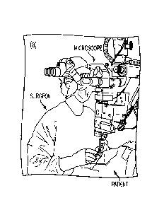
[0014] Figures 1A
and 1B show an example of retinal microsurgery: (a) position of the
patient and the lead surgeon in the operating room; and (b) the layout of the
surgical instruments
in the eye during ERM peeling.
CA 02950139 2016-11-23
WO 2015/184351 PCT/US2015/033322
[0015] Figures 2A-2D illustrate an embodiment of a multi-force sensing
instrument
according to an embodiment of the current invention. Dimensions of an example
of a multi-
function force sensing instrument are shown in (a). The section view of the
tool shaft with
the FBG sensors (b). The geometry related to tool calibration (c). The
dimension of a single
fiber with three FBG sensors (d). The center Bragg wavelengths of FBG-I, FBG-
II, and
FBG-III are 1529 nm, 1545 nm, and 1553 nm, respectively.
[0016] Figure 2E is a schematic illustration of two multi-force sensing
instruments
according to embodiments of the current invention being used in conjunction
for retinal surgery.
[0017] Figure 3 is a schematic illustration of a multi-force sensing
instrument according
to another embodiment of the current invention.
[0018] Figures 4, 5A and 5B are schematic illustrations of further
embodiments of
multi-force sensing instruments according to the current invention.
[0019] Figure 6 is a schematic illustration of a robotic system according
to an
embodiment of the current invention. It also illustrates an example of a
variable admittance
robot control scheme according to an embodiment of the current invention. The
solid lines show
the signal flow in an example implementation, dashed lines show the signals
that can also be
incorporated into the control law.
[0020] Figure 7 shows how admittance varies along with the insertion depth
for an
example according to an embodiment of the current invention. The section
between in and /õb is
the transition between pure force scaling of the sclera force and pure RCM.
[0021] Figures 8A-8D provide results of tool tip force calibration for an
example
according to an embodiment of the current invention. The calculated tool tip
force along X-axis
Ftx versus the actual value (a), its residual error (b). The calculated tool
tip force along Y-axis F4,
versus the actual value (c), and its residual error (d).
[0022] Figure 9A and 9B show results of the optimization problem. The
optimization
cost for FBG- II versus /if (a) and the optimization cost for FBG-III versus
ha (b). The red dots
indicate the minimum cost where ljj = 31.3 mm and /*/// = 37.2 mm.
6
CA 02950139 2016-11-23
WO 2015/184351 PCT/US2015/033322
[0023] Figure 10A-10D show results of sclera force calibration. The
calculated sclera
force along X-axis Fsx versus the actual value (a), its residual error (b).
The calculated sclera
force along Y-axis Fsy versus the actual value (c), and its residual error
(d).
[0024] Figures 11A and 11B show results of sclerotomy location calibration.
The
calculated distance from the tool tip to sclerotomy ds versus the actual value
(a), the RMS error
at each calibrated location versus the actual distance (b). The further the
scleratomy is located
from the tool tip, i.e., the closer it is with respect to FBG-II and FBG-III,
the smaller is the RMS
error. Data points with forces smaller than 5 ml\I in magnitude is not
included to reduce noise, as
discussed in Section II-B.
[0025] Figures 12A and 12B show sclera force estimation error due to tool
tip force.
The calculated sclera force along X-axis E., versus the applied tool tip force
along X-axis Fa
(a), and the calculated sclera force along Y-axis F,versus the applied tool
tip force along Y-
axis Fly (b).
[0026] Figures 13A-13D show results of validation experiment for sclera
contact force.
The calculated sclera force versus the actual value (a), the residual error of
force calculation (b).
The calculated distance from the tool tip to sclerotomy ds versus the actual
value (c), and its
residual error (d).
[0027] Figures 14A and 14B show setup of the pseudo pivot calibration (a)
and the
close-up with coordinate frames robot handle {I}, sclera {sit, and tool tip
{t} (b). Tool tip frame
{t} is underneath the CD, shown with dashed arrows.
[0028] Figures 15A and 15B show results of the optimization to find the
tool tip offset
from the handle along Z-axis (a). The optimum offset is at z*rt = -39.4 mm,
shown as the dot.
The corresponding trajectories of the RCM point (top) and the tool tip
(bottom) (b). The black
straight line shows the end position of the tool shaft.
[0029] Figure 16 shows the setup of the retina vein tracing experiment with
robotic
assistance.
7
[0030] Figures 17A and 17B show sclera force of one retina vein tracing
trial (a). The
corresponding trajectories of the scleratomy point (top) and the tool tip
(bottom) (b). The black
straight line shows the end position of the tool shaft.
DETAILED DESCRIPTION
[0031] Some embodiments of the current invention are discussed in
detail below. In
describing embodiments, specific terminology is employed for the sake of
clarity. However, the
invention is not intended to be limited to the specific terminology so
selected. A person skilled
in the relevant art will recognize that other equivalent components can be
employed and other
methods developed without departing from the broad concepts of the current
invention
[0032] Currently, there are a few robotics systems for the surgeries
that involve inserting
surgical instruments through access ports to perform manipulation inside the
patient. The port
location with respect to the instrument is important because the tool motion
is constrained at the
port. The contact force between the port and the tool shaft distorts the
surgeon's perception of
the tool-to-tissue interaction force exerted at the tool tip. Appropriate
solutions to these
challenges are still lacking.
[0033] Some embodiments of the current invention provide systems and
methods to
integrate multi-function force sensing into surgical instruments. It can
precisely measure the
location of the contact point between the tool shaft and the port, the contact
force, as well as the
force exerted at the tool tip. The force information can be presented directly
to the surgeon
using visual or aural display, or used in a robotic surgical system to provide
useful feedback and
intuitive motion guidance for various surgical procedures.
[0034] An embodiment of the current invention provides a new dual force
sensing
instrument that can sense not only the sclera force in transverse directions,
but also the location
of the sclera contact point on the tool shaft. This new dual force sensing
instrument can enable a
variable admittance robot control to provide intuitive robot behavior. By
varying the robot
8
Date Recue/Date Received 2021-09-30
CA 02950139 2016-11-23
WO 2015/184351
PCT/US2015/033322
admittance, the robot behavior can continuously transit from an adaptive
virtual fixture mode
that enforces RCM and adapts to the current location of the sclerotomy site,
to a force scaling
mode that provides scaled feedback of the sclera force as well as the ability
to reposition the eye.
Experiments have been conducted to calibrate the new dual force sensing
instrument, to calibrate
the tool tip position with respect to the robot, and to evaluate the force
sensor as well as the
control algorithm according to an embodiment of the current invention. The
results show the
potential to increase safety, as well as to enhance the usability and
capability of such a robotic
assistant system.
[0035] Figure 2A provides an illustration of a multi-force sensing
instrument 100
according to an embodiment of the current invention. The multi-force sensing
instrument 100
includes a tool 102 that has a tool shaft 104 having a distal end 106 and a
proximal end 108.
The multi-force sensing instrument 100 also includes a strain sensor 110
arranged at a first
position along the tool shaft 104, and at least one of a second strain sensor
112 or a torque-force
sensor (not shown in Figure 2A) arranged at a second position along the tool
shaft 104. The
second position corresponding to the second strain sensor 112 is more towards
the proximal end
108 of the tool shaft 104 than the first position corresponding to the strain
sensor 110.
[0036] The multi-force sensing instrument 100 also includes signal
processor 114
configured to communicate with the strain sensor 110 and the at least one of
the second strain
sensor or the torque-force sensor 112 to receive detection signals therefrom.
The signal
processor 114 is configured to process the signals to determine a magnitude Fs
and position ds of
a lateral component of a force applied to the tool shaft 104 when the position
ds of the applied F,
force is between the first and second positions, as is illustrated in Figure
2C. The lateral
component of the force is a component of the force that lies in a plane that
is orthogonal to the
tool shaft 104 at the position at which the force is applied.
[0037] The signal processor 114 can be incorporated within a tool handle
115 of the tool
102, as illustrated in Figure 2A. However, the invention is not limited to
this example. It could
be incorporated into a different portion of the tool 102 and/or located
externally. The signal
processor can include and/or access memory and/or data storage. The signal
processor can be a
programmable device and/or a dedicated hard-wired device, such as, but not
limited to an ASIC
or FPGA.
9
CA 02950139 2016-11-23
WO 2015/184351 PCT/US2015/033322
[0038] The multi-force sensing instrument 100 can include a wide range of
tools 102, as
long as they have a structure corresponding to the shaft 104. The tool 102 can
be, but is not
limited to a surgical tool. Figure 3 is an illustration in which two different
embodiments of
multi-force sensing instruments according to the current invention are being
used for eye
surgery. Other applications can include other form of micromanipulation in
which a tool is
extended through a narrow opening in a structure. More generally, some
embodiments can
include tools that are used to extend through a larger opening in larger
structures. Examples of
micromanipulation tools can include, but are not limited to needles, forceps,
picks, cannulas,
trocars, catheters, guide wires, light pipes, endoscopes, etc.
[0039] In some embodiments, the signal processor 114 can be further
configured to
process the signals to determine a magnitude and position of a distal force
116 applied to the
tool shaft 104 when the position of the distal force is beyond the first
position towards the distal
end 106 of the tool shaft 104.
[0040] In some embodiments, the second strain sensor 112 can be a pair of
strain sensors
displaced with respect to each other along a distal to proximal axial
direction along said tool
shaft. Furthermore, the first-mentioned strain sensor 110 and the pair of
strain sensors 112 can
include at least one optical fiber. In that case, the optical fiber includes
first, second and third
Fiber Bragg Gratings (FBG-I, FBG-II, FBG-III) written therein corresponding
respectively to
said first-mentioned strain sensor 110 and the pair of strain sensors 112 in
which the optical
fiber extends substantially parallel to the tool shaft 104. The term
"substantially parallel" is
intended to convey the fact that the fiber does not have to be perfectly
parallel. In some cases,
there may be deviations due to manufacturing tolerances. In some cases, a high
degree of
precision may not be required for the application, so a degree of eiTo can be
accepted.
[0041] Although particular embodiments describe the strain sensors as FGBs
in optical
fibers, the general concepts of the current invention are not limited to only
FBGs. Other types of
strain sensors can be used without departing from the broad scope of the
current invention.
[0042] In some embodiments, the first-mentioned strain sensor 110 and the
pair of strain
sensors 112 include a plurality of optical fibers (for example, 118, 120, 122
in Figure 2B) each
comprising first, second and third Fiber Bragg Gratings (FBG-I, FBG-II, FBG-
III) written
CA 02950139 2016-11-23
WO 2015/184351 PCT/US2015/033322
therein corresponding respectively to the first-mentioned strain sensor 110
and the pair of strain
sensors 112. The plurality of optical fibers (e.g., 118, 120, 122) each extend
substantially
parallel to the tool shaft 104 and substantially parallel to each other.
Although Figure 2B
illustrates an embodiment with three optical fibers, the plurality of optical
fibers is not limited to
that particular number. In some embodiments, there could be two, three or more
than three
optical fibers. The plurality of optical fibers (e.g., 118, 120, 122) can be
arranged substantially
equally spaced around a circumference of the tool shaft 104. In the case of
three optical fibers,
FBG-I, FBG-II, FBG-III each includes a set of three fiber Bragg gratings. In
the embodiment of
Figure 2B, the plurality of optical fibers (118, 120, 122) are three optical
fibers oriented about
120 apart around a circumference of the tool shaft 104. The dimensions
illustrated in Figures
2A-2D are for a particular embodiment and are not required in all embodiments.
[0043] The embodiment of Figures 2A-2D can be useful for being able to
determine two
force components within the plane orthogonal to the tool shaft 104 as well as
being able to
compensate for temperature changes.
[0044] In some embodiments, the first, second and third Fiber Bragg
Gratings (FBG-I,
FBG-II, FBG-III) written in the optical fiber, or fibers, can each have a
unique central reflection
wavelength under relaxed, equal temperature conditions to allow wavelength
division
multiplexing within the optical fiber.
[0045] The multi-force sensing instrument 100 can also include at least one
optical
transmitter and at least one optical receiver 124 optically coupled to the one
or more optical
fiber. The optical transmitter(s) and receiver(s) 124 can be incorporated into
the tool handle 115,
as is illustrated in Figure 2A, or can be located externally. Optical
transmitters can include, but
are not limited to, LEDs and semiconductor lasers, for example. The optical
receivers can
include, but are not limited to, photodiodes, for example.
[0046] Figures 4, 5A and 5B illustrate additional embodiments in which
multiple strain
sensors can be included to set up a plurality of sensing regions. The general
concepts of the
current invention are not limited to the particular number of sensing regions.
For example, some
embodiments could have a large number of closely spaced sensing regions to
approximate a
continuous sensing capability along the tool shaft.
11
CA 02950139 2016-11-23
WO 2015/184351
PCT/US2015/033322
[0047] The following provides a formalism for a general number of sensing
segments
and groups. Note that above we referred to a sensor in reference to FBG pairs,
i.e., groups.
Furthermore, if there are three optical fibers, each FBG pair (or group) would
have 6 sensing
elements. Figure 5A illustrates one example with six sensing segments in three
groups. Multiple
sensing segments can enable the sensor to measure multiple contact forces and
their locations on
the tool shaft. The assumption is that at most one contact force is applied
within each sensing
region. If the fiber optical strain sensor, fiber Bragg grating (FBG), is used
as the strain sensor,
then this extension does not require additional space for incorporating extra
sensors. The multi-
function sensing instrument can preserve the same form factor.
[0048] One example of a generalized model is illustrated in Figures 5A and
5B that has
six sensing segments, shown as 1-a and 1-b to 3-a and 3-b. Each sensing
segment includes three
strain sensors that are evenly placed around the circumference of the tool
shaft, with 120.
intervals. The strain sensors in this example are optical strain gauges based
on fiber Bragg
gratings (FBGs). A group of two sensing segments separates the sensing regions
on the tool
shaft. The sensing segments are numbered with the Arabic numerals indicating
the sensing
region, adjoint with -a or -b for the distal and proximal one within the
group. For example, 1-b
denotes the proximal sensing segment in the first group, as shown in Figure
5A. The portion of
the tool shaft that is located between the sensing segments are called sensing
regions, numbered
with the Arabic numerals. We assume there is at most one contact force exerted
within one
sensing region. The contact forces are also numbered with Arabic numerals. The
distance
between force Fi and sensing segment j-x is denoted as dFii-x, where i =1, 2,
,j =1, 2 === , and
x = a or b. The constant distance between the two sensors within the same
group, sensing
segments j-a and j-b, is denoted as Ali, where j =1, 2, === , and we have A/i
= 11-b ¨ ¨
dFij-b, for any F, that is distal to sensing segment j, i.e. i <j. One special
case of the first contact
force Fl is the tool-to-tissue interaction force exerted right at the tool
tip, i.e. dp7,/-x = 11-x, x = a
orb.
[0049] First, we look at one strain sensor within each sensing segment.
When transverse
force is applied to the tool shaft, the strain generated is proportional to
the moment at the sensor
location and thus proportional to the force applied:
12
CA 02950139 2016-11-23
WO 2015/184351
PCT/US2015/033322
Ed
E =-_ ____________ 1' ________________________________ (1)
E_I EI
[0050] where C is the local strain at the sensor location, M is the moment
at the sensor
location that is attributed to the transverse force, F is the transverse force
applied to the tool
shaft, d is the distance between the force and the sensor location, i.e. the
moment arm, E is the
Young's modulus, I is the moment of inertia, and r is the radial distance
between the bending
axis and the strain sensor.
[0051] The shift in Bragg wavelength of the FBG sensors is proportional to
local strain
and temperature change:
AA = kgÃ+kATAT (2)
where AA denotes the shift in the Bragg wavelength of the FBG sensor, C
denotes the local
strain at the sensor location, AT denotes the temperature change, ke and kAT
are constant
coefficients.
[0052] Second, there are three strain sensors within each sensing segment,
evenly placed
around the circumference of the tool shaft with 120 intervals. The common mode
of the three
strain sensors within the same sensing segment is caused mostly by the axial
strain and the
temperature change. It is the mean value of the wavelength shifts of the three
FBG sensors. The
remaining differential mode reflects the strain attributed to the transverse
forces. We define the
differential mode as the sensor reading. It can be calculated by subtracting
the common mode
from the FBG wavelength shifts:
3
Sj-x, k = k ¨3 E AA3-x, k (3)
k= J
13
CA 02950139 2016-11-23
WO 2015/184351
PCT/US2015/033322
AA
[0053] where Asj and -x, k - _denote respectively the sensor
reading and the wavelength shift of the FBG kin the sensing segment j-x , with
j =1, 2,
= = , x = a, b , and k =1, 2,3.
[0054] There are two sensing segments longitudinally configured proximal to
the
corresponding sensing region. With the assumption that Fi is always exerted
within the sensing
region #1, the sensor readings of sensing segment 1-a and 1-b are linearly
dependent on Fi:
AS/-x = F1MF1,i-x (4)
Fi (5)
where AS/-x = Ast-x,2, X = a and b,
denote the sensor readings of
sensing segment 1-a and 2-b, respectively, K/-x,r). are the constant
coefficient matrices that
can be obtained through calibration, and /111-
1 denoteS the moment at sensing segment /-x
attributed to force Fi, i.e.
= F1dF,1 (6)
Fi _Pi T
where - -s' 4/ denotes the transverse force exerted within the first
sensing
region, from the tool tip to the first sensing segment, and dF,,1-x denotes
the longitudinal distance
from Fi to sensing segment 1-x.
[0055] From (4), we
can compute the moments attributed to Fi at sensing segment /-a
and 1-b:
-111F1,1-x = Ktx, FlAS1-x (7)
where 0 denotes the matrix pseudo-inverse, X = a and b.
[0056] Further we can write:
14
CA 02950139 2016-11-23
WO 2015/184351
PCT/US2015/033322
MF1,1-b 1141,1-a = R1b F1AS I-b FiAS l'a
(8)
[0057] Also from (7), we can write:
, I -6 111F1,1-a = Fl(dF).,1-6
61F1tl-a) (9)
= F1A/1
(10)
[0058] Then we can calculate Fi from (8) and (10)
IVIF1, 1-b AfF1,1-a
-=
(11)
= A/1
/0 AS ¨ Kt AS
1-b, 1-1) 1-a, F1 --a
(12)
A /1
[0059] The distance from Fi to sensing segment 1-x can then be
calculated as follows:
dF1,1-x = ________________________________
(13)
where I I denotes the 2-norm of a vector.
[0060] The sensor readings of sensing segments 2-a and 2-b
reflect the strain attributed
to all forces distal to the sensing segments, i.e. both Fi and F2 contribute
to the sensor reading
S2-x:
2_x --= K2_:crFiAlF1,2-x K2-x,F2MF2,2-x
(14)
= K2-z, Fi FidF1,2-x + K2-x,F2F2dF2,2-x
(15)
where K2-x, F1 and K2-x, F2 are constant coefficient matrices that can be
obtained through calibra-
tion, MFi,2-x is the moment attributed to force Fi at sensing segment 2-X, i =
1 and 2. From (11)
and (13), we can calculate Fi and In addition, we have:
CA 02950139 2016-11-23
WO 2015/184351
PCT/US2015/033322
(117).,2-x 12-a, (16)
as shown in Figures 5A and 5B.
[0061] Therefore, we can write:
MF2,2-g; -4E,F2 (6"52-2.; MF1,2-s) - (17)
F2 (S ¨ FiFidFiA,x) (18)
¨ At, F2 (4512-X K 2- Fl (dFi , -b "4- 12)) (19)
[0062] Similarly, we can calculate F2:
A'1/02 2-6 ¨ 11'11,2,2-ts
(20)
A/2
and d.p2,2-.:
IIMF2,2-211
=
IlF211 (21)
[0063] Now we can derive the equations to calculate F./and dFi,i-x based on
the steps
above. The sensor readings reflect all forces distal to the sensing segment j-
x, where] 2, x = a
and b:
j-1
ASi-x = E 1.j-:v, FA,111Fk,i-x Kj-zr, (22)
k=1
J-1
=_ E ph,F0Fk,i_x + Ki_x, (23)
i-1
>2, (Kj.õ, (dFk,k-b + E + Itce, (24)
i=k+i
16
[0064] Then the transverse contact force Fi applied within sensing
region IV and the
distance dy-x from Fi to sensing segment j-x can be calculated as follows:
F- ¨ ¨ 25)
3 ¨
Al3
j-xll
ci = (26)
HFil
Smart Light Pipe
[0065] One useful application can be a sensorized light pipe 300 for
ophthalmological
surgery, as shown in Figure 2E. Sensor-I 302 can be used for collision
detection with other
tools, with the lens, or with the retina, etc. Sensor-II 304 and Sensor-III
306 can be used to
measure the location of the sclerotomy site and the contact force between the
light pipe and the
trocar. In current practice of ophthalmological surgery, the surgeon uses one
hand to hold a
functional tool, e.g. forceps, while the other hand holds a light pipe to
provide illumination
inside the eye. The sensorized light pipe can be held by a robot, so that the
surgeon can perfollit
bilateral manipulation using two instruments, e.g. two forceps.
[0066] This can be especially useful during vitrectomy, as well as
during complex
manipulation of eye tissue. The robot holds the sensorized light pipe, and
complies with the_eye
motion to satisfy the remote center of motion (RCM) constraint. Furthermore,
computer vision
techniques can be used to track the other tools inside the eye and the
lighting history of the eye
to adjust position and orientation of the light pipe to provide optimal
lighting with minimal light
toxicity.
Additional Embodiments
[0067] Other sensing implementations can achieve the same functionality
of the multi-
function force sensor presented above. One possible approach is to use a force
torque sensor
310 to replace two strain segments within the same group. Figure 3 illustrates
one example that
17
Date Recue/Date Received 2021-09-30
uses a handle-mounted force torque sensor to replace the two sensing segments
that are located
proximally on the tool shaft, close to the handle. The strain sensor that is
located distally, close
to the tool tip, provides information to compensate for the force exerted
close to the tool tip.
[0068] We assume that at most one contact force is applied within each
sensing region.
Let Fi denote the force exerted in the tool portion from the tool tip to the
tip strain sensor 308,
and let F2 denote the force exerted in the tool portion from tip strain sensor
to the handle
mounted force torque sensor. Let li and 12 denote the fixed distance from the
tool tip to the tip
strain sensor and the handle force torque sensor, respectively. di denotes the
distance from Fi to
the tip strain sensor, while d2 denotes the distance from F2to the handle
force torque sensor,
respectively. In this example where there is only one strain sensor at the
distal end of the tool
shaft, close to the tip. We assume the force Fi is always exerted at the tool
tip, i.e. di =11. For
the case that d 1=11, we need more than one strain sensors at the tip. Let Et
denote the strain
measured by the tip strain sensor, and let Ft, and Th denote the force and
torque measured by the
handle force torque sensor. From force balance, we can write:
= + F2 (27)
Th = F1 (di + d1) + F2d2 (28)
= F112 + F2d2 (29)
[0069] Also, we have:
ct KtiFi (30)
where Kt/ is a constant coefficient matrix that can be obtained through
calibration. Combine (28),
(29), and (30), the unknowns Fi, F2, and d2 can be easily solved:
18
Date Recue/Date Received 2021-09-30
KtlEt (31)
t
F2 = Fh ¨ F1 (32)
¨ F112
d2 (33)
F2
[0070] Figure 6 is a schematic illustration of a robotic system 200
according to an
embodiment of the current invention. The robotic system 200 includes a robot
202 that has a
tool connector. Figure 6 is an example for eye surgery using a steady-hand eye
robot 202
adapted from the inventors previous work. However, the broad concepts of the
current
invention are not limited to only surgical robots and are not limited to only
such steady hand eye
robots. The robotic system 200 also includes a multi-force sensing instrument
204 attached to
the tool connector of the robot 202, and a feedback system 206 configured to
communicate with
said multi-force sensing instrument 204 to provide at least one of feedback
control of the robot
202 or feedback information to a user 208 of said robotic system 200. The
multi-force sensing
instrument 204 can be one or more of any of the embodiments of multi-force
sensing instrument
100 described above. In some embodiments, the robotic system 200 can be a
surgical robotic
system; however, the general concepts of the current invention are not limited
to only surgical
robotic systems.
[0071] In some embodiments, the robot 202 can be a teleoperated robot.
In some
embodiments, the robot 202 can be a cooperatively controlled robot that
performs automated
functions in response to a user's actions while using said tool to at least
one of modify, assist or
prevent manual operations of said user's actions. In some embodiments, the
robot 202 can be an
RCM robot.
Detection of patient movement for emergency tool retraction
[0072] During retinal microsurgery, the surgeon needs to monitor the
unexpected patient
movement, e.g., the patient sneezes, coughs, or sits up. These unexpected
movements can cause
collision between the ophthalmic tool and the retina, and can potentially lead
to serious damage
19
Date Recue/Date Received 2021-09-30
CA 02950139 2016-11-23
WO 2015/184351
PCT/US2015/033322
to the eye tissue. When the surgeon notices clues of such movements, the
ophthalmic
instruments has to be removed from the patient's eye very quickly. Previous
work (X. He, D.
Roppenecker, D. Gierlach, M. Balicki, K. Olds, P. Gehlbach, J. Handa, R.
Taylor, and I.
Iordachita, "Toward Clinically Applicable Steady-Hand Eye Robot for
Vitreoretinal Surgery," in
ASME 2012 International Mechanical Engineering Congress and Exposition, 2012,
vol. Volume
2:, pp. 145-153.; M. Balicki, J. Handa, and R. Taylor, "Tool exchange
interface and control
algorithm for cooperative surgical robots," Patent W02012018816 A2, 2011.)
investigated the
tool quick release mechanism and robot control for tool retraction.
[0073] The standard clues for eye movements are seeing the eye move in the
microscope
or the field of view through the lens changes, seeing ''vibrations" or
oscillations of the field of
view, feeling resistance of the lateral forces from the instrument at the
scicrotomy which gets
transmitted to the instrument handle which the surgeon feels since it becomes
above tactile
sensation, subtle clues such as change in light reflection on the retina other
structures. Both the
visual and tactile cues used by the surgeon in current practice are directly
associated with the
sclera-to-tool interaction forces. Using the multi-function force sensing
instrument, the contact
force between the instrument shaft and the sclera are monitored with milli-
newton sensitivity
and kilohertz rate. This sensing capability can enable early detection of the
unexpected patient
movement. For instance, the magnitude and the first time derivative (the rate
of change) of the
sclera-to-tool interaction force can be used as metrics for prediction. When
the sclera force
magnitude crosses a given threshold, and/or exhibits high frequency
oscillations (i.e., large first
time derivative), the likelihood of unexpected patient motion is high.
Warnings can be provided
to the surgeon, e.g., using auditory/haptic feedback, to make the surgeon
aware of the potential
risk before the surgeon notices the standard visual and tactile clues. This
can be used in both
freehand and robot-assisted retinal microsurgeries. In robot-assisted
procedures, the instrument
motion can be used together with the force information to distinguish between
the force due to
the tool motion and that due to the unexpected patient movement. This can
enable faster and
more accurate detection of the unexpected motion, compared to the surgical
scenarios in which
the tool motion is not available.
Tool retraction using robotic assistance
CA 02950139 2016-11-23
WO 2015/184351 PCT/US2015/033322
[0074] When the unexpected patient movement is detected, the desired
reaction time
varies based on the speed and magnitude of movement so that the reaction time
beats removing
the instrument from proximity of tissue that it will potentially injure. For
small movements, the
surgeon simply moves the instrument away from the retina. If a patient sits up
during the
operation, the surgeon removes the instrument really fast. In robot-assisted
procedures, similar
strategies can be implemented with autonomous tool retraction methods under
the surgeon's
supervision.
[0075] Some embodiments of the current invention can provide the following
features:
1. A surgical instrument with integrated sensing to measure contact forces and
locations
of these contacts on the said surgical instrument. It can provide:
= precise measurement of the location of the contact point between the
surgical
tool shaft and the access port with respect to the tool;
= precise measurement of the contact force between the tool shaft and the
access
port in 2-D0F,
= precise measurement of the tool-to-tissue force at the tool tip in 2-DOF
and 3-
DOF,
= and the standard surgical functionality, e.g. hook, forceps, etc.
The sensing principle of the multi-function sensor is based on strain-gauges.
In some
embodiments, fiber optical sensors, i.e. fiber Bragg gratings, are used. So
the surgical
instrument can also be made MR/-compatible. Furthermore, other strain sensing
technologies can also be used for multi-function sensing according to other
embodiments of the current invention. With the same form factor, this sensor
can be
further extended with additional sensing segments to measure multiple contact
forces
and their locations.
2. A method to control a robotic manipulator using the tool forces and force
locations
from the surgical instrument. It can provide:
= useful feedback, such as ha ptic force feedback using force scaling of
the force at
the tool tip or that of the contact force between the tool shaft and the port
21
CA 02950139 2016-11-23
WO 2015/184351
PCT/US2015/033322
= intuitive motion constraints/guidance, such as guiding user's motion to
comply
with a non-static remote center-of-motion (RCM) constraint as a virtual
fixture.
This can provide many advantages over mechanical RCM or other mechanism,
such as flexibility and safety.
= improved control of the tool tip motion with minimal tool deflection,
because
the side load on the tool shaft due to the contact force at the port can be
minimized. This can be especially pertinent for hand-held robotic devices with
small actuators, because the contact force between the port and the tool shaft
can be relatively large with respect to the payload of the small actuators.
This multi-function force sensing instrument can be incorporated with various
robotic
surgical systems, e.g. cooperatively controlled robot, master/slave
teleoperated robot,
hand-held robot, etc. We developed a control strategy for a cooperatively
controlled
robot to provide safe, stable surgical manipulation. Similar control can be
implemented
on other robotic systems and can be used for various surgical procedures, such
as
minimally invasive surgery, cardiac surgery, ophthalmological surgery, etc.
One
potential application is to provide automatic illumination assistance for
ophthalmological surgery.
3. There are also other methods to present the force and force location
information to the
surgeon. Visual display, audio sensory substitution, and vibrotactile
feedback, etc.
4. The force and force location information can be used for training and
evaluation of
surgical skills for residents. Some examples are:
= The ability to control the forces below a safety threshold. For example,
in eye
surgery, forces over 7.5 mN on the retina can potentially damage the retina.
In
eye surgery and minimally invasive surgery, contact forces at the port should
be
minimized.
= The ability to perform specific surgical tasks and surgical maneuvers,
such as
obey the RCM constraint at the port.
22
CA 02950139 2016-11-23
WO 2015/184351
PCT/US2015/033322
[0076] Further embodiments of the current invention include computer
programs
configured to perform the methods of the current invention.
[0077] The following describes some examples according to particular
embodiments of
the current invention. The general concepts of this invention are not limited
to these particular
examples.
EXAMPLES
[0078] All equation numbers in this example section refer to the equations
introduced in
this section, and not the equations in the previous section of this
specification.
[0079] In following examples, we report a new design of a dual force
sensing instrument
according to an embodiment of the current invention that can sense not only
the sclera force in
transverse directions, but also the location of the sclera contact point on
the tool shaft. This new
dual force sensing instrument enables a variable admittance robot control to
provide an intuitive
robot behavior. By varying the robot admittance, the robot behavior can
continuously transit
from an adaptive virtual fixture mode that enforces RCM and adapts to the
current location of
the sclerotomy site, to a force scaling mode that provides scaled feedback of
the sclera force as
well as the ability to reposition the eye. Experiments are conducted to
calibrate the new dual
force sensing instrument, to calibrate the tool tip position with respect to
the robot, and to
evaluate the force sensor as well as the control algorithm. Results show the
potential to increase
safety, as well as to enhance the usability and capability of the robotic
assistant system.
Dual Force Sensing Instrument
[0080] Design: Some embodiments of the current invention can build on the
previous
dual force sensing instrument 36. A major assumption for this example is that
forces are only
exerted at no more than two locations: the tool tip and the sclera contact
point on the tool shaft.
The tool shaft is made of a stainless steel wire with diameter of 0.5 mm, same
as the 25 Ga
ophthalmic instrument. The tool shaft is machined to cut three longitudinal
channels with V-
shape sections. One optical fiber with three fiber Bragg grating (FBG) sensors
(Technica S.A.,
Beijing, China) is embedded into each channel in the tool shaft. Each FBG
sensor is 3 mm long.
23
CA 02950139 2016-11-23
WO 2015/184351 PCT/US2015/033322
The tool dimension, as well as the specifications of the FBG sensors are shown
in Figures 2A-
2D.
[0081] The new dual force sensing instrument includes nine FBG sensors in
total,
arranged into three segments of the tool shaft. The three FBG sensors in the
same tool shaft
segment are 120 apart, and provide strain measurements at that segment of the
tool shaft. The
first FBG sensing segment, FBG-1, typically remains inside the eye. It is used
to measure the
transverse force exerted between the tool tip and the eye tissue, because the
sclera contact force
does not generate strain at the tool tip. FBG-II and FBG-III sensing segments
are at least 30 mm
proximal from the tool tip, greater than the average diameter of human eye (25
mm). They are
dedicated to measure the transverse force exerted at the sclerotomy, and the
location of the
sclerotomy with respect to the tool. The axial force component at the
sclerotomy is mainly due
to friction, thus is correlated to the transverse force, i.e. normal force.
Axial force sensing at the
tip is not included in this prototype, but is possible as shown in our other
work 33. The total
length of the tool shaft is 45 mm. The data acquisition unit is the sm130-700
optical sensing
interrogator from Micron Optics (Atlanta, GA) with a refresh rate of 2 kHz and
a spectrum range
from 1525 nm to 1565 nm.
Algorithm to Calculate Forces and Sclerotomy Location
[0082] The algorithm to calculate the sclera and tip forces is based on the
previous
methods presented by Iordachita et al. 30 and He et al. 36. The wavelength
shift common mode
of the FBG sensors from the same sensing segment represents the strain
attributed to axial force
and temperature change. The differential mode, termed sensor reading, is
defined as follows:
Asp, =3k ¨ (1)
3 k-1 j LA- k
where Asik and AAA denotes respectively the sensor reading and the wavelength
shift of FBG
sensor kin sensing segment j, with j = 1, II, and III, and k = 1, 2, and 3.
[0083] With the assumption that tool-to-tissue interaction forces are
always exerted at
the tool tip, the sensor readings of FBG-I are linearly dependent on the
transverse force at the
tool tip:
24
CA 02950139 2016-11-23
WO 2015/184351
PCT/US2015/033322
LSI = K1F (2)
where As, = psti, Asõ, ./.5s!g]r denotes the sensor readings of FBG-I, Ft =
[Fõ Fty]T denotes the
transverse force exerted at the tool tip, and K1t is a 3 x2 matrix with
constant coefficients.
[0084] The
location where the sclera contact force is exerted on the tool shaft depends
on
the tool insertion depth inside the eye. Together with the sclera contact
force, it contributes to
the strain generated at FBG-II and FBG-III. In addition, the Fl3G sensors also
respond to tip
force, therefore:
AS. =K F + M-AS-=K-F + K- M. (3)
j t j j it t is j
= IC K-s Fs d- (4)
ft. t i
where LiSi = [As:iv Lls52, Asi3]T denotes the sensor readings of FBG-j, Fs, =
[Fsx, Fs,y1T denotes
the transverse force exerted at the sclerotomy, [11 denotes the distance from
the
sclerotomy to FBG-j along the tool shaft, Mj = [Mix, Miyir denotes the moment
attributed to Fs at FBG-j, Kit and Kis are both 3x2 constant coefficients
matrices, j = II and
III. As shown in Fig. 2, the distance L11 between FBG-II and FBG-III is
constant and is always
the difference between di/ and dm, which equals to the difference between
= ¨ = dm ¨ d11 (5)
[0086] The
coefficient matrices Kit (I = I, II, and III) and Kis (j = II and III),
as well as the distance i1 between FBG-II and FBG-III are obtained through the
tool
calibration described in more detail below.
[0087] The tip
force can be calculated using the pseudo-inverse of the coefficient matrix:
CA 02950139 2016-11-23
WO 2015/184351 PCT/US2015/033322
Ft = KittASt (6)
where k t denotes the pseudo-inverse operator.
. .
[0088] The moments attributed to the sclera contact forces at FBG-j ( j =
II and III) can
be calculated using (3) and (6):
Mi = Kis t Si ¨ Ki t t 1.1 ) (7)
[0089] The sclera contact force can be solved from the difference in
moments MI/ and
= Il1EI
(8)
[0090] The distance from the sclerotomy to the FBG-j can be obtained from
the
magnitude ratio between the moment and the force:
IlMf I
d =
(9)
where I IF denotes the vector 2-norm.
[0091] This method can calculate transverse forces exerted at the tool tip
and at the
sclerotomy, as well as the location of the sclerotomy with respect to the
tool. However, if the
magnitude of the sclera contact force is small, the location of the sclerotomy
calculated using (9)
can be subject to large error. Therefore, the sclerotomy location is updated
with the help of a
deadband on the sclera force magnitude. Only when the sclera force magnitude
exceeds a given
threshold (e.g. 5 mN), the sclerotomy location will be updated using (9),
otherwise the previous
value of d will be used.
26
CA 02950139 2016-11-23
WO 2015/184351 PCT/US2015/033322
Variable Admittance Robot Control
[0092] A variable admittance robot control scheme is devised from previous
force
scaling and admittance velocity control 3537. In addition to the surgeon's
force input at the tool
handle (robot end-effector), it utilizes the new sensing capabilities enabled
by the dual force
sensing instrument, to provide a robot behavior that is transparent and
intuitive to the surgeon.
This robot behavior enables useful feedback and virtual fixtures to increase
precision and safety
to interact with the patient and the environment. Figure 6 illustrates the
variable admittance
control scheme.
Constant Admittance Control with Force Scaling
[0093] The previous admittance velocity control is:
= aFith (10)
A= d, (11)
iwrt
where kwh and hh are the desired robot handle velocity in the robot handle
frame and that in
the world Cartesian frame, respectively, Fhh denotes user's force input
measured in the robot
handle frame, and a is a constant scalar as the admittance gain, and Ada, is
the adjoint
transformation associated with coordinate frame transformation gwh 38. If we
write
-Rwh Pwki
9wh
- , where Rwh and pwh denote the rotation and translation of
,gwh
from the local robot handle frame to the world Cartesian frame, then:
Ad P.swhflvih
(12)
awiz
0 Rwh
27
CA 02950139 2016-11-23
WO 2015/184351 PCT/US2015/033322
where r_ilvh denotes the skew symmetric matrix that is associated with the
vector pw.h.
[0094] We modify (10) using force scaling 3537 to incorporate sclera force
feedback:
hh a(Fhh yFhs) (13)
where y is a force scaling factor, and Fhs is the sclera force resolved at the
robot handle with
the following adjoint transformation:
Fhs, = AdT hs F.55 (14)
g
where Fss denotes the sclera force measured in the sclera frame which is
located at the
sclerotomy and has the same orientation as the handle frame. Let ghs [nhs
Phs]
0
denote the coordinate frame transformation from sclera frame to handle frame,
then:
RhsT 0 1
Adrs DT (15)
g h
_¨ hp p "-Ts
N
where (:)T denotes the matrix transpose. The sclerotomy is not a static point
during retinal
microsurgery. Therefore, ghs is time-varying. We assume that the tool shaft
bending due to
T
sclera force remains in a small range, then Rhs 1, Pi 0,
zji.si and Zhs can be
updated by the dual force sensing instrument.
Variable Admittance Control
28
CA 02950139 2016-11-23
WO 2015/184351 PCT/US2015/033322
[0095] The admittance in the previous control law is isotropic. Virtual
fixtures can be
rendered by commanding anisotropic admittance. We introduce diagonal
admittance matrices
into (13) and rewrite it in the sclera frame:
i1ss= ce(Ash-Psh vAs,Fss ) (16)
where is the desired velocity of where the robot/tool contact the
sclerotomy in the sclera,
and E. are the handle input force and sclera contact force resolved in the
sclera frame,
respectively, y denotes the constant scalar as the force scaling factor, ce
denotes the constant
scalar as the admittance gain, and AA. and A.õ are the diagonal admittance
matrices associated
with the handle input force and sclera contact force in the sclera frame,
respectively. If
= = .1, (16) reduces to (13) as force scaling of the sclera force.
[0096] A virtual RCM can be realized by setting Aõ. = dia9a0, 0, 1 1, 1,
If) and
Aõ = 1. The handle input force FA is resolved in the sclera frame. The
admittance matrix Ash
removes the transverse force components that can lead to undesired lateral
motion, and preserves
the 4-DOF motion that is allowed by the RCM constraints. In addition, the
sclera force
feedback is to servo the sclera contact force toward zero. This strengthens
the virtual RCM with
robustness against eye motion attributed other instrument and patient
movement.
[0097] When the surgeon is performing ER1VI peeling, the tool tip is close
to the retina,
and an RCM is desired to minimize the motion of the eye and the target
membrane. When the
surgeon needs to reposition the eye to adjust view, the tool is kept away from
the retina to avoid
collision. Therefore, the measured insertion depth of the tool can be used to
adjust the robot
admittance to provide the appropriate robot behavior. For example, we can
define:
Ash = diag ¨ , 1¨ 1, 1, 1, 11) (17)
A, = dia,g(11+ 1 +fl, 1, 1,1, 11T) (18)
29
CA 02950139 2016-11-23
WO 2015/184351
PCT/US2015/033322
where g E [0,1] varies along with the tool insertion depth as shown in Figure
7. When the
insertion depth is smaller than the given lower bound 12b, = 0 and Agh = Agg =
1. We have
the force scaling control mode that provides the freedom to reposition the eye
with scaled sclera
force feedback. When the insertion depth is larger than the given upper bound
/õ, JO = I. and it
switches to virtual RCM with doubled gain for minimizing the transverse forces
at the
sclerotomy. Alternatively, the value of JO can be controlled by the human
operator (e.g. using a
foot pedal) to select the preferred operating mode.
Experiments and Results
Calibration of the Dual Force Sensing Instrument
[0098] An automated calibration system 33 is used to carry out the
calibration.
Transverse forces are applied at different locations on the tool. The
wavelength shifts of the
FBG sensors, applied forces, and the location on the tool where the forces are
applied are
measured and recorded.
Calibration for Tip Force
[0099] The calibration for tip force is the same as for our previous dual
force sensing
tool 36. Transverse forces up to 10 mN are applied along X- and Y-axes. The
coefficient
matrices Kjõ j = 1, 2, and 3, are obtained as least square solution of (2) and
(3) with M= 0.
Figures 8A-8D illustrate the calibration results for the tip forces. Figures
8A and 8C show the
forces calculated using (6) versus the actual forces. The 45 straight line
through the origin
represents the ideal results. Figures 8B and 8D show the residual error versus
the actual forces.
The root mean square (RMS) errors are 0.35 mN for Ft, and 0.53mN for Fty,
respectively.
Calibration for Sclera Contact Force and Location
[00100] Transverse forces are applied at 16 locations on the tool shaft,
from 10 mm to 25
mm proximal from the tool tip with 1 mm intervals, shown as d, in Figure 2C.
The force
magnitude ranges from 25 mN at 10 mm from the tool tip, to 100 mN at 25 mm
from the tool
tip. Because the optical fibers are manually aligned and embedded into the
tool shaft, the
CA 02950139 2016-11-23
WO 2015/184351 PCT/US2015/033322
accurate "center" locations of FBG-II and FBG-HI, i.e. ;õ and iõ, in Figure
2C, are not known.
There is no force applied at the tool tip, hence (4) reduces to:
= K- F dI- (19)
J is s
= Kis dg) (20)
where di =Ii¨ c3 with j = II and III.
[00101] The calibration goal is to find the constant KJ, and 6. Because
they are not
linearly independent, an optimization problem is constructed to find the best
fit:
arg min/i I ASJ - KJ, Fg(li ¨ (4)11 (21)
s. t. 17j, = AS] (F, (F.] ¨ ))t (22)
25 50 (23)
[00102] The optimum 1; e 125, So] minimizes the cost function, i.e. the 2-
norm of the
residual error of the sensor reading of FBG-j. Figures 9A and 9B illustrate
the optimization
results. /7, and t;,, are 31.3 mm and 37.2 mm, respectively. The difference
between FBG-II and
FBG-III a = d ¨1;1= 5.9 mm, is consistent with the nominal value of 6 mm in a
single fiber.
[00103] The coefficient matrix icj., is calculated using (22) with U.
Calibration results
demonstrate sufficient accuracy, as shown in Figures 10A-10D. The RMS errors
are 0.82 mN
for F,õ. and 1.00 mN for F,. The location of the sclerotomy is estimated using
forces larger than
mN in magnitude. Figures 11A and 11B illustrate the estimated sclerotomy
location with
respect to the tool tip versus the actual value, and the estimation RMS error
at each calibrated
location. The further the sclerotomy is located from the tip, the closer it is
to FBG-II and FBG-
III, and the more accurate is the location estimation. As shown in next
section, low-pass
filtering can further reduce the sensing noise and smooth the estimation.
[00104] Using the sclera calibration results, we examine the tip force
cancellation from
FBG-II and FBG-III. The sensor readings of FBG-II and FBG-III from calibration
with only tip
31
CA 02950139 2016-11-23
WO 2015/184351 PCT/US2015/033322
forces are plugged into (7) and (8) to calculate the sclera force estimation
error due to tip force.
As shown in Figures 12A and 12B, the sclera force errors are not dependent on
the tip force
magnitude, and are possibly due to the system noise. The RMS errors are 0.62
mN for F and
0.74 mN for F;v, with tip forces up to 10 mN.
Validation Experiment for Sclera Contact Force
[00105] A validation experiment is carried out using the automated
calibration system to
test the results obtained from calibration for sclera force and location. The
direction and the
magnitude of the transverse forces, as well as the location on the tool shaft
where the force is
applied are generated randomly within the calibrated range. A moving average
filter with a
window size of 100 samples is applied on the location estimation of
sclerotomy. Figures 13A-
13D illustrate the results of the validation experiment. The RMS errors of Fõ
and F,s, estimations
are 0.56 mN and 1.08 mN respectively. The RMS error of the sclerotomy location
estimation is
0.57 mm, comparable to the lowest error obtained in the calibration at 25 mm
from the tool tip.
Tool to Robot Calibration
[00106] Incorporating dual force sensing capability into the robot control
requires an
accurate coordinate transformation from the local tool frame to the robot tool
holder frame. It is
reasonable to assume the tool and the tool holder are coaxial. The X- and Y-
axes of the tool and
the robot tool holder are manually aligned. The Z-offset z,, from the robot
tool holder to the tool
tip is about -40 mm measured with a caliper. A traditional pivot calibration
is not practical,
because the tool shaft is not rigid. We use variable admittance control to
enforce the RCM
constraint, and to perform a pseudo pivot calibration. Figures 14A and 14B
illustrate the
experiment setup. A piece of 0.25 mm thick, stiff paper is taped to a CD
clamped to a stable
platform. A 0.7 mm hole is punctured in the center of the paper that is
exposed through the
center hole of the CD. The dual force sensing tool is inserted into the hole
and pivoted with the
RCM constraint by the variable admittance control, as shown in Figure 14A.
[00107] The sclera location estimations cis from the dual force sensing
tool and the frame
transformations from the world Cartesian frame to the robot tool holder frame
gwr are used to
32
find the tool tip offset from the tool holder. Let grs denote the frame
transformation from the
"sclera" frame located at the RCM point 314 to the robot tool holder frame.
Because we assume
the orientation of "sclera" RCM frame is aligned with that of the robot tool
holder frame:
-Rrs
grs = Prs1
(24)
where R = 1 prs = [0, 0, z + d 1T z
rs rt. s
and rt is the Z-position of the tool tip in the
robot tool holder frame. The RCM point Pws can be considered as a static point
in the world
Cartesian frame. Ideally, all Pws computed from the kinematics should converge
to one point.
Therefore, an optimization problem that finds the rt to minimize the standard
deviation of all
Pws:
arg min llo-(Pws)
Zrt (25)
S.t. P
[P
ws =wsi == = Pwsni T (26)
rwskl , [Prskl
1 &wrk k = 1,"=,n
(27)
¨45 zrt 35 (28)
[00108]
Figure 15A shows the optimization results, rt = 39.4 mm. The corresponding
trajectories of the RCM point and the tool tip are shown in Figure 15B. The
standard deviation
of the computed RCM positions is 0.38 mm, 0.34 mm and 0.74 mm in the X-, Y-,
and Z-
direction, respectively. This demonstrates the capability of adaptive RCM
constraints enabled
by the variable admittance control.
33
Date Recue/Date Received 2021-09-30
Tracing a Retina Vein in an Eye Phantom
[00109] We further assess the performance of the robot control using an
eye phantom
318, as shown in Figure 16. The tool is inserted through a 23 Ga trocar on the
eye and is used to
trace a vein on the retina. A stereo video microscope 316 with a 3D display
320 is used for
visualization.
[00110] The task is to make a round trip above a retina vein branch that
is about 3 mm
long. Five trials are conducted with the variable admittance control. Figures
17A and 17B
illustrate the recorded sclera forces, as well as the trajectory of the
sclerotomy point and the tool
tip of one of the trials. The maximum sclera force magnitude is 3.44 0.21
mN. The
sclerotomy position is calculated using the tool-to-robot transformation
obtained above. The
standard deviation of the sclerotomy position is 0.13 0.03 mm, 0.17 0.06
mm, and 0.38
0.06 mm for X-, Y-, and Z-direction. The experiment results show the RCM
behavior with the
variable admittance control is precise and repeatable, minimizing both force
and motion of the
sclerotomy.
[00111] The same task is also attempted with the standard cooperative
control without
sclera force feedback. The user cannot feel the sclera force, and can only
rely on the visual
feedback provided by the 3D display. The severe tool deflection due to large
sclera forces (over
50mN) and the inverted motion due to RCM make it very difficult to control the
tool tip motion.
No successful trial was completed. In contrast, the variable admittance
control enables a
fulcrum at the sclerotomy, the user pivots naturally about it with precise
control of the tool tip
motion.
Discussion and Conclusion
[00112] Krupa et al. 39 used force control with the help of a force
sensor mounted on the
robot end-effector to implement an adaptive RCM behavior. However, it was
assumed that
there was no transverse forces exerted at the instrument tip, therefore the
transverse forces
measured outside the patient at the robot end-effector was the contact force
exerted between the
instrument shaft and the trocar. This assumption is not necessarily valid in
MIS. The dual force
34
Date Recue/Date Received 2021-09-30
CA 02950139 2016-11-23
WO 2015/184351 PCT/US2015/033322
sensing instrument can provide sufficiently accurate, independent measurements
of the tool tip
force and the sclera contact force, as well as the location of the sclerotomy.
Its design can also
be applied to surgical instruments for MIS, to provide additional useful
information to improve
the surgical robot control. Both impedance and admittance type robots can
utilize this sensor to
provide safe interaction with the environment. This can be especially
pertinent for bilateral
cooperative manipulation and telesurgery.
[00113] The variable admittance control takes the sensing advantage from
the dual force
sensing instrument. It reflects the natural physical interaction between the
tool and the
environment. It can adapt to the current RCM point without the assumption that
the RCM point
is static. Mechanical RCM does not provide the flexibility to vary the RCM
point, while
software virtual RCM that uses geometric constraints can incorporate the dual
force sensing
instrument to update the current RCM point. The variable admittance control
law can also be
incorporated with other virtual fixture methods, such as the constrained
optimization framework
2223. Ultimately, it should provide a transparent and intuitive interface that
can incorporate
useful feedback and natural motion guidance.
[00114] We have presented a novel multi-function force sensing instrument
designed for
vitreoretinal surgery procedures that measures not only the forces at the
instrument tip, but also
the sclera contact position and interaction force on shaft of the instrument.
A variable
admittance robot control method was developed that incorporates this
information to provide a
transparent and intuitive robot behavior that can minimize eye motion while
enabling tool
manipulation inside the eye, as well as provide useful sclera force feedback
to assist to
reposition the eye. This system can potentially provide safe, stable
micromanipulation that can
improve the outcome of the retinal microsurgery.
[00115] References
1. J. R. Wilkins, C. A. Puliafito, M. R. Hee, J. S. Duker, E. Reichel, J. G.
Coker, J. S. Schuman,
E. A. Swanson, and J. G. Fujimoto, "Characterization of epiretinal membranes
using optical
coherence tomography.," Ophthalmology, vol. 103, no. 12. pp. 2142-2151, 1996.
CA 02950139 2016-11-23
WO 2015/184351 PCT/US2015/033322
2. M. Patkin, "Ergonomics applied to the practice of microsurgery.," The
Australian and New
Zealand Journal of Surgery, vol. 47, no. 3, pp. 320-329, 1977.
3. P. Gupta, P. Jensen, and E. de Juan, "Surgical forces and tactile
perception during retinal
microsurgery," in /141CCAL 1999, vol. 1679, pp. 1218-1225.
4. R. H. Taylor, J. Funda, D. D. Grossman, J. P. Karidis, and D. A. LaRose,
"Remote center-of-
motion robot for surgery," U.S. Patent 5 397 323, Mar. 14, 1995.
5. J.-P. Hubschman, J. Son, B. Allen, S. D. Schwartz, and J.-L. Bourges,
"Evaluation of the
motion of surgical instruments during intraocular surgery," Eye (London,
England), vol. 25,
no. 7, pp. 947-953, 2011,
6. S. Charles, H. Das, T. Ohm, C. Boswell, G. Rodriguez, R. Steele, and D.
Istratc, "Dexterity-
enhanced telerobotic microsurgery," in IEEE ICRA, 1997, pp. 5-10.
7. T. Nakano, N. Sugita, T. Ueta, Y. Tamaki, and M. Mitsuishi, "A parallel
robot to assist
vitreoretinal surgery," LICARS, vol. 4, no. 6, pp. 517-526, 2009.
8. Y. Ida, N. Sugita, T. Ueta, Y. Tamaki, K. Tanimoto, and M. Mitsuishi,
"Microsurgical
robotic system for vitreoretinal surgery," LICARS, vol. 7, no. 1, pp. 27-34,
2012.
9. W. Wei, R. Goldman, and N. Simaan, "Design and theoretical evaluation of
micro-surgical
manipulators for orbital manipulation and intraocular dexterity," in IEEE
ICRA, 2007, pp.
10-14.
10. H. Yu, J. H. Shen, K. M. Joos, and N. Simaan, "Design, Calibration and
preliminary testing
of a robotic telemanipulator for OCT guided retinal surgery," in IEEE ICRA,
2013, pp. 225-
231.
11. R. A. Maclachlan, B. C. Becker, J. C. Tabar, G. W. Podnar, L. A. Lobes,
and C. N. Riviere,
"Micron: an actively stabilized handheld tool for microsurgery," IEEE
Transactions on
Robotics, vol. 28, no. 1, pp. 195-212, 2012.
12. C. Song, D. Y. Park, P. L. Gehlbach, S. J. Park, and J. U. Kang, "Fiber-
optic OCT sensor
guided 'SMART' micro-forceps for microsurgery," Biomedical Optics Express,
vol. 4, no. 7,
pp. 1045-1050, 2013.
13. M. P. Kummer, S. S. Member, J. J. Abbott, B. E. Kratochvil, R. Borer, A.
Sengul, B. J.
Nelson, and S. S. Member, "OctoMag: an electromagnetic system for 5-DOF
wireless
micromanipulation," IEEE Transactions on Robotics, vol. 26, no. 6, pp. 1006-
1017, 2010.
36
CA 02950139 2016-11-23
WO 2015/184351
PCT/US2015/033322
14. R. Taylor, P. Jensen, L. Whitcomb, A. Barnes, R. Kumar, D. Stoianovici, P.
Gupta, Z.
Wang, E. DeJuan, and L. Kavoussi, "A Steady-Hand Robotic System for
Microsurgical
Augmentation," The IJRR, vol. 18, no. 12, pp. 1201-1210,1999.
15. B. Mitchell, J. Koo, I. Iordachita, P. Kazanzides, A. Kapoor, J. Handa, G.
Hager, and R.
Taylor, "Development and application of a new steady-hand manipulator for
retinal
surgery," in IEEE ICRA, 2007, pp. 623-629.
16. A. Uneri, M. A. Balieki, J. Handa, P. Gehlbach, R. H. Taylor, and I.
Iordachita, "New
Steady-Hand Eye Robot with micro-force sensing for vitreoretinal surgery," in
IEEE
BioRob, 2010, pp. 814-819.
17. X. He, D. Roppenecker, D. Gierlach, M. Balicki, K. Olds, P. Gehlbach, J.
Handa, R. Taylor,
and I. Iordachita, "Toward clinically applicable Steady-Hand Eye Robot for
vitreoretinal
surgery," in ASME IMECE, 2012, IMECE2012-88384,
18. L. B. Rosenberg, "Virtual fixtures: Perceptual tools for telerobotic
manipulation," in IEEE
Virtual Reality Annual International Symposium, 1993, pp. 76-82.
19. J. J. Abbott and A. M. Okamura, "Virtual fixture architectures for
telemanipulation," in
IEEE ICRA, 2003, pp. 2798-2805.
20. A. Bettini, P. Marayong, S. Lang, A. M. Okamura, and G. D. Hager, "Vision-
assisted control
for manipulation using virtual fixtures," IEEE Transactions on Robotics, vol.
20, no. 6, pp.
953-966,2004.
21. P. Marayong, M. Li, A. M. Okamura, and G. D. Hager, "Spatial motion
constraints: theory
and demonstrations for robot guidance using virtual fixtures," in IEEE ICRA,
2003, vol. 2,
pp. 1954-1959.
22. M. Li, M. Ishii, and R. H. Taylor, "Spatial motion constraints using
virtual fixtures generated
by anatomy," IEEE Transactions on Robotics, vol. 23, no. 1, pp. 4-19,2007.
23. A. Kapoor and R. Taylor, "Constrained control for surgical assistant
robots," in IEEE ICRA,
2006, pp. 231-236.
24. A. Menciassi, A. Eisinberg, G. Scalari, C. Anticoli, M. Carrozza, and P.
Dario, "Force
feedback-based microinstrument for measuring tissue properties and pulse in
microsurgery,"
in IEEE ICRA, 2001, pp. 626-631.
25. U. Seibold, B. Kubler, and G. Hirzinger, "Prototype of Instrument for
Minimally Invasive
Surgery with 6-Axis Force Sensing Capability," in IEEE ICRA, 2005, pp. 496-
501.
37
CA 02950139 2016-11-23
WO 2015/184351
PCT/US2015/033322
26. J. Peirs, J. Clijnen, D. Reynaerts, H. Van Brussel, P. Herijgers, B.
Corteville, and S. Boone,
"A micro optical force sensor for force feedback during minimally invasive
robotic surgery,"
Sensors and Actuators A: Physical, vol. 115, no. 2-3, pp. 447-455, 2004.
27. P. Puangmali, H. Liu, L. D. Seneviratne, P. Dasgupta, and K. Althoefer,
"Miniature 3-Axis
Distal Force Sensor for Minimally Invasive Surgical Palpation," IEEE/ASME
Transactions
on Mechatronics, vol. 17, no, 4, pp. 646-656, 2012.
28. P. Polygerinos, A. Ataollahi, T. Schaeffter, R. Razavi, L. D. Seneviratne,
and K. Althoefer,
"MRI-compatible intensity-modulated force sensor for cardiac catheterization
procedures,"
IEEE TBME, vol. 58, no. 3, pp. 721-726, 2011.
29. P. Berkelman, L. Whitcomb, R. Taylor, and P. Jensen, "A miniature
instrument tip force
sensor for robot/human cooperative microsurgical manipulation with enhanced
force
feedback," in MICCAI, 2000, pp. 247-286.
30. 1. Iordachita, Z. Sun, M. Balicki, J. U. Kang, S. J. Phce, J. Handa, P.
Gehlbach, and R.
Taylor, "A sub-millimetric, 0.25 mN resolution fully integrated fiber-optic
force-sensing tool
for retinal microsurgery," IJCARS, vol. 4, no. 4, pp. 383-390, 2009.
31. X. He, M. Balicki, J. U. Kang, P. Gehlbach, J. Handa, R. Taylor, and I.
Iordachita, "Force
sensing micro-forceps with integrated fiber Bragg grating for vitreoretinal
surgery," in SPIE
Phontics West, 2012, 8218-82180W, pp. 1-7.
32. X. Liu, I. Iordachita, X. He, R. Taylor, and J. U. Kang, "Miniature fiber-
optic force sensor
based on low-coherence Fabry-Perot interferometry for vitreoretinal
microsurgery,"
Biomedical Optics Express, vol. 3, no. 5, pp. 1062-1076, 2012.
33. X. He, J. Handa, P. Gehlbach, R. Taylor, and I. Iordachita, "A Sub-
Millimetric 3-DOF Force
Sensing Instrument with Integrated Fiber Bragg Grating for Retinal
Microsurgery," IEEE
TBME, accepted, 2013.
34. N. Cutler, M. Balicki, M. Finkelstein, J. Wang, P. Gehlbach, J. McGready,
I. Iordachita, R.
Taylor, and J. T. Handa, "Auditory force feedback substitution improves
surgical precision
during simulated ophthalmic surgery," /01/5, vol. 54, no. 2, pp. 1316-1324,
2013.
35. M. Balicki, A. Uneri, I. Iordachita, J. Handa, P. Gehlbach, and R. Taylor,
"Micro-force
sensing in robot assisted membrane peeling for vitreoretinal surgery," in
MICCAI, 2010, vol.
13, pp. 303-310.
38
CA 02950139 2016-11-23
WO 2015/184351 PCT/US2015/033322
36, X. He, M. Balicki, P. Gehlbach, J. Handa, R. Taylor, and I. lordachita, "A
novel dual force
sensing instrument with cooperative robotic assistant for vitreoretinal
surgery," in IEEE
ICRA, 2013, pp. 213-218.
37. R. Kumar, P. Berkelman, P. Gupta, A. Barnes, P. S. Jensen, L. L. Whitcomb,
and R. H.
Taylor, "Preliminary experiments in cooperative human/robot force control for
robot assisted
microsurgical manipulation," in IEEE ICRA, 2000, pp. 610-617.
38. R. M. Murray, Z. Li, and S. S. Sastry, A Mathematical Introduction to
Robotic
Manipulation, CRC Press, 1994.
39. A. Krupa, G. Morel, and M. de Mathelin, "Achieving high-precision
laparoseopie
manipulation through adaptive force control," Advanced Robotics, vol. 18, no.
9, pp. 905¨ ,1
926, 2004.
[00116] The embodiments illustrated and discussed in this specification are
intended only
to teach those skilled in the art the best way known to the inventors to make
and use the
invention. In describing embodiments of the invention, specific terminology is
employed for the
sake of clarity. However, the invention is not intended to be limited to the
specific terminology
so selected. The above-described embodiments of the invention may be modified
or varied,
without departing from the invention, as appreciated by those skilled in the
art in light of the
above teachings. It is therefore to be understood that, within the scope of
the claims and their
equivalents, the invention may be practiced otherwise than as specifically
described.
39