Sélection de la langue

Search

Sommaire du brevet 2954500 

Énoncé de désistement de responsabilité concernant l'information provenant de tiers

Une partie des informations de ce site Web a été fournie par des sources externes. Le gouvernement du Canada n'assume aucune responsabilité concernant la précision, l'actualité ou la fiabilité des informations fournies par les sources externes. Les utilisateurs qui désirent employer cette information devraient consulter directement la source des informations. Le contenu fourni par les sources externes n'est pas assujetti aux exigences sur les langues officielles, la protection des renseignements personnels et l'accessibilité.

Disponibilité de l'Abrégé et des Revendications

L'apparition de différences dans le texte et l'image des Revendications et de l'Abrégé dépend du moment auquel le document est publié. Les textes des Revendications et de l'Abrégé sont affichés :

  • lorsque la demande peut être examinée par le public;
  • lorsque le brevet est émis (délivrance).
(12) Brevet: (11) CA 2954500
(54) Titre français: DISPOSITIF DE POSITIONNEMENT DE COTYLE PROTHETIQUE ET PROCEDE ASSOCIE
(54) Titre anglais: ACETABULAR CUP POSITIONING DEVICE AND METHOD THEREOF
Statut: Accordé et délivré
Données bibliographiques
(51) Classification internationale des brevets (CIB):
  • A61F 2/46 (2006.01)
(72) Inventeurs :
  • TERMANINI, ZAFER (Etats-Unis d'Amérique)
(73) Titulaires :
  • ZAFER TERMANINI
(71) Demandeurs :
  • ZAFER TERMANINI (Etats-Unis d'Amérique)
(74) Agent: DEETH WILLIAMS WALL LLP
(74) Co-agent:
(45) Délivré: 2018-08-21
(86) Date de dépôt PCT: 2015-05-16
(87) Mise à la disponibilité du public: 2016-01-14
Requête d'examen: 2017-04-05
Licence disponible: S.O.
Cédé au domaine public: S.O.
(25) Langue des documents déposés: Anglais

Traité de coopération en matière de brevets (PCT): Oui
(86) Numéro de la demande PCT: PCT/US2015/031275
(87) Numéro de publication internationale PCT: US2015031275
(85) Entrée nationale: 2017-01-06

(30) Données de priorité de la demande:
Numéro de la demande Pays / territoire Date
14/326,006 (Etats-Unis d'Amérique) 2014-07-08

Abrégés

Abrégé français

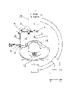
La présente invention concerne le positionnement d'un cotyle prothétique dans un alignement optimal souhaité par rapport au bassin de patients à l'aide d'un équipement de radioscopie classique facilement disponible dans des salles d'opération en association avec un gabarit métallique utilisé comme guide. Le dispositif comprend des tiges métalliques d'inclinaison à un angle de 45 degrés par rapport à l'impacteur et à la tige d'antéversion du cotyle situés à une certaine distance de la ligne médiane correspondant au degré d'inclinaison. Lorsque lesdites tiges d'inclinaison et d'antéversion sont alignées avec des structures anatomiques centrales telles que la symphyse pubienne et le milieu de la première vertèbre sacrée, cet alignement aura pour résultat une pose correcte du cotyle prothétique à l'emplacement souhaité.


Abrégé anglais

Positioning an acetabular cup in a desired optimal alignment in relation to the patients pelvis using conventional fluoroscopic equipment readily available in operating rooms in conjunction with a metallic jig as guide. The device having inclination metallic rods at 45 degrees angle to the cup impactor and anteversion rod situated at a distance from the midline that correspond to the degree of inclination. When said inclination and anteversion shafts are aligned with central anatomical structures such as symphysis pubis and middle of first sacral vertebra will result in correct placement of the acetabular cup at the desired version.

Revendications

Note : Les revendications sont présentées dans la langue officielle dans laquelle elles ont été soumises.


What is claimed:
1. An orthopedic surgical device for positioning an acetabular cup implant
in a surgically prepared acetabular cavity comprising:
a primary vertical shaft and an acetabular cup inserter shaft, said primary
vertical shaft being perpendicularly coupled to said acetabular cup inserter
shaft;
a secondary shaft perpendicularly coupled to said primary vertical shaft via a
sliding locking bracket;
an upper alignment rod horizontally secured to said secondary shaft via a
vertical central cylinder;
a lower alignment rod secured parallel and horizontal to said upper alignment
rod through said vertical central cylinder; and
an anteversion bracket having a channel able to receive anteversion rod,
arranged approximately 45 degrees to said secondary shaft, wherein said
anteversion
bracket is slidably attached to said secondary shaft.
2. The orthopedic surgical device of claim 1, wherein said rods or shafts
are made from either ceramic, radiopaque plastic, or metallic alloy.
3. The orthopedic surgical device of claim 1, wherein the angle between
the secondary shaft and any alignment rod is approximately 45 degrees.
4. The orthopedic surgical device of claim 1, wherein the central portion
of said secondary shaft further comprises a numeric scale in degrees that
correspond
to the degrees of anteversion.
5. The orthopedic surgical device of claim 2, wherein said vertical central
cylinder allows the secondary shaft to be used wherein the approximate 45
degree
angle can be either to the right or left.
8

6. The orthopedic surgical device of claim 2, wherein said upper
alignment rod and said lower alignment rod are parallel-superimposed for use
with a
portable fluoroscopy C-arm unit.
7. The orthopedic surgical device of claim 6, wherein x-ray shadows are
formed by said upper and lower alignment rods for superimposing on the midline
of
an anatomical bony pelvic landmark.
8. The orthopedic surgical device of claim 7, wherein when said upper and
lower alignment rods are parallel and horizontally aligned, the acetabular cup
inserter
shaft is automatically aligned at 45 degrees of inclination to said midline.
9. A method of providing an orthopedic surgical device to position an
acetabular cup implant in a surgically prepared acetabular cavity, comprising:
providing a primary vertical shaft, an acetabular cup inserter shaft, a
secondary shaft, an upper alignment rod, a lower alignment rod, a sliding
locking
bracket, and an anteversion bracket;
coupling said primary vertical shaft perpendicularly to said acetabular cup
inserter shaft;
coupling said a secondary shaft perpendicularly to said primary vertical shaft
via said sliding locking bracket;
securing said upper alignment rod horizontally to said secondary shaft via a
vertical central cylinder;
securing said lower alignment rod parallel and horizontal to said upper
alignment rod through said vertical central cylinder;
attaching said anteversion bracket slidably to said secondary shaft; and
inserting an anteversion rod into a channel of said anteversion bracket at
approximately 45 degrees relative to said secondary shaft.
9

10. The method of claim 9, further comprising:
removing said upper alignment rod from said vertical central cylinder; and
inserting said upper alignment rod in said anteversion bracket.
11. The method of claim 9, further comprising:
providing a portable fluoroscopy x-ray C arm unit for generating x-ray
shadows of said upper and lower alignment rods superimposed;
aligning said x-ray shadows to a midline of an anatomical bony landmark; and
wherein when said upper and lower alignment rods are vertically aligned, said
acetabular cup inserter shaft is resultantly aligned at 45 degrees of
inclination to said
midline.
12. The method of claim 11, wherein the step of aligning further comprises
rotating said inserter shaft until said upper and lower alignment rods are
superimposed
under X-ray image and appear as one.

Description

Note : Les descriptions sont présentées dans la langue officielle dans laquelle elles ont été soumises.


CA 02954500 2017-01-06
WO 2016/007226 PCT/US2015/031275
ACETABULAR CUP POSITIONING DEVICE AND METHOD THEREOF
FIELD OF THE INVENTON
[0001] The present invention relates to an orthopedic device and more
particularly to
a device used for precise acetabular cup positioning during hip surgery.
BACKGROUND OF THE INVENTION
[0002] During total hip replacement surgery, placement of the acetabular
cup is
primordial. The accurate position of the acetabular cup is critical for good
functioning of
the implant resulting in successful clinical outcome. Malpositioning if the
acetabular cup
will lead to significant complications resulting in increased risk of
dislocation, premature
wear of the bearing surfaces and release of polyethylene debris and particles
that will
result in osteolysis and acetabular component loosening. Impingement is a
major source of
loosening and metallosis, where metallic debris lead to severe local tumor
like
inflammatory reactions. This will ultimately lead to extensive revisions of
the implants.
[0003] Acetabular cup orientation is defined by the inclination and
anteversion of the
equatorial plane of said cup vis-à-vis the cardinal coronal and sagittal
planes passing
vertically through the center of the pelvis. Optimal orientation of the cup
will prevent
dislocation, increase range of motion and reduce edge loading and impingement.
Postoperative radiographs measurements have shown that 50% of the cups
inserted by
experienced surgeons were outside the desired "safe zone" of Lewinneck,
defined as a cup
placed at 45 degrees of inclination and 15 degrees of anteversion
[0004] The problem with the conventional methods of orienting the acetabular
cup
during total hip surgery is that they rely on the position of patient during
the surgical
1

CA 02954500 2017-01-06
WO 2016/007226
PCT/US2015/031275
procedure notwithstanding the fact that the position of the pelvis while
patient is covered
under surgical drapes may not be accurately flat but rotated or tilted.
Therefore, relying on
the position of the patient may lead to significant inaccuracy of the final
inclination and
the anteversion of the implanted acetabular cup. At times, surgeons use
inclination devises
attached to cup inserter and pointed in the direction of the patient's
shoulder. Again, the
position of the patient under the surgical drapes may be rotated and not
completely supine
or flat leading to malposition of the implanted cup. More invasive techniques
have been in
use recently including computerized navigation. However, these devices require
invasive
insertion of probes into anatomical references such as the iliac crest and
other anatomical
pelvic landmarks such as the anterior iliac spines. The use of these probes
requires
separate skin incisions leading and subsequent local skin irritations.
Furthermore,
navigation devices require expensive monitoring devices and assigned and
specially
trained technicians to register the specifically designated landmarks. Said
registration can
be lengthy and time consuming requiring sophisticated monitoring devices and
equipment.
SUMMARY OF THE INVENTION
[0005]
Described herein is an intraoperative guidance device for the insertion and
accurate acetabular cup orienting during hip surgery, the device comprising a
primary
vertical shaft having a first end configured to be firmly coupled orthogonally
to the
acetabular cup inserter and a second end attached to a secondary horizontal
shaft that is
slidably attached to said primary shaft at a fixed orthogonal angle. An elbow
connector
with locking nuts assures fixation of both shafts and prevent further sliding.
Two
inclination alignment metallic rods are horizontally superimposed in a
parallel fashion to
each other and connected by a connecting cylinder. The secondary horizontal
shaft
slidably transfixes said central bar through a channel situated between the
two inclination
rods. Said channel forms a 45 degrees angle with the two parallel bars.
2

CA 02954500 2017-01-06
WO 2016/007226 PCT/US2015/031275
[0006] The present invention describes a method for accurately positioning
the
acetabular cup during implantation. It utilizes X-rays fluoroscopy with image
intensification that is readily available in every operating room and
routinely used during
total hip replacement surgery. It is to be noted that surgeons currently use x-
rays to
estimate the position of the acetabular cup during insertion. However, it
remains a simple
estimate since it is not correlated with true pelvic orientation or tilt. The
inclination of 45
degrees is usually estimated, however, the anteversion estimation remains
inaccurate
unless it is correctly correlated with a fixed anatomical reference or
landmarks of the
pelvis such as the first sacral vertebrae (promontoire) and the symphysis
pubis. Said
landmarks are centrally located in the midline along a sagittal central plane.
The position
of the pelvis under the surgical drapes may not be flat or true supine and may
be rotated or
tilted. The patient may not be lying in a truly supine position and may have
rotated or
tilted to one side or the other during reaming process. The spatial
relationship between the
acetabular cavity, sacrum and symphysis pubis remain the same vis-à-vis to the
central
sagittal plane, regardless of the position of the patient while laying down on
the operating
table.
[0007] The device of this invention will provide an accurate spatial and
geometrical
relation between the above-mentioned anatomical landmarks and the true
position of the
acetabular cup-inserting device regardless the tilt or malposition of the
patient, using
conventional C-arm X-rays fluoroscopy. When the radiological shadow of the two
anteversion parallel rods is placed over the midline of the pelvis aligned and
superimposed
under fluoroscopy, the device will automatically place the cup inserter into a
45 degrees
inclination and neutral anteversion. This may require the surgeon to
manipulate and slide
the horizontal shaft so that the shadow of the two rods become on and
superimposed on
the midline anatomical structures. Once the inclination is established, the
operating
3

CA 02954500 2017-01-06
WO 2016/007226 PCT/US2015/031275
surgeon will apply the desired anteversion degree by sliding the anteversion
bracket onto
the sliding numeric anteversion scale visibly printed onto the horizontal
secondary shaft
then lock it securely into position by tightening locking knob. Subsequently,
the operating
surgeon will remove the upper metallic inclination rod and slid it into the
channel located
into the anteversion bracket. In order to maintain and secure the already
established 45
degrees inclination, the anteversion channel also forms 45 degrees angle with
the lower
inclination bar.
[0008] Subsequent to the above steps, the operating surgeon will establish
the proper
anteversion by raising and tilting the inserter to the predetermined
anteversion and align,
under fluoroscopy, the anteversion rod with the lower inclination rod and
superimpose
them with the midline pelvic anatomical structures. The anteversion process,
as described,
may displace the lower alignment rod beyond the midline structures under X-
rays, so the
operating surgeon may have to release the locking mechanism of the secondary
shaft and
slide it back to the midline so that the lower inclination rod coincide with
the center of the
pelvis.
BRIEF DESCRIPTION OF THE DRAWINGS:
[0009] Various other objects, features and attendant advantages of the
present
invention will become fully appreciated as the same become better understood
when
considered in conjunction with the accompanying drawings, in which the same
reference
number is used throughout the several views to refer to an identical or
similar, wherein:
[0010] FIG. 1 is a perspective side view of the cup positioning device and
its
attachments.
[0011] FIG. 2 is a perspective side view of the cup positioning device
inserted in the
acetabular cavity in neutral position (zero anteversion).
4

CA 02954500 2017-01-06
WO 2016/007226 PCT/US2015/031275
[0012] FIG. 3 is a perspective side view of the cup positioning device
inserted in the
acetabular cavity in 20 degrees of anteversion.
[0013] FIG. 4 is a perspective view of the cup positioning device inserted
in the
acetabular cavity in neutral position.
[0014] FIG. 5 is a perspective view of the cup positioning device inserted
into the
acetabular cavity in anteversion using the anteversion rod.
DETAILED DESCRIPTION OF THE INVENTION:
[0015] Turning now descriptively to the drawings, in which similar
references
characters denote similar elements throughout the several views, the attached
figures
illustrate the acetabular cup positioning device, which comprises two metallic
horizontal
and vertical shafts 9 and 10 perpendicular to each other and a firmly
connected by a faster
20 having two separate knobs 18 and 21 for holding the vertical and horizontal
rods in
locked position. Furthermore, said vertical shaft 10 is firmly attached to the
acetabular cup
inserter 6 via fastener 18. The end of impactor 6 will receive the acetabular
cup implant 12
that will be impacted into the acetabular cavity prepared by the surgeon in
the pelvic bone
as seen in FIGS. 2, 3, and 4.
[0016] The horizontal metallic shaft 9 is slidingly attached to fastener 20
at one end
and has on the other end two horizontal inclination rods 4 and 7 situated in a
parallel
fashion and superimposed as seen in FIG. 4. Said parallel and horizontal rods
are being
connected by vertical central cylinder 2. Furthermore, the angle between the
horizontal
shaft 9 and inclination rods 4 and 7 is 45 degree angle which is the correct
angle for
inclination (See FIG 4). The anteversion bracket 17 is slidably situated onto
horizontal
shaft 9 having a channel 5 situated at 45 degrees angel with said horizontal
shaft 9.
[0017] More descriptively, the acetabular cup being held by the orientation
device is
placed by the surgeon into the acetabular cavity. Fluoroscopy x-ray C arm 22
is used to

CA 02954500 2017-01-06
WO 2016/007226 PCT/US2015/031275
align the two parallel horizontal bars 4 and 7 along the center of the first
sacral vertebrae
and the center of the symphysis pubis readily seen on the fluoroscopy screen.
In doing so,
the angle between the impactor 6 and the two parallel horizontal bars 4 and 7
being 45
degrees will automatically make the angle of inclination of the cup inserter
45 degrees. In
other words, aligning the two parallel horizontal rods with the central
midline anatomy as
described above, will place the impactor handle at 45 degrees inclination and
neutral
anteversion or level with pelvis. The surgeon then has to determine the
anteversion angle
of the cup.
[0018] The surgeon will remove the upper inclination rod 4 from the
vertical central
cylinder 2 and insert it in the oblique channel 5 of the anteversion bracket
17. In addition,
a plurality of markings in a form of sliding scale 23 located on the central
portion of shaft
9 are used to determine the degrees of anteversion. The surgeon will slide
bracket 17 to
that number and locks it by tightening knob 19. The surgeon will then rotate
the inserter 6
of the impactor so that anteversion bar 4 and lower horizontal inclination bar
7 are
superimposed under X-ray image and appear as one. During anteversion, the
surgeon may
have to release knob 18 and slide back rod 9 in order to bring rod 7 and 4
back to the
midline (see FIG. 3).
[0019] It is to be understood that the above description is intended to be
illustrative
and not restrictive. Although numerous characteristics and advantages of
various
embodiments as described herein have been set forth in the foregoing
description, together
with details of the structure and function of various embodiments, many other
embodiments and changes to details will be apparent to those of skill in the
art upon
reviewing the above description. For example, some or all of the shafts and/or
rods can be
made out of ceramic, radiopaque plastic or metallic alloy. Therefore, the
scope of the
6

CA 02954500 2017-01-06
WO 2016/007226
PCT/US2015/031275
invention should be, therefore, determined with reference to the appended
claims, along
with the full scope of equivalents to which such claims are entitled.
7

Dessin représentatif
Une figure unique qui représente un dessin illustrant l'invention.
États administratifs

2024-08-01 : Dans le cadre de la transition vers les Brevets de nouvelle génération (BNG), la base de données sur les brevets canadiens (BDBC) contient désormais un Historique d'événement plus détaillé, qui reproduit le Journal des événements de notre nouvelle solution interne.

Veuillez noter que les événements débutant par « Inactive : » se réfèrent à des événements qui ne sont plus utilisés dans notre nouvelle solution interne.

Pour une meilleure compréhension de l'état de la demande ou brevet qui figure sur cette page, la rubrique Mise en garde , et les descriptions de Brevet , Historique d'événement , Taxes périodiques et Historique des paiements devraient être consultées.

Historique d'événement

Description Date
Lettre envoyée 2024-05-16
Paiement d'une taxe pour le maintien en état jugé conforme 2023-05-31
Inactive : TME en retard traitée 2023-05-31
Représentant commun nommé 2019-10-30
Accordé par délivrance 2018-08-21
Inactive : Page couverture publiée 2018-08-20
Préoctroi 2018-07-04
Inactive : Taxe finale reçue 2018-07-04
Requête visant le maintien en état reçue 2018-05-15
Un avis d'acceptation est envoyé 2018-01-23
Lettre envoyée 2018-01-23
month 2018-01-23
Un avis d'acceptation est envoyé 2018-01-23
Inactive : Approuvée aux fins d'acceptation (AFA) 2018-01-17
Inactive : QS réussi 2018-01-17
Lettre envoyée 2017-07-26
Exigences de rétablissement - réputé conforme pour tous les motifs d'abandon 2017-07-20
Requête en rétablissement reçue 2017-07-20
Requête visant le maintien en état reçue 2017-07-20
Lettre envoyée 2017-06-23
Inactive : Correspondance - Poursuite 2017-06-19
Inactive : Lettre officielle 2017-05-30
Réputée abandonnée - omission de répondre à un avis sur les taxes pour le maintien en état 2017-05-16
Inactive : Correspondance - PCT 2017-04-05
Exigences pour une requête d'examen - jugée conforme 2017-04-05
Toutes les exigences pour l'examen - jugée conforme 2017-04-05
Requête d'examen reçue 2017-04-05
Inactive : Page couverture publiée 2017-01-20
Inactive : Notice - Entrée phase nat. - Pas de RE 2017-01-19
Inactive : CIB en 1re position 2017-01-17
Inactive : CIB attribuée 2017-01-17
Demande reçue - PCT 2017-01-17
Exigences pour l'entrée dans la phase nationale - jugée conforme 2017-01-06
Demande publiée (accessible au public) 2016-01-14

Historique d'abandonnement

Date d'abandonnement Raison Date de rétablissement
2017-07-20
2017-05-16

Taxes périodiques

Le dernier paiement a été reçu le 2018-05-15

Avis : Si le paiement en totalité n'a pas été reçu au plus tard à la date indiquée, une taxe supplémentaire peut être imposée, soit une des taxes suivantes :

  • taxe de rétablissement ;
  • taxe pour paiement en souffrance ; ou
  • taxe additionnelle pour le renversement d'une péremption réputée.

Les taxes sur les brevets sont ajustées au 1er janvier de chaque année. Les montants ci-dessus sont les montants actuels s'ils sont reçus au plus tard le 31 décembre de l'année en cours.
Veuillez vous référer à la page web des taxes sur les brevets de l'OPIC pour voir tous les montants actuels des taxes.

Historique des taxes

Type de taxes Anniversaire Échéance Date payée
Taxe nationale de base - générale 2017-01-06
Requête d'examen - générale 2017-04-05
TM (demande, 2e anniv.) - générale 02 2017-05-16 2017-07-20
Rétablissement 2017-07-20
TM (demande, 3e anniv.) - générale 03 2018-05-16 2018-05-15
Taxe finale - générale 2018-07-04
TM (brevet, 4e anniv.) - générale 2019-05-16 2019-04-24
TM (brevet, 5e anniv.) - générale 2020-05-19 2020-04-23
TM (brevet, 6e anniv.) - générale 2021-05-17 2021-04-21
TM (brevet, 7e anniv.) - générale 2022-05-16 2022-03-22
Surtaxe (para. 46(2) de la Loi) 2024-11-18 2023-05-31
TM (brevet, 8e anniv.) - générale 2023-05-16 2023-05-31
Titulaires au dossier

Les titulaires actuels et antérieures au dossier sont affichés en ordre alphabétique.

Titulaires actuels au dossier
ZAFER TERMANINI
Titulaires antérieures au dossier
S.O.
Les propriétaires antérieurs qui ne figurent pas dans la liste des « Propriétaires au dossier » apparaîtront dans d'autres documents au dossier.
Documents

Pour visionner les fichiers sélectionnés, entrer le code reCAPTCHA :


Pour visualiser une image, cliquer sur un lien dans la colonne description du document (Temporairement non-disponible). Pour télécharger l'image (les images), cliquer l'une ou plusieurs cases à cocher dans la première colonne et ensuite cliquer sur le bouton "Télécharger sélection en format PDF (archive Zip)" ou le bouton "Télécharger sélection (en un fichier PDF fusionné)".

Liste des documents de brevet publiés et non publiés sur la BDBC .

Si vous avez des difficultés à accéder au contenu, veuillez communiquer avec le Centre de services à la clientèle au 1-866-997-1936, ou envoyer un courriel au Centre de service à la clientèle de l'OPIC.


Description du
Document 
Date
(yyyy-mm-dd) 
Nombre de pages   Taille de l'image (Ko) 
Dessin représentatif 2017-01-05 1 21
Dessins 2017-01-05 5 101
Description 2017-01-05 7 275
Abrégé 2017-01-05 2 67
Revendications 2017-01-05 3 94
Page couverture 2017-01-19 1 43
Dessin représentatif 2017-01-19 1 11
Page couverture 2018-07-26 1 41
Dessin représentatif 2018-07-26 1 10
Avis du commissaire - Non-paiement de la taxe pour le maintien en état des droits conférés par un brevet 2024-06-26 1 530
Rappel de taxe de maintien due 2017-01-16 1 113
Avis d'entree dans la phase nationale 2017-01-18 1 194
Accusé de réception de la requête d'examen 2017-06-22 1 177
Courtoisie - Lettre d'abandon (taxe de maintien en état) 2017-06-26 1 172
Avis de retablissement 2017-07-25 1 163
Avis du commissaire - Demande jugée acceptable 2018-01-22 1 163
Rapport de recherche internationale 2017-01-05 6 268
Demande d'entrée en phase nationale 2017-01-05 2 88
Déclaration 2017-01-05 1 14
Requête d'examen 2017-04-04 1 46
Correspondance reliée au PCT 2017-04-04 1 46
Courtoisie - Lettre du bureau 2017-05-29 1 38
Correspondance de la poursuite 2017-06-18 2 93
Rétablissement / Paiement de taxe périodique 2017-07-19 1 46
Paiement de taxe périodique 2018-05-14 1 39
Taxe finale 2018-07-03 1 38