Note : Les descriptions sont présentées dans la langue officielle dans laquelle elles ont été soumises.
CA 02954573 2017-01-09
WO 2016/009057
PCT/EP2015/066440
1
METHOD FOR OBTAINING A FUNCTIONAL PARAMETER OF A MUSCLE
TECHNICAL FIELD OF THE INVENTION
The invention relates to a method for obtaining at least one functional
parameter of a
muscle. The invention also concerns a method for monitoring the state of a
muscle using
such method for obtaining a functional parameter. The invention also relates
to the
associated device and system.
BACKGROUND OF THE INVENTION
The assessment of diastolic left ventricular function is critical for the
evaluation of
heart failure and ischemic cardiomyopathy. Myocardial stiffness is thought to
play a key-
role in diastolic function. For Heart Failure patients with preserved ejection
fraction
(labeled EF in the remainder of the specification), abnormalities in left
ventricle (labeled
LV in the remainder of the specification), relaxation and LV stiffness are one
of the key
pathophysiological mechanisms. Myocardial stiffness is also known to be a very
strong
prognosis parameter in hypertrophy and dilated cardiomyopathy.
In case of myocardial infarction, tissue Doppler and strain echocardiography
are
established methods to track myocardial deformation for the evaluation of the
systolic
function.
Shear wave elastography (SWE) is a more recent ultrasound technique that can
measure quantitatively in real-time the shear modulus (i.e. stiffness) of soft
tissues. SWE
can quantify myocardial stiffness and its variation during the cardiac cycle.
However, the full characterization of the myocardium requires to measure at
least
two functional parameters such as myocardial stiffness and myocardial
deformation.
SUMMARY OF THE INVENTION
The invention aims at enabling to provide a non-invasive characterization of
myocardial function or muscular function.
To this end, the invention concerns a method for obtaining at least one
functional
parameter of a muscle having at least one part, the method comprising the
steps of:
a) applying ultrasound waves to the muscle,
b) collecting the ultrasound waves retrodiffused by the muscle at a plurality
of times,
to obtain collected ultrasound waves,
characterized in that the method further comprises the steps of:
CA 02954573 2017-01-09
WO 2016/009057
PCT/EP2015/066440
2
c) determining a first plurality of values representative of stiffness values
of at least
one part of the parts of the muscle at a first plurality of times by using the
collected
ultrasound waves, the first plurality of times being included in the plurality
of times,
d) determining a second plurality of values representative of deformation
values of
said part at a second plurality of times by using the collected ultrasound
waves, the
second plurality of times being included in the plurality of times and being
associated with
the first plurality of times in a one-to-one relationship, and
e) deducing at least one functional parameter based on the first plurality of
values
and the second plurality of values.
Thanks to the invention, the diastolic myocardial stiffness is accessible in a
non-
invasive way.
Such access is notably enabled by the two distinct steps c) and d) wherein, in
a
simultaneous way, the values representative of stiffness values and the values
representative of deformation values are respectively obtained by a sequence
of
measurements carried out on the muscle. Each measurement is achieved by using
the
collected ultrasound waves.
Such approach enables to avoid the estimation of stress-strain relationship.
Stress
cannot be measured non-invasively and requires a linear approximation (Hooke's
law) to
be derived from strain measurements only. Such Hooke's law is an approximation
in the
case of biological tissues and even more in the case of a muscle. In other
words, by
avoiding the use of the Hooke's law, the variation observed between the linear
relationship and the real relationship gives access to the functional
parameter of the
muscle.
As such variation is obtained in a non-invasive way by only using ultrasound
waves,
the method for obtaining at least one functional parameter of the muscle is a
non-invasive
method.
According to further aspects of the invention which are advantageous but not
compulsory, the method for obtaining at least one functional parameter might
incorporate
one or several of the following features, taken in any technically admissible
combination:
- steps a) and b) are carried out in vivo.
- the muscle has a cycle having a temporal duration, the absolute value
of the
difference between a time of the first plurality of times and the associated
time of
the second plurality of times below or equal to 200 milliseconds modulo the
temporal duration of the cycle of the muscle.
- the functional parameter is representative of the mechanical work of the
part, the
first plurality of values and the second plurality of values forming a
stiffness-
CA 02954573 2017-01-09
WO 2016/009057
PCT/EP2015/066440
3
deformation loop and at step e), the functional parameter is the area of the
stiffness-deformation loop.
- the first plurality of values and the second plurality of values form
a stiffness-
deformation loop with four inflexion points and step e) comprises determining,
for
at least one of the inflexion point, a first value representative of stiffness
value
and a second value representative of deformation value.
- the first plurality of values and the second plurality of values form
a stiffness-
deformation loop with four inflexion points linked by four parts and step e)
comprises curve fitting at least one of the part by an exponential function
whose
coefficient is the functional parameter.
- the muscle has reflecting particles and at step a), at least one focused
ultrasound wave is applied to generate an elastic shear wave in the muscle and
a succession of ultrasound waves are applied so that at least some of said
ultrasound waves penetrate into an area of the muscle while the shear wave is
propagating in the same area and at step b), the collected ultrasound waves
are
echoes generated by the ultrasound compression waves interacting with
reflecting particles in the muscle.
- the method further comprises a step of storing the at least one deduced
functional parameter in a memory unit.
- the method further comprises a step of displaying the at least one deduced
functional parameter on a display unit.
- the muscle is the myocardium, the part is at least a segment of the
myocardium
and the functional parameter is at least one of end-diastolic passive
myocardial
stiffness, myocardium work, the passive myocardial stiffness variation with
deformation and the end-systolic myocardial stiffness.
- steps a) to e) are iterated to obtain a plurality of values for the
functional
parameter.
- each iteration of steps a) to e) corresponds to different operating
conditions for
the muscle.
It is also proposed a method for monitoring the state of a muscle comprising
the step
of carrying out the method for obtaining at least one functional parameter of
said muscle
as previously described, to obtain a plurality of values for at least one
functional
parameter of said muscle. The method for monitoring the state of a muscle also
comprises a step of comparing the plurality of values for the functional
parameter with a
plurality of expected values for the functional parameter according to a
comparison
criterion, and a step of emitting a warning in case the comparison criterion
is not met.
CA 02954573 2017-01-09
WO 2016/009057
PCT/EP2015/066440
4
According to a preferred embodiment, the muscle is the myocardium and the
state is
chosen in the group consisting of in good health, in stunning state, in
ischemia and in
infarction.
It also concerns a device for obtaining at least one functional parameter of a
muscle
having at least one part, the device comprising an applying unit adapted to
apply
ultrasound waves to the muscle and a collecting unit adapted to collect the
ultrasound
waves retrodiffused by the muscle at a plurality of times, to obtain collected
ultrasound
waves. The device also comprises a calculator being adapted to carry out the
step of
determining a first plurality of values representative of stiffness values of
at least one part
of the parts of the muscle at a first plurality of times by using the
collected ultrasound
waves, the first plurality of times being included in the plurality of times.
The calculator is
further adapted to determine a second plurality of values representative of
deformation
values of said part at a second plurality of times by using the collected
ultrasound waves,
the second plurality of times being included in the plurality of times and
being associated
with the first plurality of times in a one-to-one relationship. The calculator
is also adapted
to deduce at least one functional parameter based on the first plurality of
values and the
second plurality of values.
It is also proposed a system for monitoring the state of a muscle comprising a
device for obtaining at least one functional parameter of a muscle as
previously described,
the device being adapted to obtain a plurality of values for at least one
functional
parameter of said muscle. The system also comprises a comparator adapted to
compare
the plurality of values for the functional parameter with a plurality of
expected values for
the functional parameter according to a comparison criterion, and a warning
unit adapted
to emit a warning in case the comparison criterion is not met.
BRIEF DESCRIPTION OF THE DRAWINGS
The invention will be better understood on the basis of the following
description
which is given in correspondence with the annexed figures and as an
illustrative example,
without restricting the object of the invention. In the annexed figures:
- figure 1 is a schematic representation of a device for obtaining at least
one
myocardial functional parameter,
- figure 2 is a schematic representation of a system for monitoring the state
of
myocardium, the system comprising the device of figure 1,
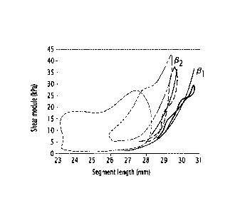
- figure 3 is graph illustrating examples of stiffness-segment loops, and
- figure 4 is a graph showing the evolution of mechanical work of the studied
myocardial segment based on figure 3.
CA 02954573 2017-01-09
WO 2016/009057
PCT/EP2015/066440
DETAILED DESCRIPTION OF SOME EMBODIMENTS
A device 10 for obtaining at least one myocardial functional parameter is
represented on figure 1.
5 Such device 10 is adapted to obtain a myocardial functional parameter.
By definition,
a functional parameter is a parameter relative to the properties of the
muscle, a parameter
relative to the metabolism of the muscle or a parameter relative to the
operation of the
muscle.
A myocardial functional parameter is, for instance, the end-diastolic passive
myocardial stiffness, myocardium work of a specific segment of the myocardium,
the
passive myocardial stiffness variation with deformation or the end-systolic
myocardial
stiffness.
The device 10 comprises an applying unit 12, a collecting unit 14 and a
computer
unit 16.
The applying unit 12 is adapted to apply ultrasound waves to the myocardium.
According to the example of figure 1, the applying unit 12 comprises an array
of
transducers.
Alternatively, the applying unit 12 comprises only one transducer.
The collecting unit 14 is adapted to collect the ultrasound waves
retrodiffused by the
muscle.
According to the example of figure 1, the collecting unit 14 comprises an
array of
transducers.
Alternatively, the collecting unit 14 comprises only one transducer.
According to another embodiment, the applying unit 12 and the collecting unit
14 are
the same unit.
The computer unit 16 comprises a calculator 18, a memory unit 20 and a
displaying
unit 22.
The calculator 18 is adapted to carry out calculation.
According to the example of figure 1, the calculator 18 is a processor.
The memory unit 20 is adapted to store data.
The displaying unit 22 is adapted to display data.
For instance, the displaying unit 22 is a screen.
According to another embodiment, the calculator 18, the memory unit 20 and the
displaying unit 22 are comprised in a watch. This enables to obtain a portable
device 10.
Operation of the device 10 is now described in reference to a method for
obtaining at
least one myocardial functional parameter.
CA 02954573 2017-01-09
WO 2016/009057
PCT/EP2015/066440
6
The applying unit 12 applies one focused ultrasound wave to the myocardium by
using the applying unit 12. The focused ultrasound wave generates an elastic
shear wave
in the muscle.
The applying unit 12 then applies a succession of ultrasound waves so that at
least
some of said ultrasound waves penetrate into a segment of the myocardium while
the
shear wave is propagating in the same segment of the myocardium.
As the myocardium has reflecting particles, echoes are generated by the
ultrasound
compression waves interacting with reflecting particles in the myocardium.
Such echoes
are called retrodiffused ultrasound waves.
In a specific embodiment, the succession of ultrasound waves is a succession
of
focused ultrasound waves. This is notably the case for ultrafast imaging.
In another embodiment, the succession of ultrasound waves is a succession of
unfocused ultrasound waves. This is notably the case for cardioscope.
The collecting unit 14 then collects the retrodiffused ultrasound waves at a
plurality
of times.
Such collection enables to obtain a plurality of images which can be analyzed
to
determine values.
The calculator 18 then determines a first plurality of values representative
of
stiffness values of at least one part of the parts of the muscle at a first
plurality of times by
using the collected ultrasound waves, the first plurality of times being
included in the
plurality of times.
A value is representative of stiffness is any physical quantity linked to the
stiffness.
For instance, the shear modulus 1..1 of the myocardium is a value
representative of
stiffness. As shear waves propagate in a muscle in an anisotropic way, such
shear
modulus E is a mean value of several shear modulus
r-direction of the myocardium along
several directions.
Alternatively, the shear modulus
r-direction of the myocardium in a specific direction is
also a value representative of stiffness. The shear modulus which is along the
direction of
the fibers of the myocardium labeled
r-parallel and the shear modulus which is along the
direction perpendicular to the direction of the fibers of the myocardium
labeled
r-perpendicular
are examples of shear modulus of the myocardium in a specific direction.
As another example, the Young's modulus E of the myocardium is a value
representative of stiffness. By definition, the Young's modulus E is linked to
the shear
modulus 1..1 by the relation E = .1.As shear waves propagate in a muscle in an
anisotropic
way, such Young's modulus 1..1 is a mean value of several Young's modulus
Ed,rect,on of the
myocardium along several directions.
CA 02954573 2017-01-09
WO 2016/009057
PCT/EP2015/066440
7
Alternatively, the Young's modulus Edirection of the myocardium in a specific
direction
is also a value representative of stiffness. The Young's modulus which is
along the
direction of the fibers of the myocardium labeled Eparallel and the Young's
modulus which is
along the direction perpendicular to the direction of the fibers of the
myocardium labeled
Eperpendicular are examples of Young's modulus of the myocardium in a specific
direction.
As another example, the propagation speed cs of shear waves in the myocardium
is
a value representative of stiffness. The propagation speed cs of shear waves
in the
myocardium is linked to the Young's modulus Eared,on by the following
relation:
cs1\7
= ¨ [1]
3p
wherein p is the density of the myocardium.
As shear waves propagate in a muscle in an anisotropic way, such propagation
speed cs of shear waves in the myocardium is a mean value of several
propagation
speeds cs direction of shear waves in the myocardium along several directions.
Alternatively, the propagation speed cs direction of shear waves in the
myocardium in a
specific direction is also a value representative of stiffness. The
propagation speed of
shear waves along the direction of the fibers in the myocardium labeled Cs
parallel and the
propagation speed of shear waves along the direction perpendicular to the
direction of the
fibers in the myocardium labeled cs perpendicular are examples of propagation
speed cs direction
of shear waves in the myocardium in a specific direction.
The calculator 18 then determines a second plurality of values representative
of
deformation values of said part at a second plurality of times by using the
collected
ultrasound waves.
A value is representative of deformation is any physical quantity linked to
the
deformation.
The cumulative deformation is an example of value representative of
deformation.
The length of the segment is an example of value representative of
deformation.
Such length is measured along any direction. The length along the direction of
the
fibers, the length along the direction perpendicular to the direction of the
fibers are specific
examples of length of the segment which may be considered.
The length of the segment which is normalized to a reference length is another
example of value representative of deformation.
The volume of the ventricle is also representative of the deformation.
The second plurality of times is included in the plurality of times and is
associated
with the first plurality of times in a one-to-one relationship.
CA 02954573 2017-01-09
WO 2016/009057
PCT/EP2015/066440
8
Preferably, the absolute value of the difference between a time of the first
plurality of
times and the associated time of the second plurality of times below or equal
to
100 milliseconds modulo the temporal duration of the cardiac cycle.
In case the measurement are carried out during the same cardiac cycle, the
absolute value of the difference between a time of the first plurality of
times and the
associated time of the second plurality of times below or equal to 200
milliseconds.
More preferably, the absolute value of the difference between a time of the
first
plurality of times and the associated time of the second plurality of times
below or equal to
20 milliseconds modulo the temporal duration of the cardiac cycle.
In case the measurement are carried out during the same cardiac cycle, the
absolute value of the difference between a time of the first plurality of
times and the
associated time of the second plurality of times below or equal to 20
milliseconds.
As visible on figure 3, the first plurality of values and the second plurality
of values
form a stiffness-deformation loop. Figure 12 illustrates examples of stiffness-
segment
loops. Stiffness measured by SWE is plotted as a function of segment length
for baseline
(in dotted line), 5 minutes after coronary occlusion (in full line), 2 hours
after occlusion (in
thick line), and 40 minutes after reperfusion (in dot-dash line).
Such loop comprises four inflexion points linked by four parts, a lower part,
an upper
part and lateral parts. When the loop is followed continuously starting from
the lower part,
the lower part is followed, then the first lateral part is followed, then the
upper part is
followed and then the second lateral part is followed.
In the specific case of myocardium, the inflexion point which is common to the
lower
part and the first lateral part is called end diastolic point whereas the
inflexion point which
is common to the upper part and the second lateral part is called end systolic
point.
The calculator 18 then deduces at least one myocardium functional parameter
based on the first plurality of values and the second plurality of values.
According to an example, the myocardium functional parameter is representative
of
the mechanical work of the segment. In such case, the myocardium functional
parameter
is obtained by calculating the area of the stiffness-deformation loop. Such
calculation is
illustrated schematically on figure 4 given the data of figure 3.
According to another example, at the step of deducing, at least one part is
curve
fitted by an exponential function whose coefficient is the myocardium
functional
parameter.
For instance, the myocardium functional parameter is representative of the end
diastolic passive myocardial stiffness and such myocardium functional
parameter is
obtained by curve-fitting the lower part. Figure 3 illustrates such step of
curve-fitting by
CA 02954573 2017-01-09
WO 2016/009057
PCT/EP2015/066440
9
showing two exemplary exponential functions which fits the lower part of two
loops. These
two exemplary exponential functions has respectively two coefficients labeled
131 and 132.
According to another example, the step of deducing comprises determining, for
at
least one of the inflexion point, a first value representative of stiffness
value (stiffness
coordinate) and a second value representative of deformation value
(deformation
coordinate).
For instance, in the case of myocardium, it is valuable to obtain the
stiffness
coordinates and the deformation coordinates of the end-diastolic point and of
the end-
systolic point.
At the end of the deducing step, at least one myocardial functional parameter
is
deduced.
Such deduced myocardial functional parameter is stored in the memory unit 20
and
displayed on the displaying unit 22.
The method for obtaining at least one myocardial functional parameter enables
to
obtain a myocardial functional parameter by using a stiffness-deformation
loop.
Such stiffness-deformation loop is obtainable in a non-invasive way. Indeed,
according to a preferred embodiment, the measurements implied in the method
for
obtaining are carried out in vivo.
In addition, the method for obtaining at least one myocardial functional
parameter
enables to access to myocardial functional parameters which are not easily
accessible to
the methods belonging to the prior art. Notably, the mechanical work of the
segment of the
myocardium is a quantity which is difficult to access for the methods of the
prior art.
Furthermore, it can be shown that the method for obtaining at least one
myocardial
functional parameter is as accurate as invasive methods.
Such accuracy has been demonstrated experimentally. Indeed, experiments using
the device 10 were performed on an open chest ovine model. After sternotomy,
the
ultrasonic transducer of the applying unit 12 was placed in front of the left
ventricular free
wall. A sequence combining shear wave imaging and strain imaging was used.
Shear
wave imaging was performed at a repetition rate of 15 Hz during 1 s to
quantify the
myocardial stiffness change over a cardiac cycle. Myocardial strain was
measured on the
ultrasound images during the same cardiac cycles. The stiffness-strain curve
loop was
obtained from these two non-invasive ultrasound based measurements. The same
experiment was performed during coronary occlusion on the ischemic wall. The
area of
the loop was strongly reduced (almost equal to 0) compared to baseline. The
area of
stiffness-strain loop correlated with the work of the segment.
CA 02954573 2017-01-09
WO 2016/009057
PCT/EP2015/066440
Such method for obtaining a functional parameter is also applicable for other
muscle. For instance, the muscle is uterus or a muscle involved in the
practicing of a
sport.
According to an embodiment, such method for obtaining a functional parameter
is
5 iterated several times to obtain a plurality of values for the myocardial
functional
parameter.
In such cases, comparisons are achievable.
The comparison may be carried out for different operating conditions for the
myocardium. For instance, the myocardium may be subjected to drug or the
person may
10 be in a different stage of physical effort.
The comparison may also be temporal such that an evolution of the functional
parameter may indicate an abnormality of operating of the myocardium.
For this, as represented on figure 2, it is proposed a system 23 for
monitoring the
state of the myocardium. By the expression "state", it is meant an evaluation
of the
operating of the myocardium. III and healthy are states of myocardium.
Intermediate state
exists. For instance, a heart murmur is also a state of myocardium.
The system 23 comprises the device 10 for obtaining at least myocardium as
illustrated on figure 1, a comparator 24 and a warning unit 26.
The device 10 is adapted to a plurality of values for at least one myocardial
functional parameter of the myocardium.
The comparator 24 is adapted to compare the plurality of values for the
myocardial
functional parameter with a plurality of expected values for the myocardial
functional
parameter according to a comparison criterion.
The comparator 24 is, for instance, a processor.
The comparison criterion may differ according to the kind of monitoring.
As an example, the comparison criterion is a predetermined threshold. For
instance,
if the mechanical work of a segment is below a given value, this means the
considered
segment is not in the healthy state.
As an example, the comparison criterion is relative to the evolution of the
myocardial
functional parameter with time and notably, the value of the derivative of the
myocardial
functional parameter with time at given time.
For preventing infarction, a comparison criterion related to the end-diastolic
passive
myocardial stiffness has shown a strong correlation between the end-diastolic
passive
myocardial stiffness and the infarction.
For detecting ischemia, a comparison criterion related to an evolution of the
mechanical work of a segment is considered
CA 02954573 2017-01-09
WO 2016/009057 PCT/EP2015/066440
11
The warning unit 26 is adapted to emit a warning in case the comparison
criterion is
not met.
The warning may be an audible alarm or a visible alarm.
Such system 23 enables to monitor efficiently the state of the myocardium.
Preferably, the system 23 is portable.
The embodiments and alternative embodiments considered here-above can be
combined to generate further embodiments of the invention.