Note : Les descriptions sont présentées dans la langue officielle dans laquelle elles ont été soumises.
CA 2,970,965
Blakes Ref: 76029/00015
1
SOFT TISSUE GRAFTS, AND METHODS OF
MAKING AND USING SAME
Technical Field
[0002] The present invention relates to soft tissue grafts, methods of
preparing
soft tissue grafts, and methods of use thereof. The present invention also
relates to
soft tissue grafts for use in mastopexy or breast reconstruction procedures.
The
present invention also relates to soft tissue grafts for use in rotator cuff
repair or
reinforcement. The present invention also relates to soft tissue grafts for
use in tendon
and ligament repair.
Background
[0003] A wide variety of soft tissue products are used in medical,
surgical,
veterinary, and other applications. These soft tissue products can be used in
load-
bearing and non-load bearing applications and can be supplied in a variety of
forms.
The intended use of the soft tissue product may dictate certain aspects of its
form such
as size, shape, or thickness. General soft tissue grafts, however, may be
unable to
meet desired dimensions, or may require substantial modification before they
are
suitable for a particular use,
Summary
[0004] Soft tissue grafts, packaged soft tissue grafts, and methods of
making
and using soft tissue grafts are disclosed.
[0005] In one example, a soft tissue graft is disclosed. The soft tissue
graft
includes processed tissue material having first and second opposed surfaces.
The first
and second opposed surfaces are bounded by first and second edges. The first
edge
has a concave shape that curves toward the second edge. The second edge has a
convex shape that curves away from the first edge. The first surface comprises
a
plurality of apertures, At least one of the apertures is formed from a multi-
directional
separation in the first surface.
[0006] In another example, another soft tissue graft is disclosed. The
soft
tissue graft includes processed tissue material having first and second
opposed
CA 2970965 2018-09-04
CA 02970965 2017-06-14
WO 2017/066568
PCT/US2016/057038
2
surfaces. The processed tissue material has a trapezoidal shape with a pair of
parallel
edges. The first surface comprises a plurality of first apertures.
[0007] In yet another example, another soft tissue graft is disclosed, The
soft
tissue graft includes processed tissue material having first and second
opposed
surfaces. The first and second opposed surfaces are bounded by first and
second
edges. The first edge has a concave shape that curves toward the second edge.
The
second edge has a convex shape that curves away from the first edge. The first
surface is meshed to form a plurality of apertures with a predetermined
density.
[0008] In still another example, a packaged soft tissue graft is disclosed.
The
packaged soft tissue graft includes a support, processed tissue material, and
packaging
material. The support has a base and a projection extending upward from the
base.
The processed tissue material has first and second opposed surfaces. The
processed
tissue material is positioned to cover at least a portion of the projection
with the first
surface facing away from the projection and the second surface facing the
projection.
The first surface comprises a plurality of apertures. The packaging material
encloses
the support arid the processed tissue material.
[0009] In yet another example, another packaged soft tissue graft is
disclosed.
The packaged soft tissue graft includes a support, processed tissue material,
a frame,
and packaging material. The processed tissue material has first and second
opposed
surfaces. The processed tissue material is positioned to cover at least a
portion of the
support with the first surface facing away from the support and the second
surface
facing the support. The first surface comprises a plurality of apertures. The
frame is
configured to surround the processed tissue material and press edges of the
processed
tissue material against the support. The frame is configured to apply a
tension to the
processed tissue material when the processed tissue material is positioned
between the
support and the frame. The packaging material encloses the support, the frame,
and
the processed tissue material.
[0010] In still another example, a method of making a soft tissue graft is
disclosed. The method includes positioning a cutting die on a surface of
tissue
material, pressing the cutting die into the tissue material to cut the tissue
material, and
processing the cut tissue material to create processed tissue material.
Brief Description of the Drawings
[0011] The drawing figures depict one CIF' more implementations in accord
with
the present concepts, by way of example only, not by way of limitations. In
the figures,
like reference numerals refer to the same or similar elements.
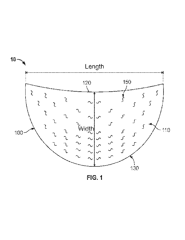
[0012] FIG. I. shows an example of a soft tissue graft,
CA 02970965 2017-06-14
WO 2017/066568
PCT/11S2016/057038
3
[0013] FIGS. 2A-2C show examples of variations in aperture layout of the
soft
tissue graft of FIG. 1.
[0014] FIGS. 3A-3F show examples of different aperture shapes for soft
tissue
grafts.
[0015] FIGS, 4A-4D show measurements of apertures for soft tissue grafts.
[0016] FIGS. 5A-5D show an example of a packaged soft tissue graft.
[0017] FIG. 6 shows a method of making a soft tissue graft.
[0018] FIG. 7 shows an example of a cutting die for use in the method of
FIG. 6.
[0019] FIG. 8 shows a graph of guidelines for pressing force relative to
tissue
thickness and cutting die blade length for the method of FIG, 6,
[0020] FIGS, 9A and 93 show examples of another soft tissue graft.
[0021] FIGS. 10A, 10B, and 10C show examples of yet another soft tissue
graft.
[0022] FIGS. 11A and 113 show examples of another soft tissue graft.
[0023] FIGS. 12A and 126 show examples of the soft tissue grafts of FIGS.
11A
and 116 including operative modifications.
Detailed Description
[0024] In the following detailed description, numerous specific details are
set
forth by way of examples in order to provide a thorough understanding of the
relevant
teachings. However, it should be apparent to those skilled in the art that the
present
teachings may be practiced without such details. In other instances, well
known
methods, procedures, components, and/or circuitry have been described at a
relatively
high-level, without detail, in order to avoid unnecessarily obscuring aspects
of the
present teachings.
[0025] The detailed description below and the accompanying drawings
disclose
examples of soft tissue grafts and methods of making and using soft tissue
grafts. The
examples of soft tissue grafts have sizes, shapes, and thicknesses selected
for
particular uses. The examples of soft tissue grafts may further include
apertures to
promote successful implantation. The soft tissue grafts may be packaged or
u n packaged.
[0026] The examples discussed below may be particularly suitable for use in
mastopexy or breast reconstruction procedures. Mastopexy, or
breast lift, is a
procedure designed to improve the appearance of sagging or ptotic breasts. For
example, one goal of the surgery is to improve the shape and position of the
breast
while minimizing visible scars. Breast reconstruction is a procedure used to
restore
form and function after mastectomy. The goals of implant-based breast
reconstruction
include: recreation of the breast mound - including defining the contour of
the lower
pole to reestablish normal ptosis and the creation of aesthetically pleasing
CA 02970965 2017-06-14
WO 2017/066568
PCT/US2016/057038
4
inframammary fold. Various procedures and modifications of mastopexy are known
in
the art.
[0027] The examples
discussed below may be particularly suitable for rotator
cuff repair or reinforcement, tendon and ligament repair or reinforcement,
and/or
capsular reconstruction. Rotator cuff repair or reinforcement is a procedure
to restore
normal form, function, and range of motion to a patient's shoulder following
partial or
complete tear of the patient's rotator cuff. Tendon and ligament repair are
procedures
for remedying partial or complete tears of a patient's ligaments or tendons.
Use of the
example grafts in rotator cuff repair can restore stability of the shoulder
joint and
resolve dysfunction and pain. Capsular reconstruction is a procedure to
recreate the
joint capsule thereby restoring normal joint biomechanics and stability. The
implanted
graft has a high ultimate load and suture retention strength. Soft tissue
repairs
augmented with the graft may demonstrate improved strength, reduce re-tear and
improved clinical outcomes. Moreover, repair of complete and chronic tendon
tears
with the graft supplements inadequate tendon tissue. Tendon augmentation can
provide a more effective treatment of chronic or acute conditions by creating
a stronger
repair construct. A stronger repair may allow for more aggressive
rehabilitation
decreasing postoperative stiffness, muscle atrophy, and repair site gapping.
[0028] Examples
discussed below and shown in the drawings improve over the
art by providing a suitable size, shape, and thickness for a predetermined
procedure,
thereby eliminating the need for substantial processing or cutting of the
graft prior to
implantation. Additionally, the examples discussed below may include apertures
to
provide increased locations for angiogenesis (formation of blood vessels), as
well as
improved tissue ingrowth following implantation, thereby speeding the post-
implantation healing process,
[0029] In preparation
of the example grafts below, soft tissue can be cut in such
a way that allows for suturing zones on the graft without adversely impacting
the
biomechanical strength of the graft, and without impacting the placement of
apertures
in the soft tissue. The processes
described below are designed to minimize
introduction of bio-burden during the process of forming the soft tissue
graft. The final
soft tissue grafts allow for intra-operative suturing at an edge of the graft,
while
eliminating risk of pull-out of sutures through apertures in the graft.
[0030] The examples
described below have a shape designed for minimum graft
size necessary to achieve desired intraoperative coverage. These examples
support the
use of smaller grafts to achieve existing procedural techniques, potentially
saving
institutions cost and shelf space. In some examples, a concave shape is
provided that
roughly mirrors natural borders of pectoralis major muscle aiding in various
CA 02970965 2017-06-14
WO 2017/066568
PCT/11S2016/057038
reconstructive techniques, potentially minimizing trauma to the pectoralis
major
muscle. Some examples are designed to allow for fat grafting around the upper
pole,
for enhanced aesthetic outcomes without impacting aperture placement and/or
ability
of the graft to enable fluid egress. Some examples have different zones of
elongation,
in order to maximize fluid egress in 3D planes in high risk areas while at the
same time
providing defined elongation in north-south plane.
[0031] The concave
design of certain examples allows for maximum utilization
of tissue, minimizing wastage of donated tissue. The concave shape allows for
intra-
operative shape adjustment based on patient requirements/physiology. The
processes
described below promote uniform and consistent handling of soft tissue to
enhance pre-
operative planning and provide a pathway to technical training to less
experienced
surgeons.
[0032] The example
soft tissue grafts described below may include apertures.
The apertures are designed to minimize stress concentrations in the soft
tissue. The
apertures may minimize the number of drains used post-operatively. The
apertures
may further maximize post-operative incorporation and revascularization of the
graft.
In some examples, linear apertures are used, which close when the tissue
tensioned
parallel to the apertures and open when tensioned obliquely or orthogonally to
the
apertures, to increase the potential vascular pathways necessary for maximum
tissue
remodeling and regeneration. These examples could lead to earlier structural
integrity
of the graft due to the increased vascular channels, resulting in more rapid
granulated
tissue and tissue ingrowth.
[0033] In some
examples, apertures may be oriented to create variable zones of
fluid egress through the soft tissue graft corresponding to anatomical zones.
Apertures
may be patterned to create consistent 2D openings in the 3D anatomical space
where
utilized, Minimizing the potential gapping or closing of the aperture
maximizes the
contact between ADM and implant, while minimizing potential dead space leading
to
post-operative complications. Apertures may be sized to maximize opening when
placed over an implant, and may be shaped for optimal opening when tensioned
in
three dimensions.
[0034] In some
examples, the soft tissue grafts are meshed. The meshing
pattern maximizes the opening area of the soft tissue graft while maintaining
biomechanical integrity through suture borders and internal graft bands. The
meshing
pattern may be designed to enhance current and contemplated techniques in
breast
reconstruction. The meshing
pattern may also be designed to provide
controlled/defined expansion in any surgical plane.
CA 02970965 2017-06-14
WO 2017/066568
PCT/1JS2016/057038
6
[0035] While the following examples are described chiefly with respect to
particular procedures (such as mastopexy, breast reconstruction, rotator cuff
repair,
tendon/ligament reconstruction, or capsular reconstruction), it should be
readily
apparent that the examples herein are not so limited. The following examples
and
variations thereof may alternatively be used in any number of procedures
requiring the
use of a soft tissue grafts. Other suitable procedures will be apparent from
the
description herein.
[0036] Definitions are set forth below to provide a clear and consistent
understanding of the specification and claims, including the scope to be given
such
terms.
[0037] And/or. It should be understood that the use of "and/or" is defined
inclusively such that the term "a, b and/or c" should be read to include all
the
combination of a, b, and c, including "a, b, and c," "a and b," "a and c," "b
and c," "a,
b, or c," "a or b," "a or c," "b or c," "a," "b," and "c." hole
[0038] Aperture. The term "aperture" as used herein is intended to
encompass
any separation in a surface of the soft tissue, including holes, slits,
cavities, voids,
fenestrations, channels, or other types of openings, regardless of whether
that
separation extends part of the way or all of the way through the soft tissue.
[0039] Biocompatible. The term "biocornpatible" as used herein is intended
to
encompass any material which does not provoke an adverse response in a
patient. For
example, a suitable biocompatible material when introduced into a patient does
not
itself provoke a significant immune response, and is not toxic to the patient.
[0040] Biomechanical strength. The term "biomechanical strength" as used
herein is intended to encompass those properties exhibited by a tissue graft,
including
loading strength, compressive strength, and tensile strength.
[0041] Impregnating. The term "impregnating" as used herein is intended to
encompass any processing conditions which result in filling the internal
matrix of a
graft with an identified material.
[0042] Internal matrix. The term "internal matrix" as used herein is
intended to
encompass the intercellular substance of such soft tissue including for
example
ligaments and tendons, including collagen and elastin fibers and base matrix
substances.
[0043] Plasticizer. The term "plasticizer" as used herein is intended to
encompass any biocompatibie compounds which can easily displace/replace water
at
the molecular level and preferably have a low molecular weight such that the
plasticizer
fits into the spaces available to water within the molecular structure of the
bone or soft
tissue. Such plasticizers are preferably not toxic to the cellular elements of
tissue into
CA 2,970,965
Blakes Ref: 76029/00015
7
which the graft is to be placed. Suitable plasticizers are described in U.S.
Patent
Na. 6,569,200.
[0044] Processed
tissue material. The term "processed tissue material" as used
herein is intended to encompass native, normal tissue that has been procured
from an
animal source (e.g. human or non--human, such as bovine, porcine, canine
including,
but not limited to, a dog, equine, ovine, or non-human primate including, but
not
limited to, ape and gorilla, in origin), preferably a mammal, and mechanically
cleaned
of attendant tissues and/or chemically cleaned of cells and cellular debris.
[00451 Soft
tissue graft. The term "soft tissue graft" as used herein is intended
to encompass load-bearing and non-load-bearing soft tissue products composed
of an
internal matrix which includes collagen, eiastin, and high molecular weight
solutes
which during cleaning may be removed.
[0046] The soft
tissue grafts disclosed herein may be derived from allogenic,
autogenic, or xenogenic sources. The tissue material used for the grafts may
be
processed from human or animal tissue, in one aspect, the processed tissue
material
may be derived from native tissues, such as stomach, intestine, dermis, fascia
lata,
pericardium, bladder, and dura mater. The processed tissue material may be,
for
example, biologically-derived collagenous materials, such as the intestinal
subrnucosa
described in U.S. Patent Application
Publication Nos. 2002/0103542 and
2008/0097601. When
implanted into a mammalian patient, the processed tissue material may undergo
controlled biodegradation occurring with adequate living cell replacement such
that the
original implanted graft is remodeled by the patient's living cells, and, in
some
examples, the graft does not interfere with radiographic imaging.
[0047] In another
aspect, the processed tissue material described herein
consists essentially of and/or consists of the one or more soft tissue(s); and
a liquid,
solution, or solvent. In some examples, the processed tissue material consists
essentially of and/or consists of components from the one or more soft
tissue(s). The
term 'essentially consisting of" defines the scope of the processed tissue
material to
include additional elements that do not materially affect the porosity or void
fraction of
the processed tissue material consisting of initial elements. For example, the
processed
tissue material consisting essentially of one or more soft tissue(s) may
include
elements in addition to the one or more soft tissue(s) that do not materially
affect the
extracellular matrix composition of the processed tissue material consisting
of the one
or more soft tissue(s),
CA 2970965 2018-09-04
CA 02970965 2017-06-14
WO 2017/066568
PCT/US20161057038
8
[0048] Reference now
is made in detail to the examples illustrated in the
accompanying drawings and discussed below. FIGS. 1 and 2A-2C illustrate an
example
of a soft tissue graft 10. Soft tissue graft 10 is formed from processed
tissue material
100. Details regarding soft tissue graft 10 are set forth below.
[0049] In one
example, processed tissue material 100 may be dermis, and the
processed tissue material may comprise a reticular lamina layer. In
additional
examples, the processed tissue material 100 comprises a basal lamina layer and
a
reticular lamina layer, and the processed tissue material 100 may comprise a
basal
lamina layer, a reticular lamina layer, and adipose tissue. In further
examples, the
processed tissue material 100 may exclude a basal lamina layer, a reticular
lamina
layer, and/or adipose tissue. The processed tissue material 100 may have one,
two,
three, or all sides on which the reticular lamina layer is exposed. For
example, when
the processed tissue material 100 consists of the reticular lamina layer, all
sides of
such a processed tissue material would be reticular sides. When at least 60,
70, 80, 90,
95, 98, 99 or 100% of a side of a material is composed of the reticular lamina
layer,
such a side may be called a reticular side, and one, two, three or all sides
of processed
tissue material 100 may be reticular sides. In some examples, processed tissue
material 100 may have top and bottom reticular sides.
[0050] As shown in
FIG, 1, processed tissue material 100 has a first major
surface 110 and a second surface (not shown) opposite the first surface.
Surface 110
is bounded by edges 120 and 130. Edge 120 has a concave shape that curves
toward
edge 130, and edge 130 has a convex shape that curves away from edge 120. The
radius of curvature of edge 120 is longer than the radius of curvature of edge
130.
Edges 120 and 130 share both ends; in other words, edges :120 and 130 start
and end
at the same points. As shown in FIG. 1, processed tissue material 100 is
symmetrical
about a line bisecting edges 120 and 130. Processed tissue material 100 may be
symmetrical about one or multiple different lines dependent on the intended
use of
graft 10.
[0051] FIG, 1 shows
lines representing the length and width of processed tissue
material 100. Length may be measured from the most distant points of processed
tissue material 100, with width measured from the most distant points of
processed
tissue material 100 along a line orthogonal to length. Processed tissue
material 100
may have a length of from 5, 6, 7, 8, 9, 10, 13 or 15 cm to 20, 23, 25, 27 or
30 cm.
Processed tissue material 100 may have a width of from 2, 5, 6, 7, 8, 9, 10 or
15 cm to
15, 20, 21, 22, 23, 24, 25 or 30 cm. Processed tissue material 100 may have a
thickness (measured from surface 110 to the opposing surface) in a range of
from
0.1 mm to 10 mm. Processed tissue material 100 may have an average thickness
of
CA 02970965 2017-06-14
WO 2017/066568
PCT/US2016/057038
9
from 0.05, 0.1, 0.5, 1, 2, 3, 4, 5 or 6 mm to 6, 7, 8, 9, 10, 11, 12, 13, 15,
or 20 mm,
The thickness of processed tissue material 100 need not be uniform and may
increase
at locations closer to edges 120 and 130 of processed tissue material 100.
[0052] As set forth above, edges 120 and 130 may have different radii of
curvature, Concave edge 120 may have a radius of curvature of from 25 cm to 50
cm,
and more preferably, from 30 cm to 46 cm. Convex edge 130 may have a radius of
curvature of from 5 cm to 15 cm, and more preferably, from 6.5 cm to 10.6 cm.
[0053] The shape and size of processed tissue material 100 shown in FIG. 1
is
selected to be suitable for a mastopexy or breast reconstruction procedure.
The shape
may facilitate the performance of these procedures by requiring little or no
pre-surgical
modification (such as cutting). It will be understood that other shapes for
processed
tissue material 100 may be selected based on the intended procedure. Examples
of
possible shapes include, for example, circles, semicircles, partial circles,
ellipses,
triangles, rectangles, trapezoids, parallelograms, squares, other regular or
irregular,
convex or concave polygons, or combinations of these shapes. A concave polygon
is
defined as a polygon with one or more interior angles greater than 180 , and a
convex
polygon is defined as a polygon with all its interior angles less than 180 .
In some
examples, the edges of the processed tissue material may be curved, so as to
form a
continuous edge lacking any corners or vertices. In additional examples, the
concave
border curves toward the convex border, and the convex border curves away from
the
concave border. Additionally, it will be understood that processed tissue
material 100
need not be symmetrical, but may have asymmetrical features in order to
correspond
to variations in the anatomy of the intended recipient (such as left/right
variations).
For one example, as shown in FIG. 1, processed tissue material 100 may have
apertures of one shape (such as an S-shape) on a right side of the graft, and
apertures
of a mirrored shape (such as a mirror S-shape) on a left side of the graft.
[0054] In the example shown in FIG. 1, the surface 110 of processed tissue
material 100 includes a number of apertures 150. Apertures 150 may extend all
of the
way through processed tissue material 100, or may extend only part of the way
through processed tissue material 100.
[0055] Apertures 150 may be formed from cutting into surface 110 of
processed
tissue material 100, or may be formed from removing at least a part of
processed
tissue material 100 from surface 110. The cutting of apertures 150 in surface
110 may
be performed, for example, with a knife, blade, scissors, press, pressurized
fluid or
pellet, or a laser. For example, the blade may be a scalpel blade (e.g. steel
or diamond
or other material), electronic scalpel or harmonic scalpel or steel rule die
or machined
cutting die blade that is pressed into the skin. A water jet or dry ice
blaster, liquid or
CA 02970965 2017-06-14
WO 2017/066568
PCT/1JS2016/057038
pellet pressurized to a small area may also be used to cut the tissue.
Examples of the
lasers include femtosecond laser and epilog laser, and other examples will be
apparent
to those skilled in the art.
[0056] Apertures 150 may each have the same shape and size, or may have
different shapes and sizes. Where the shapes and/or sizes of apertures 150
differ, the
differences may be based on the location of the aperture 150 on processed
tissue
material 100.
[0057] The shape, size, and density of apertures is selected to promote
angiogenesis and vascularization of the soft tissue graft following
implantation, without
adversely affecting a biomechanical strength of the graft. Examples of shapes,
sizes,
and layouts of apertures 150 are set forth below.
[0058] FIGS. 3A-3F show various possible shapes for apertures 150. As shown
in FIG. 3A, apertures 150 may be formed from a linear separation or cut in
surface
110. Alternatively, apertures 150 may be formed from a multi-directional
separation or
cut in surface 110. As used herein, the term "multi-directional" refers to a
separation
that extends in more than one different direction. The multi-directional
separation may
be arc-shaped, or may have another shape. Possible shapes for the multi-
directional
separation include S-shapes (as shown in FIGS, 3B and 3C), Z-shapes (as shown
in
FIG. 3D), 3-shapes, L-shapes, X-shapes (as shown in FIG. 3E), omega shapes (as
shown in FIG. 3F), or mirror images thereof. In additional examples, the multi-
directional separation may have two, three, four, five, six or more and/or
three, four,
five, six, seven or fewer directions.
[0059] The length of each aperture may be considered to be the distance
between opposite ends of the aperture, without consideration of the particular
path of
the aperture (in the case of multi-directional separations), FIG. 4A shows an
example
of a length measurement for an S-shaped aperture. An average length of the
apertures 150 may be from 0.3, 0.4, 0.5, 0.6, 0.7 or 0.8, 2, 4, 6 or 8 mm n to
10, 20,
25, 28, 30, 35 or 40 mm. Apertures 150 may each have a length in a range of
from
1 mm to 10 mm.
[0060] In addition to aperture length, multi-directional apertures may be
characterized by a path length of the aperture, i.e., a length along the
particular path
or cut of the aperture. FIG. 4B shows an example of a path length measurement
for an
S-shaped aperture. An average path length of multi-directional apertures 150
may be
from 0.3, 0.4, 0.5, 0.6, 0.7 or 0.8, 2, 4, 6 or 8 mm to 10, 20, 25, 28, 30, 35
or
40 mm,
[0061] As shown in FIGS. 4A and 4B, a multi-directional aperture will have
a
path length which is longer than the aperture length. The ratio of aperture
length to
CA 02970965 2017-06-14
W02017/066568
PCT/US2016/057038
11
path length for the multi-directional apertures may be 0.2, 0.3, 0.4, 0.5,
0.6, 0.7, 0.8,
0.9, 0.95, or 0.99 or more and/or less than 1.0, 0,99, 0.95, 0,9, 0.8, 0.7,
0.6, of 0.5,
[0062] In addition to aperture length and path length, multi-directional
apertures may also be characterized by an aperture angle, i.e., an angle
between a
central portion of the aperture and end portions of the aperture, or a
aperture path
angle, i.e., an angle between one portion of the aperture path and another
portion of
the aperture path. FIGS. 4C and 4D show an example of an angle measurement for
an
S-shaped aperture and a mirror S-shaped aperture, respectively. Average path
angles
may be independently from -300, -200, -100, -50, -10 or -5 degrees to 5, 10,
50, 100,
200 or 300 degrees,
[00631 Adjacent apertures 150 may be spaced apart at a distance of from
0.5 mm to 30 mm. The aperture length and spacing may be selected such that the
ratio of average distance between adjacent apertures to average length of
apertures is
from 0.5, 0.6, 0.7, 0.8, 0.9, 0,95, or 0.99 or more and/or less than 1.0,
0.99, 0.95,
0.9, 0,8, 0.7, or 0.6. In some examples, this ratio may be from 0,2 to 0.99,
from 0,3
to 0.9, from 0.5 to 0.8, or from 0.6 to 0.8.
[0064] Alternative or additionally, apertures 150 may be characterized by
the
area created by the aperture with or without stretching of the processed
tissue
material. It will be understood that when the aperture is formed by cutting
the tissue
material without removing any part of the tissue material, the size of the
aperture is
zero prior to any stretching of the processed tissue material. When the
aperture is
formed by removing tissue material, the aperture may have an area even without
stretching of the processed tissue material. An average area of apertures 150
formed
in processed tissue material 100 may be from 0, 0.1, 0.4, 0.5, 1, 5, 8 or 10
mm2 to 50,
100, 150, 180 or 200 mm2, Apertures 150 may all have an area in a range of
from
0.5 mm2 to 200 mm2.
[0065] The positioning and density of apertures 150 on processed tissue
material 100 may be uniform across surface :110, or may vary. Apertures may be
arrayed on surface 110 in rows and/or columns, or may be randomly dispersed on
surface 110,
[0066] In one example, apertures 150 are more concentrated in the center of
surface 110. In this example, surface 110 has a central region which may be
considered to be the portion of surface 110 closer to a line bisecting edges
120 and 130
than the ends of edges 120 and 130. The number and/or density of apertures 150
in
the central region of surface 110 is greater than the number and/or density of
apertures in the remaining area of surface 110.
CA 02970965 2017-06-14
WO 2017/066568
PCT/US2016/057038
12
[0067] In another
example, apertures 150 are more concentrated in the lower
region of surface 110. In this example, surface 110 has a lower region which
may be
considered to be the portion of surface 110 closer to edge 130 than edge 120.
The
number and/or density of apertures 150 in the lower region of surface 110 is
greater
than the number and/or density of apertures in the remaining area of surface
110.
[0068] As shown in
FIG. 2A, apertures 150 may not be positioned close to the
edges of surface 110. In one
example, no apertures are positioned within a
predetermined distance from edges 120 and 130, The predetermined distance may
be,
for example, at least 0.5, 1.0, 1.2, 1.5, 2.0, 2.5, 3.0 or 3.5 cm. Providing
an aperture
free space along the edges of surface 110 may be desirable in order to create
a suture
zone 155, e.g., a zone for steady and secure suturing of graft 10 during
implantation.
[0069] As shown in
FIG. 2B, surface 110 may also include one or more bands
160 which are free from apertures 150. Bands 160 may be located along the
edges of
processed tissue material 100, or may extend across a portion of processed
tissue
material 100, with apertures 150 provided on each side of the band 160. Bands
160
may have a width of at least 0.5, 1,0, 1.2, 1.5, 2.0, 2,5, 3.0 or 3.5 cm, for
example.
In one example, a band 160 extends from edge 120 to edge 130 of processed
tissue
material 100, as shown in FIG. 2B. In another example, a band 160 extends from
one
portion of edge 120 to another portion of edge 120, and/or from one portion of
edge
130 to another portion of edge 130, as shown in FIG. 2C. Bands 160 may be
straight
or curved. In the example shown in FIG, 2B, multiple convex bands 160 which
extend
from edge 120 to edge 130, and which curve away from a center of processed
tissue
material 100, are provided. Providing
bands 1.60 in this layout may maintain a
biornechanical strength of graft 10 during or after implantation.
[0070] Processed
tissue material 100 of graft 10 has been processed to be
suitable for implantation. Such processing may include cleaning the tissue
material,
disinfecting the tissue material, skiving the tissue material to a
predetermined
thickness, removing cellular elements and small molecular weight solutes from
the
tissue material (i.e. "decellularizing" the tissue material), plasticizing the
tissue
material, packaging the tissue material, and/or sterilizing the tissue
material. During
plasticization, the internal matrix of the tissue material is impregnated with
one or
more plasticizers.
[0071] FIGS. 5A-5D
illustrate an example of a packaged soft tissue graft 200.
The packaged soft tissue graft 200 includes processed tissue material 210, a
support
220, and packaging material 240. Processed tissue material 21.0 may be any
processed tissue material described above with respect to processed tissue
material
100. Additional details regarding packaged soft tissue graft 200 are set forth
below.
CA 02970965 2017-06-14
WO 2017/06068
PCT/11S2016/057038
13
[0072] Support 220
supports processed tissue material 210. Support 220 is
formed from a rigid, semi-rigid, flexible, porous, and/or spongy material in
order to
prevent folding, twisting, or flexing of processed tissue material 210 after
packaging.
Support 220 may be formed from a rigid biocompatible polymer, or may be
covered
with a biocompatible material, in order to prevent possible adverse reaction
following
implantation of processed tissue material 210. Suitable biocompatible
materials for use
as support 220 include, for example, metals such as stainless steel or foil,
plastic such
as polyethylene, polyester, or
acrylonitrile-butadiene-styrene (ABS),
polytetrafluoroethylene (PTFE), ceramics such as aluminum oxide, or natural
materials
as cellulose sponge, or combinations of the foregoing materials, such as
PET/AIO.
Other suitable biocompatible materials will be apparent to those of skill in
the art.
[0073] Support 220
may be formed, for example, by injection molding, vacuum
forming, or three-dimensional printing. In one example, the size of support
220 is
tailored to the dimensions of the patient that will be receiving the soft
tissue graft. In
this example, the area of the patient to receive the soft tissue graft may be
measured,
and those measurements may be used to calculate the size of support 220.
Support
220 may then be three-dimensional printed according to the desired dimensions.
This
example may be helpful in order to model the intra-operative positioning of
the soft
tissue graft prior to implantation, while the processed tissue material is
packaged.
[0074] As shown in
FIGS. 5A and 5B, support 220 may include a base 222 and a
projection 228 extending upward from the base. Base 222 has a flat lower
surface so
support 220 can sit stably on a shelf or surface. Base 222 may have a shape
matching
corresponding to a shape of processed tissue material 210. As shown in the
example
of FIG. 5A, processed tissue material has edges 212 and 214. Edge 212 has a
concave
shape that curves toward edge 2.14, and edge 214 has a convex shape that
curves
away from edge 212. Likewise, base 222 has edges 224 and 226 which correspond
in
shape to edges 212 and 214 of processed tissue material 210.
[0075] Projection 228
enables processed tissue material 210 to be maintained in
a three-dimensional form in the packaging. The shape or contour of projection
228
may be selected to correspond the shape or contour which processed tissue
material
210 is intended to take following implantation, so that processed tissue
material 210
can be stored and/or maintained in its intended position for implantation.
Support 220
and/or projection 228 may thus be designed to assist in simulating an
intraoperative
appearance of processed tissue material 210, in order to promote ease of use
of the
packaged soft tissue graft 200.
[0076] Projection 228
may be formed from one uniform surface, such as a
dome, or partial sphere, or may be formed from multiple surfaces. In one
example, as
CA 02970965 2017-06-14
WO 2017/066568
PCT/US2016/057038
14
shown in FIG. SB, projection 228 includes first and second support surfaces
230 and
232. Surfaces 230 and 232 define a ridge 234 extending between them. Ridge 234
extends from base 222 over the top of projection 228 and track down to base
222,
Processed tissue material 210 covers at least a portion of both surfaces 230
and 232
and ridge 234.
[0077] While support 220 is illustrated as having a projection 228, it will
be
understood that this is not intended to be limited. In another example, a
flat, two-
dimensional support may be used, such as when a three-dimensional positioning
of
processed tissue material 210 is not anticipated during implantation.
[0078] Processed tissue material 210 may include apertures 216 which extend
through processed tissue material 210, As shown in FIGS. 5A and SC, support
220
may be visible through apertures 216 in processed tissue material 210, In one
example, processed tissue material 210 includes an internal matrix which is
impregnated with one or more plasticizers, as set forth above. Plasticizing
the tissue
material may enable the tissue material to be manipulated, stretched, or bent
during
packaging, storage, or implantation.
[0079] in one example, a tension is applied to processed tissue material
210
prior to or during packaging. Processed tissue material 210 may be tensioned,
for
example, by being stretched overtop of the projection 228 of support 220,
Processed
tissue material 210 may also be held under tension by friction or holding
force from
support 220 and/or packaging material 240. In another example, packaging
material
240 may be crimped or pressed against processed tissue material 210 in order
to apply
a tension to processed tissue material 210. The tension may be sufficient to
stretch
apertures 216 in processed tissue material 210 such that support 220 or
packaging
material 240 is visible through apertures 216, as shown in FIG. 5A.
[0080] An amount of tension suitable for processed tissue material 210 may
be
dependent on an intended implantation location or use of processed tissue
material
210, and may be measured based on a change in any dimension (e.g. length,
width) of
processed tissue material 210. A suitable tension to be applied to processed
tissue
material 210 may be, for example, a tension that results in an elongation of a
dimension of processed tissue material 210 by 0-75% or more, including 1%, 2%,
3%,
5%, 7%, 10%, 120/0, 15%, 20%, 25%, 30%, 350/0, 40%, 45%, 50%, 60%, or 70% or
more. Providing the soft tissue material tensioned and packaged may assist in
aligning
the collagen fibrillar ultrastructure in preparation for the intended
application and
minimize time spent tensioning and processing the graft in the operating room.
With
the processed tissue material under tension in the packaging, the soft tissue
graft may
be delivered at its implant dimensions, removing guess work related to size
changes.
CA 02970965 2017-06-14
WO 2017/066568
PCT/11S2016/057038
[0081] Packaging
material 240 encloses processed tissue material 210 and
support 220. Packaging material 240 is formed from a flexible, strong material
to
facilitate easy handling and storage of the processed tissue material while
maintaining
a sterile environment therein. As shown in FIG. 5C, one or more portions of
packaging
material 240 may be transparent or translucent in order to enable viewing of
processed
tissue material 210 within packaging material 2.40. Packaging material 240 may
further be formed from a biocompatible material, in order to prevent possible
adverse
reaction following implantation of processed tissue material 210. Suitable
biocompatible materials for use as packaging material 240 include, for
example, metals
such as foil, plastic such as polyethylene, polyester, or acrylonitrile-
butadiene-styrene
(ABS), polytetrafluoroethylene (PTFE), ceramics such as aluminum oxide, or
combinations of the foregoing materials, such as PET/A10. Other suitable
biocompatible materials will be apparent to those of skill in the art.
[0082] Processed
tissue material 210 may include no apertures within a
predetermined distance from edges of processed tissue material 210. As shown
in
FIGS. 5C and 5D, packaged soft tissue graft 200 may further include a frame
250
configured to surround processed tissue material 210. Frame 250 presses the
edges of
processed tissue material 210 (which may or may not include apertures 216)
against
support 220. Frame 250 may be configured to press processed tissue material
210
such that processed tissue material 210 is held under tension on support 220.
Frame
250 may further be coupled to support 220 in order to secure processed tissue
material
210 between support 220 and frame 250, Suitable structures for coupling frame
250
to support 220 include, for example, latches or other interlocking structures.
[0083] It will be
understood that with hydrated and/or plasticized soft tissue
grafts, post-packaging events such as shipping and storage can render the
graft
wrinkled, folded, slumped, etc. Providing frame 250 may provide a benefit of
allowing
the user to ascertain the true size and/or shape of the soft tissue graft
before it is
unpackaged, at which point it must be used or discarded.
[0084] FIG, 6
illustrates an example of a method 300 for making a soft tissue
graft. The method includes positioning a .cutting die, pressing the cutting
die, and
processing tissue material. Details regarding method 300 are set. forth below.
[0085] In step 310, a
cutting die is positioned on a surface of tissue material.
The cutting die may define an outer edge of the resulting soft tissue graft,
apertures to
be cut into the soft tissue graft, or both. For cutting outer edges of the
soft tissue
graft, the blades of the cutting die may be sized to cut all of the way
through the tissue
material. For cutting apertures of the soft tissue graft, the blades of the
cutting die
may be sized to cut all of the way through the tissue material, or may be
sized to cut
CA 02970965 2017-06-14
WO 2017/066568
PCT/1JS2016/057038
16
only part of the way through the tissue material, depending on the desired
depth of the
apertures in the soft tissue graft.
[0086] An example cutting die 302 for use in method 300 is shown, for
example,
in FIG. 7. Cutting die 302 includes a first portion 304 for cutting outer
edges of the
soft tissue graft, and a plurality of second portions 306 for cutting
apertures in the soft
tissue graft. In the example of FIG. 7, first portion 304 and second portions
306 are
not coupled to one another. However, it will be understood that first portion
304 and
second portions 306 could be connected to one another or integrally formed
with one
another.
[0087] Prior to cutting, the tissue may be prepared to improve the ease or
effectiveness of cutting. Such preparation may include cooling or freezing,
freeze-
drying, crosslinking, stretching, or being placed and held between two rigid
or semi
rigid surfaces. Tissue material may also be kept hydrated and/or wet prior to
cutting in
order to promote cutting of the tissue material.
[0088] In step 320, the cutting die is pressed into the tissue material to
cut the
tissue material, The cutting die must be pressed with sufficient force to cut
through
the tissue material. In one example, the cutting die is pressed by a hydraulic
press.
The hydraulic press may press the cutting die with a force of up to 10 tons,
20 tons, 30
tons, 40 tons, 50, tons 60 tons, 70 tons, 80 tons, 90 tons, 100 tons, or more,
dependent on the thickness of the tissue material being cut and the length of
the
blades on the cutting die. An example graph of pressing force based on tissue
thickness and blade length is shown in FIG. 8,
[0089] The cutting die may cut the outer edges of the soft tissue graft,
apertures of the soft tissue graft, or both. If cutting is done in one stage,
the cutting
die may cut the outer edges and apertures simultaneously. If cutting is done
in
multiple stages, one cutting die may be used to cut outer edges of the soft
tissue graft,
and another cutting die may be used to cut the apertures of the soft tissue
graft.
[0090] For cutting outer edges of the soft tissue graft, the blades of the
cutting
die may be sized to cut all of the way through the tissue material. For
cutting
apertures of the soft tissue graft, the blades of the cutting die may be sized
to cut all of
the way through the tissue material, or may be sized to cut only part of the
way
through the tissue material, depending on the desired depth of the apertures
in the soft
tissue graft.
[0091] The process of cutting the tissue material need not be limited to
cutting
tissue material for a single soft tissue graft, but may encompass cutting a
plurality of
separate portions from a tissue material. In additional examples, the tissue
material
not used to make the processed tissue material can be cut into reinforcement
pieces to
CA 02970965 2017-06-14
WO 2017/066568
PCT/US2016/057038
17
be stitched to the processed tissue material. Thus, the method may further
comprise
cutting reinforcement pieces from the tissue material after the cutting the
plurality of
cut tissue materials. The utilization of the tissue material may be
characterized by the
percentage of the tissue material used in making the processed tissue
materials and/or
reinforcement pieces. Such tissue material utilization may be at least 60, 65,
70, 75,
80, 85, 90, 95, 98 or 99 ./0.
[0092] In step 330,
the cut tissue material is processed to create processed
tissue material. Suitable processes for step 330 are set forth above, and may
include
cleaning the cut tissue material, disinfecting the cut tissue material,
removing cellular
elements and small molecular weight solutes from the cut tissue material (i.e.
"decellularizing" the cut tissue material), plasticizing the cut tissue
material, packaging
the cut tissue material, and/or sterilizing the cut tissue material.
[0093] Examples of a
number of processes for step 330 are set forth below, It
will be understood that these processing steps may occur at any point during
making of
the processed tissue material, including before or after cutting of the tissue
material.
[0094] Processing the
tissue material may include cleaning and disinfecting the
tissue material with antibiotic and/or antimicrobial agents, and/or removing
extraneous
tissues associated with the tissue material, for example, including adipose,
epithelial or
epidermal tissues, prior to cutting the tissue material. The thickness of the
tissue may
be reduced prior to the cutting step by cutting or skiving the tissue
material, for
example, to create multiple thinner processed tissue materials for easier
press cutting.
The skived tissue material may optionally include the basement membrane and
may
also have a reticular side. Skiving may create a piece with a uniform
thickness or allow
for different thicknesses within a processed tissue material, such as thicker
boarders.
Skiving may be achieved with a rotating circular blade or an oscillating or
band saw like
straight blade or other cutting blade as described above. The tissue material
may be
held or fastened to a surface to aid in skiving by use of a vacuum table,
clamp table,
pin board, or any combination. Additionally, the skin may be prepared to
improve
cutting by cooling or freezing, free drying, or crosslinking, stretching, or
being placed
and held between two rigid or semi rigid surfaces.
[0095] Tissue
materials may be washed with distilledideionized endotoxin-free
water and/or an aqueous solution, such as isotonic saline, among others.
Multiple
"washes" or "cleaning" may be affected using volumes of aqueous solution that
are 2,
5, 10, 20 or 30 times the approximated volume of the tissue being processed,
in some
examples. The use of three such washing or cleaning steps may affect an
approximate
1:100, 1:500 or 1:1000 dilution of associated solubilizable elements rendering
the
tissue essentially free from such solubilizable elements. In another
aspect, the
CA 2,970,965
Blakes Ref: 76029/00015
18
processing step described herein may also comprise devitalizing or
decellularizing the
tissue material to remove cellular components in accordance with the methods
described in U.S. Patent Nos. 6,734,018, 7,338,757, 8,574,826, 6,743,574, and
8,563,232, and U.S. Patent Application Publication No. 2014/0065238 and
2014/0154663.
[0096] A devitalization process may be performed after cutting of the
processed
tissue material without damage to matrix and/or tissue structure of the tissue
material
and may employ detergents, sarcosinates, endonuclease, and decontaminating
agents.
The matrix structure may include collagens, hyaluronins, elastins,
mucopolysaccharides
and proteoglycans, among other components. In another aspect, the processing
described herein may also comprise sterilizing the tissue material.
Sterilization may
involve the use of ionizing radiation, in some examples. In other examples,
the
absorbed dose of ionizing radiation may be between 8,0 KGy and 50 KGy, between
8.0
KGy and 25 KGy, or between 8.0 KGy and 18 KGy. In some examples, the
sterilizing
step may include placing a packaged graft on dry ice and irradiating the
packaged
product. In certain examples, sterilization may be performed at a temperature
of
between -20 C and -50 C. The processed tissue material described herein may
be
sterilized using gamma irradiation, supercritical carbon dioxide, ethylene
oxide, or
electronic-beam.
[0097] The processing described herein may further comprise treating the
tissue
material with a water replacing agent. The water replacing agent may comprise
one or
more selected from the group consisting of glycerol (glycerin USP), adonitol,
sorbital,
ribitol, galactitol, D-galactose, 1,3-dihydroxypropanol, ethylene glycol,
triethylene
glycol, propylene glycol, glucose, sucrose, mannitol, xylitol, meso-
erythritol, adipic
acid, proline, hydroxyproline, polyethylene glycol, alcohol, and lipids. The
processing
described herein may further comprise plasticizing the tissue material
according to the
teachings of one or more of U.S. Patent Nos. 6,293,970, 6,569,200, 6,544,289,
7,063,726, or U.S. Patent Application Publication Nos. 2010/0030340,
2014/0180437,
2011/0015757, and 2013/0218294.
[0098] The processing described herein may also comprise treating the
tissue
material with one or more treatment solutions before or after freezing and/or
freeze
drying. The processing described herein may also comprise treating the tissue
material
with one or more treatment solutions after freezing and/or freeze drying
before
implantation. The treatment solution may comprise an ionic, enzymatic,
chemical
crosslinking agent, a photoactive agent, or a polymer. The ionic crosslinking
agent
may comprise one or more selected from the group consisting of calcium,
barium,
CA 2970965 2018-09-04
CA 02970965 2017-06-14
WO 2017/066568
PCT/US2016/057038
19
aluminum, strontium, copper, zinc, magnesium, manganese, cobalt, and iron. The
enzymatic crosslinking agent may comprise one or more selected from the group
consisting of transgiutaminase, ethylenediamine, lysyl oxidase family,
hexamethylene
diisocyanate (HMDIC), dirnethyl suberimidate (DMS), and dimethy1-3-3'-
dithiobispropionimidate (DTBP). The chemical crosslinking agent may comprise
one or
more selected from the group consisting of glutaraldehyde, glyceraldehyde,
genipin,
glucose or ribose, poly(ethylene glycol) diepoxide crosslinker, poly(ethylene
glycol)
diglycidyi ether, EDC and NHS, and acryl azide. The polymer may comprise one
or
more selected from the group consisting of native or modified collagen,
gelatin,
agarose, modified hyaluronic acid, fibrin, chitin, biotin, avidin,
demineralized bone
matrix, MATRIGEL , HUMAN EXTRACELLULAR MATRIX"', proteoglycans, laminin,
fibronectin, elastin, heparin, glycerol, sucrose actasulfate, polyethylene
glycol,
polymethylmethacrylate, polyurethane, acryloilmorpholine, N,N-dirnethyl
acrylamide,
N-vinyl pyrrolidone and tetrahydrofurfuryl methacrylate, hydroxyapatite,
polyurethane,
and polyiactic acid.
[0099] The processing described herein may also comprise adding one or more
bioactive supplement(s) to the tissue material, In some examples, the one or
more
bioactive supplement(s) is selected from a group consisting of a growth or
differentiation factor of the FGF family, TGF-family, IGF-1, PDGF, EGF, VEGF,
HGF,
PTHrP, Ihh, dexamethasone, insulin, transferrin, selenium, ITS, or ascorbate.
The
bioactive supplements may be growth factors, differentiation factors,
cytokines, anti-
microbial agents, or anti-inflammatory agents. The growth or differentiation
factors
may be for example, a growth factor of the FGF-family or TGF-family, IGF-1,
PDGF,
EGF, VEGF, HGF, PTHrP, Ihh (Indian Hedgehog Homolog), dexamethasone,
transferrin, selenium, ITS supplement, ascorbate, or a combination thereof.
The
cytokines may include GM-CSF, G-CSF, INF-o, IL-1.13, IL-4, IL-6, IL-8, IL-10,
SLP1,
MCP1, MIP-la, MIP-2, IL-18, angiopoietin, KGF, endothelin, IFN-o, or IFN-p.
Examples
of anti-inflammatory agents may include an IL-1R antibody, TV-a receptor
antagonist, cyclooxygenase-2 specific inhibitors, MAP kinase inhibitors, NO
synthase
inhibitors, NF-KB inhibitors, or inhibitors of MMP. There are various
fibroblast growth
factors. As an example, the human FGF-family includes 22 members, FGE-1
through
FGF-23. Examples of members of the TGF-family may include TGF-a and TGF-13
superfamily. The TGF-P superfamily includes TGF-Ps (such as TGF-p1, TGF-32,
TC'DF-
p3), activins, inhibins, bone morphogenic factors (BMPs), modified BMPs, anti-
mullerian
hormone (AMH), myostatins, and others. There are 20 isotypes of BMPs. They may
be
separated into four subfamilies, for example, (1) BMP2 and BMP4; (2) BMP3 and
CA 02970965 2017-06-14
WO 2017/066568
PCT/US2016/057038
BMP3B (also known as growth/differentiation factor 10 (GDF10)); (3) BMPs 5, 6,
7 and
8; and (4) GDFs 5, 6, and 7.
[00100] The processing
described herein may also comprise adding one or more
bioactive supplement(s) extracted from tissue comprising deminerallzed bone
matrix,
basement membrane, or submucosa matrix. In further
examples, the method
described herein may also comprise adding one or more antioxidants including,
for
instance, sodium nitroprusside, cartilage matrix glycoprotein (CMGP), vitamins
C,
vitamin E, selenium, N-Acetylcysteine (NAC) estradial, glutathione, melatonin,
resveratrol, flavonoid, carotene, aminoguanidine, or lycopene to protect
bioactive
components from oxygen-radical-induced damage antioxidants,
[00101] The processing
described herein may also comprise adding one or more
agent(s) that have bioactive supplement binding site(s) to the tissue
material, In some
examples, the agents having bioactive supplement binding site(s) may comprise
hyaluronan, heparin, heparin sulfate, keratin sulfate, dermatan sulfate,
chondroitin
sulfate, betaglycan, heparan sulfate proteoglycan, syndecan, biglycan, or
decorin. In
additional examples, the agent(s) that have bioactive supplement binding
site(s)
increases the affinity of growth factors, differentiation factors, cytokines,
anti-microbial
agents, or anti-inflammatory agents to the tissue material.
[00102] Method 300 is
not limited to the above steps, but may include alternative
or additional steps, as would be understood from the description herein,
[00103] In order to
facilitate processing of the tissue material, method 300 may
further include positioning the tissue material in a bag. In one example, the
tissue
material is positioned in a bag which may later be used for packaging the
tissue
material. The tissue material may be placed on a cutting pad within the bag to
avoid
cutting of the bag underneath the tissue material. Positioning tissue material
in a bag
allows the tissue material to be kept hydrated during the cutting process,
which may
promote cutting of the tissue material. The cutting die may be positioned in
the hag on
the surface of the tissue material. In this example, the pressing may comprise
pressing the outer surface of the bag to press the cutting die into the tissue
material.
The press may directly contact the outer surface of the bag, or may press a
plate
positioned against the outer surface of the bag, in order to avoid direct
contact
between the press and the bag. Additionally, a plate may be placed inside the
bag on
top of the cutting die to avoid accidental cutting of the top of the bag by
the top of the
cutting die. Following pressing, the cutting die and cutting pad are removed
from the
bag. The cut tissue material may then be processed in the bag, and the bag may
then
be sealed with the processed tissue material inside. Method 300 may further
comprise
storing the tissue material prior to implanting. In some examples, the
processed tissue
CA 02970965 2017-06-14
WO 2017/066568
PCT/US2016/057038
21
material is stored in a dry state, in cryopreservation, or in a wet state
within the bag.
The processed tissue material may be stored at room temperature prior to and
up until
implantation.
[00104] FIGS. 9 and 10
illustrate another example of a soft tissue graft 400. Soft
tissue graft 400 may be suitable for use in mastopexy or breast reconstruction
surgery.
Soft tissue graft 400 is formed from processed tissue material 410. Details
regarding
soft tissue graft 400 are set forth below.
[00105] As shown in
FIG. 9, processed tissue material 410 comprises a meshed
tissue material. The meshed
tissue material has a plurality of apertures 450.
Apertures 450 may have any of the shapes or sizes set forth above with respect
to
apertures 150, In one example, apertures 450 are all of substantially the same
size,
e.g,, within a size variation from an average aperture size of 10% or less.
The density
of apertures 450 in the meshed tissue material is 100, 80, 60, 40, 20, 10, 5
or 2
apertures/cm2 of the tissue material or more, and 200, 150, 90, 70, 50, 30, 10
or 5
apertures/cm2 of the tissue material or less. The density of apertures 450 in
the
meshed tissue material may also be from 2 to 200, from 5 to 10, from 10 to
100, from
1 to 300, from 15 to 150, from 15 to 40, or from 20 to 70 apertures/cm2 of the
tissue
material. In some examples, when the processed tissue material 410 comprises
the
meshed tissue material, graft 400 has a plurality of apertures that form from
1, 5, 10,
15, 20, 25, 30, 35, 40, 45, 50, 55, 60, 65 or 70 % to 35, 40, 45, 50, 55, 60,
65, 70,
75, 80, 85, 90 or 95 To opening area based on the total area of processed
tissue
material 410. Apertures 450 may form from 4% to 98%, 10% to 80%, 30% to 70%,
40"/o to 60%, or 48% to 54% opening area based on the total area of the
processed
tissue material 410.
[00106] The meshed
tissue material includes a plurality of linear apertures
arranged in closely spaced rows and/or columns. In the example of FIGS. 9A and
98,
the linear apertures in the meshed tissue material are arranged extending in a
length
(or horizontal) direction of processed tissue material 410. This arrangement
may
promote elongation of processed tissue material 410 in a width (or vertical)
direction of
processed tissue material 410, while limiting elongation of processed tissue
material in
a length (or horizontal) direction, when compared to non-meshed tissue
material.
[00107] The
orientation of apertures 450 is not intended to be limiting. In the
example of FIGS. 10A and 108, the linear apertures in the meshed tissue
material are
arranged extending in a width (or vertical) direction of processed tissue
material 410.
This arrangement may promote elongation of processed tissue material in a
length (or
horizontal) direction of processed tissue material, while limiting elongation
of processed
tissue material in a width (or vertical) direction, when compared to non-
meshed tissue
CA 02970965 2017-06-14
WO 2017/066568
PCT/US2016/057038
22
material. In the example of FIG. 10C, the linear apertures in the meshed
tissue
material are arranged different depending on their position in processed
tissue material
410. As shown in FIG. 10C, a first group of apertures 450a adjacent the lower
edge of
processed tissue material 410 may be oriented to be parallel to the lower edge
of
processed tissue material 410, and a second group of apertures 450b spaced
from the
lower edge of processed tissue material 410 may be oriented to be parallel to
the upper
edge of processed tissue material 410. Other arrangements and orientations of
apertures 450 will be apparent from the description herein.
[00108] As shown in FIGS. 9A-10C, processed issue material 410 may further
comprise a tissue frame 455 attached to the meshed tissue material to prevent
or
decrease stretching of the meshed tissue material in at least one direction.
In one
example, tissue frame 455 is formed by not meshing or reducing the number of
apertures during meshing in a frame area of a tissue material. In this
example, tissue
frame 455 may correspond in structure to the suture zone of processed tissue
material
100. Processed tissue material 410 may further include one or more bands 460
corresponding in structure to the reinforcement bands 160 of processed tissue
material
100.
[00109] In another example, tissue frame 455 may be formed separately with
the
processed tissue material described herein or with synthetic material, for
example,
including polyglycol, PTFE, polypropylene, and polyethylene, and sutured,
sewed, or
adhered to a meshed tissue material. In further examples, the frame may have a
different number and/or area of apertures as the meshed tissue material
described
herein. For example, the frame may have from 0 to 2, from 1 to 2, from 1 to
10, from
1 to 20 apertures, and/or the apertures may form from 0 to 30%, from 0 to 5%,
1 to
20%, from 3 to 10% opening area based on the total area or the frame.
Processed
tissue material 410 may comprise 0, 1, 2, 3, 4, 5, 6, 7, 8, 9, 10, 20, 30, or
40 frames,
and the total area of frames per graft may be from 1, 3, 5, 8, 10, 13, 15, 18,
20, 25,
30, 35, 40, 45 or 50% to 3, 5, 7, 10, 16, 19, 22, 24, 27, 30, 33, 36, 39, 42,
45, 48,
51, 55 or 60% based on the total area of the graft. In some examples, one or
more
frames may be located or cover at least a part of one or more suture zones, as
described above with respect to graft 10.
[00110] Processed tissue material 410 may be prepared by cutting through
the
tissue material, for example, with a mesher, For another example, a meshed
tissue
material with a frame forming a suture zone may be prepared by using a cutting
die
that has border blades to cut the outside border rim matching the shape of the
blades,
a blade-free area that render the suture zone, and blades in the center area
to cut
through and mesh the tissue material. A meshed tissue material with a frame
forming
CA 02970965 2017-06-14
WO 2017/066568
PCT/US2016/057038
23
a suture zone and a center connecting bands may be prepared by using a cutting
die
that has border blades to cut the outside border rim matching the shape of the
blades,
a blade-free area around the border rim that render the suture zone, blade-
free areas
in the center that render the bands, and blades in the center area to cut
through and
mesh the tissue material.
[00111] FIGS, 1.1A and
118 illustrate another example of a soft tissue graft 500.
Soft tissue graft 500 may be suitable for use in rotator cuff repair,
remodeling,
augmentation, or enforcement, tendon and/or ligament repair or enforcement
procedures, or capsular reconstruction. Soft tissue graft 500 is formed from
processed
tissue material 510. Details regarding soft tissue graft 500 are set forth
below.
[00112] As shown in
FIGS. 11A and 118, processed tissue material 510 has a
trapezoidal shape with parallel edges 512 and 514. However, processed tissue
material
510 is not limited to having the shape shown in FIGS, 11A and 118. Processed
tissue
material 510 may have any alternate shape suitable for the intended
implantation
procedure, including a quadrilateral or parallelogram shape.
[00113] Processed
tissue material 510 further includes a plurality of apertures
550. Apertures 550 may have any of the shape, sizes, or layouts set forth
above with
respect to apertures 150.
[00114] In one
example, processed tissue material 510 has a set of apertures
550a adjacent parallel edges 512 and 514, and a set of apertures 550b in a
central
region of processed tissue material 510. Apertures 550a may be the same or
different
from apertures 550b. In a further example, apertures 550a and 550b may extend
only
part of the way through processed tissue material 510, in order to preserve
the
biomechanical strength of graft 500. Apertures 550a may extend from an
inferior or
bottom surface of processed tissue material 510, to improve cellular
infiltration and
ingrowth at bony attachment points, Apertures 550b may extend from a superior
or
top surface of processed tissue material 510, in order to enhance cellular
infiltration
and neovascularization.
[00115] It will be
understood that the location of apertures 550 shown in
FIGS. 11A and 108 is provided for the purposes of illustration. Apertures 550
shown in
FIGS. 11A and 108 may be repositioned, removed, duplicated. In one example,
the
positioning of one or more apertures 550 in FIG. 11A may be combined with the
positioning of one or more apertures 550 in FIG. 118 in a single graft 500,
[00116] Soft tissue
graft 500 may require one or more operative modifications
during surgical implantation. FIGS. 12A and
128 show examples of operative
modifications of soft tissue graft 500 during an example superior capsular
reconstruction procedure. As shown in FIGS, 12A and 123, it may be necessary
to
CA 02970965 2017-06-14
WO 2017/066568
PCT/US2616/057038
24
form one or more suture holes 560 in graft 500 for suturing graft 500 to the
patient. It
may further be necessary to thread sutures 570 through suture holes 560 to
anchor
graft 500 in the correct position during implantation. Suture holes 560 may be
provided on graft 500 in advance of surgery, or may be created intra-
operatively
during implantation of graft 500.
[00117] A method of implanting a soft tissue graft in a patient is
disclosed. The
method comprises optionally stretching a soft tissue graft, and stitching the
soft tissue
graft on a predetermined location of the patient. The soft tissue graft may be
any of
the soft tissue grafts described herein. Jr a mastopexy or breast
reconstruction
procedure, the soft tissue graft may be stitched onto the chest wall of the
patient.
Where the graft includes a suturing zone and or reinforcement bands, the
stitching may
be performed within the suturing zone(s) and/or reinforcement band(s) of the
graft.
[00118] in one example, the soft tissue graft may be three dimensionally
stretched on the surface, for example, of a breast implant to form a stretched
graft.
Upon stretching, the graft would no longer be in a two dimensional plane, but
would be
in a three dimensional form having a contour of the site of implantation (e.g.
contour of
a synthetic breast implant at the site of the implantation). Also upon
stretching,
apertures in the graft may be stretched to form openings in the graft. An
average size
of the opening area formed by the apertures may increase upon stretching by 0,
0.1,
0.4, 0.5, 1, 5, 8, 10, or 15 mm2 to 50, 100, 150, 180, 200, 300 or 400 mm2.
[00119] In some examples, the method of implanting may further comprise
stitching one, two, three, four or more reinforcement pieces onto the
processed tissue
material of the graft. The reinforcement pieces may be stitched to any corner
and/or
borders of the processed tissue material to increase their length or width. In
other
examples, the method may further comprise stitching at least two reinforcement
pieces
to two corners of the processed tissue material to form a reinforced graft
having an
increased length compared to the graft prior to the stitching.
[00120] The method of implanting may incorporate any of multiple different
reconstructive techniques. Such techniques which utilize the described soft
tissue
grafts may include: (i) one stage sub muscular, or direct to implant
procedure, (ii) two
stage sub muscular, or tissue expander to implant procedure, and/or (iii)
immediate
implant-based prepectoral breast reconstruction.
[00121] With respect to the one stage sub muscular, or direct to implant
procedure, for example, post-mastectomy, the inferior border of the processed
tissue
material is used to recreate the inframammary fold. The superior border is
attached to
the disinserted pectoralis major to create a complete sub pectoral, sub graft
pocket for
implant placement. The processed tissue material may provide numerous
potential
CA 02970965 2017-06-14
WO 2017/966568
PCT/US2016/057038
benefits. Complete implant coverage may reduce the risk of implant exposure,
extrusion, visibility, and palpability. Tethering of the pectoral's major may
prevent the
implant from migrating and creating an unnatural breast step-off or fold
effacement,
[00122] With respect
to the two stage sub muscular, or tissue expander to
implant procedure, for example, post-mastectomy, the inferior border of the
processed
tissue material is used to recreate the inframammary fold. The superior border
may be
attached to the disinserted pectoralis major to create a complete sub
pectoral, sub
graft pocket for the expander placement. The processed tissue material may
provide
numerous potential benefits. Complete
expander coverage may allow for more
intraoperative expansion volume as well as more rapid overall expansion.
Additional
benefits after the exchange from expander to implant may include reduce risk
of
implant exposure, extrusion, visibility, and palpability. Tethering of the
pectoralis
major prevents the implant from migrating and creating an unnatural breast
step-off or
fold effacement,
[00123] With respect
to the immediate implant based prepectoral breast
reconstruction, for example, post-mastectomy the lateral skin flap may be
anchored to
the serratus and pectoralis muscle by advancing the flap medially. Processed
tissue
material may be sutured to the superior medial and lateral edges of the
pectoralis
major muscle. The inferior edge may be sutured to the fascia at the level of
the
inframammary fold, The implant may be inserted in the newly created sub
processed
tissue material pocket. Post implant filling, the subcutaneous pocket may be
dissected
inferolaterally and the injection port secured. The lateral flap may be
trimmed and
advanced beneath the medial flap and sutured into position.
[00124] Another method
of implanting a soft tissue graft in a patient is disclosed.
The following method may be usable during a rotator cuff repair, remodeling,
augmentation, or enforcement procedure, tendon and/or ligament repair or
enforcement procedure, or capsular reconstruction. In particular, the method
may
utilize soft tissue grafts for repair, augmentation, reconstruction or
enforcement of
tendons and ligaments. Surgical procedures may include repair of rotator cuff,
superior
capsule reconstruction, repair of Achilles tendon rupture, repair of ruptured
distal
biceps (Bicep brachii), distal triceps tendon repair, reconstruction of
Acromioclavicular
joint or Coracoclavicular ligaments repair, and/or augmentation or repair of
patellar
and/or quadriceps tendon, latissimus dorsi tendon transfer, pectoralis major
tendon.
[001.25] Prior to
surgery, the graft is provided preshaped to a suitable size, shape
and thickness for the application and arthroscopic surgery to minimize time
spent
cutting and processing the graft in the operating room. For a rotator cuff
repair and/or
superior capsular reconstruction procedure, the graft may be provided with a
CA 02970965 2017-06-14
WO 2017/066568
PCT/US2016/057038
26
trapezoidal shape having parallel edges. During the procedure, suture holes
may be
formed adjacent the parallel edges of the graft, and then the graft may be
anchored to
the underlying bone with sutures, Suitable suture hole locations as well as
suturing
procedures will be known to those skilled in the art,
[00126] For tendon and/or
ligament repair procedures, the graft may be wrapped
around the subject tendon or ligament to mechanically support the tendon or
ligament.
The graft may then be sutured in place wrapped around the tendon or ligament,
to
form a new outer surface for the tendon or ligament.
[00127] The graft is
provided with apertures in order to provide increased
locations for angiogenesis, enable ingrowth and remodeling without
compromising
biomechanical strength for the intended application. For rotator cuff repair
and/or
superior capsular reconstruction, apertures on the inferior surface enable
ingrowth and
remodeling at the bone tendon interface and apertures on the superior surface
provide
locations for ingrowth between soft tissues. For tendon repair, apertures
wrapped
against the tendon provide increased locations for ingrowth between the tendon
and
graft while the exterior of the graft remains smooth to maintain tendon glide
and
minimize adhesions.
[00128] It will be
understood that the terms and expressions used herein have
the ordinary meaning as is accorded to such terms and expressions with respect
to
their corresponding respective areas of inquiry and study except where
specific
meanings have otherwise been set forth herein. Relational terms such as first
and
second and the like may be used solely to distinguish one entity or action
from another
without necessarily requiring or implying any actual such relationship or
order between
such entities or actions. The terms "comprises," "comprising," "includes,"
"including,"
or any other variation thereof, are intended to cover a non-exclusive
inclusion, such
that a process, method, article, or apparatus that comprises a list of
elements does not
include only those elements but may include other elements not expressly
listed or
inherent to such process, method, article, or apparatus. An element preceded
by ''a" or
"an" does not, without further constraints, preclude the existence of
additional identical
elements in the process, method, article, or apparatus that comprises the
element.
[00129] Unless otherwise
stated, any and all measurements, values, ratings,
positions, magnitudes, sizes, and other specifications that are set forth in
this
specification, including in the claims that follow, are approximate, not
exact. They are
intended to have a reasonable range that is consistent with the functions to
which they
relate and with what is customary in the art to which they pertain.
[00130] While the foregoing
has described what are considered to be the best
mode and/or other examples, it is understood that various modifications may be
made
CA 02970965 2017-06-14
WO 2017/066568
PCT/US2016/057038
27
therein and that the subject matter disclosed herein may be implemented in
various
forms and examples, and that they may be applied in numerous applications,
only
some of which have been described herein. It is intended by the following
claims to
claim any and all modifications and variations that fall within the true scope
of the
present concepts.