Note : Les descriptions sont présentées dans la langue officielle dans laquelle elles ont été soumises.
CA 02975454 2017-07-31
WO 2016/126467
PCT/US2016/015009
VENOUS ELECTRICAL STIMULATION APPARATUS
AND METHODS AND USES THEREOF
TECHNICAL FIELD
tom ibis. disclosurerelates to medical devices for providing Unproved
venous
attr-ss to aid in the drawing of blood from, administering fluids or thugs
via, or insertion
of a peripheral intravenous cannula into, the veins of a patient
BACKGROUND
10002] The single standard practice for gaining peripheral venous access in
a medical
patient has not changed significantly in over SO years. Typically, the
standard practice
involves the use of a tourniquet applied to an upper 'portion of a patient's
arm. The
of a tourniquet stops the flow of blood to the heart and allows whatever
pressure is available hom the arteries and capillaries to till and distend the
veins. A
medical practitioner, such as a doctor, physician's assistant paramedic, or
nurse, may
then access the distended vein with a needle to draw blood, or insert a
peripheral venous
catheter or other such cannula into the distended vein to administer drugs or
other fluids.
'This is a painful, sometimes dangerous, time consuming, and inaccurate
method.
10003 In a majority of patients, this approach is sufficient for either the
drawing of
blood for hematology analysis, or for the placement of an intravenous cannula
to
administer fluids, including but not limited to volume expanders (e.g.,
colloids (e.g.,
blood, dextran, hydroxyethyl starch, stroma-free hemoglobin), crystalloids
(e.g., normal
saline, Ringer's Lactate, glucose/dextrose, Hartmann's Solution), blood-based
products
(e.g., red Kanl cells, plasma, platelets), blood substitutes (e.g., oxygen-
carrying
substitutes), buffer solutions (e.g., intravenous sodium bicatbonate, Ringer's
Lactate),
nutritional formula (e.g., peripheral .parentemal nutrition), or drugs
including but not
limited to antibiotics, analgesics or chemotherapy into the blood stream of a
patient.
However in most patients, geriatric patients or cancer treatment patients fOr
example,
gaining venous access can be difficult and problematic for any number of
reasons, which
may lead to medical practitioners requiring multiple repeated attempts to
successfully
gain intravenous access to the patient's vein(s). Repeated attempts to gain
venous access
1
CA 02975454 2017-07-31
WO 2016/126467
PCT/US2016/015009
in a patient may result in a variety of adverse issues including hematomas,
fluid
infiltration into the surrounding tissue (Which, with chemotherapy agents, can
cause
were local reactions), pain, shock, discomfort, vasoconsttiction, and in
emergency
situations, may require the practiticeer to switch to either a central venous
access
approach or a "ctit-dowe (opening the tissue) to gain access to a vein.
100041 There are many types of patients in whom these problems can result.
Elderly
or geriatric patients frequently have frail veins or are peripherally shut
down due to
dehydration_ Pedianie and neonatal (newlxirn) patients are especially
difficult to gain
venous access to, due to small veins and the significant immaturity of their
bodies.
Patients who have lost blood volume through trauma, shock, or dehydration
(such as ER
and paramedic patients, patients injured in road traffic accidents or military
combat, crush
victims, &mine victims, etc) are likely to be peripherally shut down, making
it ditlficallt
to locate and raise a vein, but are often the patients in whom medical
practitioners most
rapidly need to gain venous access. Obese patients are yet another patient
group in which
medical practitioners encounter difficulties in locating or raising a vein for
venous access.
Cancer treatment patients also present difficulties for medical practitioners
to gain venous
access due to, among other things, phlebitis.
100051 Other methodologies and devices have been employed to attempt to
locate
target veins for venipunctute or determine when a proper and successful
venipuncture has
been achieved. However, such devices and methodologies are either passive, non-
invasive devices and techniques, or they are invasive mechanical devices and
techniques
that actually first require the puncture 0 the target vein in order to
determine the position
of the needle within the vein (which does not otherwise aid in locating the
target vein or
increasing the ease of inserting the needle into the target vein). One example
of a passive
technique and device is the use of a strong source of visible or ultraviolet
light placed
against the skin of the patient in an attempt to read the reflectivity of the.
underlying iron
in the patient's red blood cells in the target vein, through the patient's
skin. While this
passive technique may help to locate a target vein, it does not increase the
ease of
achieving successful venipuncture. Additionally, the vein will often roll away
from the
needle when the medical practitioner tries to inset it. The drawback to using
active
mechanical devices that need to puncture the lumen to determine the position
therein is
2
CA 02975454 2017-07-31
WO 2016/126467
PCT/US2016/015009
that, if the machine perfonning the venipuncture goes too tin and pushes the
needle
-completely through the opposite Side of the target vein, the result is a
double penetration
of the vein requiting the tip of the needle to be withdrawn back into the
lumen of the
win. Accordingly such mechanieal techniques are flawed in that they permit the
possibility of a double penetration which may result in blood leaking from the
second.
vein puncture causing a hetnatoma in the patient.
[00061 Accordingly, there is a need for a more rapid, reliable, less
painful, more
efficient, safer and repeatable method of accessing a patient's veins in the
hands, aims,
feet or legs. to allow easier venous access by medical practitioners. In
addition, there is a
need for a medical apparatus that can cause a more rapid, reliable, and
repeatable
distension or expansion of veins in a patient's bands, arms, feet or legs
across a broader
patient spectrum including geriatric, pediatric, neonatal, and trauma
patients, to assist
medical practitioners in gaining venous access.
SUMMARY
tool in general terms, this disclosure is directed to electrical venous
stimulation. In
one possible configuration and by non-limiting example, the elecuical venous
stimulafion
is used to provide improved access to a vein without the necessity for a
tourniquet or
other means of constriction or compression. Various aspects are described in
this
disclosure, which include, but am not limited to, the following aspects.
[00081 One aspect is an electrical venous stimulation apparatus, for
causing target
veins in a subject to distend under the surface of the subject's skin,
comprising: a power
supply a signal generator powered by the power supply, the signal generator
configured
to generate a specified electrical Output signal-, and a plurality of
electrodes in electrical
communication with the signal generator and configured to be placed in
electrical
communication with the subject, wherein the electrical output signal includes
an output
voltage, electrical current, and waveform that changes with tune in a
preprogrammed
repeating cycle, the output voltage, electrical current, and waveform being
configured to
elicit a physiological response that stimulates a plurality of peripheral
nerves in the
subject, activates a venous muscle pump mechanism in one or more limbs of the
subject,
3
CA 02975454 2017-07-31
WO 2016/126467
PCT/US2016/015009
and non-invasively alter the physiology of a target vein, wherein the target
vein is caused
to distend under the surface of the sub jeet's skin
f00091 Another aspect is a method of stimulating peripheral target veins to
cause the
veins to distend under the surface of a subject's skin to facilitate
venipuncture,
comprising; generating an adjustable electrical output signal with an
electrical venous
stimulation apparatus, the signal including an adjustable output voltage, an
adjustable
current, and an adjustable output voltage wavefOrm configured to elicit a
physiological
venoms response in the subject that causes the target vein in the subject to
protrude from
under the surface of the subject's skin, the electrical stimulation apparatus
including, a
powered signal generator configured to generate the adjustable electrical
signal, and a
plurality of electrodes in electrical communication with the signal generator
and
configured to be placed in electrical communication with the subject; and
transmitting the
output signal to the subject via the plurality of electrodes.
100101 A further aspect is a method of suppressing pain signals at a.
venous needle
stick site of a subject, comprising; generating an adjustable electrical
output signal with
an electrical venous stimulation apparatus, the signal including an adjustable
output:
voltage, an adjustable current, and an adjustable output voltage waveform
configured to
elicit a physiological venous response in the subject that causes the target
vein in the
subject to distend under the surface of the subject's skin, the electrical
stimulation
apparatus including, a powered signal generator configured to generate the
adjustable
electrical signal, and a plurality of electrodes in electrical communication
with the signal
generator and configured to be placed in electrical communication with the
subject; and
transmitting the output signal to the subject via the plurality of electrodes,
and thereby
stimulating the .peripheral nerves and activating the venous pump mechanism in
at least
one limb of the subject.
1.0011] A further aspect is a method of accessing a vein of a person, the
method
comprising: receiving a portion of a limb of the person into a container;
supplying a
liquid electrolytic solution into the container, wherein the liquid
electrolytic solution is in
contact with the portion of the limb; electrically stimulating the portion of
the limb with
at least one signal generated by an electrical signal generator, the
electrical signal
provided to the electrolytic solution by at least one electrode in contact
with the liquid
4
CA 02975454 2017-07-31
WO 2016/126467
PCT/US2016/015009
electrolytic solution; causing at least one vein in the limb of the penon to
protrude in
response to the electrical stimulation; and inserting :a tip Of a needle into
the vein while it
is protruding to access the vein.
[00121 Another aspect is a venous elecuical stimulation apparatus for
temporarily
enlarging and distending the peripheral veins in the limbs of a patient to
make it easier for
a medical practitioner to gain venous access when drawing blood or when
inserting an
intravenous catmints, such as a catheter, into the vein without the necessity
for a
tourniquet or other means of constriction or compression. The venous
ekx:trical
stimulation apparatus is configured to stimulate one or more muscles that form
an.
anatomical part of the win to cause the circumference of the win's lumen to
enlarge, thus
making the target vein mass against the skin, and simultaneously creating a
vacuum in
the target vein that can help increase the total volume of blood within the
vein, which
also helps make it easier and safer to perfotin venipuncture.
[00131 Yet another aspect is an apparatus that includes a signal generator
having a
pair of electrical output terminals, a power supply in electrical
communication with the
signal generator, at least a pair of electrical leads in electrical
communication at a
proximal end with the output temiinals of the signal generator, and at least a
pair of
electrodes in electrical communication with the proximal ends of the leads,
and
configured to introduce the electrical signal into a patient (or subject). The
patient or
subject can be a mammal, and more specifically, a human.
[OM In another aspect the apparatus is configured to non-invasively alter
the.
physiology of the peripheral veins that are targeted for venipuaentre in the
limbs ofa
patient using an active electrical signal, rather than using passive means
traditionally used
or requiring the use of a tourniquet or other means of constriction or
compression. In an.
aspect of the present disclosure, an active signal imparted to the skin of a
patient by the
apparatus elicits a physiological response and a change in condition/behavior
of the target
vein, causing the vein to fill with blood and become distended/enlarged and
become more
rigid, therefore increasing the visibility of the vein. In this manner, using
such an
apparatus and methodology, it becomes easier for medical practitioners to
achieve
successful and proper venipuncture. No other active device currently exists
that non,
invasively changes the physiology of the tissue in and around the target veins
to aid in
CA 02975454 2017-07-31
WO 2016/126467
PCT/US2016/015009
locating the target vet and increasing the ease of achieving successful and
proper
venipuncture without the need for multiple attempts,
100151 In yet another aspect, the electrical .signal generator includes 8
plurality of
capacitors andresistors, and at least one potentiometer for adiusting the
output voltage.
The electrical signal generator further includes programming configured to
adjust the
output signal, which may include one or more of the output voltage, output
currant,
output voltage waveform. and/or signal frequency that is imparted to the
patient over
lime, to stimulate the venous pump action in the motor muscles of the
patient's limbs
resulting in distension of the peripheral veins of a patient. In one
embodiment, the
electrical signal generator is configured to change the output voltage and the
shape of the
output voltage waveform. The output voltage determines how many muscle fibers
are
recruited and fired (i.e. the muscle stimulation portion of the waveform), as
well as how
much energy is used to fire the nerve impulses across the synaptic junction.
The shape of
the output voltage waveform &ermines What information is communicated to the
brain.
10016] in another aspect, the electrical signal generated is an, AC signal
of less than
one milliarnp and the output voltage from the potentiometer is in the range
oft.) to 90
volts.
100171 In another aspect, the electrical signal generator generates a
specific
predefined output voltage waveform that is imparted to the skin overlying the
limbs of
the patient. One portion of the generated electrical waveform is specifically
tuned to the
frequency, duty cycle, pulse width, and voltage at which the tiny, involuntary
muscles
surrounding the target veins and the nearby voluntary muscles exhibit a
physical
response, resulting in muscular expansion and contraction. This predefined
waveform and
the resulting response in the veins makes them rigid and enlarges their
circumference.
Another portion of the predefined waveform stimulates the nearby nerves in the
skin to
override any pain signals in the body resulting from the needle stick. This
nerve
stimulation reduces the pain and anxiety usually accompanying a veuipuncture.
Still
another portion of the electrical signal stimulates the brain to release
endorphins to the
body, thereby reducing anxiety in the patient.
In another ataxel of the present disclosure is a method of providing medical
practitioners with peripheral venous access in patients while suppressing pain
signals at a
6
CA 02975454 2017-07-31
WO 2016/126467
PCT/US2016/015009
venous needle:stick site by stimulating the periplicial nerves and activating
the venom
pump mechanism in the faiths of apatient using attriMeirial elect:deal
stimulation
apparatus, thereby causing the peripheral nerves to distend.
100191 in another aspect, for non-emergency patients, one benefit to using
some
embodiments disclosed herein is the reduction of the time spent by medical
practitioners
acquiring venous access ;mil the reduction of the number of failed attempts at
venous
access in patient groups whom medical practitioners historically have had
difficulties
gaining .venous access_ Furthermore, in emergency situations and for emergency
patients,
having the ability to gain rapid venous access can increase the speed with
which vital
fluids and/or drugs may be administered, thereby potentially saving vital
minutes and
patient lives.
BRIEF DESCRIPTION OF THE DRAWINGS
100201 The figures are for illustration purposes only and not necessarily
drawn to
scale. However, the .present disclosure may he hest understood. by reference
to the
detailed description which follows when taken in conjunction with the
accompanying
drawings, wherein:
100211 FIG, I is a top front isometric view of an example embodiment of an.
electrical vein stimulation and expansion apparatus of the presem disclosure.
100221 FIG. 2 is a top isometric view of the electrical vein stimulation
and expansion
apparatus of FIG. I, showing the cover of the electrical signal generator in
tar open
position to expose the internal circuitry and electrical components of the
example
electrical signal generator.
100231 FIG, 3 is another lop isometric view of the electrical vein
stimulation and
expansion apparatus of FIG. I.
[0024j FIG. 4 is a another top front isometric view of the electrical vein
stimulation
and expansion apparatus of FIG. I, showing the apparatus ready for use wherein
a patient
has her fingertips placed in containers of electrolyte solution that are
electrically
connected to the signal generator of the apparatus.
7
CA 02975454 2017-07-31
WO 2016/126467
PCT/US2016/015009
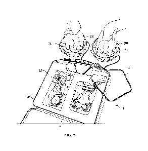
100251 FIG. S is a another top front isometric view of the electrical vein
stimulation
and expansion apparatus ofFIG. 1. showing the apparatus in use. and
illusuaningthe
distending and protruding of the patient's veins.
[00261 FIG. 6 is an electrical schematic of an embodiment of a signal
genelaitor attic
electrical vein stimulation and expansion apparatus of the present disclosure.
100271 FIG. 7 is a %Iva= graph of the output voltage vs. time for one cycle
of the
output signal, such as generated by the signal generator shown in, FIG. 6.
100281 FIG. 8 is a waveform graph illustrating another example wavelbrm.
100291 FIG. 9 is a wavefrom graph illustrating another example waveform.
100301 FIG. IO is a top view of an example embodiment of direct electrode
placement
for electrical vein stimulation.
[00311 FIG. 11 is a top view of an example embodiment of aliteornprising
the
electrical) vein stimulation and expansion apparatus of the present
disclosure.
I00321 FIG. 12 is a table illustrating example outputs from the electrical
vein
stimulation and expansion apparatus of the present disclosure.
[0033j FIG. 13 is a waveform graph illustrating another example wavefonn,
100341 FIG. 14 is an electrical schematic of another embodiment of a signal
generator
of the electrical vein stimulation and expansion apparatus of the present
disclosure.
(00351 FIG. 15 is a waveform graph illustrating another example waveform.
[00361 FIG. 16 is a %Weft= graph illustrating another example wavefiann.
DETAILED DESCRIPTION
[00371 Various embodiments will be described in detail with reference to
the
drawings, wherein like reference numerals represent like parts and assemblies
throughout
the several views. Reference to various embodiments does not limit the scope
of the
claims attached hereto. Additionally, any examples set forth in this
specification are not
.intended to be limiting and merely set forth some of the many possible
embodiments for
the appended claims.
100381 While the present disclosure is capable of embodiment in various
'brans, there
is shown in the drawings, and will be hereinafter described, one or more
presently
preferred embodiments with the understanding that the present disclosure is to
be
8
CA 02975454 2017-07-31
WO 2016/126467
PCT/US2016/015009
considered as an exemplifie26031- of the invention, and is not intended to
limit the
invention to the Specific embodiments illustrated herein. Heading* are
provided for
convenience only and are not to be-construed to limit the invention in anyway.
Embodiments illustrated under any heading may be combined with embodiments
illustrated under any other heading.
100391 Referring to FIGS. 1-5, in general, disclosed herein is one
embodiment of an
electrical stimulation apparatus 1 configured to deliver an electrical signal
through the
arms or other limbs a a patient, from one limb, up through the limb, across
the spine and
down the other limb and cause the veins in the hands, arms, legs or feet of
the patient to
distend or expand. In doing so, the stimulation apparatus makes the peripheral
veins in
the arms, hands, legs or tivt of the patient more visible, thereby providing a
medical
practitioner venous access for the drawing of blood or the insertion of a
peripheral venoms
cannula. The apparatus is generally placed in electrical communication with a
patienes
hands and/or arms Or other limbs) by a pair of electrodes or other means that
connects
the device to the patient's arms or feet to deliver a predetermined electrical
signal through
the electrically connected limbs of the patient.
10040j The veins thus become tilled with blood while being subjected to the
electrical stimulation, increasing the internal pressure within the veins,
without the
necessity for a tourniquet or other means of constriction or compression. The
increased
pressure in the veins makes them more rigid, thereby increasing the physical
resistance,
or force, required to insert a needle or other intravenous cannulas therein.
The increased
physical resistance of the target vein permits the medical practitioner to
have an
improved physical feel for the insertion of the needle into the vein, and to
better
differentiate instances when the tip of the needle has been correctly inserted
into the
=drat lumen of the vein, from instances in which the needle has pierced
through the vein
(which can cause serious medical complications).
100411 In general, the electrical stimulation apparatus I. comprises an
electrical
signal generator 10, a power supply 12 in electrical communication with the
signal
generator and configured to supply power thereto, at least a pair of
electrical leads 14
connected at a proximal end to a plurality of electrical output terminals 16
of the
9
CA 02975454 2017-07-31
WO 2016/126467
PCT/US2016/015009
electrical signal generator, and at least a pair of electrodes 18 connected to
a distal end of
each of the electrical leads 14.
[00421 The electrical power supply 12 may be a portable power supply, such
as for
example a 9-volt battery, other voltage battery, or rechargeable battery.
Alternatively, the
power supply may utilize a standard electrical power cord that plugs into a
typical power
outlet in a wall.
10943] One example of the electrical signal generator 10 is shown in FIG,
6, while
another example of the electrical signal generator 10 is shown in FIG. 14. The
electrical
signal generator 10 of FIG. 6 includes the power supply 12, electrical lead
14, container
28, and electrolytic solution 30. Some embodiments include two or more
electrical signal
generators 10, coupled to one or more leads 14, electrodes 18, and containers
28.
[0044] The electrical signal generator 10 comprises electrical circuitry 20
operable to
generate an electrical output signal, such as having a waveform illustrated
and described
with reference to FIG. 7, or another suitable waveform, such as the waveforms
shown in
FIGS. 8,9, 13, 15, and 16. In some embodiments the electrical circuitry 20
includes
electronics such as one or more of resistors, capacitors, transformers, and a
microprocessor in electrical communication with each other. In the example
shown in
FIG. 6, the electrical circuitry 20 of the electrical signal generator 10
includes a power
switch 50, oscillator 52, variable control 54, and output circuitry 56. In
this example the
oscillator 52 includes an integrated circuit, such as a microcontroller 62.
The output
circuitry 56 includes a first stage 58, such as including operational
amplifiers 64 and 66
and capacitor 68, and a second stage 60, including transformer 70. The output
of the
second stage 60 forms the output terminal 16, which can be electrically
coupled to the
lead 14 and electrode 18, to deliver the output signal to the patient.
10945] The oscillator 52 operates to generate an initial oscillating
signal. In this
example, the oscillator includes a square wave generator. One example of a
square wave
generator is a microcontroller, such as the 8-pin, flash-based 8-bit CMOS
microcontroller, part number PIC12F675, available from Microchip Technology
Inc. of
Chandler, AZ, US. Another example of a square wave generator is a 555 timer.
The
square wave generator produces a squarewave signal, which oscillates between
low and
high voltages, such as between 0 and 5 volts. In this example the square wave
has a
SUBSTITUTE SHEET (RULE 26)
CA 02975454 2017-07-31
WO 2016/126467
PCT/US2016/015009
frequency in a range from 4 Hz to 12 Hz. As one example the fiequency is. 7.83
Hz.
Frequencies in this range have been foundlo be preferred over fester
frequencies because
they give the nerves in the patient time to rgiolatize after stimulation
before the next
stimulation. The frequency can be higher fora heathly person whose nerves can
repolarize more quickly, while the frequency typically needs to be. lower for
an unhealthy
person whose nerves require more titne to repohirize.
100461 In some embodiments the signal generator 10 includes a variable
control 54,
such as one or more potentiometers 22, 24 in electrical communication with the
electrical
circuitry of the signal generator 10. The one or more variable controls 54
allow an
operator, such as a medical practitioner, the patient, or another person to
provide an input
to adjust the magnitude of the signal generated by the signal generator 10,
such as to
increase or decrease the magnitude of the signal. In this example, each
potentiometer 22,
24 that is present in the signal generator corresponds to a separate output
voltage channel
(each having its own signal generator 10) having its own leads 14 and
electrodes 18, and
whose voltage is adjusted by its own intensity adjustment knob coupled to the
variable
control 54 that adjusts/sets the output voltage of that channel that is sent
from the signal
generator 1 0 to the patient via the leads 14 and electrodes 18. The ability
to adjust the
output voltage experienced by the patient allows a patient to have the voltage
adjusted
down to a comfortable level, which therefore contributes to lowering the
patient's anxiety
over use of the device, which thus reduces the chance of any anxiety or stress
induced
vasoconstriction that can reduce the amount of blood within the targeted
veins.
100471 hi one embodiment, the signal generator 10 includes two variable
commis
(e.g., potentiometers 22. 24); and therefore may have two separate output
voltage
channels each having its own signal generator 10, with each intensity knob and
variable
control 54 separately adjusting the output voltage to be sent to the patient
along two sets
of electroikss, corresponding to each of the two output voltage channels. A
first of the two
potentiometers 22 and its respective output voltage channel impart an output
voltage to
the patient that is configured to cause the target vein to become swollen or
distended. A
second of the two potentiometers 24 and its respective output voltage channel
impart an
output voltage to the patient that is configured to stop the pain at the
needle stick site by
interntpting nerve signals associated with pain. In the present embodiment,
the two
11
CA 02975454 2017-07-31
WO 2016/126467
PCT/US2016/015009
output voltage channels are identical, but in alternate embodiments, each
potentiometer
may be configured to adjust the output voltage in differing ranges. Having two
separate
channels, each with the ability to adjust the output voltage, allows the
stimulation
apparatus 1 to be configured to adapt to target veins in the foot, neck,
elbow, or other
such target vein sites.
[00481 In this example the electronic circuitry 20 of the signal generator
10 further
includes output circuitry 56. The output circuitry operates to convert the
square wave
signal generated by the oscillator 52 into a desired output signal, such as
having a
waveform shown in FIGS. 7-9, 13, 15, or 16.
[0049] The first stage 58 of the output circuitry includes electronics
including
operational amplifiers 54- and 66, and a capacitor 68. The first stage 58 is
coupled to the
variable control 54 to receive the input from a user to adjust the magnitude
of the signal
generated by the signal generator 10. In this example, the variable control 54
is a
potentiometer that provides a variable resistance. The variable control 54 is
electrically
coupled to an input of the operational amplifier 64. The voltage of the signal
provided by
the variable control 54 changes as the variable control is adjusted. The
operational
amplifier 64 is configured as a unity gain buffer amplifier in this example.
[0050] The oscillator 52 generates a square wave output (e.g., pin 7) that
is then
supplied to the capacitor 68. The capacitor 68 converts the square wave signal
to a series
of pulses having a leading edge with a sharp voltage transition, followed by a
trailing
edge in which the voltage tapers off.
[0051] The signal is then provided to the second stage 60 where it is
further filtered
and amplified such as using the amplifier including operational amplifier 66
arranged in a
non-inverting configuration.
[0052] The amplified signal is then provided to the second stage 60,
including the
transformer 70, which operates to amplify and rectify the signal.
[0053] In some embodiments the transformer 70 has an unequal ratio of
windings.
As one example, the transformer is a 10:1 transformer, which is arranged in a
step-up
configuration to increase the voltage at the output. In other possible
embodiments the
transformer can be arranged in a step-down configuration. Other embodiments
have
12
SUBSTITUTE SHEET (RULE 26)
CA 02975454 2017-07-31
WO 2016/126467
PCT/US2016/015009
other ratios of windings, The output can also be generated in the second stage
without
using a transformer in yet other embodiments.
100541 In this example, the transformer 70 is a center tap transformer. The
oscillating
signal generated by the first stage 58 is provided to the primary winding and
the center
tap, and operates in conjunction with a pair of diodes to rectify the output
signal. The
output signal is generated at the secondary windings and supplied to the
output terminal
16. The ratio of the primary windings to the secondary windings determines the
amplification provided by the transformer 70.
[0055] In some embodiments, the circuitry 20 further includes electronic
components, and/or programming, that are configured to automatically vary the
output
signal, which may include varying one or more of the output voltage, the
output current,
shape of the output voltage waveform, and/or frequency of the output signal
over time,
without having to adjust the variable controls (e.g., potentiometers 22, 24).
In one
embodiment, the output signal may be changed over time by executing specific
computer
code or a software program in the microprocessor. In another embodiment, the
output
signal may be randomly changed inexpensively by the inclusion ofa typical
flashing light
emitting diode (LED) 63 within the circuitry of the signal generator 10.
Flashing LEDs
automatically blink when supplied with electrical power, alternating between
an "on" and
"off state, with the frequency of flashing between the two states depending on
the input
voltage. In one embodiment, the flashing LED is placed in the electrical
circuit
downstream of the microprocessor and upstream of the amplifying circuit that
is
connected to the output leads that are attached to the patient by the
electrodes. The
flashing LED, oscillating between an "on" and "off' state, is constantly
switching the
output current on and off, causing the signal generator 10 to vary the
electrical output
signal and voltage over time, according to the flashing frequency of the
flashing LED. In
this manner, the LED acts as a repetitive timer for the output signal from the
signal
generator. And because the frequency of the LED is dependent on its input
voltage,
adjusting the voltage from the potentiometer will change the frequency of the
flashing
LED, so as to provide an infinitely variable output signal to the patient,
[0056] Furthermore, the lower the quality of the components used to make
the
flashing LED, as with inexpensive flashing LEDs, the more variation or
randomness
13
SUBSTITUTE SHEET (RULE 26)
CA 02975454 2017-07-31
WO 2016/126467
PCT/US2016/015009
there will be io-the consistency or stablenessof the frequency of the flashing
for a given
voltage. Accordingly, lower quality. flashing LEDs provide a flashing pattern
that is More
random than that of higher: quality flashing LEDs. Thereline, in one
embodiment, to
achieve more randomness in the frequency of the electrical signal sent to the
patient from
the signal generator 10, it may be beneficial to use lower quality flashing
LED within the
circuitry as disclosed herein.
ion in still alternate embodiments, additional methods to vary the output
signal
and voltage over time are contemplated herein, without departing from the
scope of the
present disclosure. By varying the output signal in the manner disclosed
herein, the
patient's body is constantly reacting to the changing output signal, rather
than possibly
becoming accustomed to a constant output: signal to which the venous system
might
otherwise no longer respond afler a short exposure thereto.
pm] The signal generator 10 may also include at least one indicator 12,
such as an
LED or other lighted indicator, to indicate to the medical practitioner
utilizing the
electrical stimulation apparatus 1 as to when the power to the apparatus is
turned "on."
An additional indicator may be included to indicate when the electrical signal
is being
sent to a patient. In one embodiment, the indicator may perform both
functions, however,
in alternate embodiments, separate indicators may be utilized to communicate
each of the
two functions.
100591 The apparatus I may also include programming and/or a display screen
configured to communicate and display for the medical practitioner the real
time output
voltage and signal, an initial set output voltage and signal, fault
conditions, stimulation
apparatus fault diagnostic information, or any other such setting, output, or
11:edback
information as may be desired. In another embodiment, the apparatus 1 may
include a
display configured to graphically display the real time electrical information
(e.g. the
electrical signal and/or voltage vs. tune) being sent to the patient. In still
further
embodiments, the stimulation apparatus I may include data output programming
and
associated output connectors that are configured to permit the apparatus to be
connected
to a separate, stand-alone external display for displaying any/all of the
information
disclosed herein.
14
CA 02975454 2017-07-31
WO 2016/126467
PCT/US2016/015009
[00601 In some embodiments the electronic circuitry 20 is arranged on one
or more
circuit boards. The circuit boards include at least one substate layer, and
typically have at
least one layer of electrical traces formed thereon to make electrical
connections between
the electronic components. In some embodiments the electronic signal generator
10 is
formed on the circuit board.
100611 The output signal is sent from the signal generator 10 to the
patient's body by
two electrical leads 14 that are connected at a proximal end to the signal
generator 10,
and at a distal end to a pair of electrodes 18. In one embodiment of the
present disclosure,
the electrodes 18 may be configured as a pair of cups 28 or containers, such
as for
example, a pair of manicure nail soaking bowls or other such similar
containers, that are
configured to hold a liquid electrolyte solution 30 into which the finger and
thumb tips of
a patient are to be submerged. In some embodiments the containers include one
or more
recessed regions sized and shaped to receive at least the tips of the fingers
of a hand, or
the toes of a foot, therein. The purpose of using an electrolyte solution is
to provide a
conductive liquid medium into which the patient may place his fingers and
through which
the electrical signal may be delivered to the patient. In one embodiment, the
electrolyte
solution may be a mix of minerals and water. However, in alternate
embodiments, the
electrolyte solution may be any other type of solution used for increasing
electrical
conductivity between the electrical leads and the skin of a patient.
100621 In another embodiment of the present disclosure, the electrodes may
be
configured as a pair of conductive electrode pads having a conductive gel or
adhesive
layer disposed on one side thereof to help adhere the electrode pad to the
skin of a patient
and to aid in making good electrical contact between the conductive pad and
the patient's
skin. Such electrode pads may be similar to those used with transeutaneous
electrical
nerve stimulation (TENS) devices or portable defibrillators. In addition, the
electrode
pads may be disposable. In one example embodiment, as shown in FIG. 10, at
least one
pair of electrodes 180,182 are configured as conductive electrode pads with an
adhesive
backing on one side thereof, such that a first electrode 180 of the pair of
electrodes is
attached to a palmar surface of one hand of a patient and a second electrode
182 of the
pair of electrodes is attached to an arm, preferably to the bicep, of the
patient. In the
embodiment of FIG. 10, the arm to which the second electrode 182 is attached
is the
SUBSTITUTE SHEET (RULE 26)
CA 02975454 2017-07-31
WO 2016/126467
PCT/US2016/015009
same arm as the hand to which the first electrode 180 is attached.
Alternatively, *-
second electrode182 may be attached to the patienrsOther arm.. However, in
this case, a
greater level of intensity of the output signal would likely need to be
supplied to the
patient to achieve an efketive vein distension. As also shown in HO, 10, the
pair of
electrodes 180,182 are each connected to a distal end of an electrical lead
140, which an
each connected at a proximal end to the signal generator (not shown) of the
stimulation
apparatus. In one example embodiment, the electrode 180 attached to the palmar
surf=
clone hand of the patient will supply a positive output signal to the patient,
while the
other electrode 182 attached to the arm of the patient will supply a negative
output signal
to the patient. Alternatively, the negative output signal can be supplied to
electrode 180,
while the positive output signal can be supplied to electrode 182.
[00631 Mier attachment of the pair ofelecurides 180,182 to the patient, the
signal
generator may be turned on to supply the output signal to the patient and to
begin the
electrical stimulation. The intensity of the output signal can be increased if
no physical
response, e.g., muscle fasciculation and/or vein distension, is observed.
Alternatively, if
the patient is experiencing discomfort, the intensity of the ouput signal can
be decreased
to a level that is tolerable, but that still produces a physical response, as
discussed above.
As the output signal is sent from the signal generator to the patient's body
by the two
electrical leads 140 that are connected at a proximal end to the signal
generator (not
shown), and at a distal end to the pair of electrodes 180,182, distension of
the veins in the
patient's arm will begin and will generally last for at least about nai (10)
minutes, and
may even last for more than about r fteen (1 5) minutes. In one example
embodiment, the
electrical stimulation is continuedihr at least two (2) minutes, but for no
more than ten
(10) minutes. In particular, the electrical stimulation can be discontinued
once the target
vein is visible and/or palpable. Once the target vein has become distended,
the signal
generator may be turned oft and ventpuneture or any other medical procedure
requiring
vein distension may be performed.
[00641 In one embodiment: as shown in FIG. 11, the electrical stimulation
apparatus
of the present disclosure may comprise a kit for use by a medical practioner.
The kit can
include a signal generator 100 that is preferably battery operated, a pair of
containers 28
Ibr holding an electrolytic solution, a prefilled labeled bottle of Epsom salt
.110, a bottle
16
CA 02975454 2017-07-31
WO 2016/126467
PCT/US2016/015009
of deoniz.ed water 115, and a disposable electrode assembly 112 that includes
a pair of
electrodes and a pair of electrical leads for.connectingthe pair of
electrodesIo the signal
.genaator 100. The kit can be used for electrical stimulation of a patient by
either using
the containers 28 to which the electrodes are attached and the electrolytic
solution is
added, as discussed in one of the embodiments above, or by directly connecting
the
electrodes to the patient, as discussed in another of the embodiments alxwe.
[00651 In the case of electrical stimulation of a patient using the
containers 28 to
which the electrolytic solution is added, the electrolytic solution can be
prepared by
adding the supplied deionized water 115 to the prefilled bottle of Epsom salt
110. In one
embodiment, the Epsom salt concentration is at least about 30 gil.õ The
electrodes are
thereafter attached to the containers 28, and a patient may then place their
hands into the
containers 28, prior to the addition of the prepared electrolytic solution
into the containers
28. The electrodes are then attache4 to the signal generator 100 via the
supplied
electrical leads, and the signal generator 100 can be turned on to supply the
output signal
to the pair of electrodes. One of the electrodes can be supplied a negative
output signal,
while the other electrode can be supplied a positive output signal. As
discussed above,
once the target vein has become distended, the signal generator may be turned
oft and
venipuncture or any other medical procedure requiring vein distension may be
performed.
Prior to performing venipuncture, however, it may be preferred to wash the
patient's
hands with water in order to remove the salt solution, which may affect the
outcome of
any blood chemistry analysis.
0o661 While the previous embodiments disclosed the electrodes configured as
either
small containers lbr permitting the fingertips to be placed into an
electrolyte solution, or
conductive electrode pads, the electrodes should not be limited to such
embodiments and
in alternate ernbrxliments may have alternate configurations as desired. For
example, in
alternate embodiments, the electrodes may be alternate sized containers that
permit the
submersion of a patient's full hands, leet, or any portion of the patient's
body, including
but not limited to arms and/or legs, into an electrolyte solution in
electrical
communication with the signal generator. In still alternate embodiments, the
electrodes
may be one or more of a metal pin-typo probe or metal plate that ate contact
based
electrodes. In still alternate embodiments: the electrode may be a finger
clamp-type probe
17
CA 02975454 2017-07-31
WO 2016/126467
PCT/US2016/015009
that is similar in inethanical structure to those used to measure pulse
oximetty. In yet
additional V:111110Iiiillerliti, the electrodes may be tonthictiVegartnentsõ
or other such
coitact-based electrode having an alternate physical configuration, without
departing
from the scope of the disclosure herein. In yet an additional embodiment, the
electrodes
may be configured as one or more electromagnets that generate a magnetic
field, into
which magnetic. field the patient may place his hands, feet, or limbs. The
electromagnetic
field is configured to generate a complementary electric signal in the
patient's body via
changes to the magnetic field. In such an embodiment, the patient is not
directly
connected to the signal generator.
10067] In one embodiment, the eleatical signal output from the signal
generator 10
sent to a patient's limbs through the electrodes includes an electrical signal
that is an
alternating signal (AC). In one embodiment, the AC signal sent to the patient
has a
frequency of 7.83 Hz (or 7.83 full &mating cycles per second). This means that
the
output circuit is interrupted 7.83 times per second. This frequency of 7,83
Utz has been
selected in one embodiment to provide the nerves of the patient time to
repolarize
between successive output signals, and thus have time to get prepared for the
next
subsequent output signal. By providing adequate time to allow the nerves to
rqiolatize,
the signal generated by the signal generator 10 has a consistent effect on the
skin, nerves,
and muscles in the vicinity of the electrodes.
[00681 In another example embodiment, as shown in FIG. 12, the AC signal
sent to
the patient has a frequency of 7,9 Hz, with an assymetrical charged balanced
'Aphasic
waveform. The duration of the pulse at 1200 ohms, 1600 ohms, and 950 ohms is
68.8 is,
60,0 l's. and 77.0 es, respectively. In addition, the MOXit)11111) amplitude
at 1200 ohms,
1600 ohms, and 950 ohms is 80.4 V., 94.4 \rpm., and 70_5 V)õFik. respectively.
While
FIG, 12 displays one embodiment of theoretical standard measurements across
purely
resistive loads at maximum intensity settings, outputs may vary depending on
parameter
settings.
[00691 However, while the above embodiments operate at ficquencies of 7.83-
11z
and 7,9 Hz, respectively, the frequency of the output signal should not be
read In he.
limited only to such specified frequencies, and in alternate embodiments, the
AC or DC
signal may have a different frequency without departing from the scope of the
present
18
CA 02975454 2017-07-31
WO 2016/126467
PCT/US2016/015009
diselosure. lit alt ate enibodintents, the frequency of the output signal may
be any.
alternate liequency, depending on the Spivak eircuitiy design of the signal
generator.
For example, in an alternate embodiment, a different duty cycle or output
cyele, oreven a
different wavvform that is subsequently developed, may use a diMrent
frequency.
Furthermore, in alternate embodiments, the signal generator 10 may be
configured to
adjust the frequency or waveform of the output signal based on sensed feedback
related
to the physiological differences between patients of difiCrent ages, the
patient's
circulatoiy system patency, and other biomedical and/or bioelectrical aspects
of the
patient's body. In one embodiment, the microprocessor in the signal generator
10 may
thither contain programming that adjusts the output signal for the changes
that are
usually associated with an aging patient, such as thinner skin, more sensitive
skin, skin
that is sensitive to bleeding, etc.
100701 In one embodiment, the output voltage from the signal generator 10,
which is
set by at least one of the potentiometers 22, 24, is initially set to be
within the range of
between 0 volts and 90 volts. In another embodiment, each of the two output
voltage
channels may be set to be within the range of between 0 volts and 90 volts.
However, in
alternate embodiments, the potentiometers 22, 24 may have larger or smaller
output
voltage ranges than that disclosed herein, and may each be seleetably set: to
an initial
output voltage value, or adjusted to a new output voltage vahle, within such
larger or
smaller voltage ranges, without departing from the scope of the present
disclosure.
10071j FEEDBACK SYSTEM
100721 The signal generator 10 may tiirther include an integrated feedback
system
that is configured to measure the resistance and capacitance of the patient's
body dining
the time between each successive cycle of the output signal. hi one
embodiment, the
feedback system utilizes a ten to one (10:1) audio transformer that responds
to the
electrical and capacitive resistance (i.e., electrical back pressure) of the
patient's body, as
well as any changes thereto, in order to adjust the output signal sent to the
patient. Each
human body presents with an electrical resistance. This resistance can change
with the
body's weight, hydration, etc. This electrical resistance can also change
during the
treatment. The signal generator 10 use.s the audio transformer to measure the
electrical
resistance of the patient's body and, in response, appropriately alter the
output voltage
19
CA 02975454 2017-07-31
WO 2016/126467
PCT/US2016/015009
and/or current transmitted to the patient as part of the signal. In doing so,
the signal may
be altered battedon the feedback from the feedback system to ensure that the
signal
generator 10- is eliciting the same clinical or physiological response in the
patient's body,
even when the patient's bodily response to treatment is changing (Le. changes
to the
patient's electrical back pressure, or bodily resistance andan capacitance).
100731 A. simple transformer performs the job of monitoriag the electrical
back
pressure of the patient's body simply and inexpensively. When the
microprocessor, via
the transformer in electrical communication with the patient, detects a very
high electrical
resistance in the patient's body, then very little current will flow from the
signal generator
into the patient for a given constant output voltage .frorn the signal
generator to the
patient. If the input mama from the signal generator is very low (as when
powered by a
small battety), and if the output voltage texas do not have much resistance,
then the
battery WW1' decreases and the current drops significantly. The measured
electrical
resistance of die human body is fairly constant, bin the capacitance of the
human body
can vary greatly. This is a concern, because the sudden release of electrical
energy or
charge from the capacitor-like parts of the human body can result in the body
receiving a
painful jolt of electricity that may potentially cause damage to the patient's
nervous or
cardiac system, and otherwise interrupt the desired clinical response in the
patient's body
caused by the treatment.
100741 The transformer of the feedback system filters an output voltage of
the swat
generator, which voltage fluctuates over time according to a preprogrammed
voltage
wavefbrm to allow the specific portions of the voltage waveform that are the
most
effective at eliciting the desired vein distension response to pass through to
the patient
The electrical back pressure in the patient causes a reaction in the patient's
body that
creates a resulting electrical signal from the patient's body that can be
captured and read
by the signal generator, which can then be used as an input to adjust the
output voltage of
the next cycle of the output sitnail from the signal generator.
100751 In alternate embodiments, the feedback mechanism may be specific
programming within the microprocessor of the signal generator that is
configured to
monitor the feedback. of the patient's electrical resistance and capacitance
and, in turn,
adjust the output signal sent to the patient based on the monitored feedback.
In still
CA 02975454 2017-07-31
WO 2016/126467
PCT/US2016/015009
attenutte embodiments, the feedback system may utilize a plurality of sensors
configured
to measure the patients.reSistatiCe and capacitance, or any othersuch
electrical
component or computer code configured to measute feedback resistance and
capacitance,
without departing from die scope of the present disclosure.
100761 In one embodiment, the apparatus I can be configured to stop all
output
signals from the signal generator 10 and wait for the patient's body to react
to the last
output signal. When the patient's body reacts to the last signal, the
patient's body
produces a resulting electrical signal that can he captured by the signal
generator 10,
analyzed, and used to alter the next output signal thuri the signal generator
10 that is sent
to the patient. This can be done in mil time with the appropriate
microprocessor and
software. In an alternate embodiment, if the feedback mechanism of the signal
generator
measures a change in a patient's bioelectrical resistance or capacitance of
more than 10%
between Stict..t.iw cycles of the output signals, the signal generator is
configured to shut
off or go into a fault mode, as a change of larger than 10% may indicate that
the patient's
body is experiencing a stress response and no is longer responding to the
output signals.
In one embodiment, the signal generator would automatically adjust the output
signal
waveform, voltage, and cumin based on the individual patient's specific
physiology and
related bioelectrical properties.
01077i In still further embodiments, the signal generator includes software
to collect
physiological data from the patient using the stimulation apparatus, minding
the patient's
physiological response data. That data can then be stored and analyzed by the
signal
generator and used to change the output signal in real time, so as to optimize
the output
signal and the achieved venous response for the specific patient.
100781 Included in the signal generator may be a microprocessor having
programming therein configured to control the amount of current and voltage
being sent
to the patient via the electrodes, as well as the shape of the output voltage
waveform that
is being sent to the patient, monitor the electrical feedback received from
the patient (i.e.
the patient's internal bodily resistance and capacitance), and automatically
adjust, in real
time, any of the voltage output, the cutrent output, or the shape of the
voltage waveform
being sent to the patient. The microprocessor may be any programmable
microprocessor
having any speed or internal numnary size without departing form the scope- of
the present
21
CA 02975454 2017-07-31
WO 2016/126467
PCT/US2016/015009
diselositre. In one embodiment, the microprocessor may include a comparator
circuit
configured, to compare the Original output signal Sent10 the patient litim the
Signal
.genenatorto the returned signal from the patient. The results of the
comparison are then
used by the microprocessor to change the output signal :proportionately to
balance the
next output signal sent to the patient. In such an embodiment, the
microprocessor may
have a baseline waveform stored in its memory which is sent to the patient
with the first
signal. A response/reflex signal is then sent back to the microprocessor from
the patient
through the feedback system, which response/reflex signal is also stored in
the
microprocessor. Thereader, the microprocessor adapts the next outgoing signal
based on
the prior stored incoming response/reflex signal to gently coax the patients
nerves to
early the best waveform, voltage, and current necessary to produce the
greatest visible
presentation of the vein. This comparative process ensures that the output
signal being set
to the patient each time will continue to elicit the desired physiological and
clinical
response in the peripheral veins of the patient, preventing the patient's body
from getting
accustomed to the signal being sent.
[0079] Furthermore, the processor includes programming configured to
maintain a
predefined signal frequency. For example, in one embodiment, the
microprocessor is
programmed to maintain a preprogrammed signal frequency of 7.8311z. However,
in
alternate embodiments, alternate frequencies may be chosen without departing
from the
present disclosure. For example, in some patient groups or subsets, such as
obese
patients, geriatric patients, or neonatal patients, alternate signal
frequencies may be
needed to aid in eliciting the optimal venous presentation maths. In addition,
in an
embodiment, the microprocessor may be programmed and configured to continue to
operate properly on a constantly declining voltage, such as for example when
the power
supply is a battery that slowly runs out of power over time and continued use.
POSfil WAVEFORM GRAPH
100811 FIG. 7 shows an exemplary graph of an embodiments of active portions
of a
single cycle of a signal. The graph shows an output voltage (the Y-axis) of
the output
signal, versus time in milliseconds (the X-Axis), that is able to illicit the
desired vein
distension and pain suppression response in a patient. The shape of the
signal, including
the location and amplitude of the various peaks and valleys therein, is an
exemplary
22
CA 02975454 2017-07-31
WO 2016/126467
PCT/US2016/015009
waveform that is able to elieit active, signal-based enlargement of the target
peripheral
veins, Which aids in the performing, of Venipuneture by medical practitioners,
fur
example. FIGS. 8, 9, 13, 15, and 16-show additional exemplary waveform graphs
of
active portions of a single cycle of a signal.
100821 Referring further to FIG. 7, a plurality of points.1-9- are
identified on the
graphed wavetium showing the output signal's output voltagevs. time. Point I
on the
graph corresponds to the beginning of a new cycle of the repetitive output
signal, and
indicates the initial output voltage from the signal generator that is
selected to alert or
stimulate a patient's sensory nerve (via its dendrites in the surface of the
skin) to a change -
in condition. This initial output voltage initiates a tiny electrical signal
in the patient's
body, having a unique voltage, current, and waveform, to be sent to the
central nervous
system so the brain can monitor the extremities. In response, the brain sends
a healing
signal back to that specific sensory dendrite from Which the signal to the
brain originated.
100831 Point 2 on the graph corresponds to the primaty effective portion of
the nerve
stimulation signal. This point is the main output voltage in the nerve
stimulating portion
of the output signal that causes the peripheral nerves in the patient's limbs
to over-react
and causes a simultaneous tetany or spasm of the nearby muscles surrounding
the target
peripheral veins. This is the portion of the waveform that is adjusted via the
knob of one
of the potentiometers 22, 24 on the signal generator. In overweight patients,
the voltage
level at Point 2 is automatically suppressed by a layer of fat in the skin.
Accordingly, fir
overweight patients, in order to get the signal to reach the nerves of the
patient and
overcome the resistance of the fat layer, it may be necessary to send a higher
output
voltage to the patient. This can be accomplished by using a ten to one (10:1)
audio
transformer, or other such transformer, in the signal generator to amplify the
output
voltage signal sent to the patient. Alternatively, the increasing of the
voltage to overcome
the resistance of the fat layer so the signal may reach the nerves may also be
accomplished by the implementation of programming contained in the
microprocessor.
100841 Point 3 in the voltage waveform graph corresponds to the output
voltage that
triggers the sensory nerve in the patient to "turn oft" In this regard. Point
3 is the voltage
that triggers the nerve to be at rest and reset to its standby voltage,
waiting to be used or
triggered "on" again in the next subsequent cycle of the output signal. Point
4 in the
23
CA 02975454 2017-07-31
WO 2016/126467
PCT/US2016/015009
voltage waveform graph is the output voltage that cancels the positive portion
of the
signal and. balancesthe stimulation apparatus' nerve signal to allOw the nerve
time to
reset itself, or repolarizv....
[00851 Point 5 in the wavefotm graph corresponds to the muscle stimulation
portion
of the output signal, and is the output voltage that causes the motor muscles
to stinntlate
the venous muscle pump that in tutu causes the veins to distend and fill with
blood. In the
waveform presented in FIG. 7, the length of time during which this portion of
the signal
is active is small, however in some patients the length of time over which
this portion of
the output voltage in the output signal is active will be adjusted to achieve
the proper
amount of voluntary muscle stimulation to activate the venous muscle pump. The
longer
that this portion of the signal is active, the more that the muscles are
stimulated. in
addition, the small involuntary smooth muscles surrounding the veins rewire a
different
amount of active stimulation time to activate the venous muscle pump action
than that of
the larger muscles. This portion of the waveform also maybe adjusted from
patient to
patient to achieve the optimal venous muscle pump action in each patient.
[0086j Point 6 in the waveform graph is the point at which the motor muscle
stimulation is shut off to allow them to reset and get ready for the next
cycle of the signal.
Point 7 in the wavelbrm graph corresponds to a reflex signal back pressure
from the
patient's peripheral nervous system, indicating that the nervous system is
trying to take
over control of the nerves and muscles and stabilize the patient's muscle and
nerve
activity. Point 8 in the waveform graph corresponds to a period of zero output
voltage to
the patient, and is part of the integrated feedback loop that the peripheral
nervous system
uses to gently restore the patient's baseline electrical potential hack to its
original resting
electrical potential, or internal voltage. In comfortable, relaxed patients,
their resting
potential, or measured voltage, may be on the order of 20 millivolts. However,
in some
patients -who are anxious, their measured testing potential maybe zero volts,
or a positive
measured voltage, which are otherwise higher electrical potentials or voltages
than a
typical relaxed patient. This initial resting potential measurement is used to
setup the
basic parameters of the first and each succeeding treatment output signal from
the signal
generator.
24
CA 02975454 2017-07-31
WO 2016/126467
PCT/US2016/015009
100871 Point. 9 in the waveform graph corresponds to the patient's baseline
condition,
whereby there is no active output Signal or -voltage being-Sent to the
patient's -body, and
the patient is otherwise unaffected by any output signal -from the stimulation
apparatus.
Ibis also corresponds to the period during which the signal generator is
monitoring the
patient's internal electrical potential and preparing to initiate a new cycle
of the signal,
and adjusting the active portion of the Output signal based on the feedback
monitored
from the patient.
100881 APPARATUS OPERATION AND STIMULATION ACTION
100891 hi operation, the stimulation apparatus functions AS MIMS. The
electrodes
are placed in electrical contact with the lingers, hands, and/or limbs of a
patient. In one
embodiment, this involves the patient placing the fingertips of each hand into
separate
containers of an electrolyte solution. The electrolyte solution in each
container is placed
in electrical communication with the signal generator by separate electrical
leads that are
terminated at one end in the electrolyte solution, and at the opposite end to
output
contacts of the signal generator. In alternate embodiments, the electrodes may
be
adhesive backed pads that are affixed directly to the patient's skin.
100901 The power source supplies power to the signal generator. The medical
practitioner adjusts the output voltage to the patient by rotating an
adjustment knob of at
least one potentiometer. The signal generator is switched "on" and the
preprogrammed
electrical output signal is transmitted through the leads and electrodes to
the fingertips,
hands, and/or arms of the patient. The preprogrammed output signal includes a
repetitive
cycle of preprogrammed fluctuating output voltages at various specified points
in time for
each cycle. In one embodiment, the initial output voltage may be set between 0
and 90
'ohs and the signal delivered is less than one milliarnp. However, in
alternate
embodiments, the output voltage range may be larger or smaller, or cover a
different
voltage range than that disclosed in the present embodiment, and the output
signal may
be larger than 1 milliatrip without departing from the scope of the present
disclosure.
1009I Each cycle of the output electrical signal includes a period of
active output
voltage and a period of rest where no output voltage is being imparted to the
patient's
limbs. The preprogrammed output voltage may include sevetal phases including,
one or
more or the thflowing: an initiation phase that alerts the patient's sensory
nerve to the
CA 02975454 2017-07-31
WO 2016/126467
PCT/US2016/015009
presence- of the output voltage; a primary nerve stimulation phase that elopes
the
peripheral nerves to !bite the motor muscles surrounding the peripheral target
veins: to
contract; an end to the nerve stimulation phase that turns "off the sensory
nerve; a
balancing phase that cancels the stimulation signals that were sent to the
nerves to allow
the nerves to reset; a muscle stimulation phase that activates the venous
mimic pump; a:
shutdown phase that ends the activation of the motor muscles; an electrical
hack pressure
phase; an electrical feedback phase; and a rest phase with no active voltage
output to
allow the patient's system time to reset before the next cycle begins. This
cycling part of'
the wavelbrm in the current embodiment is not exclusive of other possible
wavelbrins.
What is envisioned is a waveform that causes all the actions described in this
application
and may vary relative to the patient's physiology, the design and limitations
of the
electronic circuitry, and/or the method used to deliver the signals to the
patient
[00921 The result of the repetitive electrical cycles in the output signal
that are
imparted to the patient is a physiological response in the patient as
follow's. One portion
of the generated electrical signal stimulates the involuntary smooth muscles
near the
electrodes to contract and relax. These muscles are circular in nature and
when they
contract they form a tube. This tube is larger than normal and creates a
vacuum which
can have the effect of drawing in whatever blood is available via the
capillaries and the
nearby arteries. hi addition, part of the waveform stimulates the adjacent
muscles which
act as a venous muscle pump to increase the local blood pressure in the veins,
thus adding
more blood to the now visually obvious and distended veins. This venoms muscle
pump is
the body's way of moving blood from the arteries and capillaries back to the
heart The
multitude of valves present in the veins prevent retrograde blood flow; thus
aiding the
enlarging of the target veins internal volume for easier access for
venipuncture. For some
patient groups, such as geriatric patients, this venous muscle pump action may
be further
aided in conjunction with the presently disclosed electrical venous
stimulation apparatus,
by the use of a tourniquet applied between the target vein and the heart.
However, the
presently disclosed electrical venous stimulation apparatus can activate the
venoms
muscle pump action without the need of a tourniquet or other means of
constriction or
compression.
26
CA 02975454 2017-07-31
WO 2016/126467
PCT/US2016/015009
100931 The electrical venous stitnulation apparatus works best to present
the veins in
the backtif thelandsõ top of the feet and the fort.-arnar. In oneumbodiment,
the dee:Weal
venous .stinntlation apparatus further operates as a TENS device in that there
is a portion
of the output voltage wax:clot:ailing is configured to numb the tissue
adjacent the
electrodes (and accordingly the target vein site), all the way to the medial
portion of the
forearm. This included functionality mikes the process of inserting a needle
into a target
vein while using the electrical Venous stimulation apparatus less painful to
the patient
when the needle stick actually OCCUIS. Iii embodiments having two
potentiometers, the
second potentiometer controls the output voltage channel that creates the TENS
device
functionality. The second output channel can be configured to attach directly
on the skin
of the patient nearby the projected needle stick site to focus the numbing
effect to a
specifically local area. The second channel can be configured to perform this
nerve
deadening function specifically. Thus in one embodiment, one output voltage
channel is
used to achieve the displaying of an enlarged, engorged vein, and the other
output voltage
channel is used to numb the area of the needle stick.
1.0094] The apparatus of the present disclosure is configured to non-
invasively alter
the physiology of the peripheral veins that are targeted for venipuncture in
the limbs of a
patient using an active electrical signal, rather than using passive means
traditionally
used, or requiting the use Of a tourniquet or another means of constriction or
compression. In an aspect of the present disclosure, an active signal imparted
to the skin
of a patient by the apparatus elicits a physiological response and a change in
condition/behavior of the target vein, causing the vein to fill with blood and
become
distended/enlarged and become more rigid, thereby increasing visibility of the
vein, as
shown in FIG. S. In this manner, using such an apparatus and methodology as
disclosed
herein, it becomes easier for medical practitioners to locate the target vein
and achieve
successful and proper veniptineture. No other active device currently exists
that non-
invasively changes the physiology of the tissue in and around the target veins
to aid in
locating the target vein and increasing the ease of achieving successful and
proper
venipuncture without the need for multiple attempts.
100951 As discussed herein, one embodiment is a method of accessing a veimi
of a
person, the method comprising: receiving a portion of a limb of the person
into a
27
CA 02975454 2017-07-31
WO 2016/126467
PCT/US2016/015009
container; .supplying a liquid electrolytic solution into the. container,
wherein the liquid
electrolvie.tiolution IS in contact with the portion of the link electrically
Stimulating-the
portion of the limb with at least one signal generated by an electrical signal
generator, the
electrical signal provided to the electrolytic solution by at least one
electrode in contact
with the liquid electrolytic solution; causing at least one vein in the litrib
of the-pawn to
protonic in response to the electrical stimulation; and inserting a tip of a
needle into the
vein while it is protruding to at.t.t.ss the vein.
100961 The various embodiments described above are plovided. by way of
illustration.
only and should not be construed to limit the claims attached hereto. Those
skilled in the
art will readily recognize various modifications and changes that may be made
without
following the example embodiments and applications illustrated and described
herein,
and without departing from the true spirit and scope of the f011owing
28