Note : Les descriptions sont présentées dans la langue officielle dans laquelle elles ont été soumises.
CA 02977406 2017-08-22
WO 2016/134436
PCT/CA2015/000106
System and Method for Delta Relaxation Enhanced Magnetic Resonance Imaging
FIELD OF THE INVENTION
[0001] The present invention relates generally to magnetic resonance imaging.
More
specifically, the present invention relates to delta relaxation enhanced
magnetic
.. resonance imaging.
BACKGROUND OF THE INVENTION
[0002] Magnetic resonance imaging (MRI) is a major imaging technique used in
medicine. MRI is capable of generating detailed images of soft tissues such as
the
brain, muscles and kidneys. Specific properties of the various compounds found
inside
.. tissues, such as water and/or fat, are used to generate images. When
subjected to a
strong magnetic field, the vector sum of the nuclear magnetic moments of a
large
number of atoms possessing a nuclear spin angular momentum, such as hydrogen,
which is abundant in water and fat, will produce a net magnetic moment in
alignment
with the externally applied field. The resultant net magnetic moment can
furthermore
precess with a well-defined frequency that is proportional to the applied
magnetic field.
After excitation by radio frequency pulses, the net magnetization will
generate a signal
that can be detected.
. .. [0003] Delta relaxation enhanced magnetic resonance imaging (DREMR)
generally
referred to as field-cycled relaxometry or field-cycled imaging is an MRI
technique that
.. offers the possibility of using underlying tissue contrast mechanism which
vary with the
strength of the applied magnetic field to generate novel image contrasts. To
achieve
DREMR contrast, the main magnetic field is varied as a function of time during
specific
portions of an MR pulse sequence. A field-shifting electromagnet coil is used
to perform
the field variation. To date the DREMR imaging methods have focused on the
effect of
main magnetic field variations on the T1 relaxation characteristic of
materials being
imaged. This, however, is a limited use of a DREMR system.
SUMMARY OF THE INVENTION
1
CA 02977406 2017-08-22
WO 2016/134436
PCT/CA2015/000106
[0004] It is an object to provide a novel system and method for an MRI
scanning
system and method that obviates and mitigates at least one of the above-
identified
disadvantages of the prior art.
[0005] According to one aspect, a method of acquiring magnetic resonance (MR)
signals at a delta-relaxation enhanced MR imaging (DREMR) system is provided.
According to the method, the DREMR system can generate a main magnetic field
with a
strength of BO andan initial pulse sequence for acquiring at least one of: T2*-
weighted
MR imaging signals; susceptibility weighted imaging (SWI) signals; and
saturation
imaging signals. The main magnetic field strength can be varied to a strength
of B1
during at least one portion of the initial pulse sequence and a first image
can be
acquired based on the initial pulse sequence.
[0006] According to another aspect, a method of acquiring MR signals at a
DREMR
system is provided. According to the method, the DREMR system can generate a
main
magnetic field with a strength of BO and an initial pulse sequence for
acquiring MR
spectroscopy signals. A first spectroscopy signal can be acquired based on the
initial
pulse sequence. A repeat pulse sequence for acquiring MR spectroscopy signals
can
also be generated, the repeat pulse sequence corresponding to the initial
pulse
sequence. The main magnetic field strength can be varied to a strength of B1
during at
least one portion of the repeat pulse sequence. A second spectroscopy signal
can be
acquired based on the repeat pulse sequence and peaks from the first and the
second
spectroscopy signals can be identified. The identified peaks can then be
correlated.
[0007] According to yet another aspect, a method of acquiring MR signals at a
DREMR system is provided. According to the method, the DREMR system can
generate a main magnetic field with a strength of BO and an initial pulse
sequence for
acquiring MR signals for fingerprinting. A first image can be acquired based
on the
initial pulse sequence. A repeat pulse sequence for acquiring MR
fingerprinting signals
can be generated, the repeat pulse sequence corresponding to the initial pulse
sequence. The main magnetic field strength can be varied to a strength of B1
during at
least one portion of the repeat pulse sequence and a second image can be
acquired
based on the repeat pulse sequence. At least one MR signal property can be
measured
2
CA 02977406 2017-08-22
WO 2016/134436
PCT/CA2015/000106
based on the first and the second images. A tissue type can be identified
based on the
at least one MR signal property.
[0008] According to a further aspect a DREMR system is provided. The system
can
comprise a main magnet operating to generate a main magnetic field with a
strength of
BO. The system can further comprise radio frequency coils having a transmit
aspect
and gradient coils operating to generate an initial pulse sequence for
acquiring at least
one of: T2*-weighted MR imaging signals; susceptibility weighted imaging (SWI)
signals; and saturation imaging signals. The system can also comprise field-
shifting
magnets operating to vary the main magnetic field strength to a strength of B1
during at
least one portion of the initial pulse sequence. The radio frequency coils can
have a
receive aspect operating to acquire a first image based on the initial pulse
sequence.
[0009] These, together with other aspects and advantages which will be
subsequently apparent, reside in the details of construction and operation as
more fully
hereinafter described and claimed, reference being had to the accompanying
drawings
.. forming a part hereof, wherein like numerals refer to like parts
throughout.
BRIEF DESCRIPTION OF THE DRAWINGS
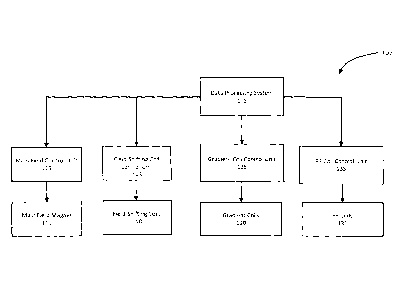
[0010] FIG. 1 shows a block diagram of functional subsystems of a delta
relaxation
magnetic resonance imaging (DREMR) system in accordance with an
implementation;
[0011] FIG. 2 an imaging volume and corresponding slice to be scanned by the
delta
relaxation magnetic resonance system of FIG. 1 in accordance with an
implementation;
[0012] FIG. 3 shows illustrative examples of Ti and T2 relaxation diagrams;
[0013] FIG. 4 shows an example pulse sequence in accordance with an
implementation;
[0014] FIG. 5 shows a schematic representation of a k-space containing one
received
line in accordance with an implementation;
[0015] FIG. 6 shows idealized frequency distribution of two materials at
different
magnetic field strengths;
3
CA 02977406 2017-08-22
WO 2016/134436
PCT/CA2015/000106
[0016] FIG. 7 shows an example pulse sequence for augmented MR signal
acquisition
using the example DREMR system of FIG. 1 based on spectral suppression;
[0017] FIG. 8 shows an example pulse sequence for augmented MR signal
acquisition
using the example DREMR system of FIG. 1 based on susceptibility weighted
imaging;
[0018] FIG. 9 shows an example pulse sequence for augmented MR signal
acquisition
using the example DREMR system of FIG. 1 based on susceptibility weighted
imaging;
[0019] FIG. 10 shows an example pulse sequence for augmented MR signal
acquisition using the example DREMR system of FIG. 1 based on T2* based
imaging;
[0020] FIG. 11 shows a conceptual illustration of T2* signal separation from 2
materials; and
[0021] FIG. 12 shows an example pulse sequence for augmented MR signal
acquisition using the example DREMR system of FIG. 1 based on T2* based
imaging;
[0022] FIG. 13 shows idealized results of performing augmented MR signal
acquisition
using the example DREMR system 100 of FIG. 1; and
[0023] FIG. 14 shows a simplified example of the effects of magnetic field
strength
changes to MR fingerprinting results.
DETAILED DESCRIPTION
[0024] Referring to FIG. 1, a block diagram of a delta relaxation magnetic
resonance
imaging (DREMR) system, in accordance with an example implementation, is shown
at
100. The example implementation of the DREMR system indicated at 100 is for
illustrative purposes only, and variations including additional, fewer and/or
varied
components are possible. Traditional magnetic resonance imaging (MRI) systems
represent an imaging modality which is primarily used to construct pictures of
magnetic
resonance (MR) signals from protons such as hydrogen atoms in an object. In
medical
MRI, typical signals of interest are MR signals from water and fat, the major
hydrogen
containing components of tissues. DREMR systems use field-shifting magnetic
resonance methods in conjunction with traditional MRI techniques to obtain
images with
different contrast than is possible with traditional MRI, including
molecularly-specific
contrast.
4
CA 02977406 2017-08-22
WO 2016/134436
PCT/CA2015/000106
[0025] As shown in FIG. 1, the illustrative DREMR system 100 comprises a data
processing system 105. The data processing system 105 can generally include
one or
more output devices such as a display, one or more input devices such as a
keyboard
and a mouse as well as one or more processors connected to a memory having
volatile
and persistent components. The data processing system 105 can further comprise
one
or more interfaces adapted for communication and data exchange with the
hardware
components of MRI system 100 used for performing a scan.
[0026] Continuing with FIG. 1, example the DREMR system 100 can also include a
main field magnet 110. The main field magnet 110 can be implemented as a
io permanent, superconducting or a resistive magnet, for example. Other
magnet types,
including hybrid magnets suitable for use in the DREMR system 100 will now
occur to a
person of skill and are contemplated. The main field magnet 110 is operable to
produce
a substantially uniform main magnetic field having a strength BO and a
direction along
an axis. The main magnetic field is used to create an imaging volume within
which
is desired atomic nuclei, such as the protons in Hydrogen within water and
fat, of an object
are magnetically aligned in preparation for a scan. In some implementations,
as in this
example implementation, a main field control unit 115 in communication with
data
processing system 105 can be used for controlling the operation of the main
field
magnet 110.
20 [0027] The DREMR system 100 can further include gradient coils 120 used
for
encoding spatial information in the main magnetic field along, for example,
three
perpendicular gradient axis. The size and configuration of the gradient coils
120 can be
such that they produce a controlled and uniform linear gradient. For example,
three
paired orthogonal current-carrying primary coils located within the main field
magnet
25 110 can be designed to produce desired linear-gradient magnetic fields.
[0028] In some implementations, the gradient coils 120 may be shielded and
include
an outer layer of shield coils which can produce a counter magnetic field to
counter the
gradient magnetic field produced by the primary gradient coils forming a
primary-shield
coils pair. In such a coil pair the "primary" coils can be responsible for
creating the
30 gradient field and the "shield" coils can be responsible for reducing
the stray field of the
5
CA 02977406 2017-08-22
WO 2016/134436
PCT/CA2015/000106
primary coil outside a certain volume such as an imaging volume. The primary-
shield
coils pair of the gradient coils 120, the primary and shield coils, may be
connected in
series. It is also possible to have more than two layers of coils for any
given gradient
axis that together form a shielded gradient coil. Shielded gradient coils 120
may reduce
.. eddy currents and other interference which can cause artefacts in the
scanned images.
Since eddy currents mainly flow in conducting components of the DREMR system
100
that are caused by magnetic fields outside of the imaging volume (fringe
fields),
reducing the fringe fields produced by the gradient coils 120 may reduce
interference.
Accordingly, the shapes and sizes, conductor wire patterns and sizes, and
current
.. amplitudes and patterns of the primary-shield coils pair can be selected so
that the net
magnetic field outside the gradient coils 120 is as close to zero as possible.
For
cylindrical magnets, for example, the two coils can be arranged in the form of
concentric
cylinders whereas for vertical field magnets, the two coils may be arranged in
coaxial
disks.
[0029] One side effect of shielding can be that the fields produced by the
primary-
shield coils pair of the gradient coils 120 may partially cancel each other
within the
imaging volume. Accordingly, more current can be required to produce a
gradient field
with a particular strength by shielded gradient coils 120 than by unshielded
gradient
coils 120. This effect can be quantified as the gradient efficiency, which may
be defined
as the achievable gradient strength for 1 Ampere of driving current. Another
important
parameter describing gradient coil performance is called the gradient slew
rate, which is
the rate of driving a gradient coil from zero to its maximum amplitude. This
term is
inversely proportional to the inductance of the gradient coil. Typically, in
order to
increase the efficiency of a shielded gradient coils 120 to be comparable to
the
efficiency of an unshielded gradient coils 120 the inductance must increase.
This
increase in inductance will decrease the maximum achievable slew rate. The
loss in
efficiency for a shielded configuration can depend on the distance and current
density
ratio between the primary and shield coils. Increasing the distance between
the
primary-shield coils pair may increase the efficiency.
[0030] The conductive components of the gradient coils 120, whether shielded
or
unshielded and including the primary and shield coils, may consist of an
electrical
6
CA 02977406 2017-08-22
WO 2016/134436
PCT/CA2015/000106
conductor (for example copper, aluminum, etc.). The internal electrical
connections can
be such that when a voltage difference is applied to the terminals of the
gradient coils
120, electric current can flow in the desired path. The conductive components
for the
three gradient axes for both the primary gradient coils and the gradient
shield coils can
s be insulated by physical separation and/or a non-conductive barrier. The
primary
gradient windings can be placed on a non-conductive substrate (for example,
G10, FR4,
epoxy or others).
[0031] In some variations, the gradient coils 120 may also be provided with
thermal
control or heat extraction mechanisms. For example, some of the windings can
be
hollow and coolant can be passed through these hollow conductors to extract
heat from
the gradient coils 120, produced, for instance, by resistive heating of the
windings when
electricity is applied. Alternatively, other methods of extracting heat can be
used, such
as inserting coolant channels within the gradient coils 120. The coolant
channels can
be in thermal contact with the gradient coil windings. The gradient coils 120
can also be
mounted in a thermally-conductive but electrically-non-conductive epoxy to
ensure that
the mechanical assembly is rigid and to limit the possibility of electrical
breakdown.
[0032] The magnetic fields produced by the gradient coils 120, in combination
and/or
sequentially, can be superimposed on the main magnetic field such that
selective
spatial excitation of objects within the imaging volume can occur. In addition
to allowing
spatial excitation, the gradient coils 120 can attach spatially specific
frequency and
phase information to the atomic nuclei placed within the imaging volume,
allowing the
resultant MR signal to be reconstructed into a useful image. A gradient coil
control unit
125 in communication with the data processing system 105 can be used to
control the
operation of the gradient coils 120.
[0033] In some implementations of the DREMR system 100, there may be
additional
electromagnet coils present, such as shim coils (traditionally, but not
limited to,
producing magnetic field profiles of 2nd order or higher spherical harmonics)
or a
uniform field offset coil or any other corrective electromagnet. To perform
active
shimming (correcting the field distortions that are introduced when different
objects are
placed within or around the system), the corrective electromagnets, such as
the shim,
7
CA 02977406 2017-08-22
WO 2016/134436
PCT/CA2015/000106
coils, carry a current that is used to provide magnetic fields that act to
make the main
magnetic field more uniform. For example, the fields produced by these coils
can aid in
the correction of inhomogeneities in the main magnetic field due to
imperfections in the
main magnet 110, or to the presence of external ferromagnetic objects, or due
to
susceptibility differences of materials within the imaging region, or any
other static or
time-varying phenomena.
[0034] The DREMR system 100 can further comprise radio frequency (RF) coils
130.
The RF coils 130 are used to establish an RF magnetic field with a strength B1
to excite
the atomic nuclei or "spins". The RF coils 130 can also detect signals emitted
from the
1.0 "relaxing" spins within the object being imaged. Accordingly, the RF
coils 130 can be in
the form of separate transmit and receive coils or a combined transmit and
receive coil
with a switching mechanism for switching between transmit and receive modes.
[0035] The RF coils 130 can be implemented as surface coils, which are
typically
receive only coils and/or volume coils which can be receive and transmit
coils. The RF
coils 130 can be integrated in the main field magnet 110 bore. Alternatively,
the RF
coils 130 can be implemented in closer proximity to the object to be scanned,
such as a
head, and can take a shape that approximates the shape of the object, such as
a close-
fitting helmet. An RF coil control unit 135 in communication with the data
processing
system 100 can be used to control the operation of the RF coils 130.
[0036] To create a contrast image in accordance with field-shifting
techniques,
DREMR system 100 can use field-shifting electromagnets 140 while generating
and
obtaining MR signals. The field-shifting electromagnets 140 can modulate the
strength
of the main magnetic field. Accordingly, the field-shifting electromagnets 140
can act as
auxiliary to the main field magnet 110 by producing a field-shifting magnetic
field that
augments or perturbs the main magnetic field. A field-shifting electromagnet
control
unit 145 in communication with the data processing system 100 can be used to
control
the operation of the field-shifting electromagnets 140.
[0037] To reduce interference and artefacts, the field-shifting electromagnets
140 may
include a shield similar to the shielded gradient coils 120 described above.
The shielded
field-shifting electromagnets 140 can have two components: an inner primary
field-
8
CA 02977406 2017-08-22
WO 2016/134436
PCT/CA2015/000106
shifting electromagnets, to produce the field shift and an outer shield field-
shifting
electromagnets, to form a shield by reducing the stray field of the primary
field-shifting
electromagnets outside a certain volume such as an imaging volume.
Implementing
field-shifting primary and shield electromagnets combination that balances the
competing needs of low inductance (faster slew rates), high efficiency
(greater magnetic
field strength for a given current amplitude), and low resistance (less
heating and
subsequent demands on cooling) is a complex electromagnetic problem.
[0038] Indeed, one side effect of shielding the field-shifting electromagnets
140 can be
that the fields produced by the primary and shield components of the shielded
field-
shifting electromagnets 140 may partially cancel each other within the imaging
volume.
Accordingly, more current can be required to produce a magnetic field with a
particular
strength by shielded field-shifting electromagnets 140 than by unshielded
field-shifting
electromagnets 140. This effect can be quantified as the field-shift
efficiency, which
may be defined as the field-shift amplitude per 1 Ampere of current passing
through the
electromagnet. The loss in efficiency for a shielded configuration depends on
the
distance and current density ratio between the shield electromagnets and the
primary
electromagnets. Increasing the distance between the primary and shield
electromagnets may increase the field-shift efficiency.
[0039] The conductive components of the field-shifting electromagnets 140,
including
the primary and shield electromagnets, may consist of an electrical conductor
(for
example copper, aluminum, etc.). The internal electrical connections can be
such that
when a voltage difference is applied to the terminals of the field-shifting
electromagnets
140, electric current can flow in the desired path. The conductive components
for both
the primary and the shield electromagnets can be insulated by physical
separation
and/or a non-conductive barrier. The field-shift windings can be placed in
layers on or
within a non-conductive substrate (for example, G10, FR4, epoxy or others).
[0040] In some variations, the field-shifting electromagnets 140 may also be
provided
with thermal control or heat extraction mechanisms. For example, where
windings are
used to form the electromagnets, the windings can be hollow and coolant can be
passed through these hollow conductors to extract heat deposited in the
electromagnet
9
CA 02977406 2017-08-22
WO 2016/134436
PCT/CA2015/000106
due to resistive heating of the windings when electricity is applied.
Alternatively, other
methods of extracting heat can be used, such as inserting coolant channels
within the
field-shifting electromagnets 140. The coolant channels can be in thermal
contact with
the field-shifting electromagnets 140. The field-shifting electromagnets 140
can also be
mounted in a thermally-conductive but electrically-non-conductive epoxy to
ensure that
the mechanical assembly is rigid and to limit the possibility of electrical
breakdown.
[0041] There are many techniques for obtaining images using the DREMR system
100, including Ti and T2 weighted images. To provide a simplified illustration
of the
DREMR system 100's functionality, simplified operations for obtaining proton
density-
.. weighted images are described as a non-limiting example. To create an image
in
accordance with the example illustration, the DREMR system 100 detects the
presence
of atomic nuclei containing spin angular momentum in an object, such as those
of
Hydrogen protons in water or fat found in tissues, by subjecting the object to
a relatively
large magnetic field. In this example implementation, the main magnetic field
has a
strength of BO and the atomic nuclei containing spin angular momentum may be
Hydrogen protons or simply protons. The main magnetic field partially
polarizes the
Hydrogen protons in the object placed in the imaging volume of the main magnet
110.
The protons are then excited with appropriately tuned RE radiation, forming an
RE
magnetic field with a strength of B1, for example. Finally, weak RE radiation
signal from
the excited protons is detected as an MR signal, as the protons "relax" from
the
magnetic interaction. The frequency of the detected MR signal is proportional
to the
magnetic field to which they are subjected. Cross-sections of the object from
which to
obtain signals can be selected by producing a magnetic field gradient across
the object
so that magnetic field values of the main magnetic field can be varied along
various
locations in the object. Given that the signal frequency is proportional to
the varied
magnetic field created, the variations allow assigning a particular signal
frequency and
phase to a location in the object. Accordingly, sufficient information can be
found in the
obtained MR signals to construct a map of the object in terms of proton
presence, which
is the basis of a traditional MRI image. For example, since proton density
varies with the
type of tissue, tissue variations can be mapped as image contrast variations
after the
obtained signals are processed.
CA 02977406 2017-08-22
WO 2016/134436
PCT/CA2015/000106
[0042] Referring now to FIG. 2, to further illustrate the example signal
acquisition
process by the DREMR system 100, it will be assumed that an object is placed
within
an imaging volume 250 of the main magnet 110 having a main magnetic field 210
with a
strength BO, pointing along the Z-axis indicated at 240. The object
subsequently has a
net magnetization vector. In this illustrative example, a slice in a plane
along the X and
Y axes, as indicated at 205, is being imaged. It should be noted that in this
example, the
slice has a finite thickness along the Z-axis, creating a volumetric slice
205.
[0043] When the object is placed in the main magnetic field BO, the individual
spins align themselves in the direction of the Z-axis. Referring to FIG. 3, at
equilibrium, the magnetization by main field BO can produce a net longitudinal
magnetization Mz, with an amplitude of MO, parallel with the main magnetic
field.
Excitation of the spins may be achieved when a radio frequency (RF) pulse that
generates the RF magnetic field with an amplitude of B1 is applied at the
Larmor
frequency, by the RF coils 130. During the application of the RF magnetic
field the net
magnetization rotates around the applied RF (B1) field and can cause the net
magnetization to rotate away from the Z-axis. The component of the rotated
magnetization that is projected in the X-Y plane is the net transverse
magnetization
Mxy. The spins can precess about the applied RE magnetic field until the RF
magnetic
field is removed.
[0044] Once the equilibrium magnetization is perturbed, spin-relaxation
processes
occur. Spin-lattice relaxation processes cause a return of magnetization to
the
equilibrium distribution along the Z-axis. Spin-lattice relaxation can thus
bring the
longitudinal magnetization Mz back toward its maximum value MO, as indicated
at 305,
with a characteristic time constant Ti. A characteristic time representing the
recovery of
the magnetization along the Z-axis by 37% is called the Ti relaxation time or
Ti time.
1/T1 is referred to as the longitudinal relaxation rate.
[0045] Spin-spin relaxation, on the other hand, can cause a loss of coherence
due to
dephasing of the net transverse magnetization. Therefore, during spin-spin
relaxation,
the transverse magnetization Mxy exponentially decays toward zero, as
indicated at
310, with a characteristic time constant T2. A characteristic time
representing the decay
11
CA 02977406 2017-08-22
WO 2016/134436
PCT/CA2015/000106
of the signal by 37%, is called the T2 relaxation time or T2 time. 1ff2 is
referred to as
the transverse relaxation rate.
[0046] Transverse relaxation (T2) can cause irreversible dephasing of the
transverse
magnetization. There is also a reversible dephasing effect caused by magnetic
field
inhomogeneities. These additional dephasing fields may arise from a variety of
sources including the main magnetic field inhomogeneity, the differences in
magnetic
susceptibility among various tissues or materials, chemical shift, and
gradients applied
for spatial encoding. The contribution to the transverse relaxation time from
these
reversible dephasing effects are typically referred to as T2'. The
characteristic
relaxation time of the combination of reversible (T2') and irreversible (T2)
dephasing
effects is typically referred to as T2* relaxation.
[0047] The difference between the time constants Ti and T2 are important for
development of contrast in MR imaging. The relaxation times can vary with the
strength of the magnetic field applied, as well as temperature. Moreover, Ti
and T2
values associated with biological tissues can vary. Generally, tissues with
shorter Ti
times, such as T1a as indicated at 315, can yield greater signal intensity at
a given point
in time (appearing brighter in images) than those with longer Ti times, such
as Tlb as
indicated at 305, due to the more rapid recovery of signal. On the other hand,
tissues
possessing short 12 times, such as T2a as indicated at 320, can yield lower
signal
intensity (appearing darker in images) due to a reduction in the detected
transverse
magnetization Mxy. The MR signal from an image can be therefore dependent on
the combination of the intrinsic tissue properties and extrinsic user-selected
imaging parameters and contrast agents.
[0048] To obtain images from the DREMR system 100 in the traditional manner,
one
or more sets of RF pulses and gradient waveforms (collectively called "pulse
sequences") are selected at the data processing system 105. The data
processing
system 105 passes the selected pulse sequence information to the RF control
unit 135
and the gradient control unit 125, which collectively generate the associated
waveforms
and timings for providing a sequence of pulses to perform a scan.
12
CA 02977406 2017-08-22
WO 2016/134436
PCT/CA2015/000106
[0049] The sequence of RF pulses and gradient waveforms, namely the type of
pulse
sequence, applied may change which relaxation times have the most influence on
the
image characteristics. For example, 12* relaxation has a significant influence
following
a 90 RF pulse which is used in a gradient-echo (GRE) sequence, whereas T2
relaxation has a more significant influence following 90 -180'sequential RF
pulses
(also known as a spin echo sequence).
[0050] Referring now to FIG. 4, an illustrative pulse sequence 400 is shown
that can
be used to acquire images using the DREMR system 100. Specifically, a timing
diagram for the example pulse sequence is indicated. The timing diagram shows
pulse
or signal magnitudes, as a function of time, for transmitted (RFt) signal,
magnetic field
gradients Gx, Gy, and Gz, received RFx signal and filed-shifting signal (FS).
An
idealized pulse sequence, simplified for illustrative purposes, can contain a
slice
selection radio frequency pulse 410 at RFt, a slice selection gradient pulse
420 at Gz, a
phase encoding gradient pulse 430 at Gy, a frequency encoding gradient pulse
440 at
Gx, as well as a detected MR signal 450 at RFx. The pulses for the three
gradients Gx,
Gy, and Gz represent the magnitude and duration of the magnetic field
gradients that
can be generated by the gradient coils 120. The slice selection pulse 410 can
be
generated by the transmit aspect of RF coils 130. Detected MR signal 450 can
be
detected by the receive aspect of the RF coils 130. In this illustrative
example it will be
assumed that transmit aspect and receive aspect of RF coils 130 are formed by
distinct
coils. Finally, the field-shifting signal FS causes the main magnetic field
strength to be
changed for the duration of the signal FS. The precise timing, amplitude,
shape and
duration of the pulses or signals may vary for different imaging techniques.
For
example, field-shifting signal FS may be applied at a time and manner that
allows image
contrast to increase for the technique used.
[0051] The first event to occur in pulse sequence 400 can be to turn on the
slice
selection gradient pulse 420. The slice selection RF pulse 410 can be applied
at the
same time. In this illustrative example, the slice selection RF pulse 410 can
be a sinc
function shaped burst of RF energy. In other implementations, other RF pulse
shapes
and durations can be used. Once the slice selection RF pulse 410 is turned
off, the
slice selection gradient pulse 420 can also be turned off and a phase encoding
gradient
13
CA 02977406 2017-08-22
WO 2016/134436
PCT/CA2015/000106
pulse 430 can be turned on. In some implementations, the field-shifting signal
460 may
also be turned on at this point to change the main magnetic field strength.
Once the
phase encoding gradient pulse 430 is turned off, a frequency encoding gradient
pulse
440 can be turned on and a detected MR signal 450 can be recorded. It should
be
noted that the shapes, magnitudes and durations of the pulses and signals
shown in
FIG. 4 are chosen for illustrative purposes, and that in implementations, one
or more of
these factors and others may be varied to achieve the desired scan results.
[0052] The pulse sequence 400 can be repeated a certain number of times or
iterations, typically 256 times, to collect all the data needed to produce one
image. The
time between each repetition of the pulse sequence 400 can be referred to as
the
repetition time (TR). Moreover, the duration between the center point of the
slice
selection pulse 410 and the peak of detected MR signal 450 can be referred to
as echo
time (TE). Both TR and TE can be varied as appropriate for a desired scan.
[0053] To further illustrate the signal acquisition process of DREMR system
100, FIG.
2 is referred to in conjunction with FIG. 4. To select a slice, the slice
selection gradient
pulse 420 can be applied along the Z-axis, satisfying the resonance condition
for the
protons located in the slice 205. Indeed, the location of the slice along the
Z-axis can be
determined based in part on the slice selective gradient pulse 420.
Accordingly, the
slice selection pulse 410, generated at the same time as the slice selection
gradient
pulse 420 can excite protons that are located within the slice 205 in this
example.
Protons located above and below the slice 205 are typically not affected by
the slice
selection pulse 410.
[0054] Continuing with the illustrative example, in accordance with the pulse
sequence
400, a phase encoding gradient pulse 430 can be applied after the slice
selection
gradient pulse 420. Assuming this is applied along the Y-axis, the spins at
different
locations along the Y-axis can begin to precess at different Larmor
frequencies. When
the phase encoding gradient pulse 420 is turned off, the net magnetization
vectors at
different locations can precess at the same rate, but possess different
phases. The
phases can be determined by the duration and magnitude of the phase encoding
gradient pulse 430.
14
CA 02977406 2017-08-22
WO 2016/134436
PCT/CA2015/000106
[0055] Once the phase encoding gradient pulse 430 is turned off, a frequency
encoding gradient pulse 440 can be turned on. In this example the frequency
encoding
gradient is in the X direction. The frequency encoding gradient can cause
protons in the
selected slice to precess at rates dependent on their X location. Accordingly,
different
spatial locations within the slice are now characterized by unique phase
angles and
precessional frequencies. RF receive coils 130 can be used to receive the
detected
signal 450 generated by the protons contained in the object being scanned
while the
frequency encoding gradient pulse 440 is turned on.
[0056] As the pulse sequence 400 is performed by DREMR system 100, the
acquired
signals can be stored in a temporary matrix referred to as k-space, as shown
in FIG 5 at
500. Typically, k-space is the collection of the detected signals measured for
a scan
and is in the spatial frequency domain. K-space can be covered by frequency
encoding data along the X-axis 520 (Kx) and phase encoding data along the Y-
axis 530
(Ky). When all the lines for the k-space matrix for a slice are received (at
the end of the
scan of a single slice, for example) the data can be mathematically processed,
for
example through a two-dimensional Fourier-transform, to produce a final image.
Thus,
k-space can hold raw data before reconstruction of the image into the spatial
domain.
Typically, k-space has the same number of rows and columns as the final image
and is
filled with raw data during the scan, usually one line per pulse sequence 400.
For
example, the first line of k-space 500, indicated at 510, is filled after the
completion of
the first iteration of the pulse sequence generated for scanning a slice and
contains the
detected signal for that pulse sequence iteration. After multiple iterations
of the pulse
sequence, the k-space can be filled. Each iteration of the pulse sequence may
be
varied slightly, so that signals for the appropriate portions of the k-space
are acquired.
.. It should be noted that based on different pulse sequences, other methods
of filling the
k-space are possible, such as in a spiral manner, and are contemplated.
[0057] The choice of specific pulse sequences with optimized parameters can be
used by the DREMR system 100 to exploit tissue contrast to obtain images that
are
able to depict different characteristics of tissue and materials. For example,
as
mentioned above, T2* relaxation has a significant contribution on relative
signal
intensities immediately following a 900 RF pulse. T2* relaxation can be one of
the main
CA 02977406 2017-08-22
WO 2016/134436
PCT/CA2015/000106
determinants of image contrast with GRE pulse sequences and forms the basis
for
many magnetic resonance (MR) applications, such as susceptibility-weighted
imaging
(SWI), perfusion MR imaging, and functional MR imaging. GRE sequences with T2*
based contrast can be used to depict hemorrhage, calcification and iron
deposition in
various tissues and lesions.
[0058] SWI uses phase information in addition to T2* relaxation based contrast
to
exploit the magnetic susceptibility differences of blood and of iron and
calcification in
various tissues. Accordingly, SWI is an MR imaging method that takes advantage
of
signal loss and phase information to allow better imaging of vessels and other
tissues.
[0059] Functional MRI (fMRI) studies rely on regional differences in cerebral
blood flow
to delineate regional activity. Blood Oxygenation Level Dependent Imaging
(BOLD) is a
technique used to generate images in function MRI studies. BOLD-fMRI is able
to
detect differences in cerebral blood flow in part due to a difference in the
paramagnetic
properties of oxygenated hemoglobin and deoxygenated hemoglobin. Deoxygenated
hemoglobin can be more strongly paramagnetic than oxygenated hemoglobin, and
therefore the former can cause greater local dephasing of protons. The local
dephasing
can reduce the MR signal from the tissues in its immediate vicinity. T2*
weighted pulse
sequences can be used to detect this change.
[0060] The DREMR system 100 can also be used to perform MR spectroscopy.
Spectroscopy is the determination of the chemical composition of a substance
by
observing the spectrum of electromagnetic energy released from a material,
including
chemical samples, or a tissue sample. MR spectroscopy is a technique whereby
MR
signals obtained from the nuclei of a material is analyzed to identify the
material's
composition. MR spectroscopy is based on the fact that components of a
material have
different resonant frequencies. Rather than displaying MR signals on a gray
scale as an
image based on the relative signal strength, MR Spectroscopy displays the MR
signal
as a spectrum graph. Accordingly, the resonance frequency of each compound is
represented on a graph as a peak.
[0061] MR spectroscopy can be performed with a variety of pulse sequences. A
basic
sequence consists of a 90 degree RE pulse followed by reception of the MR
signal by
16
CA 02977406 2017-08-22
WO 2016/134436
PCT/CA2015/000106
the receiving components of the RF coils 130, without any intervening gradient
pulses.
Moreover, many pulse sequences used for imaging, such as a spin echo sequence,
can
be used for MR spectroscopy as well.
[0062] A DREMR system 100 can enhance traditional MR images by modulating or
varying the strength BO of the main magnetic field during at least a portion
of one or
more pulse sequences. To perform field-shifting scans using a DREMR system
100,
magnetic strength level BO of the main magnetic field may be caused to
rapidly, and
uniformly change during one or more portions of one or more pulse sequences
used to
obtain image signals which can form an image. The goal is to cause shifts in
the main
filed by a predetermined field-shifting magnetic field without causing
artifacts or image
degradation due to changes in the main magnetic field
[0063] Specifically, field-shifting electromagnets 140 can be used for
obtaining a
contrast image by causing a shift in the main magnetic field strength. A field-
shifting
magnetic field can be applied during a portion of a pulse sequence causing the
main
magnetic field to be field shifted in strength. More specifically, the static
magnetic field
strength BO generated by the main magnet 110 can be either increased or
decreased
by an amount dB through the use of field-shifting electromagnets 140. The
field-shifting
magnetic field generated by the field shifting electromagnets 140 may be
applied during
part, substantially all, or all of a pulse sequence.
[0064] Field-shifting properties of DREMR system 100 can be combined with
various
traditional imaging techniques by modifying traditional pulse sequences as
appropriate,
and by including an appropriate field-shifting signal, to obtain improved
images. For
example, in certain types of MR imaging it is often desirable to suppress MR
signals
arising from different materials. A common example of this is the suppression
of MR
signals arising from fat while preserving MR signals arising from water. This
suppression can be done by making use of the fact that MR signals from
different
materials may have different frequencies of precession. For example, protons
of fat and
water have different precessional or Larmor frequencies. Thus, in a homogenous
main
magnetic field, a sufficiently narrow band RF pulse may be generated by RF
coils 130 to
excite the desired tissue type only. If such a pulse is used to excite water,
for example,
17
CA 02977406 2017-08-22
WO 2016/134436
PCT/CA2015/000106
in place of the typical slice selection transmission 410, it may primarily tip
the
magnetization of water molecules into the transverse plane. Hence the
resulting MR
signal measured will primarily be from the water molecules.
[0065] In alternative implementations, a saturation pulse may instead be
applied to
suppress signals from the unwanted tissue types, such as fat. Thus, a
sufficiently
narrow band saturation pulse may be used by the DREMR system 100 to tip the
protons
of the undesired species into the transverse plane. If such a pulse is used to
suppress
signals from fat protons, for example, then a conventional slice select pulse
combination, such as pulse 410 and 420, applied shortly thereafter can
primarily tip the
io magnetization of water protons into the transverse plane since the fat
protons would
have already been excited by the saturation pulse prior to the application of
the slice
selection pulse. Because the longitudinal magnetization of fat protons would
not have
had time to regrow, fat protons would not be available to tip into the
transverse plane at
the time the slice selection pulse is applied. Thus, the resulting measured MR
signal
is would be primarily obtained from the water protons. The selective RF
pulse used to
excite the desired species may be referred to as a saturation pulse.
[0066] One difficulty of the saturation method can be that the difference in
precessional frequencies between materials is proportional to the main
magnetic field
strength. At lower main magnetic field strengths, the separation between the
20 precessional frequencies of protons of different materials is lower. For
example where
BO is at 0.5T, the separation between precessional frequencies of fat and
water protons
(whose precessional frequencies differ by 3.5 parts per million), is
approximately 70Hz
whereas at 1.5T the separation is approximately 220Hz. Figure 6(a) illustrates
a
generic 15ms radio frequency saturation pulse response 605 for exciting water,
25 compared to signals from fat (610) and water (615) at one hypothetical
main magnetic
field strength BO strength. As illustrated in FIG. 6(b), at a lower strength
BO' and a
similar duration saturation pulse, the saturation pulse response 605 is not
sufficient for
robust saturation. It should be noted that illustrations of FIG. 6 are not to
scale and the
elements have been chosen to clarify the concepts being discussed.
18
CA 02977406 2017-08-22
WO 2016/134436
PCT/CA2015/000106
[0067] Additional problems involve criteria used for generating narrow band
saturation
pulses. Saturation pulses which are designed to affect only a narrow range of
frequencies are generated in accordance with various practical constraints
including
how sharply the frequency-dependent effect can occur, how long the RF pulse
takes,
how much RF power is needed and other criteria. Accordingly, generating
effective
narrow band saturation pulses get increasingly difficult as the Larmor
frequency
separation between tissue types decreases.
[0068] By applying a field-shifting magnetic field, generated for
example, by the field-
shifting coils 140, the strength BO of the main magnetic field can be
increased by dB
io during the spectral selective or saturation portion of the MR pulse
sequence. Thus the
separation between the precessional frequencies of different materials can be
increased, allowing the use of saturation pulses that are more practical and
effective. In
accordance, a spectrally selective saturation pulse can be designed for a main
field
strength of BO+dB where dB is the strength added by the magnetic field
generated by
field-shifting coils 140.
[0069] Referring to FIG. 7, an example method of augmented MR signal
acquisition is
illustrated. A saturation pulse can be combined with a predetermined pulse
sequence,
such as pulse 400, to effect MR image acquisition. Accordingly, at 705, the
saturation
portion of the combined pulse sequence, the saturation pulse is generated by
RF coils
130, concurrently with the field-shifting magnetic field, as generated by
field-shift coils
140 to increase the main magnetic field strength to BO+dB. The increase, in
turn, allows
a greater separation of the precession frequencies of different materials,
increasing the
efficacy of the saturation pulse. After the saturation portion 705, the
predetermined
portion 710 of the combined pulse sequence is applied. During the
predetermined
portion 710, the field-shifting field may be turned off and a predetermined
pulse
sequence such as that of pulse sequence 400 may be applied, the pulse sequence
being designed for the main magnetic field strength BO. This process may then
be
repeated as shown at 715 and 720. The repetition may last as many times as
desired
to obtain appropriate MR images. In variations, the spectral saturation
portion of the
.. combined pulse sequence may not always be provided prior to the beginning
of the
predetermined pulse sequence. In some variations, the spectral saturation
portion may
19
CA 02977406 2017-08-22
WO 2016/134436
PCT/CA2015/000106
be applied at some point within the predetermined pulse sequence. In further
variations, the field-shifting field may also be applied during at least a
portion of the
predetermined portion 710 of the pulse sequence, the pulse sequence applied
being
appropriately varied to account for the shifted strength of the main magnetic
field. The
additional application of the field-shifting field during a pulse sequence
portion may be
at a different strengths, such as dB1, than the field-shifting field applied
during a
spectral saturation portion. Moreover, each repetition may involve field-
shifting fields
that are different in strength and duration than the previous application of
the field-
shifting field.
io .. [0070] Field-shifting properties of DREMR system 100 can also be
combined with
susceptibility-weighted imaging (SWI). SWI is an MR imaging method where image
contrast is generated based on local variations in the magnetic field caused
by local
magnetic susceptibility variations of materials. SWI uses phase information in
addition
to T2*-relaxation time based contrast to exploit the magnetic susceptibility
differences of
is tissues and/or materials such as blood and iron. In other words, SWI is
an imaging
method where image contrast may be enhanced based on magnetic susceptibility
differences between tissues and/or materials.
[0071] Magnetic susceptibility is a property of material which determines an
alteration
in a magnetic field caused by a material, when that material is placed in a
magnetic
20 field, such as the main magnetic field during MR imaging. For example,
the magnetic
field strength H inside a tissue, depends on that tissue's magnetic
susceptibility which is
an inherent property of the tissue. The relationship between the strength H of
the
susceptibility altered magnetic field and the main magnetic field, BO, can be
expressed
as H = (1+x)*B0 where x is the magnetic susceptibility property of the
material. For
25 example, venous blood has a x approximately equal to -6.56x10⁻⁶ and soft
tissues have
a x approximately equal to -9.05x10⁻⁶. Accordingly, SWI imaging can be used to
image
the difference in susceptibility altered magnetic fields between venous blood
and soft
tissues as caused by susceptibility difference between the two tissue types.
[0072] As an example, venous blood and hemorrhage (bleeds) areas have a
30 susceptibility difference from soft tissue. This difference can cause
the venous blood, or
CA 02977406 2017-08-22
WO 2016/134436
PCT/CA2015/000106
hemorrhage areas, to have a signal with a shorter T2* in comparison with soft
tissues.
Accordingly, signals from venous blood/bleeds can decay away faster and
produce less
signal in a T2*-weighted pulse sequence (e.g. a GRE sequence).
[0073] The strength of the main magnetic field can be another factor that
affects the
differences in susceptibility altered magnetic field between tissues.
Accordingly,
increasing the magnetic field applied to an object during imaging through the
application
of a field-shifting magnetic field, can increase, for example, the imaged
contrast
between blood such as venous blood and other tissues obtained by SWI imaging.
For
example, a typical SWI pulse sequence can be generated while the main magnetic
field
with a strength of BO is supplemented by the field-shifting magnetic field
generated by
field electromagnets 140, increasing the main magnetic field strength to
BO+dB. The
field-shifting magnetic field may be applied during the interval between
signal excitation
and acquisition.
[0074] Referring to FIG. 8, an illustrative example method for augmenting SWI
with
the use of field-shifting magnetic field using the DREMR system 100 is
indicated.
Excitation is achieved, through application of an RF pulse by the,RF coils
130, at an
excitation portion 810 of a SWI pulse sequence 805 for acquiring an SWI image.
The
main magnetic field strength is at BO. At the phase accrual portion 815, of
the SWI
pulse sequence 805, which is the time during which much of the magnetic-
susceptibility-
based image contrast is generated, a field-shifting field is applied by the
field-shifting
coils 140, which causes the strength of the main magnetic field to be
increased to
BO+dB as indicated at 820. Next, the data acquisition portion 825 of the SWI
pulse
sequence allows acquisition of the MR signals. The process can be repeated, as
indicated at the second SWI pulse sequence 830. The repetition may occur a
predetermined number of times to obtain a desired image. It should be noted
that a
field-shifting magnetic field may be applied during portions of the pulse
sequence other
than the phase accrual portion and the pulse sequence portions adjusted as
desired in
accordance with the changed main magnetic field strength. Moreover, the
strength and
the duration of the field-shifting field applied may vary at different
portions or different
repetitions of the SWI pulse sequence.
21
CA 02977406 2017-08-22
WO 2016/134436
PCT/CA2015/000106
[0075] In variations, SWI may be used as a method that can help visualize
small
bleeds in tissue. In some situations, such as small tissue regions where
hemorrhaging
has occurred or small areas of blood, detecting the contrast difference due to
susceptibility effects can be challenging, especially at lower magnetic field
strengths
where the susceptibility effect is reduced compared to high fields. In these
situations,
the reduced variation in signal strength due to the susceptibility effect may
be enhanced
by combining images with different levels of susceptibility weighting. This
can be
achieved by acquiring images at different main magnetic field strengths. As an
example, for some tissues, the corresponding signal obtained in an SWI image
can be
.. high but may not change significantly when the image is acquired using
different main
magnetic field strengths. Furthermore, there may also be a region of a small
bleed
(background tissue into which blood has hemorrhaged) for which the
corresponding
SWI image signal can be low but may change significantly with different main
magnetic
field strengths. If the small bleed region is embedded within the background
tissue, the
image contrast between an image location containing background tissue only and
an
image location containing a region of small bleed would be proportionally
small. If two
images are acquired at two different magnetic field strengths and the images
are
subsequently subtracted, the background tissue signal would be eliminated and
the
relative contrast between the region containing background tissue only and one
.. containing background tissue and a small bleed would be increased.
[0076] As an illustrative example, an SWI image can be acquired in accordance
with a
SWI pulse sequence at a first main magnetic field strength, such as BO. The
acquisition
can be followed by the acquisition of one or more additional susceptibility
weighted
images using the same SWI pulse sequence, but at different main magnetic field
strengths as achieved through the application of a field-shifting magnetic
field by field-
shifting coils 140. The images from each of these acquisitions, each image
being
acquired at a different main magnetic field strength, can then be combined to
produce
an image that emphasizes regions where the susceptibility-induced contrast
varied from
image to image based on the variation of the main field strength to field
strength. The
two images may be combined in any manner that can increase the relative image
contrast. This may include subtracting images in pairs; summing all the images
22
CA 02977406 2017-08-22
WO 2016/134436
PCT/CA2015/000106
together; fitting the signal at each pixel location across the images to some
parametric
model; or other mathematical combinations.
[0077] Referring to FIG. 9, a simplified example of a method for visualizing
small
bleeds in tissues using the DREMR system 100 is illustrated. Excitation can be
achieved, through application of an RF pulse by the RF coils 130, at
excitation portion
910 of a SWI pulse sequence 905 for acquiring an SWI image. At the phase
accrual
portion 915, of the SWI pulse sequence 905, which is typically the time during
which
much of the magnetic-susceptibility-based image contrast can be generated, a
field-
shifting field can be applied by the field-shifting coils 140, at least during
a part of the
portion 915. The application of the field-shifting field typically causes the
strength of the
main magnetic field to be increased to BO+dB as indicated at 920. Next, the
data
acquisition portion 925 of the SWI pulse sequence can allow the acquisition of
the MR
signals and thus a portion of an MR image. The process can then be repeated,
as
indicated at the second SWI pulse sequence 930. However, during the SWI pulse
sequence 930, the field-shifting field applied by the field-shifting coils 140
as indicated
at 935 is at a strength dB1, different from the initial application of the
auxiliary field at
strength dB indicated at 920. It should be noted that pulse sequence 930 is
typically the
same pulse sequence as pulse sequence 905, altered as necessary to accommodate
the changes in the main magnetic field. The variations in main field strength
to dB1 and
dB can coincide in location and duration within the two pulse sequences. The
pulse
sequence pairs may be repeated, a predetermined number of times, as they are
varied
appropriately to obtain two complete images. In variations, the two images may
be
acquired sequentially. For example, a number of pulse sequences desired to
obtain a
first image may be applied at a first main magnetic field, and repeated at a
second main
magnetic field strength to obtain a second image. In other variations, other
methods for
obtaining two images at two different main magnetic field strengths can be
used. To
generate the final contrast enhanced image, the two images can be combined as
described above. It should be noted that a field-shifting magnetic field may
be applied
during portions of the scan other than the phase accrual portion. For example,
the field-
shifting magnetic field can remain on during data acquisition, or for part of
the data
23
CA 02977406 2017-08-22
WO 2016/134436
PCT/CA2015/000106
acquisition. In further implementations, the strength of the field-shifting
field applied
may vary within or at different portions of the SWI pulse sequence.
[0078] The process of acquiring multiple images at differing main magnetic
fields field-
shifted by field-shifting coils 140 may be repeated as many times as required.
For
example, in some implementations, more than two images may be acquired. When
more than two images are used they may be combined in any manner that can
increase
the relative image contrast. This may include subtracting images in pairs,
then summing
the subtracted images; summing all the images together; fitting the signal at
each pixel
location across all the images to some parametric model; or other mathematical
combinations. In further implementations, the strength of the field-shifting
field applied
may vary within or at different portions of the SWI pulse sequence. For
example, the
auxiliary filed can remain on during the data acquisition, or for part of the
data
acquisition.
[0079] Field-shifting properties of DREMR system 100 can also be combined with
other T2*-weighted MR imaging techniques. As discussed above, T2* relaxation
refers
to the decay of transverse magnetization caused by a combination of spin-spin
relaxation and magnetic field inhomogeneity. T2* relaxation has contributions
both
from the T2 relaxation which is an inherent tissue property, as well as
contributions from
local magnetic field inhomogeneities, commonly referred to as the decay time
T2'. The
three relaxations are related by 1/T. -= 11T2 + 1/72 where T yAB0 where ABO
measures the magnetic field inhomogeneities. Accordingly, T2* relaxation, as
described above, can be detected with gradient-echo (GRE) imaging because
transverse relaxation T2' caused by magnetic field inhomogeneities, unlike in
the case
of a 180 pulse at spin-echo imaging, is not eliminated by a GRE pulse.
.. [0080] There can be many contributions to the magnetic field
inhomogeneities
including inhomogeneities in the main magnetic field due to characteristics of
main
magnet 110, as well as magnetic susceptibility based field differences. Both
of these
effects scale linearly with the strength of the main magnetic field. Thus, the
rate of
signal decay T2', and hence T2*, may vary in different materials placed within
different
.. main magnetic fields.
24
CA 02977406 2017-08-22
WO 2016/134436
PCT/CA2015/000106
[0081] One or more T2*-weighted MR images may be acquired using known T2*
weighted imaging methods, with a field-shifting magnetic field being provided
by the
field-shifting coils 140 for at least some of the images during all or part of
the time
during which T2* decay occurs in the pulse sequence. The T2* dispersion signal
can
then be generated by observing the variation in T2*-weighted signal at each
magnetic
field strength for a given material such as tissue and/or region of the image,
for
example. Accordingly, changes in main field strength of DREMR system 100 can
be
provided through variations in the field-shifting magnetic field applied by
the filed-shifting
coils 140. The variation of T2* dispersion signal in accordance with the main
magnetic
field can then be analyzed to differentiate different tissues by identifying,
for example,
unique patterns in the relationship between T2* and magnetic field strength
or, as
another example application, determine iron content within the tissues. As
further
example, the T2* dispersion analysis can include the identification of unique
magnetic
field strengths where there is a rapid increase or decrease in the T2*
dispersion curve
is that may be a unique characteristic for a given tissue.
[0082] Referring to FIG. 10, an example method of generating a T2* dispersion
signal
using DREMR system 100 is illustrated. Excitation is achieved, through
application of
an RF pulse by the RF coils 130, at excitation portion 1010 of a 12* pulse
sequence
1005 for acquiring T2* signal. At the T2* decay portion 1015, of the T2* pulse
sequence
1005, a field-shifting magnetic field is applied by the field-shifting coils
140 as indicated
at 1020. Next, the data acquisition portion 1025 of the 12* pulse sequence
allows
acquisition of the MR signals. The process is then repeated, as indicated at
the second
T2* pulse sequence 1030 and third T2* pulse sequence 1035. However, during the
second pulse sequence 1030, and the third pulse sequence 1035 the field-
shifting
magnetic field applied by the field-shifting coils 140 as indicated at 1040
and 1045
respectively is at strengths differing from the initial application of the
field-shifting field
indicated at 1020. Specifically, at 1040, the main magnetic field strength has
been
shifted to BO+dB1 and at 1045, the main filed strength has been shifted to
BO+dB2. The
repetition may occur an additional predetermined number of times. It should be
noted
that a field-shifting magnetic field may be applied during portions of the
pulse sequence
other than the T2* decay portion. In some implementations, the strength and/or
CA 02977406 2017-08-22
WO 2016/134436
PCT/CA2015/000106
duration of the field-shifting field applied may vary within or at different
portions of the
T2* pulse sequence. For example, the field-shifting filed can remain on during
or part of
the data acquisition portion of a pulse sequence. Although this example
discusses
obtaining and comparing signals associated with a single pulse sequence
repeated at
different main magnetic field strengths, the same process can be applied to
entire
images or portions or regions of images acquired in a similar manner, using
different
main field strengths.
[0083] Once the multitude of signals or images are acquired at different main
field
strengths, they may be compared to determine changes in 12* dispersion.
Referring
to FIG. 11, a conceptual illustration of how T2* signals from 2 materials,
indicated by a
circle and a star, which could be the same (P1) at one field strength (B0+dB
indicated at
1105 and corresponding to signals acquired using T2* pulse 1005 of FIG. 10)
can be
differentiated by repeating MR signal acquisition at shifted main magnetic
fields. At
magnetic field strength B0+dB1, indicated at 1110 and corresponding to signals
acquired using T2* pulse 1030 of FIG. 10, the T2* signals for the two
materials are now
different (P2 and P4). At magnetic field strength BO+dB2, indicated at 1115
and
corresponding to signals acquired using T2* pulse 1035 of FIG. 10, the T2*
signals for
the two materials or tissues are further differentiated (P3 and P5). Based on
these
differentiations, the type of material can be determined. For example, the
differentiation
may simply indicate a specific magnetic field strength (which may be different
from the
unshifted main field strength of the MRI system) at which there is the largest
difference
in T2* values between two tissues and at which T2* based imaging would be
preferably
performed. Alternatively, the dispersion patterns for any set of tissues may
suggest
specific data processing to increase T2* based signal differentiation from the
tissues.
This could include fitting the measured 12* dispersion points to a specific
model (shape
of variation), subtraction or other linear combinations of signals or images
at specific
magnetic field strengths or other image combination methods.
[0084] As discussed above, 12' component of 12*, and accordingly, 12* varies
with
the applied magnetic field strength. For most materials or tissues, the
expected variation
of T2* with respect to main magnetic field strength is linear. Specifically,
the T2* change
caused by an increase in the main magnetic field strength may be balanced by a
T2*
26
CA 02977406 2017-08-22
WO 2016/134436
PCT/CA2015/000106
change caused by a decrease in the field strength by the same amount. For some
materials, in particular those containing iron, the variation of T2* with
respect to the field
strength can be non-linear. The DREMR system 100 can be used to take advantage
of
this non-linearity to perform enhanced iron or BOLD imaging. T2* weighted
images,
both with and without main field perturbations, can be acquired. Such pairs of
images
may be performed such that they differ in regions where the T2* response to
field
variations is non-linear. For example regions containing iron-based compounds
may
show changes in contrast.
[0085] To implement a differential acquisition, a first acquisition may be
performed
where no main field perturbation is applied. In a second acquisition of the
same MR
image, the main field strength can be varied in a manner which can alter the
image
contrast for materials having a non-linear response to the field variation. As
an example,
the main field may be changed in one direction during a 12* decay portion of a
T2*
pulse sequence, and may be changed in an equal but opposite direction, and for
equivalent duration, for another portion of the T2*decay. For materials having
a non-
linear response to main magnetic field variations, the change in T2*
dispersion when the
main magnetic field increases by a predetermined amount may not be balanced by
the
change in 12* dispersion when the main magnetic field decreases by an equal
amount
and duration. This may be in contrast to tissues or materials that vary
linearly with
respect to changes in the main magnetic field where the change in T2*
dispersion can
be the same when the main magnetic field is perturbed up and down by the same
amount and duration.
[0086] Referring to FIG. 12, an example of a method for performing Iron or
BOLD
imaging using a DREMR system 100 is illustrated. In this figure pulse sequence
1205 is
used to perform a T2*-weighted acquisition at main field strength BO, without
any main
field perturbations. Following the MR signal acquisition based on the pulse
sequence
1205, the same pulse sequence is repeated at 1210. This time, however, the
main field
strength BO is increased by dB for a period of time during which T2* decay is
occurring,
through an application of a field-shifting magnetic field by filed-shifting
coils 140.
Following the increase, the main field strength is decreased by the same
amount dB,
again through the application of an auxiliary magnetic field by field-shifting
coils 140 for
27
CA 02977406 2017-08-22
WO 2016/134436
PCT/CA2015/000106
an equivalent duration. Subsequently, the MR data is acquired. It should be
noted that
although in this example, the main magnetic field strength is first increased,
and then
decreased by the same amount for an equal duration, many different ways of
perturbing
the main magnetic field during the T2* decay portion of a pulse sequence is
possible as
long as the perturbations occur in a manner which can alter the image contrast
for
materials having a non-linear response to field variations. For example, the
main
magnetic field may be altered in a manner such that the alterations are
balanced.
There are various methods for achieving balanced alterations. For example, in
some
variations a series of increases and decreases of equivalent amounts in the
main
magnetic field strength may be applied during the T2* decay portion of the
pulse
sequence. The main magnetic field may be increased first, then decreased by an
equivalent amount and duration, increased back up, and decreased again by an
equivalent amount and duration to the last increase. Each increase-decrease
pair may
be by a different amount and duration. Moreover, the order of increase and
decrease
may change, and pairs may not be located immediately adjacent to each other.
Although this example discusses obtaining signals associated with a single
pulse
sequence repeated at different main magnetic field strengths, it is to be
understood that
a similar process can be applied to the acquisition and analysis of two or
more images.
[0087] Field-shifting properties of DREMR system 100 can also be combined with
MR
spectroscopy. As discussed above, MR spectroscopy is a method whereby MR data
is
acquired and processed to identify components of a substance that have
different
resonant frequencies. The difference in resonant frequencies may arise, for
example,
based on protons existing in different chemical environments within a compound
or
within different compounds within a material such as a tissue. MR spectroscopy
is often
used to analyze substances that are at a very low concentration and thus
generate very
low MR signals. Accordingly, a distribution of peaks at different frequencies
are
developed from MR signals to identify different tissues or materials. However,
MR
signals acquired also include significant noise. The noise is generally
uniformly
distributed across all frequencies. Due to the low concentration and low
signal of
compounds in tissues or materials, it can be difficult to identify peaks above
random
noise associated with signal acquisition. To counter the low signal-to-noise
ratio that is
28
CA 02977406 2017-08-22
WO 2016/134436
PCT/CA2015/000106
typical in these measurements, often MR signal acquisition is repeated and
averaged
together as a method of averaging down white noise.
[0088] The DREMR system 100 can allow performing improved MR spectroscopy
through acquisition of multiple MR signals at different main magnetic field
strengths.
The relative separation of peaks may be proportional to the main magnetic
field
strength. Moreover, the size of the peaks may also weakly depend on the main
magnetic field strength. On the other hand, random noise signals do not
typically vary,
in a determinate manner, with changes in the main magnetic field strength.
Accordingly,
MR signal acquisitions can be repeated using the DREMR system 100, with at
least
lo some of the repeats being made at differing main magnetic field
strengths. Any peaks
present in the acquired MR signal may thus move by known amounts based on the
known shift in the main magnetic field. Accordingly, the acquired MR signals
may be
processed to identify peaks that have shifted by the predicted amounts making
it
possible to improve the detection of desired signal peaks.
[0089] Referring to FIG. 13, an example method for performing MR spectroscopy
using the DREMR system 100 is illustrated. FIG. 13(a) illustrates 3 possible
ideal signal
peaks which are located at different frequencies for a given main field BO.
FIG. 13(b)
illustrates a characteristic random noise that is uniformly distributed over
all frequencies
and FIG. 13(c) illustrates a combined signal resulting from the combination of
the ideal
zo peaks with the characteristic noise. Figure 13(c) is indicative of the
type of signal that
would be acquired by DREMR system 100.
[0090] Continuing with the figure, FIG. 13(d) illustrates the 3 possible ideal
signal
peaks corresponding to the three signal peaks of FIG. 13(a). However, in FIG.
13(d),
the main magnetic field provided for acquisition of the MR signals is
increased by a
field-shifting magnetic field with a strength dB. Accordingly, the position
and amplitude
of all 3 peaks are scaled relative to the 3 peaks of FIG. 13(a), in proportion
to the
change of the main field strength from BO to BO+dB. For example if BO+dB is
equivalent to a main field with a strength of 2130, the position of the peaks
in FIG. 13(d)
may be scaled in frequency by a multiple of 2 relative to the peaks in FIG.
13(a). FIG.
13(e) illustrates a characteristic random noise acquired at the augmented main
29
CA 02977406 2017-08-22
WO 2016/134436
PCT/CA2015/000106
magnetic field strength BO+dB. As shown, the noise may not change with respect
to the
change in the main field strength in a predictable and systematic way.
Finally, FIG. 13(f)
illustrates a combined signal resulting from the combination of the ideal
peaks of FIG.
13(d) with the characteristic noise of FIG. 13(e). Figure 13(f) is indicative
of the type of
signal that would be acquired by DREMR system 100.
[0091] Correlating the peak movements with a change in the strength of the
main
magnetic field may help filter out randomly distributed noise which is
invariant with
respect to changes in the main magnetic field strength. For example, to
determine
whether a peak is present in a pair of MR signals acquired at different main
magnetic
io field strengths, the peak's expected locations, which would be different
at different main
magnetic field strengths, can be checked in both MR signals. A peak can be
assumed
to be present when it is found in the expected locations in both images, the
locations
determined in part on the basis of the difference in main field strengths.
[0092] Although only two acquisitions were used in this illustrative example,
additional
is acquisitions can be performed, and spectra obtained can be used in the
determination
of peak presence through the correlation of peak locations in the additional
MR signals.
In variations, each signal acquisition at a given main magnetic field strength
may also
be repeated at the same magnetic field strength, and the signals thus acquired
averaged to partially average out white noise as described above.
20 [0093] Field-shifting properties of DREMR system 100 can also be
combined with MR
fingerprinting. Any given tissue or material may be characterized based on a
set of
measured MR signal properties, referred to as the MR fingerprint of that
tissue. For
example, for a given tissue or material, multiple MR signal properties can be
quantified
on the basis of MR signals acquired for that tissue or material. Accordingly
Ti, T2, T2*
25 and/or other MR signal properties can be obtained for each tissue or
material based on
MR signals acquired using one or more pulse sequences. These obtained set of
MR
signal parameters can then be used to characterize the MR scanned tissue or
material.
[0094] MR signal properties can be dependent on the strength of the main
magnetic
field applied during signal acquisition. Accordingly, obtaining MR signal
properties at
30 multiple field strengths can add an additional dimension to the set of
parameters that
CA 02977406 2017-08-22
WO 2016/134436
PCT/CA2015/000106
could be used to characterize and differentiate tissues. Accordingly, a set of
MR signal
properties are selected, and scans are performed with the DREMR system 100
using
appropriate pulse sequences for the selected MR properties to obtain the
selected MR
signal properties. The acquisition of the MR signal properties are then
repeated with
different main magnetic field strengths. The change in main magnetic field
strength is
accomplished by applying an auxiliary magnetic field using the field-shifting
coils 140.
The field-shifting magnetic field can be can be specific to each MR signal
acquisition
and applied in a manner that make MR signal measurements sensitive to changes
in
the main magnetic field strength. Some of these techniques, for example for
the
acquisition of T2* property, are discussed above. As a further illustrative
example, to
obtain a T1 measurement, well-established MR acquisition methods for mapping
the Ti
relaxation parameter can be used. The acquisition can be repeated with an
auxiliary
field applied by field-shifting coils 140 during the inversion time (TI)
portion of the pulse
sequence.
[0095] FIG. 14 provides a simplified illustrated example of how the addition
of
magnetic field strength changes can enhance MR fingerprinting. FIG. 14(a)
shows a
distribution of values of one MR signal property, Parameter1, for two
different tissue
types at main magnetic field strength BO along the x-Axis. The y-axis has been
added
for illustrative convenience, and does not represent any values. According to
FIG.
14(a), if the measured MR signal property Parameter1 falls between 1405 and
1410, the
tissue can be identified as Tissue A. If the measured MR signal property
Parameter1
falls between 1415 and 1420, on the other hand, the tissue can be identified
as Tissue
B. The distribution of values for the two tissues overlap. Accordingly, for a
tissue of
interest, after scanning the tissue, if a MR signal Parameter value of "Vali'
was
obtained, it would not be possible to uniquely identify what tissue type that
value
represented.
[0096] Continuing with the figure, FIG. 14(b) illustrates, along the x-axis, a
distribution
of values of one MR signal property ("Parameter 1") for two different tissue
types at
main magnetic field strength BO along the x-Axis, as in FIG. 14(a). However,
in this
case, the y-Axis represents the acquisition distribution of values of the same
MR signal
property, Parameter1, for the same two tissue types at main magnetic field
strength
31
CA 02977406 2017-08-22
WO 2016/134436
PCT/CA2015/000106
BO+dB. It can be noted that when the MR signal property Parameter1 varies with
respect to changes in the main magnetic field strength, the measured MR signal
property value for Paranneter1 for a given tissue will also change.
Accordingly, the
tissue of interest discussed above that had a Parameter1 value of Vail at
magnetic field
strength BO, may have a Parameter1 value of Val2 or Val3 at magnetic-field
strength
BO+dB. Accordingly, if a tissue provides the measurement Vail when measuring
Parameter1 at BO and Val2 when measuring Parameter1 BO+dB, the tissue may be
uniquely identified. Similarly, a measurement of Val3 for Parameter1 at BO+db
may
uniquely identify the tissue of interest as being a different tissue. Note
that this example
io is for 1 measured parameter whereas in practice more than one parameters
may be
used, at least some which being magnetic field dependent. The unique
separation of
tissue types may be resolved across multiple parameter dimensions, some of
which will
include one or more measurements at varying magnetic field strengths.
[0097] The addition of field-dependent contrast agents to tissues being
scanned by
is DREMR system 100 pulse sequences can further enhance the detection of
traumatic
brain injury (TBI), which is quite difficult to image using traditional MRI
techniques. The
contrast agents used typically have a relaxation profile that varies with the
main field
strength, both in their bound and unbound state.
[0098] In some cases, an opening in the normally closed blood brain barrier
(BBB)
20 may allow albumin and fibrinogen to enter (normally inaccessible) brain
tissue, and can
be a specific cause of brain inflammation. Selectively imaging these molecules
would
enable imaging sites of albumin to and fibrinogen penetration in the brain,
which may be
used to identify areas of brain trauma. To determine sites of increased
albumin/fibrinogen in the brain, the patient can be injected with a bolus of
(appropriate)
25 .. contrast agent and imaged with a field varying gradient echo scan. By
varying the field
strength, molecular contrast can be achieved. It would be advantageous to
observe the
quantity of albumin/fibrin in the brain, as well as to observe the time course
of the
spread of albumin/fibrin in a manner similar to current perfusion imaging.
[0099] The above-described embodiments are intended to be examples and
30 alterations and modifications may be effected thereto, by those of skill
in the art, without
32
CA 02977406 2017-08-22
WO 2016/134436 PCT/CA2015/000106
departing from the scope which is defined solely by the claims appended
hereto. For
example, methods, systems and embodiments discussed can be varied and
combined,
in full or in part.
33