Note : Les descriptions sont présentées dans la langue officielle dans laquelle elles ont été soumises.
DEMANDE OU BREVET VOLUMINEUX
LA PRESENTE PARTIE DE CETTE DEMANDE OU CE BREVET COMPREND
PLUS D'UN TOME.
CECI EST LE TOME 1 DE 2
CONTENANT LES PAGES 1 A 216
NOTE : Pour les tomes additionels, veuillez contacter le Bureau canadien des
brevets
JUMBO APPLICATIONS/PATENTS
THIS SECTION OF THE APPLICATION/PATENT CONTAINS MORE THAN ONE
VOLUME
THIS IS VOLUME 1 OF 2
CONTAINING PAGES 1 TO 216
NOTE: For additional volumes, please contact the Canadian Patent Office
NOM DU FICHIER / FILE NAME:
NOTE POUR LE TOME / VOLUME NOTE:
CA 02978628 2017-09-01
WO 2016/141169 PCT/US2016/020657
MOLECULAR PROFILING FOR CANCER
CROSS-REFERENCE
This application claims the benefit of priority to United States Provisional
Patent Application Serial Nos.
62/127,769, filed on March 3, 2015, and 62/167,659, filed on May 28, 2015; all
of which applications are
incorporated by reference herein in their entirety.
BACKGROUND
[0001] Disease states in patients are typically treated with treatment
regimens or therapies that are
selected based on clinical based criteria; that is, a treatment therapy or
regimen is selected for a patient
based on the determination that the patient has been diagnosed with a
particular disease (which diagnosis
has been made from classical diagnostic assays). Although the molecular
mechanisms behind various
disease states have been the subject of studies for years, the specific
application of a diseased individual's
molecular profile in determining treatment regimens and therapies for that
individual has been disease
specific and not widely pursued.
[0002] Some treatment regimens have been determined using molecular profiling
in combination with
clinical characterization of a patient such as observations made by a
physician (such as a code from the
International Classification of Diseases, for example, and the dates such
codes were determined),
laboratory test results, x-rays, biopsy results, statements made by the
patient, and any other medical
information typically relied upon by a physician to make a diagnosis in a
specific disease. However, using
a combination of selection material based on molecular profiling and clinical
characterizations (such as
the diagnosis of a particular type of cancer) to determine a treatment regimen
or therapy presents a risk
that an effective treatment regimen may be overlooked for a particular
individual since some treatment
regimens may work well for different disease states even though they are
associated with treating a
particular type of disease state.
[0003] Patients with refractory or metastatic cancer are of particular concern
for treating physicians. The
majority of patients with metastatic or refractory cancer eventually run out
of treatment options or may
suffer a cancer type with no real treatment options. For example, some
patients have very limited options
after their tumor has progressed in spite of front line, second line and
sometimes third line and beyond)
therapies. For these patients, molecular profiling of their cancer may provide
the only viable option for
prolonging life.
[0004] More particularly, additional targets or specific therapeutic agents
can be identified assessment of
a comprehensive number of targets or molecular findings examining molecular
mechanisms, genes, gene
expressed proteins, and/or combinations of such in a patient's tumor.
Identifying multiple agents that can
treat multiple targets or underlying mechanisms would provide cancer patients
with a viable therapeutic
alternative on a personalized basis so as to avoid standar therapies, which
may simply not work or identify
therapies that would not otherwise be considered by the treating physician.
[0005] There remains a need for better theranostic assessment of cancer
vicitims, including molecular
profiling analysis that identifies at least one individual profile to provide
more informed and effective
personalized treatment options, resulting in improved patient care and
enhanced treatment outcomes. The
-1-
CA 02978628 2017-09-01
WO 2016/141169 PCT/US2016/020657
present invention provides methods and systems for identifying treatments for
these individuals by
molecular profiling a sample from the individual. The molecular profiling can
include analysis of immune
modulators such as PD-1 and/or its ligand PD-Li.
SUMMARY OF THE INVENTION
[0006] The present invention provides methods and system for molecular
profiling, using the results from
molecular profiling to identify treatments for individuals. In some
embodiments, the treatments were not
identified initially as a treatment for the disease or disease lineage. The
molecular profiling can include
analysis of a sequence of a nucleic acid. The sequence can be assessed in
multiple aspects, e.g., for the
presence or absence of any detectable chromosomal or transcript abnormality.
Such a chromosomal or
transcript abnormality may comprise without limitation a mutation, a
polymorphism, a deletion, an
insertion, a substitution, a translocation, a fusion, a break, a duplication,
an amplification, a repeat, a copy
number variant, a DNA methylation variation, a transcript expression level, a
transcript variant, and a
splice variant.
[0007] In an aspect, the invention provides a method of identifying at least
one treatment associated with
a cancer in a subject, comprising: a) determining a molecular profile for at
least one sample from the
subject by assessing a plurality of genes and/or gene products; and b)
identifying, based on the molecular
profile, at least one of: i) at least one treatment that is associated with
benefit for treatment of the cancer;
ii) at least one treatment that is associated with lack of benefit for
treatment of the cancer; and iii) at least
one treatment associated with a clinical trial. The plurality of genes and/or
gene products can be chosen
from amongst genes and or gene products (e.g., transcripts and proteins) with
efficacy known to be related
to various chemotherapeutic agents. In one non-limiting example, it may be
known that an individual with
a tumor that express a certain biomarker has likely benefit of a given
treatment whereas an individual with
a tumor that does not express that biomarker has likely lack of benefit of the
treatment. For example,
HER2+ tumors may respond to the anti-HER2 antibody whereas HER2- tumors would
likely receive no
benefit from such treatment. In another non-limiting example, a certain drug
may have likely benefit from
a tumor carrying a wild type gene but not effective against a tumor carrying a
given mutation in the same
gene. For example, tumors with EGFR wild type may be treatable with an EGFR
tyrosine kinase inhibitor
(TKI), such as gefitinib and erlotinib, whereas EGFR T790M mutants are
resistant to such treatments.
[0008] In an embodiment of the method of the invention, the cancer comprises a
bladder cancer and
assessing the plurality of genes and/or gene products comprises protein
analysis of at least one, e.g., 1, 2,
3, 4, 5, 6, 7, 8 or 9, of ERCC1, Her2/Neu, PD-L1, PTEN, RRM1, TOP2A, TOP01,
TS, TUBB3; and/or
nucleic acid analysis of at least TOP2A.
[0009] In another embodiment of the method of the invention, the cancer
comprises a breast cancer and
assessing the plurality of genes and/or gene products comprises protein
analysis of at least one, e.g., 1, 2,
3, 4, 5, 6, 7, 8,9, 10 or 11 of AR, ER, ERCC1, Her2/Neu, PD-L1, PR, PTEN,
RRM1, TLE3, TOP01, TS;
and/or nucleic acid analysis of at least one or two of Her2/Neu and TOP2A.
[0010] In still another embodiment of the method of the invention, the cancer
comprises a cancer of
unknown primary (CUP) and assessing the plurality of genes and/or gene
products comprises protein
-2-
CA 02978628 2017-09-01
WO 2016/141169 PCT/US2016/020657
analysis of at least one, e.g., 1, 2, 3, 4, 5, 6, 7, 8, 9, 10, 11 or 12 of AR,
ER, ERCC1, Her2/Neu, PD-L1,
PR, PTEN, RRM1, TOP2A, TOP01,TS, TUBB3; and/or nucleic acid analysis of at
least Her2/Neu.
[0011] In yet embodiment of the method of the invention, the cancer comprises
a cervical cancer and
assessing the plurality of genes and/or gene products comprises protein
analysis of at least one, e.g., 1, 2,
3, 4, 5, 6, 7, 8,9, 10 or 11 of ER, ERCC1, Her2/Neu, PD-L1, PR, PTEN, RRM1,
TOP2A, TOPOL TS,
TUBB3; and/or nucleic acid analysis of at least one or two of Her2/Neu and
TOP2A.
[0012] In an embodiment of the method of the invention, the cancer comprises a
colorectal cancer (CRC)
and assessing the plurality of genes and/or gene products comprises protein
analysis of at least one, e.g., 1,
2, 3, 4, 5, 6, 7, 8,9, 10 or 11 of ERCC1, HER2/Neu, MGMT, MLH1, MSH2, MSH6, PD-
L1, PMS2,
PTEN, TOP01, TS; and/or nucleic acid analysis of at least one or two of
Her2/Neu and TOP2A; and/or
MSI analysis.
[0013] In another embodiment of the method of the invention, the cancer
comprises an endometrial
cancer and assessing the plurality of genes and/or gene products comprises
protein analysis of at least one,
e.g., 1, 2, 3, 4, 5, 6, 7, 8, 9, 10, 11, 12, 13, 14 or 15 of ER, ERCC1,
Her2/Neu, MLH1, MSH2, MSH6, PD-
L1, PMS2, PR, PTEN, RRM1, TOP2A, TOPOL TS, TUBB3; and/or nucleic acid analysis
of at least
Her2/Neu; and/or MSI analysis.
[0014] In still another embodiment of the method of the invention, the cancer
comprises a
gastric/esophageal cancer and assessing the plurality of genes and/or gene
products comprises protein
analysis of at least one, e.g., 1, 2, 3, 4, 5, 6, 7 or 8 of ERCC1, Her2/Neu,
PD-L1, PTEN, TOP2A, TOP01,
TS, TUBB3; and/or nucleic acid analysis of at least Her2/Neu.
[0015] In yet another embodiment of the method of the invention, the cancer
comprises a gastrointestinal
stromal tumor (GIST) and assessing the plurality of genes and/or gene products
comprises protein analysis
of at least one, e.g., 1, 2, 3 or 4 of ERCC1, Her2/Neu, PD-L1, PTEN; and/or
nucleic acid analysis of at
least Her2/Neu.
[0016] In an embodiment of the method of the invention, the cancer comprises a
glioma and assessing
the plurality of genes and/or gene products comprises protein analysis of at
least one, e.g., 1, 2, 3, 4, 5, 6
or 7 of ERCC1, Her2/Neu, PD-L1, PTEN, TOP01, TS, TUBB3; and/or nucleic acid
analysis of at least
one or two of Her2/Neu and 1p1 9q; and/or fragment analysis of at least EGFR
Variant III; and/or MGMT
promoter methylation analysis, e.g., by pyrosequencing.
[0017] In another embodiment of the method of the invention, the cancer
comprises a head & neck
cancer and assessing the plurality of genes and/or gene products comprises
protein analysis of at least one,
e.g., 1, 2, 3, 4, 5, 6 or 7 of ERCC1, Her2/Neu, PD-L1, PTEN, RRM1, TS, TUBB3;
and/or nucleic acid
analysis of at least Her2/Neu.
[0018] In yet another embodiment of the method of the invention, the cancer
comprises a kidney cancer
and assessing the plurality of genes and/or gene products comprises protein
analysis of at least one, e.g., 1,
2, 3, 4, 5, 6, 7, 8 or 9 of ERCC1, Her2/Neu, PD-L1, PTEN, RRM1, TOP2A, TOPOL
TS, TUBB3; and/or
nucleic acid analysis of at least Her2/Neu.
-3-
CA 02978628 2017-09-01
WO 2016/141169 PCT/US2016/020657
[0019] In still another embodiment of the method of the invention, the cancer
comprises a melanoma and
assessing the plurality of genes and/or gene products comprises protein
analysis of at least one, e.g., 1, 2,
3, 4, 5, 6 or 7 of ERCC1, Her2/Neu, MGMT, PD-L1, PTEN, TS, TUBB3; and/or
nucleic acid analysis of
at least Her2/Neu.
[0020] In an embodiment of the method of the invention, the cancer comprises a
a non-small cell lung
cancer (NSCLC) and assessing the plurality of genes and/or gene products
comprises protein analysis of
at least one, e.g., 1, 2, 3, 4, 5, 6, 7, 8 or 9 of ALK, ERCC1, Her2/Neu, PD-
L1, PTEN, RRM1, TOP01,
TS, TUBB3; and/or nucleic acid analysis of at least one, e.g., 1, 2, 3 or 4 of
cMET, EGFR, Her2/Neu and
ROS1.
[0021] In another embodiment of the method of the invention, the cancer
comprises an ovarian cancer
and assessing the plurality of genes and/or gene products comprises protein
analysis of at least one, e.g., 1,
2, 3, 4, 5, 6, 7, 8, 9 or 10 of ER, ERCC1, Her2/Neu, PD-L1, PTEN, RRM1, TOP2A,
TOP01, TS,
TUBB3; and/or nucleic acid analysis of at least Her2/Neu.
[0022] In yet another embodiment of the method of the invention, the cancer
comprises a
pancreatic/hepatobiliary/cholangiocarcinoma cancer and assessing the plurality
of genes and/or gene
products comprises protein analysis of at least one, e.g., 1, 2, 3, 4, 5, 6, 7
or 8 of ERCC1, Her2/Neu, PD-
L1, PTEN, RRM1, TOP01, TS, TUBB3; and/or nucleic acid analysis of at least
Her2/Neu.
[0023] In some embodiments of the method of the invention, the cancer
comprises a prostate cancer and
assessing the plurality of genes and/or gene products comprises protein
analysis of at least one, e.g., 1, 2,
3, 4, 5, 6 or 7 of AR, ERCC1, Her2/Neu, PD-L1, PTEN, TOP2A, TUBB3; and/or
nucleic acid analysis of
at least Her2/Neu.
[0024] In an embodiment of the method of the invention, the cancer comprises a
sarcoma and assessing
the plurality of genes and/or gene products comprises protein analysis of at
least one, e.g., 1, 2, 3, 4, 5, 6,
7, 8, 9 or 10 of ERCC1, Her2/Neu, MGMT, PD-L1, PTEN, RRM1, TOP2A, TOP01, TS,
TUBB3; and/or
nucleic acid analysis of at least Her2/Neu.
[0025] In another embodiment of the method of the invention, the cancer
comprises a thyroid cancer and
assessing the plurality of genes and/or gene products comprises protein
analysis of at least one, e.g., 1, 2,
3, 4 or 5 of ERCC1, Her2/Neu, PD-L1, PTEN, TOP2A; and/or nucleic acid analysis
of at least Her2/Neu.
[0026] In still another embodiment of the method of the invention, the cancer
comprises a solid tumor
and assessing the plurality of genes and/or gene products comprises protein
analysis of at least one, e.g., 1,
2, 3, 4, 5, 6, 7 or 8 of ERCC1, Her2/Neu, PD-L1, PTEN, TOP2A, TOP01, TS,
TUBB3; and/or nucleic
acid analysis of at least Her2/Neu.
[0027] Any useful laboratory method for protein analysis and/or nucleic acid
analysis can be used to
carry out the methods of the invention. For example, proteins can be assessed
using various forms of
immunoassay, by mass based detection, or other techniques such as disclosed
herein. Nucleic acids can be
assessed by various amplification, hybridization, sequencing, or other
techniques such as disclosed herein.
In some embodiments, the protein analysis comprises immunohistochemistry (IHC)
and/or the nucleic
acid analysis comprises in situ hybridization (ISH).
-4-
CA 02978628 2017-09-01
WO 2016/141169 PCT/US2016/020657
[0028] The methods of the invention may further comprise mutational analysis
performed on any desired
panel of genes. In an embodiment, assessing the plurality of genes and/or gene
products further comprises
mutational analysis of at least one, e.g., at least 1, 2, 3, 4, 5, 6, 7, 8, 9,
10, 11, 12, 13, 14, 15, 16, 17, 18,
19, 20, 21, 22, 23, 24, 25, 26, 27, 28, 29, 30, 31, 32, 33, 34, 35, 36, 37,
38, 39, 40, 41, 42, 43, 44, 45 or 46,
of ABL1, AKT1, ALK, APC, ATM, BRAF, BRCA1, BRCA2, CDH1, CSF1R, CTNNB1, EGFR,
ERBB2
(HER2), ERBB4 (HER4), FBX7W7, FGFR1, FGFR2, FLT3, GNAll, GNAQ, GNAS, HNF1A,
HRAS,
IDH1, JAK2, JAK3, KDR (VEGFR2), KIT (cKIT), KRAS, MET (cMET), MPL, NOTCH1,
NPM1,
NRAS, PDGFRA, PIK3CA, PTEN, PTPN11, RBI, RET, SMAD4, SMARCB1, SMO, STK11, TP53
and
VHL. The mutational analysis may comprise any useful combination of these
genes.
100291 In another embodiment, assessing the plurality of genes and/or gene
products further comprises
using mutational analysis to assess at least one, e.g., 1, 2, 3, 4, 5, 6, 7,
8, 9, 10, 11, 12, 13, 14, 15, 16, 17,
18, 19, 20, 21, 22, 23, 24, 25, 26, 27, 28, 29, 30, 31, 32, 33, 34, 35, 36,
37, 38, 39, 40, 41, 42, 43, 44, 45,
46, 47, 48, 49, 50, 51, 52, 53, 54, 55, 56, 57 or 58, of ABL1, AKT1, ALK, APC,
AR, ARAF, ATM,
BAP1, BRAF, BRCA1, BRCA2, CDK4, CDKN2A, CHEK1, CHEK2, CSF1R, CTNNB1, DDR2,
EGFR,
ERBB2, ERBB3, FGFR1, FGFR2, FGFR3, FLT3, GNAll, GNAQ, GNAS, HRAS, IDH1, IDH2,
JAK2,
KDR, KIT, KRAS, MAP2K1 (MEK1), MAP2K2 (MEK2), MET, MLH1, MPL, NF1, NOTCH1,
NRAS,
NTRK1, PDGFRA, PDGFRB, PIK3CA, PTCH1, PTEN, RAF1, RET, ROS1, SMO, SRC, TP53,
VHL
and WT1. The mutational analysis may comprise any useful selection or
combination of these genes.
[0030] In still another embodiment, assessing the plurality of genes and/or
gene products further
comprises mutational analysis to assess at least one gene, e.g., at least 1,
2, 3, 4, 5, 6, 7, 8, 9, 10, 11, 12,
13, 14, 15, 16, 17, 18, 19, 20, 21, 22, 23, 24, 25, 26, 27, 28, 29, 30, 31,
32, 33, 34, 35, 36, 37, 38, 39, 40,
41, 42, 43, 44, 45, 46, 47, 48, 49, 50, 60, 70, 80, 90, 100, 110, 120, 130,
140, 150, or all, of the genes
listed in Table 12. The mutational analysis may comprise any useful selection
or combination of these
genes.
[0031] In yet another embodiment, assessing the plurality of genes and/or gene
products further
comprises mutational analysis to assess at least one, e.g., at least 1, 2, 3,
4, 5, 6, 7, 8, 9, 10, 11, 12, 13, 14,
15, 16, 17, 18, 19, 20, 21, 22, 23, 24, 25, 26, 27, 28, 29, 30, 31, 32, 33,
34, 35, 36, 37, 38, 39, 40, 41, 42,
43, 44, 45, 46, 47, 48, 49, 50, 60, 70, 80, 90, 100, 110, 120, 130, 140, 150,
160, 170, 180, 190, 200, 250,
300, 350, 400, or all, of the genes listed in Table 13. The mutational
analysis may comprise any useful
selection or combination of these genes.
[0032] In an embodiment, assessing the plurality of genes and/or gene products
further comprises
mutational analysis to assess at least one gene, e.g., at least 1, 2, 3, 4, 5,
6, 7 or 8, of the genes listed in
Table 14. The mutational analysis may comprise any useful combination of these
genes.
[0033] In another embodiment, assessing the plurality of genes and/or gene
products further comprises
mutational analysis to assess at least one, e.g., at least 1 or 2, of the
genes listed in Table 15 (EGFR vIII
and MET Exon 14 Skipping). The mutational analysis may comprise any useful
selection or combination
of these genes.
-5-
CA 02978628 2017-09-01
WO 2016/141169 PCT/US2016/020657
100341 In still another embodiment, assessing the plurality of genes and/or
gene products further
comprises mutational analysis to assess at least one, e.g., at least 1, 2, 3,
4, 5, 6, 7, 8, 9, 10, 11, 12, 13, 14,
15, 16, 17, 18, 19, 20, 21, 22, 23, 24, 25, 26, 27, 28, 29, 30, 31, 32, 33,
34, 35, 36, 37, 38, 39, 40, 41, 42,
43, 44, 45, 46, 47, 48, 49, 50, 60, 70, 80, 90, 100, 110, 120, 130, 140, 150,
160, 170, 180, 190, 200, 250,
300, 350, 400, 450, 500, 550, 600 or all, of the genes listed in Tables 12-15,
and any combination thereof
The mutational analysis may comprise any useful selection or combination of
these genes.
100351 In yet another embodiment, assessing the plurality of genes and/or gene
products further
comprises mutational analysis to assess at least one, e.g., at least 1, 2, 3,
4, 5, 6, 7, 8, 9, 10, 11, 12, 13, 14,
15, 16, 17, 18, 19, 20, 21, 22, 23, 24, 25, 26, 27, 28, 29, 30, 31, 32, 33,
34, 35, 36, 37, 38, 39, 40, 41, 42,
43, 44, 45, 46, 47, 48, 49, 50, 60, 70, 80, 90, 100, 110, 120, 130, 140, 150,
160, 170, 180, 190, 200, 250,
300, 350, 400, 450, 500, or all, of ABIL ABL2, ACSL3, ACSL6, AFF1, AFF3, AFF4,
AKAP9, AKT2,
AKT3, ALDH2, AMER1, AR, ARFRP1, ARHGAP26, ARHGEF12, ARID1A, ARID2, ARNT,
ASPSCR1, ASXL1, ATF1, ATIC, ATP1A1, ATP2B3, ATR, ATRX, AURKA, AURKB, AXIN1,
AXL,
BARD1, BCL10, BCL11A, BCL11B, BCL2, BCL2L11, BCL2L2, BCL3, BCL6, BCL7A, BCL9,
BCOR,
BCORL1, BCR, BIRC3, BLM, BMPR1A, BRD3, BRD4, BRIPL BTG1, BTK, BUB1B, Cl
lorf30,
Cl5orf21, Cl5orf55, Cl5orf65, Cl6orf75, C2orf44, CACNA1D, CALR, CAMTA1, CANT1,
CARD11,
CARS, CASC5, CASP8, CBFA2T3, CBFB, CBL, CBLB, CBLC, CCDC6, CCNB1IP1, CCND1,
CCND2, CCND3, CCNE1, CD274, CD74, CD79A, CD79B, CDC73, CDH11, CDK12, CDK4,
CDK6,
CDK8, CDKN1B, CDKN2A, CDKN2B, CDKN2C, CDX2, CEBPA, CHCHD7, CHIC2, CHN1, CIC,
CIITA, CLP1, CLTC, CLTCL1, CNBP, CNOT3, CNTRL, COL1A1, COPB1, COX6C, CREB1,
CREB3L1, CREB3L2, CREBBP, CRKL, CRLF2, CRTC1, CRTC3, CSF3R, CTCF, CTLA4,
CTNNA1,
CXCR7, CYLD, CYP2D6, DAXX, DDB2, DDIT3, DDX10, DDX5, DDX6, DEK, DICER1, DNM2,
DNMT3A, DOT1L, DUX4, EBF1, ECT2L, EIF4A2, ELF4, ELK4, ELL, ELN, EML4, EP300,
EPHA3,
EPHA5, EPHB1, EPS15, ERC1, ERCC1, ERCC2, ERCC3, ERCC4, ERCC5, ERG, ESR1, ETV1,
ETV4,
ETV5, ETV6, EWSR1, EXT1, EXT2, EZH2, EZR, FAM123B, FAM22A, FAM22B, FAM46C,
FANCA,
FANCC, FANCD2, FANCE, FANCF, FANCG, FANCL, FAS, FBX011, FCGR2B, FCRL4, FEV,
FGF10, FGF14, FGF19, FGF23, FGF3, FGF4, FGF6, FGFR1OP, FGFR3, FGFR4, FH, FHIT,
FIP1L1,
FLCN, FLI1, FLT1, FLT4, FNBP1, FOXA1, FOXL2, FOX01, FOX03, FOX04, FOXP1,
FSTL3,
FUBP1, FUS, GAS7, GATA1, GATA2, GATA3, GID4, GMPS, GNA13, GOLGA5, GOPC, GPC3,
GPHN, GPR124, GRIN2A, GSK3B, H3F3A, H3F3B, HERPUD1, HEY1, HGF, HIFI, HIST1H3B,
HIST1H4I, HLF, HMGA1, HMGA2, HNRNPA2B1, HOOK3, HOXA11, HOXA13, HOXA9, HOXC11,
HOXC13, HOXD11, HOXD13, HSP9OAA1, HSP90AB1, IGF1R, IKBKE, IKZFL IL2, IL21R,
IL6ST,
IL7R, INHBA, IRF4, IR52, ITK, JAKL JAZFL JUN, KAT6A, KCNJ5, KDM5A, KDM5C,
KDM6A,
KDSR, KEAP1, KIAA1549, KIF5B, KLF4, KLHL6, KLK2, KTN1, LASP1, LCK, LCP1, LGR5,
LHFP,
LIFR, LM01, LM02, LPP, LRIG3, LRP1B, LYL1, MAF, MAFB, MALT1, MAML2, MAP2K1
(MEK1), MAP2K2 (MEK2), MAP2K4, MAP3K1, MAX, MCL1, MDM2, MDM4, MDS2, MECOM,
MED12, MEF2B, MEN1, MITF, MKL1, MLF1, MLL, MLL2, MLL3, MLLT1, MLLT10, MLLT11,
MLLT3, MLLT4, MLLT6, MN1, MNX1, MRE11A, MSH2, MSH6, M5I2, MSN, MTCP1, MTOR,
-6-
CA 02978628 2017-09-01
WO 2016/141169 PCT/US2016/020657
MUC1, MUTYH, MYB, MYC, MYCL1, MYCN, MYD88, MYH11, MYH9, MYST4, NACA, NBN,
NCKIPSD, NCOA1, NCOA2, NCOA4, NDRG1, NF2, NFE2L2, NFIB, NFKB2, NFKBIA, NIN,
NKX2-
1, NONO, NOTCH2, NR4A3, NSD1, NT5C2, NTRK2, NTRK3, NUMA1, NUP214, NUP93,
NUP98,
OLIG2, OMD, P2RY8, PAFAH1B2, PAK3, PALB2, PATZ1, PAX3, PAX5, PAX7, PAX8,
PBRM1,
PBX1, PCM1, PCSK7, PDCD1, PDCD1LG2, PDE4DIP, PDGFB, PDGFRB, PDK1, PERL PHF6,
PHOX2B, PICALM, PIK3CG, PIK3R1, PIK3R2, PIM1, PLAG1, PML, PMS1, PMS2, POLE,
POT1,
POU2AF1, POU5F1, PPARG, PPP2R1A, PRCC, PRDM1, PRDM16, PRF1, PRKAR1A, PRKDC,
PRRX1, PSIP1, PTCH1, PTPRC, RABEP1, RAC1, RAD21, RAD50, RAD51, RAD51L1,
RALGDS,
RANBP17, RAP1GDS1, RARA, RBM15, RECQL4, REL, RHOH, RICTOR, RNF213, RNF43,
RPL10,
RPL22, RPL5, RPN1, RPTOR, RUNDC2A, RUNX1, RUNx1T1, SBDS, SDC4, SDHAF2, SDHB,
SDHC, SDHD, SEPT5, SEPT6, SEPT9, SET, SETBP1, SETD2, SF3B1, SFPQ, SFRS3,
5H2B3,
SH3GL1, 5LC34A2, 5LC45A3, SMAD2, SMARCA4, SMARCE1, SOCS1, SOX10, 50X2, SPECC1,
SPEN, SPOP, SRC, SRGAP3, SRSF2, SS18, 5518L1, SSX1, 55X2, 55X4, STAG2, STAT3,
STAT4,
STAT5B, STIL, SUFU, SUZ12, SYK, TAF15, TALL TAL2, TBL1XR1, TCEA1, TCF12, TCF3,
TCF7L2, TCL1A, TERT, TETI, TET2, TFE3, TFEB, TFG, TFPT, TFRC, TGFBR2, THRAP3,
TLX1,
TLX3, TMPRSS2, TNFAIP3, TNFRSF14, TNFRSF17, TOP1, TPM3, TPM4, TPR, TRAF7,
TRIM26,
TRIM27, TRIM33, TRIP11, TRRAP, TSC1, TSC2, TSHR, TTL, U2AF1, UBR5, USP6,
VEGFA,
VEGFB, VTI1A, WAS, WHSC1, WHSC1L1, WIF1, WISP3, WRN, WWTR1, XPA, XPC, XP01,
YWHAE, ZBTB16, ZMYM2, ZNF217, ZNF331, ZNF384, ZNF521, ZNF703 and ZRSR2. The
mutational analysis may comprise any useful selection or combination of these
genes.
[0036] In an embodiment, assessing the plurality of genes and/or gene products
further comprises using
mutational analysis to assess at least one, e.g., at least 1, 2, 3, 4, 5, 6,
7, 8, 9, 10, 11, 12, 13, 14, 15, 16, 17,
18, 19, 20, 21, 22, 23, 24, 25, 26, 27, 28, 29, 30, 31, 32, 33, 34, 35, 36,
37, 38, 39, 40, 41, 42, 43, 44, 45,
46, 47, 48, 49, 50, 60, 70, 80, 90, 100, 110, 120, 130, 140, 150, 160, 170,
180, 190, 200, 250, 300, 350,
400, 450, 500, 550, or all, of ABIL ABL1, ABL2, ACKR3, ACSL3, ACSL6, AFF1,
AFF3, AFF4,
AKAP9, AKT1, AKT2, AKT3, ALDH2, ALK, AMER1 (FAM123B), APC, AR, ARAF, ARFRP1,
ARHGAP26, ARHGEF12, ARID1A, ARID2, ARNT, ASPSCR1, ASXL1, ATF1, ATIC, ATM,
ATP1A1,
ATP2B3, ATR, ATRX, AURKA, AURKB, AXIN1, AXL, BAP1, BARD1, BCL10, BCL11A,
BCL11B,
BCL2, BCL2L11, BCL2L2, BCL3, BCL6, BCL7A, BCL9, BCOR, BCORL1, BCR, BIRC3, BLM,
BMPR1A, BRAF, BRCA1, BRCA2, BRD3, BRD4, BRIPL BTG1, BTK, BUB1B, Cl lorf30
(EMSY),
C15orf65, C2orf44, CACNA1D, CALR, CAMTA1, CANT1, CARD11, CARS, CASC5, CASP8,
CBFA2T3, CBFB, CBL, CBLB, CBLC, CCDC6, CCNB1IP1, CCND1, CCND2, CCND3, CCNE1,
CD274 (PDL1), CD74, CD79A, CD79B, CDC73, CDH1, CDH11, CDK12, CDK4, CDK6, CDK8,
CDKN1B, CDKN2A, CDKN2B, CDKN2C, CDX2, CEBPA, CHCHD7, CHEK1, CHEK2, CHIC2,
CHN1, CIC, CIITA, CLP1, CLTC, CLTCL1, CNBP, CNOT3, CNTRL, COL1A1, COPB1,
COX6C,
CREB1, CREB3L1, CREB3L2, CREBBP, CRKL, CRLF2, CRTC1, CRTC3, CSF1R, CSF3R,
CTCF,
CTLA4, CTNNA1, CTNNB1, CYLD, CYP2D6, DAXX, DDB2, DDIT3, DDR2, DDX10, DDX5,
DDX6,
DEK, DICER1, DNM2, DNMT3A, DOT1L, EBF1, ECT2L, EGFR, EIF4A2, ELF4, ELK4, ELL,
ELN,
-7-
CA 02978628 2017-09-01
WO 2016/141169 PCT/US2016/020657
EML4, EP300, EPHA3, EPHA5, EPHB1, EPS15, ERBB2 (HER2), ERBB3 (HER3), ERBB4
(HER4),
ERC1, ERCC1, ERCC2, ERCC3, ERCC4, ERCC5, ERG, ESR1, ETV1, ETV4, ETV5, ETV6,
EWSR1,
EXT1, EXT2, EZH2, EZR, FAM46C, FANCA, FANCC, FANCD2, FANCE, FANCF, FANCG,
FANCL,
FAS, FBX011, FBXW7, FCRL4, FEV, FGF10, FGF14, FGF19, FGF23, FGF3, FGF4, FGF6,
FGFR1,
FGFR1OP, FGFR2, FGFR3, FGFR4, FH, FHIT, FIP1L1, FLCN, FLI1, FLT1, FLT3, FLT4,
FNBP1,
FOXA1, FOXL2, FOX01, FOX03, FOX04, FOXP1, FSTL3, FUBP1, FUS, GAS7, GATA1,
GATA2,
GATA3, GID4 (C17orf39), GMPS, GNAll, GNA13, GNAQ, GNAS, GOLGA5, GOPC, GPC3,
GPHN,
GPR124, GRIN2A, GSK3B, H3F3A, H3F3B, HERPUD1, HEY1, HGF, HIP1, HIST1H3B,
HIST1H4I,
HLF, HMGA1, HMGA2, HMGN2P46, HNF1A, HNRNPA2B1, HOOK3, HOXA11, HOXA13, HOXA9,
HOXC11, HOXC13, HOXD11, HOXD13, HRAS, HSP9OAA1, HSP90AB1, IDH1, IDH2, IGF1R,
IKBKE, IKZFl, IL2, IL21R, IL6ST, IL7R, INHBA, IRF4, IRS2, ITK, JAK1, JAK2,
JAK3, JAZFl, JUN,
KAT6A (MYST3), KAT6B, KCNJ5, KDM5A, KDM5C, KDM6A, KDR, KDSR, KEAP1, KIAA1549,
KIF5B, KIT, KLF4, KLHL6, KLK2, KMT2A (MLL), KMT2C (MLL3), KMT2D (MLL2), KRAS,
KTN1, LASP1, LCK, LCP1, LGR5, LHFP, LIFR, LM01, LM02, LPP, LRIG3, LRP1B, LYL1,
MAF,
MAFB, MALT1, MAML2, MAP2K1, MAP2K2, MAP2K4, MAP3K1, MAX, MCL1, MDM2, MDM4,
MDS2, MECOM, MED12, MEF2B, MEN1, MET, MITF, MKL1, MLF1, MLH1, MLLT1, MLLT10,
MLLT11, MLLT3, MLLT4, MLLT6, MN1, MNX1, MPL, MRE11A, MSH2, MSH6, MSI2, MSN,
MTCP1, MTOR, MUC1, MUTYH, MYB, MYC, MYCL (MYCL1), MYCN, MYD88, MYH11, MYH9,
NACA, NBN, NCKIPSD, NCOA1, NCOA2, NCOA4, NDRG1, NF1, NF2, NFE2L2, NFIB, NFKB2,
NFKBIA, NIN, NKX2-1, NONO, NOTCH1, NOTCH2, NPM1, NR4A3, NRAS, NSD1, NT5C2,
NTRK1, NTRK2, NTRK3, NUMA1, NUP214, NUP93, NUP98, NUTM1, NUTM2B, OLIG2, OMD,
P2RY8, PAFAH1B2, PAK3, PALB2, PATZ1, PAX3, PAX5, PAX7, PAX8, PBRM1, PBX1,
PCM1,
PCSK7, PDCD1 (PD1), PDCD1LG2 (PDL2), PDE4DIP, PDGFB, PDGFRA, PDGFRB, PDK1,
PER1,
PHF6, PHOX2B, PICALM, PIK3CA, PIK3CG, PIK3R1, PIK3R2, PIM1, PLAG1, PML, PMS1,
PMS2,
POLE, POT1, POU2AF1, POU5F1, PPARG, PPP2R1A, PRCC, PRDM1, PRDM16, PRF1,
PRKAR1A,
PRKDC, PRRX1, PSIP1, PTCH1, PTEN, PTPN11, PTPRC, RABEP1, RAC1, RAD21, RAD50,
RAD51,
RAD51B, RAF1, RALGDS, RANBP17, RAP1GDS1, RARA, RB1, RBM15, RECQL4, REL, RET,
RHOH, RICTOR, RMI2, RNF213, RNF43, ROS1, RPL10, RPL22, RPL5, RPN1, RPTOR,
RSP03,
RUNX1, RUNx1T1, SBDS, SDC4, SDHAF2, SDHB, SDHC, SDHD, SEPT5, SEPT6, SEPT9,
SET,
SETBP1, SETD2, SF3B1, SFPQ, 5H2B3, SH3GL1, 5LC34A2, 5LC45A3, SMAD2, SMAD4,
SMARCA4, SMARCB1, SMARCE1, SMO, 5NX29, SOCS1, SOX10, 50X2, SPECC1, SPEN, SPOP,
SRC, SRGAP3, SRSF2, SRSF3, SS18, 5518L1, SSX1, STAG2, STAT3, STAT4, STAT5B,
STIL,
STK11, SUFU, SUZ12, SYK, TAF15, TAL1, TAL2, TBL1XR1, TCEA1, TCF12, TCF3,
TCF7L2,
TCL1A, TERT, TETI, TET2, TFE3, TFEB, TFG, TFPT, TFRC, TGFBR2, THRAP3, TLX1,
TLX3,
TMPRSS2, TNFAIP3, TNFRSF14, TNFRSF17, TOP1, TP53, TPM3, TPM4, TPR, TRAF7,
TRIM26,
TRIM27, TRIM33, TRIP11, TRRAP, TSC1, TSC2, TSHR, TTL, U2AF1, UBR5, USP6,
VEGFA,
VEGFB, VHL, VTI1A, WAS, WHSC1, WHSC1L1, WIF1, WISP3, WRN, WT1, WWTR1, XPA,
XPC,
-8-
CA 02978628 2017-09-01
WO 2016/141169 PCT/US2016/020657
XP01, YWHAE, ZBTB16, ZMYM2, ZNF217, ZNF331, ZNF384, ZNF521, ZNF703 and ZRSR2.
The
mutational analysis may comprise any useful selection or combination of these
genes.
100371 In another embodiment, assessing the plurality of genes and/or gene
products further comprises
using mutational analysis to assess at least one, e.g., at least 1, 2, 3, 4,
5, 6, 7, 8, 9, 10, 11, 12, 13, 14, 15,
16, 17, 18, 19, 20, 21, 22, 23, 24, 25, 26, 27, 28, 29, 30, 31, 32, 33, 34,
35, 36, 37, 38, 39, 40, 41, 42, 43,
44, 45, 46, 47, 48, 49, 50, 60, 70, 80, 90, 100, 110, 120, 130, 140, 150, 160,
170, 180, 190, 200, 250, 300,
350, 400, 450, 500, 550, 600, 650, 700, 750 or all, of ABCB1, ABCG2, ABIL
ABL1, ABL2, ACKR3,
ACSL3, ACSL6, ACVR1B, ACVR2A, AFF1, AFF3, AFF4, AKAP9, AKT1, AKT2, AKT3,
ALDH1A1,
ALDH2, ALK, AMER1, ANGPT1, ANGPT2, ANKRD23, APC, AR, ARAF, AREG, ARFRP1,
ARHGAP26, ARHGEF12, ARID1A, ARID1B, ARID2, ARNT, ASPSCR1, ASXL1, ATF1, ATIC,
ATM,
ATP1A1, ATP2B3, ATR, ATRX, AURKA, AURKB, AXIN1, AXL, BAP1, BARD1, BBC3, BCL10,
BCL11A, BCL11B, BCL2, BCL2L1, BCL2L11, BCL2L2, BCL3, BCL6, BCL7A, BCL9, BCOR,
BCORL1, BCR, BIRC3, BLM, BMPR1A, BRAF, BRCA1, BRCA2, BRD3, BRD4, BRINP3, BRIPL
BTG1, BTG2, BTK, BUB1B, Cl lorf30, C15orf65, C2orf44, CA6, CACNA1D, CALR,
CAMTA1,
CANT1, CARD11, CARS, CASC5, CASP8, CBFA2T3, CBFB, CBL, CBLB, CBLC, CCDC6,
CCNB1IP1, CCND1, CCND2, CCND3, CCNE1, CD19, CD22, CD274, CD38, CD4, CD70,
CD74,
CD79A, CD79B, CD83, CDC73, CDH1, CDH11, CDK12, CDK4, CDK6, CDK7, CDK8, CDK9,
CDKN1A, CDKN1B, CDKN2A, CDKN2B, CDKN2C, CDX2, CEBPA, CHCHD7, CHD2, CHD4,
CHEK1, CHEK2, CHIC2, CHN1, CHORDC1, CIC, CIITA, CLP1, CLTC, CLTCL1, CNBP,
CNOT3,
CNTRL, COL1A1, COPB1, COX6C, CRBN, CREB1, CREB3L1, CREB3L2, CREBBP, CRKL,
CRLF2,
CRTC1, CRTC3, CSF1R, CSF3R, CTCF, CTLA4, CTNNA1, CTNNB1, CUL3, CXCR4, CYLD,
CYP17A1, CYP2D6, DAXX, DDB2, DDIT3, DDR1, DDR2, DDX10, DDX3X, DDX5, DDX6, DEK,
DICER1, DI53, DLL4, DNM2, DNMT1, DNMT3A, DOT1L, DPYD, DUSP4, DUSP6, EBF1,
ECT2L,
EDNRB, EED, EGFR, EIF4A2, ELF4, ELK4, ELL, ELN, EML4, EP300, EPHA3, EPHA5,
EPHA7,
EPHA8, EPHB1, EPHB2, EPHB4, EPS15, ERBB2, ERBB3, ERBB4, ERC1, ERCC1, ERCC2,
ERCC3,
ERCC4, ERCC5, EREG, ERG, ERN1, ERRFIl, ESR1, ETV1, ETV4, ETV5, ETV6, EWSR1,
EXT1,
EXT2, EZH2, EZR, FAF1, FAIM3, FAM46C, FANCA, FANCC, FANCD2, FANCE, FANCF,
FANCG,
FANCL, FAS, FAT1, FBX011, FBXW7, FCRL4, FEV, FGF10, FGF14, FGF19, FGF2, FGF23,
FGF3,
FGF4, FGF6, FGFR1, FGFR1OP, FGFR2, FGFR3, FGFR4, FH, FHIT, FIP1L1, FKBP1A,
FLCN, FLI1,
FLT1, FLT3, FLT4, FNBP1, FOXA1, FOXL2, FOX01, FOX03, FOX04, FOXP1, FRS2,
FSTL3,
FUBP1, FUS, GABRA6, GAS7, GATA1, GATA2, GATA3, GATA4, GATA6, GID4, GLI1, GMPS,
GNAll, GNA12, GNA13, GNAQ, GNAS, GNRH1, GOLGA5, GOPC, GPC3, GPHN, GPR124,
GRIN2A, GRM3, GSK3B, GUCY2C, H3F3A, H3F3B, HCK, HDAC1, HERPUD1, HEY1, HGF,
HIFI,
HIST1H1E, HIST1H3B, HIST1H4I, HLF, HMGA1, HMGA2, HMGN2P46, HNF1A, HNMT,
HNRNPA2B1, HNRNPK, HOOK3, HOXA11, HOXA13, HOXA9, HOXC11, HOXC13, HOXD11,
HOXD13, HRAS, HSD3B1, HSP9OAA1, HSP90AB1, IAPP, ID3, IDH1, IDH2, IGF1R, IGF2,
IKBKE,
IKZFL IL2, IL21R, IL3RA, IL6, IL6ST, IL7R, INHBA, INPP4B, IRF2, IRF4, IR52,
ITGAV, ITGB1,
ITK, ITPKB, JAKL JAK2, JAK3, JAZFL JUN, KAT6A, KAT6B, KCNJ5, KDM1A, KDM5A,
KDM5C,
-9-
CA 02978628 2017-09-01
WO 2016/141169 PCT/US2016/020657
KDM6A, KDR, KDSR, KEAP1, KEL, KIAA1549, KIF5B, KIR3DL1, KIT, KLF4, KLHL6,
KLK2,
KMT2A, KMT2C, KMT2D, KRAS, KTN1, LASP1, LCK, LCP1, LGALS3, LGR5, LHFP, LIFR,
LM01,
LM02, LOXL2, LPP, LRIG3, LRP1B, LUC7L2, LYL1, LYN, LZTR1, MAF, MAFB, MAGED1,
MAGI2, MALT1, MAML2, MAP2K1, MAP2K2, MAP2K4, MAP3K1, MAPK1, MAPK11, MAX,
MCL1, MDM2, MDM4, MDS2, MECOM, MED12, MEF2B, MEN1, MET, MITF, MKI67, MKL1,
MLF1, MLH1, MLLT1, MLLT10, MLLT11, MLLT3, MLLT4, MLLT6, MMP9, MN1, MNX1, MPL,
MRE11A, MS4A1, MSH2, MSH6, MSI2, MSN, MST1R, MTCP1, MTF2, MTOR, MUC1, MUC16,
MUTYH, MYB, MYC, MYCL, MYCN, MYD88, MYH11, MYH9, NACA, NAE1, NBN, NCKIPSD,
NCOA1, NCOA2, NCOA4, NDRG1, NF1, NF2, NFE2L2, NFIB, NFKB2, NFKBIA, NIN, NKX2-
1,
NONO, NOTCH1, NOTCH2, NOTCH3, NPM1, NR4A3, NRAS, NSD1, NT5C2, NTRK1, NTRK2,
NTRK3, NUMA1, NUP214, NUP93, NUP98, NUTM1, NUTM2B, OLIG2, OMD, P2RY8,
PAFAH1B2,
PAK3, PALB2, PARK2, PARP1, PATZ1, PAX3, PAX5, PAX7, PAX8, PBRM1, PBX1, PCM1,
PCSK7,
PDCD1, PDCD1LG2, PDE4DIP, PDGFB, PDGFRA, PDGFRB, PDK1, PECAM1, PERL PHF6,
PHOX2B, PICALM, PIK3C2B, PIK3CA, PIK3CB, PIK3CD, PIK3CG, PIK3R1, PIK3R2, PIM1,
PLAG1, PLCG2, PML, PMS1, PMS2, POLD1, POLE, POT1, POU2AF1, POU5F1, PPARG,
PPP2R1A,
PRCC, PRDM1, PRDM16, PREX2, PRF1, PRKAR1A, PRKCI, PRKDC, PRLR, PRPF40B, PRRT2,
PRRX1, PRSS8, PSIP1, PSMD4, PTBP1, PTCH1, PTEN, PTK2, PTPN11, PTPRC, PTPRD,
QKI,
RABEP1, RAC1, RAD21, RADS 0, RADS 1, RADS 1B, RADS 1C, RADS 1D, RAF1, RALGDS,
RANBP17, RANBP2, RAP1GDS1, RARA, R131, RBM10, RBM15, RCOR1, RECQL4, REL, RELN,
RET, RHOA, RHOH, RICTOR, RIPKL RMI2, RNF213, RNF43, ROS1, RPL10, RPL22, RPL5,
RPN1,
RPS6KB1, RPTOR, RUNX1, RUNX1T1, S1PR2, SAMHDL SBDS, SDC4, SDHA, SDHAF2, SDHB,
SDHC, SDHD, SEPT5, SEPT6, SEPT9, SET, SETBP1, SETD2, SF1, SF3A1, SF3B1, 5F3B2,
SFPQ,
SGK1, 5H2B3, SH3GL1, SLAMF7, 5LC34A2, 5LC45A3, SLIT2, SMAD2, SMAD3, SMAD4,
SMARCA4, SMARCB1, SMARCE1, SMC1A, SMC3, SMO, SNCAIP, 5NX29, SOCS1, SOX10,
SOX11, 50X2, 50X9, SPECC1, SPEN, SPOP, SPTA1, SRC, SRGAP3, SRSF2, SRSF3, SS18,
5518L1,
SSX1, STAG2, STAT3, STAT4, STAT5B, STEAP1, STIL, STK11, SUFU, SUZ12, SYK,
TAF1, TAF15,
TALI, TAL2, TBL1XR1, TBX3, TCEA1, TCF12, TCF3, TCF7L2, TCL1A, TEK, TERC, TERT,
TET1,
TET2, TFE3, TFEB, TFG, TFPT, TFRC, TGFB1, TGFBR2, THRAP3, TIMP1, TIP', TLX1,
TLX3,
TM7SF2, TMPRSS2, TNFAIP3, TNFRSF14, TNFRSF17, TNFRSF18, TNFRSF9, TNFSF11,
TOP1,
TOP2A, TP53, TP63, TPBG, TPM3, TPM4, TPR, TRAF2, TRAF3, TRAF3IP3, TRAF7,
TRIM26,
TRIM27, TRIM33, TRIP11, TRRAP, TSC1, TSC2, TSHR, TTK, TTL, TYMS, U2AF1, U2AF2,
UBA1,
UBR5, USP6, VEGFA, VEGFB, VHL, VPS51, VTI1A, WAS, WEE1, WHSC1, WHSC1L1, WIF1,
WISP3, WNT11, WNT2B, WNT3, WNT3A, WNT4, WNT5A, WNT6, WNT7B, WRN, WT1, WWTRL
XBP1, XPA, XPC, XP01, YWHAE, YWHAZ, ZAK, ZBTB16, ZBTB2, ZMYM2, ZMYM3, ZNF217,
ZNF331, ZNF384, ZNF521, ZNF703 and ZRSR2. The mutational analysis may comprise
any useful
selection or combination of these genes.
[0038] In still another embodiment, assessing the plurality of genes and/or
gene products further
comprises using mutational analysis to assess a copy number variation in at
least one, e.g., at least 1, 2, 3,
-10-
CA 02978628 2017-09-01
WO 2016/141169 PCT/US2016/020657
4, 5, 6, 7, 8,9, 10, 11, 12, 13, 14, 15, 16, 17, 18, 19, 20, 21, 22, 23, 24,
25, 26, 27, 28, 29, 30, 31, 32, 33,
34, 35, 36, 37, 38, 39, 40, 41, 42, 43, 44, 45, 46, 47, 48, 49, 50, 60, 70,
80, 90, or all, of ABL1, AKT1,
AKT2, ALK, ANG1/ANGPT1/TM7SF2, ANG2/ANGPT2NPS51, APC, ARAF, ARID1A, ATM,
AURKA, AURKB, BBC3, BCL2, BIRC3, BRAF, BRCA1, BRCA2, CCND1, CCND3, CCNE1,
CDK4,
CDK6, CDK8, CDKN2A, CHEK1, CHEK2, CREBBP, CRKL, CSF1R, CTLA4, CTNNB1, DDR2,
EGFR, EP300, ERBB3, ERBB4, EZH2, FBXW7, FGF10, FGF3, FGF4, FGFR1, FGFR2,
FGFR3, FLT3,
GATA3, GNAll, GNAQ, GNAS, HNF1A, HRAS, IDH1, IDH2, JAK2, JAK3, KRAS, MCL1,
MDM2,
MLH1, MPL, MYC, NF1, NF2, NFKBIA, NOTCH1, NPM1, NRAS, NTRK1, PAX3, PAX5, PAX7,
PAX8, PDGFRA, PDGFRB, PIK3CA, PTCH1, PTEN, PTPN11, RAF1, RB1, RET, RICTOR,
ROS1,
SMAD4, SRC, TOP1, TOP2A, TP53, VHL and WT1. The mutational analysis may
comprise any
selection or useful combination of these genes.
[0039] In yet another embodiment, assessing the plurality of genes and/or gene
products further
comprises using mutational analysis to assess a gene fusion in at least one,
e.g., at least 1, 2, 3, 4, 5, 6, 7,
8,9, 10, 11, 12, 13, 14, 15, 16, 17, 18, 19, 20, 21, 22, 23, 24, 25, 26, 27,
28 or 29, of ALK, AR, BCR,
BRAF, ETV1, ETV4, ETV5, ETV6, EWSR1, FGFR1, FGFR2, FGFR3, FUS, MYB, NFIB,
NR4A3,
NTRK1, NTRK2, NTRK3, PDGFRA, RAF1, RARA, RET, ROS1, SSX1, SSX2, SSX4, TFE3 and
TMPRSS2. The mutational analysis may comprise any useful selection or
combination of these genes.
[0040] In an embodiment, assessing the plurality of genes and/or gene products
further comprises using
mutational analysis to assess a gene fusion in at least one, e.g., at least 1,
2, 3, 4, 5, 6, 7, 8, 9, 10, 11, 12,
13, 14, 15, 16, 17, 18, 19, 20, 21, 22, 23, 24, 25, 26, 27, 28, 29, 30, 31,
32, 33, 34, 35, 36, 37, 38, 39, 40,
41, 42, 43, 44, 45, 46, 47, 48, 49, 50, 51, 52 or 53, of AKT3, ALK, ARHGAP26,
AXL, BRAF, BRD3,
BRD4, EGFR, ERG, ESR1, ETV1, ETV4, ETV5, ETV6, EWSR1, FGFR1, FGFR2, FGFR3,
FGR, INSR,
MAML2, MAST1, MAST2, MET, MSMB, MUSK, MYB, NOTCH1, NOTCH2, NRG1, NTRK1,
NTRK2, NTRK3, NUMBL, NUTM1, PDGFRA, PDGFRB, PIK3CA, PKN1, PPARG, PRKCA, PRKCB,
RAF1, RELA, RET, ROS1, RSP02, RSP03, TERT, TFE3, TFEB, THADA and TMPRSS2. The
mutational analysis may comprise any useful selection or combination of these
genes.
[0041] In another embodiment, assessing the plurality of genes and/or gene
products further comprises
using mutational analysis to assess a gene fusion in at least one, e.g., at
least 1, 2, 3, 4, 5, 6, 7, 8, 9, 10, 11,
12, 13, 14, 15, 16, 17, 18, 19, 20, 21, 22, 23, 24, 25 or 26, of ALK, CAMTA1,
CCNB3, CIC, EPC,
EWSR1, FKHR, FUS, GLI1, HMGA2, JAZFL MEAF6, MKL2, NCOA2, NTRK3, PDGFB, PLAG1,
ROS1, SS18, STAT6, TAF15, TCF12, TFE3, TFG, USP6 and YWHAE.
[0042] In still another embodiment, assessing the plurality of genes and/or
gene products further
comprises using mutational analysis to assess a gene fusion in at least one,
e.g., at least 1, 2, 3, 4, 5, 6, 7,
8, 9, 10, 11 or 12, of ABL1, ABL2, CSF1R, PDGFRB, CRLF2, JAK2, EPOR, IL2RB,
NTRK3, PTK2B,
TSLP and TYK2. The mutational analysis may comprise any useful selection or
combination of these
genes.
[0043] The mutational analysis can be used to assess at least one, e.g., at
least 1, 2, 3, 4, 5, 6, 7, 8, 9, 10,
11, 12, 13, 14 of a mutation, a polymorphism, a deletion, an insertion, a
substitution, a translocation, a
-11-
CA 02978628 2017-09-01
WO 2016/141169 PCT/US2016/020657
fusion, a break, a duplication, an amplification, a repeat, a copy number
variation, a transcript variant, and
a splice variant. The mutational analysis can be performed using any useful
laboratory method or
combination of methods. For example, the mutational analysis can be performed
using at least one of ISH,
amplification, PCR, RT-PCR, hybridization, microarray, sequencing,
pyrosequencing, Sanger sequencing,
high throughput or Next Generation sequencing (NGS), fragment analysis or
RFLP. Other useful methods
are disclosed herein. In some embodiments, the mutational analysis comprises
Next Generation
Sequencing.
[0044] Additional genes or gene products can be assessed as desired. For
example, additional genes of
theranostic or prognostic benefit may be chosen to be assessed. The plurality
of genes and/or gene
products further comprises at least one, e.g., 1, 2, 3, 4, 5, 6, 7, 8 or 9, of
CAIX, hENT1, IDO, LAG3,
RET, NTRK1 (NTRK, TRK), PD-1, H3K36me3 and PBRM1, and any combination thereof
H3K36me3
and PBRM1 may be assessed in the case of kidney cancer. The plurality of genes
and/or gene products
can be according to any one or more of Tables 7, 8, 12, 13, 14 and 15.
[0045] As noted, any useful combination of laboratory techniques may be used
to determine the
molecular profile.
[0046] In the methods of the invention, the step of identifying based on the
molecular profile may
comprise correlating the molecular profile with treatments whose benefit has
been assessed for cancers
characterized by presence or level, overexpression, underexpression, copy
number, mutation, deletion,
insertion, translocation, amplification, rearrangement, or other molecular
alteration in at least one member
of the plurality of gene or gene products. In some embodiments, the step of
correlating the molecular
profile with treatments is according to at least one biomarker-drug
association in any of Tables 3-6,
Tables 9-10, Table 17, and Tables 22-24.
[0047] Exemplary biomarker-drug association rules include the following: a)
performing IHC on PD1 to
determine likely benefit or lack of benefit from a PD-1 modulating therapy, PD-
1 inhibitor, anti-PD-1
immunotherapy, anti-PD-1 monoclonal antibody, nivolumab, pidilizumab (CT-011,
CureTech, LTD),
pembrolizumab (lambrolizumab, MK-3475, Merck), a PD-1 antagonist, a PD-1
ligand soluble construct,
and/or AMP-224 (Amplimmune); b) performing IHC on PD-Li to determine likely
benefit or lack of
benefit from a PD-L1 modulating therapy, PD-L1 inhibitor, anti-PD-Li
immunotherapy, anti-PD-Li
monoclonal antibody, BMS-936559, MPDL3280A/RG7446, and/or MEDI4736
(MedImmune); c)
performing IHC on RRM1 to determine likely benefit or lack of benefit from an
antimetabolite and/or
gemcitabine; d) performing IHC on TS to determine likely benefit or lack of
benefit from a
antimetabolite, fluorouracil, capecitabine, and/or pemetrexed; e) performing
IHC on TOP01 to determine
likely benefit or lack of benefit from a TOP01 inhibitor, irinotecan and/or
topotecan; f) performing at
least one of IHC on MGMT, pyrosequencing for MGMT promoter methylation, and
sequencing on IDH1
to determine likely benefit or lack of benefit from an alkylating agent,
temozolomide, and/or dacarbazine;
g) performing IHC on AR to determine likely benefit or lack of benefit from an
anti-androgen,
bicalutamide, flutamide, abiraterone and/or enzalutamide; h) performing IHC on
ER to determine likely
benefit or lack of benefit from a hormonal agent, tamoxifen, fulvestrant,
letrozole, and/or anastrozole; i)
-12-
CA 02978628 2017-09-01
WO 2016/141169 PCT/US2016/020657
performing IHC on at least one of ER, PR and AR to determine likely benefit or
lack of benefit from a
hormonal agent, tamoxifen, toremifene, fulvestrant, letrozole, anastrozole,
exemestane, megestrol acetate,
leuprolide, goserelin, bicalutamide, flutamide, abiraterone, enzalutamide,
triptorelin, abarelix, and/or
degarelix; j) performing at least one of IHC on HER2 and ISH on HER2 to
determine likely benefit or
lack of benefit from a tyrosine kinase inhibitor and/or lapatinib, pertuzumab,
and/or ado-trastuzumab
emtansine (T-DM1); k) performing at least one of IHC on HER2, ISH on HER2, IHC
on PTEN and
sequencing on PIK3CA to determine likely benefit or lack of benefit from HER2
targeted therapy, and/or
trastuzumab; 1) performing at least one of ISH on TOP2A, ISH on HER2, IHC on
TOP2A and IHC on
PGP to determine likely benefit or lack of benefit from an anthracycline,
doxorubicin, liposomal-
doxorubicin, and/or epirubicin; m) performing sequencing on at least one of
cKIT and PDGFRA to
determine likely benefit or lack of benefit from a tyrosine kinase inhibitor
and/or imatinib; n) performing
at least one of ISH on ALK and ISH on ROS1 to determine likely benefit or lack
of benefit from a
tyrosine kinase inhibitor and/or crizotinib; o) performing at least one of IHC
on ER or sequencing on
PIK3CA to determine likely benefit or lack of benefit from an mTOR inhibitor,
everolimus, and/or
temsirolimus; p) performing sequencing on RET to determine likely benefit or
lack of benefit from a
tyrosine kinase inhibitor, and/or vandetanib; q) performing IHC on at least
one of TLE3, TUBB3 and PGP
to determine likely benefit or lack of benefit from a taxane, paclitaxel,
and/or docetaxel; r) performing
IHC on SPARC to determine likely benefit or lack of benefit from a taxane,
and/or nab-paclitaxel; s)
performing at least one of PCR and sequencing on BRAF to determine likely
benefit or lack of benefit
from a tyrosine kinase inhibitor, vemurafenib, dabrafenib, and/or trametinib;
t) performing at least one of
sequencing on KRAS, sequencing on BRAF, sequencing on NRAS, sequencing on
PIK3CA and IHC on
PTEN to determine likely benefit or lack of benefit from an EGFR-targeted
antibody, cetuximab, and/or
panitumumab; u) performing sequencing on EGFR to determine likely benefit or
lack of benefit from an
EGFR-targeted antibody, and/or cetuximab; v) performing at least one of
sequencing on EGFR,
sequencing on KRAS, ISH on cMET, sequencing on PIK3CA and IHC on PTEN to
determine likely
benefit or lack of benefit from a tyrosine kinase inhibitor, erlotinib, and/or
gefitinib; w) performing
sequencing on EGFR to determine likely benefit or lack of benefit from a
tyrosine kinase inhibitor, and/or
afatinib; x) performing sequencing on cKIT to determine likely benefit or lack
of benefit from a tyrosine
kinase inhibitor, and/or sunitinib; y) performing sequencing on at least one
of BRCA1, BRCA2 and/or
IHC on ERCC1 to determine likely benefit or lack of benefit from carboplatin,
cisplatin, and/or
oxaliplatin; z) performing ISH on ALK to determine likely benefit or lack of
benefit from ceritinib; and
aa) performing ISH to detect 1p19q codeletion to determine likely benefit or
lack of benefit from
procarbazine, lomustine, and/or vincristine (PCV).
[0048] Any useful methodology can be used to determine biomarker-drug
association rules. In an
embodiment, the step of correlating the molecular profile with treatments is
according to at least one
biomarker-drug association rule derived from review of the scientific
literature, data obtained from
clinical trials, and/or from previous molecular profiling results in
individuals with similar cancers.
-13-
CA 02978628 2017-09-01
WO 2016/141169 PCT/US2016/020657
[0049] The methods of the invention may further comprise identifying at least
one candidate clinical trial
for the subject based on the molecular profiling.
[0050] Any useful biological sample can be used to carry out the methods of
the invention. In some
embodiments, the sample comprises formalin-fixed paraffin-embedded (FFPE)
tissue, fixed tissue, core
needle biopsy, fine needle aspirate, unstained slides, fresh frozen (FF)
tissue, formalin samples, tissue
comprised in a solution that preserves nucleic acid or protein molecules, a
fresh sample, malignant fluid,
and/or a bodily fluid sample. Multiple samples and/or sample types can be
assessed as desired. The
sample may comprise cells from a solid tumor. The sample may also comprise a
bodily fluid. The bodily
fluid may comprise a malignant fluid. The bodily fluid may comprise a pleural
fluid or peritoneal fluid. In
some embodiments, the bodily fluid comprises peripheral blood, sera, plasma,
ascites, urine, cerebrospinal
fluid (CSF), sputum, saliva, bone marrow, synovial fluid, aqueous humor,
amniotic fluid, cerumen, breast
milk, broncheoalveolar lavage fluid, semen, prostatic fluid, cowper's fluid,
pre-ejaculatory fluid, female
ejaculate, sweat, fecal matter, tears, cyst fluid, pleural fluid, peritoneal
fluid, pericardial fluid, lymph,
chyme, chyle, bile, interstitial fluid, menses, pus, sebum, vomit, vaginal
secretions, mucosal secretion,
stool water, pancreatic juice, lavage fluids from sinus cavities,
bronchopulmonary aspirates, blastocyst
cavity fluid, or umbilical cord blood.
[0051] The at least one sample may comprise a microvesicle population. In such
cases, at least one
member of the plurality of genes and/or gene products can be associated with
the microvesicle population.
[0052] As described herein, molecular profiling of the invention may be
performed at any useful time in
the course of treatment. For example, the molecular profiling may be performed
before any treatment or
any chemotherapy has been administered to the individual for the cancer. The
molecular profiling may
also be performed after one or more prior chemotherapeutic regimen has been
administered to the
individual for the cancer. Such prior treatments may have failed. Molecular
profiling may be performed in
the salvage treatment setting. The cancer may comprise a metastatic and/or
recurrent cancer. The cancer
may be refractory to a prior treatment. In some embodiments, the prior
treatment comprises the standard
of care for the cancer. The cancer can be refractory to all known standard of
care treatments. Typically,
the subject has not previously been treated with the at least one treatment
that is associated with benefit
for treatment of the cancer. Accordingly, the molecular profiling may reveal a
new treatment option for
the individual.
[0053] Based on the results of the methods of the invention, the caregiver,
e.g., a treating physician such
as an oncologist, may determine a treatment regimen to the subject. In
preferred embodiments,
progression free survival (PFS), disease free survival (DFS), or lifespan is
extended by administration of
the at least one treatment that is associated with benefit for treatment of
the cancer to the individual.
[0054] The methods of the invention can be used to determine a molecular
profile for any desired cancer.
The cancer may comprise without limitation an acute lymphoblastic leukemia;
acute myeloid leukemia;
adrenocortical carcinoma; AIDS-related cancer; AIDS-related lymphoma; anal
cancer; appendix cancer;
astrocytomas; atypical teratoid/rhabdoid tumor; basal cell carcinoma; bladder
cancer; brain stem glioma;
brain tumor, brain stem glioma, central nervous system atypical
teratoid/rhabdoid tumor, central nervous
-14-
CA 02978628 2017-09-01
WO 2016/141169 PCT/US2016/020657
system embryonal tumors, astrocytomas, craniopharyngioma, ependymoblastoma,
ependymoma,
medulloblastoma, medulloepithelioma, pineal parenchymal tumors of intermediate
differentiation,
supratentorial primitive neuroectodermal tumors and pineoblastoma; breast
cancer; bronchial tumors;
Burkitt lymphoma; cancer of unknown primary site (CUP); carcinoid tumor;
carcinoma of unknown
primary site; central nervous system atypical teratoid/rhabdoid tumor; central
nervous system embryonal
tumors; cervical cancer; childhood cancers; chordoma; chronic lymphocytic
leukemia; chronic
myelogenous leukemia; chronic myeloproliferative disorders; colon cancer;
colorectal cancer;
craniopharyngioma; cutaneous T-cell lymphoma; endocrine pancreas islet cell
tumors; endometrial
cancer; ependymoblastoma; ependymoma; esophageal cancer;
esthesioneuroblastoma; Ewing sarcoma;
extracranial germ cell tumor; extragonadal germ cell tumor; extrahepatic bile
duct cancer; gallbladder
cancer; gastric (stomach) cancer; gastrointestinal carcinoid tumor;
gastrointestinal stromal cell tumor;
gastrointestinal stromal tumor (GIST); gestational trophoblastic tumor;
glioma; hairy cell leukemia; head
and neck cancer; heart cancer; Hodgkin lymphoma; hypopharyngeal cancer;
intraocular melanoma; islet
cell tumors; Kaposi sarcoma; kidney cancer; Langerhans cell histiocytosis;
laryngeal cancer; lip cancer;
liver cancer; malignant fibrous histiocytoma bone cancer; medulloblastoma;
medulloepithelioma;
melanoma; Merkel cell carcinoma; Merkel cell skin carcinoma; mesothelioma;
metastatic squamous neck
cancer with occult primary; mouth cancer; multiple endocrine neoplasia
syndromes; multiple myeloma;
multiple myeloma/plasma cell neoplasm; mycosis fungoides; myelodysplastic
syndromes;
myeloproliferative neoplasms; nasal cavity cancer; nasopharyngeal cancer;
neuroblastoma; Non-Hodgkin
lymphoma; nonmelanoma skin cancer; non-small cell lung cancer; oral cancer;
oral cavity cancer;
oropharyngeal cancer; osteosarcoma; other brain and spinal cord tumors;
ovarian cancer; ovarian
epithelial cancer; ovarian germ cell tumor; ovarian low malignant potential
tumor; pancreatic cancer;
papillomatosis; paranasal sinus cancer; parathyroid cancer; pelvic cancer;
penile cancer; pharyngeal
cancer; pineal parenchymal tumors of intermediate differentiation;
pineoblastoma; pituitary tumor; plasma
cell neoplasm/multiple myeloma; pleuropulmonary blastoma; primary central
nervous system (CNS)
lymphoma; primary hepatocellular liver cancer; prostate cancer; rectal cancer;
renal cancer; renal cell
(kidney) cancer; renal cell cancer; respiratory tract cancer; retinoblastoma;
rhabdomyosarcoma; salivary
gland cancer; Sezary syndrome; small cell lung cancer; small intestine cancer;
soft tissue sarcoma;
squamous cell carcinoma; squamous neck cancer; stomach (gastric) cancer;
supratentorial primitive
neuroectodermal tumors; T-cell lymphoma; testicular cancer; throat cancer;
thymic carcinoma; thymoma;
thyroid cancer; transitional cell cancer; transitional cell cancer of the
renal pelvis and ureter; trophoblastic
tumor; ureter cancer; urethral cancer; uterine cancer; uterine sarcoma;
vaginal cancer; vulvar cancer;
Waldenstrom macroglobulinemia; or Wilm's tumor. In embodiments, the cancer
comprises an acute
myeloid leukemia (AML), breast carcinoma, cholangiocarcinoma, colorectal
adenocarcinoma,
extrahepatic bile duct adenocarcinoma, female genital tract malignancy,
gastric adenocarcinoma,
gastroesophageal adenocarcinoma, gastrointestinal stromal tumor (GIST),
glioblastoma, head and neck
squamous carcinoma, leukemia, liver hepatocellular carcinoma, low grade
glioma, lung
bronchioloalveolar carcinoma (BAC), non-small cell lung cancer (NSCLC), lung
small cell cancer
-15-
CA 02978628 2017-09-01
WO 2016/141169 PCT/US2016/020657
(SCLC), lymphoma, male genital tract malignancy, malignant solitary fibrous
tumor of the pleura
(MSFT), melanoma, multiple myeloma, neuroendocrine tumor, nodal diffuse large
B-cell lymphoma, non
epithelial ovarian cancer (non-EOC), ovarian surface epithelial carcinoma,
pancreatic adenocarcinoma,
pituitary carcinomas, oligodendroglioma, prostatic adenocarcinoma,
retroperitoneal or peritoneal
carcinoma, retroperitoneal or peritoneal sarcoma, small intestinal malignancy,
soft tissue tumor, thymic
carcinoma, thyroid carcinoma, or uveal melanoma.
[0055] In some embodiments, the cancer comprises a breast cancer, triple
negative breast cancer,
metaplastic breast cancer (MpBC), head and neck squamous cell carcinoma
(HNSCC), human papilloma
virus (HPV)-positive HNSCC, HPV¨negative/TP53-mutated HNSCC, metastatic HNSCC,
oropharyngeal
HNSCC, non-oropharyngeal HNSCC, a carcinoma, a sarcoma, a melanoma, a luminal
A breast cancer, a
luminal B breast cancer, HER2+ breast cancer, a high microsatellite
instability (MSI-H) colorectal cancer,
a microsatellite stable colorectal cancer (MSS), non-small cell lung cancer
(NSCLC), chordoma, or
adrenal cortical carcinoma. The carcinoma can be a carcinoma of the breast,
colon, lung, pancreas,
prostate, Merkel cell, ovary, liver, endometrial, bladder, kidney or cancer of
unknown primary (CUP).
The sarcoma can be a liposarcoma, chondrosarcoma, extraskeletal myxoid
chondrosarcoma or uterine
sarcoma. In some embodiments, the sarcoma comprises an alveolar soft part
sarcoma (ASPS),
angiosarcoma, breast angiosarcoma, chondrosarcoma, chordoma, clear cell
sarcoma, desmoplastic small
round cell tumor (DSRCT), epithelioid hemangioendothelioma (EHE), epithelioid
sarcoma, endometrial
stromal sarcoma (ESS), ewing sarcoma, fibromatosis, fibrosarcoma, giant cell
tumour, leiomyosarcoma
(LMS), uterine LMS, liposarcoma, malignant fibrous histiocytoma (MFH/UPS),
malignant peripheral
nerve sheath tumor (MPNST), osteosarcoma, perivascular epithelioid cell tumor
(PEComa),
rhabdomyosarcoma, solitary fibrous tumor (SFT), synovial sarcoma, fibromyxoid
sarcoma, fibrous
hamartoma of infancy, hereditary leiomyomatosis, angiomyolipoma, angiomyxoma,
atypical spindle cell
lesion (with fibrohistiocytic differentiation), chondroblastoma, dendritic
cell sarcoma, granular cell tumor,
high grade myxoid sarcoma, high-grade myoepithelial carcinoma, hyalinizing
fibroblastic sarcoma,
inflammatory myofibroblastic sarcoma, interdigitating dendritic cell tumor,
intimal sarcoma, leiomyoma,
lymphangitic sarcomatosis, malignant glomus tumor, malignant myoepithelioma,
melanocytic neoplasm,
mesenchymal neoplasm, mesenteric glomangioma, metastatic histocytoid neoplasm,
myoepithelioma,
myxoid sarcoma, myxoid stromal, neurilemmoma, phyllodes, rhabdoid, round cell,
sarcoma not otherwise
specified (NOS), sarcomatous mesothelioma, schwannoma, spindle and round cell
sarcoma, spindle cell
or spinocellular mesenchymal tumor.
[0056] In a related aspect, the invention provides a method of generating a
molecular profiling report
comprising preparing a report comprising results of the determining and
identifying steps as described
above. In some embodiments, the report further comprises a list of the at
least one treatment that is
associated with benefit for treatment of the cancer, a list of the at least
one treatment that is associated
with lack of benefit for treatment of the cancer, and/or a list of at least
one treatment that is associated
with indeterminate benefit for treating the cancer. The report can further
comprise identification of the at
least one treatment as standard of care or not for the cancer, e.g., using
guidelines such as NCCN for the
-16-
CA 02978628 2017-09-01
WO 2016/141169 PCT/US2016/020657
cancer's lineage. In some embodiments, the report further comprises a list of
clinical trials for which the
subject is indicated and/or eligible based on the molecular profile. FIGs. 29A-
V present an illustrative
report according to the invention.
[0057] The report may comprise various listings and descriptions of the
molecular profiling that was
performed. In some embodiments, the report further comprises a listing of at
least one member of the
plurality of genes or gene products assessed with description of the at least
one member. For example,
such descriptions can be as provided in Table 6 herein. In embodiments, the
report comprises a listing of
the laboratory techniques used to assess the members of the plurality of genes
or gene products. For
example, the report can specify whether each member was assessed by at least
one of ISH, IHC, Next
Generation sequencing, Sanger sequencing, PCR, pyrosequencing and fragment
analysis. The report can
provide an evidentiary level for each biomarker-drug association. For example,
the report may comprises
a list of evidence supporting the identification of certain treatments as
likely to benefit the patient, not
benefit the patient, or having indeterminate benefit. See, e.g., Table 10 and
accompanying text herein.
[0058] The report can provide any desired combination of such information. In
some embodiments, the
report further comprises: 1) a list of the genes and/or gene products in the
molecular profile; 2) a
description of the molecular profile of the genes and/or gene products as
determined for the subject; 3) a
treatment associated with at least one of the genes and/or gene products in
the molecular profile; and 4)
and an indication whether each treatment is likely to benefit the patient, not
benefit the patient, or has
indeterminate benefit. The description of the molecular profile of the genes
and/or gene products as
determined for the subject may comprise the technique used to assess the gene
and/or gene products and
the results of the assessment.
[0059] In preferred embodiments, the report is computer generated. For
example, the can be a printed
report or a computer file. The report can be made accessible via a web portal.
[0060] In still another related aspect, the invention provides use of a
reagent in carrying out the methods
of the invention, and/or use of a reagent in the manufacture of a reagent or
kit for carrying out the
methods of the invention. Relatedly, the invention provides a kit comprising a
reagent for carrying out the
methods of the invention. The reagent can be any useful reagent for performing
molecular profiling. For
example, the reagent may comprise at least one of a reagent for extracting
nucleic acid from a sample, a
reagent for performing ISH, a reagent for performing IHC, a reagent for
performing PCR, a reagent for
performing Sanger sequencing, a reagent for performing next generation
sequencing, a reagent for a DNA
microarray, a reagent for performing pyrosequencing, a nucleic acid probe, a
nucleic acid primer, an
antibody, a reagent for performing bisulfite treatment of nucleic acid, and a
combination thereof
[0061] In yet another related aspect, the invention provides a report
generated by the methods of the
invention. The report can be a report as described above. For example, the can
be a printed report or a
computer file. The report can be made accessible via a web portal. The
invention also provides a computer
system for generating the report.
[0062] In an aspect, the invention provides a system for identifying at least
one treatment associated with
a cancer in a subject, comprising: a) a host server; b) a user interface for
accessing the host server to
-17-
CA 02978628 2017-09-01
WO 2016/141169 PCT/US2016/020657
access and input data; c) a processor for processing the inputted data; d) a
memory coupled to the
processor for storing the processed data and instructions for: accessing a
molecular profile generated by
the methods of the invention and identifying, based on the molecular profile,
at least one of: i) at least one
treatment that is associated with benefit for treatment of the cancer; ii) at
least one treatment that is
associated with lack of benefit for treatment of the cancer; and iii) at least
one treatment associated with a
clinical trial; and e) a display for displaying the identified at least one
of: i) at least one treatment that is
associated with benefit for treatment of the cancer; ii) at least one
treatment that is associated with lack of
benefit for treatment of the cancer; and iii) at least one treatment
associated with a clinical trial. The
display may comprise a molecular profiling report as described above.
[0063] In a related aspect, the invention provides a system for generating a
report identifying a
therapeutic agent for an individual with a cancer, comprising: a) at least one
device configured to assay a
plurality of plurality of genes and/or gene products in a biological sample
from the individual to determine
molecular profile test values for the plurality of gene or gene products,
wherein the plurality of genes
and/or gene products is selected from any of those described above; b) at
least one computer database
comprising: i) a reference value for each of the plurality of gene or gene
products; and ii) a listing of
available therapeutic agents with efficacy known to be related to at least one
of the plurality of gene or
gene products; c) a computer-readable program code comprising instructions to
input the molecular
profile test values and to compare the molecular profile test values with a
corresponding reference value
from the at least one computer database in (b)(i); d) a computer-readable
program code comprising
instructions to access the at least one computer database and to identify at
least one therapeutic agent from
the listing of available therapeutic agents in (b)(ii), wherein the comparison
to the reference in (c)
indicates a likely benefit or lack benefit of the at least one therapeutic
agent; and e) a computer-readable
program comprising instructions to generate a report that comprises a listing
of the members of the
plurality of genes and/or gene products for which the comparison to the
reference value indicated a likely
benefit or lack of benefit of the at least one therapeutic agent in (d) and
the at least one therapeutic agent
identified in (d). The at least one device may include at least one nucleic
acid sequencing device. The at
least one nucleic acid sequencing device can be configured to assess any
number of desired
characteristics, including without limitation at least one of a mutation, a
polymorphism, a deletion, an
insertion, a substitution, a translocation, a fusion, a break, a duplication,
an amplification, a repeat, a copy
number variation, a transcript variant or a splice variant. In some
embodiments, the at least one nucleic
acid sequencing device comprises a Next Generation Sequencing device. Such
device may be able to
detect many if not all of these characteristics in a single assay.
INCORPORATION BY REFERENCE
[0064] All publications and patent applications mentioned in this
specification are herein incorporated by
reference to the same extent as if each individual publication or patent
application was specifically and
individually indicated to be incorporated by reference.
-18-
CA 02978628 2017-09-01
WO 2016/141169 PCT/US2016/020657
BRIEF DESCRIPTION OF THE DRAWINGS
[0065] A better understanding of the features and advantages of the present
invention will be obtained by
reference to the following detailed description that sets forth illustrative
embodiments, in which the
principles of the invention are used, and the accompanying drawings of which:
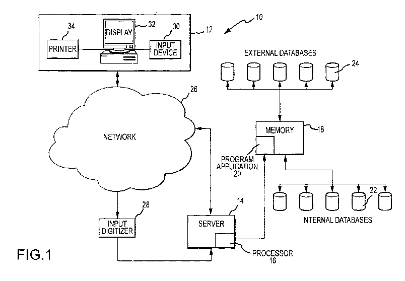
[0066] FIG. 1 illustrates a block diagram of an exemplary embodiment of a
system for determining
individualized medical intervention for a particular disease state that
utilizes molecular profiling of a
patient's biological specimen that is non disease specific.
[0067] FIG. 2 is a flowchart of an exemplary embodiment of a method for
determining individualized
medical intervention for a particular disease state that utilizes molecular
profiling of a patient's biological
specimen that is non disease specific.
[0068] FIGS. 3A through 3D illustrate an exemplary patient profile report in
accordance with step 80 of
FIG. 2.
[0069] FIG. 4 is a flowchart of an exemplary embodiment of a method for
identifying a drug
therapy/agent capable of interacting with a target.
[0070] FIGS. 5-14 are flowcharts and diagrams illustrating various parts of an
information-based
personalized medicine drug discovery system and method in accordance with the
present invention.
[0071] FIGS. 15-25 are computer screen print outs associated with various
parts of the information-
based personalized medicine drug discovery system and method shown in FIGS. 5-
14.
[0072] FIGs. 26A-D illustrate a molecular profiling service requisition using
a molecular profiling
approach as outlined in Tables 7-9 and 12-15, and accompanying text herein.
[0073] FIGs. 27A-V illustrate an exemplary patient report based on molecular
profiling for a patient
having a triple negative breast cancer.
[0074] FIG. 28 illustrates progression free survival (PFS) using therapy
selected by molecular profiling
(period B) with PFS for the most recent therapy on which the patient has just
progressed (period A). If
PFS(B) / PFS(A) ratio? 1.3, then molecular profiling selected therapy was
defined as having benefit for
patient.
[0075] FIG. 29 is a schematic of methods for identifying treatments by
molecular profiling if a target is
identified.
[0076] FIG. 30 illustrates the distribution of the patients in the study as
performed in Example 1.
[0077] FIG. 31 is graph depicting the results of the study with patients
having PFS ratio? 1.3 was 18/66
(27%).
[0078] FIG. 32 is a waterfall plot of all the patients for maximum % change of
summed diameters of
target lesions with respect to baseline diameter.
[0079] FIG. 33 illustrates the relationship between what clinician selected as
what she/he would use to
treat the patient before knowing what the molecular profiling results
suggested. There were no matches
for the 18 patients with PFS ratio? 1.3.
[0080] FIG. 34 is a schematic of the overall survival for the 18 patients with
PFS ratio? 1.3 versus all 66
patients.
-19-
CA 02978628 2017-09-01
WO 2016/141169 PCT/US2016/020657
[0081] FIG. 35 illustrates a molecular profiling system that performs analysis
of a cancer sample using a
variety of components that measure expression levels, chromosomal aberrations
and mutations. The
molecular "blueprint" of the cancer is used to generate a prioritized ranking
of druggable targets and/or
drug associated targets in tumor and their associated therapies.
[0082] FIG. 36 shows an example output of microarray profiling results and
calls made using a cutoff
value.
[0083] FIGs. 37A-F illustrate results of molecular profiling of a cohort of
126 Triple Negative (TN)
Metaplastic Breast Cancers.
[0084] FIG. 38 illustrates results of molecular profiling of PD1 and PDL1 in
HPV+ and HPV-/TP53
mutated head and neck squamous cell carcinomas.
[0085] FIGs. 39A-D illustrates a case of endometrial adenocarcinoma (FIG. 39A,
hematoxylin and eosin
stained section) exhibiting microsatellite instability caused by the loss of
MLH-1 protein [note retained
MLH-1 protein expression in the nuclei of the tumor infiltrating lymphocytes]
(FIG. 39B,
immunohistochemical stain); PD-1+ Tumor-infiltrating lymphocytes (FIG. 39C,
immunohistochemical
stain); aberrant expression of PD-Li in the tumor cells' basolateral membranes
(FIG. 39D,
immunohistochemical stain).
DETAILED DESCRIPTION OF THE INVENTION
[0086] The present invention provides methods and systems for identifying
therapeutic agents for use in
treatments on an individualized basis by using molecular profiling. The
molecular profiling approach
provides a method for selecting a candidate treatment for an individual that
could favorably change the
clinical course for the individual with a condition or disease, such as
cancer. The molecular profiling
approach provides clinical benefit for individuals, such as identifying drug
target(s) that provide a longer
progression free survival (PFS), longer disease free survival (DFS), longer
overall survival (OS) or
extended lifespan. Methods and systems of the invention are directed to
molecular profiling of cancer on
an individual basis that can provide alternatives for treatment that may be
convention or alternative to
conventional treatment regimens. For example, alternative treatment regimes
can be selected through
molecular profiling methods of the invention where, a disease is refractory to
current therapies, e.g., after
a cancer has developed resistance to a standard-of-care treatment.
Illustrative schemes for using molecular
profiling to identify a treatment regime are shown in FIGs. 2, 49A-B and 50,
each of which is described
in further detail herein. Thus, molecular profiling provides a personalized
approach to selecting candidate
treatments that are likely to benefit a cancer. In embodiments, the molecular
profiling method is used to
identify therapies for patients with poor prognosis, such as those with
metastatic disease or those whose
cancer has progressed on standard front line therapies, or whose cancer has
progressed on multiple
chemotherapeutic or hormonal regimens.
[0087] Personalized medicine based on pharmacogenetic insights, such as those
provided by molecular
profiling according to the invention, is increasingly taken for granted by
some practitioners and the lay
press, but forms the basis of hope for improved cancer therapy. However,
molecular profiling as taught
herein represents a fundamental departure from the traditional approach to
oncologic therapy where for
-20-
CA 02978628 2017-09-01
WO 2016/141169 PCT/US2016/020657
the most part, patients are grouped together and treated with approaches that
are based on findings from
light microscopy and disease stage. Traditionally, differential response to a
particular therapeutic strategy
has only been determined after the treatment was given, i.e. a posteriori. The
"standard" approach to
disease treatment relies on what is generally true about a given cancer
diagnosis and treatment response
has been vetted by randomized phase III clinical trials and forms the
"standard of care" in medical
practice. The results of these trials have been codified in consensus
statements by guidelines organizations
such as the National Comprehensive Cancer Network and The American Society of
Clinical Oncology.
The NCCN CompendiumTM contains authoritative, scientifically derived
information designed to support
decision-making about the appropriate use of drugs and biologics in patients
with cancer. The NCCN
CompendiumTM is recognized by the Centers for Medicare and Medicaid Services
(CMS) and United
Healthcare as an authoritative reference for oncology coverage policy. On-
compendium treatments are
those recommended by such guides. The biostatistical methods used to validate
the results of clinical trials
rely on minimizing differences between patients, and are based on declaring
the likelihood of error that
one approach is better than another for a patient group defined only by light
microscopy and stage, not by
individual differences in tumors. The molecular profiling methods of the
invention exploit such individual
differences. The methods can provide candidate treatments that can be then
selected by a physician for
treating a patient. In a study of such an approach presented in Example 1
herein, the results were
profound: in 66 consecutive patients, the treating oncologist never managed to
identify the molecular
target selected by the test, and 27% of patients whose treatment was guided by
molecular profiling
managed a remission 1.3x longer than their previous best response. At present,
such results are virtually
unheard of result in the salvage therapy setting.
[0088] Molecular profiling can be used to provide a comprehensive view of the
biological state of a
sample. In an embodiment, molecular profiling is used for whole tumor
profiling. Accordingly, a number
of molecular approaches are used to assess the state of a tumor. The whole
tumor profiling can be used for
selecting a candidate treatment for a tumor. Molecular profiling can be used
to select candidate
therapeutics on any sample for any stage of a disease. In embodiment, the
methods of the invention are
used to profile a newly diagnosed cancer. The candidate treatments indicated
by the molecular profiling
can be used to select a therapy for treating the newly diagnosed cancer. In
other embodiments, the
methods of the invention are used to profile a cancer that has already been
treated, e.g., with one or more
standard-of-care therapy. In embodiments, the cancer is refractory to the
prior treatment/s. For example,
the cancer may be refractory to the standard of care treatments for the
cancer. The cancer can be a
metastatic cancer or other recurrent cancer. The treatments can be on-
compendium or off-compendium
treatments.
[0089] Molecular profiling can be performed by any known means for detecting a
molecule in a biological
sample. Molecular profiling comprises methods that include but are not limited
to, nucleic acid sequencing, such as a
DNA sequencing or mRNA sequencing; immunohistochemistry (IHC); in situ
hybridization (ISH);
fluorescent in situ hybridization (FISH); chromogenic in situ hybridization
(CISH); PCR amplification
(e.g., qPCR or RT-PCR); various types of microarray (mRNA expression arrays,
low density arrays,
protein arrays, etc); various types of sequencing (Sanger, pyrosequencing,
etc); comparative genomic
-21-
CA 02978628 2017-09-01
WO 2016/141169 PCT/US2016/020657
hybridization (CGH); NextGen sequencing; Northern blot; Southern blot;
immunoassay; and any other
appropriate technique to assay the presence or quantity of a biological
molecule of interest. In various
embodiments of the invention, any one or more of these methods can be used
concurrently or subsequent
to each other for assessing target genes disclosed herein.
[0090] Molecular profiling of individual samples is used to select one or more
candidate treatments for a
disorder in a subject, e.g., by identifying targets for drugs that may be
effective for a given cancer. For
example, the candidate treatment can be a treatment known to have an effect on
cells that differentially
express genes as identified by molecular profiling techniques, an experimental
drug, a government or
regulatory approved drug or any combination of such drugs, which may have been
studied and approved
for a particular indication that is the same as or different from the
indication of the subject from whom a
biological sample is obtain and molecularly profiled.
[0091] When multiple biomarker targets are revealed by assessing target genes
by molecular profiling,
one or more decision rules can be put in place to prioritize the selection of
certain therapeutic agent for
treatment of an individual on a personalized basis. Rules of the invention
aide prioritizing treatment, e.g.,
direct results of molecular profiling, anticipated efficacy of therapeutic
agent, prior history with the same
or other treatments, expected side effects, availability of therapeutic agent,
cost of therapeutic agent, drug-
drug interactions, and other factors considered by a treating physician. Based
on the recommended and
prioritized therapeutic agent targets, a physician can decide on the course of
treatment for a particular
individual. Accordingly, molecular profiling methods and systems of the
invention can select candidate
treatments based on individual characteristics of diseased cells, e.g., tumor
cells, and other personalized
factors in a subject in need of treatment, as opposed to relying on a
traditional one-size fits all approach
that is conventionally used to treat individuals suffering from a disease,
especially cancer. In some cases,
the recommended treatments are those not typically used to treat the disease
or disorder inflicting the
subject. In some cases, the recommended treatments are used after standard-of-
care therapies are no
longer providing adequate efficacy.
[0092] The treating physician can use the results of the molecular profiling
methods to optimize a
treatment regimen for a patient. The candidate treatment identified by the
methods of the invention can be
used to treat a patient; however, such treatment is not required of the
methods. Indeed, the analysis of
molecular profiling results and identification of candidate treatments based
on those results can be
automated and does not require physician involvement.
Biological Entities
[0093] Nucleic acids include deoxyribonucleotides or ribonucleotides and
polymers thereof in either
single- or double-stranded form, or complements thereof Nucleic acids can
contain known nucleotide
analogs or modified backbone residues or linkages, which are synthetic,
naturally occurring, and non-
naturally occurring, which have similar binding properties as the reference
nucleic acid, and which are
metabolized in a manner similar to the reference nucleotides. Examples of such
analogs include, without
limitation, phosphorothioates, phosphoramidates, methyl phosphonates, chiral-
methyl phosphonates, 2-0-
methyl ribonucleotides, peptide-nucleic acids (PNAs). Nucleic acid sequence
can encompass
-22-
CA 02978628 2017-09-01
WO 2016/141169 PCT/US2016/020657
conservatively modified variants thereof (e.g., degenerate codon
substitutions) and complementary
sequences, as well as the sequence explicitly indicated. Specifically,
degenerate codon substitutions may
be achieved by generating sequences in which the third position of one or more
selected (or all) codons is
substituted with mixed-base and/or deoxyinosine residues (Batzer et al.,
Nucleic Acid Res. 19:5081
(1991); Ohtsuka et al., J. Biol. Chem. 260:2605-2608 (1985); Rossolini et al.,
Mol. Cell Probes 8:91-98
(1994)). The term nucleic acid can be used interchangeably with gene, cDNA,
mRNA, oligonucleotide,
and polynucleotide.
[0094] A particular nucleic acid sequence may implicitly encompass the
particular sequence and "splice
variants" and nucleic acid sequences encoding truncated forms. Similarly, a
particular protein encoded by
a nucleic acid can encompass any protein encoded by a splice variant or
truncated form of that nucleic
acid. "Splice variants," as the name suggests, are products of alternative
splicing of a gene. After
transcription, an initial nucleic acid transcript may be spliced such that
different (alternate) nucleic acid
splice products encode different polypeptides. Mechanisms for the production
of splice variants vary, but
include alternate splicing of exons. Alternate polypeptides derived from the
same nucleic acid by read-
through transcription are also encompassed by this definition. Any products of
a splicing reaction,
including recombinant forms of the splice products, are included in this
definition. Nucleic acids can be
truncated at the 5' end or at the 3' end. Polypeptides can be truncated at the
N-terminal end or the C-
terminal end. Truncated versions of nucleic acid or polypeptide sequences can
be naturally occurring or
created using recombinant techniques.
[0095] The terms "genetic variant" and "nucleotide variant" are used herein
interchangeably to refer to
changes or alterations to the reference human gene or cDNA sequence at a
particular locus, including, but
not limited to, nucleotide base deletions, insertions, inversions, and
substitutions in the coding and non-
coding regions. Deletions may be of a single nucleotide base, a portion or a
region of the nucleotide
sequence of the gene, or of the entire gene sequence. Insertions may be of one
or more nucleotide bases.
The genetic variant or nucleotide variant may occur in transcriptional
regulatory regions, untranslated
regions of mRNA, exons, introns, exon/intron junctions, etc. The genetic
variant or nucleotide variant can
potentially result in stop codons, frame shifts, deletions of amino acids,
altered gene transcript splice
forms or altered amino acid sequence.
[0096] An allele or gene allele comprises generally a naturally occurring gene
having a reference
sequence or a gene containing a specific nucleotide variant.
[0097] A haplotype refers to a combination of genetic (nucleotide) variants in
a region of an mRNA or a
genomic DNA on a chromosome found in an individual. Thus, a haplotype includes
a number of
genetically linked polymorphic variants which are typically inherited together
as a unit.
[0098] As used herein, the term "amino acid variant" is used to refer to an
amino acid change to a
reference human protein sequence resulting from genetic variants or nucleotide
variants to the reference
human gene encoding the reference protein. The term "amino acid variant" is
intended to encompass not
only single amino acid substitutions, but also amino acid deletions,
insertions, and other significant
changes of amino acid sequence in the reference protein.
-23-
CA 02978628 2017-09-01
WO 2016/141169 PCT/US2016/020657
[0099] The term "genotype" as used herein means the nucleotide characters at a
particular nucleotide
variant marker (or locus) in either one allele or both alleles of a gene (or a
particular chromosome region).
With respect to a particular nucleotide position of a gene of interest, the
nucleotide(s) at that locus or
equivalent thereof in one or both alleles form the genotype of the gene at
that locus. A genotype can be
homozygous or heterozygous. Accordingly, "genotyping" means determining the
genotype, that is, the
nucleotide(s) at a particular gene locus. Genotyping can also be done by
determining the amino acid
variant at a particular position of a protein which can be used to deduce the
corresponding nucleotide
variant(s).
[00100] The term "locus" refers to a specific position or site in a gene
sequence or protein. Thus, there
may be one or more contiguous nucleotides in a particular gene locus, or one
or more amino acids at a
particular locus in a polypeptide. Moreover, a locus may refer to a particular
position in a gene where one
or more nucleotides have been deleted, inserted, or inverted.
[00101] Unless specified otherwise or understood by one of skill in art, the
terms "polypeptide," "protein,"
and "peptide" are used interchangeably herein to refer to an amino acid chain
in which the amino acid
residues are linked by covalent peptide bonds. The amino acid chain can be of
any length of at least two
amino acids, including full-length proteins. Unless otherwise specified,
polypeptide, protein, and peptide
also encompass various modified forms thereof, including but not limited to
glycosylated forms,
phosphorylated forms, etc. A polypeptide, protein or peptide can also be
referred to as a gene product.
[00102] Lists of gene and gene products that can be assayed by molecular
profiling techniques are
presented herein. Lists of genes may be presented in the context of molecular
profiling techniques that
detect a gene product (e.g., an mRNA or protein). One of skill will understand
that this implies detection
of the gene product of the listed genes. Similarly, lists of gene products may
be presented in the context of
molecular profiling techniques that detect a gene sequence or copy number. One
of skill will understand
that this implies detection of the gene corresponding to the gene products,
including as an example DNA
encoding the gene products. As will be appreciated by those skilled in the
art, a "biomarker" or "marker"
comprises a gene and/or gene product depending on the context.
[00103] The terms "label" and "detectable label" can refer to any composition
detectable by spectroscopic,
photochemical, biochemical, immunochemical, electrical, optical, chemical or
similar methods. Such
labels include biotin for staining with labeled streptavidin conjugate,
magnetic beads (e.g.,
DYNABEADSTm), fluorescent dyes (e.g., fluorescein, Texas red, rhodamine, green
fluorescent protein,
and the like), radiolabels (e.g., 3H, 1251, 35s, 14,,u,
or 32P), enzymes (e.g., horse radish peroxidase, alkaline
phosphatase and others commonly used in an ELISA), and calorimetric labels
such as colloidal gold or
colored glass or plastic (e.g., polystyrene, polypropylene, latex, etc) beads.
Patents teaching the use of
such labels include U.S. Pat. Nos. 3,817,837; 3,850,752; 3,939,350; 3,996,345;
4,277,437; 4,275,149; and
4,366,241. Means of detecting such labels are well known to those of skill in
the art. Thus, for example,
radiolabels may be detected using photographic film or scintillation counters,
fluorescent markers may be
detected using a photodetector to detect emitted light. Enzymatic labels are
typically detected by
providing the enzyme with a substrate and detecting the reaction product
produced by the action of the
-24-
CA 02978628 2017-09-01
WO 2016/141169 PCT/US2016/020657
enzyme on the substrate, and calorimetric labels are detected by simply
visualizing the colored label.
Labels can include, e.g., ligands that bind to labeled antibodies,
fluorophores, chemiluminescent agents,
enzymes, and antibodies which can serve as specific binding pair members for a
labeled ligand. An
introduction to labels, labeling procedures and detection of labels is found
in Polak and Van Noorden
Introduction to Immunocytochemistry, 2nd ed., Springer Verlag, NY (1997); and
in Haugland Handbook
of Fluorescent Probes and Research Chemicals, a combined handbook and
catalogue Published by
Molecular Probes, Inc. (1996).
[00104] Detectable labels include, but are not limited to, nucleotides
(labeled or unlabelled), compomers,
sugars, peptides, proteins, antibodies, chemical compounds, conducting
polymers, binding moieties such
as biotin, mass tags, calorimetric agents, light emitting agents,
chemiluminescent agents, light scattering
agents, fluorescent tags, radioactive tags, charge tags (electrical or
magnetic charge), volatile tags and
hydrophobic tags, biomolecules (e.g., members of a binding pair
antibody/antigen, antibody/antibody,
antibody/antibody fragment, antibody/antibody receptor, antibody/protein A or
protein G, hapten/anti-
hapten, biotin/avidin, biotin/streptavidin, folic acid/folate binding protein,
vitamin B12/intrinsic factor,
chemical reactive group/complementary chemical reactive group (e.g.,
sulfhydryl/maleimide,
sulfhydryl/haloacetyl derivative, amine/isotriocyanate, amine/succinimidyl
ester, and amine/sulfonyl
halides) and the like.
[00105] The term "antibody" as used herein encompasses naturally occurring
antibodies as well as non-
naturally occurring antibodies, including, for example, single chain
antibodies, chimeric, bifunctional and
humanized antibodies, as well as antigen-binding fragments thereof, (e.g.,
Fab', F(ab1)2, Fab, Fv and rIgG).
See also, Pierce Catalog and Handbook, 1994-1995 (Pierce Chemical Co.,
Rockford, Ill.). See also, e.g.,
Kuby, J., Immunology, 3<sup>rd</sup> Ed., W. H. Freeman & Co., New York (1998). Such
non-naturally
occurring antibodies can be constructed using solid phase peptide synthesis,
can be produced
recombinantly or can be obtained, for example, by screening combinatorial
libraries consisting of variable
heavy chains and variable light chains as described by Huse et al., Science
246:1275-1281(1989), which
is incorporated herein by reference. These and other methods of making, for
example, chimeric,
humanized, CDR-grafted, single chain, and bifunctional antibodies are well
known to those skilled in the
art. See, e.g., Winter and Harris, Immunol. Today 14:243-246 (1993); Ward et
al., Nature 341:544-546
(1989); Harlow and Lane, Antibodies, 511-52, Cold Spring Harbor Laboratory
publications, New York,
1988; Hilyard et al., Protein Engineering: A practical approach (IRL Press
1992); Borrebaeck, Antibody
Engineering, 2d ed. (Oxford University Press 1995); each of which is
incorporated herein by reference.
[00106] Unless otherwise specified, antibodies can include both polyclonal and
monoclonal antibodies.
Antibodies also include genetically engineered forms such as chimeric
antibodies (e.g., humanized murine
antibodies) and heteroconjugate antibodies (e.g., bispecific antibodies). The
term also refers to
recombinant single chain Fv fragments (scFv). The term antibody also includes
bivalent or bispecific
molecules, diabodies, triabodies, and tetrabodies. Bivalent and bispecific
molecules are described in, e.g.,
Kostelny et al. (1992) J Immunol 148:1547, Pack and Pluckthun (1992)
Biochemistry 31:1579, Holliger et
al. (1993) Proc Natl Acad Sci USA. 90:6444, Gruber et al. (1994) J
Immuno1:5368, Zhu et al. (1997)
-25-
CA 02978628 2017-09-01
WO 2016/141169 PCT/US2016/020657
Protein Sci 6:781, Hu etal. (1997) Cancer Res. 56:3055, Adams etal. (1993)
Cancer Res. 53:4026, and
McCartney, etal. (1995) Protein Eng. 8:301.
[00107] Typically, an antibody has a heavy and light chain. Each heavy and
light chain contains a constant
region and a variable region, (the regions are also known as "domains"). Light
and heavy chain variable
regions contain four framework regions interrupted by three hyper-variable
regions, also called
complementarity-determining regions (CDRs). The extent of the framework
regions and CDRs have been
defined. The sequences of the framework regions of different light or heavy
chains are relatively
conserved within a species. The framework region of an antibody, that is the
combined framework regions
of the constituent light and heavy chains, serves to position and align the
CDRs in three dimensional
spaces. The CDRs are primarily responsible for binding to an epitope of an
antigen. The CDRs of each
chain are typically referred to as CDR1, CDR2, and CDR3, numbered sequentially
starting from the N-
terminus, and are also typically identified by the chain in which the
particular CDR is located. Thus, a VH
CDR3 is located in the variable domain of the heavy chain of the antibody in
which it is found, whereas a
Vi. CDR1 is the CDR1 from the variable domain of the light chain of the
antibody in which it is found.
References to VH refer to the variable region of an immunoglobulin heavy chain
of an antibody, including
the heavy chain of an Fv, scFv, or Fab. References to VL refer to the variable
region of an
immunoglobulin light chain, including the light chain of an Fv, scFv, dsFy or
Fab.
[00108] The phrase "single chain Fv" or "scFv" refers to an antibody in which
the variable domains of the
heavy chain and of the light chain of a traditional two chain antibody have
been joined to form one chain.
Typically, a linker peptide is inserted between the two chains to allow for
proper folding and creation of
an active binding site. A "chimeric antibody" is an immunoglobulin molecule in
which (a) the constant
region, or a portion thereof, is altered, replaced or exchanged so that the
antigen binding site (variable
region) is linked to a constant region of a different or altered class,
effector function and/or species, or an
entirely different molecule which confers new properties to the chimeric
antibody, e.g., an enzyme, toxin,
hormone, growth factor, drug, etc.; or (b) the variable region, or a portion
thereof, is altered, replaced or
exchanged with a variable region having a different or altered antigen
specificity.
[00109] A "humanized antibody" is an immunoglobulin molecule that contains
minimal sequence derived
from non-human immunoglobulin. Humanized antibodies include human
immunoglobulins (recipient
antibody) in which residues from a complementary determining region (CDR) of
the recipient are
replaced by residues from a CDR of a non-human species (donor antibody) such
as mouse, rat or rabbit
having the desired specificity, affinity and capacity. In some instances, Fv
framework residues of the
human immunoglobulin are replaced by corresponding non-human residues.
Humanized antibodies may
also comprise residues which are found neither in the recipient antibody nor
in the imported CDR or
framework sequences. In general, a humanized antibody will comprise
substantially all of at least one, and
typically two, variable domains, in which all or substantially all of the CDR
regions correspond to those
of a non-human immunoglobulin and all or substantially all of the framework
(FR) regions are those of a
human immunoglobulin consensus sequence. The humanized antibody optimally also
will comprise at
least a portion of an immunoglobulin constant region (Fc), typically that of a
human immunoglobulin
-26-
CA 02978628 2017-09-01
WO 2016/141169 PCT/US2016/020657
(Jones etal., Nature 321:522-525 (1986); Riechmann etal., Nature 332:323-327
(1988); and Presta, Curr.
Op. Struct. Biol. 2:593-596 (1992)). Humanization can be essentially performed
following the method of
Winter and co-workers (Jones etal., Nature 321:522-525 (1986); Riechmann
etal., Nature 332:323-327
(1988); Verhoeyen et al., Science 239:1534-1536 (1988)), by substituting
rodent CDRs or CDR sequences
for the corresponding sequences of a human antibody. Accordingly, such
humanized antibodies are
chimeric antibodies (U.S. Pat. No. 4,816,567), wherein substantially less than
an intact human variable
domain has been substituted by the corresponding sequence from a non-human
species.
[00110] The terms "epitope" and "antigenic determinant" refer to a site on an
antigen to which an antibody
binds. Epitopes can be formed both from contiguous amino acids or
noncontiguous amino acids
juxtaposed by tertiary folding of a protein. Epitopes formed from contiguous
amino acids are typically
retained on exposure to denaturing solvents whereas epitopes formed by
tertiary folding are typically lost
on treatment with denaturing solvents. An epitope typically includes at least
3, and more usually, at least 5
or 8-10 amino acids in a unique spatial conformation. Methods of determining
spatial conformation of
epitopes include, for example, x-ray crystallography and 2-dimensional nuclear
magnetic resonance. See,
e.g., Epitope Mapping Protocols in Methods in Molecular Biology, Vol. 66,
Glenn E. Morris, Ed (1996).
[00111] The terms "primer", "probe," and "oligonucleotide" are used herein
interchangeably to refer to a
relatively short nucleic acid fragment or sequence. They can comprise DNA,
RNA, or a hybrid thereof, or
chemically modified analog or derivatives thereof Typically, they are single-
stranded. However, they can
also be double-stranded having two complementing strands which can be
separated by denaturation.
Normally, primers, probes and oligonucleotides have a length of from about 8
nucleotides to about 200
nucleotides, preferably from about 12 nucleotides to about 100 nucleotides,
and more preferably about 18
to about 50 nucleotides. They can be labeled with detectable markers or
modified using conventional
manners for various molecular biological applications.
[00112] The term "isolated" when used in reference to nucleic acids (e.g.,
genomic DNAs, cDNAs,
mRNAs, or fragments thereof) is intended to mean that a nucleic acid molecule
is present in a form that is
substantially separated from other naturally occurring nucleic acids that are
normally associated with the
molecule. Because a naturally existing chromosome (or a viral equivalent
thereof) includes a long nucleic
acid sequence, an isolated nucleic acid can be a nucleic acid molecule having
only a portion of the nucleic
acid sequence in the chromosome but not one or more other portions present on
the same chromosome.
More specifically, an isolated nucleic acid can include naturally occurring
nucleic acid sequences that
flank the nucleic acid in the naturally existing chromosome (or a viral
equivalent thereof). An isolated
nucleic acid can be substantially separated from other naturally occurring
nucleic acids that are on a
different chromosome of the same organism. An isolated nucleic acid can also
be a composition in which
the specified nucleic acid molecule is significantly enriched so as to
constitute at least 10%, 20%, 30%,
40%, 50%, 60%, 70%, 80%, 90%, 95%, or at least 99% of the total nucleic acids
in the composition.
[00113] An isolated nucleic acid can be a hybrid nucleic acid having the
specified nucleic acid molecule
covalently linked to one or more nucleic acid molecules that are not the
nucleic acids naturally flanking
the specified nucleic acid. For example, an isolated nucleic acid can be in a
vector. In addition, the
-27-
CA 02978628 2017-09-01
WO 2016/141169 PCT/US2016/020657
specified nucleic acid may have a nucleotide sequence that is identical to a
naturally occurring nucleic
acid or a modified form or mutein thereof having one or more mutations such as
nucleotide substitution,
deletion/insertion, inversion, and the like.
[00114] An isolated nucleic acid can be prepared from a recombinant host cell
(in which the nucleic acids
have been recombinantly amplified and/or expressed), or can be a chemically
synthesized nucleic acid
having a naturally occurring nucleotide sequence or an artificially modified
form thereof
[00115] The term "isolated polypeptide" as used herein is defined as a
polypeptide molecule that is present
in a form other than that found in nature. Thus, an isolated polypeptide can
be a non-naturally occurring
polypeptide. For example, an isolated polypeptide can be a "hybrid
polypeptide." An isolated polypeptide
can also be a polypeptide derived from a naturally occurring polypeptide by
additions or deletions or
substitutions of amino acids. An isolated polypeptide can also be a "purified
polypeptide" which is used
herein to mean a composition or preparation in which the specified polypeptide
molecule is significantly
enriched so as to constitute at least 10% of the total protein content in the
composition. A "purified
polypeptide" can be obtained from natural or recombinant host cells by
standard purification techniques,
or by chemically synthesis, as will be apparent to skilled artisans.
[00116] The terms "hybrid protein," "hybrid polypeptide," "hybrid peptide,"
"fusion protein," "fusion
polypeptide," and "fusion peptide" are used herein interchangeably to mean a
non-naturally occurring
polypeptide or isolated polypeptide having a specified polypeptide molecule
covalently linked to one or
more other polypeptide molecules that do not link to the specified polypeptide
in nature. Thus, a "hybrid
protein" may be two naturally occurring proteins or fragments thereof linked
together by a covalent
linkage. A "hybrid protein" may also be a protein formed by covalently linking
two artificial polypeptides
together. Typically but not necessarily, the two or more polypeptide molecules
are linked or "fused"
together by a peptide bond forming a single non-branched polypeptide chain.
[00117] The term "high stringency hybridization conditions," when used in
connection with nucleic acid
hybridization, includes hybridization conducted overnight at 42 C in a
solution containing 50%
formamide, 5x SSC (750 mM NaC1, 75 mM sodium citrate), 50 mM sodium phosphate,
pH 7.6,
x Denhardt's solution, 10% dextran sulfate, and 20 microgram/ml denatured and
sheared salmon sperm
DNA, with hybridization filters washed in 0.1x SSC at about 65 C. The term
"moderate stringent
hybridization conditions," when used in connection with nucleic acid
hybridization, includes hybridization
conducted overnight at 37 C in a solution containing 50% formamide, 5x SSC
(750 mM NaC1, 75 mM
sodium citrate), 50 mM sodium phosphate, pH 7.6, 5 xDenhardt's solution, 10%
dextran sulfate, and 20
microgram/ml denatured and sheared salmon sperm DNA, with hybridization
filters washed in lx SSC at
about 50 C. It is noted that many other hybridization methods, solutions and
temperatures can be used to
achieve comparable stringent hybridization conditions as will be apparent to
skilled artisans.
[00118] For the purpose of comparing two different nucleic acid or polypeptide
sequences, one sequence
(test sequence) may be described to be a specific percentage identical to
another sequence (comparison
sequence). The percentage identity can be determined by the algorithm of
Karlin and Altschul, Proc. Natl.
Acad. Sci. USA, 90:5873-5877 (1993), which is incorporated into various BLAST
programs. The
-28-
CA 02978628 2017-09-01
WO 2016/141169 PCT/US2016/020657
percentage identity can be determined by the "BLAST 2 Sequences" tool, which
is available at the
National Center for Biotechnology Information (NCBI) website. See Tatusova and
Madden, FEMS
Microbiol. Lett., 174(2):247-250 (1999). For pairwise DNA-DNA comparison, the
BLASTN program is
used with default parameters (e.g., Match: 1; Mismatch: -2; Open gap: 5
penalties; extension gap: 2
penalties; gap x_dropoff: 50; expect: 10; and word size: 11, with filter). For
pairwise protein-protein
sequence comparison, the BLASTP program can be employed using default
parameters (e.g., Matrix:
BLOSUM62; gap open: 11; gap extension: 1; x_dropoff: 15; expect: 10.0; and
wordsize: 3, with filter).
Percent identity of two sequences is calculated by aligning a test sequence
with a comparison sequence
using BLAST, determining the number of amino acids or nucleotides in the
aligned test sequence that are
identical to amino acids or nucleotides in the same position of the comparison
sequence, and dividing the
number of identical amino acids or nucleotides by the number of amino acids or
nucleotides in the
comparison sequence. When BLAST is used to compare two sequences, it aligns
the sequences and yields
the percent identity over defined, aligned regions. If the two sequences are
aligned across their entire
length, the percent identity yielded by the BLAST is the percent identity of
the two sequences. If BLAST
does not align the two sequences over their entire length, then the number of
identical amino acids or
nucleotides in the unaligned regions of the test sequence and comparison
sequence is considered to be
zero and the percent identity is calculated by adding the number of identical
amino acids or nucleotides in
the aligned regions and dividing that number by the length of the comparison
sequence. Various versions
of the BLAST programs can be used to compare sequences, e.g., BLAST 2.1.2 or
BLAST+ 2.2.22.
[00119] A subject or individual can be any animal which may benefit from the
methods of the invention,
including, e.g., humans and non-human mammals, such as primates, rodents,
horses, dogs and cats.
Subjects include without limitation a eukaryotic organisms, most preferably a
mammal such as a primate,
e.g., chimpanzee or human, cow; dog; cat; a rodent, e.g., guinea pig, rat,
mouse; rabbit; or a bird; reptile;
or fish. Subjects specifically intended for treatment using the methods
described herein include humans. A
subject may be referred to as an individual or a patient.
[00120] Treatment of a disease or individual according to the invention is an
approach for obtaining
beneficial or desired medical results, including clinical results, but not
necessarily a cure. For purposes of
this invention, beneficial or desired clinical results include, but are not
limited to, alleviation or
amelioration of one or more symptoms, diminishment of extent of disease,
stabilized (i.e., not worsening)
state of disease, preventing spread of disease, delay or slowing of disease
progression, amelioration or
palliation of the disease state, and remission (whether partial or total),
whether detectable or undetectable.
Treatment also includes prolonging survival as compared to expected survival
if not receiving treatment
or if receiving a different treatment. A treatment can include administration
of a therapeutic agent, which
can be an agent that exerts a cytotoxic, cytostatic, or immunomodulatory
effect on diseased cells, e.g.,
cancer cells, or other cells that may promote a diseased state, e.g.,
activated immune cells. Therapeutic
agents selected by the methods of the invention are not limited. Any
therapeutic agent can be selected
where a link can be made between molecular profiling and potential efficacy of
the agent. Therapeutic
agents include without limitation drugs, pharmaceuticals, small molecules,
protein therapies, antibody
-29-
CA 02978628 2017-09-01
WO 2016/141169 PCT/US2016/020657
therapies, viral therapies, gene therapies, and the like. Cancer treatments or
therapies include apoptosis-
mediated and non-apoptosis mediated cancer therapies including, without
limitation, chemotherapy,
hormonal therapy, radiotherapy, immunotherapy, and combinations thereof.
Chemotherapeutic agents
comprise therapeutic agents and combinations of therapeutic agents that treat,
cancer cells, e.g., by killing
those cells. Examples of different types of chemotherapeutic drugs include
without limitation alkylating
agents (e.g., nitrogen mustard derivatives, ethylenimines, alkylsulfonates,
hydrazines and triazines,
nitrosureas, and metal salts), plant alkaloids (e.g., vinca alkaloids,
taxanes, podophyllotoxins, and
camptothecan analogs), antitumor antibiotics (e.g., anthracyclines,
chromomycins, and the like),
antimetabolites (e.g., folic acid antagonists, pyrimidine antagonists, purine
antagonists, and adenosine
deaminase inhibitors), topoisomerase I inhibitors, topoisomerase II
inhibitors, and miscellaneous
antineoplastics (e.g., ribonucleotide reductase inhibitors, adrenocortical
steroid inhibitors, enzymes,
antimicrotubule agents, and retinoids).
[00121] A biomarker refers generally to a molecule, including without
limitation a gene or product
thereof, nucleic acids (e.g., DNA, RNA), protein/peptide/polypeptide,
carbohydrate structure, lipid,
glycolipid, characteristics of which can be detected in a tissue or cell to
provide information that is
predictive, diagnostic, prognostic and/or theranostic for sensitivity or
resistance to candidate treatment.
Biological Samples
[00122] A sample as used herein includes any relevant biological sample that
can be used for molecular
profiling, e.g., sections of tissues such as biopsy or tissue removed during
surgical or other procedures,
bodily fluids, autopsy samples, and frozen sections taken for histological
purposes. Such samples include
blood and blood fractions or products (e.g., serum, buffy coat, plasma,
platelets, red blood cells, and the
like), sputum, malignant effusion, cheek cells tissue, cultured cells (e.g.,
primary cultures, explants, and
transformed cells), stool, urine, other biological or bodily fluids (e.g.,
prostatic fluid, gastric fluid,
intestinal fluid, renal fluid, lung fluid, cerebrospinal fluid, and the like),
etc. The sample can comprise
biological material that is a fresh frozen & formalin fixed paraffin embedded
(FFPE) block, formalin-
fixed paraffin embedded, or is within an RNA preservative + formalin fixative.
More than one sample of
more than one type can be used for each patient. In a preferred embodiment,
the sample comprises a fixed
tumor sample.
[00123] The sample used in the methods described herein can be a formalin
fixed paraffin embedded
(FFPE) sample. The FFPE sample can be one or more of fixed tissue, unstained
slides, bone marrow core
or clot, core needle biopsy, malignant fluids and fine needle aspirate (FNA).
In an embodiment, the fixed
tissue comprises a tumor containing formalin fixed paraffin embedded (FFPE)
block from a surgery or
biopsy. In another embodiment, the unstained slides comprise unstained,
charged, unbaked slides from a
paraffin block. In another embodiment, bone marrow core or clot comprises a
decalcified core. A formalin
fixed core and/or clot can be paraffin-embedded. In still another embodiment,
the core needle biopsy
comprises 1, 2, 3, 4, 5, 6, 7, 8, 9, 10 or more, e.g., 3-4, paraffin embedded
biopsy samples. An 18 gauge
needle biopsy can be used. The malignant fluid can comprise a sufficient
volume of fresh pleural/ascitic
fluid to produce a 5x5x2mm cell pellet. The fluid can be formalin fixed in a
paraffin block. In an
-30-
CA 02978628 2017-09-01
WO 2016/141169 PCT/US2016/020657
embodiment, the core needle biopsy comprises 1, 2, 3, 4, 5, 6, 7, 8, 9, 10 or
more, e.g., 4-6, paraffin
embedded aspirates.
[00124] A sample may be processed according to techniques understood by those
in the art. A sample can
be without limitation fresh, frozen or fixed cells or tissue. In some
embodiments, a sample comprises
formalin-fixed paraffin-embedded (FFPE) tissue, fresh tissue or fresh frozen
(FF) tissue. A sample can
comprise cultured cells, including primary or immortalized cell lines derived
from a subject sample. A
sample can also refer to an extract from a sample from a subject. For example,
a sample can comprise
DNA, RNA or protein extracted from a tissue or a bodily fluid. Many techniques
and commercial kits are
available for such purposes. The fresh sample from the individual can be
treated with an agent to preserve
RNA prior to further processing, e.g., cell lysis and extraction. Samples can
include frozen samples
collected for other purposes. Samples can be associated with relevant
information such as age, gender, and
clinical symptoms present in the subject; source of the sample; and methods of
collection and storage of
the sample. A sample is typically obtained from a subject.
[00125] A biopsy comprises the process of removing a tissue sample for
diagnostic or prognostic
evaluation, and to the tissue specimen itself Any biopsy technique known in
the art can be applied to the
molecular profiling methods of the present invention. The biopsy technique
applied can depend on the
tissue type to be evaluated (e.g., colon, prostate, kidney, bladder, lymph
node, liver, bone marrow, blood
cell, lung, breast, etc.), the size and type of the tumor (e.g., solid or
suspended, blood or ascites), among
other factors. Representative biopsy techniques include, but are not limited
to, excisional biopsy,
incisional biopsy, needle biopsy, surgical biopsy, and bone marrow biopsy. An
"excisional biopsy" refers
to the removal of an entire tumor mass with a small margin of normal tissue
surrounding it. An "incisional
biopsy" refers to the removal of a wedge of tissue that includes a cross-
sectional diameter of the tumor.
Molecular profiling can use a "core-needle biopsy" of the tumor mass, or a
"fine-needle aspiration biopsy"
which generally obtains a suspension of cells from within the tumor mass.
Biopsy techniques are
discussed, for example, in Harrison's Principles of Internal Medicine, Kasper,
et al., eds., 16th ed., 2005,
Chapter 70, and throughout Part V.
[00126] Standard molecular biology techniques known in the art and not
specifically described are
generally followed as in Sambrook et al., Molecular Cloning: A Laboratory
Manual, Cold Spring Harbor
Laboratory Press, New York (1989), and as in Ausubel et al., Current Protocols
in Molecular Biology,
John Wiley and Sons, Baltimore, Md. (1989) and as in Perbal, A Practical Guide
to Molecular Cloning,
John Wiley & Sons, New York (1988), and as in Watson et al., Recombinant DNA,
Scientific American
Books, New York and in Birren et al (eds) Genome Analysis: A Laboratory Manual
Series, Vols. 1-4
Cold Spring Harbor Laboratory Press, New York (1998) and methodology as set
forth in U.S. Pat. Nos.
4,666,828; 4,683,202; 4,801,531; 5,192,659 and 5,272,057 and incorporated
herein by reference.
Polymerase chain reaction (PCR) can be carried out generally as in PCR
Protocols: A Guide to Methods
and Applications, Academic Press, San Diego, Calif. (1990).
-31-
CA 02978628 2017-09-01
WO 2016/141169 PCT/US2016/020657
Vesicles
[00127] The sample can comprise vesicles. Methods of the invention can include
assessing one or more
vesicles, including assessing vesicle populations. A vesicle, as used herein,
is a membrane vesicle that is
shed from cells. Vesicles or membrane vesicles include without limitation:
circulating microvesicles
(cMVs), microvesicle, exosome, nanovesicle, dexosome, bleb, blebby,
prostasome, microparticle,
intralumenal vesicle, membrane fragment, intralumenal endosomal vesicle,
endosomal-like vesicle,
exocytosis vehicle, endosome vesicle, endosomal vesicle, apoptotic body,
multivesicular body, secretory
vesicle, phospholipid vesicle, liposomal vesicle, argosome, texasome,
secresome, tolerosome,
melanosome, oncosome, or exocytosed vehicle. Furthermore, although vesicles
may be produced by
different cellular processes, the methods of the invention are not limited to
or reliant on any one
mechanism, insofar as such vesicles are present in a biological sample and are
capable of being
characterized by the methods disclosed herein. Unless otherwise specified,
methods that make use of a
species of vesicle can be applied to other types of vesicles. Vesicles
comprise spherical structures with a
lipid bilayer similar to cell membranes which surrounds an inner compartment
which can contain soluble
components, sometimes referred to as the payload. In some embodiments, the
methods of the invention
make use of exosomes, which are small secreted vesicles of about 40-100 nm in
diameter. For a review of
membrane vesicles, including types and characterizations, see Thery et al.,
Nat Rev Immunol. 2009
Aug;9(8): 581-93. Some properties of different types of vesicles include those
in Table 1:
Table 1: Vesicle Properties
Feature Exosomes Microvesicl Ectosomes Membrane Exosome- Apoptotic
es particles like
vesicles
vesicles
Size 50-100 nm 100-1,000 50-200 nm 50-80 nm 20-50 nm 50-
500 nm
nm
Density in 1.13-1.19g/ml 1.04-1.07 1.1 giml 1.16-
1.28
sucrose giml giml
EM Cup shape Irregular Bilamellar Round Irregular
Heterogeneo
appearance shape, round shape us
electron structures
dense
Sedimentati 100,000 g 10,000 g 160,000- 100,000- 175,000
g 1,200 g,
on 200,000 g 200,000 g 10,000
g,
100,000 g
Lipid Enriched in Expose PPS Enriched in No lipid
composition cholesterol, cholesterol rafts
sphingomyelin and
and ceramide; diacylglycero
contains lipid 1; expose PPS
rafts; expose
PPS
Major Tetraspanins Integrins, CR1 and
CD133; no TNFRI Histones
protein (e.g., CD63, selectins and proteolytic CD63
markers CD9), Alix, CD40 ligand enzymes; no
TSG101 CD63
Intracellular Internal Plasma Plasma Plasma
origin compartments membrane membrane membrane
(endosomes)
Abbreviations: phosphatidylserine (PPS); electron microscopy (EM)
-32-
CA 02978628 2017-09-01
WO 2016/141169 PCT/US2016/020657
[00128] Vesicles include shed membrane bound particles, or "microparticles,"
that are derived from either
the plasma membrane or an internal membrane. Vesicles can be released into the
extracellular
environment from cells. Cells releasing vesicles include without limitation
cells that originate from, or are
derived from, the ectoderm, endoderm, or mesoderm. The cells may have
undergone genetic,
environmental, and/or any other variations or alterations. For example, the
cell can be tumor cells. A
vesicle can reflect any changes in the source cell, and thereby reflect
changes in the originating cells, e.g.,
cells having various genetic mutations. In one mechanism, a vesicle is
generated intracellularly when a
segment of the cell membrane spontaneously invaginates and is ultimately
exocytosed (see for example,
Keller et al., Immunol. Lett. 107 (2): 102-8 (2006)). Vesicles also include
cell-derived structures bounded
by a lipid bilayer membrane arising from both herniated evagination (blebbing)
separation and sealing of
portions of the plasma membrane or from the export of any intracellular
membrane-bounded vesicular
structure containing various membrane-associated proteins of tumor origin,
including surface-bound
molecules derived from the host circulation that bind selectively to the tumor-
derived proteins together
with molecules contained in the vesicle lumen, including but not limited to
tumor-derived microRNAs or
intracellular proteins. Blebs and blebbing are further described in Charras et
al., Nature Reviews
Molecular and Cell Biology, Vol. 9, No. 11, p. 730-736 (2008). A vesicle shed
into circulation or bodily
fluids from tumor cells may be referred to as a "circulating tumor-derived
vesicle." When such vesicle is
an exosome, it may be referred to as a circulating-tumor derived exosome
(CTE). In some instances, a
vesicle can be derived from a specific cell of origin. CTE, as with a cell-of-
origin specific vesicle,
typically have one or more unique biomarkers that permit isolation of the CTE
or cell-of-origin specific
vesicle, e.g., from a bodily fluid and sometimes in a specific manner. For
example, a cell or tissue specific
markers are used to identify the cell of origin. Examples of such cell or
tissue specific markers are
disclosed herein and can further be accessed in the Tissue-specific Gene
Expression and Regulation
(TiGER) Database, available at bioinfo.wilmerjhu.edu/tiged; Liu et al. (2008)
TiGER: a database for
tissue-specific gene expression and regulation. BMC Bioinformatics. 9:271;
TissueDistributionDBs,
available at genome.dkfz-heidelberg.de/menu/tissue_db/index.html.
[00129] A vesicle can have a diameter of greater than about 10 nm, 20 nm, or
30 nm. A vesicle can have a
diameter of greater than 40 nm, 50 nm, 100 nm, 200 nm, 500 nm, 1000 nm or
greater than 10,000 nm. A
vesicle can have a diameter of about 30-1000 nm, about 30-800 nm, about 30-200
nm, or about 30-100
nm. In some embodiments, the vesicle has a diameter of less than 10,000 nm,
1000 nm, 800 nm, 500 nm,
200 nm, 100 nm, 50 nm, 40 nm, 30 nm, 20 nm or less than 10 nm. As used herein
the term "about" in
reference to a numerical value means that variations of 10% above or below the
numerical value are
within the range ascribed to the specified value. Typical sizes for various
types of vesicles are shown in
Table 1. Vesicles can be assessed to measure the diameter of a single vesicle
or any number of vesicles.
For example, the range of diameters of a vesicle population or an average
diameter of a vesicle population
can be determined. Vesicle diameter can be assessed using methods known in the
art, e.g., imaging
technologies such as electron microscopy. In an embodiment, a diameter of one
or more vesicles is
determined using optical particle detection. See, e.g., U.S. Patent 7,751,053,
entitled "Optical Detection
-33-
CA 02978628 2017-09-01
WO 2016/141169 PCT/US2016/020657
and Analysis of Particles" and issued July 6, 2010; and U.S. Patent 7,399,600,
entitled "Optical Detection
and Analysis of Particles" and issued July 15, 2010.
[00130] In some embodiments, vesicles are directly assayed from a biological
sample without prior
isolation, purification, or concentration from the biological sample. For
example, the amount of vesicles in
the sample can by itself provide a biosignature that provides a diagnostic,
prognostic or theranostic
determination. Alternatively, the vesicle in the sample may be isolated,
captured, purified, or concentrated
from a sample prior to analysis. As noted, isolation, capture or purification
as used herein comprises
partial isolation, partial capture or partial purification apart from other
components in the sample. Vesicle
isolation can be performed using various techniques as described herein or
known in the art, including
without limitation size exclusion chromatography, density gradient
centrifugation, differential
centrifugation, nanomembrane ultrafiltration, immunoabsorbent capture,
affinity purification, affinity
capture, immunoassay, immunoprecipitation, microfluidic separation, flow
cytometry or combinations
thereof
[00131] Vesicles can be assessed to provide a phenotypic characterization by
comparing vesicle
characteristics to a reference. In some embodiments, surface antigens on a
vesicle are assessed. A vesicle
or vesicle population carrying a specific marker can be referred to as a
positive (biomarker+) vesicle or
vesicle population. For example, a DLL4+ population refers to a vesicle
population associated with
DLL4. Conversely, a DLL4- population would not be associated with DLL4. The
surface antigens can
provide an indication of the anatomical origin and/or cellular of the vesicles
and other phenotypic
information, e.g., tumor status. For example, vesicles found in a patient
sample can be assessed for surface
antigens indicative of colorectal origin and the presence of cancer, thereby
identifying vesicles associated
with colorectal cancer cells. The surface antigens may comprise any
informative biological entity that can
be detected on the vesicle membrane surface, including without limitation
surface proteins, lipids,
carbohydrates, and other membrane components. For example, positive detection
of colon derived
vesicles expressing tumor antigens can indicate that the patient has
colorectal cancer. As such, methods of
the invention can be used to characterize any disease or condition associated
with an anatomical or
cellular origin, by assessing, for example, disease-specific and cell-specific
biomarkers of one or more
vesicles obtained from a subject.
[00132] In embodiments, one or more vesicle payloads are assessed to provide a
phenotypic
characterization. The payload with a vesicle comprises any informative
biological entity that can be
detected as encapsulated within the vesicle, including without limitation
proteins and nucleic acids, e.g.,
genomic or cDNA, mRNA, or functional fragments thereof, as well as microRNAs
(miRs). In addition,
methods of the invention are directed to detecting vesicle surface antigens
(in addition or exclusive to
vesicle payload) to provide a phenotypic characterization. For example,
vesicles can be characterized by
using binding agents (e.g., antibodies or aptamers) that are specific to
vesicle surface antigens, and the
bound vesicles can be further assessed to identify one or more payload
components disclosed therein. As
described herein, the levels of vesicles with surface antigens of interest or
with payload of interest can be
compared to a reference to characterize a phenotype. For example,
overexpression in a sample of cancer-
-34-
CA 02978628 2017-09-01
WO 2016/141169 PCT/US2016/020657
related surface antigens or vesicle payload, e.g., a tumor associated mRNA or
microRNA, as compared to
a reference, can indicate the presence of cancer in the sample. The biomarkers
assessed can be present or
absent, increased or reduced based on the selection of the desired target
sample and comparison of the
target sample to the desired reference sample. Non-limiting examples of target
samples include: disease;
treated/not-treated; different time points, such as a in a longitudinal study;
and non-limiting examples of
reference sample: non-disease; normal; different time points; and sensitive or
resistant to candidate
treatment(s).
[00133] In an embodiment, molecular profiling of the invention comprises
analysis of microvesicles, such
as circulating microvesicles.
Micr oRNA
[00134] Various biomarker molecules can be assessed in biological samples or
vesicles obtained from
such biological samples. MicroRNAs comprise one class biomarkers assessed via
methods of the
invention. MicroRNAs, also referred to herein as miRNAs or miRs, are short RNA
strands approximately
21-23 nucleotides in length. MiRNAs are encoded by genes that are transcribed
from DNA but are not
translated into protein and thus comprise non-coding RNA. The miRs are
processed from primary
transcripts known as pri-miRNA to short stem-loop structures called pre-miRNA
and finally to the
resulting single strand miRNA. The pre-miRNA typically forms a structure that
folds back on itself in
self-complementary regions. These structures are then processed by the
nuclease Dicer in animals or
DCL1 in plants. Mature miRNA molecules are partially complementary to one or
more messenger RNA
(mRNA) molecules and can function to regulate translation of proteins.
Identified sequences of miRNA
can be accessed at publicly available databases, such as www.microRNA.org,
www.mirbase.org, or
www.mirz.unibas.ch/cgi/miRNA.cgi.
[00135] miRNAs are generally assigned a number according to the naming
convention " mir-[number]."
The number of a miRNA is assigned according to its order of discovery relative
to previously identified
miRNA species. For example, if the last published miRNA was mir-121, the next
discovered miRNA will
be named mir-122, etc. When a miRNA is discovered that is homologous to a
known miRNA from a
different organism, the name can be given an optional organism identifier, of
the form [organism
identifierl- mir-[number]. Identifiers include hsa for Homo sapiens and mmu
for Mus Musculus. For
example, a human homolog to mir-121 might be referred to as hsa-mir-121
whereas the mouse homolog
can be referred to as mmu-mir-121.
[00136] Mature microRNA is commonly designated with the prefix "miR" whereas
the gene or precursor
miRNA is designated with the prefix "mir." For example, mir-121 is a precursor
for miR-121. When
differing miRNA genes or precursors are processed into identical mature
miRNAs, the genes/precursors
can be delineated by a numbered suffix. For example, mir-121-1 and mir-121-2
can refer to distinct genes
or precursors that are processed into miR-121. Lettered suffixes are used to
indicate closely related mature
sequences. For example, mir-121a and mir-121b can be processed to closely
related miRNAs miR-121a
and miR-121b, respectively. In the context of the invention, any microRNA
(miRNA or miR) designated
-35-
CA 02978628 2017-09-01
WO 2016/141169 PCT/US2016/020657
herein with the prefix mir-* or miR-* is understood to encompass both the
precursor and/or mature
species, unless otherwise explicitly stated otherwise.
[00137] Sometimes it is observed that two mature miRNA sequences originate
from the same precursor.
When one of the sequences is more abundant that the other, a "*" suffix can be
used to designate the less
common variant. For example, miR-121 would be the predominant product whereas
miR-121* is the less
common variant found on the opposite arm of the precursor. If the predominant
variant is not identified,
the miRs can be distinguished by the suffix "Sp" for the variant from the 5'
arm of the precursor and the
suffix "3p" for the variant from the 3' arm. For example, miR-121-5p
originates from the 5' arm of the
precursor whereas miR-121-3p originates from the 3' arm. Less commonly, the 5p
and 3p variants are
referred to as the sense ("s") and anti-sense ("as") forms, respectively. For
example, miR-121-5p may be
referred to as miR-121-s whereas miR-121-3p may be referred to as miR-121-as.
[00138] The above naming conventions have evolved over time and are general
guidelines rather than
absolute rules. For example, the let- and lin- families of miRNAs continue to
be referred to by these
monikers. The mir/miR convention for precursor/mature forms is also a
guideline and context should be
taken into account to determine which form is referred to. Further details of
miR naming can be found at
www.mirbase.org or Ambros et al., A uniform system for microRNA annotation,
RNA 9:277-279 (2003).
[00139] Plant miRNAs follow a different naming convention as described in
Meyers et al., Plant Cell.
2008 20(12):3186-3190.
1001401A number of miRNAs are involved in gene regulation, and miRNAs are part
of a growing class of
non-coding RNAs that is now recognized as a major tier of gene control. In
some cases, miRNAs can
interrupt translation by binding to regulatory sites embedded in the 3'-UTRs
of their target mRNAs,
leading to the repression of translation. Target recognition involves
complementary base pairing of the
target site with the miRNA's seed region (positions 2-8 at the miRNA's 5'
end), although the exact extent
of seed complementarity is not precisely determined and can be modified by 3'
pairing. In other cases,
miRNAs function like small interfering RNAs (siRNA) and bind to perfectly
complementary mRNA
sequences to destroy the target transcript.
[00141] Characterization of a number of miRNAs indicates that they influence a
variety of processes,
including early development, cell proliferation and cell death, apoptosis and
fat metabolism. For example,
some miRNAs, such as lin-4, let-7, mir-14, mir-23, and bantam, have been shown
to play critical roles in
cell differentiation and tissue development. Others are believed to have
similarly important roles because
of their differential spatial and temporal expression patterns.
[00142] The miRNA database available at miRBase (www.mirbase.org) comprises a
searchable database
of published miRNA sequences and annotation. Further information about miRBase
can be found in the
following articles, each of which is incorporated by reference in its entirety
herein: Griffiths-Jones et al.,
miRBase: tools for microRNA genomics. NAR 2008 36(Database Issue):D154-D158;
Griffiths-Jones et
al., miRBase: microRNA sequences, targets and gene nomenclature. NAR 2006
34(Database
Issue):D140-D144; and Griffiths-Jones, S. The microRNA Registry. NAR 2004
32(Database
-36-
CA 02978628 2017-09-01
WO 2016/141169 PCT/US2016/020657
Issue):D109-D111. Representative miRNAs contained in Release 16 of miRBase,
made available
September 2010.
[00143] As described herein, microRNAs are known to be involved in cancer and
other diseases and can
be assessed in order to characterize a phenotype in a sample. See, e.g.,
Ferracin et al., Micromarkers:
miRNAs in cancer diagnosis and prognosis, Exp Rev Mol Diag, Apr 2010, Vol. 10,
No. 3, Pages 297-308;
Fabbri, miRNAs as molecular biomarkers of cancer, Exp Rev Mol Diag, May 2010,
Vol. 10, No. 4, Pages
435-444.
[00144] In an embodiment, molecular profiling of the invention comprises
analysis of microRNA.
[00145] Techniques to isolate and characterize vesicles and miRs are known to
those of skill in the art. In
addition to the methodology presented herein, additional methods can be found
in U.S. Patent Nos.
7,888,035, entitled "METHODS FOR ASSESSING RNA PATTERNS" and issued February
15, 2011;
and 7,897,356, entitled "METHODS AND SYSTEMS OF USING EXOSOMES FOR DETERMINING
PHENOTYPES" and issued March 1, 2011; and International Patent Publication
Nos. WO/2011/066589,
entitled "METHODS AND SYSTEMS FOR ISOLATING, STORING, AND ANALYZING VESICLES"
and filed November 30, 2010; WO/2011/088226, entitled "DETECTION OF
GASTROINTESTINAL
DISORDERS" and filed January 13, 2011; WO/2011/109440, entitled "BIOMARKERS
FOR
THERANOSTICS" and filed March 1, 2011; and WO/2011/127219, entitled
"CIRCULATING
BIOMARKERS FOR DISEASE" and filed April 6, 2011, each of which applications
are incorporated by
reference herein in their entirety.
Circulating Biomarkers
[00146] Circulating biomarkers include biomarkers that are detectable in body
fluids, such as blood,
plasma, serum. Examples of circulating cancer biomarkers include cardiac
troponin T (cTnT), prostate
specific antigen (PSA) for prostate cancer and CA125 for ovarian cancer.
Circulating biomarkers
according to the invention include any appropriate biomarker that can be
detected in bodily fluid,
including without limitation protein, nucleic acids, e.g., DNA, mRNA and
microRNA, lipids,
carbohydrates and metabolites. Circulating biomarkers can include biomarkers
that are not associated with
cells, such as biomarkers that are membrane associated, embedded in membrane
fragments, part of a
biological complex, or free in solution. In one embodiment, circulating
biomarkers are biomarkers that are
associated with one or more vesicles present in the biological fluid of a
subject.
[00147] Circulating biomarkers have been identified for use in
characterization of various phenotypes,
such as detection of a cancer. See, e.g., Ahmed N, et al., Proteomic-based
identification of haptoglobin-1
precursor as a novel circulating biomarker of ovarian cancer. Br. J. Cancer
2004; Mathelin et al.,
Circulating proteinic biomarkers and breast cancer, Gynecol Obstet Fertil.
2006 Jul-Aug;34(7-8):638-46.
Epub 2006 Jul 28; Ye et al., Recent technical strategies to identify
diagnostic biomarkers for ovarian
cancer. Expert Rev Proteomics. 2007 Feb;4(1):121-31; Carney, Circulating
oncoproteins HER2/neu,
EGFR and CAIX (MN) as novel cancer biomarkers. Expert Rev Mol Diagn. 2007
May;7(3):309-19;
Gagnon, Discovery and application of protein biomarkers for ovarian cancer,
Curr Opin Obstet Gynecol.
2008 Feb;20(1):9-13; Pasterkamp et al., Immune regulatory cells: circulating
biomarker factories in
-37-
CA 02978628 2017-09-01
WO 2016/141169 PCT/US2016/020657
cardiovascular disease. Clin Sci (Lond). 2008 Aug;115(4):129-31; Fabbri,
miRNAs as molecular
biomarkers of cancer, Exp Rev Mol Diag, May 2010, Vol. 10, No. 4, Pages 435-
444; PCT Patent
Publication WO/2007/088537; U.S. Patents 7,745,150 and 7,655,479; U.S. Patent
Publications
20110008808, 20100330683, 20100248290, 20100222230, 20100203566, 20100173788,
20090291932,
20090239246, 20090226937, 20090111121, 20090004687, 20080261258, 20080213907,
20060003465,
20050124071, and 20040096915, each of which publication is incorporated herein
by reference in its
entirety. In an embodiment, molecular profiling of the invention comprises
analysis of circulating
biomarkers.
Gene Expression Profiling
[00148] The methods and systems of the invention comprise expression
profiling, which includes
assessing differential expression of one or more target genes disclosed
herein. Differential expression can
include overexpression and/or underexpression of a biological product, e.g., a
gene, mRNA or protein,
compared to a control (or a reference). The control can include similar cells
to the sample but without the
disease (e.g., expression profiles obtained from samples from healthy
individuals). A control can be a
previously determined level that is indicative of a drug target efficacy
associated with the particular
disease and the particular drug target. The control can be derived from the
same patient, e.g., a normal
adjacent portion of the same organ as the diseased cells, the control can be
derived from healthy tissues
from other patients, or previously determined thresholds that are indicative
of a disease responding or not-
responding to a particular drug target. The control can also be a control
found in the same sample, e.g. a
housekeeping gene or a product thereof (e.g., mRNA or protein). For example, a
control nucleic acid can
be one which is known not to differ depending on the cancerous or non-
cancerous state of the cell. The
expression level of a control nucleic acid can be used to normalize signal
levels in the test and reference
populations. Illustrative control genes include, but are not limited to, e.g.,
13-actin, glyceraldehyde 3-
phosphate dehydrogenase and ribosomal protein Pl. Multiple controls or types
of controls can be used.
The source of differential expression can vary. For example, a gene copy
number may be increased in a
cell, thereby resulting in increased expression of the gene. Alternately,
transcription of the gene may be
modified, e.g., by chromatin remodeling, differential methylation,
differential expression or activity of
transcription factors, etc. Translation may also be modified, e.g., by
differential expression of factors that
degrade mRNA, translate mRNA, or silence translation, e.g., microRNAs or
siRNAs. In some
embodiments, differential expression comprises differential activity. For
example, a protein may carry a
mutation that increases the activity of the protein, such as constitutive
activation, thereby contributing to a
diseased state. Molecular profiling that reveals changes in activity can be
used to guide treatment
selection.
Methods of gene expression profiling include methods based on hybridization
analysis of polynucleotides,
and methods based on sequencing of polynucleotides. Commonly used methods
known in the art for the
quantification of mRNA expression in a sample include northern blotting and in
situ hybridization (Parker
& Barnes (1999) Methods in Molecular Biology 106:247-283); RNAse protection
assays (Hod (1992)
Biotechniques 13:852-854); and reverse transcription polymerase chain reaction
(RT-PCR) (Weis et al.
-38-
CA 02978628 2017-09-01
WO 2016/141169 PCT/US2016/020657
(1992) Trends in Genetics 8:263-264). Alternatively, antibodies may be
employed that can recognize
specific duplexes, including DNA duplexes, RNA duplexes, and DNA-RNA hybrid
duplexes or DNA-
protein duplexes. Representative methods for sequencing-based gene expression
analysis include Serial
Analysis of Gene Expression (SAGE), gene expression analysis by massively
parallel signature
sequencing (MPSS) and/or next generation sequencing.
[00149] RT-PCR
[00150] Reverse transcription polymerase chain reaction (RT-PCR) is a variant
of polymerase chain
reaction (PCR). According to this technique, a RNA strand is reverse
transcribed into its DNA
complement (i.e., complementary DNA, or cDNA) using the enzyme reverse
transcriptase, and the
resulting cDNA is amplified using PCR. Real-time polymerase chain reaction is
another PCR variant,
which is also referred to as quantitative PCR, Q-PCR, qRT-PCR, or sometimes as
RT-PCR. Either the
reverse transcription PCR method or the real-time PCR method can be used for
molecular profiling
according to the invention, and RT-PCR can refer to either unless otherwise
specified or as understood by
one of skill in the art.
[00151] RT-PCR can be used to determine RNA levels, e.g., mRNA or miRNA
levels, of the biomarkers
of the invention. RT-PCR can be used to compare such RNA levels of the
biomarkers of the invention in
different sample populations, in normal and tumor tissues, with or without
drug treatment, to characterize
patterns of gene expression, to discriminate between closely related RNAs, and
to analyze RNA structure.
[00152] The first step is the isolation of RNA, e.g., mRNA, from a sample. The
starting material can be
total RNA isolated from human tumors or tumor cell lines, and corresponding
normal tissues or cell lines,
respectively. Thus RNA can be isolated from a sample, e.g., tumor cells or
tumor cell lines, and compared
with pooled DNA from healthy donors. If the source of mRNA is a primary tumor,
mRNA can be
extracted, for example, from frozen or archived paraffin-embedded and fixed
(e.g. formalin-fixed) tissue
samples.
[00153] General methods for mRNA extraction are well known in the art and are
disclosed in standard
textbooks of molecular biology, including Ausubel et al. (1997) Current
Protocols of Molecular Biology,
John Wiley and Sons. Methods for RNA extraction from paraffin embedded tissues
are disclosed, for
example, in Rupp & Locker (1987) Lab Invest. 56:A67, and De Andres et al.,
BioTechniques 18:42044
(1995). In particular, RNA isolation can be performed using purification kit,
buffer set and protease from
commercial manufacturers, such as Qiagen, according to the manufacturer's
instructions (QIAGEN Inc.,
Valencia, CA). For example, total RNA from cells in culture can be isolated
using Qiagen RNeasy mini-
columns. Numerous RNA isolation kits are commercially available and can be
used in the methods of the
invention.
[00154] In the alternative, the first step is the isolation of miRNA from a
target sample. The starting
material is typically total RNA isolated from human tumors or tumor cell
lines, and corresponding normal
tissues or cell lines, respectively. Thus RNA can be isolated from a variety
of primary tumors or tumor
cell lines, with pooled DNA from healthy donors. If the source of miRNA is a
primary tumor, miRNA can
-39-
CA 02978628 2017-09-01
WO 2016/141169 PCT/US2016/020657
be extracted, for example, from frozen or archived paraffin-embedded and fixed
(e.g. formalin-fixed)
tissue samples.
[00155] General methods for miRNA extraction are well known in the art and are
disclosed in standard
textbooks of molecular biology, including Ausubel et al. (1997) Current
Protocols of Molecular Biology,
John Wiley and Sons. Methods for RNA extraction from paraffin embedded tissues
are disclosed, for
example, in Rupp & Locker (1987) Lab Invest. 56:A67, and De Andres et al.,
BioTechniques 18:42044
(1995). In particular, RNA isolation can be performed using purification kit,
buffer set and protease from
commercial manufacturers, such as Qiagen, according to the manufacturer's
instructions. For example,
total RNA from cells in culture can be isolated using Qiagen RNeasy mini-
columns. Numerous miRNA
isolation kits are commercially available and can be used in the methods of
the invention.
[00156] Whether the RNA comprises mRNA, miRNA or other types of RNA, gene
expression profiling
by RT-PCR can include reverse transcription of the RNA template into cDNA,
followed by amplification
in a PCR reaction. Commonly used reverse transcriptases include, but are not
limited to, avilo
myeloblastosis virus reverse transcriptase (AMV-RT) and Moloney murine
leukemia virus reverse
transcriptase (MMLV-RT). The reverse transcription step is typically primed
using specific primers,
random hexamers, or oligo-dT primers, depending on the circumstances and the
goal of expression
profiling. For example, extracted RNA can be reverse-transcribed using a
GeneAmp RNA PCR kit
(Perkin Elmer, Calif., USA), following the manufacturer's instructions. The
derived cDNA can then be
used as a template in the subsequent PCR reaction.
[00157] Although the PCR step can use a variety of thermostable DNA-dependent
DNA polymerases, it
typically employs the Taq DNA polymerase, which has a 5'-3' nuclease activity
but lacks a 3'-5'
proofreading endonuclease activity. TaqMan PCR typically uses the 5'-nuclease
activity of Taq or Tth
polymerase to hydrolyze a hybridization probe bound to its target amplicon,
but any enzyme with
equivalent 5' nuclease activity can be used. Two oligonucleotide primers are
used to generate an amplicon
typical of a PCR reaction. A third oligonucleotide, or probe, is designed to
detect nucleotide sequence
located between the two PCR primers. The probe is non-extendible by Taq DNA
polymerase enzyme, and
is labeled with a reporter fluorescent dye and a quencher fluorescent dye. Any
laser-induced emission
from the reporter dye is quenched by the quenching dye when the two dyes are
located close together as
they are on the probe. During the amplification reaction, the Taq DNA
polymerase enzyme cleaves the
probe in a template-dependent manner. The resultant probe fragments
disassociate in solution, and signal
from the released reporter dye is free from the quenching effect of the second
fluorophore. One molecule
of reporter dye is liberated for each new molecule synthesized, and detection
of the unquenched reporter
dye provides the basis for quantitative interpretation of the data.
[00158] TaqManTm RT-PCR can be performed using commercially available
equipment, such as, for
example, ABI PRISM 7700TM Sequence Detection SystemTM (Perkin-Elmer-Applied
Biosystems, Foster
City, Calif., USA), or LightCycler (Roche Molecular Biochemicals, Mannheim,
Germany). In one
specific embodiment, the 5' nuclease procedure is run on a real-time
quantitative PCR device such as the
ABI PRISM 7700 Sequence Detection System. The system consists of a
thermocycler, laser, charge-
-40-
CA 02978628 2017-09-01
WO 2016/141169 PCT/US2016/020657
coupled device (CCD), camera and computer. The system amplifies samples in a
96-well format on a
thermocycler. During amplification, laser-induced fluorescent signal is
collected in real-time through fiber
optic cables for all 96 wells, and detected at the CCD. The system includes
software for running the
instrument and for analyzing the data.
[00159] TaqMan data are initially expressed as Ct, or the threshold cycle. As
discussed above,
fluorescence values are recorded during every cycle and represent the amount
of product amplified to that
point in the amplification reaction. The point when the fluorescent signal is
first recorded as statistically
significant is the threshold cycle (Ct).
[00160] To minimize errors and the effect of sample-to-sample variation, RT-
PCR is usually performed
using an internal standard. The ideal internal standard is expressed at a
constant level among different
tissues, and is unaffected by the experimental treatment. RNAs most frequently
used to normalize patterns
of gene expression are mRNAs for the housekeeping genes glyceraldehyde-3-
phosphate-dehydrogenase
(GAPDH) and I3-actin.
[00161] Real time quantitative PCR (also quantitative real time polymerase
chain reaction, QRT-PCR or
Q-PCR) is a more recent variation of the RT-PCR technique. Q-PCR can measure
PCR product
accumulation through a dual-labeled fluorigenic probe (i.e., TaqMan probe).
Real time PCR is compatible
both with quantitative competitive PCR, where internal competitor for each
target sequence is used for
normalization, and with quantitative comparative PCR using a normalization
gene contained within the
sample, or a housekeeping gene for RT-PCR. See, e.g. Held et al. (1996) Genome
Research 6:986-994.
[00162] Protein-based detection techniques are also useful for molecular
profiling, especially when the
nucleotide variant causes amino acid substitutions or deletions or insertions
or frame shift that affect the
protein primary, secondary or tertiary structure. To detect the amino acid
variations, protein sequencing
techniques may be used. For example, a protein or fragment thereof
corresponding to a gene can be
synthesized by recombinant expression using a DNA fragment isolated from an
individual to be tested.
Preferably, a cDNA fragment of no more than 100 to 150 base pairs encompassing
the polymorphic locus
to be determined is used. The amino acid sequence of the peptide can then be
determined by conventional
protein sequencing methods. Alternatively, the HPLC-microscopy tandem mass
spectrometry technique
can be used for determining the amino acid sequence variations. In this
technique, proteolytic digestion is
performed on a protein, and the resulting peptide mixture is separated by
reversed-phase chromatographic
separation. Tandem mass spectrometry is then performed and the data collected
is analyzed. See Gatlin et
al., Anal. Chem., 72:757-763 (2000).
[00163] Microarray
[00164] The biomarkers of the invention can also be identified, confirmed,
and/or measured using the
microarray technique. Thus, the expression profile biomarkers can be measured
in cancer samples using
microarray technology. In this method, polynucleotide sequences of interest
are plated, or arrayed, on a
microchip substrate. The arrayed sequences are then hybridized with specific
DNA probes from cells or
tissues of interest. The source of mRNA can be total RNA isolated from a
sample, e.g., human tumors or
tumor cell lines and corresponding normal tissues or cell lines. Thus RNA can
be isolated from a variety
-41-
CA 02978628 2017-09-01
WO 2016/141169 PCT/US2016/020657
of primary tumors or tumor cell lines. If the source of mRNA is a primary
tumor, mRNA can be extracted,
for example, from frozen or archived paraffin-embedded and fixed (e.g.
formalin-fixed) tissue samples,
which are routinely prepared and preserved in everyday clinical practice.
[00165] The expression profile of biomarkers can be measured in either fresh
or paraffin-embedded tumor
tissue, or body fluids using microarray technology. In this method,
polynucleotide sequences of interest
are plated, or arrayed, on a microchip substrate. The arrayed sequences are
then hybridized with specific
DNA probes from cells or tissues of interest. As with the RT-PCR method, the
source of miRNA typically
is total RNA isolated from human tumors or tumor cell lines, including body
fluids, such as serum, urine,
tears, and exosomes and corresponding normal tissues or cell lines. Thus RNA
can be isolated from a
variety of sources. If the source of miRNA is a primary tumor, miRNA can be
extracted, for example,
from frozen tissue samples, which are routinely prepared and preserved in
everyday clinical practice.
[00166] Also known as biochip, DNA chip, or gene array, cDNA microarray
technology allows for
identification of gene expression levels in a biologic sample. cDNAs or
oligonucleotides, each
representing a given gene, are immobilized on a substrate, e.g., a small chip,
bead or nylon membrane,
tagged, and serve as probes that will indicate whether they are expressed in
biologic samples of interest.
The simultaneous expression of thousands of genes can be monitored
simultaneously.
[00167] In a specific embodiment of the microarray technique, PCR amplified
inserts of cDNA clones are
applied to a substrate in a dense array. In one aspect, at least 100, 200,
300, 400, 500, 600, 700, 800, 900,
1,000, 1,500, 2,000, 3000, 4000, 5000, 6000, 7000, 8000, 9000, 10,000, 15,000,
20,000, 25,000, 30,000,
35,000, 40,000, 45,000 or at least 50,000 nucleotide sequences are applied to
the substrate. Each sequence
can correspond to a different gene, or multiple sequences can be arrayed per
gene. The microarrayed
genes, immobilized on the microchip, are suitable for hybridization under
stringent conditions.
Fluorescently labeled cDNA probes may be generated through incorporation of
fluorescent nucleotides by
reverse transcription of RNA extracted from tissues of interest. Labeled cDNA
probes applied to the chip
hybridize with specificity to each spot of DNA on the array. After stringent
washing to remove non-
specifically bound probes, the chip is scanned by confocal laser microscopy or
by another detection
method, such as a CCD camera. Quantitation of hybridization of each arrayed
element allows for
assessment of corresponding mRNA abundance. With dual color fluorescence,
separately labeled cDNA
probes generated from two sources of RNA are hybridized pairwise to the array.
The relative abundance
of the transcripts from the two sources corresponding to each specified gene
is thus determined
simultaneously. The miniaturized scale of the hybridization affords a
convenient and rapid evaluation of
the expression pattern for large numbers of genes. Such methods have been
shown to have the sensitivity
required to detect rare transcripts, which are expressed at a few copies per
cell, and to reproducibly detect
at least approximately two-fold differences in the expression levels (Schena
et al. (1996) Proc. Natl. Acad.
Sci. USA 93(2):106-149). Microarray analysis can be performed by commercially
available equipment
following manufacturer's protocols, including without limitation the
Affymetrix GeneChip technology
(Affymetrix, Santa Clara, CA), Agilent (Agilent Technologies, Inc., Santa
Clara, CA), or Illumina
(Illumina, Inc., San Diego, CA) microarray technology.
-42-
CA 02978628 2017-09-01
WO 2016/141169 PCT/US2016/020657
[00168] The development of microarray methods for large-scale analysis of gene
expression makes it
possible to search systematically for molecular markers of cancer
classification and outcome prediction in
a variety of tumor types.
[00169] In some embodiments, the Agilent Whole Human Genome Microarray Kit
(Agilent Technologies,
Inc., Santa Clara, CA). The system can analyze more than 41,000 unique human
genes and transcripts
represented, all with public domain annotations. The system is used according
to the manufacturer's
instructions.
[00170] In some embodiments, the Illumina Whole Genome DASL assay (Illumina
Inc., San Diego, CA)
is used. The system offers a method to simultaneously profile over 24,000
transcripts from minimal RNA
input, from both fresh frozen (FF) and formalin-fixed paraffin embedded (FFPE)
tissue sources, in a high
throughput fashion.
[00171] Microarray expression analysis comprises identifying whether a gene or
gene product is up-
regulated or down-regulated relative to a reference. The identification can be
performed using a statistical
test to determine statistical significance of any differential expression
observed. In some embodiments,
statistical significance is determined using a parametric statistical test.
The parametric statistical test can
comprise, for example, a fractional factorial design, analysis of variance
(ANOVA), a t-test, least squares,
a Pearson correlation, simple linear regression, nonlinear regression,
multiple linear regression, or
multiple nonlinear regression. Alternatively, the parametric statistical test
can comprise a one-way
analysis of variance, two-way analysis of variance, or repeated measures
analysis of variance. In other
embodiments, statistical significance is determined using a nonparametric
statistical test. Examples
include, but are not limited to, a Wilcoxon signed-rank test, a Mann-Whitney
test, a Kruskal-Wallis test, a
Friedman test, a Spearman ranked order correlation coefficient, a Kendall Tau
analysis, and a
nonparametric regression test. In some embodiments, statistical significance
is determined at a p-value of
less than about 0.05, 0.01, 0.005, 0.001, 0.0005, or 0.0001. Although the
microarray systems used in the
methods of the invention may assay thousands of transcripts, data analysis
need only be performed on the
transcripts of interest, thereby reducing the problem of multiple comparisons
inherent in performing
multiple statistical tests. The p-values can also be corrected for multiple
comparisons, e.g., using a
Bonferroni correction, a modification thereof, or other technique known to
those in the art, e.g., the
Hochberg correction, Holm-Bonferroni correction, 'Sidak correction, or
Dunnett's correction. The degree
of differential expression can also be taken into account. For example, a gene
can be considered as
differentially expressed when the fold-change in expression compared to
control level is at least 1.2, 1.3,
1.4, 1.5, 1.6, 1.7, 1.8, 1.9, 2.0, 2.2, 2.5, 2.7, 3.0, 4, 5, 6, 7, 8, 9 or 10-
fold different in the sample versus the
control. The differential expression takes into account both overexpression
and underexpression. A gene
or gene product can be considered up or down-regulated if the differential
expression meets a statistical
threshold, a fold-change threshold, or both. For example, the criteria for
identifying differential expression
can comprise both a p-value of 0.001 and fold change of at least 1.5-fold (up
or down). One of skill will
understand that such statistical and threshold measures can be adapted to
determine differential expression
by any molecular profiling technique disclosed herein.
-43-
CA 02978628 2017-09-01
WO 2016/141169 PCT/US2016/020657
[00172] Various methods of the invention make use of many types of microarrays
that detect the presence
and potentially the amount of biological entities in a sample. Arrays
typically contain addressable
moieties that can detect the presence of the entity in the sample, e.g., via a
binding event. Microarrays
include without limitation DNA microarrays, such as cDNA microarrays,
oligonucleotide microarrays and
SNP microarrays, microRNA arrays, protein microarrays, antibody microarrays,
tissue microarrays,
cellular microarrays (also called transfection microarrays), chemical compound
microarrays, and
carbohydrate arrays (glycoarrays). DNA arrays typically comprise addressable
nucleotide sequences that
can bind to sequences present in a sample. MicroRNA arrays, e.g., the MMChips
array from the
University of Louisville or commercial systems from Agilent, can be used to
detect microRNAs. Protein
microarrays can be used to identify protein¨protein interactions, including
without limitation identifying
substrates of protein kinases, transcription factor protein-activation, or to
identify the targets of
biologically active small molecules. Protein arrays may comprise an array of
different protein molecules,
commonly antibodies, or nucleotide sequences that bind to proteins of
interest. Antibody microarrays
comprise antibodies spotted onto the protein chip that are used as capture
molecules to detect proteins or
other biological materials from a sample, e.g., from cell or tissue lysate
solutions. For example, antibody
arrays can be used to detect biomarkers from bodily fluids, e.g., serum or
urine, for diagnostic
applications. Tissue microarrays comprise separate tissue cores assembled in
array fashion to allow
multiplex histological analysis. Cellular microarrays, also called
transfection microarrays, comprise
various capture agents, such as antibodies, proteins, or lipids, which can
interact with cells to facilitate
their capture on addressable locations. Chemical compound microarrays comprise
arrays of chemical
compounds and can be used to detect protein or other biological materials that
bind the compounds.
Carbohydrate arrays (glycoarrays) comprise arrays of carbohydrates and can
detect, e.g., protein that bind
sugar moieties. One of skill will appreciate that similar technologies or
improvements can be used
according to the methods of the invention.
[00173] Certain embodiments of the current methods comprise a multi-well
reaction vessel, including
without limitation, a multi-well plate or a multi-chambered microfluidic
device, in which a multiplicity of
amplification reactions and, in some embodiments, detection are performed,
typically in parallel. In
certain embodiments, one or more multiplex reactions for generating amplicons
are performed in the same
reaction vessel, including without limitation, a multi-well plate, such as a
96-well, a 384-well, a 1536-well
plate, and so forth; or a microfluidic device, for example but not limited to,
a TaqManTm Low Density
Array (Applied Biosystems, Foster City, CA). In some embodiments, a massively
parallel amplifying step
comprises a multi-well reaction vessel, including a plate comprising multiple
reaction wells, for example
but not limited to, a 24-well plate, a 96-well plate, a 384-well plate, or a
1536-well plate; or a multi-
chamber microfluidics device, for example but not limited to a low density
array wherein each chamber or
well comprises an appropriate primer(s), primer set(s), and/or reporter
probe(s), as appropriate. Typically
such amplification steps occur in a series of parallel single-plex, two-plex,
three-plex, four-plex, five-plex,
or six-plex reactions, although higher levels of parallel multiplexing are
also within the intended scope of
-44-
CA 02978628 2017-09-01
WO 2016/141169 PCT/US2016/020657
the current teachings. These methods can comprise PCR methodology, such as RT-
PCR, in each of the
wells or chambers to amplify and/or detect nucleic acid molecules of interest.
[00174] Low density arrays can include arrays that detect lOs or 100s of
molecules as opposed to 1000s of
molecules. These arrays can be more sensitive than high density arrays. In
embodiments, a low density
array such as a TaqManTm Low Density Array is used to detect one or more gene
or gene product in any
of Table 2, Tables 6-9 or Tables 12-15. For example, the low density array can
be used to detect at least
1, 2, 3, 4, 5, 6, 7, 8, 9, 10, 15, 20, 25, 30, 40, 50, 60, 70, 80, 90 or 100
genes or gene products in Table 2,
Tables 6-9 or Tables 12-15.
[00175] In some embodiments, the disclosed methods comprise a microfluidics
device, "lab on a chip," or
micrototal analytical system (pTAS). In some embodiments, sample preparation
is performed using a
microfluidics device. In some embodiments, an amplification reaction is
performed using a microfluidics
device. In some embodiments, a sequencing or PCR reaction is performed using a
microfluidic device. In
some embodiments, the nucleotide sequence of at least a part of an amplified
product is obtained using a
microfluidics device. In some embodiments, detecting comprises a microfluidic
device, including without
limitation, a low density array, such as a TaqManTm Low Density Array.
Descriptions of exemplary
microfluidic devices can be found in, among other places, Published PCT
Application Nos. WO/0185341
and WO 04/011666; Kartalov and Quake, Nucl. Acids Res. 32:2873-79, 2004; and
Fiorini and Chiu, Bio
Techniques 38:429-46, 2005.
Any appropriate microfluidic device can be used in the methods of the
invention. Examples of
microfluidic devices that may be used, or adapted for use with molecular
profiling, include but are not
limited to those described in U.S. Pat. Nos. 7,591,936, 7,581,429, 7,579,136,
7,575,722, 7,568,399,
7,552,741, 7,544,506, 7,541,578, 7,518,726, 7,488,596, 7,485,214, 7,467,928,
7,452,713, 7,452,509,
7,449,096, 7,431,887, 7,422,725, 7,422,669, 7,419,822, 7,419,639, 7,413,709,
7,411,184, 7,402,229,
7,390,463, 7,381,471, 7,357,864, 7,351,592, 7,351,380, 7,338,637, 7,329,391,
7,323,140, 7,261,824,
7,258,837, 7,253,003, 7,238,324, 7,238,255, 7,233,865, 7,229,538, 7,201,881,
7,195,986, 7,189,581,
7,189,580, 7,189,368, 7,141,978, 7,138,062, 7,135,147, 7,125,711, 7,118,910,
7,118,661, 7,640,947,
7,666,361, 7,704,735; U.S. Patent Application Publication 20060035243; and
International Patent
Publication WO 2010/072410; each of which patents or applications are
incorporated herein by reference
in their entirety. Another example for use with methods disclosed herein is
described in Chen etal.,
"Microfluidic isolation and transcriptome analysis of serum vesicles," Lab on
a chip, Dec. 8, 2009 DOI:
10.1039/b916199f.
[00176] Gene Expression Analysis by Massively Parallel Signature Sequencing
(MPSS)
[00177] This method, described by Brenner et al. (2000) Nature Biotechnology
18:630-634, is a
sequencing approach that combines non-gel-based signature sequencing with in
vitro cloning of millions
of templates on separate microbeads. First, a microbead library of DNA
templates is constructed by in
vitro cloning. This is followed by the assembly of a planar array of the
template-containing microbeads in
a flow cell at a high density. The free ends of the cloned templates on each
microbead are analyzed
simultaneously, using a fluorescence-based signature sequencing method that
does not require DNA
-45-
CA 02978628 2017-09-01
WO 2016/141169 PCT/US2016/020657
fragment separation. This method has been shown to simultaneously and
accurately provide, in a single
operation, hundreds of thousands of gene signature sequences from a cDNA
library.
[00178] MPSS data has many uses. The expression levels of nearly all
transcripts can be quantitatively
determined; the abundance of signatures is representative of the expression
level of the gene in the
analyzed tissue. Quantitative methods for the analysis of tag frequencies and
detection of differences
among libraries have been published and incorporated into public databases for
SAGETM data and are
applicable to MPSS data. The availability of complete genome sequences permits
the direct comparison of
signatures to genomic sequences and further extends the utility of MPSS data.
Because the targets for
MPSS analysis are not pre-selected (like on a microarray), MPSS data can
characterize the full complexity
of transcriptomes. This is analogous to sequencing millions of ESTs at once,
and genomic sequence data
can be used so that the source of the MPSS signature can be readily identified
by computational means.
[00179] Serial Analysis of Gene Expression (SAGE)
[00180] Serial analysis of gene expression (SAGE) is a method that allows the
simultaneous and
quantitative analysis of a large number of gene transcripts, without the need
of providing an individual
hybridization probe for each transcript. First, a short sequence tag (e.g.,
about 10-14 bp) is generated that
contains sufficient information to uniquely identify a transcript, provided
that the tag is obtained from a
unique position within each transcript. Then, many transcripts are linked
together to form long serial
molecules, that can be sequenced, revealing the identity of the multiple tags
simultaneously. The
expression pattern of any population of transcripts can be quantitatively
evaluated by determining the
abundance of individual tags, and identifying the gene corresponding to each
tag. See, e.g. Velculescu et
al. (1995) Science 270:484-487; and Velculescu et al. (1997) Cell 88:243-51.
DNA Copy Number Profiling
[00181] Any method capable of determining a DNA copy number profile of a
particular sample can be
used for molecular profiling according to the invention as long as the
resolution is sufficient to identify
the biomarkers of the invention. The skilled artisan is aware of and capable
of using a number of different
platforms for assessing whole genome copy number changes at a resolution
sufficient to identify the copy
number of the one or more biomarkers of the invention. Some of the platforms
and techniques are
described in the embodiments below. In some embodiments of the invention, ISH
techniques as described
herein are also used for determining copy number / gene amplification.
[00182] In some embodiments, the copy number profile analysis involves
amplification of whole genome
DNA by a whole genome amplification method. The whole genome amplification
method can use a strand
displacing polymerase and random primers.
[00183] In some aspects of these embodiments, the copy number profile analysis
involves hybridization of
whole genome amplified DNA with a high density array. In a more specific
aspect, the high density array
has 5,000 or more different probes. In another specific aspect, the high
density array has 5,000, 10,000,
20,000, 50,000, 100,000, 200,000, 300,000, 400,000, 500,000, 600,000, 700,000,
800,000, 900,000, or
1,000,000 or more different probes. In another specific aspect, each of the
different probes on the array is
an oligonucleotide having from about 15 to 200 bases in length. In another
specific aspect, each of the
-46-
CA 02978628 2017-09-01
WO 2016/141169 PCT/US2016/020657
different probes on the array is an oligonucleotide having from about 15 to
200, 15 to 150, 15 to 100, 15
to 75, 15 to 60, or 20 to 55 bases in length.
[00184] In some embodiments, a microarray is employed to aid in determining
the copy number profile
for a sample, e.g., cells from a tumor. Microarrays typically comprise a
plurality of oligomers (e.g., DNA
or RNA polynucleotides or oligonucleotides, or other polymers), synthesized or
deposited on a substrate
(e.g., glass support) in an array pattern. The support-bound oligomers are
"probes", which function to
hybridize or bind with a sample material (e.g., nucleic acids prepared or
obtained from the tumor
samples), in hybridization experiments. The reverse situation can also be
applied: the sample can be
bound to the microarray substrate and the oligomer probes are in solution for
the hybridization. In use, the
array surface is contacted with one or more targets under conditions that
promote specific, high-affinity
binding of the target to one or more of the probes. In some configurations,
the sample nucleic acid is
labeled with a detectable label, such as a fluorescent tag, so that the
hybridized sample and probes are
detectable with scanning equipment. DNA array technology offers the potential
of using a multitude (e.g.,
hundreds of thousands) of different oligonucleotides to analyze DNA copy
number profiles. In some
embodiments, the substrates used for arrays are surface-derivatized glass or
silica, or polymer membrane
surfaces (see e.g., in Z. Guo, et al., Nucleic Acids Res, 22, 5456-65 (1994);
U. Maskos, E. M. Southern,
Nucleic Acids Res, 20, 1679-84 (1992), and E. M. Southern, et al., Nucleic
Acids Res, 22, 1368-73
(1994), each incorporated by reference herein). Modification of surfaces of
array substrates can be
accomplished by many techniques. For example, siliceous or metal oxide
surfaces can be derivatized with
bifunctional silanes, i.e., silanes having a first functional group enabling
covalent binding to the surface
(e.g., Si-halogen or Si-alkoxy group, as in --SiC13 or --Si(OCH3) 3,
respectively) and a second functional
group that can impart the desired chemical and/or physical modifications to
the surface to covalently or
non-covalently attach ligands and/or the polymers or monomers for the
biological probe array. Silylated
derivatizations and other surface derivatizations that are known in the art
(see for example U.S. Pat. No.
5,624,711 to Sundberg, U.S. Pat. No. 5,266,222 to Willis, and U.S. Pat. No.
5,137,765 to Farnsworth,
each incorporated by reference herein). Other processes for preparing arrays
are described in U.S. Pat. No.
6,649,348, to Bass et. al., assigned to Agilent Corp., which disclose DNA
arrays created by in situ
synthesis methods.
[00185] Polymer array synthesis is also described extensively in the
literature including in the following:
WO 00/58516, U.S. Pat. Nos. 5,143,854, 5,242,974, 5,252,743, 5,324,633,
5,384,261, 5,405,783,
5,424,186, 5,451,683, 5,482,867, 5,491,074, 5,527,681, 5,550,215, 5,571,639,
5,578,832, 5,593,839,
5,599,695, 5,624,711, 5,631,734, 5,795,716, 5,831,070, 5,837,832, 5,856,101,
5,858,659, 5,936,324,
5,968,740, 5,974,164, 5,981,185, 5,981,956, 6,025,601, 6,033,860, 6,040,193,
6,090,555, 6,136,269,
6,269,846 and 6,428,752, 5,412,087, 6,147,205, 6,262,216, 6,310,189,
5,889,165, and 5,959,098 in PCT
Applications Nos. PCT/U599/00730 (International Publication No. WO 99/36760)
and PCT/US01/04285
(International Publication No. WO 01/58593), which are all incorporated herein
by reference in their
entirety for all purposes.
-47-
CA 02978628 2017-09-01
WO 2016/141169 PCT/US2016/020657
[00186] Nucleic acid arrays that are useful in the present invention include,
but are not limited to, those
that are commercially available from Affymetrix (Santa Clara, Calif) under the
brand name GeneChipTM.
Example arrays are shown on the website at affymetrix.com. Another microarray
supplier is Illumina,
Inc., of San Diego, Calif with example arrays shown on their website at
illumina.com.
[00187] In some embodiments, the inventive methods provide for sample
preparation. Depending on the
microarray and experiment to be performed, sample nucleic acid can be prepared
in a number of ways by
methods known to the skilled artisan. In some aspects of the invention, prior
to or concurrent with
genotyping (analysis of copy number profiles), the sample may be amplified any
number of mechanisms.
The most common amplification procedure used involves PCR. See, for example,
PCR Technology:
Principles and Applications for DNA Amplification (Ed. H. A. Erlich, Freeman
Press, NY, N.Y., 1992);
PCR Protocols: A Guide to Methods and Applications (Eds. Innis, et al.,
Academic Press, San Diego,
Calif., 1990); Manila et al., Nucleic Acids Res. 19, 4967 (1991); Eckert et
al., PCR Methods and
Applications 1, 17 (1991); PCR (Eds. McPherson et al., IRL Press, Oxford); and
U.S. Pat. Nos. 4,683,202,
4,683,195, 4,800,159 4,965,188, and 5,333,675, and each of which is
incorporated herein by reference in
their entireties for all purposes. In some embodiments, the sample may be
amplified on the array (e.g.,
U.S. Pat. No. 6,300,070 which is incorporated herein by reference)
[00188] Other suitable amplification methods include the ligase chain reaction
(LCR) (for example, Wu
and Wallace, Genomics 4, 560 (1989), Landegren et al., Science 241, 1077
(1988) and Barringer et al.
Gene 89:117 (1990)), transcription amplification (Kwoh et al., Proc. Natl.
Acad. Sci. USA 86, 1173
(1989) and W088/10315), self-sustained sequence replication (Guatelli et al.,
Proc. Nat. Acad. Sci. USA,
87, 1874 (1990) and W090/06995), selective amplification of target
polynucleotide sequences (U.S. Pat.
No. 6,410,276), consensus sequence primed polymerase chain reaction (CP-PCR)
(U.S. Pat. No.
4,437,975), arbitrarily primed polymerase chain reaction (AP-PCR) (U.S. Pat.
Nos. 5,413,909, 5,861,245)
and nucleic acid based sequence amplification (NABSA). (See, U.S. Pat. Nos.
5,409,818, 5,554,517, and
6,063,603, each of which is incorporated herein by reference). Other
amplification methods that may be
used are described in, U.S. Pat. Nos. 5,242,794, 5,494,810, 4,988,617 and in
U.S. Ser. No. 09/854,317,
each of which is incorporated herein by reference.
[00189] Additional methods of sample preparation and techniques for reducing
the complexity of a
nucleic sample are described in Dong et al., Genome Research 11, 1418 (2001),
in U.S. Pat. Nos.
6,361,947, 6,391,592 and U.S. Ser. Nos. 09/916,135, 09/920,491 (U.S. Patent
Application Publication
20030096235), 09/910,292 (U.S. Patent Application Publication 20030082543),
and 10/013,598.
[00190] Methods for conducting polynucleotide hybridization assays are well
developed in the art.
Hybridization assay procedures and conditions used in the methods of the
invention will vary depending
on the application and are selected in accordance with the general binding
methods known including those
referred to in: Maniatis et al. Molecular Cloning: A Laboratory Manual
(2<sup>nd</sup> Ed. Cold Spring Harbor,
N.Y., 1989); Berger and Kimmel Methods in Enzymology, Vol. 152, Guide to
Molecular Cloning
Techniques (Academic Press, Inc., San Diego, Calif., 1987); Young and Davism,
P.N.A.S, 80: 1194
(1983). Methods and apparatus for carrying out repeated and controlled
hybridization reactions have been
-48-
CA 02978628 2017-09-01
WO 2016/141169 PCT/US2016/020657
described in U.S. Pat. Nos. 5,871,928, 5,874,219, 6,045,996 and 6,386,749,
6,391,623 each of which are
incorporated herein by reference.
[00191] The methods of the invention may also involve signal detection of
hybridization between ligands
in after (and/or during) hybridization. See U.S. Pat. Nos. 5,143,854,
5,578,832; 5,631,734; 5,834,758;
5,936,324; 5,981,956; 6,025,601; 6,141,096; 6,185,030; 6,201,639; 6,218,803;
and 6,225,625, in U.S. Ser.
No. 10/389,194 and in PCT Application PCT/1J599/06097 (published as
W099/47964), each of which
also is hereby incorporated by reference in its entirety for all purposes.
[00192] Methods and apparatus for signal detection and processing of intensity
data are disclosed in, for
example, U.S. Pat. Nos. 5,143,854, 5,547,839, 5,578,832, 5,631,734, 5,800,992,
5,834,758; 5,856,092,
5,902,723, 5,936,324, 5,981,956, 6,025,601, 6,090,555, 6,141,096, 6,185,030,
6,201,639; 6,218,803; and
6,225,625, in U.S. Ser. Nos. 10/389,194, 60/493,495 and in PCT Application
PCT/U599/06097
(published as W099/47964), each of which also is hereby incorporated by
reference in its entirety for all
purposes.
Immuno-based Assays
1001931 Protein-based detection molecular profiling techniques include
immunoaffinity assays based on
antibodies selectively immunoreactive with mutant gene encoded protein
according to the present
invention. These techniques include without limitation immunoprecipitation,
Western blot analysis,
molecular binding assays, enzyme-linked immunosorbent assay (ELISA), enzyme-
linked
immunofiltration assay (ELIFA), fluorescence activated cell sorting (FACS) and
the like. For example, an
optional method of detecting the expression of a biomarker in a sample
comprises contacting the sample
with an antibody against the biomarker, or an immunoreactive fragment of the
antibody thereof, or a
recombinant protein containing an antigen binding region of an antibody
against the biomarker; and then
detecting the binding of the biomarker in the sample. Methods for producing
such antibodies are known in
the art. Antibodies can be used to immunoprecipitate specific proteins from
solution samples or to
immunoblot proteins separated by, e.g., polyacrylamide gels.
Immunocytochemical methods can also be
used in detecting specific protein polymorphisms in tissues or cells. Other
well-known antibody-based
techniques can also be used including, e.g., ELISA, radioimmunoassay (RIA),
immunoradiometric assays
(IRMA) and immunoenzymatic assays (IEMA), including sandwich assays using
monoclonal or
polyclonal antibodies. See, e.g., U.S. Pat. Nos. 4,376,110 and 4,486,530, both
of which are incorporated
herein by reference.
[00194] In alternative methods, the sample may be contacted with an antibody
specific for a biomarker
under conditions sufficient for an antibody-biomarker complex to form, and
then detecting said complex.
The presence of the biomarker may be detected in a number of ways, such as by
Western blotting and
ELISA procedures for assaying a wide variety of tissues and samples, including
plasma or serum. A wide
range of immunoassay techniques using such an assay format are available, see,
e.g., U.S. Pat. Nos.
4,016,043, 4,424,279 and 4,018,653. These include both single-site and two-
site or "sandwich" assays of
the non-competitive types, as well as in the traditional competitive binding
assays. These assays also
include direct binding of a labelled antibody to a target biomarker.
-49-
CA 02978628 2017-09-01
WO 2016/141169 PCT/US2016/020657
[00195] A number of variations of the sandwich assay technique exist, and all
are intended to be
encompassed by the present invention. Briefly, in a typical forward assay, an
unlabelled antibody is
immobilized on a solid substrate, and the sample to be tested brought into
contact with the bound
molecule. After a suitable period of incubation, for a period of time
sufficient to allow formation of an
antibody-antigen complex, a second antibody specific to the antigen, labelled
with a reporter molecule
capable of producing a detectable signal is then added and incubated, allowing
time sufficient for the
formation of another complex of antibody-antigen-labelled antibody. Any
unreacted material is washed
away, and the presence of the antigen is determined by observation of a signal
produced by the reporter
molecule. The results may either be qualitative, by simple observation of the
visible signal, or may be
quantitated by comparing with a control sample containing known amounts of
biomarker.
[00196] Variations on the forward assay include a simultaneous assay, in which
both sample and labelled
antibody are added simultaneously to the bound antibody. These techniques are
well known to those
skilled in the art, including any minor variations as will be readily
apparent. In a typical forward sandwich
assay, a first antibody having specificity for the biomarker is either
covalently or passively bound to a
solid surface. The solid surface is typically glass or a polymer, the most
commonly used polymers being
cellulose, polyacrylamide, nylon, polystyrene, polyvinyl chloride or
polypropylene. The solid supports
may be in the form of tubes, beads, discs of microplates, or any other surface
suitable for conducting an
immunoassay. The binding processes are well-known in the art and generally
consist of cross-linking
covalently binding or physically adsorbing, the polymer-antibody complex is
washed in preparation for
the test sample. An aliquot of the sample to be tested is then added to the
solid phase complex and
incubated for a period of time sufficient (e.g. 2-40 minutes or overnight if
more convenient) and under
suitable conditions (e.g. from room temperature to 40 C such as between 25 C
and 32 C inclusive) to
allow binding of any subunit present in the antibody. Following the incubation
period, the antibody
subunit solid phase is washed and dried and incubated with a second antibody
specific for a portion of the
biomarker. The second antibody is linked to a reporter molecule which is used
to indicate the binding of
the second antibody to the molecular marker.
[00197] An alternative method involves immobilizing the target biomarkers in
the sample and then
exposing the immobilized target to specific antibody which may or may not be
labelled with a reporter
molecule. Depending on the amount of target and the strength of the reporter
molecule signal, a bound
target may be detectable by direct labelling with the antibody. Alternatively,
a second labelled antibody,
specific to the first antibody is exposed to the target-first antibody complex
to form a target-first antibody-
second antibody tertiary complex. The complex is detected by the signal
emitted by the reporter molecule.
By "reporter molecule", as used in the present specification, is meant a
molecule which, by its chemical
nature, provides an analytically identifiable signal which allows the
detection of antigen-bound antibody.
The most commonly used reporter molecules in this type of assay are either
enzymes, fluorophores or
radionuclide containing molecules (i.e. radioisotopes) and chemiluminescent
molecules.
[00198] In the case of an enzyme immunoassay, an enzyme is conjugated to the
second antibody,
generally by means of glutaraldehyde or periodate. As will be readily
recognized, however, a wide variety
-50-
CA 02978628 2017-09-01
WO 2016/141169 PCT/US2016/020657
of different conjugation techniques exist, which are readily available to the
skilled artisan. Commonly
used enzymes include horseradish peroxidase, glucose oxidase, 0-galactosidase
and alkaline phosphatase,
amongst others. The substrates to be used with the specific enzymes are
generally chosen for the
production, upon hydrolysis by the corresponding enzyme, of a detectable color
change. Examples of
suitable enzymes include alkaline phosphatase and peroxidase. It is also
possible to employ fluorogenic
substrates, which yield a fluorescent product rather than the chromogenic
substrates noted above. In all
cases, the enzyme-labelled antibody is added to the first antibody-molecular
marker complex, allowed to
bind, and then the excess reagent is washed away. A solution containing the
appropriate substrate is then
added to the complex of antibody-antigen-antibody. The substrate will react
with the enzyme linked to the
second antibody, giving a qualitative visual signal, which may be further
quantitated, usually
spectrophotometrically, to give an indication of the amount of biomarker which
was present in the sample.
Alternately, fluorescent compounds, such as fluorescein and rhodamine, may be
chemically coupled to
antibodies without altering their binding capacity. When activated by
illumination with light of a
particular wavelength, the fluorochrome-labelled antibody adsorbs the light
energy, inducing a state to
excitability in the molecule, followed by emission of the light at a
characteristic color visually detectable
with a light microscope. As in the ETA, the fluorescent labelled antibody is
allowed to bind to the first
antibody-molecular marker complex. After washing off the unbound reagent, the
remaining tertiary
complex is then exposed to the light of the appropriate wavelength, the
fluorescence observed indicates
the presence of the molecular marker of interest. Immunofluorescence and ETA
techniques are both very
well established in the art. However, other reporter molecules, such as
radioisotope, chemiluminescent or
bioluminescent molecules, may also be employed.
[00199] Immunohistochemistry (IHC)
[00200] IHC is a process of localizing antigens (e.g., proteins) in cells of a
tissue binding antibodies
specifically to antigens in the tissues. The antigen-binding antibody can be
conjugated or fused to a tag
that allows its detection, e.g., via visualization. In some embodiments, the
tag is an enzyme that can
catalyze a color-producing reaction, such as alkaline phosphatase or
horseradish peroxidase. The enzyme
can be fused to the antibody or non-covalently bound, e.g., using a biotin-
avadin system. Alternatively,
the antibody can be tagged with a fluorophore, such as fluorescein, rhodamine,
DyLight Fluor or Alexa
Fluor. The antigen-binding antibody can be directly tagged or it can itself be
recognized by a detection
antibody that carries the tag. Using IHC, one or more proteins may be
detected. The expression of a gene
product can be related to its staining intensity compared to control levels.
In some embodiments, the gene
product is considered differentially expressed if its staining varies at least
1.2, 1.3, 1.4, 1.5, 1.6, 1.7, 1.8,
1.9, 2.0, 2.2, 2.5, 2.7, 3.0, 4, 5, 6, 7, 8, 9 or 10-fold in the sample versus
the control.
[00201] IHC comprises the application of antigen-antibody interactions to
histochemical techniques. In an
illustrative example, a tissue section is mounted on a slide and is incubated
with antibodies (polyclonal or
monoclonal) specific to the antigen (primary reaction). The antigen-antibody
signal is then amplified
using a second antibody conjugated to a complex of peroxidase antiperoxidase
(PAP), avidin-biotin-
peroxidase (ABC) or avidin-biotin alkaline phosphatase. In the presence of
substrate and chromogen, the
-51-
CA 02978628 2017-09-01
WO 2016/141169 PCT/US2016/020657
enzyme forms a colored deposit at the sites of antibody-antigen binding.
Immunofluorescence is an
alternate approach to visualize antigens. In this technique, the primary
antigen-antibody signal is
amplified using a second antibody conjugated to a fluorochrome. On UV light
absorption, the
fluorochrome emits its own light at a longer wavelength (fluorescence), thus
allowing localization of
antibody-antigen complexes.
Epigenetic Status
[00202] Molecular profiling methods according to the invention also comprise
measuring epigenetic
change, i.e., modification in a gene caused by an epigenetic mechanism, such
as a change in methylation
status or histone acetylation. Frequently, the epigenetic change will result
in an alteration in the levels of
expression of the gene which may be detected (at the RNA or protein level as
appropriate) as an indication
of the epigenetic change. Often the epigenetic change results in silencing or
down regulation of the gene,
referred to as "epigenetic silencing." The most frequently investigated
epigenetic change in the methods
of the invention involves determining the DNA methylation status of a gene,
where an increased level of
methylation is typically associated with the relevant cancer (since it may
cause down regulation of gene
expression). Aberrant methylation, which may be referred to as
hypermethylation, of the gene or genes
can be detected. Typically, the methylation status is determined in suitable
CpG islands which are often
found in the promoter region of the gene(s). The term "methylation,"
"methylation state" or "methylation
status" may refers to the presence or absence of 5-methylcytosine at one or a
plurality of CpG
dinucleotides within a DNA sequence. CpG dinucleotides are typically
concentrated in the promoter
regions and exons of human genes.
[00203] Diminished gene expression can be assessed in terms of DNA methylation
status or in terms of
expression levels as determined by the methylation status of the gene. One
method to detect epigenetic
silencing is to determine that a gene which is expressed in normal cells is
less expressed or not expressed
in tumor cells. Accordingly, the invention provides for a method of molecular
profiling comprising
detecting epigenetic silencing.
[00204] Various assay procedures to directly detect methylation are known in
the art, and can be used in
conjunction with the present invention. These assays rely onto two distinct
approaches: bisulphite
conversion based approaches and non-bisulphite based approaches. Non-
bisulphite based methods for
analysis of DNA methylation rely on the inability of methylation-sensitive
enzymes to cleave methylation
cytosines in their restriction. The bisulphite conversion relies on treatment
of DNA samples with sodium
bisulphite which converts unmethylated cytosine to uracil, while methylated
cytosines are maintained
(Furuichi Y, Wataya Y, Hayatsu H, Ukita T. Biochem Biophys Res Commun. 1970
Dec 9;41(5):1185-
91). This conversion results in a change in the sequence of the original DNA.
Methods to detect such
changes include MS AP-PCR (Methylation-Sensitive Arbitrarily-Primed Polymerase
Chain Reaction), a
technology that allows for a global scan of the genome using CG-rich primers
to focus on the regions
most likely to contain CpG dinucleotides, and described by Gonzalgo et al.,
Cancer Research 57:594-599,
1997; MethyLightTM, which refers to the art-recognized fluorescence-based real-
time PCR technique
described by Eads et al., Cancer Res. 59:2302-2306, 1999; the
HeavyMethylTmassay, in the embodiment
-52-
CA 02978628 2017-09-01
WO 2016/141169 PCT/US2016/020657
thereof implemented herein, is an assay, wherein methylation specific blocking
probes (also referred to
herein as blockers) covering CpG positions between, or covered by the
amplification primers enable
methylation-specific selective amplification of a nucleic acid sample;
HeavyMethylTmMethyLightTm is a
variation of the MethyLightTM assay wherein the MethyLightTM assay is combined
with methylation
specific blocking probes covering CpG positions between the amplification
primers; Ms-SNuPE
(Methylation-sensitive Single Nucleotide Primer Extension) is an assay
described by Gonzalgo & Jones,
Nucleic Acids Res. 25:2529-2531, 1997; MSP (Methylation-specific PCR) is a
methylation assay
described by Herman et al. Proc. Natl. Acad. Sci. USA 93:9821-9826, 1996, and
by U.S. Pat. No.
5,786,146; COBRA (Combined Bisulfite Restriction Analysis) is a methylation
assay described by Xiong
& Laird, Nucleic Acids Res. 25:2532-2534, 1997; MCA (Methylated CpG Island
Amplification) is a
methylation assay described by Toyota et al., Cancer Res. 59:2307-12, 1999,
and in WO 00/26401A1.
[00205] Other techniques for DNA methylation analysis include sequencing,
methylation-specific PCR
(MS-PCR), melting curve methylation-specific PCR (McMS-PCR), MLPA with or
without bisulfite
treatment, QAMA, MSRE-PCR, MethyLight, ConLight-MSP, bisulfite conversion-
specific methylation-
specific PCR (BS-MSP), COBRA (which relies upon use of restriction enzymes to
reveal methylation
dependent sequence differences in PCR products of sodium bisulfite-treated
DNA), methylation-sensitive
single-nucleotide primer extension conformation (MS-SNuPE), methylation-
sensitive single-strand
conformation analysis (MS-SSCA), Melting curve combined bisulfite restriction
analysis (McCOBRA),
PyroMethA, HeavyMethyl, MALDI-TOF, MassARRAY, Quantitative analysis of
methylated alleles
(QAMA), enzymatic regional methylation assay (ERMA), QBSUPT, MethylQuant,
Quantitative PCR
sequencing and oligonucleotide-based microarray systems, Pyrosequencing, Meth-
DOP-PCR. A review of
some useful techniques is provided in Nucleic acids research, 1998, Vol. 26,
No. 10, 2255-2264; Nature
Reviews, 2003, Vol.3, 253-266; Oral Oncology, 2006, Vol. 42, 5-13, which
references are incorporated
herein in their entirety. Any of these techniques may be used in accordance
with the present invention, as
appropriate. Other techniques are described in U.S. Patent Publications
20100144836; and 20100184027,
which applications are incorporated herein by reference in their entirety.
[00206] Through the activity of various acetylases and deacetylylases the DNA
binding function of
histone proteins is tightly regulated. Furthermore, histone acetylation and
histone deactelyation have been
linked with malignant progression. See Nature, 429: 457-63, 2004. Methods to
analyze histone acetylation
are described in U.S. Patent Publications 20100144543 and 20100151468, which
applications are
incorporated herein by reference in their entirety.
Sequence Analysis
[00207] Molecular profiling according to the present invention comprises
methods for genotyping one or
more biomarkers by determining whether an individual has one or more
nucleotide variants (or amino
acid variants) in one or more of the genes or gene products. Genotyping one or
more genes according to
the methods of the invention in some embodiments, can provide more evidence
for selecting a treatment.
[00208] The biomarkers of the invention can be analyzed by any method useful
for determining alterations
in nucleic acids or the proteins they encode. According to one embodiment, the
ordinary skilled artisan
-53-
CA 02978628 2017-09-01
WO 2016/141169 PCT/US2016/020657
can analyze the one or more genes for mutations including deletion mutants,
insertion mutants, frame shift
mutants, nonsense mutants, missense mutant, and splice mutants.
[00209] Nucleic acid used for analysis of the one or more genes can be
isolated from cells in the sample
according to standard methodologies (Sambrook et al., 1989). The nucleic acid,
for example, may be
genomic DNA or fractionated or whole cell RNA, or miRNA acquired from exosomes
or cell surfaces.
Where RNA is used, it may be desired to convert the RNA to a complementary
DNA. In one embodiment,
the RNA is whole cell RNA; in another, it is poly-A RNA; in another, it is
exosomal RNA. Normally, the
nucleic acid is amplified. Depending on the format of the assay for analyzing
the one or more genes, the
specific nucleic acid of interest is identified in the sample directly using
amplification or with a second,
known nucleic acid following amplification. Next, the identified product is
detected. In certain
applications, the detection may be performed by visual means (e.g., ethidium
bromide staining of a gel).
Alternatively, the detection may involve indirect identification of the
product via chemiluminescence,
radioactive scintigraphy of radiolabel or fluorescent label or even via a
system using electrical or thermal
impulse signals (Affymax Technology; Bellus, 1994).
[00210] Various types of defects are known to occur in the biomarkers of the
invention. Alterations
include without limitation deletions, insertions, point mutations, and
duplications. Point mutations can be
silent or can result in stop codons, frame shift mutations or amino acid
substitutions. Mutations in and
outside the coding region of the one or more genes may occur and can be
analyzed according to the
methods of the invention. The target site of a nucleic acid of interest can
include the region wherein the
sequence varies. Examples include, but are not limited to, polymorphisms which
exist in different forms
such as single nucleotide variations, nucleotide repeats, multibase deletion
(more than one nucleotide
deleted from the consensus sequence), multibase insertion (more than one
nucleotide inserted from the
consensus sequence), microsatellite repeats (small numbers of nucleotide
repeats with a typical 5-1000
repeat units), di-nucleotide repeats, tri-nucleotide repeats, sequence
rearrangements (including
translocation and duplication), chimeric sequence (two sequences from
different gene origins are fused
together), and the like. Among sequence polymorphisms, the most frequent
polymorphisms in the human
genome are single-base variations, also called single-nucleotide polymorphisms
(SNPs). SNPs are
abundant, stable and widely distributed across the genome.
[00211] Molecular profiling includes methods for haplotyping one or more
genes. The haplotype is a set
of genetic determinants located on a single chromosome and it typically
contains a particular combination
of alleles (all the alternative sequences of a gene) in a region of a
chromosome. In other words, the
haplotype is phased sequence information on individual chromosomes. Very
often, phased SNPs on a
chromosome define a haplotype. A combination of haplotypes on chromosomes can
determine a genetic
profile of a cell. It is the haplotype that determines a linkage between a
specific genetic marker and a
disease mutation. Haplotyping can be done by any methods known in the art.
Common methods of
scoring SNPs include hybridization microarray or direct gel sequencing,
reviewed in Landgren et al.,
Genome Research, 8:769-776, 1998. For example, only one copy of one or more
genes can be isolated
from an individual and the nucleotide at each of the variant positions is
determined. Alternatively, an
-54-
CA 02978628 2017-09-01
WO 2016/141169 PCT/US2016/020657
allele specific PCR or a similar method can be used to amplify only one copy
of the one or more genes in
an individual, and the SNPs at the variant positions of the present invention
are determined. The Clark
method known in the art can also be employed for haplotyping. A high
throughput molecular haplotyping
method is also disclosed in Tost et al., Nucleic Acids Res., 30(19):e96
(2002), which is incorporated
herein by reference.
[00212] Thus, additional variant(s) that are in linkage disequilibrium with
the variants and/or haplotypes
of the present invention can be identified by a haplotyping method known in
the art, as will be apparent to
a skilled artisan in the field of genetics and haplotyping. The additional
variants that are in linkage
disequilibrium with a variant or haplotype of the present invention can also
be useful in the various
applications as described below.
[00213] For purposes of genotyping and haplotyping, both genomic DNA and
mRNA/cDNA can be used,
and both are herein referred to generically as "gene."
[00214] Numerous techniques for detecting nucleotide variants are known in the
art and can all be used for
the method of this invention. The techniques can be protein-based or nucleic
acid-based. In either case,
the techniques used must be sufficiently sensitive so as to accurately detect
the small nucleotide or amino
acid variations. Very often, a probe is used which is labeled with a
detectable marker. Unless otherwise
specified in a particular technique described below, any suitable marker known
in the art can be used,
including but not limited to, radioactive isotopes, fluorescent compounds,
biotin which is detectable using
streptavidin, enzymes (e.g., alkaline phosphatase), substrates of an enzyme,
ligands and antibodies, etc.
See Jablonski et al., Nucleic Acids Res., 14:6115-6128 (1986); Nguyen et al.,
Biotechniques, 13:116-123
(1992); Rigby et al., J. Mol. Biol., 113:237-251(1977).
[00215] In a nucleic acid-based detection method, target DNA sample, i.e., a
sample containing genomic
DNA, cDNA, mRNA and/or miRNA, corresponding to the one or more genes must be
obtained from the
individual to be tested. Any tissue or cell sample containing the genomic DNA,
miRNA, mRNA, and/or
cDNA (or a portion thereof) corresponding to the one or more genes can be
used. For this purpose, a
tissue sample containing cell nucleus and thus genomic DNA can be obtained
from the individual. Blood
samples can also be useful except that only white blood cells and other
lymphocytes have cell nucleus,
while red blood cells are without a nucleus and contain only mRNA or miRNA.
Nevertheless, miRNA
and mRNA are also useful as either can be analyzed for the presence of
nucleotide variants in its sequence
or serve as template for cDNA synthesis. The tissue or cell samples can be
analyzed directly without
much processing. Alternatively, nucleic acids including the target sequence
can be extracted, purified,
and/or amplified before they are subject to the various detecting procedures
discussed below. Other than
tissue or cell samples, cDNAs or genomic DNAs from a cDNA or genomic DNA
library constructed
using a tissue or cell sample obtained from the individual to be tested are
also useful.
[00216] To determine the presence or absence of a particular nucleotide
variant, sequencing of the target
genomic DNA or cDNA, particularly the region encompassing the nucleotide
variant locus to be detected.
Various sequencing techniques are generally known and widely used in the art
including the Sanger
method and Gilbert chemical method. The pyrosequencing method monitors DNA
synthesis in real time
-55-
CA 02978628 2017-09-01
WO 2016/141169 PCT/US2016/020657
using a luminometric detection system. Pyrosequencing has been shown to be
effective in analyzing
genetic polymorphisms such as single-nucleotide polymorphisms and can also be
used in the present
invention. See Nordstrom et al., Biotechnol. Appl. Biochem., 31(2):107-112
(2000); Ahmadian et al.,
Anal. Biochem., 280:103-110 (2000).
[00217] Nucleic acid variants can be detected by a suitable detection process.
Non limiting examples of
methods of detection, quantification, sequencing and the like are; mass
detection of mass modified
amplicons (e.g., matrix-assisted laser desorption ionization (MALDI) mass
spectrometry and electrospray
(ES) mass spectrometry), a primer extension method (e.g., iPLEXTM; Sequenom,
Inc.), microsequencing
methods (e.g., a modification of primer extension methodology), ligase
sequence determination methods
(e.g., U.S. Pat. Nos. 5,679,524 and 5,952,174, and WO 01/27326), mismatch
sequence determination
methods (e.g., U.S. Pat. Nos. 5,851,770; 5,958,692; 6,110,684; and 6,183,958),
direct DNA sequencing,
fragment analysis (FA), restriction fragment length polymorphism (RFLP
analysis), allele specific
oligonucleotide (ASO) analysis, methylation-specific PCR (MSPCR),
pyrosequencing analysis,
acycloprime analysis, Reverse dot blot, GeneChip microarrays, Dynamic allele-
specific hybridization
(DASH), Peptide nucleic acid (PNA) and locked nucleic acids (LNA) probes,
TaqMan, Molecular
Beacons, Intercalating dye, FRET primers, AlphaScreen, SNPstream, genetic bit
analysis (GBA),
Multiplex minisequencing, SNaPshot, GOOD assay, Microarray miniseq, arrayed
primer extension
(APEX), Microarray primer extension (e.g., microarray sequence determination
methods), Tag arrays,
Coded microspheres, Template-directed incorporation (TDI), fluorescence
polarization, Colorimetric
oligonucleotide ligation assay (OLA), Sequence-coded OLA, Microarray ligation,
Ligase chain reaction,
Padlock probes, Invader assay, hybridization methods (e.g., hybridization
using at least one probe,
hybridization using at least one fluorescently labeled probe, and the like),
conventional dot blot analyses,
single strand conformational polymorphism analysis (SSCP, e.g., U.S. Pat. Nos.
5,891,625 and 6,013,499;
Orita et al., Proc. Natl. Acad. Sci. U.S.A. 86: 27776-2770 (1989)), denaturing
gradient gel electrophoresis
(DGGE), heteroduplex analysis, mismatch cleavage detection, and techniques
described in Sheffield et al.,
Proc. Natl. Acad. Sci. USA 49: 699-706 (1991), White et al., Genomics 12: 301-
306 (1992), Grompe et
al., Proc. Natl. Acad. Sci. USA 86: 5855-5892 (1989), and Grompe, Nature
Genetics 5: 111-117 (1993),
cloning and sequencing, electrophoresis, the use of hybridization probes and
quantitative real time
polymerase chain reaction (QRT-PCR), digital PCR, nanopore sequencing, chips
and combinations
thereof. The detection and quantification of alleles or paralogs can be
carried out using the "closed-tube"
methods described in U.S. patent application Ser. No. 11/950,395, filed on
Dec. 4, 2007. In some
embodiments the amount of a nucleic acid species is determined by mass
spectrometry, primer extension,
sequencing (e.g., any suitable method, for example nanopore or
pyrosequencing), Quantitative PCR (Q-
PCR or QRT-PCR), digital PCR, combinations thereof, and the like.
[00218] The term "sequence analysis" as used herein refers to determining a
nucleotide sequence, e.g., that
of an amplification product. The entire sequence or a partial sequence of a
polynucleotide, e.g., DNA or
mRNA, can be determined, and the determined nucleotide sequence can be
referred to as a "read" or
µ`sequence read." For example, linear amplification products may be analyzed
directly without further
-56-
CA 02978628 2017-09-01
WO 2016/141169 PCT/US2016/020657
amplification in some embodiments (e.g., by using single-molecule sequencing
methodology). In certain
embodiments, linear amplification products may be subject to further
amplification and then analyzed
(e.g., using sequencing by ligation or pyrosequencing methodology). Reads may
be subject to different
types of sequence analysis. Any suitable sequencing method can be used to
detect, and determine the
amount of, nucleotide sequence species, amplified nucleic acid species, or
detectable products generated
from the foregoing. Examples of certain sequencing methods are described
hereafter.
[00219] A sequence analysis apparatus or sequence analysis component(s)
includes an apparatus, and one
or more components used in conjunction with such apparatus, that can be used
by a person of ordinary
skill to determine a nucleotide sequence resulting from processes described
herein (e.g., linear and/or
exponential amplification products). Examples of sequencing platforms include,
without limitation, the
454 platform (Roche) (Margulies, M. et al. 2005 Nature 437, 376-380), Illumina
Genomic Analyzer (or
Solexa platform) or SOLID System (Applied Biosystems; see PCT patent
application publications WO
06/084132 entitled "Reagents, Methods, and Libraries For Bead-Based
Sequencing" and W007/121,489
entitled "Reagents, Methods, and Libraries for Gel-Free Bead-Based
Sequencing"), the Helicos True
Single Molecule DNA sequencing technology (Harris TD et al. 2008 Science, 320,
106-109), the single
molecule, real-time (SMRTTm) technology of Pacific Biosciences, and nanopore
sequencing (Soni G V
and Meller A. 2007 Clin Chem 53: 1996-2001), Ion semiconductor sequencing (Ion
Torrent Systems, Inc,
San Francisco, CA), or DNA nanoball sequencing (Complete Genomics, Mountain
View, CA), VisiGen
Biotechnologies approach (Invitrogen) and polony sequencing. Such platforms
allow sequencing of many
nucleic acid molecules isolated from a specimen at high orders of multiplexing
in a parallel manner (Dear
Brief Funct Genomic Proteomic 2003; 1: 397-416; Haimovich, Methods,
challenges, and promise of next-
generation sequencing in cancer biology. Yale J Biol Med. 2011 Dec;84(4):439-
46). These non-Sanger-
based sequencing technologies are sometimes referred to as NextGen sequencing,
NGS, next-generation
sequencing, next generation sequencing, and variations thereof Typically they
allow much higher
throughput than the traditional Sanger approach. See Schuster, Next-generation
sequencing transforms
today's biology, Nature Methods 5:16-18 (2008); Metzker, Sequencing
technologies - the next generation.
Nat Rev Genet. 2010 Jan;11(1):31-46. These platforms can allow sequencing of
clonally expanded or
non-amplified single molecules of nucleic acid fragments. Certain platforms
involve, for example,
sequencing by ligation of dye-modified probes (including cyclic ligation and
cleavage), pyrosequencing,
and single-molecule sequencing. Nucleotide sequence species, amplification
nucleic acid species and
detectable products generated there from can be analyzed by such sequence
analysis platforms. Next-
generation sequencing can be used in the methods of the invention, e.g., to
determine mutations, copy
number, or expression levels, as appropriate. The methods can be used to
perform whole genome
sequencing or sequencing of specific sequences of interest, such as a gene of
interest or a fragment
thereof
[00220] Sequencing by ligation is a nucleic acid sequencing method that relies
on the sensitivity of DNA
ligase to base-pairing mismatch. DNA ligase joins together ends of DNA that
are correctly base paired.
Combining the ability of DNA ligase to join together only correctly base
paired DNA ends, with mixed
-57-
CA 02978628 2017-09-01
WO 2016/141169 PCT/US2016/020657
pools of fluorescently labeled oligonucleotides or primers, enables sequence
determination by
fluorescence detection. Longer sequence reads may be obtained by including
primers containing cleavable
linkages that can be cleaved after label identification. Cleavage at the
linker removes the label and
regenerates the 5' phosphate on the end of the ligated primer, preparing the
primer for another round of
ligation. In some embodiments primers may be labeled with more than one
fluorescent label, e.g., at least
1, 2, 3, 4, or 5 fluorescent labels.
[00221] Sequencing by ligation generally involves the following steps. Clonal
bead populations can be
prepared in emulsion microreactors containing target nucleic acid template
sequences, amplification
reaction components, beads and primers. After amplification, templates are
denatured and bead
enrichment is performed to separate beads with extended templates from
undesired beads (e.g., beads with
no extended templates). The template on the selected beads undergoes a 3'
modification to allow covalent
bonding to the slide, and modified beads can be deposited onto a glass slide.
Deposition chambers offer
the ability to segment a slide into one, four or eight chambers during the
bead loading process. For
sequence analysis, primers hybridize to the adapter sequence. A set of four
color dye-labeled probes
competes for ligation to the sequencing primer. Specificity of probe ligation
is achieved by interrogating
every 4th and 5th base during the ligation series. Five to seven rounds of
ligation, detection and cleavage
record the color at every 5th position with the number of rounds determined by
the type of library used.
Following each round of ligation, a new complimentary primer offset by one
base in the 5' direction is laid
down for another series of ligations. Primer reset and ligation rounds (5-7
ligation cycles per round) are
repeated sequentially five times to generate 25-35 base pairs of sequence for
a single tag. With mate-
paired sequencing, this process is repeated for a second tag.
[00222] Pyrosequencing is a nucleic acid sequencing method based on sequencing
by synthesis, which
relies on detection of a pyrophosphate released on nucleotide incorporation.
Generally, sequencing by
synthesis involves synthesizing, one nucleotide at a time, a DNA strand
complimentary to the strand
whose sequence is being sought. Target nucleic acids may be immobilized to a
solid support, hybridized
with a sequencing primer, incubated with DNA polymerase, ATP sulfurylase,
luciferase, apyrase,
adenosine 5' phosphosulfate and luciferin. Nucleotide solutions are
sequentially added and removed.
Correct incorporation of a nucleotide releases a pyrophosphate, which
interacts with ATP sulfurylase and
produces ATP in the presence of adenosine 5' phosphosulfate, fueling the
luciferin reaction, which
produces a chemilumine scent signal allowing sequence determination. The
amount of light generated is
proportional to the number of bases added. Accordingly, the sequence
downstream of the sequencing
primer can be determined. An illustrative system for pyrosequencing involves
the following steps: ligating
an adaptor nucleic acid to a nucleic acid under investigation and hybridizing
the resulting nucleic acid to a
bead; amplifying a nucleotide sequence in an emulsion; sorting beads using a
picoliter multiwell solid
support; and sequencing amplified nucleotide sequences by pyrosequencing
methodology (e.g., Nakano et
al., "Single-molecule PCR using water-in-oil emulsion;" Journal of
Biotechnology 102: 117-124 (2003)).
[00223] Certain single-molecule sequencing embodiments are based on the
principal of sequencing by
synthesis, and use single-pair Fluorescence Resonance Energy Transfer (single
pair FRET) as a
-58-
CA 02978628 2017-09-01
WO 2016/141169 PCT/US2016/020657
mechanism by which photons are emitted as a result of successful nucleotide
incorporation. The emitted
photons often are detected using intensified or high sensitivity cooled charge-
couple-devices in
conjunction with total internal reflection microscopy (TIRM). Photons are only
emitted when the
introduced reaction solution contains the correct nucleotide for incorporation
into the growing nucleic
acid chain that is synthesized as a result of the sequencing process. In FRET
based single-molecule
sequencing, energy is transferred between two fluorescent dyes, sometimes
polymethine cyanine dyes
Cy3 and Cy5, through long-range dipole interactions. The donor is excited at
its specific excitation
wavelength and the excited state energy is transferred, non-radiatively to the
acceptor dye, which in turn
becomes excited. The acceptor dye eventually returns to the ground state by
radiative emission of a
photon. The two dyes used in the energy transfer process represent the "single
pair" in single pair FRET.
Cy3 often is used as the donor fluorophore and often is incorporated as the
first labeled nucleotide. Cy5
often is used as the acceptor fluorophore and is used as the nucleotide label
for successive nucleotide
additions after incorporation of a first Cy3 labeled nucleotide. The
fluorophores generally are within 10
nanometers of each for energy transfer to occur successfully.
[00224] An example of a system that can be used based on single-molecule
sequencing generally involves
hybridizing a primer to a target nucleic acid sequence to generate a complex;
associating the complex with
a solid phase; iteratively extending the primer by a nucleotide tagged with a
fluorescent molecule; and
capturing an image of fluorescence resonance energy transfer signals after
each iteration (e.g., U.S. Pat.
No. 7,169,314; Braslavsky et al., PNAS 100(7): 3960-3964 (2003)). Such a
system can be used to directly
sequence amplification products (linearly or exponentially amplified products)
generated by processes
described herein. In some embodiments the amplification products can be
hybridized to a primer that
contains sequences complementary to immobilized capture sequences present on a
solid support, a bead or
glass slide for example. Hybridization of the primer-amplification product
complexes with the
immobilized capture sequences, immobilizes amplification products to solid
supports for single pair
FRET based sequencing by synthesis. The primer often is fluorescent, so that
an initial reference image of
the surface of the slide with immobilized nucleic acids can be generated. The
initial reference image is
useful for determining locations at which true nucleotide incorporation is
occurring. Fluorescence signals
detected in array locations not initially identified in the "primer only"
reference image are discarded as
non-specific fluorescence. Following immobilization of the primer-
amplification product complexes, the
bound nucleic acids often are sequenced in parallel by the iterative steps of,
a) polymerase extension in
the presence of one fluorescently labeled nucleotide, b) detection of
fluorescence using appropriate
microscopy, TIRM for example, c) removal of fluorescent nucleotide, and d)
return to step a with a
different fluorescently labeled nucleotide.
[00225] In some embodiments, nucleotide sequencing may be by solid phase
single nucleotide sequencing
methods and processes. Solid phase single nucleotide sequencing methods
involve contacting target
nucleic acid and solid support under conditions in which a single molecule of
sample nucleic acid
hybridizes to a single molecule of a solid support. Such conditions can
include providing the solid support
molecules and a single molecule of target nucleic acid in a "microreactor."
Such conditions also can
-59-
CA 02978628 2017-09-01
WO 2016/141169 PCT/US2016/020657
include providing a mixture in which the target nucleic acid molecule can
hybridize to solid phase nucleic
acid on the solid support. Single nucleotide sequencing methods useful in the
embodiments described
herein are described in U.S. Provisional Patent Application Ser. No.
61/021,871 filed Jan. 17, 2008.
[00226] In certain embodiments, nanopore sequencing detection methods include
(a) contacting a target
nucleic acid for sequencing ("base nucleic acid," e.g., linked probe molecule)
with sequence-specific
detectors, under conditions in which the detectors specifically hybridize to
substantially complementary
subsequences of the base nucleic acid; (b) detecting signals from the
detectors and (c) determining the
sequence of the base nucleic acid according to the signals detected. In
certain embodiments, the detectors
hybridized to the base nucleic acid are disassociated from the base nucleic
acid (e.g., sequentially
dissociated) when the detectors interfere with a nanopore structure as the
base nucleic acid passes through
a pore, and the detectors disassociated from the base sequence are detected.
In some embodiments, a
detector disassociated from a base nucleic acid emits a detectable signal, and
the detector hybridized to
the base nucleic acid emits a different detectable signal or no detectable
signal. In certain embodiments,
nucleotides in a nucleic acid (e.g., linked probe molecule) are substituted
with specific nucleotide
sequences corresponding to specific nucleotides ("nucleotide
representatives"), thereby giving rise to an
expanded nucleic acid (e.g., U.S. Pat. No. 6,723,513), and the detectors
hybridize to the nucleotide
representatives in the expanded nucleic acid, which serves as a base nucleic
acid. In such embodiments,
nucleotide representatives may be arranged in a binary or higher order
arrangement (e.g., Soni and Meller,
Clinical Chemistry 53(11): 1996-2001 (2007)). In some embodiments, a nucleic
acid is not expanded,
does not give rise to an expanded nucleic acid, and directly serves a base
nucleic acid (e.g., a linked probe
molecule serves as a non-expanded base nucleic acid), and detectors are
directly contacted with the base
nucleic acid. For example, a first detector may hybridize to a first
subsequence and a second detector may
hybridize to a second subsequence, where the first detector and second
detector each have detectable
labels that can be distinguished from one another, and where the signals from
the first detector and second
detector can be distinguished from one another when the detectors are
disassociated from the base nucleic
acid. In certain embodiments, detectors include a region that hybridizes to
the base nucleic acid (e.g., two
regions), which can be about 3 to about 100 nucleotides in length (e.g., about
4, 5, 6, 7, 8, 9, 10, 11, 12,
13, 14, 15, 16, 17, 18, 19, 20, 25, 30, 35, 40, 50, 55, 60, 65, 70, 75, 80,
85, 90, or 95 nucleotides in
length). A detector also may include one or more regions of nucleotides that
do not hybridize to the base
nucleic acid. In some embodiments, a detector is a molecular beacon. A
detector often comprises one or
more detectable labels independently selected from those described herein.
Each detectable label can be
detected by any convenient detection process capable of detecting a signal
generated by each label (e.g.,
magnetic, electric, chemical, optical and the like). For example, a CD camera
can be used to detect signals
from one or more distinguishable quantum dots linked to a detector.
[00227] In certain sequence analysis embodiments, reads may be used to
construct a larger nucleotide
sequence, which can be facilitated by identifying overlapping sequences in
different reads and by using
identification sequences in the reads. Such sequence analysis methods and
software for constructing larger
sequences from reads are known to the person of ordinary skill (e.g., Venter
et al., Science 291: 1304-
-60-
CA 02978628 2017-09-01
WO 2016/141169 PCT/US2016/020657
1351 (2001)). Specific reads, partial nucleotide sequence constructs, and full
nucleotide sequence
constructs may be compared between nucleotide sequences within a sample
nucleic acid (i.e., internal
comparison) or may be compared with a reference sequence (i.e., reference
comparison) in certain
sequence analysis embodiments. Internal comparisons can be performed in
situations where a sample
nucleic acid is prepared from multiple samples or from a single sample source
that contains sequence
variations. Reference comparisons sometimes are performed when a reference
nucleotide sequence is
known and an objective is to determine whether a sample nucleic acid contains
a nucleotide sequence that
is substantially similar or the same, or different, than a reference
nucleotide sequence. Sequence analysis
can be facilitated by the use of sequence analysis apparatus and components
described above.
[00228] Primer extension polymorphism detection methods, also referred to
herein as "microsequencing"
methods, typically are carried out by hybridizing a complementary
oligonucleotide to a nucleic acid
carrying the polymorphic site. In these methods, the oligonucleotide typically
hybridizes adjacent to the
polymorphic site. The term "adjacent" as used in reference to
"microsequencing" methods, refers to the 3'
end of the extension oligonucleotide being sometimes 1 nucleotide from the 5'
end of the polymorphic
site, often 2 or 3, and at times 4, 5, 6, 7, 8, 9, or 10 nucleotides from the
5' end of the polymorphic site, in
the nucleic acid when the extension oligonucleotide is hybridized to the
nucleic acid. The extension
oligonucleotide then is extended by one or more nucleotides, often 1, 2, or 3
nucleotides, and the number
and/or type of nucleotides that are added to the extension oligonucleotide
determine which polymorphic
variant or variants are present. Oligonucleotide extension methods are
disclosed, for example, in U.S. Pat.
Nos. 4,656,127; 4,851,331; 5,679,524; 5,834,189; 5,876,934; 5,908,755;
5,912,118; 5,976,802; 5,981,186;
6,004,744; 6,013,431; 6,017,702; 6,046,005; 6,087,095; 6,210,891; and WO
01/20039. The extension
products can be detected in any manner, such as by fluorescence methods (see,
e.g., Chen & Kwok,
Nucleic Acids Research 25: 347-353 (1997) and Chen et al., Proc. Natl. Acad.
Sci. USA 94/20: 10756-
10761 (1997)) or by mass spectrometric methods (e.g., MALDI-TOF mass
spectrometry) and other
methods described herein. Oligonucleotide extension methods using mass
spectrometry are described, for
example, in U.S. Pat. Nos. 5,547,835; 5,605,798; 5,691,141; 5,849,542;
5,869,242; 5,928,906; 6,043,031;
6,194,144; and 6,258,538.
Microsequencing detection methods often incorporate an amplification process
that proceeds the
extension step. The amplification process typically amplifies a region from a
nucleic acid sample that
comprises the polymorphic site. Amplification can be carried out using methods
described above, or for
example using a pair of oligonucleotide primers in a polymerase chain reaction
(PCR), in which one
oligonucleotide primer typically is complementary to a region 3' of the
polymorphism and the other
typically is complementary to a region 5' of the polymorphism. A PCR primer
pair may be used in
methods disclosed in U.S. Pat. Nos. 4,683,195; 4,683,202, 4,965,188;
5,656,493; 5,998,143; 6,140,054;
WO 01/27327; and WO 01/27329 for example. PCR primer pairs may also be used in
any commercially
available machines that perform PCR, such as any of the GeneAmpTM Systems
available from Applied
Bio systems.
-61-
CA 02978628 2017-09-01
WO 2016/141169 PCT/US2016/020657
[00229] Other appropriate sequencing methods include multiplex polony
sequencing (as described in
Shendure et al., Accurate Multiplex Polony Sequencing of an Evolved Bacterial
Genome, Sciencexpress,
Aug. 4,2005, pg 1 available at www.sciencexpress.org/4 Aug.
2005/Page1/10.1126/science.1117389,
incorporated herein by reference), which employs immobilized microbeads, and
sequencing in
microfabricated picoliter reactors (as described in Margulies et al., Genome
Sequencing in
Microfabricated High-Density Picolitre Reactors, Nature, August 2005,
available at
www.nature.cominature (published online 31 Jul. 2005, doi:10.1038/nature03959,
incorporated herein by
reference).
[00230] Whole genome sequencing may also be used for discriminating alleles of
RNA transcripts, in
some embodiments. Examples of whole genome sequencing methods include, but are
not limited to,
nanopore-based sequencing methods, sequencing by synthesis and sequencing by
ligation, as described
above.
[00231] Nucleic acid variants can also be detected using standard
electrophoretic techniques. Although the
detection step can sometimes be preceded by an amplification step,
amplification is not required in the
embodiments described herein. Examples of methods for detection and
quantification of a nucleic acid
using electrophoretic techniques can be found in the art. A non-limiting
example comprises running a
sample (e.g., mixed nucleic acid sample isolated from maternal serum, or
amplification nucleic acid
species, for example) in an agarose or polyacrylamide gel. The gel may be
labeled (e.g., stained) with
ethidium bromide (see, Sambrook and Russell, Molecular Cloning: A Laboratory
Manual 3d ed., 2001).
The presence of a band of the same size as the standard control is an
indication of the presence of a target
nucleic acid sequence, the amount of which may then be compared to the control
based on the intensity of
the band, thus detecting and quantifying the target sequence of interest. In
some embodiments, restriction
enzymes capable of distinguishing between maternal and paternal alleles may be
used to detect and
quantify target nucleic acid species. In certain embodiments, oligonucleotide
probes specific to a sequence
of interest are used to detect the presence of the target sequence of
interest. The oligonucleotides can also
be used to indicate the amount of the target nucleic acid molecules in
comparison to the standard control,
based on the intensity of signal imparted by the probe.
[00232] Sequence-specific probe hybridization can be used to detect a
particular nucleic acid in a mixture
or mixed population comprising other species of nucleic acids. Under
sufficiently stringent hybridization
conditions, the probes hybridize specifically only to substantially
complementary sequences. The
stringency of the hybridization conditions can be relaxed to tolerate varying
amounts of sequence
mismatch. A number of hybridization formats are known in the art, which
include but are not limited to,
solution phase, solid phase, or mixed phase hybridization assays. The
following articles provide an
overview of the various hybridization assay formats: Singer et al.,
Biotechniques 4:230, 1986; Haase et
al., Methods in Virology, pp. 189-226, 1984; Wilkinson, In situ Hybridization,
Wilkinson ed., IRL Press,
Oxford University Press, Oxford; and Hames and Higgins eds., Nucleic Acid
Hybridization: A Practical
Approach, IRL Press, 1987.
-62-
CA 02978628 2017-09-01
WO 2016/141169 PCT/US2016/020657
[00233] Hybridization complexes can be detected by techniques known in the
art. Nucleic acid probes
capable of specifically hybridizing to a target nucleic acid (e.g., mRNA or
DNA) can be labeled by any
suitable method, and the labeled probe used to detect the presence of
hybridized nucleic acids. One
commonly used method of detection is autoradiography, using probes labeled
with 3H, 1251, 35s, 14C, 32p,
33P, or the like. The choice of radioactive isotope depends on research
preferences due to ease of
synthesis, stability, and half-lives of the selected isotopes. Other labels
include compounds (e.g., biotin
and digoxigenin), which bind to antiligands or antibodies labeled with
fluorophores, chemiluminescent
agents, and enzymes. In some embodiments, probes can be conjugated directly
with labels such as
fluorophores, chemiluminescent agents or enzymes. The choice of label depends
on sensitivity required,
ease of conjugation with the probe, stability requirements, and available
instrumentation.
[00234] In embodiments, fragment analysis (referred to herein as "FA") methods
are used for molecular
profiling. Fragment analysis (FA) includes techniques such as restriction
fragment length polymorphism
(RFLP) and/or (amplified fragment length polymorphism). If a nucleotide
variant in the target DNA
corresponding to the one or more genes results in the elimination or creation
of a restriction enzyme
recognition site, then digestion of the target DNA with that particular
restriction enzyme will generate an
altered restriction fragment length pattern. Thus, a detected RFLP or AFLP
will indicate the presence of a
particular nucleotide variant.
[00235] Terminal restriction fragment length polymorphism (TRFLP) works by PCR
amplification of
DNA using primer pairs that have been labeled with fluorescent tags. The PCR
products are digested
using RFLP enzymes and the resulting patterns are visualized using a DNA
sequencer. The results are
analyzed either by counting and comparing bands or peaks in the TRFLP profile,
or by comparing bands
from one or more TRFLP runs in a database.
[00236] The sequence changes directly involved with an RFLP can also be
analyzed more quickly by
PCR. Amplification can be directed across the altered restriction site, and
the products digested with the
restriction enzyme. This method has been called Cleaved Amplified Polymorphic
Sequence (CAPS).
Alternatively, the amplified segment can be analyzed by Allele specific
oligonucleotide (ASO) probes, a
process that is sometimes assessed using a Dot blot.
[00237] A variation on AFLP is cDNA-AFLP, which can be used to quantify
differences in gene
expression levels.
[00238] Another useful approach is the single-stranded conformation
polymorphism assay (SSCA), which
is based on the altered mobility of a single-stranded target DNA spanning the
nucleotide variant of
interest. A single nucleotide change in the target sequence can result in
different intramolecular base
pairing pattern, and thus different secondary structure of the single-stranded
DNA, which can be detected
in a non-denaturing gel. See Orita et al., Proc. Natl. Acad. Sci. USA, 86:2776-
2770 (1989). Denaturing
gel-based techniques such as clamped denaturing gel electrophoresis (CDGE) and
denaturing gradient gel
electrophoresis (DGGE) detect differences in migration rates of mutant
sequences as compared to wild-
type sequences in denaturing gel. See Miller et al., Biotechniques, 5:1016-24
(1999); Sheffield et al., Am.
J. Hum, Genet., 49:699-706 (1991); Wartell et al., Nucleic Acids Res., 18:2699-
2705 (1990); and
-63-
CA 02978628 2017-09-01
WO 2016/141169 PCT/US2016/020657
Sheffield et al., Proc. Natl. Acad. Sci. USA, 86:232-236 (1989). In addition,
the double-strand
conformation analysis (DSCA) can also be useful in the present invention. See
Arguello et al., Nat.
Genet., 18:192-194 (1998).
[00239] The presence or absence of a nucleotide variant at a particular locus
in the one or more genes of
an individual can also be detected using the amplification refractory mutation
system (ARMS) technique.
See e.g., European Patent No. 0,332,435; Newton et al., Nucleic Acids Res.,
17:2503-2515 (1989); Fox et
al., Br. J. Cancer, 77:1267-1274 (1998); Robertson et al., Eur. Respir. J.,
12:477-482 (1998). In the ARMS
method, a primer is synthesized matching the nucleotide sequence immediately
5' upstream from the locus
being tested except that the 3'-end nucleotide which corresponds to the
nucleotide at the locus is a
predetermined nucleotide. For example, the 3'-end nucleotide can be the same
as that in the mutated locus.
The primer can be of any suitable length so long as it hybridizes to the
target DNA under stringent
conditions only when its 3'-end nucleotide matches the nucleotide at the locus
being tested. Preferably the
primer has at least 12 nucleotides, more preferably from about 18 to 50
nucleotides. If the individual
tested has a mutation at the locus and the nucleotide therein matches the 3'-
end nucleotide of the primer,
then the primer can be further extended upon hybridizing to the target DNA
template, and the primer can
initiate a PCR amplification reaction in conjunction with another suitable PCR
primer. In contrast, if the
nucleotide at the locus is of wild type, then primer extension cannot be
achieved. Various forms of ARMS
techniques developed in the past few years can be used. See e.g., Gibson et
al., Clin. Chem. 43:1336-1341
(1997).
[00240] Similar to the ARMS technique is the mini sequencing or single
nucleotide primer extension
method, which is based on the incorporation of a single nucleotide. An
oligonucleotide primer matching
the nucleotide sequence immediately 5' to the locus being tested is hybridized
to the target DNA, mRNA
or miRNA in the presence of labeled dideoxyribonucleotides. A labeled
nucleotide is incorporated or
linked to the primer only when the dideoxyribonucleotides matches the
nucleotide at the variant locus
being detected. Thus, the identity of the nucleotide at the variant locus can
be revealed based on the
detection label attached to the incorporated dideoxyribonucleotides. See
Syvanen et al., Genomics, 8:684-
692 (1990); Shumaker et al., Hum. Mutat., 7:346-354 (1996); Chen et al.,
Genome Res., 10:549-547
(2000).
[00241] Another set of techniques useful in the present invention is the so-
called "oligonucleotide ligation
assay" (OLA) in which differentiation between a wild-type locus and a mutation
is based on the ability of
two oligonucleotides to anneal adjacent to each other on the target DNA
molecule allowing the two
oligonucleotides joined together by a DNA ligase. See Landergren et al.,
Science, 241:1077-1080 (1988);
Chen et al, Genome Res., 8:549-556 (1998); Iannone et al., Cytometry, 39:131-
140 (2000). Thus, for
example, to detect a single-nucleotide mutation at a particular locus in the
one or more genes, two
oligonucleotides can be synthesized, one having the sequence just 5' upstream
from the locus with its 3'
end nucleotide being identical to the nucleotide in the variant locus of the
particular gene, the other having
a nucleotide sequence matching the sequence immediately 3' downstream from the
locus in the gene. The
oligonucleotides can be labeled for the purpose of detection. Upon hybridizing
to the target gene under a
-64-
CA 02978628 2017-09-01
WO 2016/141169 PCT/US2016/020657
stringent condition, the two oligonucleotides are subject to ligation in the
presence of a suitable ligase.
The ligation of the two oligonucleotides would indicate that the target DNA
has a nucleotide variant at the
locus being detected.
[00242] Detection of small genetic variations can also be accomplished by a
variety of hybridization-
based approaches. Allele-specific oligonucleotides are most useful. See Conner
et al., Proc. Natl. Acad.
Sci. USA, 80:278-282 (1983); Saiki et al, Proc. Natl. Acad. Sci. USA, 86:6230-
6234 (1989).
Oligonucleotide probes (allele-specific) hybridizing specifically to a gene
allele having a particular gene
variant at a particular locus but not to other alleles can be designed by
methods known in the art. The
probes can have a length of, e.g., from 10 to about 50 nucleotide bases. The
target DNA and the
oligonucleotide probe can be contacted with each other under conditions
sufficiently stringent such that
the nucleotide variant can be distinguished from the wild-type gene based on
the presence or absence of
hybridization. The probe can be labeled to provide detection signals.
Alternatively, the allele-specific
oligonucleotide probe can be used as a PCR amplification primer in an "allele-
specific PCR" and the
presence or absence of a PCR product of the expected length would indicate the
presence or absence of a
particular nucleotide variant.
[00243] Other useful hybridization-based techniques allow two single-stranded
nucleic acids annealed
together even in the presence of mismatch due to nucleotide substitution,
insertion or deletion. The
mismatch can then be detected using various techniques. For example, the
annealed duplexes can be
subject to electrophoresis. The mismatched duplexes can be detected based on
their electrophoretic
mobility that is different from the perfectly matched duplexes. See Cariello,
Human Genetics, 42:726
(1988). Alternatively, in an RNase protection assay, a RNA probe can be
prepared spanning the
nucleotide variant site to be detected and having a detection marker. See
Giunta et al., Diagn. Mol. Path.,
5:265-270 (1996); Finkelstein et al., Genomics, 7:167-172 (1990); Kinszler et
al., Science 251:1366-1370
(1991). The RNA probe can be hybridized to the target DNA or mRNA forming a
heteroduplex that is
then subject to the ribonuclease RNase A digestion. RNase A digests the RNA
probe in the heteroduplex
only at the site of mismatch. The digestion can be determined on a denaturing
electrophoresis gel based on
size variations. In addition, mismatches can also be detected by chemical
cleavage methods known in the
art. See e.g., Roberts et al., Nucleic Acids Res., 25:3377-3378 (1997).
[00244] In the mutS assay, a probe can be prepared matching the gene sequence
surrounding the locus at
which the presence or absence of a mutation is to be detected, except that a
predetermined nucleotide is
used at the variant locus. Upon annealing the probe to the target DNA to form
a duplex, the E. coli mutS
protein is contacted with the duplex. Since the mutS protein binds only to
heteroduplex sequences
containing a nucleotide mismatch, the binding of the mutS protein will be
indicative of the presence of a
mutation. See Modrich et al., Ann. Rev. Genet., 25:229-253 (1991).
1002451A great variety of improvements and variations have been developed in
the art on the basis of the
above-described basic techniques which can be useful in detecting mutations or
nucleotide variants in the
present invention. For example, the "sunrise probes" or "molecular beacons"
use the fluorescence
resonance energy transfer (FRET) property and give rise to high sensitivity.
See Wolf et al., Proc. Nat.
-65-
CA 02978628 2017-09-01
WO 2016/141169 PCT/US2016/020657
Acad. Sci. USA, 85:8790-8794 (1988). Typically, a probe spanning the
nucleotide locus to be detected are
designed into a hairpin-shaped structure and labeled with a quenching
fluorophore at one end and a
reporter fluorophore at the other end. In its natural state, the fluorescence
from the reporter fluorophore is
quenched by the quenching fluorophore due to the proximity of one fluorophore
to the other. Upon
hybridization of the probe to the target DNA, the 5' end is separated apart
from the 3'-end and thus
fluorescence signal is regenerated. See Nazarenko et al., Nucleic Acids Res.,
25:2516-2521 (1997);
Rychlik et al., Nucleic Acids Res., 17:8543-8551 (1989); Sharkey et al.,
Bio/Technology 12:506-509
(1994); Tyagi et al., Nat. Biotechnol., 14:303-308 (1996); Tyagi et al., Nat.
Biotechnol., 16:49-53 (1998).
The homo-tag assisted non-dimer system (HANDS) can be used in combination with
the molecular
beacon methods to suppress primer-dimer accumulation. See Brownie et al.,
Nucleic Acids Res., 25:3235-
3241 (1997).
[00246] Dye-labeled oligonucleotide ligation assay is a FRET-based method,
which combines the OLA
assay and PCR. See Chen et al., Genome Res. 8:549-556 (1998). TaqMan is
another FRET-based method
for detecting nucleotide variants. A TaqMan probe can be oligonucleotides
designed to have the
nucleotide sequence of the gene spanning the variant locus of interest and to
differentially hybridize with
different alleles. The two ends of the probe are labeled with a quenching
fluorophore and a reporter
fluorophore, respectively. The TaqMan probe is incorporated into a PCR
reaction for the amplification of
a target gene region containing the locus of interest using Taq polymerase. As
Taq polymerase exhibits 5'-
3' exonuclease activity but has no 3'-5' exonuclease activity, if the TaqMan
probe is annealed to the target
DNA template, the 5'-end of the TaqMan probe will be degraded by Taq
polymerase during the PCR
reaction thus separating the reporting fluorophore from the quenching
fluorophore and releasing
fluorescence signals. See Holland et al., Proc. Natl. Acad. Sci. USA, 88:7276-
7280 (1991); Kalinina et al.,
Nucleic Acids Res., 25:1999-2004 (1997); Whitcombe et al., Clin. Chem., 44:918-
923 (1998).
[00247] In addition, the detection in the present invention can also employ a
chemiluminescence-based
technique. For example, an oligonucleotide probe can be designed to hybridize
to either the wild-type or a
variant gene locus but not both. The probe is labeled with a highly
chemiluminescent acridinium ester.
Hydrolysis of the acridinium ester destroys chemiluminescence. The
hybridization of the probe to the
target DNA prevents the hydrolysis of the acridinium ester. Therefore, the
presence or absence of a
particular mutation in the target DNA is determined by measuring
chemiluminescence changes. See
Nelson et al., Nucleic Acids Res., 24:4998-5003 (1996).
[00248] The detection of genetic variation in the gene in accordance with the
present invention can also be
based on the "base excision sequence scanning" (BESS) technique. The BESS
method is a PCR-based
mutation scanning method. BESS T-Scan and BESS G-Tracker are generated which
are analogous to T
and G ladders of dideoxy sequencing. Mutations are detected by comparing the
sequence of normal and
mutant DNA. See, e.g., Hawkins et al., Electrophoresis, 20:1171-1176 (1999).
[00249] Mass spectrometry can be used for molecular profiling according to the
invention. See Graber et
al., Curr. Opin. Biotechnol., 9:14-18 (1998). For example, in the primer oligo
base extension (PROBETM)
method, a target nucleic acid is immobilized to a solid-phase support. A
primer is annealed to the target
-66-
CA 02978628 2017-09-01
WO 2016/141169 PCT/US2016/020657
immediately 5' upstream from the locus to be analyzed. Primer extension is
carried out in the presence of
a selected mixture of deoxyribonucleotides and dideoxyribonucleotides. The
resulting mixture of newly
extended primers is then analyzed by MALDI-TOF. See e.g., Monforte et al.,
Nat. Med., 3:360-362
(1997).
[00250] In addition, the microchip or microarray technologies are also
applicable to the detection method
of the present invention. Essentially, in microchips, a large number of
different oligonucleotide probes are
immobilized in an array on a substrate or carrier, e.g., a silicon chip or
glass slide. Target nucleic acid
sequences to be analyzed can be contacted with the immobilized oligonucleotide
probes on the microchip.
See Lipshutz et al., Biotechniques, 19:442-447 (1995); Chee et al., Science,
274:610-614 (1996); Kozal et
al., Nat. Med. 2:753-759 (1996); Hacia et al., Nat. Genet., 14:441-447 (1996);
Saiki et al., Proc. Natl.
Acad. Sci. USA, 86:6230-6234 (1989); Gingeras et al., Genome Res., 8:435-448
(1998). Alternatively, the
multiple target nucleic acid sequences to be studied are fixed onto a
substrate and an array of probes is
contacted with the immobilized target sequences. See Drmanac et al., Nat.
Biotechnol., 16:54-58 (1998).
Numerous microchip technologies have been developed incorporating one or more
of the above described
techniques for detecting mutations. The microchip technologies combined with
computerized analysis
tools allow fast screening in a large scale. The adaptation of the microchip
technologies to the present
invention will be apparent to a person of skill in the art apprised of the
present disclosure. See, e.g., U.S.
Pat. No. 5,925,525 to Fodor et al; Wilgenbus et al., J. Mol. Med., 77:761-786
(1999); Graber et al., Curr.
Opin. Biotechnol., 9:14-18 (1998); Hacia et al., Nat. Genet., 14:441-447
(1996); Shoemaker et al., Nat.
Genet., 14:450-456 (1996); DeRisi et al., Nat. Genet., 14:457-460 (1996); Chee
et al., Nat. Genet.,
14:610-614 (1996); Lockhart et al., Nat. Genet., 14:675-680 (1996); Drobyshev
et al., Gene, 188:45-52
(1997).
[00251] As is apparent from the above survey of the suitable detection
techniques, it may or may not be
necessary to amplify the target DNA, i.e., the gene, cDNA, mRNA, miRNA, or a
portion thereof to
increase the number of target DNA molecule, depending on the detection
techniques used. For example,
most PCR-based techniques combine the amplification of a portion of the target
and the detection of the
mutations. PCR amplification is well known in the art and is disclosed in U.S.
Pat. Nos. 4,683,195 and
4,800,159, both which are incorporated herein by reference. For non-PCR-based
detection techniques, if
necessary, the amplification can be achieved by, e.g., in vivo plasmid
multiplication, or by purifying the
target DNA from a large amount of tissue or cell samples. See generally,
Sambrook et al., Molecular
Cloning: A Laboratory Manual, 211' ed., Cold Spring Harbor Laboratory, Cold
Spring Harbor, N.Y., 1989.
However, even with scarce samples, many sensitive techniques have been
developed in which small
genetic variations such as single-nucleotide substitutions can be detected
without having to amplify the
target DNA in the sample. For example, techniques have been developed that
amplify the signal as
opposed to the target DNA by, e.g., employing branched DNA or dendrimers that
can hybridize to the
target DNA. The branched or dendrimer DNAs provide multiple hybridization
sites for hybridization
probes to attach thereto thus amplifying the detection signals. See Detmer et
al., J. Clin. Microbiol.,
34:901-907 (1996); Collins et al., Nucleic Acids Res., 25:2979-2984 (1997);
Horn et al., Nucleic Acids
-67-
CA 02978628 2017-09-01
WO 2016/141169 PCT/US2016/020657
Res., 25:4835-4841 (1997); Horn et al., Nucleic Acids Res., 25:4842-4849
(1997); Nilsen et al., J. Theor.
Biol., 187:273-284 (1997).
[00252] The InvaderTM assay is another technique for detecting single
nucleotide variations that can be
used for molecular profiling according to the invention. The InvaderTM assay
uses a novel linear signal
amplification technology that improves upon the long turnaround times required
of the typical PCR DNA
sequenced-based analysis. See Cooksey et al., Antimicrobial Agents and
Chemotherapy 44:1296-1301
(2000). This assay is based on cleavage of a unique secondary structure formed
between two overlapping
oligonucleotides that hybridize to the target sequence of interest to form a
"flap." Each "flap" then
generates thousands of signals per hour. Thus, the results of this technique
can be easily read, and the
methods do not require exponential amplification of the DNA target. The
InvaderTM system uses two short
DNA probes, which are hybridized to a DNA target. The structure formed by the
hybridization event is
recognized by a special cleavase enzyme that cuts one of the probes to release
a short DNA "flap." Each
released "flap" then binds to a fluorescently-labeled probe to form another
cleavage structure. When the
cleavase enzyme cuts the labeled probe, the probe emits a detectable
fluorescence signal. See e.g.
Lyamichev et al., Nat. Biotechnol., 17:292-296 (1999).
[00253] The rolling circle method is another method that avoids exponential
amplification. Lizardi et al.,
Nature Genetics, 19:225-232 (1998) (which is incorporated herein by
reference). For example, SniperTM, a
commercial embodiment of this method, is a sensitive, high-throughput SNP
scoring system designed for
the accurate fluorescent detection of specific variants. For each nucleotide
variant, two linear, allele-
specific probes are designed. The two allele-specific probes are identical
with the exception of the 3'-base,
which is varied to complement the variant site. In the first stage of the
assay, target DNA is denatured and
then hybridized with a pair of single, allele-specific, open-circle
oligonucleotide probes. When the 3'-base
exactly complements the target DNA, ligation of the probe will preferentially
occur. Subsequent detection
of the circularized oligonucleotide probes is by rolling circle amplification,
whereupon the amplified
probe products are detected by fluorescence. See Clark and Pickering, Life
Science News 6, 2000,
Amersham Pharmacia Biotech (2000).
1002541A number of other techniques that avoid amplification all together
include, e.g., surface-enhanced
resonance Raman scattering (SERRS), fluorescence correlation spectroscopy, and
single-molecule
electrophoresis. In SERRS, a chromophore-nucleic acid conjugate is absorbed
onto colloidal silver and is
irradiated with laser light at a resonant frequency of the chromophore. See
Graham et al., Anal. Chem.,
69:4703-4707 (1997). The fluorescence correlation spectroscopy is based on the
spatio-temporal
correlations among fluctuating light signals and trapping single molecules in
an electric field. See Eigen et
al., Proc. Natl. Acad. Sci. USA, 91:5740-5747 (1994). In single-molecule
electrophoresis, the
electrophoretic velocity of a fluorescently tagged nucleic acid is determined
by measuring the time
required for the molecule to travel a predetermined distance between two laser
beams. See Castro et al.,
Anal. Chem., 67:3181-3186 (1995).
[00255] In addition, the allele-specific oligonucleotides (ASO) can also be
used in in situ hybridization
using tissues or cells as samples. The oligonucleotide probes which can
hybridize differentially with the
-68-
CA 02978628 2017-09-01
WO 2016/141169 PCT/US2016/020657
wild-type gene sequence or the gene sequence harboring a mutation may be
labeled with radioactive
isotopes, fluorescence, or other detectable markers. In situ hybridization
techniques are well known in the
art and their adaptation to the present invention for detecting the presence
or absence of a nucleotide
variant in the one or more gene of a particular individual should be apparent
to a skilled artisan apprised
of this disclosure.
[00256] Accordingly, the presence or absence of one or more genes nucleotide
variant or amino acid
variant in an individual can be determined using any of the detection methods
described above.
[00257] Typically, once the presence or absence of one or more gene nucleotide
variants or amino acid
variants is determined, physicians or genetic counselors or patients or other
researchers may be informed
of the result. Specifically the result can be cast in a transmittable form
that can be communicated or
transmitted to other researchers or physicians or genetic counselors or
patients. Such a form can vary and
can be tangible or intangible. The result with regard to the presence or
absence of a nucleotide variant of
the present invention in the individual tested can be embodied in descriptive
statements, diagrams,
photographs, charts, images or any other visual forms. For example, images of
gel electrophoresis of PCR
products can be used in explaining the results. Diagrams showing where a
variant occurs in an individual's
gene are also useful in indicating the testing results. The statements and
visual forms can be recorded on a
tangible media such as papers, computer readable media such as floppy disks,
compact disks, etc., or on
an intangible media, e.g., an electronic media in the form of email or website
on internet or intranet. In
addition, the result with regard to the presence or absence of a nucleotide
variant or amino acid variant in
the individual tested can also be recorded in a sound form and transmitted
through any suitable media,
e.g., analog or digital cable lines, fiber optic cables, etc., via telephone,
facsimile, wireless mobile phone,
internet phone and the like.
[00258] Thus, the information and data on a test result can be produced
anywhere in the world and
transmitted to a different location. For example, when a genotyping assay is
conducted offshore, the
information and data on a test result may be generated and cast in a
transmittable form as described above.
The test result in a transmittable form thus can be imported into the U.S.
Accordingly, the present
invention also encompasses a method for producing a transmittable form of
information on the genotype
of the two or more suspected cancer samples from an individual. The method
comprises the steps of (1)
determining the genotype of the DNA from the samples according to methods of
the present invention;
and (2) embodying the result of the determining step in a transmittable form.
The transmittable form is the
product of the production method.
In Situ Hybridization
[00259] In situ hybridization assays are well known and are generally
described in Angerer et al., Methods
Enzymol. 152:649-660 (1987). In an in situ hybridization assay, cells, e.g.,
from a biopsy, are fixed to a
solid support, typically a glass slide. If DNA is to be probed, the cells are
denatured with heat or alkali.
The cells are then contacted with a hybridization solution at a moderate
temperature to permit annealing
of specific probes that are labeled. The probes are preferably labeled, e.g.,
with radioisotopes or
fluorescent reporters, or enzymatically. FISH (fluorescence in situ
hybridization) uses fluorescent probes
-69-
CA 02978628 2017-09-01
WO 2016/141169 PCT/US2016/020657
that bind to only those parts of a sequence with which they show a high degree
of sequence similarity.
CISH (chromogenic in situ hybridization) uses conventional peroxidase or
alkaline phosphatase reactions
visualized under a standard bright-field microscope.
[00260] In situ hybridization can be used to detect specific gene sequences in
tissue sections or cell
preparations by hybridizing the complementary strand of a nucleotide probe to
the sequence of interest.
Fluorescent in situ hybridization (FISH) uses a fluorescent probe to increase
the sensitivity of in situ
hybridization.
[00261] FISH is a cytogenetic technique used to detect and localize specific
polynucleotide sequences in
cells. For example, FISH can be used to detect DNA sequences on chromosomes.
FISH can also be used
to detect and localize specific RNAs, e.g., mRNAs, within tissue samples. In
FISH uses fluorescent
probes that bind to specific nucleotide sequences to which they show a high
degree of sequence similarity.
Fluorescence microscopy can be used to find out whether and where the
fluorescent probes are bound. In
addition to detecting specific nucleotide sequences, e.g., translocations,
fusion, breaks, duplications and
other chromosomal abnormalities, FISH can help define the spatial-temporal
patterns of specific gene
copy number and/or gene expression within cells and tissues.
[00262] Various types of FISH probes can be used to detect chromosome
translocations. Dual color, single
fusion probes can be useful in detecting cells possessing a specific
chromosomal translocation. The DNA
probe hybridization targets are located on one side of each of the two genetic
breakpoints. "Extra signal"
probes can reduce the frequency of normal cells exhibiting an abnormal FISH
pattern due to the random
co-localization of probe signals in a normal nucleus. One large probe spans
one breakpoint, while the
other probe flanks the breakpoint on the other gene. Dual color, break apart
probes are useful in cases
where there may be multiple translocation partners associated with a known
genetic breakpoint. This
labeling scheme features two differently colored probes that hybridize to
targets on opposite sides of a
breakpoint in one gene. Dual color, dual fusion probes can reduce the number
of normal nuclei exhibiting
abnormal signal patterns. The probe offers advantages in detecting low levels
of nuclei possessing a
simple balanced translocation. Large probes span two breakpoints on different
chromosomes. Such probes
are available as Vysis probes from Abbott Laboratories, Abbott Park, IL.
[00263] CISH, or chromogenic in situ hybridization, is a process in which a
labeled complementary DNA
or RNA strand is used to localize a specific DNA or RNA sequence in a tissue
specimen. CISH
methodology can be used to evaluate gene amplification, gene deletion,
chromosome translocation, and
chromosome number. CISH can use conventional enzymatic detection methodology,
e.g., horseradish
peroxidase or alkaline phosphatase reactions, visualized under a standard
bright-field microscope. In a
common embodiment, a probe that recognizes the sequence of interest is
contacted with a sample. An
antibody or other binding agent that recognizes the probe, e.g., via a label
carried by the probe, can be
used to target an enzymatic detection system to the site of the probe. In some
systems, the antibody can
recognize the label of a FISH probe, thereby allowing a sample to be analyzed
using both FISH and CISH
detection. CISH can be used to evaluate nucleic acids in multiple settings,
e.g., formalin-fixed, paraffin-
embedded (FFPE) tissue, blood or bone marrow smear, metaphase chromosome
spread, and/or fixed cells.
-70-
CA 02978628 2017-09-01
WO 2016/141169 PCT/US2016/020657
In an embodiment, CISH is performed following the methodology in the SPoT-
Light HER2 CISH Kit
available from Life Technologies (Carlsbad, CA) or similar CISH products
available from Life
Technologies. The SPoT-Light HER2 CISH Kit itself is FDA approved for in
vitro diagnostics and can
be used for molecular profiling of HER2. CISH can be used in similar
applications as FISH. Thus, one of
skill will appreciate that reference to molecular profiling using FISH herein
can be performed using CISH,
unless otherwise specified.
[00264] Silver-enhanced in situ hybridization (SISH) is similar to CISH, but
with SISH the signal appears
as a black coloration due to silver precipitation instead of the chromogen
precipitates of CISH.
[00265] Modifications of the in situ hybridization techniques can be used for
molecular profiling
according to the invention. Such modifications comprise simultaneous detection
of multiple targets, e.g.,
Dual ISH, Dual color CISH, bright field double in situ hybridization (BDISH).
See e.g., the FDA
approved INFORM HER2 Dual ISH DNA Probe Cocktail kit from Ventana Medical
Systems, Inc.
(Tucson, AZ); DuoCISHTM, a dual color CISH kit developed by Dako Denmark A/S
(Denmark).
[00266] Comparative Genomic Hybridization (CGH) comprises a molecular
cytogenetic method of
screening tumor samples for genetic changes showing characteristic patterns
for copy number changes at
chromosomal and subchromosomal levels. Alterations in patterns can be
classified as DNA gains and
losses. CGH employs the kinetics of in situ hybridization to compare the copy
numbers of different DNA
or RNA sequences from a sample, or the copy numbers of different DNA or RNA
sequences in one
sample to the copy numbers of the substantially identical sequences in another
sample. In many useful
applications of CGH, the DNA or RNA is isolated from a subject cell or cell
population. The comparisons
can be qualitative or quantitative. Procedures are described that permit
determination of the absolute copy
numbers of DNA sequences throughout the genome of a cell or cell population if
the absolute copy
number is known or determined for one or several sequences. The different
sequences are discriminated
from each other by the different locations of their binding sites when
hybridized to a reference genome,
usually metaphase chromosomes but in certain cases interphase nuclei. The copy
number information
originates from comparisons of the intensities of the hybridization signals
among the different locations
on the reference genome. The methods, techniques and applications of CGH are
known, such as described
in U.S. Pat. No. 6,335,167, and in U.S. App. Ser. No. 60/804,818, the relevant
parts of which are herein
incorporated by reference.
[00267] In an embodiment, CGH used to compare nucleic acids between diseased
and healthy tissues. The
method comprises isolating DNA from disease tissues (e.g., tumors) and
reference tissues (e.g., healthy
tissue) and labeling each with a different "color" or fluor. The two samples
are mixed and hybridized to
normal metaphase chromosomes. In the case of array or matrix CGH, the
hybridization mixing is done on
a slide with thousands of DNA probes. A variety of detection system can be
used that basically determine
the color ratio along the chromosomes to determine DNA regions that might be
gained or lost in the
diseased samples as compared to the reference.
-71-
CA 02978628 2017-09-01
WO 2016/141169 PCT/US2016/020657
Molecular Profiling for Treatment Selection
[00268] The methods of the invention provide a candidate treatment selection
for a subject in need thereof
Molecular profiling can be used to identify one or more candidate therapeutic
agents for an individual
suffering from a condition in which one or more of the biomarkers disclosed
herein are targets for
treatment. For example, the method can identify one or more chemotherapy
treatments for a cancer. In an
aspect, the invention provides a method comprising: performing at least one
molecular profiling technique
on at least one biomarker. Any relevant biomarker can be assessed using one or
more of the molecular
profiling techniques described herein or known in the art. The marker need
only have some direct or
indirect association with a treatment to be useful. Any relevant molecular
profiling technique can be
performed, such as those disclosed here. These can include without limitation,
protein and nucleic acid
analysis techniques. Protein analysis techniques include, by way of non-
limiting examples,
immunoassays, immunohistochemistry, and mass spectrometry. Nucleic acid
analysis techniques include,
by way of non-limiting examples, amplification, polymerase chain
amplification, hybridization,
microarrays, in situ hybridization, sequencing, dye-terminator sequencing,
next generation sequencing,
pyrosequencing, and restriction fragment analysis.
[00269] Molecular profiling may comprise the profiling of at least one gene
(or gene product) for each
assay technique that is performed. Different numbers of genes can be assayed
with different techniques.
Any marker disclosed herein that is associated directly or indirectly with a
target therapeutic can be
assessed. For example, any "druggable target" comprising a target that can be
modulated with a
therapeutic agent such as a small molecule or binding agent such as an
antibody, is a candidate for
inclusion in the molecular profiling methods of the invention. The target can
also be indirectly drug
associated, such as a component of a biological pathway that is affected by
the associated drug. The
molecular profiling can be based on either the gene, e.g., DNA sequence,
and/or gene product, e.g.,
mRNA or protein. Such nucleic acid and/or polypeptide can be profiled as
applicable as to presence or
absence, level or amount, activity, mutation, sequence, haplotype,
rearrangement, copy number, or other
measurable characteristic. In some embodiments, a single gene and/or one or
more corresponding gene
products is assayed by more than one molecular profiling technique. A gene or
gene product (also referred
to herein as "marker" or "biomarker"), e.g., an mRNA or protein, is assessed
using applicable techniques
(e.g., to assess DNA, RNA, protein), including without limitation ISH, gene
expression, IHC, sequencing
or immunoassay. Therefore, any of the markers disclosed herein can be assayed
by a single molecular
profiling technique or by multiple methods disclosed herein (e.g., a single
marker is profiled by one or
more of IHC, ISH, sequencing, microarray, etc.). In some embodiments, at least
about 1, 2, 3, 4, 5, 6, 7, 8,
9, 10, 11, 12, 13, 14, 15, 16, 17, 18, 19, 20, 21, 22, 23, 24, 25, 26, 27, 28,
29, 30, 35, 40, 45, 50, 55, 60,
65, 70, 75, 80, 85, 90, 95 or at least about 100 genes or gene products are
profiled by at least one
technique, a plurality of techniques, or using a combination of ISH, gene
expression, gene copy, IHC, and
sequencing. In some embodiments, at least about 100, 200, 300, 400, 500, 600,
700, 800, 900, 1000, 2000,
3000, 4000, 5000, 6000, 7000, 8000, 9000, 10,000, 11,000, 12,000, 13,000,
14,000, 15,000, 16,000,
17,000, 18,000, 19,000, 20,000, 21,000, 22,000, 23,000, 24,000, 25,000,
26,000, 27,000, 28,000, 29,000,
-72-
CA 02978628 2017-09-01
WO 2016/141169 PCT/US2016/020657
30,000, 31,000, 32,000, 33,000, 34,000, 35,000, 36,000, 37,000, 38,000,
39,000, 40,000, 41,000, 42,000,
43,000, 44,000, 45,000, 46,000, 47,000, 48,000, 49,000, or at least 50,000
genes or gene products are
profiled using various techniques. The number of markers assayed can depend on
the technique used. For
example, microarray and massively parallel sequencing lend themselves to high
throughput analysis.
Because molecular profiling queries molecular characteristics of the tumor
itself, this approach provides
information on therapies that might not otherwise be considered based on the
lineage of the tumor.
1002701ln some embodiments, a sample from a subject in need thereof is
profiled using methods which
include but are not limited to IHC analysis, gene expression analysis, ISH
analysis, and/or sequencing
analysis (such as by PCR, RT-PCR, pyrosequencing) for one or more of the
following: ABCC1, ABCG2,
ACE2, ADA, ADH1C, ADH4, AGT, AR, AREG, ASNS, BCL2, BCRP, BDCA1, beta III
tubulin,
BIRC5, B-RAF, BRCA1, BRCA2, CA2, caveolin, CD20, CD25, CD33, CD52, CDA,
CDKN2A,
CDKN1A, CDKN1B, CDK2, CDW52, CES2, CK 14, CK 17, CK 5/6, c-KIT, c-Met, c-Myc,
COX-2,
Cyclin D1, DCK, DHFR, DNMT1, DNMT3A, DNMT3B, E-Cadherin, ECGF1, EGFR, EML4-ALK
fusion, EPHA2, Epiregulin, ER, ERBR2, ERCC1, ERCC3, EREG, ESR1, FLT1, folate
receptor, FOLR1,
FOLR2, FSHB, FSHPRH1, FSHR, FYN, GART, GNAll, GNAQ, GNRH1, GNRHR1, GSTP1, HCK,
HDAC1, hENT-1, Her2/Neu, HGF, HIF1A, HIG1, HSP90, HSP9OAA1, HSPCA, IGF-1R,
IGFRBP,
IGFRBP3, IGFRBP4, IGFRBP5, IL13RA1, IL2RA, KDR, Ki67, KIT, K-RAS, LCK, LTB,
Lymphotoxin
Beta Receptor, LYN, MET, MGMT, MLH1, MMR, MRP1, MS4A1, MSH2, MSH5, Myc, NFKB1,
NFKB2, NFKBIA, NRAS, ODC1, OGFR, p16, p21, p27, p53, p95, PARP-1, PDGFC,
PDGFR,
PDGFRA, PDGFRB, PGP, PGR, PI3K, POLA, POLA1, PPARG, PPARGC1, PR, PTEN, PTGS2,
PTPN12, RAF1, RARA, ROS1, RRM1, RRM2, RRM2B, RXRB, RXRG, SIK2, SPARC, SRC,
SSTR1,
SSTR2, SSTR3, SSTR4, SSTR5, Survivin, TK1, TLE3, TNF, TOP1, TOP2A, TOP2B, TS,
TUBB3,
TXN, TXNRD1, TYMS, VDR, VEGF, VEGFA, VEGFC, VHL, YES1, ZAP70.
[00271] Table 2 provides a listing of gene and corresponding protein symbols
and names of many of the
molecular profiling targets that are analyzed according to the methods of the
invention. As understood by
those of skill in the art, genes and proteins have developed a number of
alternative names in the scientific
literature. Thus, the listing in Table 2 comprises an illustrative but not
exhaustive compilation. A further
listing of gene aliases and descriptions can be found using a variety of
online databases, including
GeneCards0 (www.genecards.org), HUGO Gene Nomenclature (www.genenames.org),
Entrez Gene
(www.ncbi.nlm.nih.gov/entrez/query.fcgi?db=gene), UniProtKB/Swiss-Prot
(www.uniprot.org),
UniProtKB/TrEMBL (www.uniprot.org), OMIM
(www.ncbi.nlm.nih.gov/entrez/query.fcgi?db=0MIM),
GeneLoc (genecards.weizmann.ac.il/geneloc/), and Ensembl (www.ensembl.org).
Generally, gene
symbols and names below correspond to those approved by HUGO, and protein
names are those
recommended by UniProtKB/Swiss-Prot. Common alternatives are provided as well.
Where a protein
name indicates a precursor, the mature protein is also implied. Throughout the
application, gene and
protein symbols may be used interchangeably and the meaning can be derived
from context, e.g., FISH is
used to analyze nucleic acids whereas IHC is used to analyze protein.
-73-
CA 02978628 2017-09-01
WO 2016/141169 PCT/US2016/020657
Table 2: Gene and Protein Names
Gene Gene Name Protein Protein Name
Symbol Symbol
ABCB 1, ATP-binding cassette, sub-family B AB CB1, Multidrug resistance
protein 1; P-
PGP (MDR/TAP), member 1 MDR1, PGP glycoprotein
ABCC1, ATP-binding cassette, sub-family C MRP1, Multidrug resistance-
associated protein
MRP1 (CFTR/MRP), member 1 ABCC1 1
ABCG2, ATP-binding cassette, sub-family G ABCG2 ATP-binding cassette sub-
family G
BCRP (WHITE), member 2 member 2
ACE2 angiotensin I converting enzyme ACE2 Angiotensin-converting
enzyme 2
(peptidyl-dipeptidase A) 2 precursor
ADA adenosine deaminase ADA Adenosine deaminase
ADH1C alcohol dehydrogenase 1C (class I), ADH1G Alcohol dehydrogenase 1C
gamma polypeptide
ADH4 alcohol dehydrogenase 4 (class II), pi ADH4 Alcohol dehydrogenase
4
polypeptide
AGT angiotensinogen (serpin peptidase ANGT, AGT Angiotensinogen precursor
inhibitor, clade A, member 8)
ALK anaplastic lymphoma receptor ALK ALK tyrosine
kinase receptor precursor
tyrosine kinase
AR androgen receptor AR Androgen receptor
AREG amphiregulin AREG Amphiregulin precursor
ASNS asparagine synthetase ASNS Asparagine synthetase
[glutamine-
hydrolyzing]
BCL2 B-cell CLUlymphoma 2 BCL2 Apoptosis regulator Bc1-2
BDCA1, CD1c molecule CD1C T-cell surface glycoprotein
CD1c
CD1C precursor
BIRC5 baculoviral TAP repeat-containing 5 BIRC5, Baculoviral
TAP repeat-containing
Survivin protein 5; Survivin
BRAF v-raf murine sarcoma viral oncogene B-RAF, Serine/threonine-
protein kinase B-raf
homolog B1 BRAF
BRCA1 breast cancer 1, early onset BRCA1 Breast cancer type 1
susceptibility
protein
BRCA2 breast cancer 2, early onset BRCA2 Breast cancer type 2
susceptibility
protein
CA2 carbonic anhydrase II CA2 Carbonic anhydrase 2
CAV1 caveolin 1, caveolae protein, 22kDa CAV1 Caveolin-1
CCND1 cyclin D1 CCND1, Gl/S-specific cyclin-Dl
Cyclin D1,
BCL-1
CD20, membrane-spanning 4-domains, CD20 B-lymphocyte antigen CD20
MS4A1 subfamily A, member 1
CD25, interleukin 2 receptor, alpha CD25 Interleukin-2 receptor
subunit alpha
IL2RA precursor
CD33 CD33 molecule CD33 Myeloid cell surface antigen
CD33
precursor
CD52, CD52 molecule CD52 CAMPATH-1 antigen precursor
CDW52
CDA cytidine deaminase CDA Cytidine deaminase
CDH1, cadherin 1, type 1, E-cadherin E-Cad Cadherin-1 precursor (E-
cadherin)
ECAD (epithelial)
CDK2 cyclin-dependent kinase 2 CDK2 Cell division protein kinase 2
CDKN1A, cyclin-dependent kinase inhibitor lA CDKN1A, Cyclin-dependent kinase
inhibitor 1
P21 (p21, Cipl) p21
CDKN1B cyclin-dependent kinase inhibitor 1B CDKN1B, Cyclin-dependent kinase
inhibitor 1B
(p27, Kipl) p27
-74-
CA 02978628 2017-09-01
WO 2016/141169 PCT/US2016/020657
CDKN2A, cyclin-dependent kinase inhibitor 2A CD21A, p16 Cyclin-dependent
kinase inhibitor 2A,
P16 (melanoma, p16, inhibits CDK4) isoforms 1/2/3
CES2 carboxylesterase 2 (intestine, liver) CES2, EST2 Carboxylesterase
2 precursor
CK 5/6 cytokeratin 5 / cytokeratin 6 CK 5/6 Keratin, type II
cytoskeletal 5; Keratin,
type II cytoskeletal 6
CK14, keratin 14 CK14 Keratin, type I cytoskeletal
14
KRT14
CK17, keratin 17 CK17 Keratin, type I cytoskeletal
17
KRT17
COX2, prostaglandin-endoperoxide synthase COX-2, Prostaglandin G/H
synthase 2 precursor
PTGS2 2 (prostaglandin G/H synthase and PTGS2
cyclooxygenase)
DCK deoxycytidine kinase DCK Deoxycytidine kinase
DHFR dihydrofolate reductase DHFR Dihydrofolate reductase
DNMT1 DNA (cytosine-5-)-methyltransferase DNMT1 DNA (cytosine-5)-
methyltransferase 1
1
DNMT3A DNA (cytosine-5-)-methyltransferase DNMT3A DNA (cytosine-5)-
methyltransferase 3A
3 alpha
DNMT3B DNA (cytosine-5-)-methyltransferase DNMT3B DNA (cytosine-5)-
methyltransferase 3B
3 beta
ECGF1, thymidine phosphorylase TYMP, PD- Thymidine phosphorylase
precursor
TYMP ECGF,
ECDF1
EGFR, epidermal growth factor receptor EGFR, Epidermal growth factor
receptor
ERBB 1, (erythroblastic leukemia viral (v-erb- ERBB1, precursor
HER1 b) oncogene homolog, avian) HER1
EML4 echinoderm microtubule associated EML4 Echinoderm microtubule-
associated
protein like 4 protein-like 4
EPHA2 EPH receptor A2 EPHA2 Ephrin type-A receptor 2
precursor
ER, ESR1 estrogen receptor 1 ER, ESR1 Estrogen receptor
ERBB2, v-erb-b2 erythroblastic leukemia ERBB2, Receptor tyrosine-protein
kinase erbB-2
HER2/NEU viral oncogene homolog 2, HER2, HER- precursor
neuro/glioblastoma derived oncogene 2/neu
homolog (avian)
ERCC1 excision repair cross-complementing ERCC1 DNA excision repair
protein ERCC-1
rodent repair deficiency,
complementation group 1 (includes
overlapping antisense sequence)
ERCC3 excision repair cross-complementing ERCC3 TFIIH basal
transcription factor complex
rodent repair deficiency, helicase XPB subunit
complementation group 3 (xeroderma
pigmentosum group
complementing)
EREG Epiregulin EREG Proepiregulin precursor
FLT1 fms-related tyrosine kinase 1 FLT-1, Vascular endothelial
growth factor
(vascular endothelial growth VEGFR1 receptor 1 precursor
factor/vascular permeability factor
receptor)
FOLR1 folate receptor 1 (adult) FOLR1 Folate receptor alpha
precursor
FOLR2 folate receptor 2 (fetal) FOLR2 Folate receptor beta precursor
FSHB follicle stimulating hormone, beta FSHB Follitropin subunit beta
precursor
polypeptide
FSHPRH1, centromere protein I FSHPRH1, Centromere protein I
CENP1 CENP1
FSHR follicle stimulating hormone FSHR Follicle-
stimulating hormone receptor
receptor precursor
-75-
CA 02978628 2017-09-01
WO 2016/141169 PCT/US2016/020657
FYN FYN oncogene related to SRC, FGR, FYN Tyrosine-protein kinase Fyn
YES
GART phosphoribosylglycinamide GART, Trifunctional purine
biosynthetic protein
formyltransferase, PUR2 adenosine-3
phosphoribosylglycinamide
synthetase,
phosphoribosylaminoimidazole
synthetase
GNAll, guanine nucleotide binding protein GNAll, G Guanine nucleotide-
binding protein
GAll (G protein), alpha 11 (Gq class) alpha-11, G- subunit alpha-11
protein
subunit
alpha-11
GNAQ, guanine nucleotide binding protein GNAQ Guanine nucleotide-binding
protein G(q)
GAQ (G protein), q polypeptide subunit alpha
GNRH1 gonadotropin-releasing hormone 1 GNRH1, Progonadoliberin-1
precursor
(luteinizing-releasing hormone) GON1
GNRHR1, gonadotropin-releasing hormone GNRHR1 Gonadotropin-releasing
hormone
GNRHR receptor receptor
GS TP1 glutathione 5-transferase pi 1 GSTP 1 Glutathione 5-
transferase P
HCK hemopoietic cell kinase HCK Tyrosine-protein kinase HCK
HDAC1 histone deacetylase 1 HDAC1 Histone deacetylase 1
HGF hepatocyte growth factor HGF Hepatocyte growth factor
precursor
(hepapoietin A; scatter factor)
HIF lA hypoxia inducible factor 1, alpha HIF1A Hypoxia-inducible factor
1-alpha
subunit (basic helix-loop-helix
transcription factor)
HIG1, HIG1 hypoxia inducible domain HIG1, HIG1 domain family member lA
HIGD1A, family, member lA HIGD1A,
HIG1A HIG1A
HSP90AA1 heat shock protein 90kDa alpha H5P90, Heat shock protein HSP 90-
alpha
, HSP90, (cytosolic), class A member 1 HSP90A
HSPCA
IGF 1R insulin-like growth factor 1 receptor IGF-1R Insulin-like growth
factor 1 receptor
precursor
IGFBP3, insulin-like growth factor binding IGFBP-3, Insulin-like
growth factor-binding
IGFRBP3 protein 3 IBP-3 protein 3 precursor
IGFBP4, insulin-like growth factor binding IGFBP-4, Insulin-like
growth factor-binding
IGFRBP4 protein 4 IBP-4 protein 4 precursor
IGFBP5, insulin-like growth factor binding IGFBP-5, Insulin-like
growth factor-binding
IGFRBP5 protein 5 IBP-5 protein 5 precursor
IL13RA1 interleukin 13 receptor, alpha 1 IL-13RA1 Interleukin-13
receptor subunit alpha-1
precursor
KDR kinase insert domain receptor (a type KDR, Vascular endothelial
growth factor
III receptor tyrosine kinase) VEGFR2 receptor 2 precursor
KIT, c-KIT v-kit Hardy-Zuckerman 4 feline KIT, c-KIT, Mast/stem cell growth
factor receptor
sarcoma viral oncogene homolog CD117, precursor
SCFR
KRAS v-Ki-ras2 Kirsten rat sarcoma viral K-RAS GTPase KRas precursor
oncogene homolog
LCK lymphocyte-specific protein tyrosine LCK Tyrosine-protein kinase
Lck
kinase
LTB lymphotoxin beta (TNF superfamily, LTB, TNF3 Lymphotoxin-beta
member 3)
LTBR lymphotoxin beta receptor (TNFR LTBR, Tumor necrosis factor
receptor
superfamily, member 3) LTBR3, superfamily member 3 precursor
-76-
CA 02978628 2017-09-01
WO 2016/141169 PCT/US2016/020657
TNFR
LYN v-yes-1 Yamaguchi sarcoma viral LYN Tyrosine-protein kinase Lyn
related oncogene homolog
MET, c- met proto-oncogene (hepatocyte MET,
c- Hepatocyte growth factor receptor
MET growth factor receptor) MET precursor
MGMT 0-6-methylguanine-DNA MGMT Methylated-DNA--protein-
cysteine
methyltransferase methyltransferase
MKI67, antigen identified by monoclonal Ki67, Ki-67 Antigen KI-67
KI67 antibody Ki-67
MLH1 mutL homolog 1, colon cancer, MLH1 DNA mismatch repair protein
Mlhl
nonpolyposis type 2 (E. coli)
MMR mismatch repair (refers to MLH1,
MSH2, MSH5)
MSH2 mutS homolog 2, colon cancer, MSH2 DNA mismatch repair protein
Msh2
nonpolyposis type 1 (E. coli)
MSH5 mutS homolog 5 (E. coli) MSH5, MutS protein homolog 5
hMSH5
MYC, c- v-myc myelocytomatosis viral MYC, c- Myc proto-
oncogene protein
MYC oncogene homolog (avian) MYC
NBN, P95 nibrin NBN, p95 Nibrin
NDGR1 N-myc downstream regulated 1 NDGR1 Protein NDGR1
NFKB1 nuclear factor of kappa light NFKB1 Nuclear factor NF-kappa-B p105
polypeptide gene enhancer in B-cells subunit
1
NFKB2 nuclear factor of kappa light NFKB2 Nuclear factor NF-kappa-B p100
subunit
polypeptide gene enhancer in B-cells
2 (p49/p100)
NFKBIA nuclear factor of kappa light NFKBIA NF-kappa-B inhibitor alpha
polypeptide gene enhancer in B-cells
inhibitor, alpha
NRAS neuroblastoma RAS viral (v-ras) NRAS GTPase NRas, Transforming
protein N-
oncogene homolog Ras
OD Cl ornithine decarboxylase 1 ODC Ornithine decarboxylase
OGFR opioid growth factor receptor OGFR Opioid growth factor
receptor
PARP1 poly (ADP-ribose) polymerase 1 PARP-1 Poly [ADP-ribose]
polymerase 1
PDGFC platelet derived growth factor C PDGF-C, Platelet-derived
growth factor C
VEGF-E precursor
PDGFR platelet-derived growth factor PDGFR Platelet-
derived growth factor receptor
receptor
PDGFRA platelet-derived growth
factor PDGFRA, Alpha-type platelet-derived growth
receptor, alpha polypeptide PDGFR2, factor receptor precursor
CD140 A
PDGFRB platelet-derived growth factor PDGFRB, Beta-type platelet-
derived growth factor
receptor, beta polypeptide PDGFR, receptor precursor
PDGFR1,
CD140 B
PGR progesterone receptor PR Progesterone receptor
PIK3CA phosphoinositide-3-kinase, catalytic, PI3K subunit phosphoinositide-
3-kinase, catalytic,
alpha polypeptide p 110a alpha polypeptide
POLA1 polymerase (DNA directed), alpha 1, POLA, DNA polymerase alpha
catalytic subunit
catalytic subunit; polymerase (DNA POLA1,
directed), alpha, polymerase (DNA p180
directed), alpha 1
PPARG, peroxisome proliferator-activated PPARG
Peroxisome proliferator-activated
PPARG1, receptor gamma receptor gamma
PPARG2,
-77-
CA 02978628 2017-09-01
WO 2016/141169 PCT/US2016/020657
PPAR-
gamma,
NR1C3
PPARGC1 peroxisome proliferator-activated PGC-1-
Peroxisome proliferator-activated
A, LEM6, receptor gamma, coactivator 1 alpha alpha, receptor gamma
coactivator 1-alpha;
PGC1, PPARGC-1- PPAR-gamma coactivator 1-alpha
PGC1A, alpha
PPARGC1
PSMD9, proteasome (prosome, macropain) p27 26S proteasome non-ATPase
regulatory
P27 26S subunit, non-ATPase, 9 subunit 9
PTEN, phosphatase and tensin homolog PTEN Phosphatidylinosito1-
3,4,5-trisphosphate
MMAC1, 3-phosphatase and dual-
specificity
TEP1 protein phosphatase; Mutated
in multiple
advanced cancers 1
PTPN12 protein tyrosine phosphatase, non- PTPG1 Tyrosine-protein
phosphatase non-
receptor type 12 receptor type 12; Protein-
tyrosine
phosphatase G1
RAF1 v-raf-1 murine leukemia viral RAF, RAF- RAF proto-oncogene
serine/threonine-
oncogene homolog 1 1, c-RAF protein kinase
RARA retinoic acid receptor, alpha RAR, RAR- Retinoic acid receptor
alpha
alpha,
RARA
ROS1, c-ros oncogene 1, receptor tyrosine ROS1, ROS Proto-oncogene tyrosine-
protein kinase
ROS, kinase ROS
MCF3
RRM1 ribonucleotide reductase M1 RRM1, RR1 Ribonucleoside-diphosphate
reductase
large subunit
RRM2 ribonucleotide reductase M2 RRM2, Ribonucleoside-diphosphate
reductase
RR2M, RR2 subunit M2
RRM2B ribonucleotide reductase M2 B (TP53 RRM2B, Ribonucleoside-
diphosphate reductase
inducible) P53R2 subunit M2 B
RXRB retinoid X receptor, beta RXRB Retinoic acid receptor RXR-
beta
RXRG retinoid X receptor, gamma RXRG, Retinoic acid receptor RXR-
gamma
RXRC
SIK2 salt-inducible kinase 2 SIK2, Salt-inducible protein
kinase 2;
Q9HOK1 Serine/threonine-protein
kinase 5IK2
SLC29A1 solute carrier family 29 (nucleoside ENT-1 Equilibrative nucleoside
transporter 1
transporters), member 1
SPARC secreted protein, acidic, cysteine-rich SPARC SPARC precursor;
Osteonectin
(osteonectin)
SRC v-src sarcoma (Schmidt-Ruppin A-2) SRC Proto-oncogene tyrosine-
protein kinase
viral oncogene homolog (avian) Src
SSTR1 somatostatin receptor 1 SSTR1, Somatostatin receptor type 1
SSR1, SS1R
SSTR2 somatostatin receptor 2 SSTR2, Somatostatin receptor type 2
SSR2, SS2R
SSTR3 somatostatin receptor 3 SSTR3, Somatostatin receptor type 3
SSR3, SS3R
SSTR4 somatostatin receptor 4 SSTR4, Somatostatin receptor type 4
SSR4, SS4R
SSTR5 somatostatin receptor 5 SSTR5, Somatostatin receptor type 5
SSR5, SS5R
TK1 thymidine kinase 1, soluble TK1, KITH Thymidine kinase, cytosolic
TLE3 transducin-like enhancer of split 3 TLE3 Transducin-like enhancer
protein 3
(E(spl) homolog, Drosophila)
TNF tumor necrosis factor (TNF TNF, TNF- Tumor necrosis factor
precursor
-78-
CA 02978628 2017-09-01
WO 2016/141169 PCT/US2016/020657
superfamily, member 2) alpha, TNF-a
TOP1, topoisomerase (DNA) I TOP1, DNA topoisomerase 1
TOP01 TOP01
TOP2A, topoisomerase (DNA) II alpha TOP2A, DNA topoisomerase
2-alpha;
TOPO2A 170kDa TOP2, Topoisomerase II alpha
TOPO2A
TOP2B, topoisomerase (DNA) II beta TOP2B, DNA topoisomerase
2-beta;
TOPO2B 180kDa TOPO2B Topoisomerase II beta
TP53 tumor protein p53 p53 Cellular tumor antigen p53
TUBB3 tubulin, beta 3 Beta III Tubulin beta-3 chain
tubulin,
TUBB3,
TUBB4
TXN thioredoxin TXN, TRX, Thioredoxin
TRX-1
TXNRD1 thioredoxin reductase 1 TXNRD1, Thioredoxin reductase 1,
cytoplasmic;
TXNR Oxidoreductase
TYMS, TS thymidylate synthetase TYMS, TS Thymidylate synthase
VDR vitamin D (1,25- dihydroxyvitamin VDR Vitamin D3 receptor
D3) receptor
VEGFA, vascular endothelial growth factor A VEGF-A, Vascular endothelial
growth factor A
VEGF VEGF precursor
VEGFC vascular endothelial growth factor C VEGF-C Vascular endothelial
growth factor C
precursor
VHL von Hippel-Lindau tumor suppressor VHL Von Hippel-Lindau disease
tumor
suppressor
YES 1 v-yes-1 Yamaguchi sarcoma viral YES1, Yes, Proto-oncogene tyrosine-
protein kinase
oncogene homolog 1 p61-Yes Yes
ZAP70 zeta-chain (TCR) associated protein ZAP-70 Tyrosine-protein kinase
ZAP-70
kinase 70kDa
[00272] In some embodiments, additional molecular profiling methods are
performed. These can include
without limitation PCR, RT-PCR, Q-PCR, SAGE, MPSS, immunoassays and other
techniques to assess
biological systems described herein or known to those of skill in the art. The
choice of genes and gene
products to be assayed can be updated over time as new treatments and new drug
targets are identified.
Once the expression or mutation of a biomarker is correlated with a treatment
option, it can be assessed by
molecular profiling. One of skill will appreciate that such molecular
profiling is not limited to those
techniques disclosed herein but comprises any methodology conventional for
assessing nucleic acid or
protein levels, sequence information, or both. The methods of the invention
can also take advantage of
any improvements to current methods or new molecular profiling techniques
developed in the future. In
some embodiments, a gene or gene product is assessed by a single molecular
profiling technique. In other
embodiments, a gene and/or gene product is assessed by multiple molecular
profiling techniques. In a
non-limiting example, a gene sequence can be assayed by one or more of FISH
and pyrosequencing
analysis, the mRNA gene product can be assayed by one or more of RT-PCR and
microarray, and the
protein gene product can be assayed by one or more of IHC and immunoassay. One
of skill will
appreciate that any combination of biomarkers and molecular profiling
techniques that will benefit disease
treatment are contemplated by the invention.
-79-
CA 02978628 2017-09-01
WO 2016/141169 PCT/US2016/020657
[00273] Genes and gene products that are known to play a role in cancer and
can be assayed by any of the
molecular profiling techniques of the invention include without limitation
2AR, A DISINTEGRIN,
ACTIVATOR OF THYROID AND RETINOIC ACID RECEPTOR (ACTR), ADAM 11,
ADIPOGENESIS INHIBITORY FACTOR (ADIF), ALPHA 6 INTEGRIN SUBUNIT, ALPHA V
INTEGRIN SUBUNIT, ALPHA-CATENIN, AMPLIFIED IN BREAST CANCER 1 (AIB1),
AMPLIFIED IN BREAST CANCER 3 (AIB3), AMPLIFIED IN BREAST CANCER 4 (AIB4),
AMYLOID PRECURSOR PROTEIN SECRETASE (APPS), AP-2 GAMMA, APPS, ATP-BINDING
CASSETTE TRANSPORTER (ABCT), PLACENTA-SPECIFIC (ABCP), ATP-BINDING CASSETTE
SUBFAMILY C MEMBER (ABCC1), BAG-1, BASIGIN (BSG), BCEI, B-CELL DIFFERENTIATION
FACTOR (BCDF), B-CELL LEUKEMIA 2 (BCL-2), B-CELL STIMULATORY FACTOR-2 (BSF-2),
BCL-1, BCL-2-ASSOCIATED X PROTEIN (BAX), BCRP, BETA 1 INTEGRIN SUBUNIT, BETA 3
INTEGRIN SUBUNIT, BETA 5 INTEGRIN SUBUNIT, BETA-2 INTERFERON, BETA-CATENIN,
BETA-CATENIN, BONE SIALOPROTEIN (BSP), BREAST CANCER ESTROGEN-INDUCIBLE
SEQUENCE (BCEI), BREAST CANCER RESISTANCE PROTEIN (BCRP), BREAST CANCER
TYPE 1 (BRCA1), BREAST CANCER TYPE 2 (BRCA2), BREAST CARCINOMA AMPLIFIED
SEQUENCE 2 (BCAS2), CADHERIN, EPITHELIAL CADHERIN-11, CADHERIN-ASSOCIATED
PROTEIN, CALCITONIN RECEPTOR (CTR), CALCIUM PLACENTAL PROTEIN (CAPL),
CALCYCLIN, CALLA, CAMS, CAPL, CARCINOEMBRYONIC ANTIGEN (CEA), CATENIN,
ALPHA 1, CATHEPSIN B, CATHEPSIN D, CATHEPSIN K. CATHEPSIN L2, CATHEPSIN 0,
CATHEPSIN 01, CATHEPSIN V, CD10, CD146, CD147, CD24, CD29, CD44, CD51, CD54,
CD61,
CD66e, CD82, CD 87, CD9, CEA, CELLULAR RETINOL-BINDING PROTEIN 1 (CRBP1), c-
ERBB-2,
CK7, CK8, CK18, CK19, CK20, CLAUDIN-7, c-MET, COLLAGENASE, FIBROBLAST,
COLLAGENASE, INTERSTITIAL, COLLAGENASE-3, COMMON ACUTE LYMPHOCYTIC
LEUKEMIA ANTIGEN (CALLA), CONNEXIN 26 (Cx26), CONNEXIN 43 (Cx43), CORTACTIN,
COX-2, CTLA-8, CTR, CTSD, CYCLIN D1, CYCLOOXYGENASE-2, CYTOKERATIN 18,
CYTOKERATIN 19, CYTOKERATIN 8, CYTOTOXIC T-LYMPHOCYTE-ASSOCIATED SERINE
ESTERASE 8 (CTLA-8), DIFFERENTIATION-INHIBITING ACTIVITY (DIA), DNA AMPLIFIED
IN
MAMMARY CARCINOMA 1 (DAM1), DNA TOPOISOMERASE II ALPHA, DR-NM23, E-
CADHERIN, EMMPRIN, EMS 1, ENDOTHELIAL CELL GROWTH FACTOR (ECGR), PLATELET-
DERIVED (PD-ECGF), ENKEPHALINASE, EPIDERMAL GROWTH FACTOR RECEPTOR (EGFR),
EPISIALIN, EPITHELIAL MEMBRANE ANTIGEN (EMA), ER-ALPHA, ERBB2, ERBB4, ER-BETA,
ERF-1, ERYTHROID-POTENTIATING ACTIVITY (EPA), ESR1, ESTROGEN RECEPTOR-ALPHA,
ESTROGEN RECEPTOR-BETA, ETS-1, EXTRACELLULAR MATRIX METALLOPROTEINASE
INDUCER (EMMPRIN), FIBRONECTIN RECEPTOR, BETA POLYPEPTIDE (FNRB),
FIBRONECTIN RECEPTOR BETA SUBUNIT (FNRB), FLK-1, GA15.3, GA733.2, GALECTIN-3,
GAMMA-CATENIN, GAP JUNCTION PROTEIN (26 kDa), GAP JUNCTION PROTEIN (43 kDa),
GAP JUNCTION PROTEIN ALPHA-1 (GJA1), GAP JUNCTION PROTEIN BETA-2 (GJB2), GCP1,
GELATINASE A, GELATINASE B, GELATINASE (72 kDa), GELATINASE (92 kDa),
GLIOSTATIN,
-80-
CA 02978628 2017-09-01
WO 2016/141169 PCT/US2016/020657
GLUCOCORTICOID RECEPTOR INTERACTING PROTEIN 1 (GRIP1), GLUTATHIONE S-
TRANSFERASE p, GM-CSF, GRANULOCYTE CHEMOTACTIC PROTEIN 1 (GCP1),
GRANULOCYTE-MACROPHAGE-COLONY STIMULATING FACTOR, GROWTH FACTOR
RECEPTOR BOUND-7 (GRB-7), GSTp, HAP, HEAT-SHOCK COGNATE PROTEIN 70 (HSC70),
HEAT-STABLE ANTIGEN, HEPATOCYTE GROWTH FACTOR (HGF), HEPATOCYTE GROWTH
FACTOR RECEPTOR (HGFR), HEPATOCYTE-STIMULATING FACTOR III (HSF III), HER-2,
HER2/NEU, HERMES ANTIGEN, HET, FIHM, HUMORAL HYPERCALCEMIA OF MALIGNANCY
(HEIM), ICERE-1, INT-1, INTERCELLULAR ADHESION MOLECULE-1 (ICAM-1), INTERFERON-
GAMMA-INDUCING FACTOR (IGIF), INTERLEUKIN-1 ALPHA (IL-1A), INTERLEUKIN-1 BETA
(IL-1B), INTERLEUKIN-11 (IL-11), INTERLEUKIN-17 (IL-17), INTERLEUKIN-18 (IL-
18),
INTERLEUKIN-6 (IL-6), INTERLEUKIN-8 (IL-8), INVERSELY CORRELATED WITH ESTROGEN
RECEPTOR EXPRESSION-1 (ICERE-1), KAIl, KDR, KERATIN 8, KERATIN 18, KERATIN 19,
KISS-1, LEUKEMIA INHIBITORY FACTOR (LIF), LIF, LOST IN INFLAMMATORY BREAST
CANCER (LIBC), LOT ("LOST ON TRANSFORMATION"), LYMPHOCYTE HOMING RECEPTOR,
MACROPHAGE-COLONY STIMULATING FACTOR, MAGE-3, MAMMAGLOBIN, MASPIN,
MC56, M-CSF, MDC, MDNCF, MDR, MELANOMA CELL ADHESION MOLECULE (MCAM),
MEMBRANE METALLOENDOPEPTIDASE (MME), MEMBRANE-ASSOCIATED NEUTRAL
ENDOPEPTIDASE (NEP), CYSTEINE-RICH PROTEIN (MDC), METASTASIN (MTS-1), MLN64,
MMP1, MMP2, MMP3, MMP7, MMP9, MMP11, MMP13, MMP14, MMP15, MMP16, MMP17,
MOESIN, MONOCYTE ARGININE-SERPIN, MONOCYTE-DERIVED NEUTROPHIL
CHEMOTACTIC FACTOR, MONOCYTE-DERIVED PLASMINOGEN ACTIVATOR INHIBITOR,
MTS-1, MUC-1, MUC18, MUCIN LIKE CANCER ASSOCIATED ANTIGEN (MCA), MUCIN, MUC-
1, MULTIDRUG RESISTANCE PROTEIN 1 (MDR, MDR1), MULTIDRUG RESISTANCE RELATED
PROTEIN-1 (MRP, MRP-1), N-CADHERIN, NEP, NEU, NEUTRAL ENDOPEPTIDASE,
NEUTROPHIL-ACTIVATING PEPTIDE 1 (NAP1), NM23-H1, NM23-H2, NME1, NME2, NUCLEAR
RECEPTOR COACTIVATOR-1 (NCoA-1), NUCLEAR RECEPTOR COACTIVATOR-2 (NCoA-2),
NUCLEAR RECEPTOR COACTIVATOR-3 (NCoA-3), NUCLEOSIDE DIPHOSPHATE KINASE A
(NDPKA), NUCLEOSIDE DIPHOSPHATE KINASE B (NDPKB), ONCOSTATIN M (OSM),
ORNITHINE DECARBOXYLASE (ODC), OSTEOCLAST DIFFERENTIATION FACTOR (ODF),
OSTEOCLAST DIFFERENTIATION FACTOR RECEPTOR (ODFR), OSTEONECTIN (OSN, ON),
OSTEOPONTIN (OPN), OXYTOCIN RECEPTOR (OXTR), p27/kipl, p300/CBP COINTEGRATOR
ASSOCIATE PROTEIN (p/CIP), p53, p9Ka, PAI-1, PAI-2, PARATHYROID ADENOMATOSIS 1
(PRAD1), PARATHYROID HORMONE-LIKE HORMONE (PTHLH), PARATHYROID HORMONE-
RELATED PEPTIDE (PTHrP), P-CADHERIN, PD-ECGF, PDGF, PEANUT-REACTIVE URINARY
MUCIN (PUM), P-GLYCOPROTEIN (P-GP), PGP-1, PHGS-2, PHS-2, PIP, PLAKOGLOBIN,
PLASMINOGEN ACTIVATOR INHIBITOR (TYPE 1), PLASMINOGEN ACTIVATOR INHIBITOR
(TYPE 2), PLASMINOGEN ACTIVATOR (TISSUE-TYPE), PLASMINOGEN ACTIVATOR
(UROKINASE-TYPE), PLATELET GLYCOPROTEIN Ma (GP3A), PLAU, PLEOMORPHIC
-81-
CA 02978628 2017-09-01
WO 2016/141169 PCT/US2016/020657
ADENOMA GENE-LIKE 1 (PLAGL1), POLYMORPHIC EPITHELIAL MUCIN (PEM), PRAD1,
PROGESTERONE RECEPTOR (PgR), PROGESTERONE RESISTANCE, PROSTAGLANDIN
ENDOPEROXIDE SYNTHASE-2, PROSTAGLANDIN G/H SYNTHASE-2, PROSTAGLANDIN H
SYNTHASE-2, pS2, PS6K, PSORIASIN, PTHLH, PTHrP, RAD51, RAD52, RAD54, RAP46,
RECEPTOR-ASSOCIATED COACTIVATOR 3 (RAC3), REPRESSOR OF ESTROGEN RECEPTOR
ACTIVITY (REA), 5100A4, 5100A6, 5100A7, S6K, SART-1, SCAFFOLD ATTACHMENT
FACTOR
B (SAF-B), SCATTER FACTOR (SF), SECRETED PHOSPHOPROTEIN-1 (SPP-1), SECRETED
PROTEIN, ACIDIC AND RICH IN CYSTEINE (SPARC), STANNICALCIN, STEROID RECEPTOR
COACTIVATOR-1 (SRC-1), STEROID RECEPTOR COACTIVATOR-2 (SRC-2), STEROID
RECEPTOR COACTIVATOR-3 (SRC-3), STEROID RECEPTOR RNA ACTIVATOR (SRA),
STROMELYSIN-1, STROMELYSIN-3, TENASCIN-C (TN-C), TESTES-SPECIFIC PROTEASE 50,
THROMBOSPONDIN I, THROMBOSPONDIN II, THYMIDINE PHOSPHORYLASE (TP), THYROID
HORMONE RECEPTOR ACTIVATOR MOLECULE 1 (TRAM-1), TIGHT JUNCTION PROTEIN 1
(TJP1), TIMP1, TIMP2, TIMP3, TIMP4, TISSUE-TYPE PLASMINOGEN ACTIVATOR, TN-C,
TP53,
tPA, TRANSCRIPTIONAL INTERMEDIARY FACTOR 2 (TIF2), TREFOIL FACTOR 1 (TFF1),
TSG101, TSP-1, TSP1, TSP-2, TSP2, TSP50, TUMOR CELL COLLAGENASE STIMULATING
FACTOR (TCSF), TUMOR-ASSOCIATED EPITHELIAL MUCIN, uPA, uPAR, UROKINASE,
UROKINASE-TYPE PLASMINOGEN ACTIVATOR, UROKINASE-TYPE PLASMINOGEN
ACTIVATOR RECEPTOR (uPAR), UVOMORULIN, VASCULAR ENDOTHELIAL GROWTH
FACTOR, VASCULAR ENDOTHELIAL GROWTH FACTOR RECEPTOR-2 (VEGFR2),
VASCULAR ENDOTHELIAL GROWTH FACTOR-A, VASCULAR PERMEABILITY FACTOR,
VEGFR2, VERY LATE T-CELL ANTIGEN BETA (VLA-BETA), VIMENTIN, VITRONECTIN
RECEPTOR ALPHA POLYPEPTIDE (VNRA), VITRONECTIN RECEPTOR, VON WILLEBRAND
FACTOR, VPF, VWF, WNT-1, ZAC, ZO-1, and ZONULA OCCLUDENS-1.
[00274] In some embodiments, IHC is used to detect on or more of the following
proteins, including
without limitation: ADA, AR, ASNA, BCL2, BRCA2, c-Met, CD33, CDW52, CES2,
DNMT1, EGFR,
EML4-ALK fusion, ERBB2, ERCC3, ESR1, FOLR2, GART, GSTP1, HDAC1, hENT-1, HIF1A,
HSPCA, IGF-1R, IL2RA, KIT, MLH1, MMR, MS4A1, MASH2, NFKB2, NFKBIA, OGFR, p16,
p21,
p27, PARP-1, PI3K, PDGFC, PDGFRA, PDGFRB, PGR, POLA, PTEN, PTGS2, RAF1, RARA,
RXRB,
SPARC, SSTR1, TK1, TLE3, TNF, TOP1, TOP2A, TOP2B, TXNRDL TYMS, VDR, VEGF, VHL,
or
ZAP70. The proteins can be detected by IHC using monoclonal or polyclonal
antibodies. In some
embodiments, both are used. As an illustrative example, SPARC can be detected
by anti-SPARC
monoclonal (SPARC mono, SPARC m) and/or anti-SPARC polyclonal (SPARC poly,
SPARC p)
antibodies. As described herein, the molecular characteristics of the tumor
determined can be determined
by IHC combined with one or more of gene copy number, gene expression, and
mutation analysis. The
genes and/or gene products used for IHC analysis can be at least 1, 2, 3, 4,
5, 6, 7, 8, 9, 10, 15, 20, 25, 30,
40, 50, 60, 70, 80, 90, 100 or all of the genes and/or gene products listed in
Table 2.
-82-
CA 02978628 2017-09-01
WO 2016/141169 PCT/US2016/020657
[00275] In some embodiments, the genes used for gene expression profiling
comprise one or more of:
EGFR, SPARC, C-kit, ER, PR, Androgen receptor, PGP, RRM1, TOP01, BRCP1, MRP1,
MGMT,
PDGFR, DCK, ERCC1, Thymidylate synthase, Her2/neu, TOPO2A, ADA, AR, ASNA,
BCL2, BRCA2,
CD33, CDW52, CES2, DNMT1, EGFR, ERBB2, ERCC3, ESR1, FOLR2, GART, GSTP1, HDAC1,
HIF1A, HSPCA, IL2RA, KIT, MLH1, MS4A1, MASH2, NFKB2, NFKBIA, OGFR, PDGFC,
PDGFRA,
PDGFRB, PGR, POLA, PTEN, PTGS2, RAF1, RARA, RXRB, SPARC, SSTR1, TK1, TNF,
TOP1,
TOP2A, TOP2B, TXNRD1, TYMS, VDR, VEGF, VHL, and ZAP70. One or more of the
following genes
can also be assessed by gene expression profiling: ALK, EML4, hENT-1, IGF-1R,
HSP9OAA1, MMR,
p16, p21, p27, PARP-1, PI3K and TLE3. The gene expression profiling can be
performed using a low
density microarray, an expression microarray, a comparative genomic
hybridization (CGH) microarray, a
single nucleotide polymorphism (SNP) microarray, a proteomic array an antibody
array, or other array as
disclosed herein or known to those of skill in the art. In some embodiments,
high throughput expression
arrays are used. Such systems include without limitation commercially
available systems from
Affymetrix, Agilent or Illumina, as described in more detail herein.
Expression profiling can be performed
using PCR, e.g., real-time PCR (qPCR or RT-PCR). Alternate gene expression
techniques can be used as
well. The genes and/or gene products examined gene expression profiling
analysis can be at least 1, 2, 3,
4, 5, 6, 7, 8, 9, 10, 15, 20, 25, 30, 40, 50, 60, 70, 80, 90, 100 or all of
the genes and/or gene products listed
in Table 2.
[00276] ISH analysis can be used to profile one or more of HER2, CMET, PIK3CA,
EGFR, TOP2A,
CMYC and EML4-ALK fusion. ISH may include FISH, CISH or the like. In some
embodiments, ISH is
used to detect or test for one or more of the following genes, including
without limitation: EGFR, SPARC,
C-kit, ER, PR, AR, PGP, RRM1, TOP01, BRCP1, MRP1, MGMT, PDGFR, DCK, ERCC1, TS,
HER2,
or TOPO2A. In some embodiments, ISH is used to detect or test for one or more
of EML4-ALK fusion
and IGF-1R. In some embodiments, ISH is used to detect or test various
biomarkers, including without
limitation one or more of the following: ADA, AR, ASNA, BCL2, BRCA2, c-Met,
CD33, CDW52,
CES2, DNMT1, EGFR, EML4-ALK fusion, ERBB2, ERCC3, ESR1, FOLR2, GART, GSTP1,
HDAC1,
hENT-1, HIF1A, HSPCA, IGF-1R, IL2RA, KIT, MLH1, MMR, MS4A1, MASH2, NFKB2,
NFKBIA,
OGFR, p16, p21, p27, PARP-1, PI3K, PDGFC, PDGFRA, PDGFRB, PGR, POLA, PTEN,
PTGS2,
RAF1, RARA, RXRB, SPARC, SSTR1, TK1, TLE3, TNF, TOP1, TOP2A, TOP2B, TXNRD1,
TYMS,
VDR, VEGF, VHL, or ZAP70. The genes and/or gene products used for ISH analysis
can be at least 1, 2,
3, 4, 5, 6, 7, 8,9, 10, 15, 20, 25, 30, 40, 50, 60, 70, 80, 90, 100 or all of
the genes and/or gene products
listed in Table 2.
[00277] Mutation profiling can be determined by sequencing, including Sanger
sequencing, array
sequencing, pyrosequencing, NextGen sequencing, etc. Sequence analysis may
reveal that genes harbor
activating mutations so that drugs that inhibit activity are indicated for
treatment. Alternately, sequence
analysis may reveal that genes harbor mutations that inhibit or eliminate
activity, thereby indicating
treatment for compensating therapies. In embodiments, sequence analysis
comprises that of exon 9 and 11
of c-KIT. Sequencing may also be performed on EGFR-kinase domain exons 18, 19,
20, and 21.
-83-
CA 02978628 2017-09-01
WO 2016/141169 PCT/US2016/020657
Mutations, amplifications or misregulations of EGFR or its family members are
implicated in about 30%
of all epithelial cancers. Sequencing can also be performed on PI3K, encoded
by the PIK3CA gene. This
gene is a found mutated in many cancers. Sequencing analysis can also comprise
assessing mutations in
one or more ABCC1, ABCG2, ADA, AR, ASNS, BCL2, BIRC5, BRCA1, BRCA2, CD33,
CD52, CDA,
CES2, DCK, DHFR, DNMT1, DNMT3A, DNMT3B, ECGF1, EGFR, EPHA2, ERBB2, ERCC1,
ERCC3,
ESR1, FLT1, FOLR2, FYN, GART, GNRH1, GSTP1, HCK, HDAC1, HIF1A, HSP9OAA1,
IGFBP3,
IGFBP4, IGFBP5, IL2RA, KDR, KIT, LCK, LYN, MET, MGMT, MLH1, MS4A1, MSH2,
NFKB1,
NFKB2, NFKBIA, NRAS, OGFR, PARP1, PDGFC, PDGFRA, PDGFRB, PGP, PGR, POLA1,
PTEN,
PTGS2, PTPN12, RAF1, RARA, RRM1, RRM2, RRM2B, RXRB, RXRG, 5IK2, SPARC, SRC,
SSTR1,
SSTR2, SSTR3, SSTR4, SSTR5, TK1, TNF, TOP1, TOP2A, TOP2B, TXNRD1, TYMS, VDR,
VEGFA,
VHL, YES1, and ZAP70. One or more of the following genes can also be assessed
by sequence analysis:
ALK, EML4, hENT-1, IGF-1R, HSP9OAA1, MMR, p16, p21, p27, PARP-1, PI3K and
TLE3. The genes
and/or gene products used for mutation or sequence analysis can be at least 1,
2, 3, 4, 5, 6, 7, 8, 9, 10, 15,
20, 25, 30, 40, 50, 60, 70, 80, 90, 100 or all of the genes and/or gene
products listed in Table 2, Tables 6-
9 or Tables 12-15.
[00278] In embodiments, the methods of the invention are used detect gene
fusions, such as those listed in
U.S. Patent Application 12/658,770, filed February 12, 2010; International PCT
Patent Application
PCT/U52010/000407, filed February 11, 2010; and International PCT Patent
Application
PCT/U52010/54366, filed October 27, 2010; all of which applications are
incorporated by reference
herein in their entirety. A fusion gene is a hybrid gene created by the
juxtaposition of two previously
separate genes. This can occur by chromosomal translocation or inversion,
deletion or via trans-splicing.
The resulting fusion gene can cause abnormal temporal and spatial expression
of genes, leading to
abnormal expression of cell growth factors, angiogenesis factors, tumor
promoters or other factors
contributing to the neoplastic transformation of the cell and the creation of
a tumor. For example, such
fusion genes can be oncogenic due to the juxtaposition of: 1) a strong
promoter region of one gene next to
the coding region of a cell growth factor, tumor promoter or other gene
promoting oncogenesis leading to
elevated gene expression, or 2) due to the fusion of coding regions of two
different genes, giving rise to a
chimeric gene and thus a chimeric protein with abnormal activity. Fusion genes
are characteristic of many
cancers. Once a therapeutic intervention is associated with a fusion, the
presence of that fusion in any type
of cancer identifies the therapeutic intervention as a candidate therapy for
treating the cancer.
[00279] The presence of fusion genes, e.g., those described in U.S. Patent
Application 12/658,770, filed
February 12, 2010; International PCT Patent Application PCT/U52010/000407,
filed February 11, 2010;
and International PCT Patent Application PCT/U52010/54366, filed October 27,
2010 or elsewhere
herein, can be used to guide therapeutic selection. For example, the BCR-ABL
gene fusion is a
characteristic molecular aberration in -90% of chronic myelogenous leukemia
(CML) and in a subset of
acute leukemias (Kurzrock etal., Annals of Internal Medicine 2003; 138:819-
830). The BCR-ABL results
from a translocation between chromosomes 9 and 22, commonly referred to as the
Philadelphia
chromosome or Philadelphia translocation. The translocation brings together
the 5' region of the BCR
-84-
CA 02978628 2017-09-01
WO 2016/141169 PCT/US2016/020657
gene and the 3' region of ABL1, generating a chimeric BCR-ABL1 gene, which
encodes a protein with
constitutively active tyrosine kinase activity (Mittleman et al., Nature
Reviews Cancer 2007; 7:233-245).
The aberrant tyrosine kinase activity leads to de-regulated cell signaling,
cell growth and cell survival,
apoptosis resistance and growth factor independence, all of which contribute
to the pathophysiology of
leukemia (Kurzrock et al., Annals of Internal Medicine 2003; 138:819-830).
Patients with the Philadelphia
chromosome are treated with imatinib and other targeted therapies. Imatinib
binds to the site of the
constitutive tyrosine kinase activity of the fusion protein and prevents its
activity. Imatinib treatment has
led to molecular responses (disappearance of BCR-ABL+ blood cells) and
improved progression-free
survival in BCR-ABL+ CML patients (Kantarjian et al., Clinical Cancer Research
2007; 13:1089-1097).
[00280] Another fusion gene, IGH-MYC, is a defining feature of -80% of
Burkitt's lymphoma (Ferry et
al. Oncologist 2006; 11:375-83). The causal event for this is a translocation
between chromosomes 8 and
14, bringing the c-Myc oncogene adjacent to the strong promoter of the
immunoglobulin heavy chain
gene, causing c-myc overexpression (Mittleman et al., Nature Reviews Cancer
2007; 7:233-245). The c-
myc rearrangement is a pivotal event in lymphomagenesis as it results in a
perpetually proliferative state.
It has wide ranging effects on progression through the cell cycle, cellular
differentiation, apoptosis, and
cell adhesion (Ferry et al. Oncologist 2006; 11:375-83).
[00281] A number of recurrent fusion genes have been catalogued in the
Mittleman database
(cgap.nci.nih.gov/Chromosomes/Mitelman). The gene fusions can be used to
characterize neoplasms and
cancers and guide therapy using the subject methods described herein. For
example, TMPRSS2-ERG,
TMPRSS2-ETV and SLC45A3-ELK4 fusions can be detected to characterize prostate
cancer; and ETV6-
NTRK3 and ODZ4-NRG1 can be used to characterize breast cancer. The EML4-ALK,
RLF-MYCL1,
TGF-ALK, or CD74-ROS1 fusions can be used to characterize a lung cancer. The
ACSL3-ETV1,
C150RF21-ETV1, FLJ35294-ETV1, HERV-ETV1, TMPRSS2-ERG, TMPRSS2-ETV1/4/5,
TMPRSS2-
ETV4/5, SLC5A3-ERG, SLC5A3-ETV1, SLC5A3-ETV5 or KLK2-ETV4 fusions can be used
to
characterize a prostate cancer. The GOPC-ROS1 fusion can be used to
characterize a brain cancer. The
CHCHD7-PLAG1, CTNNB1-PLAG1, FHIT-HMGA2, HMGA2-NFIB, LIFR-PLAG1, or TCEA1-
PLAG1 fusions can be used to characterize a head and neck cancer. The ALPHA-
TFEB, NONO-TFE3,
PRCC-TFE3, SFPQ-TFE3, CLTC-TFE3, or MALAT1-TFEB fusions can be used to
characterize a renal
cell carcinoma (RCC). The AKAP9-BRAF, CCDC6-RET, ERC1-RETM, GOLGA5-RET, HOOK3-
RET,
HRH4-RET, KTN1-RET, NCOA4-RET, PCM1-RET, PRKARA1A-RET, RFG-RET, RFG9-RET, Ria-
RET, TGF-NTRK1, TPM3-NTRK1, TPM3-TPR, TPR-MET, TPR-NTRK1, TRIM24-RET, TRIM27-
RET or TRIM33-RET fusions can be used to characterize a thyroid cancer and/or
papillary thyroid
carcinoma; and the PAX8-PPARy fusion can be analyzed to characterize a
follicular thyroid cancer.
Fusions that are associated with hematological malignancies include without
limitation TTL-ETV6,
CDK6-MLL, CDK6-TLX3, ETV6-FLT3, ETV6-RUNX1, ETV6-TTL, MLL-AFF1, MLL-AFF3, MLL-
AFF4, MLL-GAS7, TCBA1-ETV6, TCF3-PBX1 or TCF3-TFPT, which are characteristic
of acute
lymphocytic leukemia (ALL); BCL11B-TLX3, IL2-TNFRFS17, NUP214-ABL1, NUP98-
CCDC28A,
TALl-STIL, or ETV6-ABL2, which are characteristic of T-cell acute lymphocytic
leukemia (T-ALL);
-85-
CA 02978628 2017-09-01
WO 2016/141169 PCT/US2016/020657
ATIC-ALK, KIAA1618-ALK, MSN-ALK, MYH9-ALK, NPM1-ALK, TGF-ALK or TPM3-ALK,
which
are characteristic of anaplastic large cell lymphoma (ALCL); BCR-ABL1, BCR-
JAK2, ETV6-EVI1,
ETV6-MN1 or ETV6-TCBA1, characteristic of chronic myelogenous leukemia (CML);
CBFB-MYH11,
CHIC2-ETV6, ETV6-ABL1, ETV6-ABL2, ETV6-ARNT, ETV6-CDX2, ETV6-HLXB9, ETV6-PER1,
MEF2D-DAZAP1, AML-AFF1, MLL-ARHGAP26, MLL-ARHGEF12, MLL-CASC5, MLL-CBL,MLL-
CREBBP, MLL-DAB21P, MLL-ELL, MLL-EP300, MLL-EPS15, MLL-FNBP1, MLL-FOX03A, MLL-
GMPS, MLL-GPHN, MLL-MLLT1, MLL-MLLT11, MLL-MLLT3, MLL-MLLT6, MLL-MY01F,
MLL-PICALM, MLL-SEPT2, MLL-SEPT6, MLL-SORBS2, MYST3-SORBS2, MYST-CREBBP,
NPM1-MLF1, NUP98-HOXA13, PRDM16-EVI1, RABEP1-PDGFRB, RUNX1-EVI1, RUNX1-MDS1,
RUNX1-RPL22, RUNX1-RUNX1T1, RUNX1-SH3D19, RUNX1-USP42, RUNX1-YTHDF2, RUNX1-
ZNF687, or TAF15-ZNF-384, which are characteristic of acute myeloid leukemia
(AML); CCND1-
FSTL3, which is characteristic of chronic lymphocytic leukemia (CLL); BCL3-
MYC, MYC-BTG1,
BCL7A-MYC, BRWD3-ARHGAP20 or BTG1-MYC, which are characteristic of B-cell
chronic
lymphocytic leukemia (B-CLL); CITTA-BCL6, CLTC-ALK, IL21R-BCL6, PIM1-BCL6,
TFCR-BCL6,
IKZF1-BCL6 or SEC31A-ALK, which are characteristic of diffuse large B-cell
lymphomas (DLBCL);
FLIP1-PDGFRA, FLT3-ETV6, KIAA1509-PDGFRA, PDE4DIP-PDGFRB, NN-PDGFRB, TP53BP1-
PDGFRB, or TPM3-PDGFRB, which are characteristic of hyper eosinophilia /
chronic eosinophilia; and
IGH-MYC or LCP1-BCL6, which are characteristic of Burkitt's lymphoma. One of
skill will understand
that additional fusions, including those yet to be identified to date, can be
used to guide treatment once
their presence is associated with a therapeutic intervention.
[00282] The fusion genes and gene products can be detected using one or more
techniques described
herein. In some embodiments, the sequence of the gene or corresponding mRNA is
determined, e.g., using
Sanger sequencing, NextGen sequencing, pyrosequencing, DNA microarrays, etc.
Chromosomal
abnormalities can be assessed using FISH or PCR techniques, among others. For
example, a break apart
probe can be used for FISH detection of ALK fusions such as EiVIL4-ALK, KIF5B-
ALK and/or TFG-ALK.
As an alternate, PCR can be used to amplify the fusion product, wherein
amplification or lack thereof
indicates the presence or absence of the fusion, respectively. In some
embodiments, the fusion protein
fusion is detected. Appropriate methods for protein analysis include without
limitation mass spectroscopy,
electrophoresis (e.g., 2D gel electrophoresis or SDS-PAGE) or antibody related
techniques, including
immunoassay, protein array or immunohistochemistry. The techniques can be
combined. As a non-
limiting example, indication of an ALK fusion by FISH can be confirmed for ALK
expression using IHC,
or vice versa.
Treatment Selection
[00283] The systems and methods allow identification of one or more
therapeutic targets whose projected
efficacy can be linked to therapeutic efficacy, ultimately based on the
molecular profiling. Illustrative
schemes for using molecular profiling to identify a treatment regime are shown
in FIGs. 2, 49A-B and 50,
each of which is described in further detail herein. The invention comprises
use of molecular profiling
results to suggest associations with treatment responses. In an embodiment,
the appropriate biomarkers for
-86-
CA 02978628 2017-09-01
WO 2016/141169 PCT/US2016/020657
molecular profiling are selected on the basis of the subject's tumor type.
These suggested biomarkers can
be used to modify a default list of biomarkers. In other embodiments, the
molecular profiling is
independent of the source material. In some embodiments, rules are used to
provide the suggested
chemotherapy treatments based on the molecular profiling test results. In an
embodiment, the rules are
generated from abstracts of the peer reviewed clinical oncology literature.
Expert opinion rules can be
used but are optional. In an embodiment, clinical citations are assessed for
their relevance to the methods
of the invention using a hierarchy derived from the evidence grading system
used by the United States
Preventive Services Taskforce. The "best evidence" can be used as the basis
for a rule. The simplest rules
are constructed in the format of "if biomarker positive then treatment option
one, else treatment option
two." Treatment options comprise no treatment with a specific drug, treatment
with a specific drug or
treatment with a combination of drugs. In some embodiments, more complex rules
are constructed that
involve the interaction of two or more biomarkers. In such cases, the more
complex interactions are
typically supported by clinical studies that analyze the interaction between
the biomarkers included in the
rule. Finally, a report can be generated that describes the association of the
chemotherapy response and
the biomarker and a summary statement of the best evidence supporting the
treatments selected.
Ultimately, the treating physician will decide on the best course of
treatment.
[00284] As a non-limiting example, molecular profiling might reveal that the
EGFR gene is amplified or
overexpressed, thus indicating selection of a treatment that can block EGFR
activity, such as the
monoclonal antibody inhibitors cetuximab and panitumumab, or small molecule
kinase inhibitors
effective in patients with activating mutations in EGFR such as gefitinib,
erlotinib, and lapatinib. Other
anti-EGFR monoclonal antibodies in clinical development include zalutumumab,
nimotuzumab, and
matuzumab. The candidate treatment selected can depend on the setting revealed
by molecular profiling.
For example, kinase inhibitors are often prescribed with EGFR is found to have
activating mutations.
Continuing with the illustrative embodiment, molecular profiling may also
reveal that some or all of these
treatments are likely to be less effective. For example, patients taking
gefitinib or erlotinib eventually
develop drug resistance mutations in EGFR. Accordingly, the presence of a drug
resistance mutation
would contraindicate selection of the small molecule kinase inhibitors. One of
skill will appreciate that
this example can be expanded to guide the selection of other candidate
treatments that act against genes or
gene products whose differential expression is revealed by molecular
profiling. Similarly, candidate
agents known to be effective against diseased cells carrying certain nucleic
acid variants can be selected if
molecular profiling reveals such variants.
[00285] As another example, consider the drug imatinib, currently marketed by
Novartis as Gleevec in the
US in the form of imatinib mesylate. Imatinib is a 2-phenylaminopyrimidine
derivative that functions as a
specific inhibitor of a number of tyrosine kinase enzymes. It occupies the
tyrosine kinase active site,
leading to a decrease in kinase activity. Imatinib has been shown to block the
activity of Abelson
cytoplasmic tyrosine kinase (ABL), c-Kit and the platelet-derived growth
factor receptor (PDGFR). Thus,
imatinib can be indicated as a candidate therapeutic for a cancer determined
by molecular profiling to
overexpress ABL, c-KIT or PDGFR. Imatinib can be indicated as a candidate
therapeutic for a cancer
-87-
CA 02978628 2017-09-01
WO 2016/141169 PCT/US2016/020657
determined by molecular profiling to have mutations in ABL, c-KIT or PDGFR
that alter their activity,
e.g., constitutive kinase activity of ABLs caused by the BCR-ABL mutation. As
an inhibitor of PDGFR,
imatinib mesylate appears to have utility in the treatment of a variety of
dermatological diseases.
[00286] Cancer therapies that can be identified as candidate treatments by the
methods of the invention
include without limitation: 13-cis-Retinoic Acid, 2-CdA, 2-
Chlorodeoxyadenosine, 5-Azacitidine, 5-
Fluorouracil, 5-FU, 6-Mercaptopurine, 6-MP, 6-TG, 6-Thioguanine, Abraxane,
Accutane0, Actinomycin-
D, AdriamycinO, Adruci10, Afinitor0, AgrylinO, Ala-Cort0, Aldesleukin,
Alemtuzumab, ALIMTA,
Alitretinoin, Alkaban-AQO, AlkeranO, All-transretinoic Acid, Alpha Interferon,
Altretamine,
Amethopterin, Amifostine, Aminoglutethimide, Anagrelide, Anandron0,
Anastrozole,
Arabinosylcytosine, Ara-C, AranespO, Aredia0, Arimidex0, AromasinO, Arranon0,
Arsenic Trioxide,
Asparaginase, ATRA, AvastinO, Azacitidine, BCG, BCNU, Bendamustine,
Bevacizumab, Bexarotene,
BEXXARO, Bicalutamide, BiCNU, Blenoxane0, Bleomycin, Bortezomib, Busulfan,
Busulfex0, C225,
Calcium Leucovorin, Campath0, Camptosar0, Camptothecin-11, Capecitabine,
CaracTM, Carboplatin,
Carmustine, Carmustine Wafer, Casodex0, CC-5013, CCI-779, CCNU, CDDP, CeeNU,
Cerubidine0,
Cetuximab, Chlorambucil, Cisplatin, Citrovorum Factor, Cladribine, Cortisone,
Cosmegen0, CPT-11,
Cyclophosphamide, Cytadren0, Cytarabine, Cytarabine Liposomal, Cytosar-U ,
CytoxanO,
Dacarbazine, Dacogen, Dactinomycin, Darbepoetin Alfa, Dasatinib, Daunomycin
Daunorubicin,
Daunorubicin Hydrochloride, Daunorubicin Liposomal, DaunoXome0, Decadron,
Decitabine, Delta-
Cortef0, Deltasone0, Denileukin, Diftitox, DepoCytTM, Dexamethasone,
Dexamethasone Acetate
Dexamethasone Sodium Phosphate, Dexasone, Dexrazoxane, DHAD, DIC, Diodex
Docetaxel, Doxi10,
Doxorubicin, Doxorubicin Liposomal, DroxiaTM, DTIC, DTIC-Dome , Duralone0,
Efudex0, EligardTM,
EllenceTM, EloxatinTM, Elspar0, EmcytO, Epirubicin, Epoetin Alfa, Erbitux,
Erlotinib, Erwinia L-
asparaginase, Estramustine, Ethyol Etopophos0, Etoposide, Etoposide Phosphate,
EulexinO, Everolimus,
Evista0, Exemestane, Fareston0, Faslodex0, Femora , Filgrastim, Floxuridine,
Fludara0, Fludarabine,
Fluoroplex0, Fluorouracil, Fluorouracil (cream), Fluoxymesterone, Flutamide,
Folinic Acid, FUDRO,
Fulvestrant, G-CSF, Gefitinib, Gemcitabine, Gemtuzumab ozogamicin, Gemzar,
GleevecTM, Gliadel0
Wafer, GM-CSF, Goserelin, Granulocyte - Colony Stimulating Factor, Granulocyte
Macrophage Colony
Stimulating Factor, HalotestinO, HerceptinO, Hexadrol, Hexalen0,
Hexamethylmelamine, HMM,
HycamtinO, Hydrea0, Hydrocort Acetate , Hydrocortisone, Hydrocortisone Sodium
Phosphate,
Hydrocortisone Sodium Succinate, Hydrocortone Phosphate, Hydroxyurea,
Ibritumomab, Ibritumomab,
Tiuxetan, IdamycinO, Idarubicin, Ifex0, IFN-alpha, Ifosfamide, IL-11, IL-2,
Imatinib mesylate,
Imidazole Carboxamide, Interferon alfa, Interferon Alfa-2b (PEG Conjugate),
Interleukin - 2, Interleukin-
11, Intron At (interferon alfa-2b), Iressa0, Irinote can, Isotretinoin,
Ixabepilone, IxempraTM, Kidrolase
(t), Lanacort0, Lapatinib, L-asparaginase, LCR, Lenalidomide, Letrozole,
Leucovorin, Leukeran,
LeukineTM, Leuprolide, Leurocristine, LeustatinTM, Liposomal Ara-C Liquid
Pred0, Lomustine, L-PAM,
L-Sarcolysin, Lupron0, Lupron Depot , Matulane0, Maxidex, Mechlorethamine,
Mechlorethamine
Hydrochloride, Medralone0, Medro10, Megace0, Megestrol, Megestrol Acetate,
Melphalan,
Mercaptopurine, Mesna, MesnexTM, Methotrexate, Methotrexate Sodium,
Methylprednisolone,
-88-
CA 02978628 2017-09-01
WO 2016/141169 PCT/US2016/020657
Meticortent, Mitomycin, Mitomycin-C, Mitoxantrone, M-Prednisolt, MTC, MTX,
Mustargent,
Mustine, Mutamycint, Mylerant, MylocelTM, Mylotargt, Nave'bine , Nelarabine,
Neosart,
NeulastaTM, Neumegat, Neupogent, Nexavart, Nilandront, Nilutamide, Nipentt,
Nitrogen Mustard,
Novaldext, Novantronet, Octreotide, Octreotide acetate, Oncospart, Oncovint,
Ontakt, OnxalTM,
Oprevelkin, Orapredt, Orasonet, Oxaliplatin, Paclitaxel, Paclitaxel Protein-
bound, Pamidronate,
Panitumumab, Panretint, Paraplatint, Pediapredt, PEG Interferon, Pegaspargase,
Pegfilgrastim, PEG-
INTRONTm, PEG-L-asparaginase, PEMETREXED, Pentostatin, Phenylalanine Mustard,
Platinolt,
Platinol-AQ , Prednisolone, Prednisone, Prelone , Procarbazine, PROCRITED,
Proleukint,
Prolifeprospan 20 with Carmustine Implant, Purinetholt, Raloxifene, Revlimidt,
Rheumatrex ,
Rituxant, Rituximab, Roferon-At (Interferon Alfa-2a), Rubext, Rubidomycin
hydrochloride,
Sandostatint, Sandostatin LARO, Sargramostim, Solu-Cortef0, Solu-Medrolt,
Sorafenib, SPRYCELTM,
STI-571, Streptozocin, SU11248, Sunitinib, Sutentt, Tamoxifen, Tarcevat,
Targretint, Taxolt,
Taxoteret, Temodart, Temozolomide, Temsirolimus, Teniposide, TESPA,
Thalidomide, Thalomidt,
TheraCyst, Thioguanine, Thioguanine Tabloid , Thiophosphoamide, Thioplext,
Thiotepa, TICE ,
Toposart, Topotecan, Toremifene, Toriselt, Tositumomab, Trastuzumab, Treandat,
Tretinoin,
TrexallTm, Trisenoxt, TSPA, TYKERBED, VCR, VectibixTM, Velbant, Velcadet,
VePesidt,
Vesanoidt, ViadurTM. Vidazat, Vinblastine, Vinblastine Sulfate, Vincasar Pfst,
Vincristine,
Vinorelbine, Vinorelbine tartrate, VLB, VM-26, Vorinostat, VP-16, Vumont,
Xelodat, Zanosart,
ZevalinTM, Zinecardt, Zoladext, Zoledronic acid, Zolinza, Zometat, and any
appropriate combinations
thereof
[00287] The candidate treatments identified according to the subject methods
can be chosen from the class
of therapeutic agents identified as Anthracyclines and related substances,
Anti-androgens, Anti-estrogens,
Antigrowth hormones (e.g., Somatostatin analogs), Combination therapy (e.g.,
vincristine, bcnu,
melphalan, cyclophosphamide, prednisone (VBMCP)), DNA methyltransferase
inhibitors, Endocrine
therapy - Enzyme inhibitor, Endocrine therapy - other hormone antagonists and
related agents, Folic acid
analogs (e.g., methotrexate), Folic acid analogs (e.g., pemetrexed),
Gonadotropin releasing hormone
analogs, Gonadotropin-releasing hormones, Monoclonal antibodies (EGFR-Targeted
- e.g., panitumumab,
cetuximab), Monoclonal antibodies (Her2-Targeted - e.g., trastuzumab),
Monoclonal antibodies (Multi-
Targeted - e.g., alemtuzumab), Other alkylating agents, Other antineoplastic
agents (e.g., asparaginase),
Other antineoplastic agents (e.g., ATRA), Other antineoplastic agents (e.g.,
bexarotene), Other
antineoplastic agents (e.g., celecoxib), Other antineoplastic agents (e.g.,
gemcitabine), Other
antineoplastic agents (e.g., hydroxyurea), Other antineoplastic agents (e.g.,
irinotecan, topotecan), Other
antineoplastic agents (e.g., pentostatin), Other cytotoxic antibiotics,
Platinum compounds,
Podophyllotoxin derivatives (e.g., etoposide), Progestogens, Protein kinase
inhibitors (EGFR-Targeted),
Protein kinase inhibitors (Her2 targeted therapy - e.g., lapatinib),
Pyrimidine analogs (e.g., cytarabine),
Pyrimidine analogs (e.g., fluoropyrimidines), Salicylic acid and derivatives
(e.g., aspirin), Src-family
protein tyrosine kinase inhibitors (e.g., dasatinib), Taxanes, Taxanes (e.g.,
nab-paclitaxel), Vinca
Alkaloids and analogs, Vitamin D and analogs, Monoclonal antibodies (Multi-
Targeted - e.g.,
-89-
CA 02978628 2017-09-01
WO 2016/141169 PCT/US2016/020657
bevacizumab), Protein kinase inhibitors (e.g., imatinib, sorafenib,
sunitinib), Tyrosine Kinase inhibitors
(TKI) (e.g., vemurafenib, sorafenib, imatinib, sunitinib, erlotinib,
gefitinib, crizotinib, lapatinib).
[00288] In some embodiments, the candidate treatments identified according to
the subject methods are
chosen from at least the groups of treatments consisting of 5-fluorouracil,
abarelix, alemtuzumab,
aminoglutethimide, anastrozole, asparaginase, aspirin, ATRA, azacitidine,
bevacizumab, bexarotene,
bicalutamide, calcitriol, capecitabine, carboplatin, celecoxib, cetuximab,
chemotherapy, cholecalciferol,
cisplatin, cytarabine, dasatinib, daunorubicin, decitabine, doxorubicin,
epirubicin, erlotinib, etoposide,
exemestane, flutamide, fulvestrant, gefitinib, gemcitabine, gonadorelin,
goserelin, hydroxyurea, imatinib,
irinotecan, lapatinib, letrozole, leuprolide, liposomal-doxorubicin,
medroxyprogesterone, megestrol,
megestrol acetate, methotrexate, mitomycin, nab-paclitaxel, octreotide,
oxaliplatin, paclitaxel,
panitumumab, pegaspargase, pemetrexed, pentostatin, sorafenib, sunitinib,
tamoxifen, Taxanes,
temozolomide, toremifene, trastuzumab, VBMCP, and vincristine. The candidate
treatments can be any of
those in any one of Tables 3-6, Tables 9-10, Table 17, and Tables 22-24
herein.
Rules Engine
[00289] In some embodiments, a database is created that maps treatments and
molecular profiling results.
The treatment information can include the projected efficacy of a therapeutic
agent against cells having
certain attributes that can be measured by molecular profiling. The molecular
profiling can include
differential expression or mutations in certain genes, proteins, or other
biological molecules of interest.
Through the mapping, the results of the molecular profiling can be compared
against the database to select
treatments. The database can include both positive and negative mappings
between treatments and
molecular profiling results. In some embodiments, the mapping is created by
reviewing the literature for
links between biological agents and therapeutic agents. For example, a journal
article, patent publication
or patent application publication, scientific presentation, etc can be
reviewed for potential mappings. The
mapping can include results of in vivo, e.g., animal studies or clinical
trials, or in vitro experiments, e.g.,
cell culture. Any mappings that are found can be entered into the database,
e.g., cytotoxic effects of a
therapeutic agent against cells expressing a gene or protein. In this manner,
the database can be
continuously updated. It will be appreciated that the methods of the invention
are updated as well.
[00290] The rules can be generated by evidence-based literature review.
Biomarker research continues to
provide a better understanding of the clinical behavior and biology of cancer.
This body of literature can
be maintained in an up-to-date data repository incorporating recent clinical
studies relevant to treatment
options and potential clinical outcomes. The studies can be ranked so that
only those with the strongest or
most reliable evidence are selected for rules generation. For example, the
rules generation can employ the
grading system from the current methods of the U.S. Preventive Services Task
Force. The literature
evidence can be reviewed and evaluated based on the strength of clinical
evidence supporting associations
between biomarkers and treatments in the literature study. This process can be
performed by a staff of
scientists, physicians and other skilled reviewers. The process can also be
automated in whole or in part
by using language search and heuristics to identify relevant literature. The
rules can be generated by a
review of a plurality of literature references, e.g., tens, hundreds,
thousands or more literature articles.
-90-
CA 02978628 2017-09-01
WO 2016/141169 PCT/US2016/020657
[00291] In another aspect, the invention provides a method of generating a set
of evidence-based
associations, comprising: (a) searching one or more literature database by a
computer using an evidence-
based medicine search filter to identify articles comprising a gene or gene
product thereof, a disease, and
one or more therapeutic agent; (b) filtering the articles identified in (a) to
compile evidence-based
associations comprising the expected benefit and/or the expected lack of
benefit of the one or more
therapeutic agent for treating the disease given the status of the gene or
gene product; (c) adding the
evidence-based associations compiled in (b) to the set of evidence-based
associations; and (d) repeating
steps (a)-(c) for an additional gene or gene product thereof. The status of
the gene can include one or more
assessments as described herein which relate to a biological state, e.g., one
or more of an expression level,
a copy number, and a mutation. The genes or gene products thereof can be one
or more genes or gene
products thereof selected from Table 2, Tables 6-9 or Tables 12-15. For
example, the method can be
repeated for at least 1, e.g., at least 1, 2, 3, 4, 5, 6, 7, 8, 9, 10, 15, 20,
25, 30, 40, 50, 60, 70, 80, 90, 100,
200, 300, 400, 500, 600 or at least 700 of the genes or gene products thereof
in Table 2, Tables 6-9 or
Tables 12-15. The disease can be a disease described here, e.g., in embodiment
the disease comprises a
cancer. The one or more literature database can be selected from the group
consisting of the National
Library of Medicine's (NLM's) MEDLNETM database of citations, a patent
literature database, and a
combination thereof
[00292] Evidence-based medicine (EBM) or evidence-based practice (EBP) aims to
apply the best
available evidence gained from the scientific method to clinical decision
making. This approach assesses
the strength of evidence of the risks and benefits of treatments (including
lack of treatment) and diagnostic
tests. Evidence quality can be assessed based on the source type (from meta-
analyses and systematic
reviews of double-blind, placebo-controlled clinical trials at the top end,
down to conventional wisdom at
the bottom), as well as other factors including statistical validity, clinical
relevance, currency, and peer-
review acceptance. Evidence-based medicine filters are searches that have been
developed to facilitate
searches in specific areas of clinical medicine related to evidence-based
medicine (diagnosis, etiology,
meta-analysis, prognosis and therapy). They are designed to retrieve high
quality evidence from published
studies appropriate to decision-making. The evidence-based medicine filter
used in the invention can be
selected from the group consisting of a generic evidence-based medicine
filter, a McMaster University
optimal search strategy evidence-based medicine filter, a University of York
statistically developed search
evidence-based medicine filter, and a University of California San Francisco
systemic review evidence-
based medicine filter. See e.g., US Patent Publication 20080215570; Shojania
and Bero. Taking advantage
of the explosion of systematic reviews: an efficient MEDLINE search strategy.
Eff Clin Pract. 2001 Jul-
Aug;4(4):157-62; Ingui and Rogers. Searching for clinical prediction rules in
MEDLINE. J Am Med
Inform Assoc. 2001 Jul-Aug;8(4):391-7; Haynes et al., Optimal search
strategies for retrieving
scientifically strong studies of treatment from Medline: analytical survey.
BMJ. 2005 May
21;330(7501):1179; Wilczynski and Haynes. Consistency and accuracy of indexing
systematic review
articles and meta-analyses in medline. Health Info Libr J. 2009 Sep;26(3):203-
10; which references are
incorporated by reference herein in their entirety. A generic filter can be a
customized filter based on an
-91-
CA 02978628 2017-09-01
WO 2016/141169 PCT/US2016/020657
algorithm to identify the desired references from the one or more literature
database. For example, the
method can use one or more approach as described in US Patent 5168533 to Kato
et al., US Patent
6886010 to Kostoff, or US Patent Application Publication No. 20040064438 to
Kostoff; which references
are incorporated by reference herein in their entirety.
[00293] The further filtering of articles identified by the evidence-based
medicine filter can be performed
using a computer, by one or more expert user, or combination thereof The one
or more expert can be a
trained scientist or physician. In embodiments, the set of evidence-based
associations comprise one or
more of the rules in any of Tables 3-6, Tables 9-10, Table 17, and Tables 22-
24. For example, the set of
evidence-based associations can include at least 5, 10, 25, 50 or 100 rules in
Tables 3-6, Tables 9-10,
Table 17, and Tables 22-24. In some embodiments, the set of evidence-based
associations comprises or
consists of all of the rules in any of Tables 3-6, Tables 9-10, Table 17, and
Tables 22-24. In an aspect,
the invention provides a computer readable medium comprising the set of
evidence-based associations
generated by the subject methods. The invention further provides a computer
readable medium
comprising one or more rules in any of Tables 3-6, Tables 9-10, Table 17, and
Tables 22-24 herein. In
an embodiment, the computer readable medium comprises at least 5, 10, 25, 50
or 100 rules in any of
Tables 3-6, Tables 9-10, Table 17, and Tables 22-24. For example, the computer
readable medium can
comprise all rules in any of Tables 3-6, Tables 9-10, Table 17, and Tables 22-
24, e.g., all rules in Tables
3-6, Tables 9-10, Table 17, and Tables 22-24.
[00294] The rules for the mappings can contain a variety of supplemental
information. In some
embodiments, the database contains prioritization criteria. For example, a
treatment with more projected
efficacy in a given setting can be preferred over a treatment projected to
have lesser efficacy. A mapping
derived from a certain setting, e.g., a clinical trial, may be prioritized
over a mapping derived from
another setting, e.g., cell culture experiments. A treatment with strong
literature support may be
prioritized over a treatment supported by more preliminary results. A
treatment generally applied to the
type of disease in question, e.g., cancer of a certain tissue origin, may be
prioritized over a treatment that
is not indicated for that particular disease. Mappings can include both
positive and negative correlations
between a treatment and a molecular profiling result. In a non-limiting
example, one mapping might
suggest use of a kinase inhibitor like erlotinib against a tumor having an
activating mutation in EGFR,
whereas another mapping might suggest against that treatment if the EGFR also
has a drug resistance
mutation. Similarly, a treatment might be indicated as effective in cells that
overexpress a certain gene or
protein but indicated as not effective if the gene or protein is
underexpressed.
[00295] The selection of a candidate treatment for an individual can be based
on molecular profiling
results from any one or more of the methods described. Alternatively,
selection of a candidate treatment
for an individual can be based on molecular profiling results from more than
one of the methods
described. For example, selection of treatment for an individual can be based
on molecular profiling
results from FISH alone, IHC alone, or microarray analysis alone. In other
embodiments, selection of
treatment for an individual can be based on molecular profiling results from
IHC, FISH, and microarray
analysis; IHC and FISH; IHC and microarray analysis, or FISH and microarray
analysis. Selection of
-92-
CA 02978628 2017-09-01
WO 2016/141169 PCT/US2016/020657
treatment for an individual can also be based on molecular profiling results
from sequencing or other
methods of mutation detection. Molecular profiling results may include
mutation analysis along with one
or more methods, such as IHC, immunoassay, and/or microarray analysis.
Different combinations and
sequential results can be used. For example, treatment can be prioritized
according the results obtained by
molecular profiling. In an embodiment, the prioritization is based on the
following algorithm: 1)
IHC/FISH and microarray indicates same target as a first priority; 2) IHC
positive result alone next
priority; or 3) microarray positive result alone as last priority. Sequencing
can also be used to guide
selection. In some embodiments, sequencing reveals a drug resistance mutation
so that the effected drug is
not selected even if techniques including IHC, microarray and/or FISH indicate
differential expression of
the target molecule. Any such contraindication, e.g., differential expression
or mutation of another gene or
gene product may override selection of a treatment.
[00296] An illustrative listing of microarray expression results versus
predicted treatments is presented in
Table 3. As disclosed herein, molecular profiling is performed to determine
whether a gene or gene
product is differentially expressed in a sample as compared to a control. The
expression status of the gene
or gene product is used to select agents that are predicted to be efficacious
or not. For example, Table 3
shows that overexpression of the ADA gene or protein points to pentostatin as
a possible treatment. On
the other hand, underexpression of the ADA gene or protein implicates
resistance to cytarabine,
suggesting that cytarabine is not an optimal treatment.
Table 3: Molecular Profiling Results and Predicted Treatments
Gene Name Expression Status Candidate Agent(s)
Possible Resistance
ADA Overexpressed pentostatin
ADA Underexpressed cytarabine
AR Overexpressed abarelix, bicalutamide,
flutamide, gonadorelin,
goserelin, leuprolide
ASNS Underexpressed asparaginase,
pegaspargase
BCRP (ABCG2) Overexpressed cisplatin,
carboplatin,
irinotecan, topotecan
BRCA1 Underexpressed mitomycin
BRCA2 Underexpressed mitomycin
CD52 Overexpressed alemtuzumab
CDA Overexpressed cytarabine
CES2 Overexpressed irinotecan
c-kit Overexpressed sorafenib, sunitinib,
imatinib
COX-2 Overexpressed celecoxib
DCK Overexpressed gemcitabine cytarabine
DHFR Underexpressed methotrexate,
pemetrexed
DHFR Overexpressed methotrexate
DNMT1 Overexpressed azacitidine, decitabine
DNMT3A Overexpressed azacitidine, decitabine
DNMT3B Overexpressed azacitidine, decitabine
EGFR Overexpressed erlotinib, gefitinib,
cetuximab, panitumumab
EML4-ALK Overexpressed (present) crizotinib
-93-
CA 02978628 2017-09-01
WO 2016/141169 PCT/US2016/020657
EPHA2 Overexpressed dasatinib
ER Overexpressed anastrazole, exemestane,
fulvestrant, letrozole,
megestrol, tamoxifen,
medroxyprogesterone,
toremifene,
aminoglutethimide
ERCC1 Overexpressed carboplatin,
cisplatin
GART Underexpressed pemetrexed
HER-2 (ERBB2) Overexpressed trastuzumab, lapatinib
HIF- 1 a Overexpressed sorafenib, sunitinib,
bevacizumab
IKB-a Overexpressed bortezomib
MGMT Underexpressed temozolomide
MGMT Overexpressed temozolomide
MRP 1 (ABCC 1) Overexpressed etoposide,
paclitaxel,
docetaxel,
vinblastine,
vinorelbine,
topotecan, teniposide
P-gp (ABCB1) Overexpressed doxorubicin,
etoposide, epirubicin,
paclitaxel, docetaxel,
vinblastine,
vinorelbine,
topotecan, teniposide,
liposomal
doxorubicin
PDGFR-a Overexpressed sorafenib, sunitinib,
imatinib
PDGFR-13 Overexpressed sorafenib, sunitinib,
imatinib
PR Overexpressed exemestane, fulvestrant,
gonadorelin, goserelin,
medroxyprogesterone,
megestrol, tamoxifen,
toremifene
RARA Overexpressed ATRA
RRM1 Underexpressed gemcitabine,
hydroxyurea
RRM2 Underexpressed gemcitabine,
hydroxyurea
RRM2B Underexpressed gemcitabine,
hydroxyurea
RXR-a Overexpressed bexarotene
RXR-13 Overexpressed bexarotene
SPARC Overexpressed nab-paclitaxel
SRC Overexpressed dasatinib
SSTR2 Overexpressed octreotide
SSTR5 Overexpressed octreotide
TOPO I Overexpressed irinotecan, topotecan
TOPO IIa Overexpressed doxorubicin, epirubicin,
liposomal- doxorubicin
TOPO 110 Overexpressed doxorubicin, epirubicin,
liposomal- doxorubicin
TS Underexpressed capecitabine, 5-
-94-
CA 02978628 2017-09-01
WO 2016/141169 PCT/US2016/020657
fluorouracil, pemetrexed
TS Overexpressed capecitabine,
5-
fluorouracil
VDR Overexpressed calcitriol, cholecalciferol
VEGFR1 (F1t1) Overexpressed sorafenib, sunitinib,
bevacizumab
VEGFR2 Overexpressed sorafenib, sunitinib,
bevacizumab
VHL Underexpressed sorafenib, sunitinib
[00297] Table 4 presents a selection of illustrative rules for treatment
selection. The table is ordered by
groups of related therapeutic agents. Each row describes a rule that maps the
information derived from
molecular profiling with an indication of benefit or lack of benefit for the
therapeutic agent. Thus, the
database contains a mapping of treatments whose biological activity is known
against cancer cells that
have alterations in certain genes or gene products, including gene copy
alterations, chromosomal
abnormalities, overexpression of or underexpression of one or more genes or
gene products, or have
various mutations. For each agent, a Lineage is presented as applicable which
corresponds to a type of
cancer associated with use of the agent. In this example, the agents can be
used for all cancers. Agents
with Benefit are listed along with a Benefit Summary Statement that describes
molecular profiling
information that relates to the predicted beneficial agent. Similarly, agents
with Lack of Benefit are listed
along with a Lack of Benefit Summary Statement that describes molecular
profiling information that
relates to the lack of benefit associated with the agent. Finally, the
molecular profiling Criteria are shown.
In the criteria, results from analysis using DNA microarray (DMA), IHC, FISH,
and mutation analysis
(MA) for one or more biomarkers is listed. For microarray analysis, expression
can be reported as over
(overexpressed) or under (underexpressed). When these criteria are met
according to the application of the
molecular profiling techniques to a sample, then the therapeutic agent or
agents are predicted to have a
benefit or lack of benefit as indicated in the corresponding row.
[00298] Further drug associations and rules that can be used in embodiments of
the invention are found in
U.S. Patent Application Publication 20100304989, filed February 12, 2010;
International PCT Patent
Application W0/2010/093465, filed February 11, 2010; and International PCT
Patent Application
WO/2011/056688, filed October 27, 2010; all of which applications are
incorporated by reference herein
in their entirety. See e.g., "Table 4: Rules Summary for Treatment Selection"
of WO/2011/056688.
Table 4: Exemplary Rules Summary for Treatment Selection
Therapeutic Lineage Agents Benefit Agents Lack of Criteria
Agent with Summary with Benefit
Benefit Statement Lack of Summary
Benefit Statement
Protein kinase None sunitinib, Presence of c-
DMA: VEGFR1
inhibitors sorafenib Kit mutation
in overexpressed.
(imatinib, exon 9 has DMA: HIF1A
sorafenib, been associated overexpressed.
sunitinib) with benefit DMA: VEGFR2
from sunitinib. overexpressed.
In addition, DMA: KIT
over overexpressed.
-95-
CA 02978628 2017-09-01
WO 2016/141169 PCT/US2016/020657
expression of DMA: PDGFRA
HIF1A, overexpressed.
VEGFR1, DMA: PDGFRB
VEGFR2, c- overexpressed.
Kit, PDGFRA DMA: VHL
and PDGFRB,
underexpressed.
and under MA: c-kit
mutated
expression of - Exon 9
VHL have
been associated
with benefit
from sunitinib
and sorafenib.
Protein kinase None sunitinib, Presence of c-
DMA: VEGFR1
inhibitors sorafenib Kit mutation in overexpressed.
(imatinib, exon 9 has DMA: HIF1A
sorafenib, been associated overexpressed.
sunitinib) with benefit DMA: VEGFR2
from sunitinib. overexpressed.
In addition, DMA: KIT
over overexpressed.
expression of DMA: PDGFRA
HIF1A, overexpressed.
VEGFR1, DMA: PDGFRB
VEGFR2, c- overexpressed.
Kit, PDGFRA DMA: VHL. MA:
and PDGFRB c-kit mutated -
have been Exon 9
associated with
benefit from
sunitinib and
sorafenib.
Protein kinase None sunitinib, Presence of c-
DMA: VEGFR1
inhibitors sorafenib Kit mutation in overexpressed.
(imatinib, exon 9 has DMA: HIF1A
sorafenib, been associated overexpressed.
sunitinib) with benefit DMA: VEGFR2.
from sunitinib. DMA: KIT
In addition, overexpressed.
over DMA: PDGFRA
expression of overexpressed.
HIF1A, DMA: PDGFRB
VEGFR1, c- overexpressed.
Kit, PDGFRA DMA: VHL
and PDGFRB,
underexpressed.
and under MA: c-kit
mutated
expression of - Exon 9
VHL have
been associated
with benefit
from sunitinib
and sorafenib.
Protein kinase None sunitinib, Presence of c-
DMA: VEGFR1
inhibitors sorafenib Kit mutation in overexpressed.
(imatinib, exon 9 has DMA: HIF1A
sorafenib, been associated overexpressed.
sunitinib) with benefit DMA: VEGFR2.
-96-
CA 02978628 2017-09-01
WO 2016/141169 PCT/US2016/020657
from sunitinib. DMA: KIT
In addition, overexpressed.
over DMA: PDGFRA
expression of overexpressed.
HIF1A, DMA: PDGFRB
VEGFR1, c- overexpressed.
Kit, PDGFRA DMA: VHL. MA:
and PDGFRB c-kit mutated -
have been Exon 9,
associated with
benefit from
sunitinib and
sorafenib.
Protein kinase None sunitinib, Presence of c-
DMA: VEGFR1.
inhibitors sorafenib Kit mutation in DMA: HIF1A
(imatinib, exon 9 has overexpressed.
sorafenib, been associated DMA: VEGFR2
sunitinib) with benefit overexpressed.
from sunitinib. DMA: KIT
In addition, overexpressed.
over DMA: PDGFRA
expression of overexpressed.
HIF1A, DMA: PDGFRB
VEGFR2, c- overexpressed.
Kit, PDGFRA DMA: VHL
and PDGFRB,
underexpressed.
and under MA: c-kit
mutated
expression of - Exon 9
VHL have
been associated
with benefit
from sunitinib
and sorafenib.
Protein kinase None sunitinib, Presence of c-
DMA: VEGFR1.
inhibitors sorafenib Kit mutation in DMA: HIF1A
(imatinib, exon 9 has overexpressed.
sorafenib, been associated DMA: VEGFR2
sunitinib) with benefit overexpressed.
from sunitinib. DMA: KIT
In addition, overexpressed.
over DMA: PDGFRA
expression of overexpressed.
HIF1A, DMA: PDGFRB
VEGFR2, c- overexpressed.
Kit, PDGFRA DMA: VHL. MA:
and PDGFRB c-kit mutated -
have been Exon 9
associated with
benefit from
sunitinib and
sorafenib.
Protein kinase None sunitinib, Presence of c-
DMA: VEGFR1.
inhibitors sorafenib Kit mutation in DMA: HIF1A
(imatinib, exon 9 has overexpressed.
sorafenib, been associated DMA: VEGFR2.
sunitinib) with benefit DMA: KIT
from sunitinib. overexpressed.
-97-
CA 02978628 2017-09-01
WO 2016/141169 PCT/US2016/020657
In addition, DMA: PDGFRA
over overexpressed.
expression of DMA: PDGFRB
HIF1A, c-Kit, overexpressed.
PDGFRA and DMA: VHL
PDGFRB, and
underexpressed.
under MA: c-kit
mutated
expression of - Exon 9
VHL have
been associated
with benefit
from sunitinib
and sorafenib.
Protein kinase None sunitinib, Presence of c-
DMA: VEGFR1.
inhibitors sorafenib Kit mutation in DMA: HIF1A
(imatinib, exon 9 has overexpressed.
sorafenib, been associated DMA: VEGFR2.
sunitinib) with benefit DMA: KIT
from sunitinib. overexpressed.
In addition, DMA: PDGFRA
over overexpressed.
expression of DMA: PDGFRB
HIF1A, c-Kit, overexpressed.
PDGFRA and DMA: VHL. MA:
PDGFRB have c-kit mutated -
been associated Exon 9
with benefit
from sunitinib
and sorafenib.
Protein kinase None sunitinib, Presence of c-
DMA: VEGFR1
inhibitors sorafenib Kit mutation in overexpressed.
(imatinib, exon 9 has DMA: HIF1A
sorafenib, been associated overexpressed.
sunitinib) with benefit DMA: VEGFR2
from sunitinib. overexpressed.
In addition, DMA: KIT
over overexpressed.
expression of DMA: PDGFRA
HIF1A, overexpressed.
VEGFR1, DMA: PDGFRB.
VEGFR2, c- DMA: VHL
Kit and
underexpressed.
PDGFRA, and MA: c-kit
mutated
under - Exon 9
expression of
VHL have
been associated
with benefit
from sunitinib
and sorafenib.
[00299] The efficacy of various therapeutic agents given particular assay
results, such as those in Table 4
above, is derived from reviewing, analyzing and rendering conclusions on
empirical evidence, such as that
is available the medical literature or other medical knowledge base. The
results are used to guide the
selection of certain therapeutic agents in a prioritized list for use in
treatment of an individual. When
-98-
CA 02978628 2017-09-01
WO 2016/141169 PCT/US2016/020657
molecular profiling results are obtained, e.g., differential expression or
mutation of a gene or gene
product, the results can be compared against the database to guide treatment
selection. The set of rules in
the database can be updated as new treatments and new treatment data become
available. In some
embodiments, the rules database is updated continuously. In some embodiments,
the rules database is
updated on a periodic basis. Any relevant correlative or comparative approach
can be used to compare the
molecular profiling results to the rules database. In one embodiment, a gene
or gene product is identified
as differentially expressed by molecular profiling. The rules database is
queried to select entries for that
gene or gene product. Treatment selection information selected from the rules
database is extracted and
used to select a treatment. The information, e.g., to recommend or not
recommend a particular treatment,
can be dependent on whether the gene or gene product is over or
underexpressed, or has other
abnormalities at the genetic or protein levels as compared to a reference. In
some cases, multiple rules and
treatments may be pulled from a database comprising the comprehensive rules
set depending on the
results of the molecular profiling. In some embodiments, the treatment options
are presented in a
prioritized list. In some embodiments, the treatment options are presented
without prioritization
information. In either case, an individual, e.g., the treating physician or
similar caregiver may choose from
the available options.
[00300] The methods described herein are used to prolong survival of a subject
by providing personalized
treatment. In some embodiments, the subject has been previously treated with
one or more therapeutic
agents to treat the disease, e.g., a cancer. The cancer may be refractory to
one of these agents, e.g., by
acquiring drug resistance mutations. In some embodiments, the cancer is
metastatic. In some
embodiments, the subject has not previously been treated with one or more
therapeutic agents identified
by the method. Using molecular profiling, candidate treatments can be selected
regardless of the stage,
anatomical location, or anatomical origin of the cancer cells.
[00301] Progression-free survival (PFS) denotes the chances of staying free of
disease progression for an
individual or a group of individuals suffering from a disease, e.g., a cancer,
after initiating a course of
treatment. It can refer to the percentage of individuals in a group whose
disease is likely to remain stable
(e.g., not show signs of progression) after a specified duration of time.
Progression-free survival rates are
an indication of the effectiveness of a particular treatment. Similarly,
disease-free survival (DFS) denotes
the chances of staying free of disease after initiating a particular treatment
for an individual or a group of
individuals suffering from a cancer. It can refer to the percentage of
individuals in a group who are likely
to be free of disease after a specified duration of time. Disease-free
survival rates are an indication of the
effectiveness of a particular treatment. Treatment strategies can be compared
on the basis of the PFS or
DFS that is achieved in similar groups of patients. Disease-free survival is
often used with the term overall
survival when cancer survival is described.
[00302] The candidate treatment selected by molecular profiling according to
the invention can be
compared to a non-molecular profiling selected treatment by comparing the
progression free survival
(PFS) using therapy selected by molecular profiling (period B) with PFS for
the most recent therapy on
which the patient has just progressed (period A). See FIG. 28. In one setting,
a PFS(B)/PFS(A) ratio? 1.3
-99-
CA 02978628 2017-09-01
WO 2016/141169 PCT/US2016/020657
was used to indicate that the molecular profiling selected therapy provides
benefit for patient (Robert
Temple, Clinical measurement in drug evaluation. Edited by Wu Ningano and G.T
Thicker John Wiley
and Sons Ltd. 1995; Von Hoff D.D. Clin Can Res. 4: 1079, 1999: Dhani et al.
Clin Cancer Res. 15: 118-
123, 2009). Other methods of comparing the treatment selected by molecular
profiling to a non-molecular
profiling selected treatment include determining response rate (RECIST) and
percent of patients without
progression or death at 4 months. The term "about" as used in the context of a
numerical value for PFS
means a variation of +/- ten percent (10%) relative to the numerical value.
The PFS from a treatment
selected by molecular profiling can be extended by at least 10%, 15%, 20%,
30%, 40%, 50%, 60%, 70%,
80%, or at least 90% as compared to a non-molecular profiling selected
treatment. In some embodiments,
the PFS from a treatment selected by molecular profiling can be extended by at
least 100%, 150%, 200%,
300%, 400%, 500%, 600%, 700%, 800%, 900%, or at least about 1000% as compared
to a non-molecular
profiling selected treatment. In yet other embodiments, the PFS ratio (PFS on
molecular profiling selected
therapy or new treatment / PFS on prior therapy or treatment) is at least
about 1.3. In yet other
embodiments, the PFS ratio is at least about 1.1, 1.2, 1.3, 1.4, 1.5, 1.6,
1.7, 1.8, 1.9, or 2Ø In yet other
embodiments, the PFS ratio is at least about 3, 4, 5, 6, 7, 8, 9 or 10.
[00303] Similarly, the DFS can be compared in patients whose treatment is
selected with or without
molecular profiling. In embodiments, DFS from a treatment selected by
molecular profiling is extended
by at least 10%, 15%, 20%, 30%, 40%, 50%, 60%, 70%, 80%, or at least 90% as
compared to a non-
molecular profiling selected treatment. In some embodiments, the DFS from a
treatment selected by
molecular profiling can be extended by at least 100%, 150%, 200%, 300%, 400%,
500%, 600%, 700%,
800%, 900%, or at least about 1000% as compared to a non-molecular profiling
selected treatment. In yet
other embodiments, the DFS ratio (DFS on molecular profiling selected therapy
or new treatment / DFS
on prior therapy or treatment) is at least about 1.3. In yet other
embodiments, the DFS ratio is at least
about 1.1, 1.2, 1.3, 1.4, 1.5, 1.6, 1.7, 1.8, 1.9, or 2Ø In yet other
embodiments, the DFS ratio is at least
about 3, 4, 5, 6, 7, 8, 9 or 10.
[00304] In some embodiments, the candidate treatment of the invention will not
increase the PFS ratio or
the DFS ratio in the patient, nevertheless molecular profiling provides
invaluable patient benefit. For
example, in some instances no preferable treatment has been identified for the
patient. In such cases,
molecular profiling provides a method to identify a candidate treatment where
none is currently identified.
The molecular profiling may extend PFS, DFS or lifespan by at least 1 week, 2
weeks, 3 weeks, 4 weeks,
1 month, 5 weeks, 6 weeks, 7 weeks, 8 weeks, 2 months, 9 weeks, 10 weeks, 11
weeks, 12 weeks, 3
months, 4 months, 5 months, 6 months, 7 months, 8 months, 9 months, 10 months,
11 months, 12 months,
13 months, 14 months, 15 months, 16 months, 17 months, 18 months, 19 months,
20 months, 21 months,
22 months, 23 months, 24 months or 2 years. The molecular profiling may extend
PFS, DFS or lifespan
by at least 2 1/2 years, 3 years, 4 years, 5 years, or more. In some
embodiments, the methods of the
invention improve outcome so that patient is in remission.
[00305] The effectiveness of a treatment can be monitored by other measures. A
complete response (CR)
comprises a complete disappearance of the disease: no disease is evident on
examination, scans or other
-100-
CA 02978628 2017-09-01
WO 2016/141169 PCT/US2016/020657
tests. A partial response (PR) refers to some disease remaining in the body,
but there has been a decrease
in size or number of the lesions by 30% or more. Stable disease (SD) refers to
a disease that has remained
relatively unchanged in size and number of lesions. Generally, less than a 50%
decrease or a slight
increase in size would be described as stable disease. Progressive disease
(PD) means that the disease has
increased in size or number on treatment. In some embodiments, molecular
profiling according to the
invention results in a complete response or partial response. In some
embodiments, the methods of the
invention result in stable disease. In some embodiments, the invention is able
to achieve stable disease
where non-molecular profiling results in progressive disease.
Computer Systems
[00306] The practice of the present invention may also employ conventional
biology methods, software
and systems. Computer software products of the invention typically include
computer readable medium
having computer-executable instructions for performing the logic steps of the
method of the invention.
Suitable computer readable medium include floppy disk, CD-ROM/DVD/DVD-ROM,
hard-disk drive,
flash memory, ROM/RAM, magnetic tapes and etc. The computer executable
instructions may be written
in a suitable computer language or combination of several languages. Basic
computational biology
methods are described in, for example Setubal and Meidanis et al.,
Introduction to Computational Biology
Methods (PWS Publishing Company, Boston, 1997); Salzberg, Searles, Kasif,
(Ed.), Computational
Methods in Molecular Biology, (Elsevier, Amsterdam, 1998); Rashidi and
Buehler, Bioinformatics
Basics: Application in Biological Science and Medicine (CRC Press, London,
2000) and Ouelette and
Bzevanis Bioinformatics: A Practical Guide for Analysis of Gene and Proteins
(Wiley & Sons, Inc.,
2<sup>nd</sup> ed., 2001). See U.S. Pat. No. 6,420,108.
[00307] The present invention may also make use of various computer program
products and software for
a variety of purposes, such as probe design, management of data, analysis, and
instrument operation. See,
U.S. Pat. Nos. 5,593,839, 5,795,716, 5,733,729, 5,974,164, 6,066,454,
6,090,555, 6,185,561, 6,188,783,
6,223,127, 6,229,911 and 6,308,170.
[00308] Additionally, the present invention relates to embodiments that
include methods for providing
genetic information over networks such as the Internet as shown in U.S. Ser.
Nos. 10/197,621, 10/063,559
(U.S. Publication Number 20020183936), 10/065,856, 10/065,868, 10/328,818,
10/328,872, 10/423,403,
and 60/482,389. For example, one or more molecular profiling techniques can be
performed in one
location, e.g., a city, state, country or continent, and the results can be
transmitted to a different city, state,
country or continent. Treatment selection can then be made in whole or in part
in the second location. The
methods of the invention comprise transmittal of information between different
locations.
[00309] Conventional data networking, application development and other
functional aspects of the
systems (and components of the individual operating components of the systems)
may not be described in
detail herein but are part of the invention. Furthermore, the connecting lines
shown in the various figures
contained herein are intended to represent illustrative functional
relationships and/or physical couplings
between the various elements. It should be noted that many alternative or
additional functional
relationships or physical connections may be present in a practical system.
-101-
CA 02978628 2017-09-01
WO 2016/141169 PCT/US2016/020657
[00310] The various system components discussed herein may include one or more
of the following: a
host server or other computing systems including a processor for processing
digital data; a memory
coupled to the processor for storing digital data; an input digitizer coupled
to the processor for inputting
digital data; an application program stored in the memory and accessible by
the processor for directing
processing of digital data by the processor; a display device coupled to the
processor and memory for
displaying information derived from digital data processed by the processor;
and a plurality of databases.
Various databases used herein may include: patient data such as family
history, demography and
environmental data, biological sample data, prior treatment and protocol data,
patient clinical data,
molecular profiling data of biological samples, data on therapeutic drug
agents and/or investigative drugs,
a gene library, a disease library, a drug library, patient tracking data, file
management data, financial
management data, billing data and/or like data useful in the operation of the
system. As those skilled in
the art will appreciate, user computer may include an operating system (e.g.,
Windows NT, 95/98/2000,
0S2, UNIX, Linux, Solaris, MacOS, etc.) as well as various conventional
support software and drivers
typically associated with computers. The computer may include any suitable
personal computer, network
computer, workstation, minicomputer, mainframe or the like. User computer can
be in a home or
medical/business environment with access to a network. In an illustrative
embodiment, access is through a
network or the Internet through a commercially-available web-browser software
package.
[00311] As used herein, the term "network" shall include any electronic
communications means which
incorporates both hardware and software components of such. Communication
among the parties may be
accomplished through any suitable communication channels, such as, for
example, a telephone network,
an extranet, an intranet, Internet, point of interaction device, personal
digital assistant (e.g., Palm Pilot ,
Blackberry ), cellular phone, kiosk, etc.), online communications, satellite
communications, off-line
communications, wireless communications, transponder communications, local
area network (LAN), wide
area network (WAN), networked or linked devices, keyboard, mouse and/or any
suitable communication
or data input modality. Moreover, although the system is frequently described
herein as being
implemented with TCP/IP communications protocols, the system may also be
implemented using IPX,
Appletalk, IP-6, NetBIOS, OSI or any number of existing or future protocols.
If the network is in the
nature of a public network, such as the Internet, it may be advantageous to
presume the network to be
insecure and open to eavesdroppers. Specific information related to the
protocols, standards, and
application software used in connection with the Internet is generally known
to those skilled in the art and,
as such, need not be detailed herein. See, for example, DILIP NAIK, INTERNET
STANDARDS AND
PROTOCOLS (1998); JAVA 2 COMPLETE, various authors, (Sybex 1999); DEBORAH RAY
AND ERIC RAY,
MASTERING HTML 4.0 (1997); and LosmN, TCP/IP CLEARLY EXPLAINED (1997) and
DAVID GOURLEY
AND BRIAN TOTTY, HTTP, THE DEFINITIVE GUIDE (2002), the contents of which are
hereby incorporated
by reference.
[00312] The various system components may be independently, separately or
collectively suitably coupled
to the network via data links which includes, for example, a connection to an
Internet Service Provider
(ISP) over the local loop as is typically used in connection with standard
modem communication, cable
-102-
CA 02978628 2017-09-01
WO 2016/141169 PCT/US2016/020657
modem, Dish networks, ISDN, Digital Subscriber Line (DSL), or various wireless
communication
methods, see, e.g., GILBERT HELD, UNDERSTANDING DATA COMMUNICATIONS (1996),
which is hereby
incorporated by reference. It is noted that the network may be implemented as
other types of networks,
such as an interactive television (ITV) network. Moreover, the system
contemplates the use, sale or
distribution of any goods, services or information over any network having
similar functionality described
herein.
[00313] As used herein, "transmit" may include sending electronic data from
one system component to
another over a network connection. Additionally, as used herein, "data" may
include encompassing
information such as commands, queries, files, data for storage, and the like
in digital or any other form.
[00314] The system contemplates uses in association with web services, utility
computing, pervasive and
individualized computing, security and identity solutions, autonomic
computing, commodity computing,
mobility and wireless solutions, open source, biometrics, grid computing
and/or mesh computing.
[00315] Any databases discussed herein may include relational, hierarchical,
graphical, or object-oriented
structure and/or any other database configurations. Common database products
that may be used to
implement the databases include DB2 by IBM (White Plains, NY), various
database products available
from Oracle Corporation (Redwood Shores, CA), Microsoft Access or Microsoft
SQL Server by
Microsoft Corporation (Redmond, Washington), or any other suitable database
product. Moreover, the
databases may be organized in any suitable manner, for example, as data tables
or lookup tables. Each
record may be a single file, a series of files, a linked series of data fields
or any other data structure.
Association of certain data may be accomplished through any desired data
association technique such as
those known or practiced in the art. For example, the association may be
accomplished either manually or
automatically. Automatic association techniques may include, for example, a
database search, a database
merge, GREP, AGREP, SQL, using a key field in the tables to speed searches,
sequential searches
through all the tables and files, sorting records in the file according to a
known order to simplify lookup,
and/or the like. The association step may be accomplished by a database merge
function, for example,
using a "key field" in pre-selected databases or data sectors.
[00316] More particularly, a "key field" partitions the database according to
the high-level class of objects
defined by the key field. For example, certain types of data may be designated
as a key field in a plurality
of related data tables and the data tables may then be linked on the basis of
the type of data in the key
field. The data corresponding to the key field in each of the linked data
tables is preferably the same or of
the same type. However, data tables having similar, though not identical, data
in the key fields may also
be linked by using AGREP, for example. In accordance with one embodiment, any
suitable data storage
technique may be used to store data without a standard format. Data sets may
be stored using any suitable
technique, including, for example, storing individual files using an ISO/IEC
7816-4 file structure;
implementing a domain whereby a dedicated file is selected that exposes one or
more elementary files
containing one or more data sets; using data sets stored in individual files
using a hierarchical filing
system; data sets stored as records in a single file (including compression,
SQL accessible, hashed vione
or more keys, numeric, alphabetical by first tuple, etc.); Binary Large Object
(BLOB); stored as
-103-
CA 02978628 2017-09-01
WO 2016/141169 PCT/US2016/020657
ungrouped data elements encoded using ISO/IEC 7816-6 data elements; stored as
ungrouped data
elements encoded using ISO/IEC Abstract Syntax Notation (ASN.1) as in ISO/IEC
8824 and 8825; and/or
other proprietary techniques that may include fractal compression methods,
image compression methods,
etc.
[00317] In one illustrative embodiment, the ability to store a wide variety of
information in different
formats is facilitated by storing the information as a BLOB. Thus, any binary
information can be stored in
a storage space associated with a data set. The BLOB method may store data
sets as ungrouped data
elements formatted as a block of binary via a fixed memory offset using either
fixed storage allocation,
circular queue techniques, or best practices with respect to memory management
(e.g., paged memory,
least recently used, etc.). By using BLOB methods, the ability to store
various data sets that have different
formats facilitates the storage of data by multiple and unrelated owners of
the data sets. For example, a
first data set which may be stored may be provided by a first party, a second
data set which may be stored
may be provided by an unrelated second party, and yet a third data set which
may be stored, may be
provided by a third party unrelated to the first and second party. Each of
these three illustrative data sets
may contain different information that is stored using different data storage
formats and/or techniques.
Further, each data set may contain subsets of data that also may be distinct
from other subsets.
[00318] As stated above, in various embodiments, the data can be stored
without regard to a common
format. However, in one illustrative embodiment, the data set (e.g., BLOB) may
be annotated in a
standard manner when provided for manipulating the data. The annotation may
comprise a short header,
trailer, or other appropriate indicator related to each data set that is
configured to convey information
useful in managing the various data sets. For example, the annotation may be
called a "condition header",
"header", "trailer", or "status", herein, and may comprise an indication of
the status of the data set or may
include an identifier correlated to a specific issuer or owner of the data.
Subsequent bytes of data may be
used to indicate for example, the identity of the issuer or owner of the data,
user, transaction/membership
account identifier or the like. Each of these condition annotations are
further discussed herein.
[00319] The data set annotation may also be used for other types of status
information as well as various
other purposes. For example, the data set annotation may include security
information establishing access
levels. The access levels may, for example, be configured to permit only
certain individuals, levels of
employees, companies, or other entities to access data sets, or to permit
access to specific data sets based
on the transaction, issuer or owner of data, user or the like. Furthermore,
the security information may
restrict/permit only certain actions such as accessing, modifying, and/or
deleting data sets. In one
example, the data set annotation indicates that only the data set owner or the
user are permitted to delete a
data set, various identified users may be permitted to access the data set for
reading, and others are
altogether excluded from accessing the data set. However, other access
restriction parameters may also be
used allowing various entities to access a data set with various permission
levels as appropriate. The data,
including the header or trailer may be received by a standalone interaction
device configured to add,
delete, modify, or augment the data in accordance with the header or trailer.
-104-
CA 02978628 2017-09-01
WO 2016/141169 PCT/US2016/020657
[00320] One skilled in the art will also appreciate that, for security
reasons, any databases, systems,
devices, servers or other components of the system may consist of any
combination thereof at a single
location or at multiple locations, wherein each database or system includes
any of various suitable security
features, such as firewalls, access codes, encryption, decryption,
compression, decompression, and/or the
like.
[00321] The computing unit of the web client may be further equipped with an
Internet browser connected
to the Internet or an intranet using standard dial-up, cable, DSL or any other
Internet protocol known in
the art. Transactions originating at a web client may pass through a firewall
in order to prevent
unauthorized access from users of other networks. Further, additional
firewalls may be deployed between
the varying components of CMS to further enhance security.
[00322] Firewall may include any hardware and/or software suitably configured
to protect CMS
components and/or enterprise computing resources from users of other networks.
Further, a firewall may
be configured to limit or restrict access to various systems and components
behind the firewall for web
clients connecting through a web server. Firewall may reside in varying
configurations including Stateful
Inspection, Proxy based and Packet Filtering among others. Firewall may be
integrated within an web
server or any other CMS components or may further reside as a separate entity.
[00323] The computers discussed herein may provide a suitable website or other
Internet-based graphical
user interface which is accessible by users. In one embodiment, the Microsoft
Internet Information Server
(IIS), Microsoft Transaction Server (MTS), and Microsoft SQL Server, are used
in conjunction with the
Microsoft operating system, Microsoft NT web server software, a Microsoft SQL
Server database system,
and a Microsoft Commerce Server. Additionally, components such as Access or
Microsoft SQL Server,
Oracle, Sybase, Informix MySQL, Interbase, etc., may be used to provide an
Active Data Object (ADO)
compliant database management system.
[00324] Any of the communications, inputs, storage, databases or displays
discussed herein may be
facilitated through a website having web pages. The term "web page" as it is
used herein is not meant to
limit the type of documents and applications that might be used to interact
with the user. For example, a
typical website might include, in addition to standard HTML documents, various
forms, Java applets,
JavaScript, active server pages (ASP), common gateway interface scripts (CGI),
extensible markup
language (XML), dynamic HTML, cascading style sheets (CSS), helper
applications, plug-ins, and the
like. A server may include a web service that receives a request from a web
server, the request including a
URL (http://yahoo.com/stockquotes/ge) and an IP address (123.56.789.234). The
web server retrieves the
appropriate web pages and sends the data or applications for the web pages to
the IP address. Web
services are applications that are capable of interacting with other
applications over a communications
means, such as the internet. Web services are typically based on standards or
protocols such as XML,
XSLT, SOAP, WSDL and UDDI. Web services methods are well known in the art, and
are covered in
many standard texts. See, e.g., ALEX NGHIEM, IT WEB SERVICES: A ROADMAP FOR
THE ENTERPRISE
(2003), hereby incorporated by reference.
-105-
CA 02978628 2017-09-01
WO 2016/141169 PCT/US2016/020657
[00325] The web-based clinical database for the system and method of the
present invention preferably
has the ability to upload and store clinical data files in native formats and
is searchable on any clinical
parameter. The database is also scalable and may use an EAV data model
(metadata) to enter clinical
annotations from any study for easy integration with other studies. In
addition, the web-based clinical
database is flexible and may be XML and XSLT enabled to be able to add user
customized questions
dynamically. Further, the database includes exportability to CDISC ODM.
[00326] Practitioners will also appreciate that there are a number of methods
for displaying data within a
browser-based document. Data may be represented as standard text or within a
fixed list, scrollable list,
drop-down list, editable text field, fixed text field, pop-up window, and the
like. Likewise, there are a
number of methods available for modifying data in a web page such as, for
example, free text entry using
a keyboard, selection of menu items, check boxes, option boxes, and the like.
[00327] The system and method may be described herein in terms of functional
block components, screen
shots, optional selections and various processing steps. It should be
appreciated that such functional
blocks may be realized by any number of hardware and/or software components
configured to perform the
specified functions. For example, the system may employ various integrated
circuit components, e.g.,
memory elements, processing elements, logic elements, look-up tables, and the
like, which may carry out
a variety of functions under the control of one or more microprocessors or
other control devices.
Similarly, the software elements of the system may be implemented with any
programming or scripting
language such as C, C++, Macromedia Cold Fusion, Microsoft Active Server
Pages, Java, COBOL,
assembler, PERL, Visual Basic, SQL Stored Procedures, extensible markup
language (XML), with the
various algorithms being implemented with any combination of data structures,
objects, processes,
routines or other programming elements. Further, it should be noted that the
system may employ any
number of conventional techniques for data transmission, signaling, data
processing, network control, and
the like. Still further, the system could be used to detect or prevent
security issues with a client-side
scripting language, such as JavaScript, VBScript or the like. For a basic
introduction of cryptography and
network security, see any of the following references: (1) "Applied
Cryptography: Protocols, Algorithms,
And Source Code In C," by Bruce Schneier, published by John Wiley & Sons
(second edition, 1995); (2)
"Java Cryptography" by Jonathan Knudson, published by O'Reilly & Associates
(1998); (3)
"Cryptography & Network Security: Principles & Practice" by William Stallings,
published by Prentice
Hall; all of which are hereby incorporated by reference.
[00328] As used herein, the term "end user", "consumer", "customer", "client",
"treating physician",
"hospital", or "business" may be used interchangeably with each other, and
each shall mean any person,
entity, machine, hardware, software or business. Each participant is equipped
with a computing device in
order to interact with the system and facilitate online data access and data
input. The customer has a
computing unit in the form of a personal computer, although other types of
computing units may be used
including laptops, notebooks, hand held computers, set-top boxes, cellular
telephones, touch-tone
telephones and the like. The owner/operator of the system and method of the
present invention has a
computing unit implemented in the form of a computer-server, although other
implementations are
-106-
CA 02978628 2017-09-01
WO 2016/141169 PCT/US2016/020657
contemplated by the system including a computing center shown as a main frame
computer, a mini-
computer, a PC server, a network of computers located in the same of different
geographic locations, or
the like. Moreover, the system contemplates the use, sale or distribution of
any goods, services or
information over any network having similar functionality described herein.
[00329] In one illustrative embodiment, each client customer may be issued an
"account" or "account
number". As used herein, the account or account number may include any device,
code, number, letter,
symbol, digital certificate, smart chip, digital signal, analog signal,
biometric or other identifier/indicia
suitably configured to allow the consumer to access, interact with or
communicate with the system (e.g.,
one or more of an authorization/access code, personal identification number
(PIN), Internet code, other
identification code, and/or the like). The account number may optionally be
located on or associated with
a charge card, credit card, debit card, prepaid card, embossed card, smart
card, magnetic stripe card, bar
code card, transponder, radio frequency card or an associated account. The
system may include or
interface with any of the foregoing cards or devices, or a fob having a
transponder and RFID reader in RF
communication with the fob. Although the system may include a fob embodiment,
the invention is not to
be so limited. Indeed, system may include any device having a transponder
which is configured to
communicate with RFID reader via RF communication. Typical devices may
include, for example, a key
ring, tag, card, cell phone, wristwatch or any such form capable of being
presented for interrogation.
Moreover, the system, computing unit or device discussed herein may include a
"pervasive computing
device," which may include a traditionally non-computerized device that is
embedded with a computing
unit. The account number may be distributed and stored in any form of plastic,
electronic, magnetic, radio
frequency, wireless, audio and/or optical device capable of transmitting or
downloading data from itself to
a second device.
[00330] As will be appreciated by one of ordinary skill in the art, the system
may be embodied as a
customization of an existing system, an add-on product, upgraded software, a
standalone system, a
distributed system, a method, a data processing system, a device for data
processing, and/or a computer
program product. Accordingly, the system may take the form of an entirely
software embodiment, an
entirely hardware embodiment, or an embodiment combining aspects of both
software and hardware.
Furthermore, the system may take the form of a computer program product on a
computer-readable
storage medium having computer-readable program code means embodied in the
storage medium. Any
suitable computer-readable storage medium may be used, including hard disks,
CD-ROM, optical storage
devices, magnetic storage devices, and/or the like.
[00331] The system and method is described herein with reference to screen
shots, block diagrams and
flowchart illustrations of methods, apparatus (e.g., systems), and computer
program products according to
various embodiments. It will be understood that each functional block of the
block diagrams and the
flowchart illustrations, and combinations of functional blocks in the block
diagrams and flowchart
illustrations, respectively, can be implemented by computer program
instructions.
[00332] These computer program instructions may be loaded onto a general
purpose computer, special
purpose computer, or other programmable data processing apparatus to produce a
machine, such that the
-107-
CA 02978628 2017-09-01
WO 2016/141169 PCT/US2016/020657
instructions that execute on the computer or other programmable data
processing apparatus create means
for implementing the functions specified in the flowchart block or blocks.
These computer program
instructions may also be stored in a computer-readable memory that can direct
a computer or other
programmable data processing apparatus to function in a particular manner,
such that the instructions
stored in the computer-readable memory produce an article of manufacture
including instruction means
which implement the function specified in the flowchart block or blocks. The
computer program
instructions may also be loaded onto a computer or other programmable data
processing apparatus to
cause a series of operational steps to be performed on the computer or other
programmable apparatus to
produce a computer-implemented process such that the instructions which
execute on the computer or
other programmable apparatus provide steps for implementing the functions
specified in the flowchart
block or blocks.
[00333] Accordingly, functional blocks of the block diagrams and flowchart
illustrations support
combinations of means for performing the specified functions, combinations of
steps for performing the
specified functions, and program instruction means for performing the
specified functions. It will also be
understood that each functional block of the block diagrams and flowchart
illustrations, and combinations
of functional blocks in the block diagrams and flowchart illustrations, can be
implemented by either
special purpose hardware-based computer systems which perform the specified
functions or steps, or
suitable combinations of special purpose hardware and computer instructions.
Further, illustrations of the
process flows and the descriptions thereof may make reference to user windows,
web pages, websites,
web forms, prompts, etc. Practitioners will appreciate that the illustrated
steps described herein may
comprise in any number of configurations including the use of windows, web
pages, web forms, popup
windows, prompts and the like. It should be further appreciated that the
multiple steps as illustrated and
described may be combined into single web pages and/or windows but have been
expanded for the sake of
simplicity. In other cases, steps illustrated and described as single process
steps may be separated into
multiple web pages and/or windows but have been combined for simplicity.
Molecular Profiling Methods
[00334] FIG. 1 illustrates a block diagram of an illustrative embodiment of a
system 10 for determining
individualized medical intervention for a particular disease state that uses
molecular profiling of a
patient's biological specimen. System 10 includes a user interface 12, a host
server 14 including a
processor 16 for processing data, a memory 18 coupled to the processor, an
application program 20 stored
in the memory 18 and accessible by the processor 16 for directing processing
of the data by the processor
16, a plurality of internal databases 22 and external databases 24, and an
interface with a wired or wireless
communications network 26 (such as the Internet, for example). System 10 may
also include an input
digitizer 28 coupled to the processor 16 for inputting digital data from data
that is received from user
interface 12.
[00335] User interface 12 includes an input device 30 and a display 32 for
inputting data into system 10
and for displaying information derived from the data processed by processor
16. User interface 12 may
also include a printer 34 for printing the information derived from the data
processed by the processor 16
-108-
CA 02978628 2017-09-01
WO 2016/141169 PCT/US2016/020657
such as patient reports that may include test results for targets and proposed
drug therapies based on the
test results.
[00336] Internal databases 22 may include, but are not limited to, patient
biological sample/specimen
information and tracking, clinical data, patient data, patient tracking, file
management, study protocols,
patient test results from molecular profiling, and billing information and
tracking. External databases 24
nay include, but are not limited to, drug libraries, gene libraries, disease
libraries, and public and private
databases such as UniGene, OMIM, GO, TIGR, GenBank, KEGG and Biocarta.
[00337] Various methods may be used in accordance with system 10. FIG. 2 shows
a flowchart of an
illustrative embodiment of a method 50 for determining individualized medical
intervention for a
particular disease state that uses molecular profiling of a patient's
biological specimen that is non disease
specific. In order to determine a medical intervention for a particular
disease state using molecular
profiling that is independent of disease lineage diagnosis (i.e. not single
disease restricted), at least one
test is performed for at least one target from a biological sample of a
diseased patient in step 52. A target
is defined as any molecular finding that may be obtained from molecular
testing. For example, a target
may include one or more genes, one or more gene expressed proteins, one or
more molecular
mechanisms, and/or combinations of such. For example, the expression level of
a target can be determined
by the analysis of mRNA levels or the target or gene, or protein levels of the
gene. Tests for finding such
targets may include, but are not limited, fluorescent in-situ hybridization
(FISH), in-situ hybridization
(ISH), and other molecular tests known to those skilled in the art. PCR-based
methods, such as real-time
PCR or quantitative PCR can be used. Furthermore, microarray analysis, such as
a comparative genomic
hybridization (CGH) micro array, a single nucleotide polymorphism (SNP)
microarray, a proteomic array,
or antibody array analysis can also be used in the methods disclosed herein.
In some embodiments,
microarray analysis comprises identifying whether a gene is up-regulated or
down-regulated relative to a
reference with a significance of p<0.001. Tests or analyses of targets can
also comprise
immunohistochemical (IHC) analysis. In some embodiments, IHC analysis
comprises determining
whether 30% or more of a sample is stained, if the staining intensity is +2 or
greater, or both.
[00338] Furthermore, the methods disclosed herein also including profiling
more than one target. For
example, the expression of a plurality of genes can be identified.
Furthermore, identification of a plurality
of targets in a sample can be by one method or by various means. For example,
the expression of a first
gene can be determined by one method and the expression level of a second gene
determined by a
different method. Alternatively, the same method can be used to detect the
expression level of the first and
second gene. For example, the first method can be IHC and the second by
microarray analysis, such as
detecting the gene expression of a gene.
[00339] In some embodiments, molecular profiling can also including
identifying a genetic variant, such
as a mutation, polymorphism (such as a SNP), deletion, or insertion of a
target. For example, identifying a
SNP in a gene can be determined by microarray analysis, real-time PCR, or
sequencing. Other methods
disclosed herein can also be used to identify variants of one or more targets.
-109-
CA 02978628 2017-09-01
WO 2016/141169 PCT/US2016/020657
[00340] Accordingly, one or more of the following may be performed: an IHC
analysis in step 54, a
microanalysis in step 56, and other molecular tests know to those skilled in
the art in step 58.
[00341] Biological samples are obtained from diseased patients by taking a
biopsy of a tumor, conducting
minimally invasive surgery if no recent tumor is available, obtaining a sample
of the patient's blood, or a
sample of any other biological fluid including, but not limited to, cell
extracts, nuclear extracts, cell
lysates or biological products or substances of biological origin such as
excretions, blood, sera, plasma,
urine, sputum, tears, feces, saliva, membrane extracts, and the like.
[00342] In step 60, a determination is made as to whether one or more of the
targets that were tested for in
step 52 exhibit a change in expression compared to a normal reference for that
particular target. In one
illustrative method of the invention, an IHC analysis may be performed in step
54 and a determination as
to whether any targets from the IHC analysis exhibit a change in expression is
made in step 64 by
determining whether 30% or more of the biological sample cells were +2 or
greater staining for the
particular target. It will be understood by those skilled in the art that
there will be instances where +1 or
greater staining will indicate a change in expression in that staining results
may vary depending on the
technician performing the test and type of target being tested. In another
illustrative embodiment of the
invention, a micro array analysis may be performed in step 56 and a
determination as to whether any
targets from the micro array analysis exhibit a change in expression is made
in step 66 by identifying
which targets are up-regulated or down-regulated by determining whether the
fold change in expression
for a particular target relative to a normal tissue of origin reference is
significant at p< 0.001. A change in
expression may also be evidenced by an absence of one or more genes, gene
expressed proteins,
molecular mechanisms, or other molecular findings.
[00343] After determining which targets exhibit a change in expression in step
60, at least one non-disease
specific agent is identified that interacts with each target having a changed
expression in step 70. An agent
may be any drug or compound having a therapeutic effect. A non-disease
specific agent is a therapeutic
drug or compound not previously associated with treating the patient's
diagnosed disease that is capable
of interacting with the target from the patient's biological sample that has
exhibited a change in
expression. Some of the non-disease specific agents that have been found to
interact with specific targets
found in different cancer patients are shown in Table 5 below.
Table 5: Illustrative target-drug associations
Patients Target(s) Found Treatment(s)
Trastuzumab
Advanced Pancreatic Cancer HER 2Ineu
Advanced Pancreatic Cancer EGFR, HIF la Cetuximab, Sirolimus
Advanced Ovarian Cancer ERCC3 Irofulven
Advanced Adenoid Cystic Vitamin D receptors, Calcitriol, Flutamide
Carcinoma Androgen receptors
[00344] Finally, in step 80, a patient profile report may be provided which
includes the patient's test
results for various targets and any proposed therapies based on those results.
An illustrative patient profile
-110-
CA 02978628 2017-09-01
WO 2016/141169 PCT/US2016/020657
report 100 is shown in FIGS. 3A-3D. Patient profile report 100 shown in FIG.
3A identifies the targets
tested 102, those targets tested that exhibited significant changes in
expression 104, and proposed non-
disease specific agents for interacting with the targets 106. Patient profile
report 100 shown in FIG. 3B
identifies the results 108 of immunohistochemical analysis for certain gene
expressed proteins 110 and
whether a gene expressed protein is a molecular target 112 by determining
whether 30% or more of the
tumor cells were +2 or greater staining. Report 100 also identifies
immunohistochemical tests that were
not performed 114. Patient profile report 100 shown in FIG. 3C identifies the
genes analyzed 116 with a
micro array analysis and whether the genes were under expressed or over
expressed 118 compared to a
reference. Finally, patient profile report 100 shown in FIG. 3D identifies the
clinical history 120 of the
patient and the specimens that were submitted 122 from the patient. Molecular
profiling techniques can be
performed anywhere, e.g., a foreign country, and the results sent by network
to an appropriate party, e.g.,
the patient, a physician, lab or other party located remotely.
[00345] FIG. 4 shows a flowchart of an illustrative embodiment of a method 200
for identifying a drug
therapy/agent capable of interacting with a target. In step 202, a molecular
target is identified which
exhibits a change in expression in a number of diseased individuals. Next, in
step 204, a drug
therapy/agent is administered to the diseased individuals. After drug
therapy/agent administration, any
changes in the molecular target identified in step 202 are identified in step
206 in order to determine if the
drug therapy/agent administered in step 204 interacts with the molecular
targets identified in step 202. If it
is determined that the drug therapy/agent administered in step 204 interacts
with a molecular target
identified in step 202, the drug therapy/agent may be approved for treating
patients exhibiting a change in
expression of the identified molecular target instead of approving the drug
therapy/agent for a particular
disease.
[00346] FIGS. 5-14 are flowcharts and diagrams illustrating various parts of
an information-based
personalized medicine drug discovery system and method in accordance with the
present invention. FIG.
is a diagram showing an illustrative clinical decision support system of the
information-based
personalized medicine drug discovery system and method of the present
invention. Data obtained through
clinical research and clinical care such as clinical trial data,
biomedical/molecular imaging data,
genomics/proteomics/chemical library/literature/expert curation, biospecimen
tracking/LIMS, family
history/environmental records, and clinical data are collected and stored as
databases and datamarts within
a data warehouse. FIG. 6 is a diagram showing the flow of information through
the clinical decision
support system of the information-based personalized medicine drug discovery
system and method of the
present invention using web services. A user interacts with the system by
entering data into the system via
form-based entry/upload of data sets, formulating queries and executing data
analysis jobs, and acquiring
and evaluating representations of output data. The data warehouse in the web
based system is where data
is extracted, transformed, and loaded from various database systems. The data
warehouse is also where
common formats, mapping and transformation occurs. The web based system also
includes datamarts
which are created based on data views of interest.
-111-
CA 02978628 2017-09-01
WO 2016/141169 PCT/US2016/020657
1003471A flow chart of an illustrative clinical decision support system of the
information-based
personalized medicine drug discovery system and method of the present
invention is shown in FIG. 7.
The clinical information management system includes the laboratory information
management system and
the medical information contained in the data warehouses and databases
includes medical information
libraries, such as drug libraries, gene libraries, and disease libraries, in
addition to literature text mining.
Both the information management systems relating to particular patients and
the medical information
databases and data warehouses come together at a data junction center where
diagnostic information and
therapeutic options can be obtained. A financial management system may also be
incorporated in the
clinical decision support system of the information-based personalized
medicine drug discovery system
and method of the present invention.
[00348] FIG. 8 is a diagram showing an illustrative biospecimen tracking and
management system which
may be used as part of the information-based personalized medicine drug
discovery system and method of
the present invention. FIG. 8 shows two host medical centers which forward
specimens to a tissue/blood
bank. The specimens may go through laboratory analysis prior to shipment.
Research may also be
conducted on the samples via micro array, genotyping, and proteomic analysis.
This information can be
redistributed to the tissue/blood bank. FIG. 9 depicts a flow chart of an
illustrative biospecimen tracking
and management system which may be used with the information-based
personalized medicine drug
discovery system and method of the present invention. The host medical center
obtains samples from
patients and then ships the patient samples to a molecular profiling
laboratory which may also perform
RNA and DNA isolation and analysis.
[00349] A diagram showing a method for maintaining a clinical standardized
vocabulary for use with the
information-based personalized medicine drug discovery system and method of
the present invention is
shown in FIG. 10. FIG. 10 illustrates how physician observations and patient
information associated with
one physician's patient may be made accessible to another physician to enable
the other physician to use
the data in making diagnostic and therapeutic decisions for their patients.
[00350] FIG. 11 shows a schematic of an illustrative microarray gene
expression database which may be
used as part of the information-based personalized medicine drug discovery
system and method of the
present invention. The micro array gene expression database includes both
external databases and internal
databases which can be accessed via the web based system. External databases
may include, but are not
limited to, UniGene, GO, TIGR, GenBank, KEGG. The internal databases may
include, but are not
limited to, tissue tracking, LIMS, clinical data, and patient tracking. FIG.
12 shows a diagram of an
illustrative micro array gene expression database data warehouse which may be
used as part of the
information-based personalized medicine drug discovery system and method of
the present invention.
Laboratory data, clinical data, and patient data may all be housed in the
micro array gene expression
database data warehouse and the data may in turn be accessed by public/private
release and used by data
analysis tools.
[00351] Another schematic showing the flow of information through an
information-based personalized
medicine drug discovery system and method of the present invention is shown in
FIG. 13. Like FIG. 7,
-112-
CA 02978628 2017-09-01
WO 2016/141169 PCT/US2016/020657
the schematic includes clinical information management, medical and literature
information management,
and financial management of the information-based personalized medicine drug
discovery system and
method of the present invention. FIG. 14 is a schematic showing an
illustrative network of the
information-based personalized medicine drug discovery system and method of
the present invention.
Patients, medical practitioners, host medical centers, and labs all share and
exchange a variety of
information in order to provide a patient with a proposed therapy or agent
based on various identified
targets.
[00352] FIGS. 15-25 are computer screen print outs associated with various
parts of the information-
based personalized medicine drug discovery system and method shown in FIGS. 5-
14. FIGS. 15 and 16
show computer screens where physician information and insurance company
information is entered on
behalf of a client. FIGS. 17-19 show computer screens in which information can
be entered for ordering
analysis and tests on patient samples.
[00353] FIG. 20 is a computer screen showing micro array analysis results of
specific genes tested with
patient samples. This information and computer screen is similar to the
information detailed in the patient
profile report shown in FIG. 3C. FIG. 22 is a computer screen that shows
immunohistochemistry test
results for a particular patient for various genes. This information is
similar to the information contained
in the patient profile report shown in FIG. 3B.
[00354] FIG. 21 is a computer screen showing selection options for finding
particular patients, ordering
tests and/or results, issuing patient reports, and tracking current
cases/patients.
[00355] FIG. 23 is a computer screen which outlines some of the steps for
creating a patient profile report
as shown in FIGS. 3A through 3D. FIG. 24 shows a computer screen for ordering
an
immunohistochemistry test on a patient sample and FIG. 25 shows a computer
screen for entering
information regarding a primary tumor site for micro array analysis. It will
be understood by those skilled
in the art that any number and variety of computer screens may be used to
enter the information necessary
for using the information-based personalized medicine drug discovery system
and method of the present
invention and to obtain information resulting from using the information-based
personalized medicine
drug discovery system and method of the present invention.
[00356] The systems of the invention can be used to automate the steps of
identifying a molecular profile
to assess a cancer. In an aspect, the invention provides a method of
generating a report comprising a
molecular profile. The method comprises: performing a search on an electronic
medium to obtain a data
set, wherein the data set comprises a plurality of scientific publications
corresponding to plurality of
cancer biomarkers; and analyzing the data set to identify a rule set linking a
characteristic of each of the
plurality of cancer biomarkers with an expected benefit of a plurality of
treatment options, thereby
identifying the cancer biomarkers included within a molecular profile. The
method can further comprise
performing molecular profiling on a sample from a subject to assess the
characteristic of each of the
plurality of cancer biomarkers, and compiling a report comprising the assessed
characteristics into a list,
thereby generating a report that identifies a molecular profile for the
sample. The report can further
comprise a list describing the expected benefit of the plurality of treatment
options based on the assessed
-113-
CA 02978628 2017-09-01
WO 2016/141169 PCT/US2016/020657
characteristics, thereby identifying candidate treatment options for the
subject. The sample from the
subject may comprise cancer cells. The cancer can be any cancer disclosed
herein or known in the art.
[00357] The characteristic of each of the plurality of cancer biomarkers can
be any useful characteristic
for molecular profiling as disclosed herein or known in the art. Such
characteristics include without
limitation mutations (point mutations, insertions, deletions, rearrangements,
etc), epigenetic
modifications, copy number, nucleic acid or protein expression levels, post-
translational modifications,
and the like.
[00358] In an embodiment, the method further comprises identifying a priority
list as amongst said
plurality of cancer biomarkers. The priority list can be sorted according to
any appropriate priority
criteria. In an embodiment, the priority list is sorted according to strength
of evidence in the plurality of
scientific publications linking the cancer biomarkers to the expected benefit.
In another embodiment, the
priority list is sorted according to strength of the expected benefit. In
still another embodiment, the
priority list is sorted according to strength of the expected benefit. One of
skill will appreciate that the
priority list can be sorted according to a combination of these or other
appropriate priority criteria. The
candidate treatment options can be sorted according to the priority list,
thereby identifying a ranked list of
treatment options for the subject.
[00359] The candidate treatment options can be categorized by expected benefit
to the subject. For
example, the candidate treatment options can categorized as those that are
expected to provide benefit,
those that are not expected to provide benefit, or those whose expected
benefit cannot be determined.
[00360] The candidate treatment options can include regulatory approved and/or
on-compendium
treatments for the cancer. The candidate treatment options can include
regulatory approved but off-label
treatments for the cancer, such as a treatment that has been approved for a
cancer of another lineage. The
candidate treatment options can include treatments that are under development,
such as in ongoing clinical
trials. The report may identify treatments as approved, on- or off-compendium,
in clinical trials, and the
like.
[00361] In some embodiments, the method further comprises analyzing the data
set to select a laboratory
technique to assess the characteristics of the biomarkers, thereby designating
a technique that can be used
to assess the characteristic for each of the plurality of biomarkers. In other
embodiments, the laboratory
technique is chosen based on its applicability to assess the characteristic of
each of the biomarkers. The
laboratory techniques can be those disclosed herein, including without
limitation FISH for gene copy
number or mutation analysis, IHC for protein expression levels, RT-PCR for
mutation or expression
analysis, sequencing or fragment analysis for mutation analysis. Sequencing
includes any useful
sequencing method disclosed herein or known in the art, including without
limitation Sanger sequencing,
pyrosequencing, or next generation sequencing methods.
[00362] In a related aspect, the invention provides a method comprising:
performing a search on an
electronic medium to obtain a data set comprising a plurality of scientific
publications corresponding to
plurality of cancer biomarkers; analyzing the data set to select a method to
assess a characteristic of each
of the cancer biomarkers, thereby designating a method for characterizing each
of the biomarkers; further
-114-
CA 02978628 2017-09-01
WO 2016/141169 PCT/US2016/020657
analyzing the data set to select a rule set that identifies a priority list as
amongst the biomarkers;
performing tumor profiling on a tumor sample from a subject comprising the
selected methods to
determine the status of the characteristic of each of the biomarkers; and
compiling the status in a report
according to said priority list; thereby generating a report that identifies a
tumor profile.
Molecular Profiling Targets
[00363] The present invention provides methods and systems for analyzing
diseased tissue using
molecular profiling as previously described above. Because the methods rely on
analysis of the
characteristics of the tumor under analysis, the methods can be applied in for
any tumor or any stage of
disease, such an advanced stage of disease or a metastatic tumor of unknown
origin. As described herein,
a tumor or cancer sample is analyzed for molecular characteristics in order to
predict or identify a
candidate therapeutic treatment. The molecular characteristics can include the
expression of genes or gene
products, assessment of gene copy number, or mutational analysis. Any relevant
determinable
characteristic that can assist in prediction or identification of a candidate
therapeutic can be included
within the methods of the invention.
[00364] The biomarker patterns or biomarker signature sets can be determined
for tumor types, diseased
tissue types, or diseased cells including without limitation adipose, adrenal
cortex, adrenal gland, adrenal
gland ¨ medulla, appendix, bladder, blood vessel, bone, bone cartilage, brain,
breast, cartilage, cervix,
colon, colon sigmoid, dendritic cells, skeletal muscle, endometrium,
esophagus, fallopian tube, fibroblast,
gallbladder, kidney, larynx, liver, lung, lymph node, melanocytes, mesothelial
lining, myoepithelial cells,
osteoblasts, ovary, pancreas, parotid, prostate, salivary gland, sinus tissue,
skeletal muscle, skin, small
intestine, smooth muscle, stomach, synovium, joint lining tissue, tendon,
testis, thymus, thyroid, uterus,
and uterus corpus.
[00365] The methods of the present invention can be used for selecting a
treatment of any cancer or tumor
type, including but not limited to breast cancer (including HER2+ breast
cancer, HER2- breast cancer,
ER/PR+, HER2- breast cancer, or triple negative breast cancer), pancreatic
cancer, cancer of the colon
and/or rectum, leukemia, skin cancer, bone cancer, prostate cancer, liver
cancer, lung cancer, brain cancer,
cancer of the larynx, gallbladder, parathyroid, thyroid, adrenal, neural
tissue, head and neck, stomach,
bronchi, kidneys, basal cell carcinoma, squamous cell carcinoma of both
ulcerating and papillary type,
metastatic skin carcinoma, osteo sarcoma, Ewing's sarcoma, veticulum cell
sarcoma, myeloma, giant cell
tumor, small-cell lung tumor, islet cell carcinoma, primary brain tumor, acute
and chronic lymphocytic
and granulocytic tumors, hairy-cell tumor, adenoma, hyperplasia, medullary
carcinoma,
pheochromocytoma, mucosal neuroma, intestinal ganglioneuroma, hyperplastic
corneal nerve tumor,
marfanoid habitus tumor, Wilm's tumor, seminoma, ovarian tumor, leiomyoma,
cervical dysplasia and in
situ carcinoma, neuroblastoma, retinoblastoma, soft tissue sarcoma, malignant
carcinoid, topical skin
lesion, mycosis fungoides, rhabdomyosarcoma, Kaposi's sarcoma, osteogenic and
other sarcoma,
malignant hypercalcemia, renal cell tumor, polycythermia vera, adenocarcinoma,
glioblastoma
multiforma, leukemias, lymphomas, malignant melanomas, and epidermoid
carcinomas. The cancer or
tumor can comprise, without limitation, a carcinoma, a sarcoma, a lymphoma or
leukemia, a germ cell
-115-
CA 02978628 2017-09-01
WO 2016/141169 PCT/US2016/020657
tumor, a blastoma, or other cancers. Carcinomas that can be assessed using the
subject methods include
without limitation epithelial neoplasms, squamous cell neoplasms, squamous
cell carcinoma, basal cell
neoplasms basal cell carcinoma, transitional cell papillomas and carcinomas,
adenomas and
adenocarcinomas (glands), adenoma, adenocarcinoma, linitis plastica
insulinoma, glucagonoma,
gastrinoma, vipoma, cholangiocarcinoma, hepatocellular carcinoma, adenoid
cystic carcinoma, carcinoid
tumor of appendix, prolactinoma, oncocytoma, hurthle cell adenoma, renal cell
carcinoma, grawitz tumor,
multiple endocrine adenomas, endometrioid adenoma, adnexal and skin appendage
neoplasms,
mucoepidermoid neoplasms, cystic, mucinous and serous neoplasms, cystadenoma,
pseudomyxoma
peritonei, ductal, lobular and medullary neoplasms, acinar cell neoplasms,
complex epithelial neoplasms,
warthin's tumor, thymoma, specialized gonadal neoplasms, sex cord stromal
tumor, thecoma, granulosa
cell tumor, arrhenoblastoma, sertoli leydig cell tumor, glomus tumors,
paraganglioma,
pheochromocytoma, glomus tumor, nevi and melanomas, melanocytic nevus,
malignant melanoma,
melanoma, nodular melanoma, dysplastic nevus, lentigo maligna melanoma,
superficial spreading
melanoma, and malignant acral lentiginous melanoma. Sarcoma that can be
assessed using the subject
methods include without limitation Askin's tumor, botryodies, chondrosarcoma,
Ewing's sarcoma,
malignant hemangio endothelioma, malignant schwannoma, osteosarcoma, soft
tissue sarcomas including:
alveolar soft part sarcoma, angiosarcoma, cystosarcoma phyllodes,
dermatofibrosarcoma, desmoid tumor,
desmoplastic small round cell tumor, epithelioid sarcoma, extraskeletal
chondrosarcoma, extraskeletal
osteosarcoma, fibrosarcoma, hemangiopericytoma, hemangiosarcoma, kaposi's
sarcoma, leiomyosarcoma,
liposarcoma, lymphangiosarcoma, lymphosarcoma, malignant fibrous histiocytoma,
neurofibrosarcoma,
rhabdomyosarcoma, and synovialsarcoma. Lymphoma and leukemia that can be
assessed using the
subject methods include without limitation chronic lymphocytic leukemia/small
lymphocytic lymphoma,
B-cell prolymphocytic leukemia, lymphoplasmacytic lymphoma (such as
waldenstrom
macroglobulinemia), splenic marginal zone lymphoma, plasma cell myeloma,
plasmacytoma, monoclonal
immunoglobulin deposition diseases, heavy chain diseases, extranodal marginal
zone B cell lymphoma,
also called malt lymphoma, nodal marginal zone B cell lymphoma (nmzl),
follicular lymphoma, mantle
cell lymphoma, diffuse large B cell lymphoma, mediastinal (thymic) large B
cell lymphoma, intravascular
large B cell lymphoma, primary effusion lymphoma, burkitt lymphoma/leukemia, T
cell prolymphocytic
leukemia, T cell large granular lymphocytic leukemia, aggressive NK cell
leukemia, adult T cell
leukemia/lymphoma, extranodal NK/T cell lymphoma, nasal type, enteropathy-type
T cell lymphoma,
hepatosplenic T cell lymphoma, blastic NK cell lymphoma, mycosis fungoides /
sezary syndrome,
primary cutaneous CD30-positive T cell lymphoproliferative disorders, primary
cutaneous anaplastic
large cell lymphoma, lymphomatoid papulosis, angioimmunoblastic T cell
lymphoma, peripheral T cell
lymphoma, unspecified, anaplastic large cell lymphoma, classical Hodgkin
lymphomas (nodular sclerosis,
mixed cellularity, lymphocyte-rich, lymphocyte depleted or not depleted), and
nodular lymphocyte-
predominant Hodgkin lymphoma. Germ cell tumors that can be assessed using the
subject methods
include without limitation germinoma, dysgerminoma, seminoma, nongerminomatous
germ cell tumor,
embryonal carcinoma, endodermal sinus turmor, choriocarcinoma, teratoma,
polyembryoma, and
-116-
CA 02978628 2017-09-01
WO 2016/141169 PCT/US2016/020657
gonadoblastoma. Blastoma includes without limitation nephroblastoma,
medulloblastoma, and
retinoblastoma. Other cancers include without limitation labial carcinoma,
larynx carcinoma,
hypopharynx carcinoma, tongue carcinoma, salivary gland carcinoma, gastric
carcinoma,
adenocarcinoma, thyroid cancer (medullary and papillary thyroid carcinoma),
renal carcinoma, kidney
parenchyma carcinoma, cervix carcinoma, uterine corpus carcinoma, endometrium
carcinoma, chorion
carcinoma, testis carcinoma, urinary carcinoma, melanoma, brain tumors such as
glioblastoma,
astrocytoma, meningioma, medulloblastoma and peripheral neuroectodermal
tumors, gall bladder
carcinoma, bronchial carcinoma, multiple myeloma, basalioma, teratoma,
retinoblastoma, choroidea
melanoma, seminoma, rhabdomyosarcoma, craniopharyngeoma, osteosarcoma,
chondrosarcoma,
myosarcoma, liposarcoma, fibrosarcoma, Ewing sarcoma, and plasmocytoma.
[00366] In an embodiment, the cancer may be a acute myeloid leukemia (AML),
breast carcinoma,
cholangiocarcinoma, colorectal adenocarcinoma, extrahepatic bile duct
adenocarcinoma, female genital
tract malignancy, gastric adenocarcinoma, gastroesophageal adenocarcinoma,
gastrointestinal stromal
tumors (GIST), glioblastoma, head and neck squamous carcinoma, leukemia, liver
hepatocellular
carcinoma, low grade glioma, lung bronchioloalveolar carcinoma (BAC), lung non-
small cell lung cancer
(NSCLC), lung small cell cancer (SCLC), lymphoma, male genital tract
malignancy, malignant solitary
fibrous tumor of the pleura (MSFT), melanoma, multiple myeloma, neuroendocrine
tumor, nodal diffuse
large B-cell lymphoma, non epithelial ovarian cancer (non-EOC), ovarian
surface epithelial carcinoma,
pancreatic adenocarcinoma, pituitary carcinomas, oligodendroglioma, prostatic
adenocarcinoma,
retroperitoneal or peritoneal carcinoma, retroperitoneal or peritoneal
sarcoma, small intestinal
malignancy, soft tissue tumor, thymic carcinoma, thyroid carcinoma, or uveal
melanoma.
[00367] In a further embodiment, the cancer may be a lung cancer including non-
small cell lung cancer
and small cell lung cancer (including small cell carcinoma (oat cell cancer),
mixed small cell/large cell
carcinoma, and combined small cell carcinoma), colon cancer, breast cancer,
prostate cancer, liver cancer,
pancreas cancer, brain cancer, kidney cancer, ovarian cancer, stomach cancer,
skin cancer, bone cancer,
gastric cancer, breast cancer, pancreatic cancer, glioma, glioblastoma,
hepatocellular carcinoma, papillary
renal carcinoma, head and neck squamous cell carcinoma, leukemia, lymphoma,
myeloma, or a solid
tumor.
[00368] In embodiments, the cancer comprises an acute lymphoblastic leukemia;
acute myeloid leukemia;
adrenocortical carcinoma; AIDS-related cancers; AIDS-related lymphoma; anal
cancer; appendix cancer;
astrocytomas; atypical teratoid/rhabdoid tumor; basal cell carcinoma; bladder
cancer; brain stem glioma;
brain tumor (including brain stem glioma, central nervous system atypical
teratoid/rhabdoid tumor, central
nervous system embryonal tumors, astrocytomas, craniopharyngioma,
ependymoblastoma, ependymoma,
medulloblastoma, medulloepithelioma, pineal parenchymal tumors of intermediate
differentiation,
supratentorial primitive neuroectodermal tumors and pineoblastoma); breast
cancer; bronchial tumors;
Burkitt lymphoma; cancer of unknown primary site; carcinoid tumor; carcinoma
of unknown primary site;
central nervous system atypical teratoid/rhabdoid tumor; central nervous
system embryonal tumors;
cervical cancer; childhood cancers; chordoma; chronic lymphocytic leukemia;
chronic myelogenous
-117-
CA 02978628 2017-09-01
WO 2016/141169 PCT/US2016/020657
leukemia; chronic myeloproliferative disorders; colon cancer; colorectal
cancer; craniopharyngioma;
cutaneous T-cell lymphoma; endocrine pancreas islet cell tumors; endometrial
cancer;
ependymoblastoma; ependymoma; esophageal cancer; esthesioneuroblastoma; Ewing
sarcoma;
extracranial germ cell tumor; extragonadal germ cell tumor; extrahepatic bile
duct cancer; gallbladder
cancer; gastric (stomach) cancer; gastrointestinal carcinoid tumor;
gastrointestinal stromal cell tumor;
gastrointestinal stromal tumor (GIST); gestational trophoblastic tumor;
glioma; hairy cell leukemia; head
and neck cancer; heart cancer; Hodgkin lymphoma; hypopharyngeal cancer;
intraocular melanoma; islet
cell tumors; Kaposi sarcoma; kidney cancer; Langerhans cell histiocytosis;
laryngeal cancer; lip cancer;
liver cancer; malignant fibrous histiocytoma bone cancer; medulloblastoma;
medulloepithelioma;
melanoma; Merkel cell carcinoma; Merkel cell skin carcinoma; mesothelioma;
metastatic squamous neck
cancer with occult primary; micropapillary urothelial carcinoma; mouth cancer;
multiple endocrine
neoplasia syndromes; multiple myeloma; multiple myeloma/plasma cell neoplasm;
mycosis fungoides;
myelodysplastic syndromes; myeloproliferative neoplasms; nasal cavity cancer;
nasopharyngeal cancer;
neuroblastoma; Non-Hodgkin lymphoma; nonmelanoma skin cancer; non-small cell
lung cancer; oral
cancer; oral cavity cancer; oropharyngeal cancer; osteosarcoma; other brain
and spinal cord tumors;
ovarian cancer; ovarian epithelial cancer; ovarian germ cell tumor; ovarian
low malignant potential tumor;
pancreatic cancer; papillomatosis; paranasal sinus cancer; parathyroid cancer;
pelvic cancer; penile
cancer; pharyngeal cancer; pineal parenchymal tumors of intermediate
differentiation; pineoblastoma;
pituitary tumor; plasma cell neoplasm/multiple myeloma; pleuropulmonary
blastoma; primary central
nervous system (CNS) lymphoma; primary hepatocellular liver cancer; prostate
cancer; rectal cancer;
renal cancer; renal cell (kidney) cancer; renal cell cancer; respiratory tract
cancer; retinoblastoma;
rhabdomyosarcoma; salivary gland cancer; Sezary syndrome; small cell lung
cancer; small intestine
cancer; soft tissue sarcoma; squamous cell carcinoma; squamous neck cancer;
stomach (gastric) cancer;
supratentorial primitive neuroectodermal tumors; T-cell lymphoma; testicular
cancer; throat cancer;
thymic carcinoma; thymoma; thyroid cancer; transitional cell cancer;
transitional cell cancer of the renal
pelvis and ureter; trophoblastic tumor; ureter cancer; urethral cancer;
uterine cancer; uterine sarcoma;
vaginal cancer; vulvar cancer; Waldenstrom macroglobulinemia; or Wilm's tumor.
[00369] The methods of the invention can be used to determine biomarker
patterns or biomarker signature
sets in a number of tumor types, diseased tissue types, or diseased cells
including accessory, sinuses,
middle and inner ear, adrenal glands, appendix, hematopoietic system, bones
and joints, spinal cord,
breast, cerebellum, cervix uteri, connective and soft tissue, corpus uteri,
esophagus, eye, nose, eyeball,
fallopian tube, extrahepatic bile ducts, other mouth, intrahepatic bile ducts,
kidney, appendix-colon,
larynx, lip, liver, lung and bronchus, lymph nodes, cerebral, spinal, nasal
cartilage, excl. retina, eye, nos,
oropharynx, other endocrine glands, other female genital, ovary, pancreas,
penis and scrotum, pituitary
gland, pleura, prostate gland, rectum renal pelvis, ureter, peritonem,
salivary gland, skin, small intestine,
stomach, testis, thymus, thyroid gland, tongue, unknown, urinary bladder,
uterus, nos, vagina & labia, and
vulva,nos.
-118-
CA 02978628 2017-09-01
WO 2016/141169 PCT/US2016/020657
1003701111 some embodiments, the molecular profiling methods are used to
identify a treatment for a
cancer of unknown primary (CUP). Approximately 40,000 CUP cases are reported
annually in the US.
Most of these are metastatic and/or poorly differentiated tumors. Because
molecular profiling can identify
a candidate treatment depending only upon the diseased sample, the methods of
the invention can be used
in the CUP setting. Moreover, molecular profiling can be used to create
signatures of known tumors,
which can then be used to classify a CUP and identify its origin. In an
aspect, the invention provides a
method of identifying the origin of a CUP, the method comprising performing
molecular profiling on a
panel of diseased samples to determine a panel of molecular profiles that
correlate with the origin of each
diseased sample, performing molecular profiling on a CUP sample, and
correlating the molecular profile
of the CUP sample with the molecular profiling of the panel of diseased
samples, thereby identifying the
origin of the CUP sample. The identification of the origin of the CUP sample
can be made by matching
the molecular profile of the CUP sample with the molecular profiles that
correlate most closely from the
panel of disease samples. The molecular profiling can use any of the
techniques described herein, e.g.,
IHC, FISH, microarray and sequencing. The diseased samples and CUP samples can
be derived from a
patient sample, e.g., a biopsy sample, including a fine needle biopsy. In one
embodiment, DNA
microarray and IHC profiling are performed on the panel of diseased samples,
DNA microarray is
performed on the CUP samples, and then IHC is performed on the CUP sample for
a subset of the most
informative genes as indicated by the DNA microarray analysis. This approach
can identify the origin of
the CUP sample while avoiding the expense of performing unnecessary IHC
testing. The IHC can be used
to confirm the microarray findings.
1003711 The biomarker patterns or biomarker signature sets of the cancer or
tumor can be used to
determine a therapeutic agent or therapeutic protocol that is capable of
interacting with the biomarker
pattern or signature set. For example, with advanced breast cancer,
immunohistochemistry analysis can be
used to determine one or more gene expressed proteins that are overexpressed.
Accordingly, a biomarker
pattern or biomarker signature set can be identified for advanced stage breast
cancer and a therapeutic
agent or therapeutic protocol can be identified which is capable of
interacting with the biomarker pattern
or signature set.
1003721 The biomarker patterns and/or biomarker signature sets can comprise at
least one biomarker. In
yet other embodiments, the biomarker patterns or signature sets can comprise
at least 2, 3, 4, 5, 6, 7, 8, 9,
or 10 biomarkers. In some embodiments, the biomarker signature sets or
biomarker patterns can comprise
at least 15, 20, 30, 40, 50, or 60 biomarkers. In some embodiments, the
biomarker signature sets or
biomarker patterns can comprise at least 70, 80, 90, 100, 200, 300, 400, 500,
600, 700, 800, 900, 1000,
2000, 3000, 4000, 5000, 6000, 7000, 8000, 9000, 10,000, 15,000, 20,000,
25,000, 30,000, 35,000, 40,000,
45,000 or 50,000 biomarkers. Analysis of the one or more biomarkers can be by
one or more methods. For
example, analysis of 2 biomarkers can be performed using microarrays.
Alternatively, one biomarker may
be analyzed by IHC and another by microarray. Any such combinations of methods
and biomarkers are
contemplated herein.
-119-
CA 02978628 2017-09-01
WO 2016/141169 PCT/US2016/020657
[00373] The one or more biomarkers can be selected from the group consisting
of, but not limited to:
Her2/Neu, ER, PR, c-kit, EGFR, MLH1, MSH2, CD20, p53, Cyclin D1, bc12, COX-2,
Androgen
receptor, CD52, PDGFR, AR, CD25, VEGF, HSP90, PTEN, RRM1, SPARC, Survivin,
TOP2A, BCL2,
HIF1A, AR, ESR1, PDGFRA, KIT, PDGFRB, CDW52, ZAP70, PGR, SPARC, GART, GSTP1,
NFKBIA, MSH2, TXNRD1, HDAC1, PDGFC, PTEN, CD33, TYMS, RXRB, ADA, TNF, ERCC3,
RAF1, VEGF, TOP1, TOP2A, BRCA2, TK1, FOLR2, TOP2B, MLH1, IL2RA, DNMT1, HSPCA,
ERBR2, ERBB2, SSTR1, VHL, VDR, PTGS2, POLA, CES2, EGFR, OGFR, ASNS, NFKB2,
RARA,
MS4A1, DCK, DNMT3A, EREG, Epiregulin, FOLR1, GNRH1, GNRHR1, FSHB, FSHR,
FSHPRH1,
folate receptor, HGF, HIG1, IL13RA1, LTB, ODC1, PPARG, PPARGC1, Lymphotoxin
Beta Receptor,
Myc, Topoisomerase II, TOPO2B, TXN, VEGFC, ACE2, ADH1C, ADH4, AGT, AREG, CA2,
CDK2,
caveolin, NFKB1, ASNS, BDCA1, CD52, DHFR, DNMT3B, EPHA2, FLT1, HSP9OAA1, KDR,
LCK,
MGMT, RRM1, RRM2, RRM2B, RXRG, SRC, SSTR2, SSTR3, SSTR4, SSTR5, VEGFA, or
YES1.
[00374] For example, a biological sample from an individual can be analyzed to
determine a biomarker
pattern or biomarker signature set that comprises a biomarker such as HSP90,
Survivin, RRM1, SSTRS3,
DNMT3B, VEGFA, SSTR4, RRM2, SRC, RRM2B, HSP9OAA1, STR2, FLT1, SSTR5, YES1,
BRCA1,
RRM1, DHFR, KDR, EPHA2, RXRG, or LCK. In other embodiments, the biomarker
SPARC, HSP90,
TOP2A, PTEN, Survivin, or RRM1 forms part of the biomarker pattern or
biomarker signature set. In yet
other embodiments, the biomarker MGMT, SSTRS3, DNMT3B, VEGFA, SSTR4, RRM2,
SRC,
RRM2B, HSP9OAA1, STR2, FLT1, SSTR5, YES1, BRCA1, RRM1, DHFR, KDR, EPHA2, RXRG,
CD52, or LCK is included in a biomarker pattern or biomarker signature set. In
still other embodiments,
the biomarker hENT1, cMet, P21, PARP-1, TLE3 or IGF1R is included in a
biomarker pattern or
biomarker signature set.
[00375] The expression level of HSP90, Survivin, RRM1, SSTRS3, DNMT3B, VEGFA,
SSTR4, RRM2,
SRC, RRM2B, HSP9OAA1, STR2, FLT1, SSTR5, YES1, BRCA1, RRM1, DHFR, KDR, EPHA2,
RXRG, or LCK can be determined and used to identify a therapeutic for an
individual. The expression
level of the biomarker can be used to form a biomarker pattern or biomarker
signature set. Determining
the expression level can be by analyzing the levels of mRNA or protein, such
as by microarray analysis or
IHC. In some embodiments, the expression level of a biomarker is performed by
IHC, such as for
SPARC, TOP2A, or PTEN, and used to identify a therapeutic for an individual.
The results of the IHC can
be used to form a biomarker pattern or biomarker signature set. In yet other
embodiments, a biological
sample from an individual or subject is analyzed for the expression level of
CD52, such as by determining
the mRNA expression level by methods including, but not limited to, microarray
analysis. The expression
level of CD52 can be used to identify a therapeutic for the individual. The
expression level of CD52 can
be used to form a biomarker pattern or biomarker signature set. In still other
embodiments, the biomarkers
hENT1, cMet, P21, PARP-1, TLE3 and/or IGF1R are assessed to identify a
therapeutic for the individual.
[00376] As described herein, the molecular profiling of one or more targets
can be used to determine or
identify a therapeutic for an individual. For example, the expression level of
one or more biomarkers can
be used to determine or identify a therapeutic for an individual. The one or
more biomarkers, such as
-120-
CA 02978628 2017-09-01
WO 2016/141169 PCT/US2016/020657
those disclosed herein, can be used to form a biomarker pattern or biomarker
signature set, which is used
to identify a therapeutic for an individual. In some embodiments, the
therapeutic identified is one that the
individual has not previously been treated with. For example, a reference
biomarker pattern has been
established for a particular therapeutic, such that individuals with the
reference biomarker pattern will be
responsive to that therapeutic. An individual with a biomarker pattern that
differs from the reference, for
example the expression of a gene in the biomarker pattern is changed or
different from that of the
reference, would not be administered that therapeutic. In another example, an
individual exhibiting a
biomarker pattern that is the same or substantially the same as the reference
is advised to be treated with
that therapeutic. In some embodiments, the individual has not previously been
treated with that
therapeutic and thus a new therapeutic has been identified for the individual.
[00377] Molecular profiling according to the invention can take on a biomarker-
centric or a therapeutic-
centric point of view. Although the approaches are not mutually exclusive, the
biomarker-centric
approach focuses on sets of biomarkers that are expected to be informative for
a tumor of a given tumor
lineage, whereas the therapeutic-centric point approach identifies candidate
therapeutics using biomarker
panels that are lineage independent. In a biomarker-centric view, panels of
specific biomarkers are run on
different tumor types. This approach provides a method of identifying a
candidate therapeutic by
collecting a sample from a subject with a cancer of known origin, and
performing molecular profiling on
the cancer for specific biomarkers depending on the origin of the cancer. The
molecular profiling can be
performed using any of the various techniques disclosed herein. As an example,
biomarker panels may
include those for breast cancer, ovarian cancer, colorectal cancer, lung
cancer, and a "complete" profile to
run on any cancer. Markers can be assessed using various techniques such as
mutational analysis (e.g.,
sequencing approaches), ISH (e.g., FISH/CISH), and for protein expression,
e.g., using IHC. DNA
microarray profiling can be performed on any sample. The candidate therapeutic
can be selected based on
the molecular profiling results according to the subject methods. A potential
advantage to the bio-marker
centric approach is only performing assays that are most likely to yield
informative results in a given
lineage. Another postentional advantage is that this approach can focus on
identifying therapeutics
conventionally used to treat cancers of the specific lineage. In a therapeutic-
centric approach, the
biomarkers assessed are not dependent on the origin of the tumor. Rather, this
approach provides a
method of identifying a candidate therapeutic by collecting a sample from a
subject with any given
cancer, and performing molecular profiling on the cancer for a panel of
biomarkers without regards to the
origin of the cancer. The molecular profiling can be performed using any of
the various techniques
disclosed herein, e.g., such as described above. The candidate therapeutic is
selected based on the
molecular profiling results according to the subject methods. A potential
advantage to the therapeutic-
marker centric approach is that the most promising therapeutics are identified
only taking into account the
molecular characteristics of the tumor itself Another advantage is that the
method can be preferred for a
cancer of unidentified primary origin (CUP). In some embodiments, a hybrid of
biomarker-centric and
therapeutic-centric points of view is used to identify a candidate
therapeutic. This method comprises
identifying a candidate therapeutic by collecting a sample from a subject with
a cancer of known origin,
-121-
CA 02978628 2017-09-01
WO 2016/141169 PCT/US2016/020657
and performing molecular profiling on the cancer for a comprehensive panel of
biomarkers, wherein a
portion of the markers assessed depend on the origin of the cancer. For
example, consider a breast cancer.
A comprehensive biomarker panel may be run on the breast cancer, e.g., that
for any solid tumor as
described herein, but additional sequencing analysis is performed on one or
more additional markers, e.g.,
BRCA1 or any other marker with mutations informative for theranosis or
prognosis of the breast cancer.
Theranosis can be used to refer to the likely efficacy of a therapeutic
treatment. Prognosis refers to the
likely outcome of an illness. One of skill will apprecitate that the hybrid
approach can be used to identify
a candidate therapeutic for any cancer having additional biomarkers that
provide theranostic or prognostic
information, including the cancers disclosed herein.
[00378] Methods for providing a theranosis of disease include selecting
candidate therapeutics for various
cancers by assessing a sample from a subject in need thereof (i.e., suffering
from a particular cancer). The
sample is assessed by performing an immunohistochemistry (IHC) to determine of
the presence or level
of: AR, BCRP, c-KIT, ER, ERCC1, HER2, IGF1R, MET (also referred to herein as
cMet), MGMT,
MRP1, PDGFR, PGP, PR, PTEN, RRM1, SPARC, TOPOL TOP2A, TS, COX-2, CK5/6, CK14,
CK17,
Ki67, p53, CAV-1, CYCLIN D1, EGFR, E-cadherin, p95, TLE3 or a combination
thereof; performing a
microarray analysis on the sample to determine a microarray expression profile
on one or more (such as at
least five, 10, 15, 20, 25, 30, 40, 50, 60, 70 or all) of: ABCC1, ABCG2, ADA,
AR, ASNS, BCL2, BIRC5,
BRCA1, BRCA2, CD33, CD52, CDA, CES2, DCK, DHFR, DNMT1, DNMT3A, DNMT3B, ECGF1,
EGFR, EPHA2, ERBB2, ERCC1, ERCC3, ESR1, FLT1, FOLR2, FYN, GART, GNRH1, GSTP1,
HCK,
HDAC1, HIF1A, HSP9OAA1, IGFBP3, IGFBP4, IGFBP5, IL2RA, KDR, KIT, LCK, LYN,
MET,
MGMT, MLH1, MS4A1, MSH2, NFKB1, NFKB2, NFKBIA, OGFR, PARP1. PDGFC, PDGFRA,
PDGFRB, PGP, PGR, POLA1, PTEN, PTGS2, PTPN12, RAF1, RARA, RRM1, RRM2, RRM2B,
RXRB, RXRG, 5IK2, SPARC, SRC, SSTR1, SSTR2, SSTR3, SSTR4, SSTR5, TK1, TNF,
TOP1,
TOP2A, TOP2B, TXNRDL TYMS, VDR, VEGFA, VHL, YES1, and ZAP70; comparing the
results
obtained from the IHC and microarray analysis against a rules database,
wherein the rules database
comprises a mapping of candidate treatments whose biological activity is known
against a cancer cell that
expresses one or more proteins included in the IHC expression profile and/or
expresses one or more genes
included in the microarray expression profile; and determining a candidate
treatment if the comparison
indicates that the candidate treatment has biological activity against the
cancer.
[00379] Assessment can further comprise determining a fluorescent in-situ
hybridization (FISH) profile of
EGFR, HER2, cMYC, TOP2A, MET, or a combination thereof, comparing the FISH
profile against a
rules database comprising a mapping of candidate treatments predetermined as
effective against a cancer
cell having a mutation profile for EGFR, HER2, cMYC, TOP2A, MET, or a
combination thereof, and
determining a candidate treatment if the comparison of the FISH profile
against the rules database
indicates that the candidate treatment has biological activity against the
cancer.
[00380] As explained further herein, the FISH analysis can be performed based
on the origin of the
sample. This can avoid unnecessary laboratory procedures and concomitant
expenses by targeting analysis
of genes that are known to play a role in a particular disorder, e.g., a
particular type of cancer. In an
-122-
CA 02978628 2017-09-01
WO 2016/141169 PCT/US2016/020657
embodiment, EGFR, HER2, cMYC, and TOP2A are assessed for breast cancer. In
another embodiment,
EGFR and MET are assessed for lung cancer. Alternately, FISH analysis of all
of EGFR, HER2, cMYC,
TOP2A, MET can be performed on a sample. The complete panel may be assessed,
e.g., when a sample is
of unknown or mixed origin, to provide a comprehensive view of an unusual
sample, or when economies
of scale dictate that it is more efficient to perform FISH on the entire panel
than to make individual
assessments.
[00381] In an additional embodiment, the sample is assessed by performing
nucleic acid sequencing on
the sample to determine a presence of a mutation of KRAS, BRAF, NRAS, PIK3CA
(also referred to as
PI3K), c-Kit, EGFR, or a combination thereof, comparing the results obtained
from the sequencing
against a rules database comprising a mapping of candidate treatments
predetermined as effective against
a cancer cell having a mutation profile for KRAS, BRAF, NRAS, PIK3CA, c-Kit,
EGFR, or a
combination thereof; and determining a candidate treatment if the comparison
of the sequencing to the
mutation profile indicates that the candidate treatment has biological
activity against the cancer.
[00382] As explained further herein, the nucleic acid sequencing can be
performed based on the origin of
the sample. This can avoid unnecessary laboratory procedures and concomitant
expenses by targeting
analysis of genes that are known to play a role in a particular disorder,
e.g., a particular type of cancer. In
an embodiment, the sequences of PIK3CA and c-KIT are assessed for breast
cancer. In another
embodiment, the sequences of KRAS and BRAF are assessed for GI cancers such as
colorectal cancer. In
still another embodiment, the sequences of KRAS, BRAF and EGFR are assessed
for lung cancer.
Alternately, sequencing of all of KRAS, BRAF, NRAS, PIK3CA, c-Kit, EGFR can be
performed on a
sample. The complete panel may be sequenced, e.g., when a sample is of unknown
or mixed origin, to
provide a comprehensive view of an unusual sample, or when economies of scale
dictate that it is more
efficient to sequence the entire panel than to make individual assessments.
[00383] The genes and gene products used for molecular profiling, e.g., by
microarray, IHC, FISH,
sequencing, and/or PCR (e.g., qPCR), can be selected from those listed in
Table 2, Tables 6-9 or Tables
12-15. In an embodiment, IHC is performed for one or more, e.g., 2, 3, 4, 5,
6,7, 8, 9, 10, 15, 20 or more,
of: AR, BCRP, CAV-1, CD20, CD52, CK 5/6, CK14, CK17, c-kit, CMET, COX-2,
Cyclin D1, E-Cad,
EGFR, ER, ERCC1, HER-2, IGF1R, Ki67, MGMT, MRP1, P53, p95, PDGFR, PGP, PR,
PTEN, RRM1,
SPARC, TLE3, TOP01, TOPO2A, TS, TUBB3; expression analysis (e.g., microarray
or RT-PCR) is
performed on one or more, e.g. 2, 3, 4, 5, 6,7, 8, 9, 10, 15, 20, 25, 30, 40,
50 or more, of: ABCC1,
ABCG2, ADA, AR, ASNS, BCL2, BIRC5, BRCA1, BRCA2, CD33, CD52, CDA, CES2, cKit,
c-MYC,
DCK, DHFR, DNMT1, DNMT3A, DNMT3B, ECGF1, EGFR, EPHA2, ERCC1, ERCC3, ESR1,
FLT1,
FOLR2, FYN, GART, GNRH1, GSTP1, HCK, HDAC1, HER2/ERBB2, HIF1A, HSP90, IGFBP3,
IGFBP4, IGFBP5, IL2RA, KDR, LCK, LYN, MET, MGMT, MLH1, MS4A1, MSH2, NFKB1,
NFKB2,
NFKBIA, OGFR, PARP1, PDGFC, PDGFRa, PDGFRA, PDGFRB, PGP, PGR, POLA1, PTEN,
PTGS2,
RAF1, RARA, ROS1, RRM1, RRM2, RRM2B, RXRB, RXRG, 5IK2, SRC, SSTR1, SSTR2,
SSTR3,
SSTR4, SSTR5, SPARC, TK1, TNF, TOP2B, TOP2A, TOP01, TXNRD1, TYMS, VDR, VEGFA,
VHL,
YES1, and ZAP70; fluorescent in-situ hybridization (FISH) is performed on 1,
2, 3, 4, 5, 6 or 7 of ALK,
-123-
CA 02978628 2017-09-01
WO 2016/141169 PCT/US2016/020657
cMET, c-MYC, EGFR, HER-2, PIK3CA, and TOPO2A; and DNA sequencing or PCR are
performed on
1, 2, 3, 4, 5 or 6 of BRAF, c-kit, EGFR, KRAS, NRAS, and PIK3CA. In an
embodiment, all of these
genes and/or the gene products thereof are assessed.
[00384] Assessing one or more biomarkers disclosed herein can be used for
characterizing any of the
cancers disclosed herein. Characterizing includes the diagnosis of a disease
or condition, the prognosis of
a disease or condition, the determination of a disease stage or a condition
stage, a drug efficacy, a
physiological condition, organ distress or organ rejection, disease or
condition progression, therapy-
related association to a disease or condition, or a specific physiological or
biological state.
[00385] A cancer in a subject can be characterized by obtaining a biological
sample from a subject and
analyzing one or more biomarkers from the sample. For example, characterizing
a cancer for a subject or
individual may include detecting a disease or condition (including pre-
symptomatic early stage detecting),
determining the prognosis, diagnosis, or theranosis of a disease or condition,
or determining the stage or
progression of a disease or condition. Characterizing a cancer can also
include identifying appropriate
treatments or treatment efficacy for specific diseases, conditions, disease
stages and condition stages,
predictions and likelihood analysis of disease progression, particularly
disease recurrence, metastatic
spread or disease relapse. Characterizing can also be identifying a distinct
type or subtype of a cancer. The
products and processes described herein allow assessment of a subject on an
individual basis, which can
provide benefits of more efficient and economical decisions in treatment.
[00386] In an aspect, characterizing a cancer includes predicting whether a
subject is likely to respond to a
treatment for the cancer. As used herein, a "responder" responds to or is
predicted to respond to a
treatment and a "non-responder" does not respond or is predicted to not
respond to the treatment.
Biomarkers can be analyzed in the subject and compared to biomarker profiles
of previous subjects that
were known to respond or not to a treatment. If the biomarker profile in a
subject more closely aligns with
that of previous subjects that were known to respond to the treatment, the
subject can be characterized, or
predicted, as a responder to the treatment. Similarly, if the biomarker
profile in the subject more closely
aligns with that of previous subjects that did not respond to the treatment,
the subject can be characterized,
or predicted as a non-responder to the treatment.
[00387] The sample used for characterizing a cancer can be any disclosed
herein, including without
limitation a tissue sample, tumor sample, or a bodily fluid. Bodily fluids
that can be used included without
limitation peripheral blood, sera, plasma, ascites, urine, cerebrospinal fluid
(CSF), sputum, saliva, bone
marrow, synovial fluid, aqueous humor, amniotic fluid, cerumen, breast milk,
broncheoalveolar lavage
fluid, semen (including prostatic fluid), Cowper's fluid or pre-ejaculatory
fluid, female ejaculate, sweat,
fecal matter, hair, tears, cyst fluid, pleural and peritoneal fluid,
pericardial fluid, malignant effusion,
lymph, chyme, chyle, bile, interstitial fluid, menses, pus, sebum, vomit,
vaginal secretions, mucosal
secretion, stool water, pancreatic juice, lavage fluids from sinus cavities,
bronchopulmonary aspirates or
other lavage fluids. In an embodiment, the sample comprises vesicles. The
biomarkers can be associated
with the vesicles. In some embodiments, vesicles are isolated from the sample
and the biomarkers
associated with the vesicles are assessed.
-124-
CA 02978628 2017-09-01
WO 2016/141169 PCT/US2016/020657
Comprehensive and Standard-of-Care Molecular Profiling
[00388] Molecular profiling according to the invention can be used to guide
treatment selection for
cancers at any stage of disease or prior treatment. Molecular profiling
comprises assessment of DNA
mutations, gene rearrangements, gene copy number variation, RNA expression,
protein expression, as
well as assessment of other biological entities and phenomena that can inform
clinical decision making. In
some embodiments, the methods herein are used to guide selection of candidate
treatments using the
standard of care treatments for a particular type or lineage of cancer.
Profiling of biomarkers that
implicate standard-of-care treatments may be used to assist in treatment
selection for a newly diagnosed
cancer having multiple treatment options. Such profiling may be referred to
herein as "select" profiling.
Standard-of-care treatments may comprise NCCN on-compendium treatments or
other standard
treatments used for a cancer of a given lineage. One of skill will appreciate
that such profiles can be
updated as the standard of care and/or availability of experimental agents for
a given disease lineage
change. In other embodiments, molecular profiling is performed for additional
biomarkers to identify
treatments as beneficial or not beyond that go beyond the standard-of-care for
a particular lineage or stage
of the cancer. Such "comprehensive" profiling can be performed to assess a
wide panel of druggable or
drug-associated biomarker targets for any biological sample or specimen of
interest. One of skill will
appreciate that the select profiles generally comprise subsets of the
comprehensive profile. The
comprehensive profile can also be used to guide selection of candidate
treatments for any cancer at any
point of care. The comprehensive profile may also be preferable when standard-
of-care treatments not
expected to provide further benefit, such as in the salvage treatment setting
for recurrent cancer or
wherein all standard treatments have been exhausted. For example, the
comprehensive profile may be
used to assist in treatment selection when standard therapies are not an
option for any reason including,
without limitation, when standard treatments have been exhausted for the
patient. The comprehensive
profile may be used to assist in treatment selection for highly aggressive or
rare tumors with uncertain
treatment regimens. For example, a comprehensive profile can be used to
identify a candidate treatment
for a newly diagnosed case or when the patient has exhausted standard of care
therapies or has an
aggressive disease. In practice, molecular profiling according to the
invention has indeed identified
beneficial therapies for a cancer patient when all standard-of-care treatments
were exhausted the treating
physician was unsure ofwhat treatment to select next. See the Examples herein.
One of skill in the art will
appreciate that by its very nature a comprehensive molecular profiling can be
used to select a therapy for
any appropriate indication independent of the nature of the indication (e.g.,
source, stage, prior treatment,
etc). However, in some embodiments, a comprehensive molecular profile is
tailored for a particular
indication. For example, biomarkers associated with treatments that are known
to be ineffective for a
cancer from a particular lineage or anatomical origin may not be assessed as
part of a comprehensive
molecular profile for that particular cancer. Similarly, biomarkers associated
with treatments that have
been previously used and failed for a particular patient may not be assessed
as part of a comprehensive
molecular profile for that particular patient. In yet another non-limiting
example, biomarkers associated
with treatments that are only known to be effective for a cancer from a
particular anatomical origin may
-125-
CA 02978628 2017-09-01
WO 2016/141169 PCT/US2016/020657
only be assessed as part of a comprehensive molecular profile for that
particular cancer. One of skill will
further appreciate that the comprehensive molecular profile can be updated to
reflect advancements, e.g.,
new treatments, new biomarker-drug associations, and the like, as available.
Molecular Intelligence Profiles
[00389] The invention provides molecular intelligence (MI) molecular profiles
using a variety of
techniques to assess panels of biomarkers in order to select or not select a
candidate therapeutic for
treating a cancer. Such techniques comprise IHC for expression profiling,
CISH/FISH for DNA copy
number, and Sanger, Pyrosequencing, PCR, RFLP, fragment analysis and Next
Generation sequencing for
mutational analysis. Exemplary profiles are described in Tables 7-8 herein.
The profiling can be
performed using the biomarker - drug associations and related rules for the
various cancer lineages as
described, e.g., in any one of Tables 3-6, Tables 9-10, Table 17, and Tables
22-24. In some
embodiments, the associations are according to Tables 6 and/or 9. Additional
biomarker - drug
associations can be found in the following International Patent Applications,
each of which is
incorporated herein by reference in its entirety: PCT/U52007/69286, filed May
18, 2007; PCT/
U52009/60630, filed October 14, 2009; PCT/ 2010/000407, filed February 11,
2010; PCT/U512/41393,
filed June 7,2012; PCT/US2013/073184, filed December 4, 2013;
PCT/U52010/54366, filed October 27,
2010; PCT/US11/67527, filed December 28, 2011; and PCT/U515/13618, filed
January 29, 2015.
Molecular intelligence profiles may include analysis of a panel of genes
linked to known therapies and
clinical trials, as well as genes that are known to be involved in cancer and
have alternative clinical
utilities including predictive, prognostic or diagnostic uses, as shown in
Table 8. The panel may be
assessed using Next Generation sequencing analysis. In some cases, the MI
molecular profiles include
analysis of an expanded panel of genes such as in Tables 12-15.
[00390] The biomarkers which comprise the molecular intelligence molecular
profiles can include genes
or gene products that are known to be associated directly with a particular
drug or class of drugs. The
biomarkers can also be genes or gene products that interact with such drug
associated targets, e.g., as
members of a common pathway. The biomarkers can be selected from Table 2. In
some embodiments, the
genes and/or gene products included in the molecular intelligence (MI)
molecular profiles are selected
from Table 6. For example, the molecular profiles can be performed for at
least one, e.g., at least 1, 2, 3,
4, 5, 6, 7, 8,9, 10, 11, 12, 13, 14, 15, 16, 17, 18, 19, 20, 21, 22, 23, 24,
25, 26, 27, 28, 29, 30, 31, 32, 33,
34, 35, 36, 37, 38, 39, 40, 41, 42, 43, 44, 45, 46, 47, 48, 49, 50, 51, 52,
53, 54, 55, 56, 57, 58, 59, 60, 61,
62, 63, 64, 65, 66, 67, 68, 69, 70, 71, 72, 73, 74, 75 or 76 of 1p19q, ABL1,
AKT1, ALK, APC, AR,
AREG, ATM, BRAF, BRCA1, BRCA2, CDH1, CSF1R, CTNNB1, EGFR, EGFRvIII, ER, ERBB2,
ERBB3, ERBB4, ERCC1, EREG, FBMV7, FGFR1, FGFR2, FLT3, GNAll, GNAQ, GNAS,
H3K36me3, HNF1A, HRAS, IDH1, IDH2, JAK2, JAK3, KDR, KIT (cKit), KRAS, MET
(cMET),
MGMT, MLH1, MPL, MSH2, MSH6, MSI, NOTCH1, NPM1, NRAS, PBRM1, PDGFRA, PD-1, PD-
L1,
PGP, PIK3CA (PI3K), PMS2, PR, PTEN, PTPN11, RB1, RET, ROS1, RRM1, SMAD4,
SMARCB1,
SMO, SPARC, STK11, TLE3, TOP2A, TOP01, TP53, TS, TUBB3, VHL, and VEGFR2. The
biomarkers
-126-
CA 02978628 2017-09-01
WO 2016/141169 PCT/US2016/020657
can be assessed using the laboratory methods as listed in Tables 7-8, or using
similar analysis
methodology such as disclosed herein.
Table 6: Exemplary Genes and Gene Products and Related Therapies
1p19q 1p19q codeletions result from an unbalanced translocation between
the p and q arms in
chromosomes 1 and 19, respectively. Along with IDH mutations, 1p19q deletions
are
associated with oligodendroglioma tumorigenesis. Rates of 1p19q codeletion are
especially high in low-grade and anaplastic oligodendroglioma. By contrast,
1p19q
codeletions are lower in high grade gliomas like anaplastic astrocytoma and
glioblastoma multiforme. NCCN Central Nervous System Guidelines mention 1p19q
codeletions are indicative of a better prognosis in oligodendroglioma.
Prospective
studies indicate 1p19q codeletions are associated with potential benefit to
PCV
(procarbazine, CCNU Pomustinel, vincristine) chemotherapy in anaplastic
oligodendroglial tumors.
ABL1 ABL1 also known as Abelson murine leukemia homolog 1. Most CML
patients have a
chromosomal abnormality due to a fusion between Abelson (Abl) tyrosine kinase
gene
at chromosome 9 and break point cluster (Bcr) gene at chromosome 22 resulting
in
constitutive activation of the Bcr-Abl fusion gene. Imatinib is a Bcr-Abl
tyrosine kinase
inhibitor commonly used in treating CML patients. Mutations in the ABL1 gene
are
common in imatinib resistant CML patients which occur in 30-90% of patients.
However, more than 50 different point mutations in the ABL1 kinase domain may
be
inhibited by the second generation kinase inhibitors, dasatinib, bosutinib and
nilotinib.
The gatekeeper mutation, T315I that causes resistance to all currently
approved TKIs
accounts for about 15% of the mutations found in patients with imatinib
resistance.
BCR-ABL1 mutation analysis is recommended to help facilitate selection of
appropriate therapy for patients with CML after treatment with imatinib fails.
AKT1 AKT1 gene (v-akt murine thymoma viral oncogene homologue 1)
encodes a
serine/threonine kinase which is a pivotal mediator of the PI3K-related
signaling
pathway, affecting cell survival, proliferation and invasion. Dysregulated AKT
activity
is a frequent genetic defect implicated in tumorigenesis and has been
indicated to be
detrimental to hematopoiesis. Activating mutation El 7K has been described in
breast
(2-4%), endometrial (2-4%), bladder cancers (3%), NSCLC (1%), squamous cell
carcinoma of the lung (5%) and ovarian cancer (2%). This mutation in the
pleckstrin
homology domain facilitates the recruitment of AKT to the plasma membrane and
subsequent activation by altering phosphoinositide binding. A mosaic
activating
mutation E17K has also been suggested to be the cause of Proteus syndrome.
Mutation
E49K has been found in bladder cancer, which enhances AKT activation and shows
transforming activity in cell lines.
ALK ALK or anaplastic lymphoma receptor tyrosine kinase belongs to
the insulin receptor
superfamily. It has been found to be rearranged or mutated in tumors including
anaplastic large cell lymphomas, neuroblastoma, anaplastic thyroid cancer and
non-
small cell lung cancer. EML4-ALK fusion or point mutations of ALK result in
the
constitutively active ALK kinase, causing aberrant activation of downstream
signaling
pathways including RAS-ERK, JAK3-STAT3 and PI3K-AKT. Patients with an EML4-
ALK rearrangement are likely to respond to the ALK-targeted agent crizotinib
and
ceritinib. ALK secondary mutations found in NSCLC have been associated with
acquired resistance to ALK inhibitor, crizotinib and ceritinib.
AR The androgen receptor (AR) gene encodes for the androgen receptor
protein, a member
of the steroid receptor family. Like other members of the nuclear steroid
receptor
family, AR is a DNA-binding transcription factor activated by specific
hormones, in
-127-
CA 02978628 2017-09-01
WO 2016/141169 PCT/US2016/020657
this case testosterone or DHT. Mutations of this gene are not often found in
untreated,
localized prostate cancer. Instead, they occur more frequently in hormone-
refractory,
androgen-ablated, and metastatic tumors. Recent findings indicate that
specific
mutations in AR (e.g. F876L, AR-V7) are associated with resistance to newer-
generation, AR-targeted therapies such as enzalutamide.
APC APC or adenomatous polyposis coli is a key tumor suppressor gene
that encodes for a
large multi-domain protein. This protein exerts its tumor suppressor function
in the
Wnt/B-catenin cascade mainly by controlling the degradation of B-catenin, the
central
activator of transcription in the Wnt signaling pathway. The Wnt signaling
pathway
mediates important cellular functions including intercellular adhesion,
stabilization of
the cytoskeleton, and cell cycle regulation and apoptosis, and it is important
in
embryonic development and oncogenesis. Mutation in APC results in a truncated
protein product with abnormal function, lacking the domains involved in B-
catenin
degradation. Somatic mutation in the APC gene can be detected in the majority
of
colorectal tumors (80%) and it is an early event in colorectal tumorigenesis.
APC wild
type patients have shown better disease control rate in the metastatic setting
when
treated with oxaliplatin, while when treated with fluoropyrimidine regimens,
APC wild
type patients experience more hematological toxicities. APC mutation has also
been
identified in oral squamous cell carcinoma, gastric cancer as well as
hepatoblastoma
and may contribute to cancer formation. Germline mutation in APC causes
familial
adenomatous polyposis, which is an autosomal dominant inherited disease that
will
inevitably develop to colorectal cancer if left untreated. COX-2 inhibitors
including
celecoxib may reduce the recurrence of adenomas and incidence of advanced
adenomas
in individuals with an increased risk of CRC. Turcot syndrome and Gardner's
syndrome
have also been associated with germline APC defects. Germline mutations of the
APC
have also been associated with an increased risk of developing desmoid
disease,
papillary thyroid carcinoma and hepatoblastoma.
AREG AREG, also known as amphiregulin, is a ligand of the epidermal
growth factor
receptor. Overexpression of AREG in primary colorectal cancer patients has
been
associated with increased clinical benefit from cetuximab in KRAS wildtype
patients.
ATM ATM or ataxia telangiectasia mutated is activated by DNA double-
strand breaks and
DNA replication stress. It encodes a protein kinase that acts as a tumor
suppressor and
regulates various biomarkers involved in DNA repair, which include p53, BRCA1,
CHK2, RAD17, RAD9, and NBS1. Although ATM is associated with hematologic
malignancies, somatic mutations have been found in colon (18%), head and neck
(14%), and prostate (12%) cancers. Inactivating ATM mutations make patients
potentially more susceptible to PARP inhibitors. Germline mutations in ATM are
associated with ataxia-telangiectasia (also known as Louis-Bar syndrome) and a
predisposition to malignancy.
BRAF BRAF encodes a protein belonging to the raf/mil family of
serine/threonine protein
kinases. This protein plays a role in regulating the MAP kinase/ERK signaling
pathway
initiated by EGFR activation, which affects cell division, differentiation,
and secretion.
BRAF somatic mutations have been found in melanoma (43%), thyroid (39%),
biliary
tree (14%), colon (12%), and ovarian tumors (12%). A BRAF enzyme inhibitor,
vemurafenib, was approved by FDA to treat unresectable or metastatic melanoma
patients harboring BRAF V600E mutations. BRAF inherited mutations are
associated
with Noonan/Cardio-Facio-Cutaneous (CFC) syndrome, syndromes associated with
short stature, distinct facial features, and potential heart/skeletal
abnormalities.
BRCA1 BRCA1 or breast cancer type 1 susceptibility gene encodes a
protein involved in cell
growth, cell division, and DNA-damage repair. It is a tumor suppressor gene
which
-128-
CA 02978628 2017-09-01
WO 2016/141169 PCT/US2016/020657
plays an important role in mediating double-strand DNA breaks by homologous
recombination (HR). Tumors with BRCA1 mutation may be more sensitive to
platinum
agents and PARP inhibitors.
BRCA2 BRCA2 or breast cancer type 2 susceptibility gene encodes a
protein involved in cell
growth, cell division, and DNA-damage repair. It is a tumor suppressor gene
which
plays an important role in mediating double-strand DNA breaks by homologous
recombination (HR). Tumors with BRCA2 mutation may be more sensitive to
platinum
agents and PARP inhibitors.
CDH1 This gene is a classical cadherin from the cadherin superfamily.
The encoded protein is
a calcium dependent cell-cell adhesion glycoprotein comprised of five
extracellular
cadherin repeats, a transmembrane region and a highly conserved cytoplasmic
tail. The
protein plays a major role in epithelial architecture, cell adhesion and cell
invasion.
Mutations in this gene are correlated with gastric, breast, colorectal,
thyroid and
ovarian cancer. Loss of function is thought to contribute to progression in
cancer by
increasing proliferation, invasion, and/or metastasis. The ectodomain of this
protein
mediates bacterial adhesion to mammalian cells and the cytoplasmic domain is
required
for internalization.
CSF1R CSF1R or colony stimulating factor 1 receptor gene encodes a
transmembrane tyrosine
kinase, a member of the CSF1/PDGF receptor family. CSF1R mediates the cytokine
(CSF-1) responsible for macrophage production, differentiation, and function.
Although associated with hematologic malignancies, mutations of this gene are
associated with cancers of the liver (21%), colon (13%), prostate (3%),
endometrium
(2%), and ovary (2%). It is suggested that patients with CSF1R mutations could
respond to imatinib.Germline mutations in CSF1R are associated with diffuse
leukoencephalopathy, a rapidly progressive neurodegenerative disorder.
CTNNB1 CTNNB1 or cadherin-associated protein, beta 1, encodes for B-
catenin, a central
mediator of the Wnt signaling pathway which regulates cell growth, migration,
differentiation and apoptosis. Mutations in CTNNB1 (often occurring in exon 3)
prevent the breakdown of B-catenin, which allows the protein to accumulate
resulting
in persistent transactivation of target genes, including c-myc and cyclin-D1.
Somatic
CTNNB1 mutations occur in 1-4% of colorectal cancers, 2-3% of melanomas, 25-
38%
of endometrioid ovarian cancers, 84-87% of sporadic desmoid tumors, as well as
the
pediatric cancers, hepatoblastoma, medulloblastoma and Wilms' tumors.
EGFR EGFR or epidermal growth factor receptor, is a transmembrane
receptor tyrosine kinase
belonging to the ErbB family of receptors. Upon ligand binding, the activated
receptor
triggers a series of intracellular pathways (Ras/MAPK, PI3K/Akt, JAK-STAT)
that
result in cell proliferation, migration and adhesion. EGFR mutations have been
observed in 20-25% of non-small cell lung cancer (NSCLC), 10% of endometrial
and
peritoneal cancers. Somatic gain-of-function EGFR mutations, including in-
frame
deletions in exon 19 or point mutations in exon 21, confer sensitivity to
first- and
second-generation tyrosine kinase inhibitors (TKIs, e.g., erlotinib, gefitinib
and
afatinib), whereas the secondary mutation, T790M in exon 20, confers reduced
response. Non-small cell lung cancer cancer patients overexpressing EGFR
protein
have been found to respond to the EGFR monoclonal antibody, cetuximab.
Germline
mutations and polymorphisms of EGFR have been associated with familial lung
adenocarcinomas.
EGFRvIII EGFRvIII is a mutated form of EGFR with deletion of exon 2 to 7
on the extracellular
ligand-binding domain. This genetic alteration has been found in about 30% of
glioblastoma, 30% of head and neck squamous cell cancer, 30% of breast cancer
and
15% of NSCLC, and has not been found in normal tissue. EGFRvIII can form homo-
-129-
CA 02978628 2017-09-01
WO 2016/141169 PCT/US2016/020657
dimers or heterodimers with EGFR or ERBB2, resulting in constitutive
activation in
the absence of ligand binding, activating various downstream signaling
pathways
including the PI3K and MAPK pathways, leading to increased cell proliferation
and
motility as well as inhibition of apoptosis. Preliminary studies have shown
that
EGFRvIII expression may associate with higher sensitivity to erlotinib and
gefitinib, as
well as to pan-Her inhibitors including neratinib and dacomitinib. EGFRvIII
peptide
vaccine rindopepimut (CDX-110) and monoclonal antibodies specific to EGFRvIII
including ABT-806 and AMG595 are being investigated in clinical trials.
ER The estrogen receptor (ER) is a member of the nuclear hormone
family of intracellular
receptors which is activated by the hormone estrogen. It functions as a DNA
binding
transcription factor to regulate estrogen-mediated gene expression. Estrogen
receptors
overexpressing breast cancers are referred to as 'ER positive.' Estrogen
binding to ER
on cancer cells leads to cancer cell proliferation. Breast tumors over-
expressing ER are
treated with hormone-based anti-estrogen therapy. For example, everolimus
combined
with exemestane may improve survival in ER positive Her2 negative breast
cancer
patients who are resistant to aromatase inhibitors.
ERBB2 ERBB2 (HER2 (human epidermal growth factor receptor 2)) or v-erb-
b2 erythroblastic
leukemia viral oncogene homolog 2, encodes a member of the epidermal growth
factor
(EGF) receptor family of receptor tyrosine kinases. This gene binds to other
ligand-
bound EGF receptor family members to form a heterodimer and enhances kinase-
mediated activation of downstream signaling pathways, leading to cell
proliferation.
Most common mechanism for activation of HER2 are gene amplification and over-
expression with somatic mutations being rare. Her2 is overexpressed in 15-30%
of
newly diagnosed breast cancers. Clinically, Her2 is a target for the
monoclonal
antibodies trastuzumab and pertuzumab which bind to the receptor
extracellularly; the
kinase inhibitor lapatinib binds and blocks the receptor intracellularly.
ERBB3 ERBB3 encodes a protein (HER3 (human epidermal growth factor
receptor 3)) that is a
member of the EGFR family of protein tyrosine kinases. ERBB3 protein does not
actually contain a kinase domain itself, but it can activate other members of
the EGFR
kinase family by forming heterodimers. Heterodimerization with other kinases
triggers
an intracellular cascade increasing cell proliferation. Mutations in ERBB3
have been
observed primarily in gastric cancer and cancer of the gall bladder. Other
tissue types
known to harbor ERBB3 mutations include hormone-positive breast cancer,
glioblastoma, ovarian, colon, head and neck and lung.
ERBB4 ERBB4 (HER4) is a member of the Erbb receptor family known to
play a pivotal role
in cell-cell signaling and signal transduction regulating cell growth and
development.
The most commonly affected signaling pathways are the PI3K-Akt and MAP kinase
pathways. Erbb4 was found to be somatically mutated in 19% of melanomas and
Erbb4
mutations may confer "oncogene addiction" on melanoma cells. Erbb4 mutations
have
also been observed in various other cancer types, including, gastric
carcinomas (2%),
colorectal carcinomas (1-3%), non-small cell lung cancer (2-5%) and breast
carcinomas
(1%).
ERCC1 ERCC1, or excision repair cross-complementation group 1, is a key
component of the
nucleotide excision repair (NER) pathway. NER is a DNA repair mechanism
necessary
for the repair of DNA damage from a variety of sources including platinum
agents.
Tumors with low expression of ERCC1 have impaired NER capacity and may be more
sensitive to platinum agents.
EREG EREG, also known as epiregulin, is a ligand of the epidermal
growth factor receptor.
Overexpression of EREG in primary colorectal cancer patients has been related
to
clinical outcome in KRAS wildtype patients treated with cetuximab indicating
ligand
-130-
CA 02978628 2017-09-01
WO 2016/141169 PCT/US2016/020657
driven autocrine oncogenic EGFR signaling.
FBXW7 FBXW7 or E3 ligase F-box and WD repeat domain containing 7, also
known as Cdc4,
encodes three protein isoforms which constitute a component of the ubiquitin-
proteasome complex. Mutation of FBXW7 occurs in hotspots and disrupts the
recognition of and binding with substrates which inhibits the proper targeting
of
proteins for degradation (e.g. Cyclin E, c-Myc, SREBP1, c-Jun, Notch-1, mTOR
and
MCL1). Mutation frequencies identified in cholangiocarcinomas, acute T-
lymphoblastic leukemia/lymphoma, and carcinomas of endometrium, colon and
stomach are 35%, 31%, 9%, 9%, and 6%, respectively. Targeting an oncoprotein
downstream of FBXW7, such as mTOR or c-Myc, may provide a therapeutic
strategy.
Tumor cells with mutated FBXW7 may be sensitive to rapamycin treatment,
suggesting FBXW7 loss (mutation) may be a predictive biomarker for treatment
with
inhibitors of the mTOR pathway. In addition, it has been proposed that loss of
FBXW7
confers resistance to tubulin-targeting agents like paclitaxel or vinorelbine,
by
interfering with the degradation of MCL1, a regulator of apoptosis.
FGFR1 FGFR1 or fibroblast growth factor receptor 1, encodes for FGFR1
which is important
for cell division, regulation of cell maturation, formation of blood vessels,
wound
healing and embryonic development. Somatic activating mutations are rare, but
have
been documented in melanoma, glioblastoma, and lung tumors. Germline, gain-of-
function mutations in FGFR1 result in developmental disorders including
Kallmann
syndrome and Pfeiffer syndrome. Preclinical studies suggest that FGFR1
amplification
may be associated with endocrine resistance in breast cancer. FGFR1
amplification has
been observed in various cancer types including breast cancer, squamous cell
lung
cancer, head and neck squamous cell cancer and esophageal cancer and may
indicate
sensitivity to FGFR-targeted therapies.
FGFR2 FGFR2 is a receptor for fibroblast growth factor. Activation of
FGFR2 through
mutation and amplification has been noted in a number of cancers. Somatic
mutations
of the fibroblast growth factor receptor 2 (FGFR2) tyrosine kinase are present
in
endometrial carcinoma, lung squamous cell carcinoma, cervical carcinoma, and
melanoma. In the endometrioid histology of endometrial cancer, the frequency
of
FGFR2 mutation is 16% and the mutation is associated with shorter disease free
survival in patients diagnosed with early stage disease. Loss of function
FGFR2
mutations occur in about 8% melanomas and contribute to melanoma pathogenesis.
Germline mutations in FGFR2 are associated with numerous medical conditions
that
include congenital craniofacial malformation disorders, Apert syndrome and the
related
Pfeiffer and Crouzon syndromes. Amplification of FGFR2 has been shown in 5-10%
of
gastric cancer and breast cancer and may indicate sensitivity to FGFR-targeted
therapies.
FLT3 FLT3 or Fms-like tyrosine kinase 3 receptor is a member of class
III receptor tyrosine
kinase family, which includes PDGFRA/B and KIT. Signaling through FLT3 ligand-
receptor complex regulates hematopoiesis, specifically lymphocyte development.
The
FLT3 internal tandem duplication (FLT3-ITD) is the most common genetic lesion
in
acute myeloid leukemia (AML), occurring in 25% of cases. FLT3 mutations are
uncommon in solid tumors; however they have been documented in breast cancer.
GNAll GNAll is a proto-oncogene that belongs to the Gq family of the G
alpha family of G
protein coupled receptors. Known downstream signaling partners of GNAll are
phospholipase C beta and RhoA and activation of GNAll induces MAPK activity.
Over half of uveal melanoma patients lacking a mutation in GNAQ exhibit
somatic
mutations in GNAll. Activating mutations of GNAll have not been found in other
malignancies.
-131-
CA 02978628 2017-09-01
WO 2016/141169 PCT/US2016/020657
GNAQ This gene encodes the Gq alpha subunit of G proteins. G proteins
are a family of
heterotrimeric proteins coupling seven-transmembrane domain receptors.
Oncogenic
mutations in GNAQ result in a loss of intrinsic GTPase activity, resulting in
a
constitutively active Galpha subunit. This results in increased signaling
through the
MAPK pathway. Somatic mutations in GNAQ have been found in 50% of primary
uveal melanoma patients and up to 28% of uveal melanoma metastases.
GNAS GNAS (or GNAS complex locus) encodes a stimulatory G protein
alpha-subunit. These
guanine nucleotide binding proteins (G proteins) are a family of
heterotrimeric proteins
which couple seven-transmembrane domain receptors to intracellular cascades.
Stimulatory G-protein alpha-subunit transmits hormonal and growth factor
signals to
effector proteins and is involved in the activation of adenylate cyclases.
Mutations of
GNAS gene at codons 201 or 227 lead to constitutive cAMP signaling. GNAS
somatic
mutations have been found in pituitary (28%), pancreatic (20%), ovarian (11%),
adrenal gland (6%), and colon (6%) cancers. Patients with somatic GNAS
mutations
may derive benefit from clinical trials with MEK inhibitors. Germline
mutations of
GNAS have been shown to be the cause of McCune-Albright syndrome (MAS), a
disorder marked by endocrine, dermatologic, and bone abnormalities. GNAS is
usually
found as a mosaic mutation in patients. Loss of function mutations are
associated with
pseudohypoparathyroidism and pseudopseudohypoparathyroidism.
H3K36me3 Trimethylated histone H3 lysine 36 (H3K36me3) is a chromatin
regulatory protein that
regulates gene expression. A loss of H3K36me3 protein correlates with loss of
expression or mutation of SETD2 which is a member of the SET domain family of
histone methyltransferases. Loss of SETD2 as well as H3K36m3 protein has been
detected in various solid tumors including renal cell carcinoma and breast
cancer and
leads to poor prognosis.
HRAS HRAS (homologous to the oncogene of the Harvey rat sarcoma
virus), together with
KRAS and NRAS, belong to the superfamily of RAS GTPase. RAS protein activates
RAS-MEK-ERK/MAPK kinase cascade and controls intracellular signaling pathways
involved in fundamental cellular processes such as proliferation,
differentiation, and
apoptosis. Mutant Ras proteins are persistently GTP-bound and active, causing
severe
dysregulation of the effector signaling. HRAS mutations have been identified
in
cancers from the urinary tract (10%-40%), skin (6%) and thyroid (4%) and they
account for 3% of all RAS mutations identified in cancer. RAS mutations
(especially
HRAS mutations) occur (5%) in cutaneous squamous cell carcinomas and
keratoacanthomas that develop in patients treated with BRAF inhibitor
vemurafenib,
likely due to the paradoxical activation of the MAPK pathway. Germline
mutation in
HRAS has been associated with Costello syndrome, a genetic disorder that is
characterized by delayed development and mental retardation and distinctive
facial
features and heart abnormalities.
IDH1 IDH1 encodes for isocitrate dehydrogenase in cytoplasm and is
found to be mutated in
60-90% of secondary gliomas, 75% of cartilaginous tumors, 17% of thyroid
tumors,
15% of cholangiocarcinoma, 12-18% of patients with acute myeloid leukemia, 5%
of
primary gliomas, 3% of prostate cancer, as well as in less than 2% in
paragangliomas,
colorectal cancer and melanoma. Mutated IDH1 results in impaired catalytic
function
of the enzyme, thus altering normal physiology of cellular respiration and
metabolism.
IDH2 IDH2 encodes for the mitochondrial form of isocitrate
dehydrogenase, a key enzyme in
the citric acid cycle, which is essential for cell respiration. Mutation in
IDH2 not only
results in impaired catalytic function of the enzyme, but also causes the
overproduction
of an onco-metabolite, 2-hydroxy-glutarate, which can extensively alter the
methylation profile in cancer. IDH2 mutation is mutually exclusive of IDH1
mutation,
-132-
CA 02978628 2017-09-01
WO 2016/141169 PCT/US2016/020657
and has been found in 2% of gliomas and 10% of AML, as well as in
cartilaginous
tumors and cholangiocarcinoma. In gliomas, IDH2 mutations are associated with
lower
grade astrocytomas, oligodendrogliomas (grade II/III), as well as secondary
glioblastoma (transformed from a lower grade glioma), and are associated with
a better
prognosis. In secondary glioblastoma, preliminary evidence suggests that IDH2
mutation may associate with a better response to alkylating agent
temozolomide. IDH
mutations have also been suggested to associate with a benefit from using
hypomethylating agents in cancers including AML. Germline IDH2 mutation has
been
indicated to associate with a rare inherited neurometabolic disorder D-2-
hydroxyglutaric aciduria.
JAK2 JAK2 or Janus kinase 2 is a part of the JAK/STAT pathway which
mediates multiple
cellular responses to cytokines and growth factors including proliferation and
cell
survival. It is also essential for numerous developmental and homeostatic
processes,
including hematopoiesis and immune cell development. Mutations in the JAK2
kinase
domain result in constitutive activation of the kinase and the development of
chronic
myeloproliferative neoplasms such as polycythemia vera (95%), essential
thrombocythemia (50%) and myelofibrosis (50%). JAK2 mutations were also found
in
BCR-ABL1-negative acute lymphoblastic leukemia patients and the mutated
patients
show a poor outcome. Germline mutations in JAK2 have been associated with
myeloproliferative neoplasms and thrombocythemia.
JAK3 JAK3 or Janus activated kinase 3 is an intracellular tyrosine
kinase involved in
cytokine signaling, while interacting with members of the STAT family. Like
JAK1,
JAK2, and TYK2, JAK3 is a member of the JAK family of kinases. When activated,
kinase enzymes phosphorylate one or more signal transducer and activator of
transcription (STAT) factors, which translocate to the cell nucleus and
regulate the
expression of genes associated with survival and proliferation. JAK3 signaling
is
related to T cell development and proliferation. This biomarker is found in
malignancies including without limitation head and neck (21%) colon (7%),
prostate
(5%), ovary (4%), breast (2%), lung (1%), and stomach (1%) cancer. Its
prognostic and
predictive utility is under investigation. Germline mutations of JAK3 are
associated
with severe, combined immunodeficiency disease (SCID).
KDR KDR (kinase insert domain receptor), also known as VEGFR2
(vascular endothelial
growth factor 2), is one of three main subtypes of VEGFR and is expressed on
almost
all endothelial cells. This protein is an important signaling protein in
angiogenesis.
VEGFR2 copy number changes are frequently observed in lung, glioma and triple
negative breast cancer. Evidence suggests that increased levels of VEGFR2 may
be
predictive of response to anti-angiogenic drugs and multi-targeted kinase
inhibitors.
Several VEGFR antagonists are either FDA-approved or in clinical trials (i.e.
bevacizumab, cabozantinib, regorafenib, pazopanib, and vandetanib).
KIT (cKit) c-KIT is a receptor tyrosine kinase expressed by hematopoietic
stem cells, interstitial
cells of cajal (pacemaker cells of the gut) and other cell types. Upon binding
of c-KIT
to stem cell factor (SCF), receptor dimerization initiates a phosphorylation
cascade
resulting in proliferation, apoptosis, chemotaxis and adhesion. C-KIT mutation
has
been identified in various cancer types including gastrointestinal stromal
tumors
(GIST) (up to 85%) and melanoma (chronic sun damage type, acral or mucosal)
(20-
40%). C-KIT is inhibited by multi-targeted agents including imatinib and
sunitinib.
KRAS KRAS or V-Ki-ras2 Kirsten rat sarcoma viral oncogene homolog
encodes a signaling
intermediate involved in many signaling cascades including the EGFR pathway.
KRAS
somatic mutations have been found in pancreatic (57%), colon (35%), lung
(16%),
biliary tract (28%), and endometrial (15%) cancers. Mutations at activating
hotspots are
-133-
CA 02978628 2017-09-01
WO 2016/141169 PCT/US2016/020657
associated with resistance to EGFR tyrosine kinase inhibitors (erlotinib,
gefitinib) in
NSCLC and monoclonal antibodies (cetuximab, panitumumab) in CRC patients.
Patients with KRAS G13D mutation have been shown to derive benefit from anti-
EGFR monoclonal antibody therapy in CRC patients. Several germline mutations
of
KRAS (V14I, T58I, and D153V amino acid substitutions) are associated with
Noonan
syndrome.
MET (cMET) MET is a proto-oncogene that encodes the tyrosine kinase
receptor, cMET, of
hepatocyte growth factor (HGF) or scatter factor (SF). cMet mutations cause
aberrant
MET signaling in various cancer types including renal papillary,
hepatocellular, head
and neck squamous, gastric carcinomas and non-small cell lung cancer.
Specifically,
mutations in the juxtamembrane domain (exon 14, 15) results in the
constitutive
activation and show enhanced tumorigenicity. Germline mutations in cMET have
been
associated with hereditary papillary renal cell carcinoma.
MGMT 0-6-methylguanine-DNA methyltransferase (MGMT) encodes a DNA
repair enzyme.
MGMT expression is mainly regulated at the epigenetic level through CpG island
promoter methylation which in turn causes functional silencing of the gene.
MGMT
methylation and/or low expression has been correlated with response to
alkylating
agents like temozolomide and dacarbazine.
MLH1 MLH1 or mutL homolog 1, colon cancer, nonpolyposis type 2 (E.
coli) gene encodes a
mismatch repair (MMR) protein which repairs DNA mismatches that occur during
replication. Although the frequency is higher in colon cancer (10%), MLH1
somatic
mutations have been found in esophageal (6%), ovarian (5%), urinary tract
(5%),
pancreatic (5%), and prostate (5%) cancers. Germline mutations of MLH1 are
associated with Lynch syndrome, also known as hereditary non-polyposis
colorectal
cancer (HNPCC). Patients with Lynch syndrome are at increased risk for various
malignancies, including intestinal, gynecologic, and upper urinary tract
cancers and in
its variant, Muir-Tone syndrome, with sebaceous tumors.
MPL MPL or myeloproliferative leukemia gene encodes the
thrombopoietin receptor, which
is the main humoral regulator of thrombopoiesis in humans. MPL mutations cause
constitutive activation of JAK-STAT signaling and have been detected in 5-7%
of
patients with primary myelofibrosis (PMF) and 1% of those with essential
thrombocythemia (ET).
MSH2 This locus is frequently mutated in hereditary nonpolyposis colon
cancer (HNPCC).
When cloned, it was discovered to be a human homolog of the E. coli mismatch
repair
gene mutS, consistent with the characteristic alterations in microsatellite
sequences
found in HNPCC. The protein product is a component of the DNA mismatch repair
system (MMR), and forms two different heterodimers: MutS alpha (MSH2-MSH6
heterodimer) and MutS beta (MSH2-MSH3 heterodimer) which binds to DNA
mismatches thereby initiating DNA repair. After mismatch binding, MutS alpha
or beta
forms a ternary complex with the MutL alpha heterodimer, which is thought to
be
responsible for directing the downstream MMR events. MutS alpha may also play
a
role in DNA homologous recombination repair.
MSH6 This gene encodes a member of the DNA mismatch repair MutS
family. Mutations in
this gene may be associated with hereditary nonpolyposis colon cancer,
colorectal
cancer, and endometrial cancer. The protein product is a component of the DNA
mismatch repair system (MMR), and heterodimerizes with MSH2 to form MutS
alpha,
which binds to DNA mismatches thereby initiating DNA repair. MutS alpha may
also
play a role in DNA homologous recombination repair. Recruited on chromatin in
G1
and early S phase via its PWWP domain that specifically binds trimethylated
'Lys-36'
of histone H3 (H3K36me3): early recruitment to chromatin to be replicated
allowing a
-134-
CA 02978628 2017-09-01
WO 2016/141169 PCT/US2016/020657
quick identification of mismatch repair to initiate the DNA mismatch repair
reaction.
MSI Microsatellites are short, tandem repeated DNA sequences from 1-6
base pairs in
length. These repeats are distributed throughout the genome and often vary in
length
from one individual to another due to differences in the number of tandem
repeats at
each locus. They can be used to detect a form of genomic instability called
microsatellite instability. MSI is a change in length of a microsatellite
allele due to
insertion or deletion of repeat units during DNA replication and failure of
the DNA
mis-match repair system to correct these errors. Therefore, the presence of
MSI is
indicative of a loss of mismatch repair (MMR) activity.
NOTCH1 NOTCH1 or notch homolog 1, translocation-associated, encodes a
member of the
Notch signaling network, an evolutionary conserved pathway that regulates
developmental processes by regulating interactions between physically adjacent
cells.
Mutations in NOTCH1 play a central role in disruption of micro environmental
communication, potentially leading to cancer progression. Due to the dual, bi-
directional signaling of NOTCH1, activating mutations have been found in acute
lymphoblastic leukemia and chronic lymphocytic leukemia, however loss of
function
mutations in NOTCH1 are prevalent in 11-15% of head and neck squamous cell
carcinoma. NOTCH1 mutations have also been found in 2% of glioblastomas, 1% of
ovarian cancers, 10% lung adenocarcinomas, 8% of squamous cell lung cancers
and
5% of breast cancers. Notch pathway-directed therapy approaches differ
depending on
whether the tumor harbors gain or loss of function mutations, thus are
classified as
Notch pathway inhibitors or activators, respectively.
NPM1 NPM1 or nucleophosmin is a nucleolar phosphoprotein belonging to
a family of
nuclear chaperones with proliferative and growth-suppressive roles. In several
hematological malignancies, the NPM locus is lost or translocated, leading to
expression of oncogenic proteins. NPM1 is mutated in one-third of patients
with adult
acute myeloid leukemia (AML) leading to activation of downstream pathways
including JAK/STAT, RAS/ERK, and PI3K. Although there are few NPM-directed
therapies currently being investigated, research shows AML tumor cells with
mutant
NPM are more sensitive to chemotherapeutic agents, including daunorubicin and
camptothecin.
NRAS NRAS is an oncogene and a member of the (GTPase) ras family,
which includes KRAS
and HRAS. This biomarker has been detected in multiple cancers including
melanoma
(15%), colorectal cancer (4%), AML (10%) and bladder cancer (2%). Evidence
suggests that an acquired mutation in NRAS may be associated with resistance
to
vemurafenib in melanoma patients. In colorectal cancer patients NRAS mutation
is
associated with resistance to EGFR-targeted monoclonal antibodies. Germline
mutations in NRAS have been associated with Noonan syndrome, autoimmune
lymphoproliferative syndrome and juvenile myelomonocytic leukemia.
PB RM1 This locus encodes a subunit of ATP-dependent chromatin-
remodeling complexes. The
encoded protein has been identified as in integral component of complexes
necessary
for ligand-dependent transcriptional activation by nuclear hormone receptors.
Mutations at this locus have been associated with primary clear cell renal
cell
carcinoma.
PD -1 PD-1 (programmed death 1) is a co-inhibitory receptor expressed
on activated T, B and
NK cells, and tumor infiltrating lymphocytes (TIL). PD-1 is a negative
regulator of the
immune system and inhibits the proliferation and effector function of the
lymphocytes
after binding with its ligands including PD-Li. PD-1/PD-L1 signaling pathway
functions to attenuate or escape antitumor immunity by maintaining an
immunosuppressive tumor microenvironment. Studies show that the presence of PD-
1+
-135-
CA 02978628 2017-09-01
WO 2016/141169 PCT/US2016/020657
TIL is associated with a poor prognosis in various cancer types including
lymphoma
and breast cancer.
PD-Li PD-Li (programmed cell death ligand 1; also known as cluster of
differentiation 274
(CD274) or B7 homolog 1 (B7-H1)) is a glycoprotein expressed in various tumor
types
and is associated with poor outcome. Upon binding to its receptor, PD-1, the
PD-1/PD-
Li interaction functions to negatively regulate the immune system, attenuating
antitumor immunity by maintaining an immunosuppressive tumor microenvironment.
PD-Li expression is upregulated in tumor cells through activation of common
oncogenic pathways or exposure to inflammatory cytokines. Assessment of PD-Li
offers information on patient prognosis and also represents a target for
immune
manipulation in treatment of solid tumors. Clinical trials are currently
recruiting
patients with various tumor types testing immunomodulatory agents.
PDGFRA PDGFRA is the alpha-type platelet-derived growth factor receptor,
a surface tyrosine
kinase receptor structurally homologous to c-KIT, which activates PIK3CA/AKT,
RAS/MAPK and JAK/STAT signaling pathways. PDGFRA mutations are found in 5-
8% of patients with gastrointestinal stromal tumors (GIST) and increases to
30% in
KIT wildtype GIST. Germline mutations in PDGFRA have been associated with
Familial gastrointestinal stromal tumors and Hypereosinophillic Syndrome
(HES).
PGP P-glycoprotein (MDR1, ABCB1) is an ATP-dependent, transmembrane
drug efflux
pump with broad substrate specificity, which pumps antitumor drugs out of
cells. Its
expression is often induced by chemotherapy drugs and is thought to be a major
mechanism of chemotherapy resistance. Overexpression of p-gp is associated
with
resistance to anthracylines (doxorubicin, epirubicin). P-gp remains the most
important
and dominant representative of Multi-Drug Resistance phenotype and is
correlated with
disease state and resistant phenotype.
PIK3CA (PI3K) PIK3CA (phosphoinositide-3-kinase catalytic alpha polypeptide)
encodes a protein in
the PI3 kinase pathway. This pathway is an active target for drug development.
PIK3CA somatic mutations have been found in breast (26%), endometrial (23%),
urinary tract (19%), colon (13%), and ovarian (11%) cancers. PIK3CA exon 20
mutations have been associated with benefit from mTOR inhibitors (everolimus,
temsirolimus). Evidence suggests that breast cancer patients with activation
of the
PI3K pathway due to PTEN loss or PIK3CA mutation/amplification have a
significantly shorter survival following trastuzumab treatment. PIK3CA mutated
colorectal cancer patients are less likely to respond to EGFR targeted
monoclonal
antibody therapy. Somatic mosaic activating mutations in PIK3CA are said to
cause
CLOVES syndrome.
PMS2 This gene encodes the postmeiotic segregation increased 2 (PMS2)
protein involved in
DNA mismatch repair. PMS2 forms a heterodimer with MLH1 and, together, this
complex interacts with other complexes bound to mismatched bases. Loss of PMS2
leads to mismatch repair deficiency and microsatellite instability.
Inactivating
mutations in this gene are associated with protein loss and hereditary Lynch
syndrome,
the latter being linked with a lifetime risk for various malignancies,
especially
colorectal and endometrial cancer.
PR The progesterone receptor (PR or PGR) is an intracellular steroid
receptor that
specifically binds progesterone, an important hormone that fuels breast cancer
growth.
PR positivity in a tumor indicates that the tumor is more likely to be
responsive to
hormone therapy by anti-estrogens, aromatase inhibitors and progestogens.
PTEN PTEN or phosphatase and tensin homolog is a tumor suppressor gene
that prevents
cells from proliferating. PTEN is an important mediator in signaling
downstream of
EGFR, and loss of PTEN gene function/expression due to gene mutations or
allele loss
-136-
CA 02978628 2017-09-01
WO 2016/141169 PCT/US2016/020657
is associated with reduced benefit to EGFR-targeted monoclonal antibodies.
Mutation
in PTEN is found in 5-14% of colorectal cancer and 7% of breast cancer. PTEN
mutation leads to loss of function of the encoded phosphatase, and an
upregulation of
the PIK3CA/AKT pathway. Germline PTEN mutations associate with Cowden disease
and Bannayan-Riley-Ruvalcaba syndrome. These dominantly inherited disorders
belong to a family of hamartomatous polyposis syndromes which feature multiple
tumor-like growths (hamartomas) accompanied by an increased risk of breast
carcinoma, follicular carcinoma of the thyroid, glioma, prostate and
endometrial
cancer. Trichilemmoma, a benign, multifocal neoplasm of the skin is also
associated
with PTEN germline mutations.
PTPN11 PTPN11 or tyrosine-protein phosphatase non-receptor type 11 is a
proto-oncogene that
encodes a signaling molecule, Shp-2, which regulates various cell functions
like
mitogenic activation and transcription regulation. PTPN11 gain-of-function
somatic
mutations have been found to induce hyperactivation of the Akt and MAPK
networks.
Because of this hyperactivation, Ras effectors, such as Mek and PI3K, are
potential
targets for novel therapeutics in those with PTPN11 gain-of-function
mutations.
PTPN11 somatic mutations are found in hematologic and lymphoid malignancies
(8%),
gastric (2%), colon (2%), ovarian (2%), and soft tissue (2%) cancers. Germline
mutations of PTPN11 are associated with Noonan syndrome, which itself is
associated
with juvenile myelomonocytic leukemia (IMMO. PTPN11 is also associated with
LEOPARD syndrome, which is associated with neuroblastoma and myeloid leukemia.
RB1 RB1 or retinoblastoma-1 is a tumor suppressor gene whose protein
regulates the cell
cycle by interacting with various transcription factors, including the E2F
family (which
controls the expression of genes involved in the transition of cell cycle
checkpoints).
Besides ocular cancer, RB1 mutations have also been detected in other
malignancies,
such as ovarian (10%), bladder (41%), prostate (8%), breast (6%), brain (6%),
colon
(5%), and renal (2%) cancers. RB1 status, along with other mitotic
checkpoints, has
been associated with the prognosis of GIST patients. Germline mutations of RB1
are
associated with the pediatric tumor, retinoblastoma. Inherited retinoblastoma
is usually
bilateral. Studies indicate patients with a history of retinoblastoma are at
increased risk
for secondary malignancies.
RET RET or rearranged during transfection gene, located on chromosome
10, activates cell
signaling pathways involved in proliferation and cell survival. RET mutations
are
found in 23-69% of sporadic medullary thyroid cancers (MTC), but RET fusions
are
common in papillary thyroid cancer, and more recently have been found in 1-2%
of
lung adenocarcinoma. Germline activating mutations of RET are associated with
multiple endocrine neoplasia type 2 (MEN2), which is characterized by the
presence of
medullary thyroid carcinoma, bilateral pheochromocytoma, and primary
hyperparathyroidism. Germline inactivating mutations of RET are associated
with
Hirschsprung's disease.
RO S1 The proto-oncogene ROS1 is a receptor tyrosine kinase of the
insulin receptor family.
The ligand and function of ROS1 are unknown. Dimerization of ROS1-fused
proteins
results in constitutive activation of the receptor kinase, leading to cell
proliferation and
survival. Clinical data show that ROS-rearranged NSCLC patients have increased
sensitivity and improved response to the MET/ALK/ROS inhibitor, crizotinib.
RRM1 Ribonucleotide reductase subunit M1 (RRM1) is a component of the
ribonucleotide
reductase holoenzyme consisting of M1 and M2 subunits. The ribonucleotide
reductase
is a rate-limiting enzyme involved in the production of nucleotides required
for DNA
synthesis. Gemcitabine is a deoxycitidine analogue which inhibits
ribonucleotide
reductase activity. High RRM1 level is associated with resistance to
gemcitabine.
-137-
CA 02978628 2017-09-01
WO 2016/141169 PCT/US2016/020657
SMAD4 SMAD4 or mothers against decapentaplegic homolog 4, is one of
eight proteins in the
SMAD family, involved in multiple signaling pathways and are key modulators of
the
transcriptional responses to the transforming growth factor-B (TGFB) receptor
kinase
complex. SMAD4 resides on chromosome 18q21, one of the most frequently deleted
chromosomal regions in colorectal cancer. Smad4 stabilizes Smad DNA-binding
complexes and also recruits transcriptional coactivators such as histone
acetyltransferases to regulatory elements. Dysregulation of SMAD4 occurs late
in
tumor development, and occurs through mutations of the MH1 domain which
inhibits
the DNA-binding function, thus dysregulating TGFBR signaling. Mutated
(inactivated)
SMAD4 is found in 50% of pancreatic cancers and 10-35% of colorectal cancers.
Germline mutations in SMAD4 are associated with juvenile polyposis (JP) and
combined syndrome of JP and hereditary hemorrhagic teleangiectasia (JP-HI-IT).
SMARCB 1 SMARCB1 also known as SWI/SNF related, matrix associated, actin
dependent
regulator of chromatin, subfamily b, member 1, is a tumor suppressor gene
implicated
in cell growth and development. Loss of expression of SMARCB1 has been
observed
in tumors including epithelioid sarcoma, renal medullary carcinoma,
undifferentiated
pediatric sarcomas, and a subset of hepatoblastomas. Germline mutation in
SMARCB1
causes about 20% of all rhabdoid tumors which makes it important for
clinicians to
facilitate genetic testing and refer families for genetic counseling. Germline
SMARCB1 mutations have also been identified as the pathogenic cause of a
subset of
schwannomas and meningiomas.
SMO SMO (smoothened) is a G protein-coupled receptor which plays an
important role in
the Hedgehog signaling pathway. It is a key regulator of cell growth and
differentiation
during development, and is important in epithelial and mesenchymal interaction
in
many tissues during embryogenesis. Dysregulation of the Hedgehog pathway is
found
in cancers including basal cell carcinomas (12%) and medulloblastoma (1%). A
gain-
of-function mutation in SMO results in constitutive activation of hedgehog
pathway
signaling, contributing to the genesis of basal cell carcinoma. SMO mutations
have
been associated with the resistance to SMO antagonist GDC-0449 in
medulloblastoma
patients by blocking the binding to SMO. SMO mutation may also contribute
partially
to resistance to SMO antagonist LDE225 in BCC. Various clinical trials (on
www.clinicaltrials.gov) investigating SMO antagonists may be available for SMO
mutated patients.
SPARC SPARC (secreted protein acidic and rich in cysteine) is a calcium-
binding matricellular
glycoprotein secreted by many types of cells. Studies indicate SPARC over-
expression
improves the response to the anticancer drug, nab-paclitaxel. The improved
response is
thought to be related to SPARC's role in accumulating albumin and albumin-
targeted
agents within tumor tissue.
STK11 STK11 also known as LKB1, is a serine/threonine kinase. It is
thought to be a tumor
suppressor gene which acts by interacting with p53 and CDC42. It modulates the
activity of AMP-activated protein kinase, causes inhibition of mTOR, regulates
cell
polarity, inhibits the cell cycle, and activates p53. Somatic mutations in
this gene are
associated with a history of smoking and KRAS mutation in NSCLC patients. The
frequency of STK11 mutation in lung adenocarcinomas ranges from 7%-30%. STK11
loss may play a role in development of metastatic disease in lung cancer
patients.
Mutations of this gene also drive progression of HPV-induced dysplasia to
invasive,
cervical cancer and hence STK11 status may be exploited clinically to predict
the
likelihood of disease recurrence. Germline mutations in STK11 are associated
with
Peutz-Jeghers syndrome which is characterized by early onset hamartomatous
gastro-
intestinal polyps and increased risk of breast, colon, gastric and ovarian
cancer.
-138-
CA 02978628 2017-09-01
WO 2016/141169 PCT/US2016/020657
TLE3 TLE3 is a member of the transducin-like enhancer of split (TLE)
family of proteins that
have been implicated in tumorigenesis. It acts downstream of APC and beta-
catenin to
repress transcription of a number of oncogenes, which influence growth and
microtubule stability. Studies indicate that TLE3 expression is associated
with response
to taxane therapy.
TOP2A TOPOIIA is an enzyme that alters the supercoiling of double-
stranded DNA and allows
chromosomal segregation into daughter cells. Due to its essential role in DNA
synthesis and repair, and frequent overexpression in tumors, TOPOIIA is an
ideal
target for antineoplastic agents. Amplification of TOPOIIA with or without
HER2 co-
amplification, as well as high protein expression of TOPOIIA, have been
associated
with benefit from anthracycline based therapy.
TOP01 Topoisomerase I is an enzyme that alters the supercoiling of
double-stranded DNA.
TOPOI acts by transiently cutting one strand of the DNA to relax the coil and
extend
the DNA molecule. Expression of TOPOI has been associated with response to
TOPOI
inhibitors including irinotecan and topotecan.
TP53 TP53, or p53, plays a central role in modulating response to
cellular stress through
transcriptional regulation of genes involved in cell-cycle arrest, DNA repair,
apoptosis,
and senescence. Inactivation of the p53 pathway is essential for the formation
of the
majority of human tumors. Mutation in p53 (TP53) remains one of the most
commonly
described genetic events in human neoplasia, estimated to occur in 30-50% of
all
cancers. Generally, presence of a disruptive p53 mutation is associated with a
poor
prognosis in all types of cancers, and diminished sensitivity to radiation and
chemotherapy. In addition, various clinical trials (on www.clinicaltrials.gov)
investigating agents which target p53's downstream or upstream effectors may
have
clinical utility depending on the p53 status. For example, for p53 mutated
patients,
Chk 1 inhibitors in advanced cancer and Wee 1 inhibitors in ovarian cancer
have been
investigated. For p53 wildtype patients with sarcoma, mdm2 inhibitors have
been
investigated. Germline p53 mutations are associated with the Li-Fraumeni
syndrome
(LFS) which may lead to early-onset of several forms of cancer currently known
to
occur in the syndrome, including sarcomas of the bone and soft tissues,
carcinomas of
the breast and adrenal cortex (hereditary adrenocortical carcinoma), brain
tumors and
acute leukemias.
TS Thymidylate synthase (TS) is an enzyme involved in DNA synthesis
that generates
thymidine monophosphate (dTMP), which is subsequently phosphorylated to
thymidine triphosphate for use in DNA synthesis and repair. Low levels of TS
are
predictive of response to fluoropyrimidines and other folate analogues.
TUBB3 Class III B-Tubulin (TUBB3) is part of a class of proteins that
provide the framework
for microtubules, major structural components of the cytoskeleton. Due to
their
importance in maintaining structural integrity of the cell, microtubules are
ideal targets
for anti-cancer agents. Low expression of TUBB3 is associated with potential
clinical
benefit to taxane therapy.
VHL VHL or von Hippel-Lindau gene encodes for tumor suppressor
protein pVHL, which
polyubiquitylates hypoxia-inducible factor. Absence of pVHL causes
stabilization of
HIF and expression of its target genes, many of which are important in
regulating
angiogenesis, cell growth and cell survival. VHL somatic mutation has been
seen in 20-
70% of patients with sporadic clear cell renal cell carcinoma (ccRCC) and the
mutation
may imply a poor prognosis, adverse pathological features, and increased tumor
grade
or lymph-node involvement. Renal cell cancer patients with a 'loss of
function'
mutation in VHL show a higher response rate to therapy (bevacizumab or
sorafenib)
than is seen in patients with wild type VHL. Germline mutations in VHL cause
von
-139-
CA 02978628 2017-09-01
WO 2016/141169 PCT/US2016/020657
Hippel-Lindau syndrome, associated with clear-cell renal-cell carcinomas,
central
nervous system hemangioblastomas, pheochromocytomas and pancreatic tumors.
[00391] Table 7 shows exemplary MI molecular profiles for various tumor
lineages. In the table, the
lineage is shown in the column "Tumor Type." The remaining columns show
various biomarkers that can
be assessed using the indicated methodology (i.e., immunohistochemistry (IHC),
ISH or other
techniques). One of skill will appreciate that similar methodology can be
employed as desired. For
example, other suitable protein analysis methods can be used instead of IHC,
other suitable nucleic acid
analysis methods can be used instead of ISH (e.g., that assess copy number
and/or rearrangements,
translocations and the like), and other suitable nucleic acid analysis methods
can be used instead of
fragment analysis. Similarly, FISH and CISH are generally interchangeable and
the choice may be made
based upon probe availability, resources, and the like. Table 8 presents a
panel of genes that can be
assessed as part of the MI molecular profiles using Next Generation Sequencing
(NGS) analysis. One of
skill will appreciate that other nucleic acid analysis methods can be used
instead of NGS analysis, e.g.,
other sequencing, hybridization (e.g., microarray, Nanostring) and/or
amplification (e.g., PCR based)
methods.
[00392] In an embodiment, the invention provides a MI molecular profile for
bladder cancer. The
molecular profile may comprise IHC analysis of at least one, e.g., 1, 2, 3, 4,
5, 6, 7, 8 or 9, of ERCC1,
Her2/Neu, PD-L1, PTEN, RRM1, TOP2A, TOP01, TS, TUBB3; and/or ISH analysis of
at least TOP2A.
The molecular profile may further comprise NGS analysis of at least one, e.g.,
at least 1, 2, 3, 4, 5, 6, 7, 8,
9, 10, 11, 12, 13, 14, is, 16, 17, 18, 19, 20, 21, 22, 23, 24, 25, 26, 27, 28,
29, 30, 31, 32, 33, 34, 35, 36,
37, 38, 39, 40, 41, 42, 43, 44, 45 or 46, of ABL1, AKT1, ALK, APC, ATM, BRAF,
BRCA1, BRCA2,
CDH1, CSF1R, CTNNB1, EGFR, ERBB2 (HER2), ERBB4 (HER4), FBMV7, FGFR1, FGFR2,
FLT3,
GNAll, GNAQ, GNAS, HNF1A, HRAS, IDH1, JAK2, JAK3, KDR (VEGFR2), KIT (cKIT),
KRAS,
MET (cMET), MPL, NOTCH1, NPM1, NRAS, PDGFRA, PIK3CA, PTEN, PTPN11, RB1, RET,
SMAD4, SMARCB1, SMO, STK11, TP53, and VHL.
[00393] In an embodiment, the invention provides a MI molecular profile for
breast cancer. The molecular
profile may comprise IHC analysis of at least one, e.g., 1, 2, 3, 4, 5, 6, 7,
8, 9, 10 or 11 of AR, ER,
ERCC1, Her2/Neu, PD-L1, PR, PTEN, RRM1, TLE3, TOP01, TS; and/or ISH analysis
of at least one or
two of Her2/Neu and TOP2A. The molecular profile may further comprise NGS
analysis of at least one,
e.g., at least 1, 2, 3, 4, 5, 6, 7, 8, 9, 10, 11, 12, 13, 14, 15, 16, 17, 18,
19, 20, 21, 22, 23, 24, 25, 26, 27, 28,
29, 30, 31, 32, 33, 34, 35, 36, 37, 38, 39, 40, 41, 42, 43, 44, 45 or 46, of
ABL1, AKT1, ALK, APC, ATM,
BRAF, BRCA1, BRCA2, CDH1, CSF1R, CTNNB1, EGFR, ERBB2 (HER2), ERBB4 (HER4),
FBMV7,
FGFR1, FGFR2, FLT3, GNAll, GNAQ, GNAS, HNF1A, HRAS, IDH1, JAK2, JAK3, KDR
(VEGFR2),
KIT (cKIT), KRAS, MET (cMET), MPL, NOTCH1, NPM1, NRAS, PDGFRA, PIK3CA, PTEN,
PTPN11, RB1, RET, SMAD4, SMARCB1, SMO, STK11, TP53, and VHL.
[00394] In an embodiment, the invention provides a MI molecular profile for a
cancer of unknown
primary (CUP). The molecular profile may comprise IHC analysis of at least
one, e.g., 1, 2, 3, 4, 5, 6, 7, 8,
9, 10, 11 or 12 of AR, ER, ERCC1, Her2/Neu, PD-L1, PR, PTEN, RRM1, TOP2A,
TOP01,TS, TUBB3;
-140-
CA 02978628 2017-09-01
WO 2016/141169 PCT/US2016/020657
and/or ISH analysis of at least Her2/Neu. The molecular profile may further
comprise NGS analysis of at
least one, e.g., at least 1, 2, 3, 4, 5, 6, 7, 8, 9, 10, 11, 12, 13, 14, 15,
16, 17, 18, 19, 20, 21, 22, 23, 24, 25,
26, 27, 28, 29, 30, 31, 32, 33, 34, 35, 36, 37, 38, 39, 40, 41, 42, 43, 44, 45
or 46, of ABL1, AKT1, ALK,
APC, ATM, BRAF, BRCA1, BRCA2, CDH1, CSF1R, CTNNB1, EGFR, ERBB2 (HER2), ERBB4
(HER4), FBMV7, FGFR1, FGFR2, FLT3, GNAll, GNAQ, GNAS, HNF1A, HRAS, IDH1, JAK2,
JAK3, KDR (VEGFR2), KIT (cKIT), KRAS, MET (cMET), MPL, NOTCH1, NPM1, NRAS,
PDGFRA,
PIK3CA, PTEN, PTPN11, RB1, RET, SMAD4, SMARCB1, SMO, STK11, TP53, and VHL.
[00395] In an embodiment, the invention provides a MI molecular profile for a
cervical cancer. The
molecular profile may comprise IHC analysis of at least one, e.g., 1, 2, 3, 4,
5, 6, 7, 8, 9, 10 or 11 of ER,
ERCC1, Her2/Neu, PD-L1, PR, PTEN, RRM1, TOP2A, TOP01, TS, TUBB3; and/or ISH
analysis of at
least one or two of Her2/Neu and TOP2A. The molecular profile may further
comprise NGS analysis of at
least one, e.g., at least 1, 2, 3, 4, 5, 6, 7, 8, 9, 10, 11, 12, 13, 14, 15,
16, 17, 18, 19, 20, 21, 22, 23, 24, 25,
26, 27, 28, 29, 30, 31, 32, 33, 34, 35, 36, 37, 38, 39, 40, 41, 42, 43, 44, 45
or 46, of ABL1, AKT1, ALK,
APC, ATM, BRAF, BRCA1, BRCA2, CDH1, CSF1R, CTNNB1, EGFR, ERBB2 (HER2), ERBB4
(HER4), FBMV7, FGFR1, FGFR2, FLT3, GNAll, GNAQ, GNAS, HNF1A, HRAS, IDH1, JAK2,
JAK3, KDR (VEGFR2), KIT (cKIT), KRAS, MET (cMET), MPL, NOTCH1, NPM1, NRAS,
PDGFRA,
PIK3CA, PTEN, PTPN11, RB1, RET, SMAD4, SMARCB1, SMO, STK11, TP53, and VHL.
[00396] In an embodiment, the invention provides a MI molecular profile for a
colorectal cancer (CRC).
The molecular profile may comprise IHC analysis of at least one, e.g., 1, 2,
3, 4, 5, 6, 7, 8, 9, 10 or 11 of
ERCC1, HER2/Neu, MGMT, MLH1, MSH2, MSH6, PD-L1, PMS2, PTEN, TOP01, TS; and/or
ISH
analysis of at least one or two of Her2/Neu and TOP2A; and/or MSI analysis.
The molecular profile may
further comprise NGS analysis of at least one, e.g., at least 1, 2, 3, 4, 5,
6, 7, 8, 9, 10, 11, 12, 13, 14, 15,
16, 17, 18, 19, 20, 21, 22, 23, 24, 25, 26, 27, 28, 29, 30, 31, 32, 33, 34,
35, 36, 37, 38, 39, 40, 41, 42, 43,
44,45 or 46, of ABL1, AKT1, ALK, APC, ATM, BRAF, BRCA1, BRCA2, CDH1, CSF1R,
CTNNB1,
EGFR, ERBB2 (HER2), ERBB4 (HER4), FBMV7, FGFR1, FGFR2, FLT3, GNAll, GNAQ,
GNAS,
HNF1A, HRAS, IDH1, JAK2, JAK3, KDR (VEGFR2), KIT (cKIT), KRAS, MET (cMET),
MPL,
NOTCH1, NPM1, NRAS, PDGFRA, PIK3CA, PTEN, PTPN11, RB1, RET, SMAD4, SMARCB1,
SMO,
STK11, TP53, and VHL.
[00397] In an embodiment, the invention provides a MI molecular profile for an
endometrial cancer. The
molecular profile may comprise IHC analysis of at least one, e.g., 1, 2, 3, 4,
5, 6, 7, 8, 9, 10, 11, 12, 13, 14
or 15 of ER, ERCC1, Her2/Neu, MLH1, MSH2, MSH6, PD-L1, PMS2, PR, PTEN, RRM1,
TOP2A,
TOP01, TS, TUBB3; and/or ISH analysis of at least Her2/Neu; and/or MSI
analysis. The molecular
profile may further comprise NGS analysis of at least one, e.g., at least 1,
2, 3, 4, 5, 6, 7, 8, 9, 10, 11, 12,
13, 14, 15, 16, 17, 18, 19, 20, 21, 22, 23, 24, 25, 26, 27, 28, 29, 30, 31,
32, 33, 34, 35, 36, 37, 38, 39, 40,
41, 42, 43, 44, 45 or 46, of ABL1, AKT1, ALK, APC, ATM, BRAF, BRCA1, BRCA2,
CDH1, CSF1R,
CTNNB1, EGFR, ERBB2 (HER2), ERBB4 (HER4), FBMV7, FGFR1, FGFR2, FLT3, GNAll,
GNAQ,
GNAS, HNF1A, HRAS, IDH1, JAK2, JAK3, KDR (VEGFR2), KIT (cKIT), KRAS, MET
(cMET), MPL,
-141-
CA 02978628 2017-09-01
WO 2016/141169 PCT/US2016/020657
NOTCH1, NPM1, NRAS, PDGFRA, PIK3CA, PTEN, PTPN11, RB1, RET, SMAD4, SMARCB1,
SMO,
STK11, TP53, and VHL.
[00398] In an embodiment, the invention provides a MI molecular profile for a
gastric/esophageal cancer.
The molecular profile may comprise IHC analysis of at least one, e.g., 1, 2,
3, 4, 5, 6, 7 or 8 of ERCC1,
Her2/Neu, PD-L1, PTEN, TOP2A, TOP01, TS, TUBB3; and/or ISH analysis of at
least Her2/Neu. The
molecular profile may further comprise NGS analysis of at least one, e.g., at
least 1, 2, 3, 4, 5, 6, 7, 8, 9,
10, 11, 12, 13, 14, is, 16, 17, 18, 19, 20, 21, 22, 23, 24, 25, 26, 27, 28,
29, 30, 31, 32, 33, 34, 35, 36, 37,
38, 39, 40, 41, 42, 43, 44, 45 or 46, of ABL1, AKT1, ALK, APC, ATM, BRAF,
BRCA1, BRCA2, CDH1,
CSF1R, CTNNB1, EGFR, ERBB2 (HER2), ERBB4 (HER4), FBMV7, FGFR1, FGFR2, FLT3,
GNAll,
GNAQ, GNAS, HNF1A, HRAS, IDH1, JAK2, JAK3, KDR (VEGFR2), KIT (cKIT), KRAS, MET
(cMET), MPL, NOTCH1, NPM1, NRAS, PDGFRA, PIK3CA, PTEN, PTPN11, RB1, RET,
SMAD4,
SMARCB1, SMO, STK11, TP53, and VHL.
[00399] In an embodiment, the invention provides a MI molecular profile for a
gastrointestinal stromal
tumor (GIST). The molecular profile may comprise IHC analysis of at least one,
e.g., 1, 2, 3 or 4 of
ERCC1, Her2/Neu, PD-L1, PTEN; and/or ISH analysis of at least Her2/Neu. The
molecular profile may
further comprise NGS analysis of at least one, e.g., at least 1, 2, 3, 4, 5,
6, 7, 8, 9, 10, 11, 12, 13, 14, 15,
16, 17, 18, 19, 20, 21, 22, 23, 24, 25, 26, 27, 28, 29, 30, 31, 32, 33, 34,
35, 36, 37, 38, 39, 40, 41, 42, 43,
44,45 or 46, of ABL1, AKT1, ALK, APC, ATM, BRAF, BRCA1, BRCA2, CDH1, CSF1R,
CTNNB1,
EGFR, ERBB2 (HER2), ERBB4 (HER4), FBMV7, FGFR1, FGFR2, FLT3, GNAll, GNAQ,
GNAS,
HNF1A, HRAS, IDH1, JAK2, JAK3, KDR (VEGFR2), KIT (cKIT), KRAS, MET (cMET),
MPL,
NOTCH1, NPM1, NRAS, PDGFRA, PIK3CA, PTEN, PTPN11, RB1, RET, SMAD4, SMARCB1,
SMO,
STK11, TP53, and VHL.
[00400] In an embodiment, the invention provides a MI molecular profile for a
glioma. The molecular
profile may comprise IHC analysis of at least one, e.g., 1, 2, 3, 4, 5, 6 or 7
of ERCC1, Her2/Neu, PD-L1,
PTEN, TOP01, TS, TUBB3; and/or ISH analysis of at least one or two of Her2/Neu
and 1p19q; and/or
fragment analysis of at least EGFR Variant III; and/or MGMT promoter
methylation analysis, e.g., by
pyrosequencing. The molecular profile may further comprise NGS analysis of at
least one, e.g., at least 1,
2, 3, 4, 5, 6, 7, 8,9, 10, 11, 12, 13, 14, 15, 16, 17, 18, 19, 20, 21, 22, 23,
24, 25, 26, 27, 28, 29, 30, 31, 32,
33, 34, 35, 36, 37, 38, 39, 40, 41, 42, 43, 44, 45 or 46, of ABL1, AKT1, ALK,
APC, ATM, BRAF,
BRCA1, BRCA2, CDH1, CSF1R, CTNNB1, EGFR, ERBB2 (HER2), ERBB4 (HER4), FBMV7,
FGFR1,
FGFR2, FLT3, GNAll, GNAQ, GNAS, HNF1A, HRAS, IDH1, JAK2, JAK3, KDR (VEGFR2),
KIT
(cKIT), KRAS, MET (cMET), MPL, NOTCH1, NPM1, NRAS, PDGFRA, PIK3CA, PTEN,
PTPN11,
RB1, RET, SMAD4, SMARCB1, SMO, STK11, TP53, and VHL.
[00401] In an embodiment, the invention provides a MI molecular profile for a
head & neck cancer. The
molecular profile may comprise IHC analysis of at least one, e.g., 1, 2, 3, 4,
5, 6 or 7 of ERCC1,
Her2/Neu, PD-L1, PTEN, RRM1, TS, TUBB3; and/or ISH analysis of at least
Her2/Neu. The molecular
profile may further comprise NGS analysis of at least one, e.g., at least 1,
2, 3, 4, 5, 6, 7, 8, 9, 10, 11, 12,
13, 14, 15, 16, 17, 18, 19, 20, 21, 22, 23, 24, 25, 26, 27, 28, 29, 30, 31,
32, 33, 34, 35, 36, 37, 38, 39, 40,
-142-
CA 02978628 2017-09-01
WO 2016/141169 PCT/US2016/020657
41, 42, 43, 44, 45 or 46, of ABL1, AKT1, ALK, APC, ATM, BRAF, BRCA1, BRCA2,
CDH1, CSF1R,
CTNNB1, EGFR, ERBB2 (HER2), ERBB4 (HER4), FBXW7, FGFR1, FGFR2, FLT3, GNAll,
GNAQ,
GNAS, HNF1A, HRAS, IDH1, JAK2, JAK3, KDR (VEGFR2), KIT (cKIT), KRAS, MET
(cMET), MPL,
NOTCH1, NPM1, NRAS, PDGFRA, PIK3CA, PTEN, PTPN11, RB1, RET, SMAD4, SMARCB1,
SMO,
STK11, TP53, and VHL.
[00402] In an embodiment, the invention provides a MI molecular profile for a
kidney cancer. The
molecular profile may comprise IHC analysis of at least one, e.g., 1, 2, 3, 4,
5, 6, 7, 8 or 9 of ERCC1,
Her2/Neu, PD-L1, PTEN, RRM1, TOP2A, TOP01, TS, TUBB3; and/or ISH analysis of
at least
Her2/Neu. The molecular profile may further comprise NGS analysis of at least
one, e.g., at least 1, 2, 3,
4, 5, 6, 7, 8,9, 10, 11, 12, 13, 14, 15, 16, 17, 18, 19, 20, 21, 22, 23, 24,
25, 26, 27, 28, 29, 30, 31, 32, 33,
34, 35, 36, 37, 38, 39, 40, 41, 42, 43, 44, 45 or 46, of ABL1, AKT1, ALK, APC,
ATM, BRAF, BRCA1,
BRCA2, CDH1, CSF1R, CTNNB1, EGFR, ERBB2 (HER2), ERBB4 (HER4), FBXW7, FGFR1,
FGFR2,
FLT3, GNAll, GNAQ, GNAS, HNF1A, HRAS, IDH1, JAK2, JAK3, KDR (VEGFR2), KIT
(cKIT),
KRAS, MET (cMET), MPL, NOTCH1, NPM1, NRAS, PDGFRA, PIK3CA, PTEN, PTPN11, RB1,
RET,
SMAD4, SMARCB1, SMO, STK11, TP53, and VHL.
[00403] In an embodiment, the invention provides a MI molecular profile for a
melanoma. The molecular
profile may comprise IHC analysis of at least one, e.g., 1, 2, 3, 4, 5, 6 or 7
of ERCC1, Her2/Neu, MGMT,
PD-L1, PTEN, TS, TUBB3; and/or ISH analysis of at least Her2/Neu. The
molecular profile may further
comprise NGS analysis of at least one, e.g., at least 1, 2, 3, 4, 5, 6, 7, 8,
9, 10, 11, 12, 13, 14, 15, 16, 17,
18, 19, 20, 21, 22, 23, 24, 25, 26, 27, 28, 29, 30, 31, 32, 33, 34, 35, 36,
37, 38, 39, 40, 41, 42, 43, 44, 45 or
46, of ABL1, AKT1, ALK, APC, ATM, BRAF, BRCA1, BRCA2, CDH1, CSF1R, CTNNB1,
EGFR,
ERBB2 (HER2), ERBB4 (HER4), FBXW7, FGFR1, FGFR2, FLT3, GNAll, GNAQ, GNAS,
HNF1A,
HRAS, IDH1, JAK2, JAK3, KDR (VEGFR2), KIT (cKIT), KRAS, MET (cMET), MPL,
NOTCH1,
NPM1, NRAS, PDGFRA, PIK3CA, PTEN, PTPN11, RB1, RET, SMAD4, SMARCB1, SMO,
STK11,
TP53, and VHL.
[00404] In an embodiment, the invention provides a MI molecular profile for a
non-small cell lung cancer
(NSCLC). The molecular profile may comprise IHC analysis of at least one,
e.g., 1, 2, 3, 4, 5, 6, 7, 8 or 9
of ALK, ERCC1, Her2/Neu, PD-L1, PTEN, RRM1, TOP01, TS, TUBB3; and/or ISH
analysis of at least
one, e.g., 1, 2, 3 or 4 of cMET, EGFR, Her2/Neu and ROS1. The molecular
profile may further comprise
NGS analysis of at least one, e.g., at least 1, 2, 3, 4, 5, 6, 7, 8, 9, 10,
11, 12, 13, 14, 15, 16, 17, 18, 19, 20,
21, 22, 23, 24, 25, 26, 27, 28, 29, 30, 31, 32, 33, 34, 35, 36, 37, 38, 39,
40, 41, 42, 43, 44, 45 or 46, of
ABL1, AKT1, ALK, APC, ATM, BRAF, BRCA1, BRCA2, CDH1, CSF1R, CTNNB1, EGFR,
ERBB2
(HER2), ERBB4 (HER4), FBXW7, FGFR1, FGFR2, FLT3, GNAll, GNAQ, GNAS, HNF1A,
HRAS,
IDH1, JAK2, JAK3, KDR (VEGFR2), KIT (cKIT), KRAS, MET (cMET), MPL, NOTCH1,
NPM1,
NRAS, PDGFRA, PIK3CA, PTEN, PTPN11, RB1, RET, SMAD4, SMARCB1, SMO, STK11,
TP53, and
VHL.
[00405] In an embodiment, the invention provides a MI molecular profile for an
ovarian cancer. The
molecular profile may comprise IHC analysis of at least one, e.g., 1, 2, 3, 4,
5, 6, 7, 8, 9 or 10 of ER,
-143-
CA 02978628 2017-09-01
WO 2016/141169 PCT/US2016/020657
ERCC1, Her2/Neu, PD-L1, PTEN, RRM1, TOP2A, TOP01, TS, TUBB3; and/or ISH
analysis of at least
Her2/Neu. The molecular profile may further comprise NGS analysis of at least
one, e.g., at least 1, 2, 3,
4, 5, 6, 7, 8,9, 10, 11, 12, 13, 14, is, 16, 17, 18, 19, 20, 21, 22, 23, 24,
25, 26, 27, 28, 29, 30, 31, 32, 33,
34, 35, 36, 37, 38, 39, 40, 41, 42, 43, 44, 45 or 46, of ABL1, AKT1, ALK, APC,
ATM, BRAF, BRCA1,
BRCA2, CDH1, CSF1R, CTNNB1, EGFR, ERBB2 (HER2), ERBB4 (HER4), FBMV7, FGFR1,
FGFR2,
FLT3, GNAll, GNAQ, GNAS, HNF1A, HRAS, IDH1, JAK2, JAK3, KDR (VEGFR2), KIT
(cKIT),
KRAS, MET (cMET), MPL, NOTCH1, NPM1, NRAS, PDGFRA, PIK3CA, PTEN, PTPN11, RB1,
RET,
SMAD4, SMARCB1, SMO, STK11, TP53, and VHL.
1004061ln an embodiment, the invention provides a MI molecular profile for a
pancreatic/hepatobiliary/cholangiocarcinoma cancer. The molecular profile may
comprise IHC analysis of
at least one, e.g., 1, 2, 3, 4, 5, 6, 7 or 8 of ERCC1, Her2/Neu, PD-L1, PTEN,
RRM1, TOP01, TS,
TUBB3; and/or ISH analysis of at least Her2/Neu. The molecular profile may
further comprise NGS
analysis of at least one, e.g., at least 1, 2, 3, 4, 5, 6, 7, 8, 9, 10, 11,
12, 13, 14, 15, 16, 17, 18, 19, 20, 21,
22, 23, 24, 25, 26, 27, 28, 29, 30, 31, 32, 33, 34, 35, 36, 37, 38, 39, 40,
41, 42, 43, 44, 45 or 46, of ABL1,
AKT1, ALK, APC, ATM, BRAF, BRCA1, BRCA2, CDH1, CSF1R, CTNNB1, EGFR, ERBB2
(HER2),
ERBB4 (HER4), FBMV7, FGFR1, FGFR2, FLT3, GNAll, GNAQ, GNAS, HNF1A, HRAS, IDH1,
JAK2, JAK3, KDR (VEGFR2), KIT (cKIT), KRAS, MET (cMET), MPL, NOTCH1, NPM1,
NRAS,
PDGFRA, PIK3CA, PTEN, PTPN11, RB1, RET, SMAD4, SMARCB1, SMO, STK11, TP53, and
VHL.
[00407] In an embodiment, the invention provides a MI molecular profile for a
prostate cancer. The
molecular profile may comprise IHC analysis of at least one, e.g., 1, 2, 3, 4,
5, 6 or 7 of AR, ERCC1,
Her2/Neu, PD-L1, PTEN, TOP2A, TUBB3; and/or ISH analysis of at least Her2/Neu.
The molecular
profile may further comprise NGS analysis of at least one, e.g., at least 1,
2, 3, 4, 5, 6, 7, 8, 9, 10, 11, 12,
13, 14, 15, 16, 17, 18, 19, 20, 21, 22, 23, 24, 25, 26, 27, 28, 29, 30, 31,
32, 33, 34, 35, 36, 37, 38, 39, 40,
41, 42, 43, 44, 45 or 46, of ABL1, AKT1, ALK, APC, ATM, BRAF, BRCA1, BRCA2,
CDH1, CSF1R,
CTNNB1, EGFR, ERBB2 (HER2), ERBB4 (HER4), FBMV7, FGFR1, FGFR2, FLT3, GNAll,
GNAQ,
GNAS, HNF1A, HRAS, IDH1, JAK2, JAK3, KDR (VEGFR2), KIT (cKIT), KRAS, MET
(cMET), MPL,
NOTCH1, NPM1, NRAS, PDGFRA, PIK3CA, PTEN, PTPN11, RB1, RET, SMAD4, SMARCB1,
SMO,
STK11, TP53, and VHL.
[00408] In an embodiment, the invention provides a MI molecular profile for a
sarcoma. The molecular
profile may comprise IHC analysis of at least one, e.g., 1, 2, 3, 4, 5, 6, 7,
8, 9 or 10 of ERCC1, Her2/Neu,
MGMT, PD-L1, PTEN, RRM1, TOP2A, TOP01, TS, TUBB3; and/or ISH analysis of at
least Her2/Neu.
The molecular profile may further comprise NGS analysis of at least one, e.g.,
at least 1, 2, 3, 4, 5, 6, 7, 8,
9, 10, 11, 12, 13, 14, 15, 16, 17, 18, 19, 20, 21, 22, 23, 24, 25, 26, 27, 28,
29, 30, 31, 32, 33, 34, 35, 36,
37, 38, 39, 40, 41, 42, 43, 44, 45 or 46, of ABL1, AKT1, ALK, APC, ATM, BRAF,
BRCA1, BRCA2,
CDH1, CSF1R, CTNNB1, EGFR, ERBB2 (HER2), ERBB4 (HER4), FBMV7, FGFR1, FGFR2,
FLT3,
GNAll, GNAQ, GNAS, HNF1A, HRAS, IDH1, JAK2, JAK3, KDR (VEGFR2), KIT (cKIT),
KRAS,
MET (cMET), MPL, NOTCH1, NPM1, NRAS, PDGFRA, PIK3CA, PTEN, PTPN11, RB1, RET,
SMAD4, SMARCB1, SMO, STK11, TP53, and VHL.
-144-
CA 02978628 2017-09-01
WO 2016/141169 PCT/US2016/020657
[00409] In an embodiment, the invention provides a MI molecular profile for a
thyroid cancer. The
molecular profile may comprise IHC analysis of at least one, e.g., 1, 2, 3, 4
or 5 of ERCC1, Her2/Neu,
PD-L1, PTEN, TOP2A; and/or ISH analysis of at least Her2/Neu. The molecular
profile may further
comprise NGS analysis of at least one, e.g., at least 1, 2, 3, 4, 5, 6, 7, 8,
9, 10, 11, 12, 13, 14, is, 16, 17,
18, 19, 20, 21, 22, 23, 24, 25, 26, 27, 28, 29, 30, 31, 32, 33, 34, 35, 36,
37, 38, 39, 40, 41, 42, 43, 44, 45 or
46, of ABL1, AKT1, ALK, APC, ATM, BRAF, BRCA1, BRCA2, CDH1, CSF1R, CTNNB1,
EGFR,
ERBB2 (HER2), ERBB4 (HER4), FBXW7, FGFR1, FGFR2, FLT3, GNAll, GNAQ, GNAS,
HNF1A,
HRAS, IDH1, JAK2, JAK3, KDR (VEGFR2), KIT (cKIT), KRAS, MET (cMET), MPL,
NOTCH1,
NPM1, NRAS, PDGFRA, PIK3CA, PTEN, PTPN11, RB1, RET, SMAD4, SMARCB1, SMO,
STK11,
TP53, and VHL.
[00410] In an embodiment, the invention provides a MI molecular profile for
other tumors than those
listed above. The molecular profile may comprise IHC analysis of at least one,
e.g., 1, 2, 3, 4, 5, 6, 7 or 8
of ERCC1, Her2/Neu, PD-L1, PTEN, TOP2A, TOP01, TS, TUBB3; and/or ISH analysis
of at least
Her2/Neu. The molecular profile may further comprise NGS analysis of at least
one, e.g., at least 1, 2, 3,
4, 5, 6, 7, 8,9, 10, 11, 12, 13, 14, 15, 16, 17, 18, 19, 20, 21, 22, 23, 24,
25, 26, 27, 28, 29, 30, 31, 32, 33,
34, 35, 36, 37, 38, 39, 40, 41, 42, 43, 44, 45 or 46, of ABL1, AKT1, ALK, APC,
ATM, BRAF, BRCA1,
BRCA2, CDH1, CSF1R, CTNNB1, EGFR, ERBB2 (HER2), ERBB4 (HER4), FBXW7, FGFR1,
FGFR2,
FLT3, GNAll, GNAQ, GNAS, HNF1A, HRAS, IDH1, JAK2, JAK3, KDR (VEGFR2), KIT
(cKIT),
KRAS, MET (cMET), MPL, NOTCH1, NPM1, NRAS, PDGFRA, PIK3CA, PTEN, PTPN11, RB1,
RET,
SMAD4, SMARCB1, SMO, STK11, TP53, and VHL.
[00411] Tables 7-8 provide various biomarkers that can be assessed for the
indicated tumor lineages.
Table 9 presents a view of associations between the biomarkers assessed and
various therapeutic agents.
Such associations can be determined by correlating the biomarker assessment
results with drug
associations from sources such as the NCCN, literature reports and clinical
trials. The columns headed
"Agent" provide candidate agents (e.g., drugs) or biomarker status to be
included in the report. In some
cases, the agent comprises clinical trials that can be matched to a biomarker
status. Where agents are
indicated, the association of the agent with the indicated biomarker can
included in the MI report. In
certain cases, multiple biomarkers are associated with a given agent or
agents. For example, carboplatin,
cisplatin, oxaliplatin are associated with BRCA1, BRCA2 and ERCC1. Platform
abbreviations are as used
throughout the application, e.g., IHC: immunohistochemistry; FISH: fluorescent
in situ hybridication;
CISH: colorimetric in situ hybridization; NGS: next generation sequencing;
PCR: polymerase chain
reaction. The candidate agents may comprise those undergoing clinical trials,
as indicated. As will be
evident to one of skill, the same biomarkers in Table 7 can be assessed using
the indicated methodology
for both MI and MI Plus molecular profiling.
[00412] As described herein, the invention further provides a report
comprising results of the molecular
profiling and corresponding candidate treatments that are identified as likely
beneficial or likely not
beneficial.
-145-
CA 02978628 2017-09-01
WO 2016/141169
PCT/US2016/020657
Table 7- Molecular Profile and Report Parameters
Tumor Type Immunohistochemistry (IHC) in situ Other
Hybridization
(ISH)
Bladder ERCC1, Her2/Neu, PD-L1, PTEN, TOP2A (CISH)
RRM1, TOP2A, TOP01, TS,
TUBB3
Breast AR, ER, ERCC1, Her2/Neu, PD- Her2/Neu, TOP2A
Li, PR, PTEN, RRM1, TLE3, (CISH)
TOP01, TS
Cancer of Unknown AR, ER, ERCC1, Her2/Neu, PD- Her2/Neu (CISH)
Primary Li, PR, PTEN, RRM1, TOP2A,
TOP01,TS, TUBB3
Cervix ER, ERCC1, Her2/Neu, PD-L1, PR, Her2/Neu, TOP2A
PTEN, RRM1, TOP2A, TOPO 1, (CISH)
TS, TUBB3
Colorectal ERCC1, HER2/Neu, MGMT, Her2/Neu, TOP2A MSI (Fragment
MLH1, MSH2, MSH6, PD-L1, (CISH) Analysis)
PMS2, PTEN, TOP01, TS
Endometrial ER, ERCC1, Her2/Neu, MLH1, Her2/Neu (CISH) MSI (Fragment
MSH2, MSH6, PD-L1, PMS2, PR, Analysis)
PTEN, RRM1, TOP2A, TOPO 1,
TS, TUBB3
Gastric/Esophageal ERCC1, Her2/Neu, PD-L1, PTEN, Her2/Neu (CISH)
TOP2A, TOP01, TS, TUBB3
GIST ERCC1, Her2/Neu, PD-L1, PTEN Her2/Neu (CISH)
Glioma ERCC1, Her2/Neu, PD-L1, PTEN, Her2/Neu (CISH); EGFR Variant
III
TOP01, TS, TUBB3 1 p 1 9q (FISH) (Fragment
Analysis),
MGMT Methylation
(Pyro Sequencing)
Head & Neck ERCC1, Her2/Neu, PD-L1, PTEN, Her2/Neu (CISH)
RRM1, TS, TUBB3
Kidney ERCC1, Her2/Neu, PD-L1, PTEN, Her2/Neu (CISH)
RRM1, TOP2A, TOP01, TS,
TUBB3
Melanoma ERCC1, Her2/Neu, MGMT, PD-L1, Her2/Neu (CISH)
PTEN, TS, TUBB3
Non-Small Cell Lung ALK, ERCC1, Her2/Neu, PD-L1, cMET, EGFR,
PTEN, RRM1, TOPO 1, TS, Her2/Neu (CISH);
TUBB3 ROS-1 (FISH)
Ovarian ER, ERCC1, Her2/Neu, PD-L1, Her2/Neu (CISH)
PTEN, RRM1, TOP2A, TOPO 1,
TS, TUBB3
Pancreatic/ ERCC1, Her2/Neu, PD-L1, PTEN, Her2/Neu (CISH)
Hepatobiliary/ RRM1, TOP01, TS, TUBB3
Cholangiocarcinoma
Prostate AR, ERCC1, Her2/Neu, PD-L1, Her2/Neu (CISH)
PTEN, TOP2A, TUBB3
-146-
CA 02978628 2017-09-01
WO 2016/141169 PCT/US2016/020657
Sarcoma ERCC1, Her2/Neu, MGMT, PD-L1, Her2/Neu (CISH)
PTEN, RRM1, TOP2A, TOPO 1,
TS, TUBB3
Thyroid ERCC1, Her2/Neu, PD-L1, PTEN, Her2/Neu (CISH)
TOP2A
Other Tumors ERCC1, Her2/Neu, PD-L1, PTEN, Her2/Neu (CISH)
TOP2A, TOP01, TS, TUBB3
Table 8 ¨ Next Generation Sequencing Markers
ABL1 CTNNB 1 GNAS MPL SMAD4
AKT 1 EGFR HNF lA NOTCH 1 SMARCB 1
ALK ERBB2 (HER2) HRAS NPM 1 SMO
APC ERBB4 (HER4) IDH1 NRAS STK11
ATM FBXW7 JAK2 PDGFRA TP5 3
BRCA 1 FGFR 1 JAK3 PIK3 CA VHL
BRCA2 FGFR2 KDR (VEGFR2) PTEN
BRAF FLT3 KIT (cKIT) PTPN 1 1
CDH 1 GNA 1 1 KRAS RB 1
CSF 1R GNAQ MET (cMET) RET
Table 9 ¨ Therapeutic Agent ¨ Biomarker Associations
Agent Biomarker Platform
aspirin (assoc. in CRC only) PIK3 CA NGS
afatinib (assoc. in NSCLC only) EGFR NGS
ERBB2 (HER2) NGS
afatinib + cetuximab EGFR T790M NGS
(combination assoc. in NSCLC
only)
cabozantinib (assoc. in NSCLC cMET NGS
only)
capecitabine, fluorouracil, TS IHC
pemetrexed
carboplatin, cisplatin. BRCA 1 NGS
oxaliplatin BRCA2 NGS
ERCC 1 IHC
ceritinib ALK IHC
cetuximab, BRAF NGS
panitumumab (assoc. in CRC KRAS NGS
only) NRAS NGS
PIK3 CA NGS
PTEN IHC
cetuximab (assoc. in NSCLC EGFR CISH
only)
crizotinib ALK IHC
cMET CISH,NGS
ROS 1 FISH
dabrafenib, vemurafenib BRAF NGS
-147-
CA 02978628 2017-09-01
WO 2016/141169
PCT/US2016/020657
dacarbazine, temozolomide MGMT IHC
MGMT-Methylation Pyrosequencing
IDH1 (assoc. in High Grade Glioma only) NGS
docetaxel, paclitaxel, nab- TLE3 IHC
paclitaxel TUBB3 IHC
doxorubicin, liposomal- HER2/Neu CISH
doxorubicin, epirubicin TOP2A IHC
CISH
erlotinib, gefitinib EGFR NGS
(assoc. in NSCLC only) KRAS NGS
PIK3 CA NGS
cMET CISH
PTEN IHC
everolimus, temsirolimus ER (assoc. in Breast only) IHC
PIK3 CA NGS
gemcitabine RRM 1 IHC
hormone therapies AR IHC
ER IHC
PR IHC
imatinib cKIT NGS
PDGFRA NGS
irinote can TOPO 1 IHC
topotecan (excluding Breast,
CRC, NSCLC)
lapatinib, pertuzumab, T-DM1 HER2/Neu IHC;
CISH
lomustine, procarbazine, 1p19q FISH
vincristine
mitomycin-c BRCA 1 NGS
BRCA2
nivolumab, pembrolizumab PD-Li IHC
(assoc. in Bladder, Kidney,
Melanoma, NSCLC only)
olaparib BRCA 1 NGS
(assoc. in Ovarian only) BRCA2
osimertinib EGFR T790M NGS
(assoc. in NSCLC only)
palbociclib ER IHC
(assoc. in Breast only) HER2/Neu IHC ; CI SH
sunitinib (assoc. in GIST only) cKIT NGS
trametinib (assoc. in Melanoma BRAF NGS
only)
trastuzumab ERBB2 (HER2) NGS
(assoc. in NSCLC only)
HER2/Neu IHC; CISH
PTEN (assoc. in Breast only) IHC
PIK3CA (assoc. in Breast only) NGS
vandetanib RET NGS
clinical trials EGFR PTEN IHC
-148-
CA 02978628 2017-09-01
WO 2016/141169 PCT/US2016/020657
clinical trials EGFRvIII Fragment
Analysis
clinical trials cMET CISH; NGS
clinical trials MLH1, MSH2, MSH6, PMS2 IHC
MSI Fragment
Analysis
clinical trials ABL1, AKT1, ALK, APC, ATM, CSF1R, NGS
CTNNB1, EGFR, ERBB2 (Her2), FGFR1,
FGFR2, FLT3, GNAll, GNAQ, GNAS,
HRAS, IDH1, JAK2, KDR (VEGFR2), KRAS,
MPL, NOTCH1, NRAS, PTEN, SMO, TP53,
VHL
[00413] With regard to MI) molecular profiles, cetuximab/panitumumab,
vemurafenib/dabrafenib, and
trametinib may be reported in combination for CRC. Hormone therapies may
include: tamoxifen,
toremifene, fulvestrant, letrozole, anastrozole, exemestane, megestrol
acetate, leuprolide, goserelin,
bicalutamide, flutamide, abiraterone, enzalutamide, triptorelin, abarelix,
degarelix.
[00414] The biomarker - treatment associations can follow certain rules. The
rules comprise a predicted
likelihood of benefit or lack of benefit of a certain treatment for the cancer
given an assessment of one or
more biomarker. Exemplary associations/rules are presented in Table 10.
Additional biomarker - drug
associations can be found in the following International Patent Applications,
each of which is
incorporated herein by reference in its entirety: PCT/US2007/69286, filed May
18, 2007; PCT/
US2009/60630, filed October 14, 2009; PCT/ 2010/000407, filed February 11,
2010; PCT/US12/41393,
filed June 7,2012; PCT/US2013/073184, filed December 4, 2013;
PCT/US2010/54366, filed October 27,
2010; PCT/US11/67527, filed December 28, 2011; and PCT/US15/13618, filed
January 29, 2015. In
Table 10, the class of drug and illustrative drugs of the indicated class are
indicated in the columns "Class
of Drugs" and "Drugs," respectively. The columns headed "Biomarker Result"
illustrate illustrative
methods of profiling the indicated biomarkers, wherein the results are
generally true ("T") or false ("F"),
"Any," or "No Data." The data can also be labeled "Equivocal," "Equivocal
Low," or "Equivocal High,"
e.g., for IHC where the observed expression level is near or at the threshold
set to determine whether a
protein is under-expressed, over-expressed, or expressed at normal levels. For
mutations, in some cases a
particular mutation (e.g., BRAF V600E or V600K) or region / mutational hotspot
is called out (e.g., c-KIT
exonll or exon13). In some cases, a particular mutation is called out from
others in the "Biomarker
Result." For example, in the case of cKIT, the V654A mutation or mutations in
exon 14, exon 17, or exon
18 are called out in the rules for the tyrosine kinase inhibitor ("TKI")
imatinib. Similarly, in the case of
PDGFRA mutations, the PDGFRA D842V mutation may be called out in the tables
apart from other
PDGFRA mutations. One of skill will appreciate that alternative methods can be
used to analyze the
biomarkers as appropriate. For example, sequencing analysis performed by Next
Generation methodology
could also be performed by Sanger sequencing or other forms of sequence
analysis method such as those
described herein or known in the art that yield similar biological information
(e.g., an expression or
mutation status). The biomarker results combine to predict a benefit or lack
of benefit from treatment with
the indicated candidate drugs. Abbreviations used in Table 10 include:
tyrosine kinase inhibitor ("TKI");
Sequencing ("Seq."); Indeterminate ("Indet."); True ("T"); False ("F").
-149-
CA 02978628 2017-09-01
WO 2016/141169 PCT/US2016/020657
[00415] As an example in Table 10, consider that PIK3CA exon20 is mutated as
determined by
sequencing (PIK3CA Mutatedlexon20 = T), then the mTOR inhibitor agents
everolimus and/or
temsirolimus are predicted to have treatment benefit (Overall Benefit = T).
However, if PIK3CA exon20
mutation is determined to be false ("F") or is not determined ("No Data"),
then the overall benefit of the
mTOR inhibitors is indeterminate. As another example in Table 10, consider
that the sample is
determined to be ER positive by IHC. In such case, overall benefit from the
hormonal agents leuprolide
and/or megestrol acetate is expected to be likely (i.e., true or "T"). These
results are independent of the
status of PR as also determined by IHC. If ER is determined to not be
overexpressed (i.e., false "F") or no
data is available, and PR is determined to be positive by IHC, then overall
benefit from the indicated
hormonal agents such as leuprolide and megestrol acetate is also expected to
be likely (i.e., true or "T"). If
neither ER nor PR are expressed (i.e., ER Positive = false ("F") and PR
Positive = false ("F")), then
overall benefit from the hormonal agents leuprolide and/or megestrol acetate
is expected to be not likely
(i.e., false or "F"). The expected overall benefit from the hormonal agents is
indeterminate (i.e., "Indet.")
in either of the following situations: 1) ER is not expressed or data is
unavailable (i.e., ER Positive = "No
Data") and data is unavailable for PR (i.e., PR Positive = "No Data"); or 2)
data is unavailable for ER
(i.e., ER Positive = "No Data") and PR is not expressed (i.e., PR Positive =
"F").
[00416] In addition to the columns in the tables above, Table 10 provides a
predicted benefit level and an
evidence level, and list of references for each biomarker-drug association
rule in the table. The benefit
level is ranked from 1-5, wherein the levels indicate the predicted strength
of the biomarker-drug
association based on the indicated evidence. All relevant published studies
were evaluated using the U.S.
Preventive Services Task Force ("USPSTF") grading scheme for study design and
validity. See, e.g.,
www.uspreventiveservicestaskforce.org/uspstf/grades.htm. The benefit level in
the table ("Bene. Level")
corresponds to the following:
10041711: Expected benefit.
10041812: Expected reduced benefit.
[00419] 3: Expected lack of benefit.
10042014: No data is available.
[00421] 5: Data is available but no expected benefit or lack of benefit
reported because the biomarker in
this case is the not principal driver of that specific rule.
[00422] The evidence level in the table ("Evid. Level") corresponds to the
following:
10042311: Very high level of evidence. For example, the treatment comprises
the standard of care.
1004241Z: High level of evidence but perhaps insufficient to be considered for
standard of care.
[00425[3: Weaker evidence ¨ fewer publications or clinical studies, or perhaps
some controversial
evidence.
[00426] Abbreviations used in Table 10 include: Bene. (Benefit); Evid.
(Evidence); Indet.
(Indeterminate); Equiv. (Equivocal); Seq. (Sequencing). In the column "Drugs,"
under the section for
Taxanes, the following abbreviations are used: PDN (paclitaxel, docetaxel, nab-
paclitaxel) and N (nab-
paclitaxel).
-150-
CA 02978628 2017-09-01
WO 2016/141169 PCT/US2016/020657
[00427] The column "Partial Report Overall Benefit" in Table 10 is to make
drug association in a
preliminary molecular profiling report if all the biomarker assessment results
are not ready. For example,
a preliminary report may be produced when requested by the treating physician.
Interpretation of benefit
of lack of benefit of the various drugs may be more cautious in these
scenarios to avoid potential change
in drug association from benefit or lack of benefit or vice versa between the
preliminary report and a final
report that is produced when all biomarker results become available. Hence
there are some indeterminate
scenarios.
-151-
Table 10¨ Solid Tumor Drug ¨ Biomarker Associations
Partial
Report
Biomarker Bene. Evid. Ref. Biomarker Bene. Evid. Ref. Biomarker Bene. Evid.
Ref. Biomarker Bene. Evid. Ref. Biomarker Bene. Evid. Ref. Overall Overall 0
Class of Drugs Drugs Result Level Level No. Result Level Level No.
Result Level Level No. Result Level Level No. Result Level Level No. Bene.
Bene. l=-)
o
1¨,
Partial If,
RRM1
Report 4,t,
Negative Bene. Evid.
Overall Overall
cA
Antimetabolites gemcitabine
OHC) Level Level 1 Bene. Bene.
T 1 2
T T
F 3 2
F F
No Data 4
Indet. Indet.
Partial
fluorouracil,
TS Report
capecitabine, Negative Bene. Evid.
Overall Overall
Antimetabolites pemetrexed
OHC) Level Level 2 Bene. Bene.
T 1 2
T T
F 3 2
F F P
No Data 4
Indet. Indet. "
,
.
.3
u,
Partial "
.
. TOP01
Report N,
Topo 1 hinotecan,
Positive Bene. Evid. Overall Overall ,
,
,
inhibitors topotecan OHC) Level Level 3
Bene. Bene.
,
T 1 2
T T 0
,
F 3 2
F F
No Data 4
Indet. Indet.
Partial
MGMT
Report
Alkylating temozolomide, Negative Bene. Evid.
Overall Overall
agents dacarbazine
OHC) Level Level 4 Bene. Bene.
T 1 2
T T
F 3 2
F F IV
(.0)
No Data 4
Indet. Indet. 1-3
bicalutamide,
Partial c6
flutamide,
AR Report la
abiraterone, Positive Bene. Evid.
Overall Overall IA
-1
Anti-androgens enzalutamide OHC) Level Level 5
Bene. Bene. n.)
o
T 1 2
T T cA
un
F 3 2
F F --.1
No Data 4
Indet. Indet.
tamoxifen,
to remifene,
fulvestrant,
letrozole,
anastrozole,
Partial 0
exemestane, ER PR
Report n.)
o
Hormonal megestrol Positive Bene. Evid.
Positive Bene. Evid. Overall
Overall
o
Agents acetate (IHC) Level Level 6
(IHC) Level Level 7 Bene. Bene. ---
1¨,
T 1 1 T 1 1
T T
I-,
T 1 1 F 2 1
T T
T 1 1 No Data 4
T T
F 2 1 T 1 1
T T
F 3 1 F 3 1
F F
F 3 1 No Data
4 Indet. Indet.
No Data 4 T 1 1
T T
No Data 4 F 3
1 Indet. Indet.
No Data 4 No Data
4 Indet. Indet.
Pending
HER2 HER2
Report
P
Positive Bene. Evid. Amplified Bene. Evid.
Overall Overall .
TM lapatinib (IHC) Level Level 8
(ISH) Level Level 9 Bene. Bene.
,
. T 1 1 T 1 1
T T .3
T 1 1 F 5
T T .
.
NO
Equiv.
1-
T 1 1 High 1 1
T T ,
1
0
0
Equiv.
1
0
T 1 1 Low 5
T T 1-
T 1 1 No Data 4
T T
F 5 T 1 1
T T
F 3 1 F 3 1
F F
Equiv.
F 5 High 1 1
T T
Equiv.
F 3 1 Low 3 1
F F
F 3 1 No Data
4 Indet. Indet. 'V
n
Equiv. 5 T 1
1 T T ei
Equiv. 5 F 3
1 F F
ci)
Equiv.
n.)
Equiv. 5 High 1
1 T o
T
o
Equiv.
C3
Equiv. 5 Low 3 1
F F w
o
Equiv. 5 No Data
4 Indet. Indet.
No Data 4 T 1 1
T T -4
No Data 4 F 3
1 Indet. Indet.
Equiv.
No Data 4 High 1
1 T T
Equiv.
No Data 4 Low 3 1
Indet. Indet.
No Data 4 No Data 4
Indet. Indet. 0
tµ..)
o
trastuzumab,
cA
pertuzumab,
Monoclonal ado-
Partial 41
antibodies trastuzumab HER2 HER21-,
Report cA
(Her2- emtansine (T- Positive Bene. Evid.
Amplified Bene. Evid. Overall
Overall
Targeted) DM1) (IHC) Level Level 10
(ISH) Level Level 11 Bene. Bene.
T 1 1 T 1 1
T T
T 1 1 F 5
T T
T 1 1 Equiv. low 5
T T
T 1 1 Equiv. high 1
1 T T
T 1 1 No Data 4
T T
F 5 T 1 1
T T
F 3 1 F 3 1
F F
F 3 1 Equiv. low 3
1 F F Q
F 5 Equiv. high 1
1 T T
..,
0
. F 3 1 No Data 4
Indet. Indet. 4
.
0
c.41
0
f Equiv. 5 T 1 1
T T ..,
0
Equiv. 5 F 3 1
F F N,
0
Equiv. 5 Equiv. low 3
1 F F 1-
,
,
Equiv. 5 Equiv. high 1
1 T T
0
Equiv. 5 No Data 4
Indet. Indet. 0
.-.
No Data 4 T 1 1
T T
No Data 4 F 3 1
Indet. Indet.
No Data 4 Equiv. low 3
1 Indet. Indet.
No Data 4 Equiv. high 1
1 T T
No Data 4 No Data 4
Indet. Indet.
doxorubicin,
Partial
Anthracyclines liposomal- TOP2A Her2 TOP2A
PGP Report
and related doxorubicin, Amplified Bene. Evid. Amplified Bene.
Evid. Positive Bene. Evid.
Positive Bene. Evid. Overall Overall A
substances epirubicin (ISH) Level Level 12 (ISH) Level Level
13 (IHC) Level Level 14 (IHC) Level Level 15
Bene. Bene. 1-3
T 1 1 T 1 1 T 1 2
T 2 2 T T
ci)
T 1 1 T 1 1 T 1 2
F 1 2 T T t-4
o
T 1 1 T 1 1 T 1 2
No Data 4 T T
o
T 1 1 T 1 1 F 2 2
T 2 2 T T C:"3
tµ..)
T 1 1 T 1 1 F 2 2
F 1 2 T T o
o
T 1 1 T 1 1 F 2 2
No Data 4 T T ul
-4
T 1 1 T 1 1 No Data 4
T 2 2 T T
T 1 1 T 1 1 No Data 4
F 1 2 T T
CA 02978628 2017-09-01
WO 2016/141169 PCT/US2016/020657
H H H H H H H H H H H H H H H H H H H H H H H H H H H H H H H H H H H H H H
H H H H H H
H H H H H H H H H H H H H H H H H H H H H H H H H H H H H H H H H H H H H H
H H H H H H
NN NN NN NN NN NN NN NN NN NN NN NN NN NN N
'71-N,¨.71-N,-1=71-N,-1=71-N,-1=71-N,-1=71-N,-1,1-N,-1,1-N,-1,1-N,-1=71-N,-
1=71-N,-1=71-N,-1,1-N,-1=71-N,-1,1-N
ct ct ct ct ct ct ct ct ct ct ct ct
ct ct ct ct ct ct ct ct ct ct ct ct ct
ct ct
H H 4-, IZ) H 4-, IZ) H H 4-, IZ) H 4-, IZ) H
H 4-, 1Z) H H 4-, IZ) H 4-, 1Z) H 4-, IZ) H H
o o o o o o o o o o o o o o
o
Z Z Z Z Z Z Z Z Z Z Z Z Z Z Z
NNNNNN NNNNNN NNNNNN NNNNNN NNNNNN
,-1 ,-1,¨INNN=71-.71-.71-,-1,-1 ,¨INNN=71-.71-.71-,-1,-1,¨INNN=71-.71-.71-,-1,-
1,¨INNN=71-.71-.71-,-1,-1,-1NNN,1-
,c-5 ,c-5 ,c-5
+-+
ct cz: cz: cz: cz: cz: cz: cz: cz: cz: cz: cz: cz:
ct
121 E-, E-, E-, H H H H H H H E-' H H H H 4" 4-'
o o o o o o o o o o o o o
o
Z Z Z Z Z Z Z Z Z Z Z Z Z
Z
,-1NNN -------------------------------------------- NNN -----------------
,¨INNNNNNNNN=71-.71-.71-.71-.71-.71-.71-.71-.71- --------------- NNNNNNNNN -
-
ct ct ct ct ct ct ct ct ct t t t t t t t t t
c'74 c'74 c'74 c'74 rt c'74 c'74 c'74 c'74
HHHHHHH
ZZ ZZZZZ ZZ VVVVVVVVVVC,":7":7":7":7":7":7"V
V-UI-1 V-IV-IWV-IV-IV-IV-IWWWWWWWWW
----------------------------------------------------------------------------
eqc..1 eq,-1 ,-1,-1,-1
----------------------------------------------------------------------------
NNNNNNN
HHHHHHHHHHHHHHHHHHHHHHHHHHHHHHHHHHHHHV-1 V-1 V-1 V-1
-155-
CA 02978628 2017-09-01
W02016/141169 PCT/US2016/020657
ttt tttttt ttt
HHHHHwww-0-0-0HHH-0-0-0-0-0HHHHHHHHHHwww-0-0-0E.E.E.E.E.E.
HHHHHHHHHHHHHHHHHHHHHHHHHH
N NN NN NN NN NN NN NN NN NN NN NN NN NN NN
--1,YN-1.7YmN,YmN,YN-1,kmN,YmN,YN-1.7YN-1.7YN-1,YN-1,kmN,YmN,YN-1,kN,--1,k
V.IZ)E¨,¨E¨E¨,E.V.-41)E.V.11-4E¨,E.E.V.-4
00 0 0 0 0 0 0 0 0 0 0 0 0
0
Z Z Z Z Z Z Z Z Z Z Z Z Z Z Z
NNNNNN NNNNNN NNNNNN NNNNNN NNNNNN
.7k,kmmm.7kmmm,k,NNN=71-71-mm.71NNN
,W
cz: cz: cz: cz: cz: cz: cz: cz: cz: cz: cz: cz: cz: cz:
1Z)1Z)E.E.E.IE.E.E.1.41E.E.E.1.¨,E.E.1-¨,E.E.
O 00 0 0 0 0 0 0 0 0 0 0 0
ZZ ZZZ ZZZ ZZZ ZZZ
,¨.,--INNN ------------------------------------- NNN --------------------
,--1,¨INNNmmmmmm,f f f f f f f f fNNNmmmmmm ......
-.,..--.
cccb.0b.0b.0b0b.0""""
c'7474.E:E:EC":":":":":":":":'
E.E.4.41=1Z)1.,E.E.E.E.E.E.
000000000'
ZZZZZZZZZVVVVVVVVVVVVVVVVVV
WWWWWWWWWWWWWWWWWW
,--1,¨INNN ------ NNN ----------- NNN ----------- NNN ----------
NNNNNmmmmmmNNNmmmmmmNNNNNNNNNNNNmmmmmm,
cz: cz: cz: cz: cz: cz:
4.44-44-44.4.11-44-44-41=
000000
ZZZ ZZ Z
-156-
No Data 4 T 1 1 No Data 4 T 2 2
T T
No Data 4 T 1 1 No Data 4 F 1 2
T T
No Data 4 T 1 1 No Data 4 No Data 4
T T
No Data 4 F 2 2 T 1 2 T 2 2
T T 0
No Data 4 F 2 2 T 1 2 F 1 2
T T 0
No Data 4 F 2 2 T 1 2 No Data 4
T T n.)
o
No Data 4 F 3 1 F 3 2 T 3
2 F Indet. V
No Data 4 F 3 1 F 3 2 F 2
2 F Indet. 4t;
No Data 4 F 3 1 F 3 2
No Data 4 F Indet. It
No Data 4 F 3 1 No Data 4 T 3 2
F Indet. S
No Data 4 F 3 1 No Data 4 F 2
2 F Indet.
No Data 4 F 3 1 No Data 4 No Data 4
F Indet.
No Data 4 No Data 4 T 1 2 T 2 2
T T
No Data 4 No Data 4 T 1 2 F 1 2
T T
No Data 4 No Data 4 T 1 2 No Data 4
T T
No Data 4 No Data 4 F 3 2 T
3 2 F Indet.
No Data 4 No Data 4 F 3 2 F 2 2
F Indet.
No Data 4 No Data 4 F 3 2 No Data 4
F Indet.
No Data 4 No Data 4 No Data 4 T 3 2
F Indet.
No Data 4 No Data 4 No Data 4 F 1
2 T Indet. P
No Data 4 No Data 4 No Data 4 No Data
4 Indet. Indet. N,
-JNo Data 4 Equiv. high 1 1 T 1 2
T 2 2 T T
u,
cn
-ia No Data 4 Equiv. high 1 1
T 1 2 F 1 2 T T N,
No Data 4 Equiv. high 1 1 T 1
2 No Data 4 T T IV
0
F'
-JNo Data 4 Equiv. high 1 1 F 2 2
T 2 2 T T
1
.
No Data 4 Equiv. high 1 1 F 2 2
F 1 2 T T
1
.
No Data 4 Equiv. high 1 1 F 2
2 No Data 4 T T
No Data 4 Equiv. high 1 1 No Data 4 T
2 2 T T
No Data 4 Equiv. high 1 1 No Data 4
F 1 2 T T
No Data 4 Equiv. high 1 1 No Data 4
No Data 4 T T
No Data 4 Equiv. low 2 2 T 1 2 T
2 2 T T
No Data 4 Equiv. low 2 2 T 1 2 F
1 2 T T
No Data 4 Equiv. low 2 2 T 1
2 No Data 4 T T
No Data 4 Equiv. low 3 1 F 3 2
T 3 2 F Indet.
No Data 4 Equiv. low 3 1 F 3 2
F 2 2 F Indet. 't
n
No Data 4 Equiv. low 3 1 F 3
2 No Data 4 F Indet. 1-3
No Data 4 Equiv. low 3 1 No Data 4
T 3 2 F Indet. c)
No Data 4 Equiv. low 3 1 No Data 4
F 2 2 F Indet. n.)
No Data 4 Equiv. low 3 1 No Data 4
No Data 4 Fo
Indet.
cA
C i 5
P D G F R A
n.)
o
c-KIT exon 121
Partial IA
un
exonl 1 1 exon 141
Report --1
exon13 Bene. Evid. exon 18 Bene. Evid.
Overall Overall
TM imatinib (Seq.) Level Level 16
(Seq.) - Level Level 17 Bene. Bene.
T 1 2 T 1 2
T T
T 1 2 F 5
T T
T 2 2 D842V 3 2 F F
T 1 2 No Data 4
T Indet.
F 2 2 T 1 2
T T 0
F 3 2 F 3 2
Indet. Indet. a'
F 3 2 D842V 3 2 F F
cA
F 3 2 No Data 4
Indet. Indet.
.6.
V654A 3 2 T 2 2
F F
1-,
V654A 3 2 F 3 2
F F cA
V654A 3 2 D842V 3 2 F F
V654A 3 2 No Data 4
F F
exon 14 5 T 1 2
T T
exon 14 5 F 3 2
Indet. Indet.
exon 14 5 D842V 3 2
F F
exon 14 5 No Data 4
Indet. Indet.
exon 17 or
18 5 T 1 2
T T
exon 17 or
18 5 F 3 2
Indet. Indet. P
exon 17 or
N,
u,
....]
. 18 5 D842V 3 2
F F 00
ao exon 17 or
N,
18 5 No Data 4
Indet. Indet. N,
No Data 4 T 1 2
T Indet. ...]
,
No Data 4 F 3 2
Indet. Indet. u,
,
No Data 4 D842V 3
2 F F
No Data 4 No Data 4
Indet. Indet.
Pending
ALK ROS1
Report
Positive Bene. Evid. Positive Bene. Evid.
Overall Overall
TM (mizotinib) crizotinib (ISH) Level Level 18
(ISH) Level Level 19 Bene. Bene.
T 1 2 T 1 2
T T
F 5 T 1 2
T T IV
No Data 4 T 1 2
T T n
T 1 2 F 5
T T 1-3
F 3 2 F 3 2
F F c)
No Data 4 F 3 2
Indet. Indet. 64
T 1 2 No Data 4
TT
Indet.
F 3 2 No Data 4
F Ind
No Data 4 No Data 4
Indet. Indet. g
u,
-.,
mTOR everolimus, PIK3CA Bene. Evid.
Overall Partial
inhibitors tern sirolimus exon20 Level Level 20
Bene. Report
(Seq.)
Overall
Bene.
T 1 2
T T
F 3 2
Indet. Indet.
No Data 4
Indet. Indet. 0
t=.)
o
Partial
cA
RET
Report 1--,
TKI (RE T-
Mutated Bene. Evid..6.
Overall Overall 1--,
1--,
targeted) vandetanib (Seq.) Level Level 21
Bene. Bene. cA
T 1 1
T T
F 5
Indet. Indet.
No Data 4
Indet. Indet.
Partial
cisplatin, BRCA1 BRCA2
Report
Platinum carboplatin, mutated Bene. Evid.
mutated Bene. Evid. Overall
Overall
compounds oxaliplatin (Seq.) Level Level 22
(Seq.) Level Level 23 Bene. Benefft
T 1 2 T 1 2
T T
T 1 2 F 5
T T
P
T 1 2 No Data 4
T T ,D
F 5 T 1 2
T T w
...,
. F 3 2 F 3 2
Indet. Indet.
u,
F 3 2 No Data 4
Indet. Indet.
,
No Data 4 T 1 2
T T ,D
,
...,
,
No Data 4 F 3 2
Indet. Indet. ,D
'
No Data 4 No Data 4
Indet. Indet. ,D
,
goserelin,
leuprolide,
Partial
triptorelin, AR ER
Report
GnRH agonists, abarelix, Positive Bene. Evid. Positive
PR Bene. Evid. Overall
Overall
antagonists degarelix (IHC) Level Level 24 (IHC)
25 Positive Level Level 25 Bene.
Benefft
T 1 2 T 1 2 T 1 2
T T
T 1 2 T 1 2 F 2 2
T T
T 1 2 T 1 2 No Data 4
T T 't
n
T 1 2 F 2 2 T 1 2
T T y
T 1 2 F 2 2 F 2 2
T T
cp
T 1 2 F 2 2 No Data 4
T T n.)
o
T 1 2 No Data 4 T 1 2
T T 1--,
T 1 2 No Data 4 F 2 2
T T
T 1 2 No Data 4 No Data 4
T t=.)
T o
F 2 2 T 1 2 T 1 2
T T un
-4
F 2 2 T 1 2 F 2 2
T T
F 2 2 T 1 2 No Data 4
T T
F 2 2 F 2 2 T 1 2
T T
F 3 2 F 3 2 F 3 2
F F
F 3 2 F 3 i 2 No Data
4 F Indet.
F 2 2 No Data 4 _ T 1 2
T T
F 3 2 No Data 4 _ F 3 2
F Indet. 0
F 3 2 No Data , 4No Data 4
F n.)
o
1¨,
No Data 4 T , 1 -
Indet.
2 T 1 2 T T
cA
No Data 4 T , 1 -2 F 2 2
T Indet. r.
No Data 4 T , 1 - 2 No Data
4 T Indet.
. cA
No Data 4 F _ 2 - 2 T 1 2
T T
No Data 4 F _ 32 F 3 2
F Indet.
No Data 4 F _ 3 - _ _ 2
No Data _
4
F Indet.
No Data 4 No Data _ 4 _ T 1 2_
T T
_
No Data 4 No Data _ 4 _ F 3 2_
F Indet.
_
No Data 4 No Data 4 No Data 4
Indet. Indet.
Partial
TLE3 TUBB3 PGP
Report
docetaxel, Positive Bene. Evid. Positive Bene. Evid.
Positive Bene. Evid. Overall
Overall
Taxanes paclitaxel
(IHC) Level Level 26 (IHC) Level Level 27
(IHC) Level Level 28Bene. Benefft P
_ .
T 1 2 T 2 2 T 2 3
T T "
-J,
T 1 2 F 1 2 T 2 3
T T
_
0,
_
cn
N,
F T 1 2 No Data 4 T 2 3
T T 0
_
_
N,
F 3 2 T 3 2 T 3 3
F F 0
,
_
F 2 2 F 1 - 2 T 2 3
T T _
_
...]
1
.
F 3 2 No Data 4 T 3 3
F Indet.
1
0
No Data 4 T 32 T 3 3
F Indet. ,
-
No Data 4 F 1 -_ 2 T 2 3
T T
-
No Data 4 No Data 4 _ T 3 3
Indet. Indet.
-
T 1 2 T 2 2 F 1 3
T T
T 1 2 F 1 2 F 1 _ 3
T T
T 1 2 No Data 4 F 1_ 3
T T
_ -
F 3 2 T 3 2 F 1 3
F F
_
-
F 2 2 F 1 2 F 1 _ 3
T T
_
F 3 2 No Data 4 F 1 _ 3
F Indet. e
n
No Data 4 T 3 2 F 1 _ 3
F Indet. 1-3
No Data 4 F 1 2 F 1 3
T T
_
- cp
No Data 4 No Data 4 F 1_ 3
Indet. Indet. n.)
-
o
T 1 2 T 2 2 No Data 4 _
T T
_
cA
T 1 2 F 1 2 No Data 4 _
T T
T 1 2 No Data 4 No Data 4 _
_ T T n.)
o
cA
F 3 2 T 3 2 No Data 4_
F F un
F 2 2 F 1 2 No Data 4_
_ T T
F 3 2 No Data 4 No Data 4
F Indet.
No Data 4 T 3 2 No Data 4
F Indet.
No Data 4 F 1 2 No Data 4
T T
No Data 4 No Data 4 No Data 4
Indet. Indet.
Partial 0
SPARC SPARC
Report 64
Taxanes (nab- IHC Mono Bene. Eyid.
IHC Poly Bene. Eyid. Overall
Overall
paclitaxel) nab-paclitaxel Pos. Level Level 29
Pos. Level Level 29 Bene. Benefft 1--
,
.6.
T 1 2 T 1 2
T T
1¨,
T 1 2 F 2 2
T T cA
T 1 2 No Data 4
T T
F 2 2 T 1 2
T T
F 3 2 F 3 2
Indet. Indet.
F 3 2 No Data 4
Indet. Indet.
No Data 4 T 1 2
T T
No Data 4 F 3 2
Indet. Indet.
No Data 4 No Data 4
Indet. Indet.
BRAF
Partial
yemurafenib, V600E
Report P
dabrafenib, (PCR or Bene. Eyid.
Overall Overall "
TM trametinib seq.) Level Level 30
Bene. Benefft ,
00
cf, T 1 2
T T .
N,
03
F 3 2
F F "
,
No Data 4
Indet. Indet. ,
,
u,
,
Partial
Report
ALK Bene. Eyid.
Overall Overall
TM ceritinib Positive Level Level 31
Bene. Benefft
T 1 2
T T
F 3 2
F F
No Data 4
Indet. Indet.
IV
n
,-i
cp
t..,
=
cA
-,i-:--,
t..,
=
cA
u,
-4
CA 02978628 2017-09-01
WO 2016/141169 PCT/US2016/020657
[00428] Table 11 contains the references used to predict benefit level and
provide an evidence level as
shown in Table 10 above. The "Ref. No." column in Table 11 corresponds to the
"Ref. No." columns in
Table 10. Specifically, the reference numbers in Table 10 include those
references indicated in Table 11.
Table 11 - References for Solid Tumor Molecular Profile
Ref. References
No.
1 Gong, W., J. Dong, et. al. (2012). "RRM1 expression and clinical
outcome of gemcitabine-
containing chemotherapy for advanced non-small-cell lung cancer: A meta-
analysis." Lung
Cancer. 75:374-380.
2 Qiu, L.X., M.H. Zheng, et. al. (2008). "Predictive value of thymidylate
synthase expression in
advanced colorectal cancer patients receiving fluoropyrimidine-based
chemotherapy: Evidence
from 24 studies." Int. J. Cancer: 123, 2384-2389.
Chen, C.-Y., P.-C. Yang, et al. (2011). "Thymidylate synthase and
dihydrofolate reductase
expression in non-small cell lung carcinoma: The association with treatment
efficacy of
pemetrexed." Lung Cancer 74(1): 132-138.
Lee, S.J., Y.H. Im, et. al. (2010). "Thymidylate synthase and thymidine
phosphorylase as
predictive markers of capecitabine monotherapy in patients with anthracycline-
and taxane-
pretreated metastatic breast cancer." Cancer Chemother. Pharmacol. DOT
10.1007/s00280-010-
1545-0.
3 Braun, M.S., M.T. Seymour, et. al. (2008). "Predictive biomarkers of
chemotherapy efficacy in
colorectal cancer: results from the UK MRC FOCUS trial." J. Clin. Oncol.
26:2690-2698.
Kostopoulos, I., G. Fountzilas, et. al. (2009). "Topoisomerase I but not
thymidylate synthase is
associated with improved outcome in patients with resected colorectal cancer
treated with
irinotecan containing adjuvant chemotherapy." BMC Cancer. 9:339.
Ataka, M., K. Katano, et. al. (2007). "Topoisomerase I protein expression and
prognosis of
patients with colorectal cancer." Yonago Acta medica. 50:81-87.
4 Chinot, 0. L., M. Barrie, et al. (2007). "Correlation between 06-
methylguanine-DNA
methyltransferase and survival in inoperable newly diagnosed glioblastoma
patients treated
with neoadjuvant temozolomide." J Clin Oncol 25(12): 1470-5.
Kulke, M.H., M.S. Redston, et al. (2008). "06-Methylguanine DNA
Methyltransferase
Deficiency and Response to Temozolomide-Based Therapy in Patients with
Neuroendocrine
Tumors." Clin Cancer Res 15(1): 338-345.
El Sheikh, S. S., H. M. Romanska, et. al. (2008). "Predictive value of PTEN
and AR
coexpression of sustained responsiveness to hormonal therapy in prostate
cancer--a pilot
study." Neoplasia. 10(9): 949-53.
6 Lewis, J.D., M.J. Edwards, et al. (2010). "Excellent outcomes with
adjuvant toremifene or
tamoxifen in early stage breast cancer." Cancer116:2307-15.
Bartlett, J.M.S., D. Rea, et al. (2011). "Estrogen receptor and progesterone
receptor as
predictive biomarkers of response to endocrine therapy: a prospectively
powered pathology
study in the Tamoxifen and Exemestane Adjuvant Multinational trial." J Clin
Oncol 29
(12):1531-1538.
Dowsett, M., C. Allred, et al. (2008). "Relationship between quantitative
estrogen and
progesterone receptor expression and human epidermal growth factor receptor 2
(HER-2)
status with recurrence in the Arimidex, Tamoxifen, Alone or in Combination
trial." J Clin
Oncol 26(7): 1059-65.
Viale, G., M. M. Regan, et al. (2008). "Chemoendocrine compared with endocrine
adjuvant
therapies for node-negative breast cancer: predictive value of centrally
reviewed expression of
estrogen and progesterone receptors-International Breast Cancer Study Group."
J Clin Oncol
26(9): 1404-10.
Anderson, H., M. Dowsett, et. al. (2011). "Relationship between estrogen
receptor,
progesterone receptor, HER-2 and Ki67 expression and efficacy of aromatase
inhibitors in
advanced breast cancer. Annals of Oncology. 22:1770-1776.
Coombes, R.C., J.M. Bliss, et al. (2007). "Survival and safety of exemestane
versus tamoxifen
after 2-3 years' tamoxifen treatment (Intergroup Exemestane Study): a
randomized controlled
trial." The Lancet 369:559-570.
-162-
CA 02978628 2017-09-01
WO 2016/141169 PCT/US2016/020657
Stuart, N.S.A., H. Earl, et. al. (1996). "A randomized phase III cross-over
study of tamoxifen
versus megestrol acetate in advanced and recurrent breast cancer." European
Journal of
Cancer. 32(11):1888-1892.
Thurlimann, B., A. Goldhirsch, et al. (1997). "Formestane versus Megestrol
Acetate in
Postmenopausal Breast Cancer Patients After Failure of Tamoxifen: A Phase III
Prospective
Randomised Cross Over Trial of Second-line Hormonal Treatment (SAKK 20/90). E
J Cancer
33 (7): 1017-1024.
Cuzick J,LHRH-agonists in Early Breast Cancer Overview group. (2007). "Use of
luteinising-
hormone-releasing hormone agonists as adjuvant treatment in premenopausal
patients with
hormone-receptor-positive breast cancer: a meta-analysis of individual patient
data from
randomised adjuvant trials." The Lancet 369: 1711-1723.
7 Lewis, J.D., M.J. Edwards, et al. (2010). "Excellent outcomes with
adjuvant toremifene or
tamoxifen in early stage breast cancer." Cancer116:2307-15.
Stendahl, M., L. Ryden, et al. (2006). "High progesterone receptor expression
correlates to the
effect of adjuvant tamoxifen in premenopausal breast cancer patients." Clin
Cancer Res 12(15):
4614-8.
Bartlett, J.M.S., D. Rea, et al. (2011). "Estrogen receptor and progesterone
receptor as
predictive biomarkers of response to endocrine therapy: a prospectively
powered pathology
study in the Tamoxifen and Exemestane Adjuvant Multinational trial." J Clin
Oncol 29
(12):1531-1538.
Dowsett, M., C. Allred, et al. (2008). "Relationship between quantitative
estrogen and
progesterone receptor expression and human epidermal growth factor receptor 2
(HER-2)
status with recurrence in the Arimidex, Tamoxifen, Alone or in Combination
trial." J Clin
Oncol 26(7): 1059-65.
Coombes, R.C., J.M. Bliss, et al. (2007). "Survival and safety of exemestane
versus tamoxifen
after 2-3 years' tamoxifen treatment (Intergroup Exemestane Study): a
randomized controlled
trial." The Lancet 369:559-570.
Yamashita, H., Y. Yando, et al. (2006). "Immunohistochemical evaluation of
hormone receptor
status for predicting response to endocrine therapy in metastatic breast
cancer." Breast Cancer
13(1): 74-83.
Stuart, N.S.A., H. Earl, et. al. (1996). "A randomized phase III cross-over
study of tamoxifen
versus megestrol acetate in advanced and recurrent breast cancer." European
Journal of
Cancer. 32(11):1888-1892.
Thurlimann, B., A. Goldhirsch, et al. (1997). "Formestane versus Megestrol
Acetate in
Postmenopausal Breast Cancer Patients After Failure of Tamoxifen: A Phase III
Prospective
Randomised Cross Over Trial of Second-line Hormonal Treatment (SAKK 20/90). E
J Cancer
33 (7): 1017-1024.
Cuzick J,LHRH-agonists in Early Breast Cancer Overview group. (2007). "Use of
luteinising-
hormone-releasing hormone agonists as adjuvant treatment in premenopausal
patients with
hormone-receptor-positive breast cancer: a meta-analysis of individual patient
data from
randomised adjuvant trials." The Lancet 369: 1711-1723.
8 Amir, E. et. al. (2010). "Lapatinib and HER2 status: results of a meta-
analysis of randomized
phase III trials in metastatic breast cancer." Cancer Treatment Reviews.
36:410-415.
Johnston, S., Pegram M., et. al. (2009). "Lapatinib combined with letrozole
versus letrozole
and placebo as first-line therapy for postmenopausal hormone receptor-positive
metastatic
breast cancer. Journal of Clinical Oncology. Published ahead of print on
September 28, 2009
as 10.12005C0.2009.23.3734.
Press, M. F., R. S. Finn, et al. (2008). "HER-2 gene amplification, HER-2 and
epidermal
growth factor receptor mRNA and protein expression, and lapatinib efficacy in
women with
metastatic breast cancer." Clin Cancer Res 14(23): 7861-70.
9 Amir, E. et. al. (2010). "Lapatinib and HER2 status: results of a meta-
analysis of randomized
phase III trials in metastatic breast cancer." Cancer Treatment Reviews.
36:410-415.
Johnston, S., Pegram M., et. al. (2009). "Lapatinib combined with letrozole
versus letrozole
and placebo as first-line therapy for postmenopausal hormone receptor-positive
metastatic
breast cancer. Journal of Clinical Oncology. Published ahead of print on
September 28, 2009
as 10.12005C0.2009.23.3734.
Press, M. F., R. S. Finn, et al. (2008). "HER-2 gene amplification, HER-2 and
epidermal
-163-
CA 02978628 2017-09-01
WO 2016/141169 PCT/US2016/020657
growth factor receptor mRNA and protein expression, and lapatinib efficacy in
women with
metastatic breast cancer." Clin Cancer Res 14(23): 7861-70.
Bartlett, J.M.S., K. Miller, et. al. (2011). "A UK NEQAS ISH multicenter ring
study using the
Ventana HER2 dual-color ISH assay." Am. J. Clin. Pathol. 135:157-162.
Slamon, D., M. Buyse, et. al. (2011). "Adjuvant trastuzumab in HER2-positive
breast cancer."
N. Engl. J. Med. 365:1273-83.
Yin, W., J. Lu, et. al. (2011). "Trastuzumab in adjuvant treatment HER2-
positive early breast
cancer patients: A meta-analysis of published randomized controlled trials."
PLoS ONE 6(6):
e21030. doi:10.1371/journal.pone.0021030.
Cortes, J., J. Baselga, et. al. (2012). "Pertuzumab monotherapy after
trastuzumab-based
treatment and subsequent reintroduction of trastuzumab: activity and
tolerability in patients
with advanced human epidermal growth factor receptor-2-positive breast
cancer." J. Clin.
Oncol. 30. DOT: 10.12005C0.2011.37.4207.
Bang, Y-J., Y-K. Kang, et. al. (2010). "Trastuzumab in combination with
chemotherapy versus
chemotherapy alone for treatment of HER2-positive advanced gastric or gastro-
oesophageal
junction cancer (ToGA): a phase 3, open-label, randomised controlled trial."
Lancet. 376:687-
97.
Baselga, J., S.M. Swain, et. al. (2012). "Pertuzumab plus trastuzumab plus
docetaxel for
metastatic breast cancer". N. Engl. J. Med. 36:109-119.
Verma, S., K. Blackwell, et. al. (2012) "Trastuzumab Emtansine for HER2-
Positive Advanced
Breast Cancer" N Engl J Med. 367(19):1783-91.
Hurvitz, S.A., E.A. Perez, et. al. (2013) "Phase II randomized study of
trastuzumab emtansine
versus trastuzumab plus docetaxel in patients with human epidermal growth
factor receptor 2-
positive metastatic breast cancer." J Clin Onco1.31(9):1157-63
11 Slamon, D., M. Buyse, et. al. (2011). "Adjuvant trastuzumab in HER2-
positive breast cancer."
N. Engl. J. Med. 365:1273-83.
Yin, W., J. Lu, et. al. (2011). "Trastuzumab in adjuvant treatment HER2-
positive early breast
cancer patients: A meta-analysis of published randomized controlled trials."
PLoS ONE 6(6):
e21030. doi:10.1371/journal.pone.0021030.
Cortes, J., J. Baselga, et. al. (2012). "Pertuzumab monotherapy after
trastuzumab-based
treatment and subsequent reintroduction of trastuzumab: activity and
tolerability in patients
with advanced human epidermal growth factor receptor-2-positive breast
cancer." J. Clin.
Oncol. 30. DOT: 10.12005C0.2011.37.4207.
Bang, Y-J., Y-K. Kang, et. al. (2010). "Trastuzumab in combination with
chemotherapy versus
chemotherapy alone for treatment of HER2-positive advanced gastric or gastro-
oesophageal
junction cancer (ToGA): a phase 3, open-label, randomised controlled trial."
Lancet. 376:687-
97.
Bartlett, J.M.S., K. Miller, et. al. (2011). "A UK NEQAS ISH multicenter ring
study using the
Ventana HER2 dual-color ISH assay." Am. J. Clin. Pathol. 135:157-162.
Baselga, J., S.M. Swain, et. al. (2012). "Pertuzumab plus trastuzumab plus
docetaxel for
metastatic breast cancer". N. Engl. J. Med. 36:109-119.
Verma, S., K. Blackwell, et. al. (2012) "Trastuzumab Emtansine for HER2-
Positive Advanced
Breast Cancer" N Engl J Med. 367(19):1783-91.
Hurvitz, S.A., E.A. Perez, et. al. (2013) "Phase II randomized study of
trastuzumab emtansine
versus trastuzumab plus docetaxel in patients with human epidermal growth
factor receptor 2-
positive metastatic breast cancer." J Clin Onco1.31(9):1157-63
12 Press, M.F., Slamon, D.J., et. al. (2011)."Alteration of topoisomerase
II-alpha gene in human
breast cancer: association with responsiveness to anthracycline based
chemotherapy." J. Clin.
Oncol, 29(7):859-67.
Du, Y., J. Lu, et. al. (2011). "The role of topoisomerase II a in predicting
sensitivity to
anthracyclines in breast cancer patients: a meta-analysis of published
literatures." Breast Can
Res Treat. 129(3):839-848.
O'Malley, F.P., K.I. Pritchard, et. al (2009) "Topoisomerase II alpha and
responsiveness of
breast cancer to adjuvant chemotherapy." J Natl Can Inst. 101: 644-650.
Tanner, M., J. Bergh, et al. (2006). "Topoisomerase II-a Gene Amplification
Predicts
Favorable Treatment Response to Tailored and Dose-Escalated Anthracycline-
Based Adjuvant
Chemotherapy in HER-2/neu-Amplified Breast Cancer: Scandinavian Breast Group
Trial
-164-
CA 02978628 2017-09-01
WO 2016/141169 PCT/US2016/020657
9401." J Clin Oncol 24(16):2428-2436.
13 Press, M.F., Slamon, D.J., et. al. (2011)."Alteration of topoisomerase
II-alpha gene in human
breast cancer: association with responsiveness to anthracycline based
chemotherapy." J. Clin.
Oncol, 29(7):859-67.
Gennari, A., P. Bruzzi, et. al (2008) "HER2 status and efficacy of adjuvant
anthracyclines in
early breast cancer: a pooled analysis of randomized trials." J Natl Can Inst.
100:14-20.
14 O'Malley, F.P., K.I. Pritchard, et al. (2011). "Topoisomerase II alpha
protein and resposiveness
of breast cancer to adjuvant chemotherapy with CEF compared to CMF in the NCIC
CTG
randomized MA.5 adjuvant trial." Breast Can Res Treat. 128, 401-409.
Rodrigo, R.S., C. Axel le, et. al. (2011). "Topoisomerase II-alpha protein
expression and
histological response following doxorubicin-based induction chemotherapy
predict survival of
locally advanced soft tissues sarcomas." Eur J of Can. 47, 1319-1327.
15 Chintamani, J.P., Singh, et. al. (2005). "Role of p-glycoprotein
expression in predicting
response to neoadjuvant chemotherapy in breast cancer - a prospective clinical
study." World J.
Surg. Oncol. 3:61.
Akimoto, M., H, Saisho, et al. (2006). "Relationship between therapeutic
efficacy of arterial
infusion chemotherapy and expression of P-glycoprotein and p53 protein in
advanced
hepatocellular carcinoma." World J of Gastroenterol, 12(6), 868-873.
16 Carvajal, R.D., G.K. Schwartz, et. al. (2011). "KIT as a therapeutic
target in metastatic
melanoma." JAMA. 305(22):2327-2334.
Guo, Q.Z., Z.J. Wang, et. al. (2010). "High expression of ERCC1 is a poor
prognostic factor in
Chinese patients with non-small cell lung cancer receiving cisplatin-based
therapy." Chin. J.
Cancer Res. 22(4):296-302.
17 Cassier, P.A., P. Hohenberger, et al. (2012). "Outcome of Patients with
Platelet-Derived
Growth Factor Receptor Alpha-Mutated Gastrointestinal Stromal Tumors in the
Tyrosine
Kinase Inhibitor Era." Clin Cancer Res 18:4458-4464.
Heinrich, M.C., J.A. Fletcher, et. al. (2008). "Correlation of kinase genotype
and clinical
outcome in North American Intergroup phase III trial of imatinib mesylate for
treatment of
advanced gastrointestinal stromal tumor: CALGB 150105 study by Cancer and
Leukemia
Group B and Southwest Oncology Group." J Clin Oncol. 26(33): 5360-5367.
Debiec-Rychter, M., I. Judson, et al. (2006). "KIT mutations and dose
selection for imatinib in
patients with advanced gastrointestinal stromal tumours." Eur J Cancer 42:1093-
1103.
18 Kwak, E.L., A.J. Iafrate, et. al. (2010). "Anaplastic lymphoma kinase
inhibition in non-small
cell lung cancer." N. Engl. J. Med. 363:1693-703.
Lin, E., Modrusan, Z., (2009). Exon array profiling detects EML4-ALK fusion in
breast,
colorectal and non-small lung cancers, Mol. Cancer Res. 7(9):1466-76.
19 Bergethon, K., A.J. Iafrate, et. al. (2012) "ROS1 Rearrangements Define
a Unique Molecular
Class of
Lung Cancers." J. Clin. Oncol. 30(8):863-70.
Davies, K.D., R.C. Deobele, et. al. (2012) "Identifying and Targeting ROS1
Gene Fusions in
Non-Small Cell Lung Cancer." Clin. Cancer Res. 18(17): 4570-9.
Shaw, A.T., S.I. Ou, et. al. (2012) "Clinical activity of crizotinib in
advanced non-small cell
lung cancer (NSCLC) harboring ROS1 gene rearrangement." J Clin Oncol 30
(suppl; abstr
7508).
National Comprehensive Cancer Network. NCCN Clinical Practice Guidelines in
Oncology.
Non-Small Cell Lung Cancer 2.2013. 2013; National Comprehensive Cancer
Network.
20 Janku, F., R. Kurzrock, et. al. (2011). "PIK3CA mutations in patients
with advanced cancers
treated with PI3K/AKT/mTOR axis inhibitors." Molecular Cancer Therapeutics.
10(3):558-65.
Janku, F., R. Kurzrock, et. al. (2012). "PI3K/Akt/mTOR inhibitors in patients
with breast and
gynecologic malignancies harboring PIK3CA mutations." Journal of Clinical
Oncology. DOI:
10.12005C0.2011.36.1196.
Moroney, J.W., R. Kurzrock, et. al. (2011). "A phase I trial of liposomal
doxorubicin,
bevacizumab, and temsirolimus in patients with advanced gynecologic and breast
malignancies." Clin. Cancer Res. 17:6840-6846.
21 Wells, S.A., M.J. Schlumberger, et al. (2012). "Vandetanib in Patients
with Locally Advanced
or Metastatic Medullary Thyroid Cancer: A Randomized, Double-Blind Phase III
Trial." J Clin
Oncol 30: 134-141.
-165-
CA 02978628 2017-09-01
WO 2016/141169 PCT/US2016/020657
22 Lowery, M.A., et al. (2011) "An emerging entity: pancreatic
adenocarcinoma associated with a
known BRCA mutation: clinical descriptors, treatment implications, and future
directions."
Oncologist. 16(10):1397-402.
Tan, D.S.P., et al. (2008) "BRCAness" syndrome in ovarian cancer: a case-
control study
describing the clinical features and outcome of patients with epithelial
ovarian cancer
associated with BRCA1 and BRCA2 mutations." J Clin Oncol. 26(34):5530-6
Hennessy, B.T., et al. (2010) "Somatic mutations in BRCA1 and BRCA2 could
expand the
number of patients that benefit from poly (ADP ribose) polymerase inhibitors
in ovarian
cancer" J Clin Oncol. 28(22):3570-6
Byrski, T., S. Narod, et al. (2009) "Pathologic complete response rates in
young women with
BRCA1-positive breast cancers after neoadjuvant chemotherapy." J Clin Oncol.
28(3):275-9
23 Lowery, M.A., et al. (2011) "An emerging entity: pancreatic
adenocarcinoma associated with a
known BRCA mutation: clinical descriptors, treatment implications, and future
directions."
Oncologist. 16(10):1397-402.
Tan, D.S.P., et al. (2008) "BRCAness" syndrome in ovarian cancer: a case-
control study
describing the clinical features and outcome of patients with epithelial
ovarian cancer
associated with BRCA1 and BRCA2 mutations." J Clin Oncol. 26(34):5530-6
Hennessy, B.T., et al. (2010) "Somatic mutations in BRCA1 and BRCA2 could
expand the
number of patients that benefit from poly (ADP ribose) polymerase inhibitors
in ovarian
cancer" J Clin Oncol. 28(22):3570-6
24 El Sheikh, S.S. et al. (2008). "Predictive value of PTEN and AR
coexpression of sustained
responsiveness to hormonal therapy in prostate cancer--a pilot study."
Neoplasia. 10(9): 949-53
25 Cuzick J, LHRH-agonists in Early Breast Cancer Overview group. (2007).
"Use of luteinising-
hormone-releasing hormone agonists as adjuvant treatment in premenopausal
patients with
hormone-receptor-positive breast cancer: a meta-analysis of individual patient
data from
randomised adjuvant trials." The Lancet 369: 1711-1723.
26 Kulkarni, S.A., D.T. Ross, et. al. (2009). "TLE3 as a candidate
biomarker of response to taxane
therapy". Breast Cancer Research. 11:R17 (doi:10.1186/bcr2241).
27 Zhang, H.-L., X.-W. Zhou, et al. (2012). "Association between class III
0-tubulin expression
and response to paclitaxel/vinorelbine-based chemotherapy for non-small cell
lung cancer: A
meta-analysis." Lung Cancer 77: 9-15.
Seve, P., C. Dumontet, et al. (2005). "Class III 0-tubulin expression in tumor
cells predicts
response and outcome in patients with non-small cell lung cancer receiving
paclitaxel." Mol
Cancer Ther 4(12): 2001-2007.
Gao, S., J. Gao, et al. (2012). "Clinical implications of REST and TUBB3 in
ovarian cancer
and its relationship to paclitaxel resistance." Tumor Biol 33:1759-1765.
Ploussard, G., A. de la Taille, et al. (2010). "Class III 0-Tubulin Expression
Predicts Prostate
Tumor Aggressiveness and Patient Response to Docetaxel-Based Chemotherapy."
Clin Cancer
Res 70(22): 9253-9264.
28 Penson, R.T., M.V. Seiden, et al. (2004). "Expression of multidrug
resistance-1 protein
inversely correlates with paclitaxel response and survival in ovarian cancer
patients: a study in
serial samples." Gynecologic Oncology 93:98-106.
Yeh, J.J., A. Kao, et al. (2003). "Predicting Chemotherapy Response to
Paclitaxel-Based
Therapy in Advanced Non-Small-Cell Lung Cancer with P-Glycoprotein
Expression."
Respiration 70:32-35.
29 Desai, N., Soon-Shiong, P., et al. (2009). "SPARC Expression Correlates
with Tumor
Response to Albumin-Bound Paclitaxel in Head and Neck Cancer Patients."
Translational
Oncology 2(2): 59-64.
Von Hoff, D.D., M. Hidalgo, et. al. (2011). "Gemcitabine plus nab-paclitaxel
is an active
regimen in patients with advanced pancreatic cancer: a phase I/II trial." J.
Clin. Oncol. DOT:
10.12005C0.2011.36.5742.
30 Chapman, P.B., et al. (2011). "Improved survival with vemurafenib in
melanoma with BRAF
V600E mutation." N. Engl. J. Med. This article (10.1056/NEJMoa1103782) was
published on
June 5,2011, at nejm.org.
Hauschild, A., et al. (2012). "Dabrafenib in BRAF-mutated metastatic melanoma:
a
multicentre, open-label, phase 3 randomised controlled trial." Lancet 358-365
Falchook, G.S., et al. (2012). "Dabrafenib in patients with melanoma,
untreated brain
-166-
CA 02978628 2017-09-01
WO 2016/141169 PCT/US2016/020657
metastases, and other solid tumours: a phase I dose-escalation trial." Lancet
379:1893-901.
Flaherty, K.T., et al. (2010). "Inhibition of Mutated, Activated BRAF in
Metastatic
Melanoma." N Engl J Med 363:809-819.
31 Shaw, A.T., et al. (2014). "Ceritinib in ALK-Rearranged Non-small-Cell
Lung Cancer". N
Engl J Med. 370:1189-1197.
[00429] Any of the biomarker assays herein, including without limitation those
listed in 7-8 or 12-15, can
be performed individually as desired. Additional biomarkers can also be made
available for individual
testing, e.g., selected from Tables 2 or 6. One of skill will appreciate that
any combination of the
individual biomarker assays could be performed. For example, a treating
physician may choose to order
one or more of the following to profile a particular patient's tumor: IHC for
at least 1, 2, 3, 4, 5, 6, 7, 8, 9,
10, 11, 12, 13, 14, 15, 16, 17, 18, 19, 20, 21, 22, 23, 24, 25 or 26 of ALK,
AR, cMET, EGFR, ER,
ERCC1, H3K36me3, Her2/Neu, MGMT, PBRM1, MLH1, MSH2, MSH6, PD-1, PD-Li, PGP,
PMS2,
PR, PTEN, RRM1, SPARC, TLE3, TOP2A, TOP01, TS and TUBB3; ISH (e.g., FISH or
CISH) for at
least 1, 2, 3, 4, 5, 6, 7 or 8 of 1p19q, ALK, cMET, EGFR, HER2, MDM2, ROS1 and
TOP2A; Mutational
Analysis of 1, 2, 3 or 4 of BRAF (e.g., cobas0 PCR), IDH2 (e.g., Sanger
Sequencing), MGMT-Me (e.g.,
by PyroSequencing); EGFR (e.g., fragment analysis to detect EGFRvIII); MSI
detection by fragment
analysis; and/or Mutational Analysis (e.g., by Next-Generation Sequencing) of
at least 1, 2, 3, 4, 5, 6, 7, 8,
9, 10, 11, 12, 13, 14, 15, 16, 17, 18, 19, 20, 21, 22, 23, 24, 25, 26, 27, 28,
29, 30, 31, 32, 33, 34, 35, 36,
37, 38, 39, 40, 41, 42, 43, 44, 45 or 46 of ABL1, AKT1, ALK, APC, ATM, BRAF,
BRCA1,
BRCA2, CDH1, CSF1R, CTNNB1, EGFR, ERBB2 (HER2), ERBB4 (HER4), FBW7, FGFR1,
FGFR2, FLT3, GNAll, GNAQ, GNAS, HNF1A, HRAS, IDH1, JAK2, JAK3, KDR (VEGFR2),
KIT (cKIT), KRAS, MET (cMET), MPL, NOTCH1, NPM1, NRAS, PDGFRA, PIK3CA, PTEN,
PTPN11, RB1, RET, SMAD4, SMARCB1, SMO, STK11, TP53, VHL. In some embodiments,
a
selection of individual tests is made when insufficient tumor sample is
available for performing all
molecular profiling tests in Tables 7-8.
[00430] In certain embodiments, ERCC1 is assessed according to the profiles of
the invention, such as
described in any of Tables 7-8. Lack of ERCC1 expression, e.g., as determined
by IHC, can indicate
positive benefit for platinum compounds (cisplatin, carboplatin, oxaliplatin),
and conversely positive
expression of ERCC1 can indicate lack of benefit of these drugs. The presence
of EGFRvIII may be
assessed using expression analysis at the protein or mRNA level, e.g., by
either IHC or PCR, respectively.
Expression of EGFRvIII can suggest treatment with EGFR inhibitors. Mutational
analysis can be
performed for IDH2, e.g., by Sanger sequencing, pyrosequencing or by next
generation sequencing
approaches. IDH2 mutations suggest the same therapy indications as IDH1
mutations, e.g., for
decarbazine and temozolomide. In some cases, the analysis performed for each
biomarker can depend on
the lineage as desired. For example, EGFR IHC results may be assessed using H-
SCORE for NSCLC but
not other lineages.
[00431] Additional biomarkers that may be assessed according to the molecular
profiling of the invention
include BAP1 (BRCA1 Associated Protein-1 (Ubiquitin Carboxy-Terminal
Hydrolase)), SETD2 (SET
Domain Containing 2). In some embodiments of the invention, their expression
is assessed at the protein
-167-
CA 02978628 2017-09-01
WO 2016/141169 PCT/US2016/020657
and/or mRNA level. For example, IHC can be used to assess the protein
expression of one or more of
these biomarkers. PBRM1 and H3K36me3 may be assessed in kidney cancer, e.g.,
at the protein level
such as by IHC. Molecular profiling of the invention can include at least one
of TOP2A by CISH,
Chromosome 17 by CISH, PBRM1 (PB1/BAF180) by IHC, BAP1 by IHC, SETD2 (ANTI-
HISTONE
H3) by IHC, MDM2 by CISH, Chromosome 12 by CISH, ALK by IHC, CTLA4 by IHC, CD3
by IHC,
NY-ESO-1 by IHC, MAGE-A by IHC, TP by IHC, and EGFR by CISH.
Nucleic Acid Mutational Analysis
1004321 Nucleic acid analysis may be performed to assess various aspects of a
gene. For example, nucleic
acid analysis can include, but is not limited to, mutational analysis, fusion
analysis, variant analysis, splice
variants, SNP analysis and gene copy number/amplification. Such analysis can
be performed using any
number of techniques described herein or known in the art, including without
limitation sequencing (e.g.,
Sanger, Next Generation, pyrosequencing), PCR, variants of PCR such as RT-PCR,
fragment analysis,
and the like. NGS techniques may be used to detect mutations, fusions,
variants and copy number of
multiple genes in a single assay. Table 6 describes a number of biomarkers
including genes bearing
mutations that have been identified in various cancer lineages. Unless
otherwise stated herein, a
"mutation" are used herein may comprise any change in a gene as compared to
its wild type, including
without limitation a mutation, polymorphism, deletion, insertion, indels
(i.e., insertions or deletions),
substitution, translocation, fusion, break, duplication, amplification,
repeat, or copy number variation. In
an aspect, the invention provides a molecular profile comprising mutational
analysis of one or more genes
in Table 8. In one embodiment, the genes are assessed using Next Generation
sequencing methods, e.g.,
using a TruSeq/MiSeq/HiSeq/NexSeq system offered by Illumina Corporation or an
Ion Torrent system
from Life Technologies.
[00433] The MI molecular profiles of the invention may comprise mutational
analysis of additional genes
as desired. Exemplary genes are listed in Tables 12-15. As desired, different
analyses may be performed
for different sets of genes. For example, Table 12 lists various genes that
may be assessed for point
mutations and indels, Table 13 lists various genes that may be assessed for
point mutations, indels and
copy number variations, Table 14 lists various genes that may be assessed for
gene fusions, and Table 15
lists genes that can be assessed for transcript variants. Gene fusion and
transcript analysis may be
performed by analysis of RNA transcripts as desired.
Table 12 ¨ Point Mutations and Indels
ABIl CRLF2 HOXC11 MUC1 RHOH
ABL1 DDB2 HOXC13 MUTYH RNF213
ACKR3 DDIT3 HOXD11 MYCL (MYCL1) RPL10
AKT1 DNM2 HOXD13 NBN SEPT5
AMER1 DNMT3A HRAS NDRG1 SEPT6
(FAM123B)
AR EIF4A2 IKBKE NKX2-1 SFPQ
ARAF ELF4 INHBA NONO 5LC45A3
ATP2B3 ELN IR52 NOTCH1 SMARCA4
-168-
CA 02978628 2017-09-01
WO 2016/141169
PCT/US2016/020657
ATRX ERCC1 JUN NRAS SOCS 1
BCL11B ETV4 KAT6A NUMA1 SOX2
(MYST3)
BCL2 FAM46C KAT6B NUTM2B SPOP
BCL2L2 FANCF KCNJ5 OLIG2 SRC
BCOR FEV KDM5 C OMD SSX1
BCORL1 FOXL2 KDM6A P2RY8 STAG2
BRD3 FOX03 KDSR PAFAH1B2 TAL 1
BRD4 FOX04 KLF4 PAK3 TAL2
BTG1 FSTL3 KLK2 PATZ 1 TBL1XR1
BTK GATA1 LASP1 PAX8 TCEA1
C15orf65 GATA2 LMO 1 PDE4DIP TCL1A
CBLC GNAll LMO2 PHF 6 TERT
CD79B GPC3 MAFB PHOX2B TFE3
CDH1 HEY1 MAX PIK3 CG TFPT
CDK12 HIST1H3B MECOM PLAG1 THRAP3
CDKN2B HIST1H4I MED12 PMS1 TLX3
CDKN2C HLF MKL1 POU5F 1 TMPRS S2
CEBPA HMGN2P46 MLLT11 PPP2R1A UBR5
CHCHD7 HNFlA MN1 PRF 1 VHL
CNOT3 HOXAll MPL PRKDC WAS
COL1A1 HOXA13 MSN RAD21 ZBTB16
COX6C HOXA9 MTCP 1 RECQL4 ZRSR2
Table 13 ¨ Point Mutations, Indels and Copy Number Variations
ABL2 COPB 1 FUS MYB RUNX1
ACSL3 CREB 1 GAS7 MYC RUNX1T1
ACSL6 CREB3L1 GATA3 MYCN SBDS
AFF 1 CREB3L2 GID4 (C17orf39) MYD88 SDC4
AFF3 CREBBP GMPS MYH11 SDHAF2
AFF4 CRKL GNA13 MYH9 SDHB
AKAP9 CRTC1 GNAQ NACA SDHC
AKT2 CRTC3 GNAS NCKIPSD SDHD
AKT3 CSF 1R GOLGA5 NCOA1 SEPT9
ALDH2 CSF3R GOPC NCOA2 SET
ALK CTCF GPHN NCOA4 SETBP 1
APC CTLA4 GPR124 NF 1 SETD2
ARFRP 1 CTNNA1 GRIN2A NF2 SF3B 1
ARHGAP26 CTNNB1 GSK3B NFE2L2 5H2B3
ARHGEF 12 CYLD H3F3A NFIB SH3GL1
ARID 1 A CYP2D6 H3F3B NFKB2 5LC34A2
ARID2 DAXX HERPUD1 NFKBIA SMAD2
-169-
CA 02978628 2017-09-01
WO 2016/141169
PCT/US2016/020657
ARNT DDR2 HGF NIN SMAD4
ASPSCR1 DDX10 HIP1 NOTCH2 SMARCB1
ASXL1 DDX5 HMGA1 NPM1 SMARCE1
ATF1 DDX6 HMGA2 NR4A3 SMO
ATIC DEK HNRNPA2B 1 NSD1 SNX29
ATM DICER1 HOOK3 NT5C2 SOX10
ATP1A1 DOT1L HSP9OAA1 NTRK1 SPECC1
ATR EBF1 HSP90AB1 NTRK2 SPEN
AURKA ECT2L IDH1 NTRK3 SRGAP3
AURKB EGFR IDH2 NUP214 SRSF2
AXIN1 ELK4 IGF1R NUP93 SRSF3
AXL ELL IKZF1 NUP98 SS18
BAP1 EML4 IL2 NUTM1 SS18L1
BARD1 EP300 IL21R PALB2 STAT3
BCL10 EPHA3 IL6ST PAX3 STAT4
BCL11A EPHA5 IL7R PAX5 STAT5B
BCL2L11 EPHB1 IRF4 PAX7 STIL
BCL3 EPS15 ITK PBRM1 STK11
BCL6 ERBB2 (HER2) JAK1 PBX1 SUFU
BCL7A ERBB3 (HER3) JAK2 PCM1 SUZ12
BCL9 ERBB4 (HER4) JAK3 PCSK7 SYK
BCR ERC1 JAZF1 PDCD1 (PD1) TAF15
BIRC3 ERCC2 KDM5A PDCD1LG2 TCF12
(PDL2)
BLM ERCC3 KDR (VEGFR2) PDGFB TCF3
BMPR1A ERCC4 KEAP1 PDGFRA TCF7L2
BRAF ERCC5 KIAA1549 PDGFRB TETI
BRCA1 ERG KIF5B PDK1 TET2
BRCA2 ESR1 KIT PER1 TFEB
BRIP1 ETV1 KLHL6 PICALM TFG
BUB1B ETV5 KMT2A (MLL) PIK3CA TFRC
C 1 lorf30 ETV6 KMT2C (MLL3) PIK3R1 TGFBR2
(EMSY)
C2orf44 EWSR1 KMT2D (MLL2) PIK3R2 TLX1
CACNA1D EXT1 KRAS PIM1 TNFAIP3
CALR EXT2 KTN1 PML TNFRSF14
CAMTA1 EZH2 LCK PMS2 TNFRSF17
CANT1 EZR LCP1 POLE TOP1
CARD11 FANCA LGR5 POT1 TP53
CARS FANCC LHFP POU2AF1 TPM3
CASC5 FANCD2 LIFR PPARG TPM4
CASP8 FANCE LPP PRCC TPR
CBFA2T3 FANCG LRIG3 PRDM1 TRAF7
-170-
CA 02978628 2017-09-01
WO 2016/141169 PCT/US2016/020657
CBFB FANCL LRP1B PRDM16 TRIM26
CBL FAS LYL1 PRKAR1A TRIM27
CBLB FBX011 MAF PRRX1 TRIM33
CCDC6 FBXW7 MALT1 P SIP1 TRIP 11
CCNB1IP1 FCRL4 MAML2 PTCH1 TRRAP
CCND1 FGF10 MAP2K1 PTEN TSC1
CCND2 FGF14 MAP2K2 PTPN11 TSC2
CCND3 FGF19 MAP2K4 PTPRC TSHR
CCNE1 FGF23 MAP3K1 RABEP1 TTL
CD274 (PDL1) FGF3 MCL1 RAC1 U2AF1
CD74 FGF4 MDM2 RAD50 USP6
CD79A FGF6 MDM4 RADS 1 VEGFA
CDC73 FGFR1 MDS2 RAD51B VEGFB
CDH11 FGFR1OP MEF2B RAF1 VTI1A
CDK4 FGFR2 MEN1 RALGDS WHSC1
CDK6 FGFR3 MET (cMET) RANBP17 WHSC1L1
CDK8 FGFR4 MITF RAP1GDS1 WIF1
CDKN1B FH MLF1 RARA WISP3
CDKN2A FHIT MLH1 RB1 WRN
CDX2 FIP1L1 MLLT1 RBM15 WT1
CHEK1 FLCN MLLT10 REL WWTR1
CHEK2 FLI 1 MLLT3 RET XPA
CHIC2 FLT1 MLLT4 RICTOR XPC
CHN1 FLT3 MLLT6 RMI2 XPO1
CIC FLT4 MNX1 RNF43 YWHAE
CIITA FNBP1 MREllA ROS1 ZMYM2
CLP1 FOXA1 MSH2 RPL22 ZNF217
CLTC FOX01 MSH6 RPL5 ZNF331
CLTCL1 FOXP1 MSI2 RPN1 ZNF384
CNBP FUBP1 MTOR RPTOR ZNF521
CNTRL ZNF703
Table 14 ¨ Gene Fusions
ALK BRAF NTRK1 NTRK2 NTRK3 RET ROS1 RSPO3
Table 15 ¨ Variant Transcripts
EGFR vIII MET Exon 14 Skipping
[00434] In an aspect, the invention provides a molecular profile for a cancer
which comprises mutational
analysis of a panel of genes. In some embodiments, the panel of genes is
selected from Table 8 as
described herein. For example, the molecular profile may comprise mutational
analysis of at least one,
e.g., at least 1, 2, 3, 4, 5, 6, 7, 8,9, 10, 11, 12, 13, 14, 15, 16, 17, 18,
19, 20, 21, 22, 23, 24, 25, 26, 27, 28,
-171-
CA 02978628 2017-09-01
WO 2016/141169 PCT/US2016/020657
29, 30, 31, 32, 33, 34, 35, 36, 37, 38, 39, 40, 41, 42, 43, 44, 45 or 46, of
ABL1, AKT1, ALK, APC, ATM,
BRAF, BRCA1, BRCA2, CDH1, CSF1R, CTNNB1, EGFR, ERBB2 (HER2), ERBB4 (HER4),
FBXW7,
FGFR1, FGFR2, FLT3, GNAll, GNAQ, GNAS, HNF1A, HRAS, IDH1, JAK2, JAK3, KDR
(VEGFR2),
KIT (cKIT), KRAS, MET (cMET), MPL, NOTCH1, NPM1, NRAS, PDGFRA, PIK3CA, PTEN,
PTPN11, RBI, RET, SMAD4, SMARCB1, SMO, STK11, TP53, and VHL. The status of the
genes can
be linked to drug efficacy (e.g., predicted benefit or lack of benefit) or
clinical trial enrollment as desired.
See, e.g., Table 9.
[00435] In other embodiments, the panel of genes assessed as part of the MI
molecular profiling is
expanded to include additional biomarkers. Such a molecular profile may be
referred to as an "MI Profile
X" profile. In an embodiment, the additional biomarkers assessed by mutational
analysis include at least
one, e.g., at least 1, 2, 3, 4, 5, 6, 7, 8, 9, 10, 11, 12, 13, 14, 15, 16, 17,
18, 19, 20, 21, 22, 23, 24, 25, 26,
27, 28, 29, 30, 31, 32, 33, 34, 35, 36, 37, 38, 39, 40, 41, 42, 43, 44, 45,
46, 47, 48, 49, 50, 55, 60, 65, 70,
75, 80, 85, 90, 95, 100, 110, 120, 130, 140, 150, 160, 170, 180, 190, 200,
250, 300, 350, 400, 450, 500,
550 or all genes listed in Tables 12-15. The molecular profile may comprise
analysis of at least one, e.g.,
1, 2, 3, 4, 5, 6, 7, 8,9, 10, 11, 12, 13, 14, 15, 16, 17, 18, 19, 20, 21, 22,
23, 24, 25, 26, 27, 28, 29, 30, 31,
32, 33, 34, 35, 36, 37, 38, 39, 40, 41, 42, 43, 44, 45, 46, 47, 48, 49, 50,
55, 60, 65, 70, 75, 80, 85, 90, 95,
100, 110, 120, 130, 140, or all of ABIL ABL1, ACKR3, AKT1, AMER1 (FAM123B),
AR, ARAF,
ATP2B3, ATRX, BCL11B, BCL2, BCL2L2, BCOR, BCORL1, BRD3, BRD4, BTG1, BTK,
C15orf65,
CBLC, CD79B, CDH1, CDK12, CDKN2B, CDKN2C, CEBPA, CHCHD7, CNOT3, COL1A1, COX6C,
CRLF2, DDB2, DDIT3, DNM2, DNMT3A, EIF4A2, ELF4, ELN, ERCC1, ETV4, FAM46C,
FANCF,
FEV, FOXL2, FOX03, FOX04, FSTL3, GATA1, GATA2, GNAll, GPC3, HEY1, HIST1H3B,
HIST1H4I, HLF, HMGN2P46, HNF1A, HOXA11, HOXA13, HOXA9, HOXC11, HOXC13, HOXD11,
HOXD13, HRAS, IKBKE, INHBA, IRS2, JUN, KAT6A (MYST3), KAT6B, KCNJ5, KDM5C,
KDM6A,
KDSR, KLF4, KLK2, LASP1, LM01, LM02, MAFB, MAX, MECOM, MED12, MKL1, MLLT11,
MN1, MPL, MSN, MTCP1, MUC1, MUTYH, MYCL (MYCL1), NBN, NDRG1, NKX2-1, NONO,
NOTCH1, NRAS, NUMA1, NUTM2B, OLIG2, OMD, P2RY8, PAFAH1B2, PAK3, PATZ1, PAX8,
PDE4DIP, PHF6, PHOX2B, PIK3CG, PLAG1, PMS1, POU5F1, PPP2R1A, PRF1, PRKDC,
RAD21,
RECQL4, RHOH, RNF213, RPL10, SEPT5, SEPT6, SFPQ, 5LC45A3, SMARCA4, SOCS1,
50X2,
SPOP, SRC, SSX1, STAG2, TALL TAL2, TBL1XR1, TCEA1, TCL1A, TERT, TFE3, TFPT,
THRAP3,
TLX3, TMPRSS2, UBR5, VHL, WAS, ZBTB16 and ZRSR2. Such genes can be assessed,
e.g., for point
mutations and indels, or other characteristics as desired. The molecular
profile may comprise analysis of
at least one, e.g., 1, 2, 3, 4, 5, 6, 7, 8, 9, 10, 11, 12, 13, 14, 15, 16, 17,
18, 19, 20, 21, 22, 23, 24, 25, 26,
27, 28, 29, 30, 31, 32, 33, 34, 35, 36, 37, 38, 39, 40, 41, 42, 43, 44, 45,
46, 47, 48, 49, 50, 55, 60, 65, 70,
75, 80, 85, 90, 95, 100, 110, 120, 130, 140, 150, 160, 170, 180, 190, 200,
250, 300, 350, 400 or all, of
ABL2, ACSL3, ACSL6, AFF1, AFF3, AFF4, AKAP9, AKT2, AKT3, ALDH2, ALK, APC,
ARFRP1,
ARHGAP26, ARHGEF12, ARID1A, ARID2, ARNT, ASPSCR1, ASXL1, ATF1, ATIC, ATM,
ATP1A1,
ATR, AURKA, AURKB, AXINL AXL, BAP1, BARD1, BCL10, BCL11A, BCL2L11, BCL3, BCL6,
BCL7A, BCL9, BCR, BIRC3, BLM, BMPR1A, BRAF, BRCA1, BRCA2, BRIPL BUB1B,
Cllorf30
-172-
CA 02978628 2017-09-01
WO 2016/141169 PCT/US2016/020657
(EMSY), C2orf44, CACNA1D, CALR, CAMTA1, CANT1, CARD11, CARS, CASC5, CASP8,
CBFA2T3, CBFB, CBL, CBLB, CCDC6, CCNB1IP1, CCND1, CCND2, CCND3, CCNE1, CD274
(PDL1), CD74, CD79A, CDC73, CDH11, CDK4, CDK6, CDK8, CDKN1B, CDKN2A, CDX2,
CHEK1,
CHEK2, CHIC2, CHN1, CIC, CIITA, CLP1, CLTC, CLTCL1, CNBP, CNTRL, COPB1, CREB1,
CREB3L1, CREB3L2, CREBBP, CRKL, CRTC1, CRTC3, CSF1R, CSF3R, CTCF, CTLA4,
CTNNA1,
CTNNB1, CYLD, CYP2D6, DAXX, DDR2, DDX10, DDX5, DDX6, DEK, DICER1, DOT1L, EBF1,
ECT2L, EGFR, ELK4, ELL, EML4, EP300, EPHA3, EPHA5, EPHB1, EPS15, ERBB2 (HER2),
ERBB3
(HER3), ERBB4 (HER4), ERC1, ERCC2, ERCC3, ERCC4, ERCC5, ERG, ESR1, ETV1, ETV5,
ETV6,
EWSR1, EXT1, EXT2, EZH2, EZR, FANCA, FANCC, FANCD2, FANCE, FANCG, FANCL, FAS,
FBX011, FBXW7, FCRL4, FGF10, FGF14, FGF19, FGF23, FGF3, FGF4, FGF6, FGFR1,
FGFR1OP,
FGFR2, FGFR3, FGFR4, FH, FHIT, FIP1L1, FLCN, FLI1, FLT1, FLT3, FLT4, FNBP1,
FOXA1,
FOX01, FOXP1, FUBP1, FUS, GAS7, GATA3, GID4 (C17orf39), GMPS, GNA13, GNAQ,
GNAS,
GOLGA5, GOPC, GPHN, GPR124, GRIN2A, GSK3B, H3F3A, H3F3B, HERPUD1, HGF, HIP1,
HMGA1, HMGA2, HNRNPA2B 1, HO OK3, HSP9OAA1, HSP90AB 1, IDH1, IDH2, IGF1R,
IKZFl, IL2,
IL21R, IL6ST, IL7R, IRF4, ITK, JAK1, JAK2, JAK3, JAZFl, KDM5A, KDR (VEGFR2),
KEAP1,
KIAA1549, KIF5B, KIT, KLHL6, KMT2A (MLL), KMT2C (MLL3), KMT2D (MLL2), KRAS,
KTN1,
LCK, LCP1, LGR5, LHFP, LIFR, LPP, LRIG3, LRP1B, LYL1, MAF, MALT1, MAML2,
MAP2K1,
MAP2K2, MAP2K4, MAP3K1, MCL1, MDM2, MDM4, MDS2, MEF2B, MEN1, MET (cMET), MITF,
MLF1, MLH1, MLLT1, MLLT10, MLLT3, MLLT4, MLLT6, MNX1, MRE11A, MSH2, MSH6,
M5I2,
MTOR, MYB, MYC, MYCN, MYD88, MYH11, MYH9, NACA, NCKIPSD, NCOA1, NCOA2,
NCOA4, NF1, NF2, NFE2L2, NFIB, NFKB2, NFKBIA, NIN, NOTCH2, NPM1, NR4A3, NSD1,
NT5C2, NTRK1, NTRK2, NTRK3, NUP214, NUP93, NUP98, NUTM1, PALB2, PAX3, PAX5,
PAX7,
PBRM1, PBX1, PCM1, PCSK7, PDCD1 (PD1), PDCD1LG2 (PDL2), PDGFB, PDGFRA, PDGFRB,
PDK1, PER1, PICALM, PIK3CA, PIK3R1, PIK3R2, PIM1, PML, PMS2, POLE, POT1,
POU2AF1,
PPARG, PRCC, PRDM1, PRDM16, PRKAR1A, PRRX1, PSIP1, PTCH1, PTEN, PTPN11, PTPRC,
RABEP1, RAC1, RADS 0, RAD51, RADS 1B, RAF1, RALGDS, RANBP17, RAP1GDS1, RARA,
RB1,
RBM15, REL, RET, RICTOR, RMI2, RNF43, ROS1, RPL22, RPL5, RPN1, RPTOR, RUNX1,
RUNX1T1, SBDS, SDC4, SDHAF2, SDHB, SDHC, SDHD, SEPT9, SET, SETBP1, SETD2,
SF3B1,
5H2B3, SH3GL1, 5LC34A2, SMAD2, SMAD4, SMARCB1, SMARCE1, SMO, 5NX29, SOX10,
SPECC1, SPEN, SRGAP3, SRSF2, SRSF3, SS18, 5518L1, STAT3, STAT4, STAT5B, STIL,
STK11,
SUFU, SUZ12, SYK, TAF15, TCF12, TCF3, TCF7L2, TETI, TET2, TFEB, TFG, TFRC,
TGFBR2,
TLX1, TNFAIP3, TNFRSF14, TNFRSF17, TOP1, TP53, TPM3, TPM4, TPR, TRAF7, TRIM26,
TRIM27, TRIM33, TRIP 11, TRRAP, TSC1, TSC2, TSHR, TTL, U2AF1, USP6, VEGFA,
VEGFB,
VTI1A, WHSC1, WHSC1L1, WIF1, WISP3, WRN, WT1, WWTR1, XPA, XPC, XP01, YWHAE,
ZMYM2, ZNF217, ZNF331, ZNF384, ZNF521 and ZNF703. Such genes can be assessed,
e.g., for point
mutations, indels and copy number, or other characteristics as desired. The
molecular profile may
comprise analysis of at least one, e.g., 1, 2, 3, 4, 5, 6, 7 or 8 of ALK,
BRAF, NTRK1, NTRK2, NTRK3,
RET, ROS1 and RSP03. Such genes can be assessed for gene fusions or other
characteristics as desired.
-173-
CA 02978628 2017-09-01
WO 2016/141169 PCT/US2016/020657
The molecular profile may comprise analysis of EGFR vIII and/or MET Exon 14
Skipping. Such analysis
may include identification of variant transcripts. In some embodiments, all
genes listed in Tables 12-15
are analyzed as indicated in the table headers. NGS sequencing may be used to
perform such analysis in a
high throughput manner. Any useful combinations such as those listed in this
paragraph may be assessed
by mutational analysis.
[00436] In an embodiment, the plurality of genes and/or gene products
comprises mutational analysis of at
least one, e.g., 1, 2, 3, 4, 5, 6, 7, 8, 9, 10, 11, 12, 13, 14, 15, 16, 17,
18, 19, 20, 21, 22, 23, 24, 25, 26, 27,
28, 29, 30, 31, 32, 33, 34, 35, 36, 37, 38, 39, 40, 41, 42, 43, 44, 45, 46,
47, 48, 49, 50, 51, 52, 53, 54, 55,
56,57 or 58, of ABL1, AKT1, ALK, APC, AR, ARAF, ATM, BAP1, BRAF, BRCA1, BRCA2,
CDK4,
CDKN2A, CHEK1, CHEK2, CSF1R, CTNNB1, DDR2, EGFR, ERBB2, ERBB3, FGFR1, FGFR2,
FGFR3, FLT3, GNAll, GNAQ, GNAS, HRAS, IDH1, IDH2, JAK2, KDR, KIT, KRAS, MAP2K1
(MEK1), MAP2K2 (MEK2), MET, MLH1, MPL, NF1, NOTCH1, NRAS, NTRK1, PDGFRA,
PDGFRB,
PIK3CA, PTCH1, PTEN, RAF1, RET, ROS1, SMO, SRC, TP53, VHL, WT1. The genes
assessed by
mutational analysis may further comprise at least 1, 2, 3, 4, 5, 6, 7, 8, 9,
10, 11, 12, 13, 14, 15, 16, 17, 18,
19, 20, 21, 22, 23, 24, 25, 26, 27, 28, 29, 30, 31, 32, 33, 34, 35, 36, 37,
38, 39, 40, 41, 42, 43, 44, 45, 46,
47, 48, 49, 50, 60, 70, 80, 90, 100, 110, 120, 130, 140, 150, 160, 170, 180,
190, 200, 250, 300, 350, 400,
450, 500, or all genes, selected from the group consisting of ABIl, ABL2,
ACSL3, ACSL6, AFF1, AFF3,
AFF4, AKAP9, AKT2, AKT3, ALDH2, AMER1, AR, ARFRP1, ARHGAP26, ARHGEF12, ARID1A,
ARID2, ARNT, ASPSCR1, ASXL1, ATF1, ATIC, ATP1A1, ATP2B3, ATR, ATRX, AURKA,
AURKB,
AXIN1, AXL, BARD1, BCL10, BCL11A, BCL11B, BCL2, BCL2L11, BCL2L2, BCL3, BCL6,
BCL7A,
BCL9, BCOR, BCORL1, BCR, BIRC3, BLM, BMPR1A, BRD3, BRD4, BRIP1, BTG1, BTK,
BUB1B,
Cllorf30, Cl5orf21, C15orf55, C15orf65, C16orf75, C2orf44, CACNA1D, CALR,
CAMTA1, CANT1,
CARD11, CARS, CASC5, CASP8, CBFA2T3, CBFB, CBL, CBLB, CBLC, CCDC6, CCNB1IP1,
CCND1, CCND2, CCND3, CCNE1, CD274, CD74, CD79A, CD79B, CDC73, CDH11, CDK12,
CDK4,
CDK6, CDK8, CDKN1B, CDKN2A, CDKN2B, CDKN2C, CDX2, CEBPA, CHCHD7, CHIC2, CHN1,
CIC, CIITA, CLP1, CLTC, CLTCL1, CNBP, CNOT3, CNTRL, COL1A1, COPB1, COX6C,
CREB1,
CREB3L1, CREB3L2, CREBBP, CRKL, CRLF2, CRTC1, CRTC3, CSF3R, CTCF, CTLA4,
CTNNA1,
CXCR7, CYLD, CYP2D6, DAXX, DDB2, DDIT3, DDX10, DDX5, DDX6, DEK, DICER1, DNM2,
DNMT3A, DOT1L, DUX4, EBF1, ECT2L, EIF4A2, ELF4, ELK4, ELL, ELN, EML4, EP300,
EPHA3,
EPHA5, EPHB1, EPS15, ERC1, ERCC1, ERCC2, ERCC3, ERCC4, ERCC5, ERG, ESR1, ETV1,
ETV4,
ETV5, ETV6, EWSR1, EXT1, EXT2, EZH2, EZR, FAM123B, FAM22A, FAM22B, FAM46C,
FANCA,
FANCC, FANCD2, FANCE, FANCF, FANCG, FANCL, FAS, FBX011, FCGR2B, FCRL4, FEV,
FGF10, FGF14, FGF19, FGF23, FGF3, FGF4, FGF6, FGFR1OP, FGFR3, FGFR4, FH, FHIT,
FIP1L1,
FLCN, FLI1, FLT1, FLT4, FNBP1, FOXA1, FOXL2, FOX01, FOX03, FOX04, FOXP1,
FSTL3,
FUBP1, FUS, GAS7, GATA1, GATA2, GATA3, GID4, GMPS, GNA13, GOLGA5, GOPC, GPC3,
GPHN, GPR124, GRIN2A, GSK3B, H3F3A, H3F3B, HERPUD1, HEY1, HGF, HIP1, HIST1H3B,
HIST1H4I, HLF, HMGA1, HMGA2, HNRNPA2B1, HOOK3, HOXA11, HOXA13, HOXA9, HOXC11,
HOXC13, HOXD11, HOXD13, HSP9OAA1, HSP90AB1, IGF1R, IKBKE, IKZFl, IL2, IL21R,
IL6ST,
-174-
CA 02978628 2017-09-01
WO 2016/141169 PCT/US2016/020657
IL7R, INHBA, IRF4, IRS2, ITK, JAK1, JAZFl, JUN, KAT6A, KCNJ5, KDM5A, KDM5C,
KDM6A,
KDSR, KEAP1, KIAA1549, KIF5B, KLF4, KLHL6, KLK2, KTN1, LASP1, LCK, LCP1, LGR5,
LHFP,
LIFR, LM01, LM02, LPP, LRIG3, LRP1B, LYL1, MAF, MAFB, MALT1, MAML2, MAP2K1
(MEK1), MAP2K2 (MEK2), MAP2K4, MAP3K1, MAX, MCL1, MDM2, MDM4, MDS2, MECOM,
MED12, MEF2B, MEN1, MITF, MKL1, MLF1, MLL, MLL2, MLL3, MLLT1, MLLT10, MLLT11,
MLLT3, MLLT4, MLLT6, MN1, MNX1, MRE11A, MSH2, MSH6, MSI2, MSN, MTCP1, MTOR,
MUC1, MUTYH, MYB, MYC, MYCL1, MYCN, MYD88, MYH11, MYH9, MYST4, NACA, NBN,
NCKIPSD, NCOA1, NCOA2, NCOA4, NDRG1, NF2, NFE2L2, NFIB, NFKB2, NFKBIA, NIN,
NKX2-
1, NONO, NOTCH2, NR4A3, NSD1, NT5C2, NTRK2, NTRK3, NUMA1, NUP214, NUP93,
NUP98,
OLIG2, OMD, P2RY8, PAFAH1B2, PAK3, PALB2, PATZ1, PAX3, PAX5, PAX7, PAX8,
PBRM1,
PBX1, PCM1, PCSK7, PDCD1, PDCD1LG2, PDE4DIP, PDGFB, PDGFRB, PDK1, PER1, PHF6,
PHOX2B, PICALM, PIK3CG, PIK3R1, PIK3R2, PIM1, PLAG1, PML, PMS1, PMS2, POLE,
POT1,
POU2AF1, POU5F1, PPARG, PPP2R1A, PRCC, PRDM1, PRDM16, PRF1, PRKAR1A, PRKDC,
PRRX1, PSIP1, PTCH1, PTPRC, RABEP1, RAC1, RAD21, RAD50, RAD51, RAD51L1,
RALGDS,
RANBP17, RAP1GDS1, RARA, RBM15, RECQL4, REL, RHOH, RICTOR, RNF213, RNF43,
RPL10,
RPL22, RPL5, RPN1, RPTOR, RUNDC2A, RUNX1, RUNx1T1, SBDS, SDC4, SDHAF2, SDHB,
SDHC, SDHD, SEPT5, SEPT6, SEPT9, SET, SETBP1, SETD2, SF3B1, SFPQ, SFRS3,
5H2B3,
SH3GL1, 5LC34A2, 5LC45A3, SMAD2, SMARCA4, SMARCE1, SOCS1, SOX10, 50X2, SPECC1,
SPEN, SPOP, SRC, SRGAP3, SRSF2, SS18, 5518L1, SSX1, 55X2, 55X4, STAG2, STAT3,
STAT4,
STAT5B, STIL, SUFU, SUZ12, SYK, TAF15, TAL1, TAL2, TBL1XR1, TCEA1, TCF12,
TCF3,
TCF7L2, TCL1A, TERT, TETI, TET2, TFE3, TFEB, TFG, TFPT, TFRC, TGFBR2, THRAP3,
TLX1,
TLX3, TMPRSS2, TNFAIP3, TNFRSF14, TNFRSF17, TOP1, TPM3, TPM4, TPR, TRAF7,
TRIM26,
TRIM27, TRIM33, TRIP11, TRRAP, TSC1, TSC2, TSHR, TTL, U2AF1, UBR5, USP6,
VEGFA,
VEGFB, VTI1A, WAS, WHSC1, WHSC1L1, WIF1, WISP3, WRN, WWTR1, XPA, XPC, XP01,
YWHAE, ZBTB16, ZMYM2, ZNF217, ZNF331, ZNF384, ZNF521, ZNF703 and ZRSR2. Any
useful
combinations such as those listed in this paragraph may be assessed by
mutational analysis.
[00437] The genes assessed by mutational analysis may comprise at least 1, 2,
3, 4, 5, 6, 7, 8, 9, 10, 11,
12, 13, 14, 15, 16, 17, 18, 19, 20, 21, 22, 23, 24, 25, 26, 27, 28, 29, 30,
31, 32, 33, 34, 35, 36, 37, 38, 39,
40, 41, 42, 43, 44, 45, 46, 47, 48, 49, 50, 60, 70, 80, 90, 100, 110, 120,
130, 140, 150, 160, 170, 180, 190,
200, 250, 300, 350, 400, 450, 500, or all genes, selected from the group
consisting of ABIl, ABL1,
ABL2, ACKR3, ACSL3, ACSL6, AFF1, AFF3, AFF4, AKAP9, AKT1, AKT2, AKT3, ALDH2,
ALK,
AMER1 (FAM123B), APC, AR, ARAF, ARFRP1, ARHGAP26, ARHGEF12, ARID1A, ARID2,
ARNT,
ASPSCR1, ASXL1, ATF1, ATIC, ATM, ATP1A1, ATP2B3, ATR, ATRX, AURKA, AURKB,
AXIN1,
AXL, BAP1, BARD1, BCL10, BCL11A, BCL11B, BCL2, BCL2L11, BCL2L2, BCL3, BCL6,
BCL7A,
BCL9, BCOR, BCORL1, BCR, BIRC3, BLM, BMPR1A, BRAF, BRCA1, BRCA2, BRD3, BRD4,
BRIP1, BTG1, BTK, BUB1B, Cllorf30 (EMSY), C15orf65, C2orf44, CACNA1D, CALR,
CAMTA1,
CANT1, CARD11, CARS, CASC5, CASP8, CBFA2T3, CBFB, CBL, CBLB, CBLC, CCDC6,
CCNB1IP1, CCND1, CCND2, CCND3, CCNE1, CD274 (PDL1), CD74, CD79A, CD79B, CDC73,
-175-
CA 02978628 2017-09-01
WO 2016/141169 PCT/US2016/020657
CDH1, CDH11, CDK12, CDK4, CDK6, CDK8, CDKN1B, CDKN2A, CDKN2B, CDKN2C, CDX2,
CEBPA, CHCHD7, CHEK1, CHEK2, CHIC2, CHN1, CIC, CIITA, CLP1, CLTC, CLTCL1,
CNBP,
CNOT3, CNTRL, COL1A1, COPB1, COX6C, CREB1, CREB3L1, CREB3L2, CREBBP, CRKL,
CRLF2, CRTC1, CRTC3, CSF1R, CSF3R, CTCF, CTLA4, CTNNA1, CTNNB1, CYLD, CYP2D6,
DAXX, DDB2, DDIT3, DDR2, DDX10, DDX5, DDX6, DEK, DICER1, DNM2, DNMT3A, DOT1L,
EBF1, ECT2L, EGFR, EIF4A2, ELF4, ELK4, ELL, ELN, EML4, EP300, EPHA3, EPHA5,
EPHB1,
EPS15, ERBB2 (HER2), ERBB3 (HER3), ERBB4 (HER4), ERC1, ERCC1, ERCC2, ERCC3,
ERCC4,
ERCC5, ERG, ESR1, ETV1, ETV4, ETV5, ETV6, EWSR1, EXT1, EXT2, EZH2, EZR,
FAM46C,
FANCA, FANCC, FANCD2, FANCE, FANCF, FANCG, FANCL, FAS, FBX011, FBXW7, FCRL4,
FEV, FGF10, FGF14, FGF19, FGF23, FGF3, FGF4, FGF6, FGFR1, FGFR1OP, FGFR2,
FGFR3,
FGFR4, FH, FHIT, FIP1L1, FLCN, FLI1, FLT1, FLT3, FLT4, FNBP1, FOXA1, FOXL2,
FOX01,
FOX03, FOX04, FOXP1, FSTL3, FUBP1, FUS, GAS7, GATA1, GATA2, GATA3, GID4
(C17orf39),
GMPS, GNAll, GNA13, GNAQ, GNAS, GOLGA5, GOPC, GPC3, GPHN, GPR124, GRIN2A,
GSK3B,
H3F3A, H3F3B, HERPUD1, HEY1, HGF, HIP1, HIST1H3B, HIST1H4I, HLF, HMGA1, HMGA2,
HMGN2P46, HNF1A, HNRNPA2B1, HOOK3, HOXA11, HOXA13, HOXA9, HOXC11, HOXC13,
HOXD11, HOXD13, HRAS, HSP9OAA1, HSP90AB1, IDH1, IDH2, IGF1R, IKBKE, IKZFl,
IL2,
IL21R, IL6ST, IL7R, INHBA, IRF4, IRS2, ITK, JAK1, JAK2, JAK3, JAZFl, JUN,
KAT6A (MYST3),
KAT6B, KCNJ5, KDM5A, KDM5C, KDM6A, KDR, KDSR, KEAP1, KIAA1549, KIF5B, KIT,
KLF4,
KLHL6, KLK2, KMT2A (MLL), KMT2C (MLL3), KMT2D (MLL2), KRAS, KTN1, LASP1, LCK,
LCP1, LGR5, LHFP, LIFR, LM01, LM02, LPP, LRIG3, LRP1B, LYL1, MAF, MAFB, MALT1,
MAML2, MAP2K1, MAP2K2, MAP2K4, MAP3K1, MAX, MCL1, MDM2, MDM4, MDS2, MECOM,
MED12, MEF2B, MEN1, MET, MITF, MKL1, MLF1, MLH1, MLLT1, MLLT10, MLLT11, MLLT3,
MLLT4, MLLT6, MN1, MNX1, MPL, MRE11A, MSH2, MSH6, MSI2, MSN, MTCP1, MTOR,
MUC1,
MUTYH, MYB, MYC, MYCL (MYCL1), MYCN, MYD88, MYH11, MYH9, NACA, NBN, NCKIPSD,
NCOA1, NCOA2, NCOA4, NDRG1, NF1, NF2, NFE2L2, NFIB, NFKB2, NFKBIA, NIN, NKX2-
1,
NONO, NOTCH1, NOTCH2, NPM1, NR4A3, NRAS, NSD1, NT5C2, NTRK1, NTRK2, NTRK3,
NUMA1, NUP214, NUP93, NUP98, NUTM1, NUTM2B, OLIG2, OMD, P2RY8, PAFAH1B2, PAK3,
PALB2, PATZ1, PAX3, PAX5, PAX7, PAX8, PBRM1, PBX1, PCM1, PCSK7, PDCD1 (PD1),
PDCD1LG2 (PDL2), PDE4DIP, PDGFB, PDGFRA, PDGFRB, PDK1, PER1, PHF6, PHOX2B,
PICALM, PIK3CA, PIK3CG, PIK3R1, PIK3R2, PIM1, PLAG1, PML, PMS1, PMS2, POLE,
POT1,
POU2AF1, POU5F1, PPARG, PPP2R1A, PRCC, PRDM1, PRDM16, PRF1, PRKAR1A, PRKDC,
PRRX1, PSIP1, PTCH1, PTEN, PTPN11, PTPRC, RABEP1, RAC1, RAD21, RADS 0, RADS 1,
RAD51B, RAF1, RALGDS, RANBP17, RAP1GDS1, RARA, RB1, RBM15, RECQL4, REL, RET,
RHOH, RICTOR, RMI2, RNF213, RNF43, ROS1, RPL10, RPL22, RPL5, RPN1, RPTOR,
RSP03,
RUNX1, RUNx1T1, SBDS, SDC4, SDHAF2, SDHB, SDHC, SDHD, SEPT5, SEPT6, SEPT9,
SET,
SETBP1, SETD2, SF3B1, SFPQ, 5H2B3, SH3GL1, 5LC34A2, 5LC45A3, SMAD2, SMAD4,
SMARCA4, SMARCB1, SMARCE1, SMO, 5NX29, SOCS1, SOX10, 50X2, SPECC1, SPEN, SPOP,
SRC, SRGAP3, SRSF2, SRSF3, SS18, 5518L1, SSX1, STAG2, STAT3, STAT4, STAT5B,
STIL,
-176-
CA 02978628 2017-09-01
WO 2016/141169 PCT/US2016/020657
STK11, SUFU, SUZ12, SYK, TAF15, TAL1, TAL2, TBL1XR1, TCEA1, TCF12, TCF3,
TCF7L2,
TCL1A, TERT, TETI, TET2, TFE3, TFEB, TFG, TFPT, TFRC, TGFBR2, THRAP3, TLX1,
TLX3,
TMPRSS2, TNFAIP3, TNFRSF14, TNFRSF17, TOP1, TP53, TPM3, TPM4, TPR, TRAF7,
TRIM26,
TRIM27, TRIM33, TRIP11, TRRAP, TSC1, TSC2, TSHR, TTL, U2AF1, UBR5, USP6,
VEGFA,
VEGFB, VHL, VTI1A, WAS, WHSC1, WHSC1L1, WIF1, WISP3, WRN, WT1, WWTR1, XPA,
XPC,
XP01, YWHAE, ZBTB16, ZMYM2, ZNF217, ZNF331, ZNF384, ZNF521, ZNF703 and ZRSR2.
Any
useful combinations such as those listed in this paragraph may be assessed by
mutational analysis.
[00438] The genes assessed by mutational analysis may comprise at least 1, 2,
3, 4, 5, 6, 7, 8, 9, 10, 11,
12, 13, 14, 15, 16, 17, 18, 19, 20, 21, 22, 23, 24, 25, 26, 27, 28, 29, 30,
31, 32, 33, 34, 35, 36, 37, 38, 39,
40, 41, 42, 43, 44, 45, 46, 47, 48, 49, 50, 60, 70, 80, 90, 100, 110, 120,
130, 140, 150, 160, 170, 180, 190,
200, 250, 300, 350, 400, 450, 500, 550, 600, 650, 700, or all genes, selected
from the group consisting of
ABCB1, ABCG2, ABIl, ABL1, ABL2, ACKR3, ACSL3, ACSL6, ACVR1B, ACVR2A, AFF1,
AFF3,
AFF4, AKAP9, AKT1, AKT2, AKT3, ALDH1A1, ALDH2, ALK, AMER1, ANGPT1, ANGPT2,
ANKRD23, APC, AR, ARAF, AREG, ARFRP1, ARHGAP26, ARHGEF12, ARID1A, ARID1B,
ARID2,
ARNT, ASPSCR1, ASXL1, ATF1, ATIC, ATM, ATP1A1, ATP2B3, ATR, ATRX, AURKA,
AURKB,
AXIN1, AXL, BAP1, BARD1, BBC3, BCL10, BCL11A, BCL11B, BCL2, BCL2L1, BCL2L11,
BCL2L2, BCL3, BCL6, BCL7A, BCL9, BCOR, BCORL1, BCR, BIRC3, BLM, BMPR1A, BRAF,
BRCA1, BRCA2, BRD3, BRD4, BRINP3, BRIP1, BTG1, BTG2, BTK, BUB1B, Cllorf30,
C15orf65,
C2orf44, CA6, CACNA1D, CALR, CAMTA1, CANT1, CARD11, CARS, CASC5, CASP8,
CBFA2T3,
CBFB, CBL, CBLB, CBLC, CCDC6, CCNB1IP1, CCND1, CCND2, CCND3, CCNE1, CD19,
CD22,
CD274, CD38, CD4, CD70, CD74, CD79A, CD79B, CD83, CDC73, CDH1, CDH11, CDK12,
CDK4,
CDK6, CDK7, CDK8, CDK9, CDKN1A, CDKN1B, CDKN2A, CDKN2B, CDKN2C, CDX2, CEBPA,
CHCHD7, CHD2, CHD4, CHEK1, CHEK2, CHIC2, CHN1, CHORDC1, CIC, CIITA, CLP1,
CLTC,
CLTCL1, CNBP, CNOT3, CNTRL, COL1A1, COPB1, COX6C, CRBN, CREB1, CREB3L1,
CREB3L2,
CREBBP, CRKL, CRLF2, CRTC1, CRTC3, CSF1R, CSF3R, CTCF, CTLA4, CTNNA1, CTNNB1,
CUL3, CXCR4, CYLD, CYP17A1, CYP2D6, DAXX, DDB2, DDIT3, DDR1, DDR2, DDX10,
DDX3X,
DDX5, DDX6, DEK, DICER1, DI53, DLL4, DNM2, DNMT1, DNMT3A, DOT1L, DPYD, DUSP4,
DUSP6, EBF1, ECT2L, EDNRB, EED, EGFR, EIF4A2, ELF4, ELK4, ELL, ELN, EML4,
EP300,
EPHA3, EPHA5, EPHA7, EPHA8, EPHB1, EPHB2, EPHB4, EPS15, ERBB2, ERBB3, ERBB4,
ERC1,
ERCC1, ERCC2, ERCC3, ERCC4, ERCC5, EREG, ERG, ERN1, ERRFIl, ESR1, ETV1, ETV4,
ETV5,
ETV6, EWSR1, EXT1, EXT2, EZH2, EZR, FAF1, FAIM3, FAM46C, FANCA, FANCC, FANCD2,
FANCE, FANCF, FANCG, FANCL, FAS, FAT1, FBX011, FBXW7, FCRL4, FEV, FGF10,
FGF14,
FGF19, FGF2, FGF23, FGF3, FGF4, FGF6, FGFR1, FGFR1OP, FGFR2, FGFR3, FGFR4, FH,
FHIT,
FIP1L1, FKBP1A, FLCN, FLI1, FLT1, FLT3, FLT4, FNBP1, FOXA1, FOXL2, FOX01,
FOX03,
FOX04, FOXP1, FRS2, FSTL3, FUBP1, FUS, GABRA6, GAS7, GATA1, GATA2, GATA3,
GATA4,
GATA6, GID4, GLI1, GMPS, GNAll, GNA12, GNA13, GNAQ, GNAS, GNRH1, GOLGA5, GOPC,
GPC3, GPHN, GPR124, GRIN2A, GRM3, GSK3B, GUCY2C, H3F3A, H3F3B, HCK, HDAC1,
HERPUD1, HEY1, HGF, HIP1, HIST1H1E, HIST1H3B, HIST1H4I, HLF, HMGA1, HMGA2,
-177-
CA 02978628 2017-09-01
WO 2016/141169 PCT/US2016/020657
HMGN2P46, HNF1A, HNMT, HNRNPA2B1, HNRNPK, HOOK3, HOXA11, HOXA13, HOXA9,
HOXC11, HOXC13, HOXD11, HOXD13, HRAS, HSD3B1, HSP9OAA1, HSP90AB1, IAPP, ID3,
IDH1,
IDH2, IGF1R, IGF2, IKBKE, IKZFl, IL2, IL21R, IL3RA, IL6, IL6ST, IL7R, INHBA,
INPP4B, IRF'2,
IRF'4, IRS2, ITGAV, ITGB1, ITK, ITPKB, JAK1, JAK2, JAK3, JAZFl, JUN, KAT6A,
KAT6B, KCNJ5,
KDM1A, KDM5A, KDM5C, KDM6A, KDR, KDSR, KEAP1, KEL, KIAA1549, KIF5B, KIR3DL1,
KIT, KLF4, KLHL6, KLK2, KMT2A, KMT2C, KMT2D, KRAS, KTN1, LASP1, LCK, LCP1,
LGALS3,
LGR5, LHFP, LIFR, LM01, LM02, LOXL2, LPP, LRIG3, LRP1B, LUC7L2, LYL1, LYN,
LZTR1,
MAF, MAFB, MAGED1, MAGI2, MALT1, MAML2, MAP2K1, MAP2K2, MAP2K4, MAP3K1,
MAPK1, MAPK11, MAX, MCL1, MDM2, MDM4, MDS2, MECOM, MED12, MEF2B, MEN1, MET,
MITF, MKI67, MKL1, MLF1, MLH1, MLLT1, MLLT10, MLLT11, MLLT3, MLLT4, MLLT6,
MMP9,
MN1, MNX1, MPL, MRE11A, MS4A1, MSH2, MSH6, MSI2, MSN, MST1R, MTCP1, MTF2,
MTOR,
MUC1, MUC16, MUTYH, MYB, MYC, MYCL, MYCN, MYD88, MYH11, MYH9, NACA, NAE1,
NBN, NCKIPSD, NCOA1, NCOA2, NCOA4, NDRG1, NF1, NF2, NFE2L2, NFIB, NFKB2,
NFKBIA,
NN, NKX2-1, NONO, NOTCH1, NOTCH2, NOTCH3, NPM1, NR4A3, NRAS, NSD1, NT5C2,
NTRK1, NTRK2, NTRK3, NUMA1, NUP214, NUP93, NUP98, NUTM1, NUTM2B, OLIG2, OMD,
P2RY8, PAFAH1B2, PAK3, PALB2, PARK2, PARP1, PATZ1, PAX3, PAX5, PAX7, PAX8,
PBRM1,
PBX1, PCM1, PCSK7, PDCD1, PDCD1LG2, PDE4DIP, PDGFB, PDGFRA, PDGFRB, PDK1,
PECAM1, PER1, PHF6, PHOX2B, PICALM, PIK3C2B, PIK3CA, PIK3CB, PIK3CD, PIK3CG,
PIK3R1, PIK3R2, PIM1, PLAG1, PLCG2, PML, PMS1, PMS2, POLD1, POLE, POT1,
POU2AF1,
POU5F1, PPARG, PPP2R1A, PRCC, PRDM1, PRDM16, PREX2, PRF1, PRKAR1A, PRKCI,
PRKDC,
PRLR, PRPF40B, PRRT2, PRRX1, PRSS8, PSIP1, PSMD4, PTBP1, PTCH1, PTEN, PTK2,
PTPN11,
PTPRC, PTPRD, QKI, RABEP1, RAC', RAD21, RAD50, RADS', RAD51B, RAD51C, RAD51D,
RAF', RALGDS, RANBP17, RANBP2, RAP1GDS1, RARA, RB1, RBM10, RBM15, RCOR1,
RECQL4, REL, RELN, RET, RHOA, RHOH, RICTOR, RIPK1, RMI2, RNF213, RNF43, ROS1,
RPL10,
RPL22, RPL5, RPN1, RPS6KB1, RPTOR, RUNX1, RUNX1T1, S1PR2, SAMHD1, SBDS, SDC4,
SDHA, SDHAF2, SDHB, SDHC, SDHD, SEPT5, SEPT6, SEPT9, SET, SETBP1, SETD2, SF1,
SF3A1,
SF3B1, 5F3B2, SFPQ, SGK1, 5H2B3, SH3GL1, SLAMF7, 5LC34A2, SLC45A3, SLIT2,
SMAD2,
SMAD3, SMAD4, SMARCA4, SMARCB1, SMARCE1, SMC1A, SMC3, SMO, SNCAIP, 5NX29,
SOCS1, SOX10, SOX11, 50X2, 50X9, SPECC1, SPEN, SPOP, SPTA1, SRC, SRGAP3,
SRSF2,
SRSF3, SS18, 5518L1, SSX1, STAG2, STAT3, STAT4, STAT5B, STEAP1, STIL, STK11,
SUFU,
SUZ12, SYK, TAF1, TAF15, TALI, TAL2, TBL1XR1, TBX3, TCEA1, TCF12, TCF3,
TCF7L2,
TCL1A, TEK, TERC, TERT, TETI, TET2, TFE3, TFEB, TFG, TFPT, TFRC, TGFB1,
TGFBR2,
THRAP3, TIMP1, TJP1, TLX1, TLX3, TM7SF2, TMPRSS2, TNFAIP3, TNFRSF14, TNFRSF17,
TNFRSF18, TNFRSF9, TNFSF11, TOP1, TOP2A, TP53, TP63, TPBG, TPM3, TPM4, TPR,
TRAF2,
TRAF3, TRAF3IP3, TRAF7, TRIM26, TRIM27, TRIM33, TRIP 11, TRRAP, TSC1, TSC2,
TSHR, TTK,
TTL, TYMS, U2AF1, U2AF2, UBA1, UBR5, USP6, VEGFA, VEGFB, VHL, VPS51, VTI1A,
WAS,
WEE1, WI-1SC1, WFISC1L1, WIF1, WISP3, WNT11, WNT2B, WNT3, WNT3A, WNT4, WNT5A,
WNT6, WNT7B, WRN, WT1, WWTR1, XBP1, XPA, XPC, XP01, YWHAE, YWHAZ, ZAK, ZBTB16,
-178-
CA 02978628 2017-09-01
WO 2016/141169 PCT/US2016/020657
ZBTB2, ZMYM2, ZMYM3, ZNF217, ZNF331, ZNF384, ZNF521, ZNF703 and ZRSR2. As
noted, a
selection of genes can be assessed for copy number variation. For example, the
genes assessed by
mutational analysis for copy number variants may comprise at least 1, 2, 3, 4,
5, 6, 7, 8, 9, 10, 11, 12, 13,
14, 15, 16, 17, 18, 19, 20, 21, 22, 23, 24, 25, 26, 27, 28, 29, 30, 31, 32,
33, 34, 35, 36, 37, 38, 39, 40, 41,
42, 43, 44, 45, 46, 47, 48, 49, 50, 60, 70, 80, 90, or all of ABL1, AKT1,
AKT2, ALK,
ANG1/ANGPT1/TM7SF2, ANG2/ANGPT2NPS51, APC, ARAF, ARID1A, ATM, AURKA, AURKB,
BBC3, BCL2, BIRC3, BRAF, BRCA1, BRCA2, CCND1, CCND3, CCNE1, CDK4, CDK6, CDK8,
CDKN2A, CHEK1, CHEK2, CREBBP, CRKL, CSF1R, CTLA4, CTNNB1, DDR2, EGFR, EP300,
ERBB3, ERBB4, EZH2, FBXW7, FGF10, FGF3, FGF4, FGFR1, FGFR2, FGFR3, FLT3,
GATA3,
GNAll, GNAQ, GNAS, HNF1A, HRAS, IDH1, IDH2, JAK2, JAK3, KRAS, MCL1, MDM2,
MLH1,
MPL, MYC, NF1, NF2, NFKBIA, NOTCH1, NPM1, NRAS, NTRK1, PAX3, PAX5, PAX7, PAX8,
PDGFRA, PDGFRB, PIK3CA, PTCH1, PTEN, PTPN11, RAF1, RB1, RET, RICTOR, ROS1,
SMAD4,
SRC, TOP1, TOP2A, TP53, VHL, and WT1. As noted, a selection of genes can be
assessed for gene
fusions. For example, the genes assessed by mutational analysis for fusion may
comprise at least 1, 2, 3, 4,
5, 6, 7, 8, 9, 10, 11, 12, 13, 14, 15, 16, 17, 18, 19, 20, 21, 22, 23, 24, 25,
26, 27, 28 or 29 of ALK, AR,
BCR, BRAF, ETV1, ETV4, ETV5, ETV6, EWSR1, FGFR1, FGFR2, FGFR3, FUS, MYB, NFIB,
NR4A3, NTRK1, NTRK2, NTRK3, PDGFRA, RAF1, RARA, RET, ROS1, SSX1, SSX2, SSX4,
TFE3,
and TMPRSS2.
[00439] In still other embodiments, the molecular profile comprises mutational
analysis of 1, 2, 3, 4, 5, 6,
7, 8, 9, 10, 11, 12, 13, 14, 15, 16, 17, 18, 19 or 20 of ALK, BRAF, BRCA1,
BRCA2, EGFR, ERRB2,
GNAll, GNAQ, IDH1, IDH2, KIT, KRAS, MET, NRAS, PDGFRA, PIK3CA, PTEN, RET, SRC
and
TP53. The molecular profile may comprise mutational analysis of 1, 2, 3, 4, 5,
6, 7, 8, 9, 10, 11, 12, 13,
14, 15, 16, 17, 18, 19, 20, 21, 22, 23, 24, 25, 26, 27 or 28 of AKT1, HRAS,
GNAS, MEK1, MEK2,
ERK1, ERK2, ERBB3, CDKN2A, PDGFRB, IFG1R, FGFR1, FGFR2, FGFR3, ERBB4, SMO,
DDR2,
GRB1, PTCH, SHH, PD1, UGT1A1, BIM, ESR1, MLL, AR, CDK4 and SMAD4. The
molecular profile
may also comprise mutational analysis of 1, 2, 3, 4, 5, 6, 7, 8, 9, 10, 11,
12, 13, 14, 15, 16, 17, 18, 19, 20
or 21 of ABL, APC, ATM, CDH1, CSFR1, CTNNB1, FBXW7, FLT3, HNF1A, JAK2, JAK3,
KDR,
MLH1, MPL, NOTCH1, NPM1, PTPN11, RB1, SMARCB1, STK11 and VHL. The genes
assessed by
mutational analysis may comprise at least 1, 2, 3, 4, 5, 6, 7, 8, 9, 10, 11,
12, 13, 14, 15, 16, 17, 18, 19, 20,
21, 22, 23, 24, 25, 26, 27, 28, 29, 30, 31, 32, 33, 34, 35, 36, 37, 38, 39,
40, 41, 42, 43, 44, 45, 46, 47, 48,
49, 50, 60, 70, 80, 90, 100, 110, 120, 130, 140, 150, 160, 170, 180, 190, 200,
250, 300, or all genes,
selected from the group consisting of ABL1, ABL2, ACVR1B, AKT1, AKT2, AKT3,
ALK, AMER1
(FAM123B), APC, AR, ARAF, ARFRP1, ARID1A, ARID1B, ARID2, ASXL1, ATM, ATR,
ATRX,
AURKA, AURKB, AXIN1, AXL, BAP1, BARD1, BCL2, BCL2L1, BCL2L2, BCL6, BCOR,
BCORL1,
BCR, BLM, BRAF, BRCA1, BRCA2, BRD4, BRIP1, BTG1, BTK, Cl lorf30 (EMSY),
CARD11, CBFB,
CBL, CCND1, CCND2, CCND3, CCNE1, CD274, CD79A, CD79B, CDC73, CDH1, CDK12,
CDK4,
CDK6, CDK8, CDKN1A, CDKN1B, CDKN2A, CDKN2B, CDKN2C, CEBPA, CHD2, CHD4, CHEK1,
CHEK2, CIC, CREBBP, CRKL, CRLF2, CSF1R, CTCF, CTNNA1, CTNNB1, CUL3, CYLD,
DAXX,
-179-
CA 02978628 2017-09-01
WO 2016/141169 PCT/US2016/020657
DDR2, DICER1, DNMT3A, DOT1L, EGFR, EP300, EPHA3, EPHA5, EPHA7, EPHB1, ERBB2,
ERBB3, ERBB4, ERG, ERRFIl, ESR1, ETV1, ETV4, ETV5, ETV6, EZH2, FAM46C, FANCA,
FANCC, FANCD2, FANCE, FANCF, FANCG, FANCL, FAS, FAT1, FBXW7, FGF10, FGF14,
FGF19,
FGF23, FGF3, FGF4, FGF6, FGFR1, FGFR2, FGFR3, FGFR4, FH, FLCN, FLT1, FLT3,
FLT4, FOXL2,
FOXP1, FRS2, FUBP1, GABRA6, GATA1, GATA2, GATA3, GATA4, GATA6, GID4
(C17orf39),
GLI1, GNAll, GNA13, GNAQ, GNAS, GPR124, GRIN2A, GRM3, GSK3B, H3F3A, HGF,
HNF1A,
HRAS, HSD3B1, HSP9OAA1, IDH1, IDH2, IGF1R, IGF2, IKBKE, IKZFl, IL7R, INHBA,
INPP4B,
IRF2, IRF4, IRS2, JAK1, JAK2, JAK3, JUN, KAT6A (MYST3), KDM5A, KDM5C, KDM6A,
KDR,
KEAP1, KEL, KIT, KLHL6, KMT2A (MLL), KMT2C (MLL3), KMT2D (MLL2), KRAS, LM01,
LRP1B, LYN, LZTR1, MAGI2, MAP2K1, MAP2K2, MAP2K4, MAP3K1, MCL1, MDM2, MDM4,
MED12, MEF2B, MEN1, MET, MITF, MLH1, MPL, MRE11A, MSH2, MSH6, MTOR, MUTYH,
MYB, MYC, MYCL (MYCL1), MYCN, MYD88, NF1, NF2, NFE2L2, NFKBIA, NKX2-1, NOTCH1,
NOTCH2, NOTCH3, NPM1, NRAS, NSD1, NTRK1, NTRK2, NTRK3, NUP93, PAK3, PALB2,
PARK2, PAX5, PBRM1, PDCD1LG2, PDGFRA, PDGFRB, PDK1, PIK3C2B, PIK3CA, PIK3CB,
PIK3CG, PIK3R1, PIK3R2, PLCG2, PMS2, POLD1, POLE, PPP2R1A, PRDM1, PREX2,
PRKAR1A,
PRKCI, PRKDC, PRSS8, PTCH1, PTEN, PTPN11, QKI, RAC1, RAD50, RAD51, RAF1,
RANBP2,
RARA, RB1, RBM10, RET, RICTOR, RNF43, ROS1, RPTOR, RUNX1, RUNX1T1, SDHA, SDHB,
SDHC, SDHD, SETD2, SF3B1, SLIT2, SMAD2, SMAD3, SMAD4, SMARCA4, SMARCB1, SMO,
SNCAIP, SOCS1, SOX10, SOX2, SOX9, SPEN, SPOP, SPTA1, SRC, STAG2, STAT3, STAT4,
STK11,
SUFU, SYK, TAF1, TBX3, TERC, TERT, TET2, TGFBR2, TMPRSS2, TNFAIP3, TNFRSF14,
TOP1,
TOP2A, TP53, TSC1, TSC2, TSHR, U2AF1, VEGFA, VHL, WISP3, WT1, XP01, ZBTB2,
ZNF217,
ZNF703. The mutational analysis may be performed to detect a gene
rearrangement, e.g., a rearrangement
in at least 1, e.g., at least 1, 2, 3,4, 5, 6, 7, 8, 9, 10, 11, 12, 13, 14,
15, 16, 17, 18, 19, 20, 21, 22, 23, 24,
25, 26, 27, 28 or 29, of ALK, BCR, BCL2, BRAF, BRCA1, BRCA2, BRD4, EGFR, ETV1,
ETV4,
ETV5, ETV6, EWSR1, FGFR1, FGFR2, FGFR3, KIT, MSH2, MLL, MYB, MYC, NTRK1,
NTRK2,
PDGFRA, RAF1, RARA, RET, ROS1, TMPRSS2.
[00440] As noted, various cancers are characterized by chromosomal
translocations and gene fusions. For
example, acute lymphoblastic leukemia has been characterized by a number of
kinase fusions. See, e.g,
Table 16; G. Roberts et al., Targetable kinase-activating lesions in Ph-like
acute lymphoblastic leukemia.
N. Engl. J. Med. 371, 1005-1015 (2014), which reference is incorporated herein
in its entirety. Crizotinib
and imatinib target specific tyrosine kinases that form chimeric fusions.
Crizotinib is FDA approved for
ALK positive fusions in NSCLC and imatinib induces remission in leukemia
patients that are positive for
BCR-ABL fusions. In an embodiment, the molecular profile of the invention
comprises mutational
analysis to assess a gene fusion in at least one, e.g., at least 1, 2, 3, 4,
5, 6, 7, 8, 9, 10, 11 or 12, of ABL1,
ABL2, CSF1R, PDGFRB, CRLF2, JAK2, EPOR, IL2RB, NTRK3, PTK2B, TSLP and TYK2.
Kinase
fusions and other gene fusions have been observed in a number of carcinomas.
See, e.g., N. Stransky, E.
Cerami, S. Schalm, J. L. Kim, C. Lengauer, The landscape of kinase fusions in
cancer. Nat Commun 5,
4846 (2014), which reference is incorporated herein in its entirety. In
another embodiment, mutational
-180-
CA 02978628 2017-09-01
WO 2016/141169 PCT/US2016/020657
analysis is used to assess a gene fusion in at least one ,e.g., at least 1, 2,
3, 4, 5, 6, 7, 8, 9, 10, 11, 12, 13,
14, 15, 16, 17, 18, 19, 20, 21, 22, 23, 24, 25, 26, 27, 28, 29, 30, 31, 32,
33, 34, 35, 36, 37, 38, 39, 40, 41,
42, 43, 44, 45, 46, 47, 48, 49, 50, 51, 52 or 53, of AKT3, ALK, ARHGAP26, AXL,
BRAF, BRD3, BRD4,
EGFR, ERG, ESR1, ETV1, ETV4, ETV5, ETV6, EWSR1, FGFR1, FGFR2, FGFR3, FGR,
INSR,
MAML2, MAST1, MAST2, MET, MSMB, MUSK, MYB, NOTCH1, NOTCH2, NRG1, NTRK1,
NTRK2, NTRK3, NUMBL, NUTM1, PDGFRA, PDGFRB, PIK3CA, PKN1, PPARG, PRKCA, PRKCB,
RAF1, RELA, RET, ROS1, RSP02, RSP03, TERT, TFE3, TFEB, THADA and TMPRSS2.
Fusions with
any desired number of these genes can be detected in carcinomas of various
lineages. Similarly, a number
of gene fusions have been detected in a variety of sarcomas. In an embodiment,
mutational analysis is
used to assess a gene fusion in at least one, e.g., at least 1, 2, 3, 4, 5, 6,
7, 8, 9, 10, 11, 12, 13, 14, 15, 16,
17, 18, 19, 20, 21, 22, 23, 24, 25 or 26, of ALK, CAMTA1, CCNB3, CIC, EPC,
EWSR1, FKHR, FUS,
GLI1, HMGA2, JAZFL MEAF6, MKL2, NCOA2, NTRK3, PDGFB, PLAG1, ROS1, SS18, STAT6,
TAF15, TCF12, TFE3, TFG, USP6 and YWHAE. Any desired number of fusions in
these genes can be
detected in various sarcomas. Additional gene fusions that can be detected as
part of the molecular
profiling of the invention are described in M. J. Annala, B. C. Parker, W.
Zhang, M. Nykter, Fusion genes
and their discovery using high throughput sequencing. Cancer Lett. 340, 192-
200 (2013), which reference
is incorporated herein in its entirety. Gene fusions can be detected by
various technologies, including
without limitation IHC (e.g., to detect mutant proteins produced by gene
fusions), ISH, PCR (e.g., RT-
PCR), microarrays and sequencing analysis. In an embodiment, the fusions are
detected using Next
Generation Sequencing technology.
Table 16 - Kinase gene fusions
Kinase Gene 5' Genes
ABL1 ETV6, NUP214, RCSD1, RANBP2, SNX2, ZMIZ1
ABL2 PAG1, RCSD1
CSF1R SSBP2
PDGFRB EBF1, SSBP2, TNIP1, ZEB2
CRLF2 P2RY8
JAK2 ATF7IP, BCR, ETV6, PAX5, PPFIBP1, SSBP2, STRN3, TERF2, TPR
EPOR IGH, IGK
IL2RB MYH9
NTRK3 ETV6
PTK2B KDM6A, STAG2
TSLP IQGAP2
-181-
CA 02978628 2017-09-01
WO 2016/141169 PCT/US2016/020657
Kinase Gene 5' Genes
TYK2 MYB
[00441] Various cancer genes disclosed in the COSMIC (Catalogue Of Somatic
Mutations In Cancer)
database (available at cancer.sanger.ac.uk/cancergenome/projects/cosmic/) can
be assessed as well.
Lab Technique Substitution
[00442] One of skill will appreciate that the laboratory techniques of the
molecular profiles herein can be
substituted by alternative techniques if appropriate, including alternative
techniques as disclosed herein or
known in the art. For example, FISH and CISH are generally interchangeable
methods so that one can
often be used in place of the other. Similarly, Dual ISH methods such as
described herein can be
substituted for conventional ISH methods. In an embodiment, the FDA approved
INFORM HER2 Dual
ISH DNA Probe Cocktail kit from Ventana Medical Systems, Inc. (Tucson, AZ) is
used for FISH/CISH
analysis of HER2. This kit allows the determination of the HER2 gene status by
enumeration of the ratio
of the HER2 gene to Chromosome 17. The HER2 and Chromosome 17 probes are
detected using two
color chromogenic in situ hybridization (CISH) reactions. A number of methods
can be used to assess
nucleic acid sequences, and any alterations thereof, including without
limitation point mutations,
insertions, deletions, translocations, rearrangements. Nucleic acid analysis
methods include Sanger
sequencing, next generation sequencing, polymerase chain reaction (PCR), real-
time PCR (qPCR; RT-
PCR), a low density microarray, a DNA microarray, a comparative genomic
hybridization (CGH)
microarray, a single nucleotide polymorphism (SNP) microarray, fragment
analysis, RFLP,
pyrosequencing, methylation specific PCR, mass spec, Southern blotting,
hybridization, and related
methods such as described herein. Similarly, a number of methods can be used
to assess gene expression,
including without limitation next generation sequencing, polymerase chain
reaction (PCR), real-time PCR
(qPCR; RT-PCR), a low density microarray, a DNA microarray, a comparative
genomic hybridization
(CGH) microarray, a single nucleotide polymorphism (SNP) microarray, proteomic
arrays, antibody
arrays or mass spec. The presence or level of a protein can also be assessed
using multiple methods as
appropriate, including without limitation IHC, immunocapture, immunoblotting,
Western analysis,
ELISA, immunoprecipitation, flow cytometry, and the like. The desired
laboratory technique can be
chosen based of multiple criteria, including without limitation accuracy,
precision, reproduceability, cost,
amount of sample available, type of sample available, time to perform the
technique, regulatory approval
status of the technique platform, regulatory approval status of the particular
test, and the like.
[00443] In some embodiments, more than one technique is used to assess a same
biomarker. For example,
results of profiling both gene expression and protein expression can provide
confirmatory results. In other
cases, a certain method may provide optimal results depending on the available
sample. In some
embodiments, sequencing is used to assess EGFR if the sample is more than 50%
tumor. Fragment
analysis (FA) can also be used to assess EGFR. In some embodiments, FA, e.g.,
RFLP, is used to assess
EGFR if the sample is less than 50% tumor. In still other cases, one technique
may indicate a desire to
-182-
CA 02978628 2017-09-01
WO 2016/141169 PCT/US2016/020657
perform another technique, e.g., a less expensive technique or one that
requires lesser sample quantity
may indicate a desire to perform a more expensive technique or one that
consumes more sample. In an
embodiment, FA of ALK is performed first, and then FISH or PCR is performed if
the FA indicates the
presence of a particular ALK alteration such as an ALK fusion. The FISH and/or
PCR assay can be
designed such that only certain fusion products are detected, e.g., EML4-ALK.
The alternate methods
may also provide different information about the biomarker. For example,
sequence analysis may reveal
the presence of a mutant protein, whereas IHC of the protein may reveal its
level and/or cellular location.
As another example, gene copy number or gene expression at the RNA level may
be elevated, but the
presence of interfering RNAs may still downregulate protein expression. As
still another example, a
biomarker can be assessed using a same technique but with different reagents
that provide actionable
results. As an example, SPARC can be assessed by IHC using either a polyclonal
or a monoclonal
antibody. This context is identified herein, e.g., as SPARCp, SPARC poly, or
variants thereof for SPARC
detected using a polyclonal antibody), and as SPARCm, SPARC mono, or variants
thereof, for SPARC
detected using a monoclonal antibody). SPARC (m/p) and similar derivations can
be used to refer to IHC
performed using both polyclonal and monoclonal antibodies.
[00444] One of skill will appreciate that molecular profiles of the invention
can be updated as new
evidence becomes available. For example, new evidence may appear in the
literature describing an
association between a treatment and potential benefit for cancer or a certain
lineage of cancer. This
information can be incorporated into an appropriate molecular profile. As
another example, new evidence
may be presented for a biomarker that is already assessed according to the
invention. Consider the BRAF
V600E mutation that is currently FDA approved for directed treatment with
vemurafenib for melanoma. If
the treatment is determined to be effective in another setting, e.g., for
another lineage of cancer, BRAF
V600E can be added to an appropriate molecular profile for that setting.
Clinical Trial Connector
[00445] Thousands of clinical trials for therapies are underway in the United
States, with several hundred
of these tied to biomarker status. In an embodiment, the molecular
intelligence molecular profiles of the
invention include molecular profiling of markers that are associated with
ongoing clinical trials. Thus, the
molecular profile can be linked to clinical trials of therapies that are
correlated to a subject's biomarker
profile. The method can further comprise identifying trial location(s) to
facilitate patient enrollment. The
database of ongoing clinical trials can be obtained from
www.clinicaltrials.gov in the United States, or
similar source in other locations. The molecular profiles generated by the
methods of the invention can be
linked to ongoing clinical trials and updated on a regular basis, e.g., daily,
bi-weekly, weekly, monthly, or
other appropriate time period.
[00446] Although significant advances in cancer treatment have been made in
recent years, not all patients
can be effectively treated within the standard of care paradigm. Many patients
are eligible for clinical
trials participation, yet less than 3 percent are actually enrolled in a
trial, according to recent National
Cancer Institute (NCI) statistics. The Clinical Trials Connector allows
caregivers such as physicians to
quickly identify and review global clinical trial opportunities in real-time
that are molecularly targeted to
-183-
CA 02978628 2017-09-01
WO 2016/141169 PCT/US2016/020657
each patient. In embodiments, the Clinical Trials Connector has one or more of
the following features:
Examines thousands of open and enrolling clinical trials; Individualizes
clinical trials based on molecular
profiling as described herein; Includes interactive and customizable trial
search filters by: Biomarker,
Mechanism of action, Therapy, Phase of study, and other clinical factors (age,
sex, etc.). The Clinical
Trials Connector can be a computer database that is accessed once molecular
profiling results are
available. In some embodiments, the database comprises the EmergingMed
database (EmergingMed, New
York, NY).
[00447] Tables 6 and Tables 9-10 herein indicates an association of certain
biomarkers in the molecular
profiles of the invention with ongoing clinical trials. Profiling of the
specified markers can provide an
indication that a subject is a candidate for a clinical trial, e.g., by
suggesting that an agent in a clinical trial
may benefit the subject. For example, Table 9 indicates that molecular
profiling of the following
biomarkers may provide an indication that an individual meets inclusion
criteria for an ongoing clinical
trial: EGFR or PTEN by IHC; detection of EGFR vIII (e.g., by fragment
analysis); MET by ISH or
sequence analysis (e.g., NGS), MLH1, MSH2, MSH6, PMS2 by IHC or dection of MSI
(e.g., by fragment
analysis); and/or mutational analysis of at least one of ABL1, AKT1, ALK, APC,
ATM, CSF1R,
CTNNB1, EGFR, ERBB2 (Her2), FGFR1, FGFR2, FLT3, GNAll, GNAQ, GNAS, HRAS, IDH1,
JAK2,
KDR (VEGFR2), KRAS, MPL, NOTCH1, NRAS, PTEN, SMO, TP53, VHL, and any
combination
thereof (e.g., by NGS). One of skill can identify appropriate clinical trials,
e.g., by searching
www.clinicaltrials.gov by the various biomarkers of interest and determining
whether the molecular
profiling results indicated the patient meets eligibility criteria for the
identified trials.
[00448] In an aspect, the invention provides a set of rules for matching of
clinical trials to biomarker status
as determined by the molecular profiling described herein. In some
embodiments, the matching of clinical
trials to biomarker status is performed using one or more pre-specified
criteria: 1) Trials are matched
based on the OFF NCCN Compendia drug/drug class associated with potential
benefit by the molecular
profiling rules; 2) Trials are matched based on biomarker driven eligibility
requirement of the trial; and 3)
Trials are matched based on the molecular profile of the patient, the biology
of the disease and the
associated signaling pathways. In the latter case, i.e. item 3, clinical trial
matching may comprise further
criteria as follows. First, for directly targetable markers, match trials with
agents directly targeting the
gene (e.g., FGFR results map to anti-FGFR therapy trials; ERBB2 results map to
anti-HER2 agents, etc).
In addition, for directly targetable markers, trial matching considers
downstream markers under the
following scenarios: a) a known resistance mechanism is available (e.g., cMET
inhibitors for EGFR
gene); b) clinical evidence associates the (mutated) biomarker with drugs
targeting downstream pathways
(e.g., mTOR inhibitors when PIK3CA is mutated); and c) active clinical trials
are enrolling patients (with
the biomarker aberration in the inclusion criteria) with drugs targeting the
downstream pathways (e.g.,
SMO inhibitors for BCR-ABL mutation T315I). In the case of markers that are
not directly targetable by a
known therapeutic agent, trial matching may consider alternative, downstream
markers (e.g., platinum
agents for ATM gene; MEK inhibitors for GNAS/GNAQ/GNAll mutation). The
clinical trials that are
matched may be identified based on results of "pathogenic," "presumed
pathogenic," or variant of
-184-
CA 02978628 2017-09-01
WO 2016/141169 PCT/US2016/020657
uncertain (or unknown) significance ("VUS"). In some embodiments, the decision
to
incorporate/associate a drug class with a biomarker mutation can further
depend on one or more of the
following: 1) Clinical evidence; 2) Preclinical evidence; 3) Understanding of
the biological pathway
affected by the biomarker; and 4) expert analysis. In some embodiments, the
mutation of biomarkers in
the above section "Mutational Analysis" is linked to clinical trials using one
or more of these criteria.
[00449] The guiding principle above can be used to identify classes of drugs
that are linked to certain
biomarkers. The biomarkers can be linked to various clinical trials that are
studying these biomarkers,
including without limitation requiring a certain biomarker status for clinical
trial inclusion. Clinical trials
studying the drug classes and/or specific agents listed can be matched to the
biomarker. In an aspect, the
invention provides a method of selecting a clinical trial for enrollment of a
patient, comprising performing
molecular profiling of one or more biomarker on a sample from the patient
using the methods described
herein. For example, the profiling can be performed for one on more biomarker
in any of Tables 6-10 or
12-15 using the technique indicated in the table. The results of the profiling
are matched to classes of
drugs using the above criteria. Clinical trials studying members of the
classes of drugs are identified. The
patient is a potential candidate for the so-identified clinical trials.
Report
[00450] In an embodiment, the methods of the invention comprise generating a
molecular profile report.
The report can be delivered to the treating physician or other caregiver of
the subject whose cancer has
been profiled. The report can comprise multiple sections of relevant
information, including without
limitation: 1) a list of the genes and/or gene products in the molecular
profile; 2) a description of the
molecular profile of the genes and/or gene products as determined for the
subject; 3) a treatment
associated with one or more of the genes and/or gene products in the molecular
profile; and 4) and an
indication whether each treatment is likely to benefit the patient, not
benefit the patient, or has
indeterminate benefit. The list of the genes and/or gene products in the
molecular profile can be those
presented herein for the molecular intelligence profiles of the invention. The
description of the molecular
profile of the genes and/or gene products as determined for the subject may
include such information as
the laboratory technique used to assess each biomarker (e.g., RT-PCR,
FISH/CISH, IHC, PCR, FA/RFLP,
sequencing, etc) as well as the result and criteria used to score each
technique. By way of example, the
criteria for scoring a protein as positive or negative for IHC may comprise
the amount of staining and/or
percentage of positive cells, the criteria for scoring a nucleic acid RT-PCR
may be a cycle number
indicating whether the level of the appropriate nucleic acid is differentially
regulated as compared to a
control sample, or criteria for scoring a mutation may be a presence or
absence. The treatment associated
with one or more of the genes and/or gene products in the molecular profile
can be determined using a
biomarker-drug association rule set as described herein, e.g., in any one of
Tables 3-6, Tables 9-10,
Table 17, and Tables 22-24. The indication whether each treatment is likely to
benefit the patient, not
benefit the patient, or has indeterminate benefit may be weighted. For
example, a potential benefit may be
a strong potential benefit or a lesser potential benefit. Such weighting can
be based on any appropriate
-185-
CA 02978628 2017-09-01
WO 2016/141169 PCT/US2016/020657
criteria, e.g., the strength of the evidence of the biomarker-treatment
association, or the results of the
profiling, e.g., a degree of over- or underexpression.
[00451] Various additional components can be added to the report as desired.
In an embodiment, the
report comprises a list having an indication of whether one or more of the
genes and/or gene products in
the molecular profile are associated with an ongoing clinical trial. The
report may include identifiers for
any such trials, e.g., to facilitate the treating physician's investigation of
potential enrollment of the
subject in the trial. In some embodiments, the report provides a list of
evidence supporting the association
of the genes and/or gene products in the molecular profile with the reported
treatment. The list can contain
citations to the evidentiary literature and/or an indication of the strength
of the evidence for the particular
biomarker-treatment association. In still another embodiment, the report
comprises a description of the
genes and/or gene products in the molecular profile. The description of the
genes and/or gene products in
the molecular profile may comprise without limitation the biological function
and/or various treatment
associations.
[00452] FIGs. 27A-X herein presents an illustrative patient report according
to the invention. The
illustrative report was derived from molecular profiling of a triple negative
breast cancer with mutational
analysis using an expanded Next Generation sequencing panel as described
herein (see, e.g., Tables 8 and
12-15).
[00453] As noted herein, the same biomarker may be assessed by one or more
technique. In such cases,
the results of the different analysis may be prioritized in case of
inconsistent results. For example, the
different methods may detect different aspects of a single biomarker (e.g.,
expression level versus
mutation), or one method may be more sensitive than another. In one example,
consider that molecular
profiling esults obtained using the FDA approved cobas PCR (Roche Diagnostics)
can be prioritized over
Next Generation sequencing results. However, if the sequencing detects a
mutation, e.g., V600E, V600E2
or V600K, when PCR either detects wild type or is not determinable, the report
may contain a note
describing both sets of results including any therapy that may be implicated.
In the case of melanoma,
when the result of BRAF cobas PCR is "Wild type" or "no data" whereas BRAF
sequencing is "V600E"
or "V600E2", the report may comprise a note that BRAF mutation was not
detected by the FDA-approved
Cobas PCR test, however, a V600E/E2 mutation was detected by alternative
methods (next generation/
Sanger sequencing) and that evidence suggests that the presence of a V600E
mutation associates with
potential clinical benefit from vemurafenib, dabrafenib or trametinib therapy.
Similarly, when the result of
BRAF cobas PCR is "Wild type" or "no data" and BRAF sequencing is "V600K", the
report may
comprise a note that BRAF mutation was not detected by the FDA-approved Cobas
PCR test, however, a
V600K mutation was detected by alternative methods (next generation/ Sanger
sequencing) and that
evidence suggests that the presence of a V600K mutation associates with
potential clinical benefit from
trametinib therapy.
[00454] The molecular profiling report can be delivered to the caregiver for
the subject, e.g., the
oncologist or other treating physician. The caregiver can use the results of
the report to guide a treatment
regimen for the subject. For example, the caregiver may use one or more
treatments indicated as likely
-186-
CA 02978628 2017-09-01
WO 2016/141169 PCT/US2016/020657
benefit in the report to treat the patient. Similarly, the caregiver may avoid
treating the patient with one or
more treatments indicated as likely lack of benefit in the report.
Immune Modulators
[00455] PD1 (programmed death-1, PD-1) is a transmembrane glycoprotein
receptor that is expressed on
CD4-/CD8-thymocytes in transition to CD4+/CD8+ stage and on mature T and B
cells upon activation. It
is also present on activated myeloid lineage cells such as monocytes,
dendritic cells and NK cells. In
normal tissues, PD-1 signaling in T cells regulates immune responses to
diminish damage, and counteracts
the development of autoimmunity by promoting tolerance to self-antigens. PD-Li
(programmed cell death
1 ligand 1, PDL1, cluster of differentiation 274, CD274, B7 homolog 1, B7-H1,
B7H1) and PD-L2
(programmed cell death 1 ligand 2, PDL2, B7-DC, B7DC, CD273, cluster of
differentiation 273) are PD1
ligands. PD-Li is constitutively expressed in many human cancers including
without limitation
melanoma, ovarian cancer, lung cancer, clear cell renal cell carcinoma (CRCC),
urothelial carcinoma,
HNSCC, and esophageal cancer. Blockade of PD-1 which is expressed in tumor-
infiltrating T cells (TILs)
has created an important rationale for development to monoclonal antibody
therapy to target blockade of
PD1/PDL-1 pathway. Tumor cell expression of PD-Li is used as a mechanism to
evade
recognition/destruction by the immune system as in normal cells the PD1/PDL1
interplay is an immune
checkpoint. Monoclonal antibodies targeting PD-1/PD-L1 that boost the immune
system are being
developed for the treatment of cancer. See, e.g., Flies eta!, Blockade of the
B7-H1/PD-1 pathway for
cancer immunotherapy. Yale J Biol Med. 2011 Dec;84(4):409-21; Sznol and Chen,
Antagonist Antibodies
to PD-1 and B7-H1 (PD-L1) in the Treatment of Advanced Human Cancer, Clin
Cancer Res; 19(5) March
1, 2013; Momtaz and Postow, Immunologic checkpoints in cancer therapy: focus
on the programmed
death-1 (PD-1) receptor pathway. Pharmgenomics Pers Med. 2014 Nov 15;7:357-65;
Shin and Ribas, The
evolution of checkpoint blockade as a cancer therapy: what's here, what's
next?, Curr Opin Immunol.
2015 Jan 23;33C:23-35; which references are incorporated by reference herein
in their entirety. Several
drugs are in clinical development that affect the PDL1/PD1 pathway include: 1)
Nivolumab
(BM5936558/MDX-1106), an anti-PD1 drug from Bristol Myers Squib drug which was
approved by the
U.S. FDA in late 2014 under the brand name OPDIVO for the treatment of
patients with unresectable or
metastatic melanoma and disease progression following ipilimumab and, if BRAF
V600 mutation
positive, a BRAF inhibitor; 2) Pembrolizumab (formerly lambrolizumab, MK-3475,
trade name
Keytruda), an anti-PD1 drug from Merck approved in late 2014 for use following
treatment with
ipilimumab, or after treatment with ipilimumab and a BRAF inhibitor in
patients who carry a BRAF
mutation; 3) BMS-936559/MDX-1105, an anti-PDL1 drug from Bristol Myers Squib
with initial evidence
in advanced solid tumors; and 4) MPDL3280A, an anti-PDL1 drug from Roche with
initial evidence in
NSCLC.
[00456] Expression of PD1, PD-Li and/or PD-L2 expression can be assessed at
the protein and/or mRNA
level according to the methods of the invention. For example, IHC can be used
to assess their protein
expression. Expression may indicate likely benefit of inhibitors of the B7-
H1/PD-1 pathway, whereas lack
of expression may indicate lack of benefit thereof. In some embodiments,
expression of both PD-1 and
-187-
CA 02978628 2017-09-01
WO 2016/141169 PCT/US2016/020657
PD-Li is assessed and likely benefit of inhibitors of the B7-H1/PD-1 pathway
is determined only upon
co-expression of both of these immunosuppressive components. Certain cells
express PD-Li mRNA, but
not the protein, due to translational suppression by microRNA miR-513.
Therefore, analysis of PD-Li
protein may be desirable for molecular profiling. Molecular profiling may also
include that of miR-513.
Expression of miR-513 above a certain threshold may indicate lack of benefit
of immune modulation
therapy.
[00457] In an aspect, the invention provides a method of identifying at least
one treatment associated with
a cancer in a subject, comprising: a) determining a molecular profile for at
least one sample from the
subject by assessing a plurality of gene or gene products, wherein the
plurality of genes and/or gene
products comprises at least one of PD-1 and PD-Li; and b) identifying, based
on the molecular profile, at
least one of: i) at least one treatment that is associated with benefit for
treatment of the cancer; ii) at least
one treatment that is associated with lack of benefit for treatment of the
cancer; and iii) at least one
treatment associated with a clinical trial. Expression of PD-1 and/or PD-Li
may be performed along with
that of additional biomarkers that guide treatment selection according to the
invention. Such additional
biomarkers can be additional immune modulators including without limitation
CTL4A, ID01, COX2,
CD80, CD86, CD8A, Granzyme A, Granzyme B, CD19, CCR7, CD276, LAG-3, TIM-3, and
a
combination thereof The additional biomarkers could also comprise other useful
biomarkers disclosed
herein, such any of Tables 2, 6, or 12-15. For example, the additional
biomarkers may comprise at least
one of 1p19q, ABL1, AKT1, ALK, APC, AR, ATM, BRAF, BRCA1, BRCA2, cKIT, cMET,
CSF1R,
CTNNB1, EGFR, EGFRvIII, ER, ERBB2 (HER2), FGFR1, FGFR2, FLT3, GNAll, GNAQ,
GNAS,
HER2, HRAS, IDH1, IDH2, JAK2, KDR (VEGFR2), KRAS, MGMT, MGMT-Me, MLH1, MPL,
NOTCH1, NRAS, PDGFRA, Pgp, PIK3CA, PR, PTEN, RET, RRM1, SMO, SPARC, TLE3,
TOP2A,
TOP01, TP53, TS, TUBB3, VHL, CDH1, ERBB4, FBMV7, HNF1A, JAK3, NPM1, PTPN11,
RB1,
SMAD4, SMARCB1, STK1, MLH1, MSH2, MSH6, PMS2, microsatellite instability
(MSI), ROS1 and
ERCC1. These additional analyses may suggest combinations of therapies likely
to benefit the patient,
such as a PD-1/PD-L1 pathway inhibitor and another therapy suggested by the
molecular profiling. See,
e.g., additional biomarker-drug associations in any of Tables 3-6, Tables 9-
10, Table 17, and Tables 22-
24. In some embodiments, anti-CTLA-4 therapy, including without limitation
ipilimumab, is administered
with PD-1/PD-L1 pathway therapy.
[00458] The invention further provides association of immune modulation
therapy, including without
limitation PD-1/PD-L1 pathway inhibitor treatments, with molecular profiling
of biomarkers in addition
to PD-1/PD-L1 themselves. In an embodiment of the invention, beneficial
treatment of the cancer with
immunotherapy targeting at least one of PD-1, PD-L1, CTLA-4, IDO-1, and CD276,
is associated with a
molecular profile indicating that the cancer is AR-/HER2-/ER-/PR- (quadruple
negative) and/or carries a
mutation in BRCAl. In some embodiments, the invention provides associating
beneficial treatment of the
cancer with immunotherapy targeting immune modulating therapy wherein the
molecular profile indicates
that the cancer carries a mutation in at least one cancer-related gene. The
cancer-related gene can include
at least one, e.g., at least 1, 2, 3, 4, 5, 6, 7, 8, 9, 10, 11, 12, 13, 14,
15, 16, 17, 18, 19, 20, 21, 22, 23, 24,
-188-
CA 02978628 2017-09-01
WO 2016/141169 PCT/US2016/020657
25, 26, 27, 28, 29, 30, 31, 32, 33, 34, 35, 36, 37, 38, 39, 40, 41, 42, 43,
44, 45, 46 or 47, of ABL1, AKT1,
ALK, APC, ATM, BRAF, BRCA1, BRCA2, cKIT, cMET, CSF1R, CTNNB1, EGFR, ERBB2,
FGFR1,
FGFR2, FLT3, GNAll, GNAQ, GNAS, HRAS, IDH1, JAK2, KDR (VEGFR2), KRAS, MLH1,
MPL,
NOTCH1, NRAS, PDGFRA, PIK3CA, PTEN, RET, SMO, TP53, VHL, CDH1, ERBB4, FBMV7,
HNF1A, JAK3, NPM1, PTPN11, RBI, SMAD4, SMARCB1 and STK1. Other cancer related
genes, such
as those disclosed herein or in the COSMIC (Catalogue Of Somatic Mutations In
Cancer) database
(available at cancer.sanger.ac.uk/cancergenome/projects/cosmic/), can be
assessed as well. See Example
herein. It will be apparent to one of skill that such profiling may be
performed independently of direct
assessment of immune modulators themselves. As an illustrative example, a
tumor determined to carry a
mutation in BRCA1 may be a candidate for anti-PD-1 and/or anti-PD-Li therapy.
Thus, in a related
aspect, the invention provides a method of identifying at least one treatment
associated with a cancer in a
subject, comprising: a) determining a molecular profile for at least one
sample from the subject by
assessing a plurality of genes and/or gene products other than PD-1 and/or PD-
Li; and b) identifying,
based on the molecular profile, that the cancer is likely to benefit from anti-
PD-1 or anti-PD-Li therapy.
[00459] Expression of PD-1 is generally assessed in tumor infiltrating
lymphocytes (TILs). PD-Li may be
expressed in various cells in the tumor microenvironment. In addition to tumor
cells, PD-Li can be
expressed by T cells, natural killer (NK) cells, macrophages, myeloid
dendritic cells (DCs), B cells,
epithelial cells, and vascular endothelial cells. In some cases, the response
to anti-PD-1/PD-L1 therapy
may be dependent on which cells in the tumor microenvironment express PD-Li.
Thus, in some
embodiments of the invention, the tumor microenvionment is assessed to
determine the expression
patterns of PD-Li and the likely benefit or lack thereof is dependent on the
cells determined to express
PD-Li. Such PD-Li expression can be determined in various cells, including
without limitation one or
more of T cells, natural killer (NK) cells, macrophages, myeloid dendritic
cells (DCs), B cells, epithelial
cells, and endothelial cells.
[00460] Certain tumor cells may also more susceptible to immune modulating
therapy and thus more
likely associated with likely treatment benefit. An "immune modulating
therapy" can include antagonists
such as antibodies to PD-1, PD-L1, PD-L2, CTL4A, IDOL COX2, CD80, CD86, CD8A,
Granzyme A,
Granzyme B, CD19, CCR7, CD276, LAG-3 or TIM-3. The antagonist could also be a
soluble ligand or
small molecule inhibitor. As a non-limiting example, a soluble PD-Li construct
may bind PD-1 and thus
block its immunosuppressive activity. In an embodiment, the invention provides
for determining the
apoptotic or necrotic environment of the tumor. Apoptotic or necrotic cells
may be associated with likely
treatment benefit from immune modulating therapy. Thus, the invention provides
a method of identifying
at least one treatment associated with a cancer in a subject, comprising: a)
determining a molecular profile
for at least one sample from the subject by assessing tumor necrosis or
apoptosis; and b) associating the
cancer with likely to benefit from immune modulating therapy, including
without limitation anti-PD-1 or
anti-PD-Li therapy, if apoptotic or necrotic tumor cells are identified.
-189-
CA 02978628 2017-09-01
WO 2016/141169 PCT/US2016/020657
EXAMPLES
Example 1: Molecular profilin2 to find tar2ets and select treatments for
refractory cancers
[00461] The primary objective was to compare progression free survival (PFS)
using a treatment regimen
selected by molecular profiling with the PFS for the most recent regimen the
patient progressed on (e.g.
patients are their own control) (FIG. 28). The molecular profiling approach
was deemed of clinical
benefit for the individual patient who had a PFS ratio (PFS on molecular
profiling selected therapy/PFS
on prior therapy) of >1.3.
[00462] The study was also performed to determine the frequency with which
molecular profiling by IHC,
FISH and microarray yielded a target against which there is a commercially
available therapeutic agent
and to determine response rate (RECIST) and percent of patients without
progression or death at 4
months.
[00463] The study was conducted in 9 centers throughout the United States. An
overview of the method is
depicted in FIG. 29. As can be seen in FIG. 29, the patient was screened and
consented for the study.
Patient eligibility was verified by one of two physician monitors. The same
physicians confirmed whether
the patients had progressed on their prior therapy and how long that PFS (TTP)
was. A tumor biopsy was
then performed, as discussed below. The tumor was assayed using IHC, FISH (on
paraffin-embedded
material) and microarray (on fresh frozen tissue) analyses.
[00464] The results of the IHC/FISH and microarray were given to two study
physicians who in general
used the following algorithm in suggesting therapy to the physician caring for
the patient: 1) IHC/FISH
and microarray indicated same target was first priority; 2) IHC positive
result alone next priority; and 3)
microarray positive result alone the last priority.
[00465] The patient's physician was informed of the suggested treatment and
the patient was treated with
the suggested agent(s) (package insert recommendations). The patient's disease
status was assessed every
8 weeks and adverse effects were assessed by the NCI CTCAE version 3Ø
[00466] To be eligible for the study, the patient was required to: 1) provide
informed consent and HIPAA
authorization; 2) have any histologic type of metastatic cancer; 3) have
progressed by RECIST criteria on
at least 2 prior regimens for advanced disease; 4) be able to undergo a biopsy
or surgical procedure to
obtain tumor samples; 5) be >18 years, have a life expectancy > 3 months, and
an Eastern Cooperative
Oncology Group (ECOG) Performance Status or 0-1; 6) have measurable or
evaluable disease; 7) be
refractory to last line of therapy (documented disease progression under last
treatment; received >6 weeks
of last treatment; discontinued last treatment for progression); 8) have
adequate organ and bone marrow
function; 9) have adequate methods of birth control; and 10) if CNS metastases
then adequately
controlled. The ECOG performance scale is described in Oken, M.M., Creech,
R.H., Tormey, D.C.,
Horton, J., Davis, T.E., McFadden, E.T., Carbone, P.P.: Toxicity And Response
Criteria Of The Eastern
Cooperative Oncology Group. Am J Clin Oncol 5:649-655, 1982, which is
incorporated by reference in its
entirety. Before molecular profiling was performed, the principal investigator
at the site caring for the
patient must designate what they would treat the patient with if no molecular
profiling results were
available.
-190-
CA 02978628 2017-09-01
WO 2016/141169 PCT/US2016/020657
[00467] Methods
[00468] All biopsies were performed at local investigators' sites. For needle
biopsies, 2-3 18 gauge needle
core biopsies were performed. For DNA microarray (MA) analysis, tissue was
immediately frozen and
shipped on dry ice via FedEx to a central CLIA certified laboratory, Caris MPI
in Phoenix, Arizona. For
IHC, paraffin blocks were shipped on cold packs. IHC was considered positive
for target if 2+ in? 30%
of cells. The MA was considered positive for a target if the difference in
expression for a gene between
tumor and control organ tissue was at a significance level of p<0.001.
[00469] Ascertainment of the Time to Progression to Document the Progression-
Free Survival Ratio
[00470] Time to progression under the last line of treatment was documented by
imaging in 58 patients
(88%). Among these 58 patients, documentation by imaging alone occurred in 49
patients (74%), and
documentation by imaging with tumor markers occurred in nine patients (14%;
ovarian cancer, n 3;
colorectal, n 1; pancreas, n 1; prostate, n 3; breast, n 1). Patients with
clinical proof of progression were
accepted when the investigator reported the assessment of palpable and
measurable lesions (i.e.,
inflammatory breast cancer, skin/subcutaneous nodules, or lymph nodes), which
occurred in six patients
(9%). One patient (2%) with prostate cancer was included with progression by
tumor marker. In one
patient (2%) with breast cancer, the progression was documented by increase of
tumor marker and
worsening of bone pain. The time to progression achieved with a treatment
based on molecular profiling
was documented by imaging in 44 patients (67%) and by clinical events detected
between two scheduled
tumor assessments in 20 patients. These clinical events were reported as
serious adverse events related to
disease progression (e.g., death, bleeding, bowel obstruction,
hospitalization), and the dates of reporting
were censored as progression of disease. The remaining two patients were
censored at the date of last
follow-up.
[00471] IHC/FISH
[00472] For IHC studies, the formalin fixed, paraffin embedded tumor samples
had slices from these
blocks submitted for IHC testing for the following proteins: EGFR, SPARC, C-
kit, ER, PR, Androgen
receptor, PGP, RRM1, TOP01, BRCP1, MRP1, MGMT, PDGFR, DCK, ERCC1, Thymidylate
synthase,
Her2/neu and TOPO2A. IHCs for all proteins were not carried out on all
patients' tumors.
[00473] Formalin-fixed paraffin-embedded patient tissue blocks were sectioned
(4um thick) and mounted
onto glass slides. After deparaffination and rehydration through a series of
graded alcohols, pretreatment
was performed as required to expose the targeted antigen.
[00474] Human epidermal growth factor receptor 2 (HER2) and epidermal growth
factor receptor (EGFR)
were stained as specified by the vendor (DAKO, Denmark). All other antibodies
were purchased from
commercial sources and visualized with a DAB biotin-free polymer detection
kit. Appropriate positive
control tissue was used for each antibody. Negative control slides were
stained by replacing the primary
antibody with an appropriately matched isotype negative control reagent. All
slides were counterstained
with hematoxylin as the final step and cover slipped. Tissue microarray
sections were analyzed by FISH
for EGFR and HER-2/neu copy number per the manufacturer's instructions. FISH
for HER-2/neu (was
-191-
CA 02978628 2017-09-01
WO 2016/141169 PCT/US2016/020657
done with the PathVysion HER2 DNA Probe Kit (Abbott Molecular, Abbott Park,
IL). FISH for EGFR
was done with the LSI EGFR/CEP 7 Probe (Abbott Molecular).
[00475] All slides were evaluated semi-quantitatively by a first pathologist,
who confirmed the original
diagnosis as well as read each of the immunohistochemical stains using a light
microscope. Some lineage
immunohistochemical stains were performed to confirm the original diagnosis,
as necessary. Staining
intensity and extent of staining were determined; both positive, tumor-
specific staining of tumor cells and
highly positive (>2+), pervasive (>30%) tumor specific staining results were
recorded. IHC was
considered positive for target if staining was > 2+ in? 30% of cells. Rather
than look for a positive signal
without qualification, this approach raises the stringency of the cut point
such that it would be a
significant or more demonstrative positive. A higher positive is more likely
to be associated with a
therapy that would affect the time to progression. The cut point used (i.e.,
staining was > 2+ in? 30% of
cells) is similar to some cut points used in breast cancer for HER2/neu. When
IHC cut points were
compared with evidence from the tissue of origin of the cancer, the cut points
were equal to or higher
(more stringent) than the evidence cut points. A standard 10% quality control
was performed by a second
pathologist.
[00476] Microarray
[00477] Tumor samples obtained for microarray were snap frozen within 30
minutes of resection and
transmitted to Caris-MPI on dry ice. The frozen tumor fragments were placed on
a 0.5mL aliquot of
frozen 0.5M guanidine isothiocyanate solution in a glass tube, and
simultaneously thawed and
homogenized with a Covaris S2 focused acoustic wave homogenizer (Covaris,
Woburn, MA). A 0.5mL
aliquot of TriZol was added, mixed and the solution was heated to 65 C for 5
minutes then cooled on ice
and phase separated by the addition of chloroform followed by centrifugation.
An equal volume of 70%
ethanol was added to the aqueous phase and the mixture was chromatographed on
a Qiagen RNeasy
column (Qiagen, Germantown, MD). RNA was specifically bound and then eluted.
The RNA was tested
for integrity by assessing the ratio of 28S to 18S ribosomal RNA on an Agilent
BioAnalyzer (Agilent,
Santa Clara, CA). Two to five micrograms of tumor RNA and two to five
micrograms of RNA from a
sample of a normal tissue representative of the tumor's tissue of origin were
separately converted to
cDNA and then labeled during T7 polymerase amplification with contrasting
fluor tagged (Cy3, Cy5)
cytidine triphosphate. The labeled tumor and its tissue of origin reference
were hybridized to an Agilent
H1Av2 60-mer olio array chip with 17,085 unique probes.
[00478] The arrays contain probes for 50 genes for which there is a possible
therapeutic agent that would
potentially interact with that gene (with either high expression or low
expression). Those 50 genes
included: ADA, AR, ASNA, BCL2, BRCA2, CD33, CDW52, CES2, DNMT1, EGFR, ERBB2,
ERCC3,
ESR1, FOLR2, GART, GSTP1, HDAC1, HIF1A, HSPCA, IL2RA, KIT, MLH1, MS4A1, MASH2,
NFKB2, NFKBIA, OGFR, PDGFC, PDGFRA, PDGFRB, PGR, POLA, PTEN, PTGS2, RAF1,
RARA,
RXRB, SPARC, SSTR1, TK1, TNF, TOP1, TOP2A, TOP2B, TXNRD1, TYMS, VDR, VEGF,
VHL, and
ZAP70.
-192-
CA 02978628 2017-09-01
WO 2016/141169 PCT/US2016/020657
[00479] The chips were hybridized from 16 to 18 hours at 60 C and then washed
to remove non-
stringently hybridized probe and scanned on an Agilent Microarray Scanner.
Fluorescent intensity data
were extracted, normalized, and analyzed using Agilent Feature Extraction
Software. Gene expression
was judged to be different from its reference based on an estimate of the
significance of the extent of
change, which was estimated using an error model that takes into account the
levels of signal to noise for
each channel, and uses a large number of positive and negative controls
replicated on the chip to condition
the estimate. Expression changes at the level of p<0.001 were considered as
significantly different.
[00480] Statistical Considerations
[00481] The protocol called for a planned 92 patients to be enrolled of which
an estimated 64 patients
would be treated with therapy assigned by molecular profiling. The other 28
patients were projected to not
have molecular profiling results available because of (a) inability to biopsy
the patient; (b) no target
identified by the molecular profiling; or (c) deteriorating performance
status. Sixty four patients were
required to receive molecular profiling treatment in order to reject the null
hypothesis (Ho) that: <15% of
patients would have a PFS ratio of >1.3 (e.g. a non-promising outcome).
[00482] Treatment Selection
[00483] Treatment for the patients based on molecular profiling results was
selected using the following
algorithm: 1) IHC/FISH and microarray indicates same target; 2) IHC positive
result alone; 3) microarray
positive result alone. The patient's physician was informed of suggested
treatment and the patient was
treated based on package insert recommendations. Disease status was assessed
every 8 weeks. Adverse
effects were assessed by NCI CTCAE version 3Ø
[00484] The targets and associated drugs are listed in Table 17.
Table 17: Pairings of Targets and Drugs
Potential Target Agents Suggested as Interacting With the
Target
IHC
EGFR Cetuximab, erlotinib, gefitinib
SPARC Nanoparticle albumin-bound paclitaxel
c-KIT Imatinib, sunitinib, sorafenib
ER Tamoxifen, aromatase inhibitors, toremifene,
progestational agent
PR Progestational agents, tamoxifen, aromatase
inhibitor, goserelin
Androgen receptor Flutamide, abarelix, bicalutamide, leuprolide,
goserelin
PGP Avoid natural products, doxorubicin, etoposide,
docetaxel,
vinorelbine
HER2/NEU Trastuzumab
PDGFR Sunitinib, imatinib, sorafenib
CD52 Alemtuzumab
CD25 Denileukin diftitox
HSP90 Geldanamycin, CNF2024
TOP2A Doxorubicin, epirubicin, etoposide
Microarray
ADA Pentostatin, cytarabine
AR Flutamide, abarelix, bicalutamide, leuprolide,
goserelin
ASNA Asparaginase
BCL2 Oblimersen sodiumt
BRCA2 Mitomycin
CD33 Gemtuzumab ozogamicin
-193-
CA 02978628 2017-09-01
WO 2016/141169 PCT/US2016/020657
CDW5 2 Alemtuzumab
CES-2 Irinotecan
DCK Gemcitabine
DNMT 1 Azacitidine, decitabine
EGFR Cetuximab, erlotinib, gefitinib
ERBB2 Trastuzumab
ERCC1 Cisplatin, carboplatin, oxaliplatin
ESR1 Tamoxifen, aromatase inhibitors, toremifene,
progestational agent
FOLR2 Methotrexate, pemetrexed
GART Pemetrexed
GSTP 1 Platinum
HDAC 1 Vorinostat
HIF 1 a Bevacizumab, sunitinib, sorafenib
HSPCA Geldanamycin, CNF2024
IL2RA Aldesleukin
KIT Imatinib, sunitinib, sorafenib
MLH-1 Gemcitabine, oxaliplatin
MSH1 Gemcitabine
MSH2 Gemcitabine, oxaliplatin
NFKB2 Bortezomib
NFKB1 Bortezomib
OGFR Opioid growth factor
PDGFC Sunitinib, imatinib, sorafenib
PDGFRA Sunitinib, imatinib, sorafenib
PDGFRB Sunitinib, imatinib, sorafenib
PGR Progestational agents, tamoxifen, aromatase
inhibitors, goserelin
POLA Cytarabine
PTEN Rapamycin (if low)
PTGS2 Celecoxib
RAF] Sorafenib
RARA Bexarotene, all-trans-retinoic acid
RXRB Bexarotene
SPARC Nanoparticle albumin-bound paclitaxel
SSTR1 Octreotide
TK1 Capecitabine
TNF Infliximab
TOP] Irinotecan, topotecan
TOP 2A Doxorubicin, etoposide, mitoxantrone
TOP 2B Doxorubicin, etoposide, mitoxantrone
ITNRD 1 Px12
TYMS Fluorouracil, capecitabine
VDR Calcitriol
VEGF Bevacizumab, sunitinib, sorafenib
VHL Bevacizumab, sunitinib, sorafenib
ZAP 70 Geldanamycin, CNF2024
[00485] Results
[00486] The distribution of the patients is diagrammed in FIG. 30 and the
characteristics of the patients
shown in Tables 18 and 19. As can be seen in FIG. 30, 106 patients were
consented and evaluated. There
were 20 patients who did not proceed with molecular profiling for the reasons
outlined in FIG. 30 (mainly
worsening condition or withdrawing their consent or they did not want any
additional therapy). There
were 18 patients who were not treated following molecular profiling (mainly
due to worsening condition
-194-
CA 02978628 2017-09-01
WO 2016/141169 PCT/US2016/020657
or withdrawing consent because they did not want additional therapy). There
were 68 patients treated,
with 66 of them treated according to molecular profiling results and 2 not
treated according to molecular
profiling results. One of the two was treated with another agent because the
clinician caring for the patient
felt a sense of urgency to treat and the other was treated with another agent
because the insurance
company would not cover the molecular profiling suggested treatment.
[00487] The median time for molecular profiling results being made accessible
to a clinician was 16 days
from biopsy (range 8 to 30 days) and a median of 8 days (range 0 to 23 days)
from receipt of the tissue
sample for analysis. Some modest delays were caused by the local teams not
sending the patients' blocks
immediately (due to their need for a pathology workup of the specimen).
Patient tumors were sent from 9
sites throughout the United States including: Greenville, Sc; Tyler, TX;
Beverly Hills, CA; Huntsville,
AL; Indianapolis, IN; San Antonio, TX; Scottsdale, AZ and Los Angeles, CA.
[00488] Table 19 details the characteristics of the 66 patients who had
molecular profiling performed on
their tumors and who had treatment according to the molecular profiling
results. As seen in Table 19, of
the 66 patients the majority were female, with a median age of 60 (range 27-
75). The number of prior
treatment regimens was 2-4 in 53% of patients and 5-13 in 38% of patients.
There were 6 patients (9%),
who had only 1 prior therapy because no approved active 211' line therapy was
available. Twenty patients
had progressed on prior phase I therapies. The majority of patients had an
ECOG performance status of 1.
Table 18: Patient Characteristics (n=66)
Characteristic
Gender
Female 43 65
Male 23 35
Age
Median (range) 60 (27-75)
Number of Prior Treatments
2-4* 35 53
5-13 25 38
ECOG
0 18 27
1 48 73
*Note: 6 patients (9%) had 1 prior
[00489] As seen in Table 19, tumor types in the 66 patients included breast
cancer 18 (27%), colorectal 11
(17%), ovarian 5 (8%), and 32 patients (48%) were in the miscellaneous
categories. Many patients had the
more rare types of cancers.
Table 19: Patient Tumor Types (n=66)
Tumor Type
Breast 18 27
Colorectal 11 17
Ovarian 5 8
Miscellaneous 32 48
Prostate 4 6
Lung 3 5
Melanoma 2 3
Small cell (esophairetroperit) 2 3
-195-
CA 02978628 2017-09-01
WO 2016/141169 PCT/US2016/020657
Cholangiocarcinoma 2 3
Mesothelioma 2 3
H&N (SCC) 2 3
Pancreas 2 3
Pancreas neuroendocrine 1 1.5
Unknown (SCC) 1 1.5
Gastric 1 1.5
Peritoneal pseudomyxoma 1 1.5
Anal Canal (SCC) 1 1.5
Vagina (SCC) 1 1.5
Cervis 1 1.5
Renal 1 1.5
Eccrine seat adenocarinoma 1 1.5
Salivary gland adenocarinoma 1 1.5
Soft tissue sarcoma (uterine) 1 1.5
GIST (Gastric) 1 1.5
Thyroid-Anaplastic 1 1.5
[00490] Primary Endpoint: PFS Ratio> 1.3
[00491] As far as the primary endpoint for the study is concerned (PFS ratio
of >1.3), in the 66 patients
treated according to molecular profiling results, the number of patients with
PFS ratio greater or equal to
1.3 was 18 out of the 66 or 27%, 95% CI 17-38% one-sided, one-sample non
parametric test p=0.007. The
null hypothesis was that <15% of this patient population would have a PFS
ratio of >1.3. Therefore, the
null hypothesis is rejected and our conclusion is that this molecular
profiling approach is beneficial. FIG.
31 details the comparison of PFS on molecular profiling therapy (the bar)
versus PFS (TTP) on the
patient's last prior therapy (the boxes) for the 18 patients. The median PFS
ratio is 2.9 (range 1.3-8.15).
[00492] If the primary endpoint is examined, as shown in Table 20, a PFS ratio
>1.3 was achieved in 8/18
(44%) of patients with breast cancer, 4/11(36%) patients with colorectal
cancer, 1/5 (20%) of patients
with ovarian cancer and 5/32 (16%) patients in the miscellaneous tumor types
(note that miscellaneous
tumor types with PFS ratio >1.3 included: lung 1/3, cholangiocarcinoma 1/3,
mesothelioma 1/2, eccrine
sweat gland tumor 1/1, and GIST (gastric) 1/1).
Table 20: Primary Endpoint ¨ PFS Ratio? 1.3 By Tumor Type
Tumor Type Total Treated Number with PFS Ratio? cyo
1.3
Breast 18 8 44
Colorectal 11 4 36
Ovarian 5 1 20
Miscellaneous* 32 5 16
Total 66 18 27
*lung 1/3, cholangiocarcinoma1/2, mesothelioma1/2, eccrine sweat 1/1, GIST
(gastric) 1/1
[00493] The treatment that the 18 patients with the PFS >1.3 received based on
profiling is detailed in
Table 21. As can be seen in that table for breast cancer patients, the
treatment ranged from
diethylstibesterol to nab paclitaxel + gemcitabine to doxorubicin. Treatments
for patients with other tumor
types are also detailed in Table 21. The table further shows a comparison of
the drugs that the responding
patients received versus the drugs that would have been suggested without
molecular profiling and
-196-
CA 02978628 2017-09-01
WO 2016/141169 PCT/US2016/020657
indicates which targets were used to suggest the therapies. Overall, 14 were
treated with combinations and
4 were treated with single agents.
Table 21: Targets Noted in Patients' Tumors, Treatment Suggested on the Basis
of These Results,
and Treatment Investigator Would Use if No Target Was
Identified (in patients with PFS ratio > 1.3)
Location of Primary Targets Used to Treatment Suggested
Treatment the
Tumor Suggest Treatment on Basis of Patient's
Investigator Would
and Method Used Tumor Molecular Have Used if No
Profiling
Results From
Molecular Profiling
Breast ESR1: I; ESR1: M DES 5 mg TID Investigational
Cholangiocarcinoma EGFR: I; TOP]: M CPT-11 350 mg/m2 Investigational
every 3 weeks;
cetuximab 400 mg/m2
day 1, 250 mg/m2 every
week
Breast SPARC: I; SPARC, NAB paclitaxel 260 Docetaxel, trastuzumab
ERBB2: M mg/m2 every 3 weeks;
trastuzumab 6 mg/kg
every 3 weeks
Eccrine sweat gland c-KIT: I; c-KIT:M Sunitinib 50 mg/d, 4
Best supportive care
(right forearm) weeks on/2 weeks off
Ovary HER2/NEU, ER: I; Lapatinib 1,250 mg PO Bevacizumab
HER2/NEU: M days 1-21; tamoxifen 20
mg PO
Colon/rectum PDGFR, c-KIT: I I; CPT-11 70 mg/m2 Cetuximab
PDGFR, TOP]: M weekly for 4 weeks on/2
weeks off; sorafenib
400 mg BID
Breast SPARC: I; DCK: M NAB paclitaxel 90 Mitomycin
mg/m2 every 3 weeks;
gemcitabine 750 mg/m2
days 1, 8, 15, every 3
weeks
Breast ER: I; ER, TYMS: M Letrozole 2.5 mg daily; Capecitabine
capecitabine 1,250
mg/m2 BID, 2 weeks
on/1 week off
Malignant me sothe lioma MLH1, MLH2: I; Gemcitabine 1,000
Gemcitabine
RI:6/12B, RI611, RI6/12, mg/m2 days 1 and 8,
TOP2B: M every 3 weeks;
etoposide 50 mg/m2 3
days every 3 weeks
Breast MSH2 Oxaliplatin 85 mg/m2 Investigational
every 2 weeks;
fluorouracil (5FU)
1,200 mg/m2 days 1 and
2, every 2 weeks;
trastuzumab 4 mg/kg
day 1, 2 mg/kg every
week
Non¨small-cell lung EGFR: I; EGFR Cetuximab 400 mg/m2 Vinorelbine
cancer day 1, 250 mg/m2 every
week; CPT-11 125
-197-
CA 02978628 2017-09-01
WO 2016/141169 PCT/US2016/020657
mg/m2 weekly for 4
weeks on/2 weeks off
Colon/rectum MGMT Temozolomide 150 Capecitabine
mg/m2 for 5 days every
4 weeks; bevacizumab 5
mg/kg every 2 weeks
Colon/rectum PDGFR, c-KIT: I; Mitomycin 10 mg once Capecitabine
PDGFR: KDR, HIF1A, every 4-6 weeks;
BRCA2: M sunitinib 37.5 mg/d, 4
weeks on/2 weeks off
Breast DCK, DHFR: M Gemcitabine 1,000 Best supportive
care
mg/m2 days 1 and 8
every 3 weeks;
pemetrexed 500 mg/m2
days 1 and 8, every 3
weeks
Breast TOP2A: I; TOP2A: M Doxorubicin 50 mg/m2 Vinorelbine
every 3 weeks
Colon/rectum MGMT, VEGFA, Temozolomide 150 Panitumumab
HIF1A: M mg/m2 for 5 days every
4 weeks; sorafenib 400
mg BID
Breast ESR1, PR: I; ESR1, PR: Exemestane 25 mg
Doxorubicin liposomal
every day
GIST (stomach) EGFR: I; EGFR, Gemcitabine 1,000
None
RRiVI2: M mg/m2 days 1, 8, and 15
every 4 weeks;
cetuximab 400 mg/m2
day 1, 250 mg/m2 every
week
* Abbreviations used in Table 21: I, immunohistochemistry; M, microarray; DES,
diethylstilbestrol;
CPT-11, irinotecan; TID, three times a day; NAB, nanoparticle albumin bound;
PO, orally; BID, twice a
day; GIST, GI stromal tumor.
[00494] Secondary Endpoints
[00495] The results for the secondary endpoint for this study are as follows.
The frequency with which
molecular profiling of a patients' tumor yielded a target in the 86 patients
where molecular profiling was
attempted was 84/86 (98%). Broken down by methodology, 83/86 (97%) yielded a
target by IHC/FISH
and 81/86 (94%) yielding a target by microarray. RNA was tested for integrity
by assessing the ratio of
28S to 18S ribosomal RNA on an Agilent BioAnalyzer. 83/86 (97%) specimens had
ratios of 1 or greater
and gave high intra-chip reproducibility ratios. This demonstrates that very
good collection and shipment
of patients' specimens throughout the United States and excellent technical
results can be obtained.
[00496] By RECIST criteria in 66 patients, there was 1 complete response and 5
partial responses for an
overall response rate of 10% (one CR in a patient with breast cancer and PRs
in breast, ovarian, colorectal
and NSCL cancer patients). Patients without progression at 4 months included
14 out of 66 or 21%.
[00497] In an exploratory analysis, a waterfall plot for all patients for
maximum % change of the summed
diameters of target lesions with respect to baseline diameters was generated.
The patients who had
progression and the patients who had some shrinkage of their tumor sometime
during their course along
-198-
CA 02978628 2017-09-01
WO 2016/141169 PCT/US2016/020657
with those partial responses by RECIST criteria is demonstrated in FIG. 32.
There is some shrinkage of
patient's tumors in over 47% of the patients (where 2 or more evaluations were
completed).
[00498] Other Analyses ¨ Safety
[00499] As far as safety analyses there were no treatment related deaths.
There were nine treatment related
serious adverse events including anemia (2 patients), neutropenia (2
patients), dehydration (1 patient),
pancreatitis (1 patient), nausea (1 patient), vomiting (1 patient), and
febrile neutropenia (1 patient). Only
one patient (1.5%) was discontinued due to a treatment related adverse event
of grade 2 fatigue.
[00500] Other Analyses ¨ Relationship between What the Clinician Caring for
the Patient Would Have
Selected versus What the Molecular Profiling Selected
[00501] The relationship between what the clinician selected to treat the
patient before knowing what
molecular profiling results suggested for treatment was also examined. As
detailed in FIG. 33, there is no
pattern between the two. More specifically, no matches for the 18 patients
with PFS ratio >1.3 were noted.
[00502] The overall survival for the 18 patients with a PFS ratio of >1.3
versus all 66 patients is shown in
FIG. 34. This exploratory analysis was done to help determine if the PFS ratio
had some clinical
relevance. The overall survival for the 18 patients with the PFS ratio of >1.3
is 9.7 months versus 5
months for the whole population ¨ log rank 0.026. This exploratory analysis
indicates that the PFS ratio is
correlated with the clinical parameter of survival.
[00503] Conclusions
[00504] This prospective multi-center pilot study demonstrates: (a) the
feasibility of measuring molecular
targets in patients' tumors from 9 different centers across the US with good
quality and sufficient tumor
collection ¨ and treat patients based on those results; (b) this molecular
profiling approach gave a longer
PFS for patients on a molecular profiling suggested regimen than on the
regimen they had just progressed
on for 27% of the patients (confidence interval 17-38%) p = 0.007; and (c)
this is a promising result
demonstrating use and benefits of molecular profiling.
[00505] The results also demonstrate that patients with refractory cancer can
commonly have simple
targets (such as ER) for which therapies are available and can be beneficial
to them. Molecular profiling
for patients who have exhausted other therapies and who are perhaps candidates
for phase I or II trials
could have this molecular profiling performed.
Example 2: Molecular Profiling System
[00506] Molecular profiling is performed to determine a treatment for a
disease, typically a cancer. Using
a molecular profiling approach, molecular characteristics of the disease
itself are assessed to determine a
candidate treatment. Thus, this approach provides the ability to select
treatments without regard to the
anatomical origin of the diseased tissue, or other "one-size-fits-all"
approaches that do not take into
account personalized characteristics of a particular patient's affliction. The
profiling comprises
determining gene and gene product expression levels, gene copy number and
mutation analysis.
Treatments are identified that are indicated to be effective against diseased
cells that overexpress certain
genes or gene products, underexpress certain genes or gene products, carry
certain chromosomal
aberrations or mutations in certain genes, or any other measureable cellular
alterations as compared to
-199-
CA 02978628 2017-09-01
WO 2016/141169 PCT/US2016/020657
non-diseased cells. Because molecular profiling is not limited to choosing
amongst therapeutics intended
to treat specific diseases, the system has the power to take advantage of any
useful technique to measure
any biological characteristic that can be linked to a therapeutic efficacy.
The end result allows caregivers
to expand the range of therapies available to treat patients, thereby
providing the potential for longer life
span and/or quality of life than traditional "one-size-fits-all" approaches to
selecting treatment regimens.
[00507] FIG. 35 illustrates a molecular profiling system that performs
analysis of a cancer sample using a
variety of components that measure expression levels, chromosomal aberrations
and mutations. The
molecular "blueprint" of the cancer is used to generate a prioritized ranking
of druggable targets and/or
drug associated targets in tumor and their associated therapies.
[00508] A system for carrying out molecular profiling according to the
invention comprises the
components used to perform molecular profiling on a patient sample, identify
potentially beneficial and
non-beneficial treatment options based on the molecular profiling, and return
a report comprising the
results of the analysis to the treating physician or other appropriate
caregiver.
[00509] Formalin-fixed paraffin-embedded (FFPE) are reviewed by a pathologist
for quality control
before subsequent analysis. Nucleic acids (DNA and RNA) are extracted from
FFPE tissues after
microdissection of the fixed slides. Nucleic acids are extracted using phenol-
chlorform extraction or a kit
such as the QIAamp DNA FFPE Tissue kit according to the manufacturer's
instructions (QIAGEN Inc.,
Valencia, CA).
[00510] Gene expression analysis is performed using an expression microarray
or qPCR (RT-PCR). The
qPCR can be performed using a low density microarray. In addition to gene
expression analysis, the
system can perform a set of immunohistochemistry assays on the input sample.
Gene copy number is
determined for a number of genes via FISH (fluorescence in situ hybridization)
and mutation analysis is
done by DNA sequencing (including sequence sensitive PCR assays and fragment
analysis such as RFLP,
as desired) for a several specific mutations. All of this data is stored for
each patient case. Data is reported
from the expression, IHC, FISH and DNA sequencing analysis. All laboratory
experiments are performed
according to Standard Operating Procedures (SOPs).
[00511] Expression can be measured using real-time PCR (qPCR, RT-PCR). The
analysis can employ a
low density microarray. The low density microarray can be a PCR-based
microarray, such as a TaqmanTm
Low Density Microarray (Applied Biosystems, Foster City, CA).
[00512] Expression can be measured using a microarray. The expression
microarray can be an Agilent
44K chip (Agilent Technologies, Inc., Santa Clara, CA). This system is capable
of determining the
relative expression level of roughly 44,000 different sequences through RT-PCR
from RNA extracted
from fresh frozen tissue. Alternately, the system uses the Illumina Whole
Genome DASL assay (Illumina
Inc., San Diego, CA), which offers a method to simultaneously profile over
24,000 transcripts from
minimal RNA input, from both fresh frozen (FF) and formalin-fixed paraffin
embedded (FFPE) tissue
sources, in a high throughput fashion. The analysis makes use of the Whole-
Genome DASL Assay with
UDG (Illumina, cat#DA-903-1024/DA-903-1096), the Illumina Hybridization Oven,
and the Illumina
iScan System according to the manufacturer's protocols. FIG. 36 shows results
obtained from microarray
-200-
CA 02978628 2017-09-01
WO 2016/141169 PCT/US2016/020657
profiling of an FFPE sample. Total RNA was extracted from tumor tissue and was
converted to cDNA.
The cDNA sample was then subjected to a whole genome (24K) microarray analysis
using the Illumina
Whole Genome DASL process. The expression of a subset of 80 genes was then
compared to a tissue
specific normal control and the relative expression ratios of these 80 target
genes indicated in the figure
was determined as well as the statistical significance of the differential
expression.
[00513] Polymerase chain reaction (PCR) amplification is performed using the
ABI Veriti Thermal Cycler
(Applied Biosystems, cat#9902). PCR is performed using the Platinum Taq
Polymerase High Fidelity Kit
(Invitrogen, cat#11304-029). Amplified products can be purified prior to
further analysis with Sanger
sequencing, pyrosequencing or the like. Purification is performed using
CleanSEQ reagent, (Beckman
Coulter, cat#000121), AMPure XP reagent (Beckman Coulter, cat#A63881) or
similar. Sequencing of
amplified DNA is performed using Applied Biosystem's ABI Prism 3730x1 DNA
Analyzer and BigDye0
Terminator V1.1 chemistry (Life Technologies Corporation, Carlsbad, CA). The
BRAF V600E mutation
is assessed using the FDA approved cobas0 4800 BRAF V600 Mutation Test from
Roche Molecular
Diagnostics (Roche Diagnostics, Indianapolis, IN). NextGeneration sequencing
is performed using the
MiSeq platform from Illumina Corporation (San Diego, California, USA)
according to the manufacturer's
recommended protocols.
[00514] For RFLP, ALK fragment analysis is performed on reverse transcribed
mRNA isolated from a
formalin-fixed paraffin-embedded tumor sample using FAM-linked primers
designed to flank and amplify
EML4-ALK fusion products. The assay is designed to detect variants vi, v2,
v3a, v3b, 4, 5a, 5b, 6, 7, 8a
and 8b. Other rare translocations may be detected by this assay; however,
detection is dependent on the
specific rearrangement. This test does not detect ALK fusions to genes other
than EML4.
[00515] IHC is performed according to standard protocols. IHC detection
systems vary by marker and
include Dako's Autostainer Plus (Dako North America, Inc., Carpinteria, CA),
Ventana Medical Systems
Benchmark XT (Ventana Medical Systems, Tucson, AZ), and the LeicaNision
Biosystems Bond
System (Leica Microsystems Inc., Bannockburn, IL). All systems are operated
according to the
manufacturers' instructions.
[00516] FISH is performed on formalin-fixed paraffin-embedded (FFPE) tissue.
FFPE tissue slides for
FISH must be Hematoxylin and Eosion (H & E) stained and given to a pathologist
for evaluation.
Pathologists will mark areas of tumor to be FISHed for analysis. The
pathologist report must show tumor
is present and sufficient enough to perform a complete analysis. FISH is
performed using the Abbott
Molecular VP2000 according to the manufacturer's instructions (Abbott
Laboratories, Des Plaines, IA).
ALK is assessed using the Vysis ALK Break Apart FISH Probe Kit from Abbott
Molecular, Inc. (Des
Plaines, IL). HER2 is assessed using the INFORM HER2 Dual ISH DNA Probe
Cocktail kit from
Ventana Medical Systems, Inc. (Tucson, AZ) and/or SPoT-Light HER2 CISH Kit
available from Life
Technologies (Carlsbad, CA).
[00517] DNA for mutation analysis is extracted from formalin-fixed paraffin-
embedded (FFPE) tissues
after macrodissection of the fixed slides in an area that % tumor nuclei? 10%
as determined by a
pathologist. Extracted DNA is only used for mutation analysis if % tumor
nuclei? 10%. DNA is extracted
-201-
CA 02978628 2017-09-01
WO 2016/141169 PCT/US2016/020657
using the QIAamp DNA FFPE Tissue kit according to the manufacturer's
instructions (QIAGEN Inc.,
Valencia, CA). DNA can also be extracted using the QuickExtractTM FFPE DNA
Extraction Kit according
to the manufacturer's instructions (Epicentre Biotechnologies, Madison, WI).
The BRAF Mutector I
BRAF Kit (TrimGen, cat#MH1001-04) is used to detect BRAF mutations (TrimGen
Corporation, Sparks,
MD). Roche's Cobas PCR kit can be used to assess the BRAF V600E mutation. The
DxS KRAS
Mutation Test Kit (DxS, #KR-03) is used to detect KRAS mutations (QIAGEN Inc.,
Valencia, CA).
BRAF and KRAS sequencing of amplified DNA is performed using Applied
Biosystems' BigDye
Terminator V1.1 chemistry (Life Technologies Corporation, Carlsbad, CA).
1005181 Next generation sequencing is performed using a
TruSeq/MiSeq/HiSeq/NexSeq system offered by
Illumina Corporation (San Diego, CA) or an Ion Torrent system from Life
Technologies (Carlsbad, CA, a
division of Thermo Fisher Scientific Inc.) according to the manufacturer's
instructions.
Example 3: Molecular Profilin2 Reports
[00519] An exemplary report generated by the molecular profiling systems and
methods of the invention
is shown in FIGs. 27A-V. The figures illustrate an exemplary patient report
based on molecular profiling
the tumor of an individual having triple negative breast cancer. Note that the
molecular profiling results
indicate ER/PR/HER2 negative on pages 3-4 (i.e., FIGs. 27C-D). FIG. 27A
illustrates a cover page of a
report indicating patient and specimen information for the patient. FIG. 27A
also displays a summary of
agents associated with potential benefit and potential lack of benefit. Agents
associated with potential
benefit are highlighted in bold if on NCCN CompendiumTM (i.e., recommended by
NCCN guidelines for
the particular tumor lineage) or in plain text off NCCN CompendiumTM (i.e.,
not part of the NCCN
guidelines for the particular tumor lineage). FIG. 27A also lists clinical
trials which may be available
given the molecular profiling results, here no trials were matched. FIG. 27B
continues from FIG. 27A
and lists agents with indeterminate benefit, indicating that the molecular
profiling results were deemed not
definitive for potential benefit and potential lack of benefit for the
indicated agent. FIGs. 27C-D provide a
summary of biomarker results from the indicated assays. FIG. 27E provides more
detailed information for
biomarker profiling used to associate agents with potential benefit, whereas
FIGs. 27F-G illustrate more
detailed information for biomarker profiling used to associate agents with
lack of potential benefit. FIG.
27H illustrates more detailed information for biomarker profiling used to
associate agents with
indeterminate benefit. FIG. 271 illustrates more detailed information for
biomarker profiling matched to
potential clinical trials. FIG. 27J, FIG. 27K, FIG. 27L, FIG. 27M and FIG. 27N
provide a listing of
published references used to provide evidence of the biomarker ¨ agent
association rules used to construct
the report. FIG. 270 presents a description of the specimen/s received and a
disclaimer, e.g., that ultimate
treatment decisions reside solely within the discretion of the treating
physician. FIG. 27P and FIG. 27Q
provide more information about the mutational analysis such as point mutations
performed by Next
Generation sequencing. FIG. 27R provides more information about gene copy
number variations detected
by Next Generation Sequencing and FIG. 27S provides more information about
gene fusions and
transcript variants detect by NGS analysis of RNA transcripts. FIG. 27T
provides more information about
the IHC analysis performed on the patient sample, e.g., the staining threshold
and results for each marker.
-202-
CA 02978628 2017-09-01
WO 2016/141169 PCT/US2016/020657
FIG. 27U provides more information about the ISH analysis performed on the
patient sample, which
comprised CISH for this tumor. FIG. 27V provides the framework used for the
literature level of evidence
as included in the report.
Example 4: Molecular Profiling Service
[00520] FIGs. 26A-D illustrate a molecular profiling service requisition using
a molecular profiling
approach as outlined in Tables 7-9 and 12-15, and accompanying text herein.
Such requisition presents
choices for molecular profiling that can be presented to a caregiver, e.g., a
medical oncologist who may
prescribe a therapeutic regimen to a cancer patient. FIG. 26A shows a choice
of MI Profile panel that is
assessed using multiple technologies, e.g., according to Tables 7-9, or and MI
Profile X, e.g., according to
Tables 7-9 with the expanded set of gene analysis presented in Tables 12-15.
Alternately, individual
biomarkers can be assessed, as shown in FIG. 26B. The individual markers may
include those in addition
to the marker panels listed in Tables 7-9 and 12-15. For example, H3K36me3,
PBRM1 and PD-1 may be
made available. FIG. 26C amd FIG. 26D illustrate sample requirements that can
be used to perform
molecular profiling on a patient tumor sample according to the biomarker
choices in FIGs. 26A-B. FIG.
26C provides requirements for formalin fixed paraffin embedded (FFPE) and FIG.
26D provides
requirements for fresh samples. In the event that insufficient quantity or
tissue, bodily fluid or percent
tumor is available to perform all tests desired to be performed, certain tests
can be prioritized, e.g.,
according to physician preference or experience with the various biomarkers in
similar tumor types.
Example 5: Biomarker- Drug Associations
[00521] Molecular profiling according to the invention leverages multiple
technologies to provide
evidence-based, clinically actionable information FDA approved cancer drugs.
This Example summarizes
exemplary biomarker ¨ drug associations available with Level 1 or Level 2
evidence. As described above,
Level 1 evidence comprises very high level of evidence. For example, the
treatment comprises the
standard of care. Level 2 evidence comprises high level of evidence but
perhaps insufficient to be
considered for standard of care. Table 22 lists 32 drugs whose biomarker ¨
drug associations are based on
IHC or IHC/ISH combination. Table 23 lists 9 drugs whose biomarker ¨ drug
associations are based on
sequencing/IHC combination. Table 24 lists 7 drugs whose biomarker ¨ drug
associations are based on
sequencing alone. The sequencing can comprise, e.g., Next Generation
Sequencing (NGS), Sanger
sequencing, qPCR, or any combination thereof
[00522] For each row in Tables 22-24, the markers and technologies are listed
in respective order. For
example, in the fourth row in Table 22, drug name "ado-trastuzumab emtansine
(T-DM1)", the markers
"Her2/Neu, Her2/Neu" are assessed by "FISH" and "IHC," respectively. As
another example, in the
eighth row in Table 22, drug name "crizotinib", the markers "ALK, ROS1" are
assessed by "FISH" and
"FISH," respectively.
Table 22: Drugs Associations supported by Evidence by IHC and ISH
Drug Name Markers
Illustrative
Technologies
abarelix Androgen Receptor IHC
-203-
CA 02978628 2017-09-01
WO 2016/141169 PCT/US2016/020657
abiraterone Androgen Receptor IHC
ado-trastuzumab emtansine (T- Her2/Neu,
Her2/Neu FISH, IHC
DM1)
anastrozole ER, PR IHC, IHC
bicalutamide Androgen Receptor IHC
capecitabine TS IHC
crizotinib ALK, ROS 1 FISH, FISH
degarelix Androgen Receptor IHC
docetaxel SPARC Polyclonal, TUBB3, SPARC IHC, IHC,
IHC, IHC,
Monoclonal, PGP, TLE3 IHC
doxorubicin TOP2A, TOP2A, Her2/Neu, PGP FISH, IHC,
FISH,
IHC
enzalutamide Androgen Receptor IHC
epirubicin TOP2A, PGP, TOP2A, Her2/Neu FISH, IHC,
IHC,
FISH
exemestane ER, PR IHC, IHC
fluorouracil TS IHC
flutamide Androgen Receptor IHC
fulvestrant ER, PR IHC, IHC
gemcitabine RRM 1 IHC
goserelin PR, ER, AR IHC, IHC,
IHC
irinotecan TOP01 IHC
lapatinib Her2/Neu, Her2/Neu FISH, IHC
letrozole ER, PR IHC, IHC
leuprolide ER, PR IHC, IHC
liposomal-doxorubicin TOP2A, TOP2A, PGP, Her2/Neu FISH, IHC,
IHC,
FISH
megestrol acetate PR, ER IHC, IHC
nab-paclitaxel SPARC Monoclonal, SPARC Polyclonal, TLE3, IHC,
IHC, IHC, IHC,
PGP, TUBB3 IHC
paclitaxel TUBB3, SPARC Polyclonal, TLE3, SPARC IHC, IHC,
IHC, IHC,
Monoclonal, PGP IHC
pemetrexed TS IHC
pertuzumab Her2/Neu, Her2/Neu IHC, FISH
tamoxifen PR, ER IHC, IHC
topotecan TOPO 1 IHC
toremifene PR, ER IHC, IHC
triptorelin Androgen Receptor IHC
Table 23: Drugs Associations supported by evidence by IHC, ISH and Sequencing
Drug Name Markers Illustrative
Technologies
cetuximab PTEN, EGFR, BRAF, KRAS, IHC, IHC, Sanger SEQ/NGS,
Sanger
NRAS, PIK3CA SEQ/NGS, Sanger SEQ/NGS, Sanger
SEQ/NGS
dacarbazine MGMT, MGMT, MGMT, IDH1 MGMT Methylation, Pyro SEQ, IHC,
NGS
erlotinib PTEN, KRAS, cMET, PIK3CA, IHC, Sanger SEQ/NGS, FISH,
Sanger
EGFR SEQ/NGS, Sanger SEQ/NGS
everolimus Her2/Neu, PIK3CA, Her2/Neu, IHC, Sanger SEQ/NGS, Sanger
SEQ/NGS,
ER FISH, Sanger SEQ/NGS, IHC, IHC
gefitinib PTEN, EGFR, PIK3CA, cMET, IHC, Sanger SEQ/NGS, Sanger
SEQ/NGS,
KRAS, cMET IHC, Sanger SEQ/NGS, FISH
panitumumab KRAS, BRAF, NRAS, PTEN, Sanger SEQ/NGS, Sanger SEQ/NGS,
Sanger
PIK3CA SEQ/NGS, IHC, Sanger SEQ/NGS
temozolomide MGMT, MGMT, IDH1, MGMT MGMT Methylation, Pyro SEQ, NGS,
IHC
temsirolimus PIK3CA, Her2/Neu, Her2/Neu, Sanger SEQ/NGS, Sanger
SEQ/NGS, Sanger
-204-
CA 02978628 2017-09-01
WO 2016/141169 PCT/US2016/020657
ER SEQ/NGS, FISH, IHC, IHC, IHC
trastuzumab Her2/Neu, PTEN, PIK3CA, FISH, IHC, Sanger SEQ/NGS, IHC
Her2/Neu
Table 24: Drugs Associations supported by evidence by Sequencing
Drug Name Markers
Illustrative Technologies
afatinib EGFR Sanger SEQ/NGS
dabrafenib BRAF, BRAF Sanger SEQ/NGS, qPCR
imatinib c-KIT, PDGFRA NGS, NGS
sunitinib c-KIT NGS
trametinib BRAF, BRAF Sanger SEQ/NGS, qPCR
vandetanib RET NGS
vemurafenib BRAF, BRAF Sanger SEQ/NGS, qPCR
[00523] Biomarker ¨ drug associations can be updated as additional information
becomes available. For
example, new literature reports, clinical trial listings or clinical trial
data may provide new and/or updated
biomarker ¨ drug associations or clinical trial associations. The invention
may also rely upon previous
molecular profiling results to update biomarker ¨ drug associations. For
example, comparison of
molecular profiling results against actual treatments and outcomes may suggest
updated biomarker ¨ drug
associations where the status of certain biomarkers correlates with benefit or
lack of benefit for certain
drugs.
Example 6: Molecular Profiling Reagents
[00524] Molecular profiling according to the invention is performed using
various analysis methods as
described herein. The analysis includes sequence variant analysis (e.g.,
Sanger sequencing, Next
Generation Sequencing (NGS) or pyrosequencing), immunohistochemistry (protein
expression), CISH or
FISH (gene amplification), and/or RNA fragment analysis (FA). Various reagents
used for IHC and ISH
analysis as described herein are shown in Table 25.
Table 25: Reagents used for molecular profiling
Product Name Vendor Catalog Number Clone
AR antibody LEICA NCL-AR-318 AR27
Chr7 CISH probe VENTANA 760-1219 (PROBE)
cMet antibody VENTANA 790-4430 5P44
cMet CISH probe VENTANA 760-1228 (PROBE)
EGFR antibody DAKO K1494 2-18C9
ER antibody VENTANA 790-4325 SP1
ERCC1 antibody ABCAM AB2356 8F1
HER2 CISH probe VENTANA 780-4422 (PROBE)
HER2/neu antibody VENTANA 790-2991 4B5
MGMT antibody INVITROGEN 18-7337 MT23.2
NEGATIVE MOUSE VENTANA 760-2014 MOPC21
NEGATIVE MOUSE DAKO IR750
NEGATIVE RABBIT VENTANA 760-1029 (POLY)
NEGATIVE RABBIT DAKO IR600
PGP (MDR1) antibody INVITROGEN 18-7243 C494
PR antibody VENTANA 790-4296 1E2
PTEN antibody DAKO M 3627 6H2.1
-205-
CA 02978628 2017-09-01
WO 2016/141169 PCT/US2016/020657
RRM1 antibody PROTEINTECH 10526-1-AP (POLY)
SPARC-MONO antibody R&D SYSTEMS MAB941 122511
SPARC-POLY antibody EXALPHA X1867P (POLY)
TLE3 antibody SANTA CRUZ SC-9124 (POLY)
TOPO2A antibody LEICA NCL-TOP011A 3F6
TOP01 antibody LEICA NCL-TOP01 1D6
TS antibody INVITROGEN 18-0405 T5106/4H4B1
TUBB3 antibody COVANCE PRB-435P (POLY)
MLH-1 antibody VENTANA 790-4535 M1
MSH-2 antibody VENTANA (CELL 760-4265 G219-1129
MARQUE)
MSH-6 antibody VENTANA 790-4455 44
PMS-2 antibody VENTANA (CELL 760-4531 EPR3947
MARQUE)
PD-1 antibody BD PHARMINGEN 562138 EH12.1
PD-Li antibody R&D SYSTEMS MAB1561 130021
PBRM1 (PB1/BAF180) BETHYL A301-591A (POLY)
antibody LABORATORIES
BAP1 antibody SANTA CRUZ SC-28383 C-4
SETD2 (ANTI-HISTONE ABCAM AB9050 (POLY)
H3) antibody
[00525] The reagents may be updated as improvements become available.
Example 7: Molecular Profilin2 of Immune checkpoint related genes
[00526] Clinical response to immune checkpoint inhibitor therapy ranges from
18% to 28% by tumor
type. There is unmet clinical need for laboratory tests that can identify
patients likely to respond to such
therapy. Reports indicate that 36% of transgenic tumors with PD-1 expression
responded to anti-PD1
therapy while no PD-1 negative cases responded. Estimated objective responses
for tumors expressing
FoxP3 and IDO by IHC were 10.38 and 8.72 respectively. This Example used
microarray expression data
to characterize the presence of immune response modulators in human tumors and
possibly identify a
subset of cases as the candidates for immune checkpoint inhibitor therapy.
[00527] A retrospective analysis of gene expression microarray data for immune
related genes was
performed on 9,025 qualifying paraffin embedded human tumor specimens (HumanHT-
12 v4 beadChip
Illumina Inc., San Diego, CA). Samples from LN metastases were excluded from
analysis. Immune
checkpoint-related genes examined included CTLA4, its binding partners CD80
and CD86, PD-L1,
CD276 (B7-H3), Granzymes A and B, CD8a, CD19 and the chemokine receptor CCR7.
The normalized
expression values for these genes were plotted by tumor types to compare
relative expression levels and
Principal Component Analysis was performed.
[00528] The results of this analysis showed that PD-Li expression was above
the 90th percentile of
normal control tissue in 4% of breast cancers, 3% of renal cancers, 7% of
NSCLC, 3% ovarian cancer and
5% of colon cancer tumors. Principal component analysis of the immune
checkpoint-related genes showed
the greatest percentage of "distinct" cases within ovarian, melanoma, colon,
gastric and pancreatic
cancers.
-206-
CA 02978628 2017-09-01
WO 2016/141169 PCT/US2016/020657
[00529] Microarray analysis can identify tumors with unique immune components
that are more likely to
respond to immune checkpoint therapy.
Example 8: Genomic and Protein Alterations in Triple Negative (TN) Metaplastic
Breast Cancer
[00530] Metaplastic breast cancer ("MpBC") is a rare subtype (less than 1% of
all breast cancers), is
generally ER, PR and HER2-negative (triple negative, "TN"), demonstrates a
claudin-low gene
expression profile, and is poorly responsive to cytotoxic therapy. Little is
known about the genomic
alterations (GA) in MpBC nor about overexpressed proteins that may be amenable
to targeted therapy.
[00531] Molecular profiling according as described herein was used to assess
126 cases of TN MpBCs.
Specific testing was performed per physician request and included sequencing
(Sanger or next generation
sequencing [NGS1), protein expression (immunohistochemistry [IHC1), and/or
gene amplification (CISH
or FISH) as described herein.
[00532] The 126 member patient cohort had a median age of 60 years old, range
21-94 (6 patients <50
years old). 81% of patients had documented metastatic disease. Sites of
metastasis included 12 in the chest
wall/skin/soft tissue, eight in the lung, four in the lymph nodes, one in the
bone, and 61 unreported. By
ICD-0 code, 55 patients had metaplastic carcinoma, NOS, 23 patients had an
adenocarcinoma with
spindle cell metaplasia, 20 had an adenocarcinoma with squamous features and 8
had an adenocarcinoma
with cartilage elements.
[00533] Table 26 shows the percentage of gene mutations, amplifications, and
IHC findings for
biomarkers that were different between TNBC and MpBCs, as a percentage of
total patients tested.
Table 26: Molecular Profiling differences between TNBC and MpBCs
Gene Mutation, % ISH' .% IHC, % Positive
Positive
TP53 PIK3CA HRAS cMET EGFR PTENAR cMET Ki67 TOP01
loss
TNBC 64 13 0 0 22 66 17 13 85 70
Metaplastic 32 39 21 4 17 44 8 3 95 49
P value 0.101 0.002 0.002 0.430 0.801 0.001 0.046
0.250 0.650 0.147
[00534] The above analysis revealed that the biomarker profile of MpBC was
more similar to non-TNBC
than to TNBC (data not shown). mTOR pathway involvement (PIK3CA mutated and
PTEN loss) was
significantly different between TNBC and MpBC. In the MpBC cohort, 2 of 14
cases had PIK3CA and
TP53 co-mutated (14%), whereas in the TNBC cohort, 26 of 55 cases had PIK3CA
and TP53 co-mutated
(47%).
[00535] Table 27 shows the results of IHC profiling of the MpBCs in more
detail. In the table, a
symbol next to the biomarker name indicates that expression of the biomarker
below the threshold is
considered predictive of response to therapy. In all other cases, expression
above the threshold is
considered predictive of response to therapy. Thresholds are set for each
biomarker based on staining
intensity and/or percentage of positive cells.
-207-
CA 02978628 2017-09-01
WO 2016/141169 PCT/US2016/020657
Table 27: IHC profiling of MpBCs
Total Positive Total Cases Evaluated % Positive
AR 8 97 8.2
BCRP 7 11 63.6
cKit 5 57 8.8
cMET 1 37 2.7
EGFR 7 9 77.8
ER 0 98 0
ERCC1 19 40 47.5
HER2 0 99 0
MGMT$ 39 69 56.5
MRP1 46 54 85.2
p53 20 42 48.6
PDGFR 5 22 22.7
PGP 8 82 9.8
PR 2 98 2
PTEN$ 55 100 55
RRM1$ 20 63 31.7
SPARC 40 92 43.5
TLE3 32 87 36.8
TOP2A 37 58 63.8
TOP01 28 56 50
TS$ 42 81 51.9
TUBB3s 17 25 68
[00536] FIG. 37A shows selected results of mutational analysis deteted by
Sanger sequencing or NGS
along with suggested therapy. Mutations were not detected in this cohort in
the following genes: ABL1,
AKT1, ALK, APC, ATM, CDH1, cKIT, CSF1R, CTNNB1, EGFR, ERBB2, ERBB4, FBMV7,
FGFR1,
FGFR2, FLT3, GNAll, GNAQ, GNAS, HNF1A, IDH1, JAK2, KDR, KRAS, MPL, NOTCH1,
NPM1,
NRAS, PDGFRA, RET, SMAD4, SMARCB1, SMO, STK11, VHL. A breakdown of specific
mutations
in the genes indicated in FIG. 37A is shown in Table 28:
Table 28: Mutations observed in MpBCs
Gene Mutation # Observed Exon
APC L11295 1 16
BRAF N581I 1 15
cMET T1010I 1 14
HRAS G12D 1 2
Gl3V 1 2
Q61L 1 3
MLH1 5406N 1 12
PIK3CA E545K 1 9
G106R 1 1
H1047L 2 20
H1047R 10 20
N345K 1 4
PTEN R233X 1 7
JAK3 V722I 1 16
TP53 5106R 1 4
Y163C 1 5
R213X 1 6
G244S 1 7
-208-
CA 02978628 2017-09-01
WO 2016/141169 PCT/US2016/020657
Y236S 1 7
D281E 1 8
R273H 1 8
R333fs 1 10
[00537] FIG. 37B and Table 29 present comparison of p53 Mutated, PIK3CA
Mutated, and EGFR
amplified MpBC patients. Table 29 shows patient characteristics of those
harboring mutations in PIK3CA
and p53/TP53, and amplification of EGFR. FIG. 37B shows a selection of
molecular alterations detected
in these tumors as indicated.
Table 29: Patient Characteristics
PIK3CA MT, n=14 TP53 MT, n=8 EGFR Amp, n=4
Median Age 65 51 63
Percent Metastatic 85.7% 87.5% 75.0%
Histology, Metaplastic Sarcomatoid=2
Sarcomatoid=0 Sarcomatoid=2
Squamous=6 Squamous=2 Squamous=1
NOS=5 NOS=6 NOS=1
[00538] FIGs. 37C-D present further omparison of PIK3CA mutant vs. TP53 mutant
vs. EGFR amplified
MpBC for individual patients. FIG. 37C presents data for fourteen PIK3CA
mutant patients. Under
"Demographics," "Met?" indicates whether the cancer is metastatic. Under the
different analysis
techniques (i.e., IHC, ISH and DNA Sequencing), the biomarker is indicated,
"n/t" means non-tested, and
a check mark indicates an actionable finding (i.e., suggesting potential drug
therapy). Under Ki67%, the
percentage Ki67 positivity is indicated and under PIK3CA, the specific
mutation is indicated. FIG. 37D is
similar to FIG. 37C except that data for TP53 mutant MpBCs and EGFR amplified
MpBCs is shown.
These data suggest that subgroups within MpBC may have different pathways of
origin and therapy
oppportunities. For example, EGFR Amplified MpBCs may have lower incidence of
MGMT
underexpression but higher incidence of SPARC expression as compared to PIK3CA
and TP53 mutants.
[00539] FIG. 37E presents further Ki67 analysis, a proliferative marker, for
64 patients. The median Ki67
percent positive cells was 46.7%. Proliferation of MpBC is highly variable,
reflective of the indolent to
highly proliferative spectrum of progression seen in MpBC, compared to TNBC,
which tends to be more
proliferative. Six cases were both AR positive/Ki67>20% (median Ki67 for AR+
MpBC=24).
[00540] FIG. 37F indicates potential therapeutic strategies suggested by
molecular evaluation of MpBC
by IHC (immunohistochemistry) and/or ISH (in situ hybridization). The figure
shows results for a
selection of biomarkers assessed by IHC and EGFR by ISH (bar labeled "EGFR
amp"). The x-axis
indicates the biomarker and whether it was detected as high or low (depending
on actionability) in the
indicated number of patients. Potential targeted drug therapies are shown
above each bar. The Ki67
spectrum reflects variable history and spread between indolent and aggressive
progression. PD-1 (71%)
and PD-Li (100%) were expressed at high levels in the samples tested.
[00541] Comparison of the genomic and protein expression profiles highlights
some differences between
the two cancers. Although poorly responsive to cytotoxic therapies, molecular
alterations identified in
97% of cases in this large series by multiplatform profiling points to many
potential therapeutic strategies
for MpBCs, including: mTOR pathways inhibitors suggested by gene alterations
in the PI3K pathway
-209-
CA 02978628 2017-09-01
WO 2016/141169 PCT/US2016/020657
(52% of cases had PTEN/PIK3CA mutations or PTEN loss); immunomodulatory
agents, approved or
currently in clinical trials, suggested by the presence of PD-1/PD-Li;
gemcitabine treatment suggested by
low RRM1 expression in 68% of MpBCs; imitinab or anti-androgen therapies
suggested by cKIT (9%)
and AR protein overexpression (8%); MEK inhibitors suggested by HRAS mutations
(21%) and BRAF
mutations (2%). Other potential therapeutically targetable gene alterations
were present at low incidence,
thus indicating a benefit of comprehensive molecular profiling in these
patients. These results highlight
the benefit of comprehensive molecular profiling of the invention to identify
both common and potentially
rare tumor characteristics that can guide therapeutic strategy.
[00542] References:
1. Song, Y, et al. Unique clinicopathological features of metaplastic
breast carcinoma compared
with invasive ductal carcinoma and poor prognostic indicators. World Journal
of Surgical
Oncology 2013, 11:129.
2. Cooper et al. Molecular alterations in metaplastic breast carcinoma. J
Clin Pathol. 2013
Jun;66(6):522-8.
3. Hu et al. Current progress in the treatment of metaplastic breast
carcinoma. Asian Pac J Cancer
Prey. 2013;14(11):6221-5.
Example 9: PD1 and PDL1 in HPV+ and HPV-/TP53 mutated head and neck souamous
cell
carcinomas
[00543] This Example investigated the role of the programmed death 1 (PD1) and
programmed death
ligand 1 (PDL1) immunomodulatory axis in head and neck squamous cell carcinoma
(HNSCC), a cancer
with viral and non-viral etiologies. Determination of the impact of this
testing in human papilloma virus
(HPV)-positive and HPV¨negative/TP53-mutated HNSCC carries great importance
due to the
development of new immunomodulatory agents.
[00544] Thirty-four HNSCC cases, including 16 HPV+ and 18 HPV-/TP53 mutant,
were analyzed for the
PD1/PDL1 immunomodulatory axis by immunohistochemical methods. HNSCC arising
in the following
anatomic sites were assessed: pharynx, larynx, mouth, parotid gland, paranasal
sinuses, tongue and
metastatic SCC consistent with head and neck primary.
[00545] Results are summarized in FIG. 38. 8/34 (24%) HNSCC were positive for
cancer cells expression
of PDL1, and 13/34 (38%) HNSCC were positive for PD1+ tumor infiltrating
lymphocytes (TILs). 3/34
(8.8%) were positive for both components of the PD1/PDL1 axis. Comparison of
PD1 and PDL1
expression in HPV + and HPV-/TP53mutant HNSCC showed PD1 + TILs were more
frequent in HPV+
vs. HPV¨ HNSCC (56% vs. 22%; p=0.07), whereas PDL1+ tumor cells more frequent
in HPV¨ vs. HPV+
HNSCC (38% vs. 13%; p=0.14). PD1 and PDL1 were expressed in both oropharyngeal
and non-
oropharyngeal HNSCC: 33% vs. 39% for PD1 + TILs, respectively, and 11% and 33%
for PDL-1,
respectively. To examine the role of PD1 and PDL1 in progression of disease,
expression was compared
between metastatic and non-metastatic HNSCC. PD1 + TILs were detected in 45%
of metastatic vs. 25%
non-metastatic HNSCC (p=0.29), and PDL1 was detected in 27% vs. 17% of
metastatic vs. non-metastatic
HNSCC. Interestingly, the three cases that were positive for both PD1 and PDL1
were metastatic
HNSCC, including a tumor of the mandible which had metastasized to the bone of
the arm, and two
-210-
CA 02978628 2017-09-01
WO 2016/141169 PCT/US2016/020657
unknown primary consistent with head and neck primary, one metastatasized to
the lymph nodes and the
other metastasized to the lung.
[00546] Immune evasion through the PD1/PDL1 axis is relevant to both viral
(HPV) and non-viral (TP53)
etiologies of HNSCC. Expression of both axis components was less frequently
observed across HNSCC
tumor sites, and elevated expression of both PD1 and PLD1 was seen at a higher
frequency in metastatic
HNSCC. In summary, we observed that: 1) PDL1 + TILs were more frequent (56%)
in HPV + HNSCC;
2) PD1 expression was more frequent (38%) in HPV-/TP53 mutated HNSCC; 3)
elevation of both
components of the axis (PD1 and PDL1), occurs at low frequency (8%); 4)
expression of PDL1 and PD1
occurs in head and neck cancers that occur in oropharyngeal and non-
oropharyngeal sites; and 5) the
PD1/PDL1 pathway is more frequently expressed in metastatic cases vs. non-
metastatic HNSCC.
Example 10: Pro2rammed cell death 1 (PD-1) and its li2and (PD-L1) in common
cancers and their
correlation with molecular cancer type
[00547] Programmed death-1 (PD-1, CD279) is an immune suppressive molecule
that is upregulated on
activated T cells and other immune cells. It is activated by binding to its
ligand PD-Li (B7-H1, CD274),
which results in intracellular responses that reduce T-cell activation.
Aberrant PD-Li expression had been
observed on cancer cells, leading to the development of PD-1/PD-Li-directed
cancer therapies, which
have shown promising results in late phase clinical trials. Blockade of the PD-
1 and PD-Li interaction led
to good clinical responses in several, but not all cancer types, and the
heterogeneous cellular expression of
PD-1/PD-L1 may underlie these selective responses (1-6).
[00548] PD-1/PD-L1 expression has been studied by various methods in different
cancer subtypes (7).
Most of the published papers focused on prognostic relevance of PD-1/PD-L1 and
less is known about
their predictive value as well as their relationship to molecular genetic
alterations in solid tumors (1). In
this Example, we analyzed distribution of PD-1+ tumor-infiltrating lymphocytes
(TIL) and PD-Li
expression in the most common solid cancers and further correlated these
biomarkers with genotypic and
phenotypic characteristics of tumors.
[00549] Material and Methods
[00550] Tumor samples
[00551] The study cohort consisted of 437 tumor samples (both primary and
metastatic) representing both
major and some rare solid cancer types: 380 carcinomas (breast, colon, lung,
pancreas, prostate, Merkel
cell, ovary, liver, endometrial, bladder, kidney and cancers of unknown
primary [CUP]), 33 soft tissue
sarcomas (liposarcomas, chondrosarcomas, extraskeletal myxoid chondrosarcomas
and uterine sarcomas)
and 24 malignant cutaneous melanomas.
[00552] Molecular methods
[00553] Tumor samples were evaluated using a commercial multiplatform approach
consisting of protein
analysis (immunohistochemistry), gene copy number analysis (in-situ
hybridization) and gene sequencing
(Next-Generation Sequencing with the Illumina MiSeq platform) as described
herein. See also reference
8.
-211-
CA 02978628 2017-09-01
WO 2016/141169 PCT/US2016/020657
[00554] The presence of PD-1+ lymphocytes was evaluated with monoclonal
antibody NAT105 (Cell
Marque) while the expression of PD-Li was analyzed with B7-H1 antibody (R&D
Systems), using
automated immunohistochemical methods.
[00555] Due to the biopsy size-related dependence on the detection of PD-1
TILs (9, 10), we evaluated
their density using a hot-spot approach, analogous to the previously described
method for measuring
neoangiogenesis (11). The whole tumor sample was reviewed at a low power (4x
objective) and the area
of highest density of TILs in direct contact with malignant cells of the tumor
at 400x visual field (40x
objective x 10x ocular) was enumerated (number of PD-1+ TIL/ high power fields
(hpf)). The intensity of
the cancer cells expression of PD-Li was recorded on a semiquantitative scale
(0-3+): 0 for no staining,
1+ for weak cytoplasmic staining, 2+ moderate membranous and cytoplasmic
staining and 3+ strong
membranous and cytoplasmic staining. Percent of tumor cells expressing PD-Li
at the highest intensity
was recorded.
[00556] Statistical methods
[00557] The 2-tail Fisher's exact test and Chi-square test were applied for
the correlation between the
variables (p<0.05).
[00558] Results
[00559] PD-1 and PD-Li expression in solid tumors
[00560[ PD-1 and PD-Li expression in solid tumors and their subtypes are
summarized in Tables 30-33.
Table 30: Overview over PD-1 and PD-Li expression in various types of solid
tumors
Tumor types (n=437 total) PD-1 expression PD-
Li (tumor cells) Concurrent PD-1 and
(% and range) (%) PD-
Li expression (%)
Carcinomas (n=380 total):
a) Breast (n=116) 51% (1-
20) 45% 29%
b) Colon (n=87) 50%(i->20) 21%
12%
c) Non-small cell lung
cancer (n=44) 75% (1-20) 50% 43%
d) Pancreas (n=23) 43% (1-16) 23%
9%
e) Prostate (n=20) 35% (1-
6) 25% 5%
f) Merkel cell carcinoma
(n=19) 17% (1-4) 0% 0%
g) Endometrium (n=16) 86% (1-13) 88%
79%
h) Ovary (n=14) 93% (1-16) 43%
36%
i) Liver (n=13) 38% (1-
5) 8% 0%
j) Bladder (n=11) 73% (1-
10) 55% 55%
k) Kidney (n=11) 36%(i-3) 67%
33%
1) CUP (n=6) 50% (1-4) 33% 33%
Sarcomas (n=33 total) 30% (1->10) 97% 31%
Melanoma (n=24 total) 58% (1-15) 92% 58%
Table 31: PD-1 and PD-Li expression in breast cancer according to the
molecular subtype
Breast cancer subtypes PD-1 expression/HPF PD-
Li (tumor Concurrent PD-1 and
(n=116) (TILs) ( /0 and range) cells) ( /0) PD-Li
expression ( /0)
Luminal tumors (n=58)
a) Lumina' A (n=33) 25% (1->10) 33% 13%
-212-
CA 02978628 2017-09-01
WO 2016/141169
PCT/US2016/020657
b) Luminal B (n=25) 44% (1-20) 33% 17%
HER2 positive (n=5) 60% (1-9) 20% 20%
Triple-negative (n=53) 70% (1-20)* 59%* 45%*
*Significantly higher than in luminal tumors.
[00561] Table 32 shows that PD-1 and PD-Li exhibited higher expression in
tumors with high
microsatellite instability ("MSI-H") versus microsatellite stable tumors
("MSS"). The MSI-H cases here
comprised Lynch syndrome and sporadic colon cancers.
Table 32: PD-1 and PD-Li expression in colorectal carcinomas in relationship
to the microsatellite
instability status
Colon cancer subtypes PD-1 expression/HPF PD-Li (tumor cells) Concurrent
PD-1/
(n=87) (TILs) ( /0 and range) CYO PD-Li expression
CYO
MSS colon cancers 39% (1-11) 13% 4%
(n=60)
MSI-H colon cancers 77% (1->20)* 38%*
32%*
(n=27)
*Significantly higher (p<0.05)
Table 33: Overview over PD-1 and PD-Li expression in sarcoma subtypes
Sarcoma subtypes (n=33) PD-1 expression/HPF PD-Li (tumor cells) Concurrent
PD-1
(TILs) ( /0 and range) CYO and
PD-Li
expression ( /0)
Liposarcoma (n=20) 45% (1->10) 100% 45%
Chondrosarcoma (n=8) 12% (1-) 100% 12%
Extraskeletal myxoid 0% 67% 0%
chondrosarcoma (n=3)
Uterine sarcoma (n=2) 0% 100% 0%
1005621 PD-i+ lymphocytes were consistently identified in reactive, peri-
tumoral lymphoid follicles
which served as an internal positive control.
1005631 PD-i+ TILs in direct contact with cancer cells were uncommon in some
cancer types (e.g. 0%
observed in extraskeletal myxoid chondrosarcoma in this cohort), although
triple-negative breast cancer
(TNBC), bladder cancer, microsatellite instability high (MSI-H) colon cancer,
non-small cell lung cancer
(NSCLC), endometrial and ovarian cancer were frequently (70-100%) infiltrated
with PD-1+ TILs. When
present, PD-1+ TILs density varied from 1 to >20/hpf. See Table 30.
[00564] PD-Li was consistently expressed in the tumor microenvironment
including endothelial cells,
macrophages and dendritic cells, at strong (2+/3+) intensity and was used as
internal positive control. In
contrast, the cancer cells expressed PD-Li at widely varying levels and
proportions. Consistent, strong
membranous staining was a feature of only a few, specific cancer types
including endometrial carcinomas
(see FIGs. 39A-D) and malignant melanomas (88% and 92%, respectively),
metaplastic breast
carcinomas, chondrosarcomas and liposarcomas (both 100%). See Tables 30 and
33.
-213-
CA 02978628 2017-09-01
WO 2016/141169 PCT/US2016/020657
[00565] Simultaneous expression of PD-Li in tumor cells and presence of PD-1+
TILs was frequently
observed in kidney cancer (33%), ovarian cancer (36%), NSCLC (43%), TNBC
(45%), dedifferentiated
liposarcomas (50%), bladder cancer (55%), malignant melanomas (58%),
endometrial cancer (79%), but
was infrequent in other cancer types in our cohort, e.g., 0% in liver cancer
and Merkel cell carcinoma, 4%
microsatellite-stable (MSS) colon cancer, 5% prostate cancer, 8% liver cancer,
9% pancreatic cancer, and
13% in luminal A breast cancer. See Table 30.
[00566] Association of PD-1 and PD-Li expression with genotypic and phenotypic
characteristics of the
tumors
[00567] In the sample set used in this Example, expression of PD-1+ TILs was
associated with an
increasing number of mutations in tumor cells (p=0.029, Fisher's exact test)
whereas PD-Li status
showed the opposite association (p=0.004, Fisher's exact test). Consequently,
co-presence of PD-1+ TILs
and cancer cells expressing PD-Li showed no association with overall
mutational status (p=0.67, Fisher's
exact test).
[00568] In breast cancer PD-1+ TILs were significantly more common in TNBC
than in luminal-type
tumors (70% vs. 25-44%, p<0.001, Chi-square test). See Table 31. Similarly, PD-
Li expression was the
highest in TNBC as compared to other subtypes (59% vs. 33% in luminal tumors,
p=0.017). Among
TNBC, 9 cases were metaplastic breast carcinomas and all were positive for PD-
Li. Consequently, co-
expression of PD-1+TIL/cancer cells PD-L1+ was the highest in the TNBC
subgroup (45% vs. 13-17%
non-TNBC, p=0.001, Chi-Square test). Similarly, TP53 mutated breast cancers
exhibited significantly
higher PD-1 TIL positivity compared with breast cancers that harbored other
mutations (e.g. PIK3CA
mutations) or breast tumors without mutations (42% vs. 10%, p=0.002, Chi-
square test). In contrast, PD-
L1+ did not correlate with any of the detected mutations in breast cancer.
[00569] In the colon cancer cohort, MSI-H tumors exhibited a significantly
higher rate of positivity for
PD-1+ TILs than MSS colon cancers (77% vs. 39%, p=0.002, Fisher's exact test).
See Table 32. Also, the
proportion of PD-L1+ cancers was significantly higher in MSI-H than in the MSS
colon cancers (38% vs.
13%, p=0.02, Fisher's exact test). Of note, MSI-H cases were predominantly
stage I and 11 (75%) whereas
the majority of the MSS cases were at advanced stage (III and IV, 93%)
(p<0.001). Both PD-1 and PD-Li
positivity significantly decreased with the tumor stage in CRC (p=0.021 and
0.031, respectively).
[00570] In NSCLC, PD-1+TILs and PD-Li expressing tumor cells were seen in
18/42 cases (43%) of
which 8 cases lacked other biological targets (such as activating EGFR
mutations, HER2, cMET, ALK or
ROS1 rearrangements).
[00571] Discussion
[00572] Recent clinical trials have demonstrated that blocking of the PD-1/PD-
L1 pathway induces an
objective and durable remission in patients with advanced solid tumors (2-6).
The efficacy of these agents
has been primarily linked to the expression of PD-Li in the tumor cells and PD-
1 on activated T
lymphocytes (12-14). Expression of both markers has already been explored in
several human
malignancies, particularly in renal cell carcinomas, malignant melanoma and
NSCLC (13-15). Our PD-Li
results for these three cancer types are comparable with the data provided by
Taube et al (13). Consistent
-214-
CA 02978628 2017-09-01
WO 2016/141169 PCT/US2016/020657
with a previous report by Vanderstraeten et al. (16), endometrial cancer
appears to be abundantly enriched
with both PD-1 and PD-Li.
[00573] The broad array of tumors screened for this study also allowed the
assessment of PD-1/PD-L1
expression in several less common cancer types. Our study revealed a low
expression of both PD-1 and
PD-Li in several highly aggressive tumors including Merkel cell carcinoma,
hepatocellular and pancreatic
carcinoma. In contrast, PD-Li expression was particularly high in
dedifferentiated liposarcomas, which is
in line with a recent report by Kim et al. (17). We also found PD-Li
positivity in chondrosarcomas and
extraskeletal myxoid chondrosarcomas. Furthermore, PD-1 and PD-Li positivity
was observed in cancers
of unknown primary, a group of cancers with particularly difficult treatment
decisions.
[00574] Marked variations in PD-1/PD-L1 positivity have also been observed
within general histologic
types, but subtype analysis revealed significant correlations. For example, PD-
1/PD-L1 were differently
expressed in molecular subtypes of breast (TNBC vs. non-TNBC) and colon cancer
(MSI-H vs. MSS
cases) providing an indication for potential benefit of targeted immunotherapy
in aggressive subtypes of
breast and colon cancers for which no targeted therapy is currently available.
We found PD-Li expression
to be the highest in TNBC (59%) whereas a recent study that reported the
highest frequency (34%) in
HER2-positive breast cancers (18). The difference may be due to the cohorts
analyzed. Our cohort was
enriched (8%) for rare metaplastic TNBC, which were all PD-Li positive whereas
we analyzed only 5
HER2-positive breast cases. Of note, TP53 mutated breast carcinomas exhibited
significantly higher PD-1
expression in comparison with breast carcinomas harboring other types of
mutations. High PD-1+TILs
had been recently associated with a more aggressive phenotype and poorer
outcome in operable breast
cancers (19).
[00575] Upon interferon-gamma (IFN-y) stimulation, PD-Li is expressed on T-
cells, NK-cells,
macrophages and vascular endothelial cells, all present in tumor
microenvironment and detected in nearly
all of our cases. Some immunogenic tumors (e.g. MSI-H CRC) attract TILs which
produce IFN-y and
could upregulate PD-Li on tumor epithelial cells. IFN-y receptor (IFN-yRa)
expressed on tumor epithelial
cells plays a critical role in tumor immunoediting (20), including acquisition
of stem cell-like phenotype
(21) and resistance to granule-mediated cytotoxic T-lymphocyte killing (22).
[00576] Our data for colon cancer also appear to differ from those reported by
Droeser et al. who reported
more frequent expression of PD-Li in the MSS than in MSI-H colon cancers (23).
The discrepancy may
be caused by the fact that tested MSI-H and MSS cases differed significantly
in regards to the tumor stage
as the majority of MSI-H was at stage I and II while MSS tumors were
predominantly stage III and IV.
Overall, the expression of both PD-1 and PD-Li in colon cancer inversely
correlated with the tumor stage.
[00577] Another relevant finding in our study is that a substantial proportion
of NSCLCs with PD-1/PD-
Li positivity were devoid of the most common and targetable alterations (e.g.
EGFR, HER2, cMET,
ALK, ROS1). In contrast to previous studies, we did not find any association
between PD-1/PD-L1
expression and EGFR alterations in lung cancer (24, 25).
[00578] Without being bound by theory, low percentage of intra-tumoral PD-1+
lymphocytes and PD-Li
cancer cells in certain solid tumors (see Tables 30-33) may explain¨in whole
or in part¨the observed
-215-
CA 02978628 2017-09-01
WO 2016/141169 PCT/US2016/020657
lack of a benefit from therapies targeting this pathway. Also without being
bound by theory, these data are
consistent with the idea that PD-1 lymphocytes that are in direct contact with
(PD-Li expressing cancer
cell) are most relevant for response to PD-1 / PD-Li targeted therapies. Thus,
cell-to-cell contact (PD-1
lymphocytes with PD-Li cancer cell) may be used as a potential biomarker of
response. Such interactions
in a tumor may indicate the efficacy of PD-1 / PD-Li pathway modulators. Dual
IHC and/or flow
cytometry may provide such a signal. See, e.g., Segal and Stephany, The
Measurement of Specific
Cell:Cell Interactions by Dual-Parameter Flow Cytometry, Cytometry 5:169-181
(1984).
[00579] In summary, our survey demonstrated expression of two potentially
targetable immune
checkpoint proteins (PD-1/PD-L1) in a substantial proportion of solid tumors
including some aggressive
subtypes that lack targeted treatment modalities. In some other tumor types,
expression of the immune
checkpoint proteins was rare. Taken together, these data indicate that
molecular profiling can be used to
assess likely benefit of PD-1 and PD-Li therapies across a broad variety of
tumor types.
[00580] Literature
1. Sznol M, Chen L. Antagonist antibodies to PD-1 and B7-H1 (PD-L1) in the
treatment of
advanced human cancer. Clin Cancer Res 2013;19:1021-34.
2. Hodi FS, O'Day SJ, McDermott DF, Weber RW, Sosman JA, Haanen JB, et al.
Improved survival
with ipilimumab in patients with metastatic melanoma. N Engl J Med 2010;
363:711-23.
3. Hamid 0, Robert C, Daud A, Hodi FS, Hwu WJ, Kefford J, et al. Safety and
tumor responses
with lambrolizumab (anti-PD-1) in melanoma. N Engl J Med 2013;369:134-44.
4. Brahmer JR, Tykodi SS, Chow LQ, Hwu WJ, Topalian SL, Hwu P, et al. Safety
and activity of
anti-PD-Li antibody in patients with advanced cancer. N Engl J Med 2012;
366:2455-65.
5. Topalian SL, Hodi FS, Brahmer JR, Gettinger SN, Smith DC, McDermott DF,
et al. Safety,
activity, and immune correlates of anti-PD-1 antibody in cancer. N Engl J Med
2012;366:2443-54.
6. Topalian SL, Sznol M, McDermott DF, Kluger HM, Carvajal RD, et al.
Survival, durable tumor
remission, and long-term safety in patients with advanced melanoma receiving
nivolumab. J Clin Oncol
2014;32:1020-30.
7. Velcheti V, Schalper KA, Carvajal DE, Anagnostou VK, Syrigos KN, Sznol M,
et al.
Programmed death ligand-1 expression in non-small cell lung cancer. Lab Invest
2014; 94:107-16.
8. Millis S, Bryant D, Basu G, Bender R, Vranic S, Gatalica Z, Vogelzang N.
Molecular profiling of
infiltrating urothelial carcinomaof the bladder. Clin Genitourin Cancer 2014
Aug.1
DOI:10.1016/j.clgc2014.07.010.
9. Ghebeh H, Mohammed S, Al-Omair A, Qattan A, Lehe C, Al-Quadihi G, et al.
The B7¨H1
(PDL1) T lymphocyte-inhibitory molecule is expressed in breast cancer patients
with infiltrating ductal
carcinoma: correlation with important high-risk prognostic factors. Neoplasia
2006;8:190-8.
10. Muenst S, Soysal SD, Gao F, Obermann EC, Oertli, Gillanders WE. The
presence of programmed
death 1 (PD-1)-positive tumor-infiltrating lymphocytes is associated with poor
prognosis in human breast
cancer. Breast Cancer Res Treat 2013;139:667-76.
11. Weidner N. Measuring intratumoral microvessel density. Methods Enzymol
2008; 444:305-23.
-216-
DEMANDE OU BREVET VOLUMINEUX
LA PRESENTE PARTIE DE CETTE DEMANDE OU CE BREVET COMPREND
PLUS D'UN TOME.
CECI EST LE TOME 1 DE 2
CONTENANT LES PAGES 1 A 216
NOTE : Pour les tomes additionels, veuillez contacter le Bureau canadien des
brevets
JUMBO APPLICATIONS/PATENTS
THIS SECTION OF THE APPLICATION/PATENT CONTAINS MORE THAN ONE
VOLUME
THIS IS VOLUME 1 OF 2
CONTAINING PAGES 1 TO 216
NOTE: For additional volumes, please contact the Canadian Patent Office
NOM DU FICHIER / FILE NAME:
NOTE POUR LE TOME / VOLUME NOTE: