Sélection de la langue

Search

Sommaire du brevet 2981563 

Énoncé de désistement de responsabilité concernant l'information provenant de tiers

Une partie des informations de ce site Web a été fournie par des sources externes. Le gouvernement du Canada n'assume aucune responsabilité concernant la précision, l'actualité ou la fiabilité des informations fournies par les sources externes. Les utilisateurs qui désirent employer cette information devraient consulter directement la source des informations. Le contenu fourni par les sources externes n'est pas assujetti aux exigences sur les langues officielles, la protection des renseignements personnels et l'accessibilité.

Disponibilité de l'Abrégé et des Revendications

L'apparition de différences dans le texte et l'image des Revendications et de l'Abrégé dépend du moment auquel le document est publié. Les textes des Revendications et de l'Abrégé sont affichés :

  • lorsque la demande peut être examinée par le public;
  • lorsque le brevet est émis (délivrance).
(12) Demande de brevet: (11) CA 2981563
(54) Titre français: SYSTEME ET SONDE PORTABLE POUR ANALYSE PAR RESONANCE MAGNETIQUE NON INVASIVE EN TEMPS REEL DE TISSUS CORPORELS
(54) Titre anglais: SYSTEM AND HAND-HELD PROBE FOR NON-INVASIVE REAL TIME MAGNETIC RESONANCE ANALYSIS OF BODY TISSUE
Statut: Réputée abandonnée et au-delà du délai pour le rétablissement - en attente de la réponse à l’avis de communication rejetée
Données bibliographiques
(51) Classification internationale des brevets (CIB):
  • G01R 33/28 (2006.01)
  • A61B 05/055 (2006.01)
  • G01R 33/38 (2006.01)
  • G01R 33/44 (2006.01)
(72) Inventeurs :
  • LEVY, YINON (Israël)
(73) Titulaires :
  • SHMRI HAIM LTD.
(71) Demandeurs :
  • SHMRI HAIM LTD. (Israël)
(74) Agent: BROUILLETTE LEGAL INC.
(74) Co-agent:
(45) Délivré:
(86) Date de dépôt PCT: 2016-03-30
(87) Mise à la disponibilité du public: 2016-10-06
Requête d'examen: 2021-03-26
Licence disponible: S.O.
Cédé au domaine public: S.O.
(25) Langue des documents déposés: Anglais

Traité de coopération en matière de brevets (PCT): Oui
(86) Numéro de la demande PCT: PCT/IL2016/050337
(87) Numéro de publication internationale PCT: IL2016050337
(85) Entrée nationale: 2017-09-29

(30) Données de priorité de la demande:
Numéro de la demande Pays / territoire Date
238034 (Israël) 2015-03-30

Abrégés

Abrégé français

L'invention concerne un système d'analyse non invasive d'un tissu, comprenant une sonde portable non ferromagnétique (15) faisant la distinction entre les nerfs, les muscles et les vaisseaux sanguins. Une source de champ magnétique dans la sonde génère un champ magnétique uniforme invariable dans le temps à l'intérieur d'un volume de tissu et contient une bobine d'induction (19) qui reçoit des signaux d'excitation RF dans une plage de fréquence spécifique se rapportant à l'intensité de champ, ce qui permet de générer des signaux d'excitation perpendiculaires à une direction du champ magnétique et de magnétiser une tranche du tissu vivant ayant une profondeur et une épaisseur prédéfinies par l'intensité de champ, seul un tissu vivant dans chaque tranche répondant à des signaux d'écho de spin nucléaire. Une unité de commande génère des motifs temporels des signaux d'excitation et extrait des données révélant des signaux d'écho de spin nucléaire depuis le tissu vivant, détermine des courbes de relaxation à constantes de temps T1, T2 et effectue un traitement statistique des courbes de relaxation pour déterminer le type de tissu excité par la sonde.


Abrégé anglais


A system for non-invasive analysis of tissue includes a non-ferromagnetic
handheld probe (15) distinguishing
between nerves, muscles and blood vessels. A magnetic field source in the
probe generates a uniform, time-invariant magnetic field
within a volume of tissue and contains an inductive coil (19) that receives RF
excitation signals in a specific frequency range related
to the field strength thereby generating excitation signals perpendicular to a
direction of the magnetic field and magnetizing a slice of
the living tissue having depth and thickness predetermined by the field
strength whereby only living tissue in each slice responds to
nuclear spin echo signals. A control unit generates time patterns of the
excitation signals and extracts data indicative of the nuclear
spin echo signals from the living tissue, determines relaxation curves having
time constants T1, T2 and performs statistical
processing of the relaxation curves to determine the type of tissue excited by
the probe.

Revendications

Note : Les revendications sont présentées dans la langue officielle dans laquelle elles ont été soumises.


- 9 -
CLAIMS:
1. A system (10) for non-
invasive analysis of tissue of an examined subject in order
to distinguish between different types of tissue including nerves, muscles and
blood
vessels, the system comprising:
a hand-held probe (15) having a casing (16) formed of non-ferromagnetic
material
and having a working end (17);
a magnetic field source unit (18) within the casing and being configured to
generate a substantially uniform, time-invariant magnetic field within a
volume of tissue
having a magnetic field strength in a range of 0.02 to 0.75 Tesla within said
volume of
tissue; and
at least one inductive coil (19) inside the magnetic field source unit and
configured
to receive RF excitation signals in a specific frequency range related to the
magnetic field
strength, said at least one inductive coil thereby responding to the magnetic
field and to
the RF excitation signals by generation of electromagnetic excitation signals
in a direction
substantially perpendicular to a direction of said magnetic field to thereby
magnetize a
slice of the living tissue, said slice having a depth and a thickness that are
a predetermined
function of the magnetic field strength whereby only living tissue in said
slice generates
an electromagnetic response to nuclear spin echo signals;
a signal generator (20) coupled to the at least one inductive coil and being
configured for generating an RF excitation frequency that is a function of the
respective
electromagnetic response of each slice;
a receiver unit (21) coupled to the at least one inductive coil and being
configured
to receive therefrom the electromagnetic response and generate measured data
indicative
thereof; and
a control unit (25) connected to the signal generator so as to generate
predetermined time patterns of the excitation RF signals, the control unit
being further
connected to the receiver unit and responsive to the electromagnetic response
for
processing the measured data and extracting data indicative of nuclear spin
echo signals
from the living tissue, to determine relaxation curves having time constants
T1 and T2
and statistically process the relaxation curves in order to determine
therefrom the type of
tissue excited by the probe;
characterized in that:

- 10 -
more than one inductive coil is provided and the signal generator (20) is
configured to feed to each inductive coil a respective RF excitation signal in
mutually
different frequency ranges for simultaneously exciting multiple slices at
successive
depths of body tissue, and
the control unit (25) is responsive to the electromagnetic responses received
from
said multiple slices for determining relaxation curves for each slice and
determining
therefrom which type of tissue is excited by the probe in each slice within a
group
consisting of nerves, muscles and blood vessels.
2. The system according to claim 1, wherein the control unit is configured
in real
time to access predetermined data characterizing multiple groups of relaxation
curves
each group defining at least one characteristic curve corresponding to a
specific known
tissue type and decaying exponentially at a respective predetermined time
constant T2
and to analyze measured data in real time to determine to which group of
relaxation curves
the measured data is best fit in order to identify the tissue type
corresponding to the
measured data.
3. The system according to claim 1 or 2, wherein the control unit is
configured in
real time to access predetermined data characterizing multiple groups of
relaxation curves
each group defming at least two characteristic curves corresponding to a
specific known
tissue type and having predetermined time constants T1 and T2 and to analyze
measured
data in real time to determine to which group of relaxation curves the
measured data is
best fit in order to identify the tissue type corresponding to the measured
data.
4. The system according to claim 2 or 3, wherein the control unit is
configured to
augment said predetermined data with time constants and weightings for
different
exponents determined from the measured data so as to allow successive
measurements to
be analyzed based on an ever-growing database accumulated with on-going usage
and
thereby reduce processing time.
5. The system according to claim 4, wherein the control unit is configured
to
determine to which group of relaxation curves the measured data is best fit in
less than 5
seconds from start of measurement.

- 11 -
6. The system according to any one of claims 2 to 5, wherein the probe is
calibrated
prior to use in order to obtain and store said relaxation curves.
7. The system according to any one of claims 1 to 6, wherein the working
end (17)
of the casing (16) has a cylindrical cross-section.
8. The system according to claim 7, wherein the working end of the casing
has a
diameter of 2-30 mm.
9. The system according to any one of claims 1 to 8, wherein the RF
excitation
frequency lies in a frequency range of 2 to 20 MHz.
10. The system according to any one of claims 1 to 9, wherein at least one
of said
inductive coils (19) is commonly coupled to the signal generator (20) and to
the receiver
unit (21) via a duplexer (26) so that the at least one inductive coil applies
the RF excitation
frequency and receives the response in different time slots.
11. The system according to any one of claims 1 to 9, including multiple
inductive
coils wherein at least one first inductive coilis coupled to the signal
generator and at least
one second inductive coil is coupled to the receiver unit, said first and
second inductive
coils being operative during different time slots for applying the RF
excitation frequency
and receiving the response.
12. The system according to any one of claims 1 to 11, wherein the magnetic
field
source unit (18) comprises a pair of outer arcuate segments (30) both of a
first magnetic
polarity and a pair of inner segments (31) both of a second magnetic polarity
opposite to
the first magnetic polarity, the at least one inductive coil being disposed
within an annular
gap (32) between the outer and inner segments.
13. The system according to claim 12, wherein the outer and inner segments
have
respective contours that lie on circles of different radii.
14. The system according to claim 13, wherein the respective radius of the
outer
segments is less than 10 mm.

- 12 -
15. The system according to any one of claims 12 to 14, wherein the inner
segments
(31) are shorter in height than the outer segments (30) and are supported so
that respective
end faces of the inner and outer segments are co-planar.
16. The system according to any one of claims 1 to 15, wherein the multiple
coils are
arranged in a vertical stack with the annular gap between the inner and outer
segments.
17. The system according to any one of claims 1 to 16, having multiple
probes each
directed for exciting a different portion of body tissue.
18. A hand-held probe (15) for non-invasive analysis of tissue of an
examined subject
in order to distinguish between different types of tissue including nerves,
muscles and
blood vessels, the hand-held comprising:
a casing formed (16) of non-ferromagnetic material and having a working end;
a magnetic field source unit (18) within the casing and being configured to
generate a substantially uniform, time-invariant magnetic field within a
volume of tissue
having a magnetic field strength in a range of 0.02 to 0.75 Tesla within said
volume of
tissue; and
at least one inductive coil (19) inside the magnetic field source unit and
configured
to receive RF excitation signals in a specific frequency range related to the
magnetic field
strength, said at least one inductive coil thereby responding to the magnetic
field and to
the RF excitation signals by generation of electromagnetic excitation signals
in a direction
substantially perpendicular to a direction of said magnetic field to thereby
magnetize a
slice of the living tissue, said slice having a depth and a thickness that are
a predetermined
function of the magnetic field strength whereby only living tissue in said
slice generates
an electromagnetic response to nuclear spin echo signals;
the at least one inductive coil being configured for receiving an RF
excitation
frequency that is a function of the respective electromagnetic response of
each slice and
for directing RF excitation frequency to body tissue of said subject; and
the at least one inductive coil being further configured to receive the
electro-
magnetic response from the body tissue and to convey the electromagnetic
response to an
external unit for subsequent analysis;
characterized in that:

- 13 -
the magnetic field source unit (18) comprises a pair of outer arcuate segments
(30)
both of a first magnetic polarity and a pair of inner segments (31) both of a
second
magnetic polarity opposite to the first magnetic polarity, the at least one
inductive coil
being disposed within an annular gap (32) between the outer and inner
segments.
19. The probe according to claim 18, wherein the working end (17) of the
casing (16)
has a cylindrical cross-section.
20. The probe according to claim 19, wherein the working end of the casing
has a
diameter of 3-8 mm.
21. The probe according to any one of claims 18 to 20, wherein the outer
and inner
segments have respective contours that lie on circles of different radii.
22. The probe according to claim 21, wherein the respective radius of the
outer
segments is less than 10 mm.
23. The probe according to any one of claims 18 to 22, wherein the inner
segments
(31) are shorter in height than the outer segments (30) and are supported so
that respective
end faces of the inner and outer segments are co-planar.
24. The probe according to any one of claims 18 to 23, wherein the multiple
coils are
arranged in a vertical stack with the annular gap between the inner and outer
segments.
25. The probe according to any one of claims 18 to 24, wherein the magnetic
source
unit (18) has a generally pyramidal shape with an apex (33) and at least one
coil (19)
disposed at the apex.
26. The probe according to any one of claims 18 to 24, wherein the
magnetic source
unit (18) has a truncated conical shape having a tip (34) with at least one
coil (19) disposed
at the tip.
27. The probe according to any one of claims 18 to 24, wherein the magnetic
source
unit (18) has a general shape of a banana (35) in the middle of which and at
opposite ends
of which are provided respective coils (19).

- 14 -
28. The probe according to any one of claims 18 to 24, wherein the magnetic
source
unit (18) has a general shape of a hollow bagel (36) along whose internal
surfaces are
provided respective coils (19).
29. The probe according to claim 28, including at least four coils each at
opposite
ends of mutually perpendicular inner diameters of the bagel.
30. The probe according to any one of claims to 24, wherein the magnetic
source unit
(18) includes an inner cylinder (37) and an outer cylinder (38), and at least
one coil (19)
mounted at an end of the inner cylinder (37).
31. The probe according to any one of claims 18 to 24, wherein the magnetic
source
unit (18) includes opposing side walls (39, 40) each supporting coils (19) and
dimensioned for insertion of a patient's limb.

Description

Note : Les descriptions sont présentées dans la langue officielle dans laquelle elles ont été soumises.


WO 2016/157182 PCT/1L2016/050337
- 1 -
SYSTEM AND HAND-HELD PROBE FOR NON-INVASIVE REAL TIME
MAGNETIC RESONANCE ANALYSIS OF BODY TISSUE
FIELD OF THE INVENTION
This invention relates to analysis and detection of body tissue using nuclear
magnetic resonance (NMR).
BACKGROUND OF THE INVENTION
Nuclear magnetic resonance is a non-invasive method which is well known for
its use in medical imaging (MRI). Its effectiveness for radiological diagnosis
is a result
of the ability to differentiate between different types of tissues based on
the different
responses of the water content of the tissue to sequences of RF pulses.
Specifically the
contrasts in MR images are a result of different relaxation times of the
tissue after RF
excitation known as TI and T2.
US 2015/0018638 provides techniques for non-invasive measurement of blood
related parameters based on NMR (nuclei) relaxation techniques carried out
using a
relatively low constant magnetic field in the range of 0.15 to 0.5 Tesla. A
plurality of
electromagnetic excitation pulse sequences having relatively low
radiofrequencies are
applied over a living tissue placed in the magnetic field and blood related
parameters of
the examined subject are determined using a plurality of nuclear spin echo
signals
received from the tissue in response to the applied excitation sequences,
thereby
allowing to improve the accuracy of the obtained signals and substantially
reducing the
time duration of the process.
The apparatus described in US 2015/0018638 is in essence a reduced-profile
NMR system that is configured for measuring only a single limb of a patient
rather than
the whole body and requires insertion of the limb into a receptacle surrounded
by a pair
of permanent magnets.
CA 2981563 2017-09-29

PCT/IL 2016/050 337 ¨ 08.05.2017
-2- =
US 20050040823 discloses a NMR probe manufactured by Dune Medical
Devices Ltd. of Caesarea, Israel. At least one magnet generates a constant
time-
invariant polarization field BO in a material to be analyzed and current
conductors
forming a radio frequency oscillation circuit for generating a pulsed radio
frequency
magnetic excitation field B1 which is superimposed on the polarization field
NO in the
material. The circuit conductors generate adjacent excitation fields each
separated by a
distance which causes a certain penetration depth in the material to be
analyzed and
whose echoes provide measurement values which are characteristic for the
material
being analyzed. In use, the surgeon applies the probe to the specimen and the
FFS
to senses minute differences in bioelectric properties, enabling it to
accurately capture the
tissue's electromagnetic signature (healthy or cancerous).
Depth profiling is also discussed by Blumich B. et al: "Mobile single-sided
NMR", Progress in Nuclear Magnetic Resonance Spectroscopy, Pergamon Press,
Oxford, GB, vol. 52, no. 4, 1 May 2008, pages 197 - 269, XP022589395. This
publication also describes the use of the Ti and '12 parameter images to
discriminate
between flesh, bone and marrow.
US 2007/222433 discloses a sensor array mounted on a probe body having a
distal portion which can be inserted through a minimally invasive aperture for
NMR
mapping of body tissue.
10 Reference is also made to an article entitled "NMR Properties of
Human Median
Nerve at 3 T:Proton Density, T1, 12, and Magnetization Transfer" in JOURNAL OF
MAGNETIC RESONANCE IMAGING 29:982-986 (2009) by Giulio Gambarota et al. This
article discusses the measurement of MRI-relevant properties, such as proton
density
(PD). Ti and T2 relaxation times, and magnetization transfer (M1') in human
median
nerve at 3 T in order to distinguish between nerve and muscle tissue. The
authors
conclude that discrimination between median nerve and muscle tissue is
difficult.
There is a need for a portable probe that allows real-time detemiination of
tissue
type during surgery. Such a probe would allow discrimination between body
tissue such
as muscle, sinews and the like on the one hand, which can safely be cut during
surgery
and will heal thereafter and nerves, on the other hand, which should not be
damaged
during surgery and whose inadvertent damage may be permanent. This need has
not
been addressed still less met by the prior art.
AMENDED SHEET
CA 2981563 2017-09-29

PCT/IL 2016/050 337 - 13-12-2016
- 2a -
SUMMARY OF THE INVENTION
It is therefore an object of the invention to provide a portable probe that
allows
real-time determination of tissue type during surgery.
This object is realized in accordance with the invention by a system and a
probe
having the features of the respective independent claims.
AMENDED SHEET
CA 2981563 2017-09-29

WO 2016/157182 PCU1L2016/050337
- 3 -
The system according to the invention allows differentiation between nerve
tissue and other tissue types such as muscle etc. based on recording and
analyzing the
Ti and T2 relaxation curves. To this end, the system includes a probe
comprising a
permanent magnet and an RF coil creating a magnetic field in the range of 0.05
Tesla ¨
0.5 Testa at the surface of the probe. The probe is placed on the tissue and
full Ti and
T2 relaxation curves are recorded as well as magnetization transfer
coefficients. The
relaxation curve data enables differentiation between the muscle and nerve in
real time,
which is not possible in an image. Specifically, the relaxation curves are
best fit using
statistical processing to appropriate single and multi-exponential functions
for Ti and
T2 respectively. The resulting time constants and weightings for the different
exponents
are then analyzed based on an ever-growing database accumulated with on-going
usage
and state of the art clustering algorithms.
BRIEF DESCRIPTION OF THE DRAWINGS
In order to understand the invention and to see how it may be carded out in
practice, embodiments will now be described, by way of non-limiting example
only,
with reference to the accompanying drawings, in which:
Fig. 1 is a block diagram showing the functionality of a system according to
the
invention;
Figs. 2a, 2b and 2c are graphical representations useful in explaining
operation
of the system;
Fig. 3 is a pictorial representation of a probe for use in the system of Fig.
1;
Fig. 4 shows pictorially a detail of the magnetic source unit of the probe
shown
in Fig. 2; and
Figs. 5a to 5f arc schematic representations showing magnetic source units
having different geometries.
DETAILED DESCRIPTION OF EMBODIMENTS
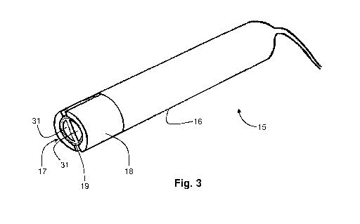
Fig. 1 is a block diagram showing the functionality of a system 10 according
to
the invention for non-invasive analysis of tissue of an examined subject in
order to
distinguish between different types of tissue. The system 10 comprises a hand-
held
probe 15 shown pictorially in Figs. 3 and 4 having a casing 16 formed of non-
fcrro-
CA 2981563 2017-09-29

WO 2016/157182 PCT/IL2016/050337
- 4 -
magnetic material and having a predetermined cross-section at a working end 17
thereof. A magnetic field source unit 18 within the casing is configured to
generate a
substantially uniform, time-invariant magnetic field within a volume of tissue
having a
cross-section equal to that of the casing at the working end 17 and having a
magnetic
field strength in a range of 0.05 to 0.5 Tesla. Disposed within the magnetic
field source
unit 18 is at least one inductive coil 19 configured to receive RF excitation
signals in a
specific frequency range typically in the range of 2-20 MHz, the excitation
frequency
being related to the magnetic field strength such that for each Tesla of
magnetic field
strength the RF excitation iignal is 42 MHz. The inductive coil responds to
the
magnetic field and to the RF excitation signals by generation of
electromagnetic
excitation signals in a direction substantially perpendicular to a direction
of the
magnetic field to thereby magnetize a slice of the living tissue. The slice
has a depth and
a thickness that are a predetermined function of the magnetic field strength
whereby
only living tissue in the thus excited slice generates an electromagnetic
response to
nuclear spin echo signals.
A signal generator 20 is coupled to the inductive coil 19 and is configured
for
generating an RF excitation frequency that is a function of the respective
electro-
magnetic response of the slice of tissue excited by the probe 10. Likewise, a
receiver
unit 21 is coupled to the inductive coil 19 and is configured to receive
therefrom the
electromagnetic response and generate measured data indicative thereof. A
control unit
is connected to or contains the signal generator 20 so as to generate
predetermined
time patterns of the excitation RF signals, the control unit 25 being further
connected to
the receiver unit 21 and responsive to the electromagnetic response for
processing the
measured data and extracting data indicative of the nuclear spin echo signals
from the
25 living tissue, to determine relaxation times and determine therefrom the
type of tissue
excited by the probe.
In some embodiments, the inductive coil 19 is commonly coupled to the signal
generator 20 and to the receiver unit 21 via a duplexer 26 so that the at
least one
inductive coil applies the RF excitation frequency and receives the response
in different
time slots.
In some embodiments, the cross-section at the working end 17 of the casing 16
is cylindrical having a diameter of 2-30 mm. In a prototype reduced to
practice, the
CA 2981563 2017-09-29

W02016/157182
PCTAL2016/050337
- 5 -
diameter of casing 16 at its working end 17 was 20 mm and permitted analysis
of tissue
to a depth of between 3.2-9.5 mm at a magnetic field strength BO of 42-62 mT.
In order to excite and receive response signals from multiple slices at
successive
depths of body tissue, two different approaches may be employed that may be
comple-
mentary or used in combination. Both approaches rely on generating and
applying via
the probe 15 signals of different frequencies, each adapted to magnetize a
different slice
of the tissue whose depth and a thickness are a predetermined function of the
magnetic
field strength. In one approach, there are provided more than one inductive
coil each
configured to receive a respective RF excitation signal in mutually different
frequency
ranges so that different slices of tissue are excited simultaneously. In
another approach,
the signal generator 20 generates time-varying excitation signals that are
applied
successively to the same induction coil. Likewise, although in Fig. 1 the same
induction
coil is used to both apply the excitation signal and receive the response
signal, the
duplexer 26 serving to direct the signal flow appropriately, multiple
induction coils may
alternatively be used: one to transmit and one to receive thus obviating the
need for the
duplexer 26. In either approach, the control unit 25 includes a pulse
programmer 27
coupled to the signal generator 20 for obtaining the correct time-varying
excitation
signals as described below with reference to Figs. 2a to 2c of the drawings.
The signal
generator 20 and the pulse programmer 27 together constitute a signal
processor 28.
Figs. 3 and 4 show an embodiment of the magnetic field source unit 18 which
comprises a pair of outer arcuate segments 30 both of a first magnetic
polarity and a
pair of inner segments 31 both of a second magnetic polarity opposite to the
first
= magnetic polarity defining an annular gap 32. At least one inductive coil
19 is disposed
within the annular gap 32 between the outer and inner segments. The outer and
inner
segments 30, 31 have respective contours that lie on circles of different
radii, the radius
of the outer segments being preferably less than 10 mm. The inner segments 31
may be
shorter in height than the outer segments 30, in which case they may be
supported so
that respective end faces of the inner and outer segments are co-planar. When
multiple
coils are provided, they may be arranged in a vertical stack with the annular
gap 32
between the inner and outer segments.
Figs. 5a to 5f are schematic representations showing magnetic source units 18
having different geometries. In all cases, one or more coils are mounted at a
working
CA 2981563 2017-09-29

WO 2016/157182 PCT/1L2016/050337
- 6 -
end of the magnetic source unit 18, which is held against the patient's tissue
or, in some
cases, into which the patient's tissue is inserted. Thus, in Fig. 5a there is
shown a
magnetic source unit 18 having a generally pyramidal shape with an apex 33
between
4-25 mm and a coil 19 disposed at the apex 33. Fig. 5b shows a magnetic source
unit 18
having a conical shape with a coil 19 disposed at a truncated tip 34 of
diameter between
4-30 mm. Either of these magnetic source units 18 replaces the head of the
probe shown
in Fig. 3. Fig. 5c shows a magnetic source unit 18 having the general shape of
a banana
35 in the middle of which and at opposite ends of which are provided
respective coils
19. Tip-to-tip dimensions are between 50-300 mm. Fig. 5d shows a magnetic
source
unit 18 having the general shape of a hollow bagel 36 along whose internal
surfaces are
provided respective coils 19, four such coils being shown each at opposite
ends of
mutually perpendicular inner diameters of the bagel of dimensions between 50-
300 mm.
Fig. 5e shows a magnetic source unit 18 having inner and outer cylinders 37
and 38,
respectively, a coil 19 being mounted at an end of the inner cylinder 37 of
diameter
between 4-30 mm. Fig. 5f shows a magnetic source unit 18 having opposing side
walls
39 and 40 spaced apart between 50-300 mm, each of which supports coils 19 and
between which a patient's limb may be inserted. In all cases, a single coil 19
is shown
for clarity, it being understood that in practice multiple coils can be
employed.
The system 10 may employ multiple probes 15 each directed for exciting a
different portion of body tissue.
Having described the elements of the system 10 and thc probe 15, we will now
briefly describe the manner in which measurement and analysis arc carried out
by the
control unit 25 with particular reference to Figs 2a to 2c.
In an embodiment of the invention the control unit 25 is configured in real
time
to access predetermined data characterizing multiple groups of relaxation
curves each
group defining at least two characteristic curves corresponding to a specific
known
tissue type and decaying exponentially at respective predetermined time
constants T1
and T2 and to analyze measured data in real time to determine to which group
of
relaxation curves the measured data is best fit in order to identify the
tissue type
corresponding to the measured data.
The derivation of the time constants TI and T2 is described below it being
understood that the characteristic curves have different time constants Ti and
T2 that
CA 2981563 2017-09-29

WO 2016/157182 PCU1L2016/050337
=
- 7 -
are characteristic of a specific type of body tissue. The relaxation curves
for each
different type of body tissue are determined off-line and data representative
of the time
constants Ti and T2 are stored in a memory of the control unit 25 and
statistically
processed. During actual measurement, the response signals returned by each
slice of
tissue to the probe are best fit to an appropriate exponential function
resulting in an
estimate of the time constant, whereupon it can be established which tissue
type
corresponds to the time constant obtained by the fit for the slice being
measured.
It should be noted that the simultaneous matching of two or more relaxation
curves each having predetermined time constants that are stored in the memory
of the
control unit 25 speeds up the convergence of the best-fit process. For this
reason there
are many surgical applications where it is essential to use two or more
relaxation curves.
In many such procedures time is of the essence and the use of two or more
relaxation
curves allows determination of tissue type in less than 5 seconds. But there
are also
applications where time is less critical. For example, the probe may be used
in non-
invasive diagnostic procedures where the diagnostician can afford to wait
significantly
longer, even a minute or more. In such applications, it may not be essential
to best-fit
the measured response to both curves and use may be made of the T1
characteristic on
its own since although it is slower than matching the T2 characteristic it may
be
sufficient to identify the tissue type while T2 on its own although faster
cannot.
A. Ti measurement with pulse inversion
The purpose of measurement is to measure quantitatively the spin relaxation
time Ti of the material. At the beginning of the measurement, before applying
the
pulses, the system is in a state of thermal equilibrium in which the
magnetization is
aligned in the direction of the external magnetic field referred to as BO. The
first pulse
operates to invert the magnetization so that it is aligned in a direction
opposite to the
magnetic field. As a result, the system strives to return to equilibrium,
whereby the time
it takes for the system to return to equilibrium is referred to as TI. The
purpose of the
second and third pulses is to measure the magnetization state at a time t
following the
first pulse. The measurement is carried out with the aid of an echo signal
generated by a
combination of 90 and 1800 pulses. Repeating this series of pulses with
different
values of t produces the curve shown in Fig. 2 from which the time constant Ti
can be
derived.
CA 2981563 2017-09-29

WO 2016/157182 PCT/IL2016/050337
- 8 -
B. T2 measurement by a series of 1800 pulses
The purpose of this measurement is to measure quantitatively the signal decay
time of the magnetic resonance known as T2. This decay is the result of local
magnetic
fields and spins in the material forming a spread in precession frequencies
and causing
the signal to decay. This measurement can be made in a single measurement
without the
need for a series of measurements using the series of pulses shown in Fig. 2c.
A suitable
algorithm is disclosed by S. Meiboom and D. Gil in "Modified spin-echo method
for
measuring nuckar relaxation times" Rev. Sci. lnstrum. 29, 688 (1958). This
series
commences with a 90 pulse that shifts the magnetization perpendicular to the
direction
tO of the external field. The magnetization now undergoes rotation
(precession) around the
external field axis. In order not to be subject to inhomogcncity of the
external field, use
is made of a 180 pulse known as an echo, at the commencement of which
frequency
spread as the result of a lack of uniformity of the external field is canceled
and all the
spins are aligned and form a strong signal. Using a series of 180 pulses a
series of
echoes may be produced thus obtaining from the signal measurement at the start
of the
echo for the duration of the echo pulses the decay curve as shown in Fig. 2b
in which
the time constant of the decay is termed T2.
The control unit according to the invention may be a suitably programmed
computer. Likewise, the invention contemplates a computer program being
readable by
a computer for executing the method of the invention. The invention further
contem-
plates a machine-readable memory tangibly embodying a program of instructions
executable by the machine for executing the method of the invention.
Features that are described with reference to one or more embodiments are
described by way of example rather than by way of limitation to those
embodiments.
Thus, unless stated otherwise or unless particular combinations arc clearly
inadmissible,
optional features that are described with reference to only some embodiments
arc
assumed to be likewise applicable to all other embodiments also.
CA 2981563 2017-09-29

Dessin représentatif
Une figure unique qui représente un dessin illustrant l'invention.
États administratifs

2024-08-01 : Dans le cadre de la transition vers les Brevets de nouvelle génération (BNG), la base de données sur les brevets canadiens (BDBC) contient désormais un Historique d'événement plus détaillé, qui reproduit le Journal des événements de notre nouvelle solution interne.

Veuillez noter que les événements débutant par « Inactive : » se réfèrent à des événements qui ne sont plus utilisés dans notre nouvelle solution interne.

Pour une meilleure compréhension de l'état de la demande ou brevet qui figure sur cette page, la rubrique Mise en garde , et les descriptions de Brevet , Historique d'événement , Taxes périodiques et Historique des paiements devraient être consultées.

Historique d'événement

Description Date
Demande non rétablie avant l'échéance 2023-07-25
Inactive : Morte - Aucune rép à dem par.86(2) Règles 2023-07-25
Lettre envoyée 2023-03-30
Réputée abandonnée - omission de répondre à un avis sur les taxes pour le maintien en état 2022-10-03
Réputée abandonnée - omission de répondre à une demande de l'examinateur 2022-07-25
Lettre envoyée 2022-03-30
Inactive : Rapport - Aucun CQ 2022-03-23
Rapport d'examen 2022-03-23
Lettre envoyée 2021-04-12
Requête d'examen reçue 2021-03-26
Exigences pour une requête d'examen - jugée conforme 2021-03-26
Toutes les exigences pour l'examen - jugée conforme 2021-03-26
Représentant commun nommé 2020-11-07
Requête pour le changement d'adresse ou de mode de correspondance reçue 2020-03-31
Requête visant le maintien en état reçue 2020-03-31
Inactive : COVID 19 - Délai prolongé 2020-03-29
Représentant commun nommé 2019-10-30
Inactive : CIB attribuée 2017-10-24
Inactive : Page couverture publiée 2017-10-20
Inactive : Notice - Entrée phase nat. - Pas de RE 2017-10-18
Inactive : CIB en 1re position 2017-10-17
Inactive : CIB attribuée 2017-10-17
Demande reçue - PCT 2017-10-12
Inactive : CIB attribuée 2017-10-12
Déclaration du statut de petite entité jugée conforme 2017-10-02
Exigences pour l'entrée dans la phase nationale - jugée conforme 2017-09-29
Déclaration du statut de petite entité jugée conforme 2017-09-29
Demande publiée (accessible au public) 2016-10-06

Historique d'abandonnement

Date d'abandonnement Raison Date de rétablissement
2022-10-03
2022-07-25

Taxes périodiques

Le dernier paiement a été reçu le 2021-03-22

Avis : Si le paiement en totalité n'a pas été reçu au plus tard à la date indiquée, une taxe supplémentaire peut être imposée, soit une des taxes suivantes :

  • taxe de rétablissement ;
  • taxe pour paiement en souffrance ; ou
  • taxe additionnelle pour le renversement d'une péremption réputée.

Les taxes sur les brevets sont ajustées au 1er janvier de chaque année. Les montants ci-dessus sont les montants actuels s'ils sont reçus au plus tard le 31 décembre de l'année en cours.
Veuillez vous référer à la page web des taxes sur les brevets de l'OPIC pour voir tous les montants actuels des taxes.

Historique des taxes

Type de taxes Anniversaire Échéance Date payée
Taxe nationale de base - petite 2017-09-29
TM (demande, 2e anniv.) - petite 02 2018-04-03 2018-03-28
TM (demande, 3e anniv.) - petite 03 2019-04-01 2019-03-29
TM (demande, 4e anniv.) - petite 04 2020-03-30 2020-03-31
TM (demande, 5e anniv.) - petite 05 2021-03-30 2021-03-22
Requête d'examen - petite 2021-03-30 2021-03-26
Titulaires au dossier

Les titulaires actuels et antérieures au dossier sont affichés en ordre alphabétique.

Titulaires actuels au dossier
SHMRI HAIM LTD.
Titulaires antérieures au dossier
YINON LEVY
Les propriétaires antérieurs qui ne figurent pas dans la liste des « Propriétaires au dossier » apparaîtront dans d'autres documents au dossier.
Documents

Pour visionner les fichiers sélectionnés, entrer le code reCAPTCHA :


Pour visualiser une image, cliquer sur un lien dans la colonne description du document. Pour télécharger l'image (les images), cliquer l'une ou plusieurs cases à cocher dans la première colonne et ensuite cliquer sur le bouton "Télécharger sélection en format PDF (archive Zip)" ou le bouton "Télécharger sélection (en un fichier PDF fusionné)".

Liste des documents de brevet publiés et non publiés sur la BDBC .

Si vous avez des difficultés à accéder au contenu, veuillez communiquer avec le Centre de services à la clientèle au 1-866-997-1936, ou envoyer un courriel au Centre de service à la clientèle de l'OPIC.


Description du
Document 
Date
(aaaa-mm-jj) 
Nombre de pages   Taille de l'image (Ko) 
Dessin représentatif 2017-09-28 1 6
Abrégé 2017-09-28 1 59
Revendications 2017-09-28 6 206
Description 2017-09-28 9 334
Dessins 2017-09-28 4 46
Avis d'entree dans la phase nationale 2017-10-17 1 194
Rappel de taxe de maintien due 2017-12-03 1 111
Courtoisie - Réception de la requête d'examen 2021-04-11 1 425
Avis du commissaire - non-paiement de la taxe de maintien en état pour une demande de brevet 2022-05-10 1 561
Courtoisie - Lettre d'abandon (R86(2)) 2022-10-02 1 548
Courtoisie - Lettre d'abandon (taxe de maintien en état) 2022-11-13 1 549
Avis du commissaire - non-paiement de la taxe de maintien en état pour une demande de brevet 2023-05-10 1 560
Correspondance reliée au PCT 2017-09-28 17 641
Correspondance reliée au PCT 2017-09-28 3 86
Rapport d'examen préliminaire international 2017-10-01 36 1 320
Paiement de taxe périodique 2018-03-27 1 26
Paiement de taxe périodique 2019-03-28 1 26
Changement à la méthode de correspondance 2020-03-30 4 91
Paiement de taxe périodique 2020-03-30 4 90
Paiement de taxe périodique 2021-03-21 1 27
Requête d'examen 2021-03-25 3 95
Demande de l'examinateur 2022-03-22 3 199