Note : Les descriptions sont présentées dans la langue officielle dans laquelle elles ont été soumises.
CA 02986562 2017-11-20
WO 2016/186503 PCT/NL2016/050361
Title: Microfluidic device for in vitro 3D cell culture experimentation
The present invention relates to a microfluidic device for in vitro 3D cell
culture experimentation comprising a body in which is provided a cell
culture chamber at least partly filled with a scaffolding substance for
maintaining a cell culture, and a fluid path communicating with the cell
culture chamber for directing a fluid stream along the scaffolding substance.
The present invention further relates to a method for in vitro 3D cell culture
experimentation, including complex living tissue reconstruction, using the
microfluidic device of the present invention.
In vitro cell culture experimentation is important in biological and medical
sciences for allowing investigation of cellular behavior of individual cells
or
of cells as part of larger cell cultures. For instance the investigation of
uptake of biomolecules by cells may lead to improved knowledge and
understanding of the effect of such biomolecules on a cellular, tissue, organ
and subject level, which in turn may lead for example to the development of
personalized medicine. Currently pharmacokinetic and toxicological
evaluation of drug candidates relies largely on costly, labor-intensive, time-
consuming and ethically questionable animal test systems, which show only
very limited predictive value for clinical efficacy and toxicity.
Many methods and devices for culturing, expanding and differentiating cells
in vitro have thus been developed. A conventional and still often used
method is growth and maintenance of cells or cell cultures on a suitable
growth surface such as a cell culture dish filled with liquid or jellified
culture medium. The culture medium may comprise specific constituents
which affect the growth and maintenance of the cells or cell culture in
desired ways. However the predictive value of these two dimensional (2D)
cell culture models for some application may be still very limited, because of
the loss of physiological context.
CA 02986562 2017-11-20
WO 2016/186503 PCT/NL2016/050361
2
With 3D scaffolds, for example cells incapsulated in a scaffolding substance
such as hydrogel, tissue-like connectivity may be achieved, but there are
limits in controlling the cell culture conditions. The 3D models mostly lack
the complexity required for pharmacokinetic studies. For many applications
in such models there is a limited nutrient supply to the cell culture and an
accumulation of metabolic waste products that can confound cell responses
to drugs. The 3D models also fail to mimic spatiotemporal biochemical
gradients existing in vivo, and lack the provision of mechanical cues such as
flow, perfusion, pressure, mechanical stress. It is also problematic for real-
time imaging, and biochemical analysis can hardly be performed in live cells
due to reaction- diffusion phenomena. Furthermore, it is not easily possible
to engineer microsystems that integrate multiple organ/tissue mimetics
with active vascular conduits and barrier tissues.
Microfluiclic devices such as microfluiclic chips allow for addressing these
limitations. With microfluiclic devices fluid flow may be controlled in the
micrometer and nanoliter scale in precisely defined geometries. Because of
the micro geometrical dimensions, the flow of fluids is laminar, and
placement of fluid volumes in very low amounts is possible. The ability of
exactly timing fluid flow allows precise chemical and physical control of the
microenvironment. For cell cultures in microfluiclic devices the doses
delivered to cells can be measured in nanoliters or less, representing a
significant improvement in precision. Small volume effects of fluids mimic
physiological conditions of cells or cell-populations in tissues more
appropriately than cells that are cultured in larger volumes. Microfluiclic
systems also allow detailed analysis of cell migration in a social context.
Controlling the spatiotemporal cues of the microenvironment and the ability
to shape the geometry of cultured cells for instance allows studying of
primary neuronal cells and cell lines in microfluidic chips.
CA 02986562 2017-11-20
WO 2016/186503
PCT/NL2016/050361
3
Integration of microfluiclics with 3D scaffolding systems renders it possible
to adapt culture conditions both biochemically and biomechanically, such as
creating dynamic 3D structures, and provides a microenvironment that
allows formation of artificial tissues from cultured cells. Microfluiclic cell
culture devices allow precise control of cell numbers and cell density in a
given area or volume, and can provide placement of cells in complex
geometries. Because cells can be organized into three-dimensional
geometries in scaffolding substances such as hydrogels in the microfluidic
devices, it is possible to culture cells in 3D structures resembling those in
tissues. Homotypic tissue culture models may be achieved in microfluidic
devices as well as heterotypic tissue culture models that mimic the
respective tissue closely both from a histologic as well as from a
physiological and functional standpoint. This allows for instance for high-
throughput pharmacological studies and might result in using microfluidic
cell culture systems also for regenerative purposes.
The small dimensions of spatially separated microfluiclic compartments in
microfluidic cell culture devices allow assembly of a multitude of
individually controllable cell cultures in chambers on a single device. This
facilitates high parallelization of experiments, high throughput of samples
and reactions and thus improvement of reproducibility, as well as a
reduction in reagent costs.
Resulting from the above-mentioned advantages, microfluidics has become
particularly valuable for analysis of single cell dynamics. With the help of
microfluidic devices cell growth and regulation of cell size can be directly
observed and lineages of single cells can be tracked for several generations.
On a molecular level microfluidics allow the characterization of
transcription factor and gene expression dynamics in single-cells thereby
CA 02986562 2017-11-20
WO 2016/186503 PCT/NL2016/050361
4
adding substantially to our understanding of the function of biological
systems.
The presently available microfluidic devices for in vitro 3D cell culture
experimentation comprise a closed system to shield the cell culture and the
culture conditions from possible outside influences, and provide a limited
accessibility to the cell culture grown in the culture chamber of the device.
Thus the known devices render the simultaneous manipulation and analysis
of cultured cells rather difficult, particularly monitoring of cells in
complex
geometries with high spatial and temporal resolution and their individual
retrieval during or following experiments.
As a result, there is a need for an improved microfluidic system for cell
culture investigation which may be particularly applied in drug studies,
vaccine development and other types of medical research. The present
invention thus provides a new device and method for cell culture
investigation, with which it is possible to investigate all types of cells
such
as vascular cells and organ cells individually or in functional units of
tissues
and organs in vitro. These and other aspects of the invention are evident
from the specification and claims hereinafter.
In one aspect of the present invention a microfluidic device for in vitro
complex living tissue reconstruction is provided. It is proposed that the in
vitro complex living tissue reconstructed by the fluidic device of the present
invention closely mimics the in vivo tissue of a living multicellular organism
such as a plant or animal. The present invention thus provides a
microfluidic device for in vitro 3D cell culture experimentation according to
the preamble, which microfluidic device is characterized in that the culture
chamber above the scaffolding substance opens into an access port provided
at an outer top surface of the body for direct access to the scaffolding
CA 02986562 2017-11-20
WO 2016/186503 PCT/NL2016/050361
substance which forms a fluid flow barrier separating the fluid path from
the access port. The access port in the device according to the invention
provides a direct access to the culture chamber there below. Thus the
scaffolding substance may be conveniently provided in the culture chamber
5 through the access port and cells cultured in the device or conditions in
the
culture chamber may be easily manipulated via the access port. The access
port for instance allows easy seeding or retrieval of cells in the culture
chamber.
If needed the cell culture environment in the microfluidic device may be
closed off from an outside environment by placing a suitable cover or lid over
the access port. The cover or lid may be an integral part of the microfluidic
device or a separate body. The cover or lid is preferably detachably
connected with the body of the microfluidic device, which renders it possible
to close or open the access port at any time as desired.
The term 'fluid flow barrier' as used herein refers to any means which forms
a restriction for a fluid flow. A fluid flow barrier may be used to redirect a
free flow of a fluid to a certain extent. The fluid flow barrier may be a
complete restriction in that no flow of fluid there through is allowed, or may
be a partial restriction in that some fluid flow there through is possible.
The
fluid flow barrier may in any event allow movement of the fluid or parts
thereof, for example substances, particles, or other components in the fluid,
through the fluid flow barrier by means of diffusion. Because of the
provision of a scaffolding substance in the culture chamber of the
microfluidic device which forms a fluid flow barrier separating the fluid path
from the access port, any fluid flowing through the fluid path in the
microfluidic device is for a larger part directed along the culture chamber,
preventing a strong flow of fluid through the culture chamber. As a
consequence only cells exposed on an outside of the scaffolding substance
CA 02986562 2017-11-20
WO 2016/186503
PCT/NL2016/050361
6
will possibly experience shear stress across the surface, whereas this will
not or hardly be the case for a cell culture captured in the scaffolding
substance.
The provision of the fluid flow barrier may be used to mimic the mechanical
forces that help govern the architecture of tissues such as the lung, bone,
articular cartilage, and vascular tissues. Importantly many cell types
including fibroblasts, smooth muscle cells, osteocytes, and chondrocytes,
reside within a three-dimensional environment and are exposed to
interstitial fluid forces. Physiological interstitial flow is the movement of
fluid through the extracellular matrix of a tissue, often between blood
vessels and lymphatic capillaries. It provides convection necessary for the
transport of large proteins through the interstitial space and constitutes an
important component of the microcirculation. Interstitial flow also provides
a specific mechanical environment to cells in the interstitium that could
play an important role in determining interstitial organization and
architecture. Thus the microfluiclic device according to the invention may
employ a scaffolding substance that forms a fluid flow barrier between the
fluid flow path and the culture chamber which allows for a flow of fluid from
the fluid flow path through the scaffolding substance mimicking that of
interstitial flow, in order to expose the cultured cells within the
scaffolding
substance to interstitial fluid forces and to provide the cells with the
necessary or intended nutrients and/or other biomolecules. Types of cells,
such as endothelial and epithelial cells, that in tissue form a monolayer to
create a lumen or surface and are exposed to shear stresses across the
surface, may be seeded on the outside of the scaffolding substance to be
exposed to the fluid flow of fluid moving through the fluid flow path of the
device.
According to a preferred embodiment the microfluidic device of the present
invention is characterized in that the body comprises a set of fluid paths
CA 02986562 2017-11-20
WO 2016/186503 PCT/NL2016/050361
7
each fluid path communicating with the cell culture chamber for directing a
respective fluid stream along the scaffolding substance, wherein the
scaffolding substance separates the respective fluid paths. The set of fluid
paths, consisting of two, three, four, five or even more fluid paths, in the
microfluidic device according to the invention allows for the supply of fluid
or components of the fluid to the culture chamber at different locations of
the scaffolding substance, thus mimicking a network of supply vessels such
as blood vessels and capillary lymph channels for a tissue. Different or
identical fluids may be flown through each fluid path in order to supply
components of interest such as nutrients, chemicals, signaling proteins
and/or other biomolecules and factors, to the cell culture in the culture
chamber.
According to a further preferred embodiment the microfluiclic device of the
present invention is characterized in that the access port is provided
directly
above the cell culture chamber. Because of such provision of the access port
directly above the cell culture chamber, the access port provides an
unobstructed view to the cell culture chamber. Thus it is possible to monitor
cells in the culture chamber in complex geometries with high spatial and
temporal resolution. Additionally, by providing a medium, such as a fluid, in
the access port on top of the scaffolding substance, a medium pressure is
applied on the scaffolding substance stimulating perfusion of the fluid
through the scaffolding substance.
A particular embodiment of the microfluidic device of the present invention
is in this respect characterized in that the access port has a height
dimension extending between the outer top surface of the body and an
opening to the culture chamber which height dimension is larger than a
height dimension of the inlet opening of each channel extending between the
outer surface of the body and a bottom of the respective channel. For
CA 02986562 2017-11-20
WO 2016/186503 PCT/NL2016/050361
8
instance the access port may be open at the outer top surface of the body
which surface is elevated with respect to an outer surface of the body in
which the inlet opening of a channel is provided. The outer surface of the
body in which the inlet opening of the channel is provided may be any
surface of the body lower positioned than the outer top surface of the body,
e.g. a lower horizontal surface, a recessed surface, an angled or vertical
side
surface of the body etcetera. A column height of fluid applied in the access
port may thus differ from that of each fluid applied in the fluid channels of
the device. As a result the diffusion and/or perfusion rate of the fluids or
fluid components of the fluid channels to the culture chamber may be
controlled as desired by applying less or more fluid in the access port.
In a further preferred embodiment the microfluidic device according to the
present invention is characterized in that the fluid paths are channels in the
body each channel extending between a respective inlet opening at an outer
surface of the body and an outlet opening at an outer surface of the body.
The channels may direct a fluid flow from the inlet opening along the
scaffolding substance to the outlet opening with the fluid being in micro
amounts, thus allowing precise control of amounts of fluid or fluid
components being supplied to the cell culture in the culture chamber.
A further preferred embodiment of the microfluidic device according to the
present invention is characterized in that the body comprises a restricting
wall between the access port and the culture chamber, the restricting wall
comprising at least one passage opening. The restricting wall poses a means
of guidance of a substance applied in the access port to the culture chamber,
while providing some protection of the thereunder provided scaffolding
substance and cell culture. For instance the access port may be dimensioned
such that introduction of the tip of an injection means, such as a syringe or
pipette, is possible up to the restricting wall, thus preventing such
injection
CA 02986562 2017-11-20
WO 2016/186503 PCT/NL2016/050361
9
means from affecting an integrity of the scaffolding substance in the culture
chamber. Preferably such access port guides the injection means to the
passage opening to facilitate the supply of substance or material to the
culture chamber. In this respect the microfluiclic device of the present
invention in a particular embodiment is characterized in that the restricting
wall defines a conical shape of the access port.
It is further preferred that the restricting wall between the access port and
the culture chamber is positioned above the scaffolding substance leaving a
free space above a complete upper surface of the scaffolding substance.
Accordingly when the access port is filled with a fluid to apply a fluid
pressure to the scaffolding substance, the fluid will accumulate via the at
least one passage opening in the restricting wall in the free space above the
upper surface of the scaffolding substance to supply the complete surface
thereof with the fluid or components of the fluid and to apply an at least
almost equal fluid pressure over the complete surface thereof. The term
restricting wall as used herein refers to any means with which a cross
sectional size of a space in the body defined by the access port and the
culture chamber may be restricted locally to define a restricted opening
between the access port and the culture. Such means may comprise an
integral part of the body facing the space defined by the access port and the
culture chamber and extending inward in said space. The means may
alternatively or in addition comprise separate means which are provided in
the space in the body defined by the access port and the culture chamber.
The access port and the culture chamber of the microfluiclic device may be
identical in width dimension or may differ in width. Particularly a width of
the access port may be narrower or greater than a width of the culture
chamber. An access port with a greater width than the culture chamber may
be separated from the culture chamber by a restricting wall comprising a
CA 02986562 2017-11-20
WO 2016/186503 PCT/NL2016/050361
passage opening with a width dimension approximately equal to the width
of the culture chamber. Accordingly the culture chamber is directly
accessible over a complete width thereof through the passage opening,
allowing filling and emptying of the whole culture chamber, whereas the
5 broader access port allows for easy guiding of filling material to the
passage
opening and provides for an unobstructed view of the culture chamber
beneath the passage opening.
The culture chamber and fluid paths of the microfluidic device may be of
10 any shape, and may with respect to each other also differ in shape. For
instance in a preferred embodiment of the microfluidic device according to
the invention one or more of the fluid paths have a smaller or bigger
dimension as compared to the other fluid paths, thus allowing for different
amounts of fluids being guided to the culture chamber and along the
scaffolding substance to mimic different supply channels such as different
sized blood vessels and/or lymph capillaries.
In a further preferred embodiment the microfluidic device according to the
present invention is characterized in that the scaffolding substance rests on
a bottom part of the body opposite the outer top surface. Thus the
scaffolding substance after being provided in the culture chamber may be
maintained in the culture chamber simply by resting on the bottom part.
A further preferred embodiment of the microfluidic device according to the
present invention is characterized in that the fluid paths communicate with
the scaffolding substance at a lateral side of the culture chamber. Nutrients
or other substances from the fluids flowing through the fluid paths along the
lateral sides of the scaffolding substance will diffuse in the scaffolding
substance and particularly form a gradient throughout the scaffolding
substance in a width direction of the culture chamber. Cells cultured in the
CA 02986562 2017-11-20
WO 2016/186503 PCT/NL2016/050361
11
scaffolding substance will thus be provided with different amounts of such
nutrients or other substances depending particularly on their position in the
culture chamber in the width dimension. The effects of such gradient on the
resulting cell culture may be easily viewed from above through the access
port.
The fluid paths may be dimensioned to communicate with the scaffolding
substance over a full height of the scaffolding substance, e.g. a height of
each channel defining a fluid path may be identical to a height of the
scaffolding substance in the culture chamber or may be identical to a height
of the culture chamber. Alternatively the fluid paths may also be configured
to communicate with a section of the scaffolding substance, for instance only
over a part of the total height of the scaffolding substance in the culture
chamber. A height of the channel defining the fluid path may be less than
the total height of the scaffolding substance, or the channel may have a
height approximately similar to a height of the scaffolding substance, with a
restricting element provided between the channel and culture chamber to
restrict a communication surface between the fluid path and scaffolding
substance. The restricting element may be a further restricting wall, or a
column or other shaped part of the body of the microfluidic device. In
particular the restricting element is a vertically oriented wall of the body
extending between the culture chamber and channel and defining a
communication opening there between which communication opening allows
fluid communication between the corresponding fluid path and the
scaffolding substance.
In a further preferred embodiment the microfluiclic device according to the
present invention is characterized in that the respective fluid paths
communicate with the scaffolding substance at mutual different heights
between the bottom part of the body and the outer top surface. Accordingly
CA 02986562 2017-11-20
WO 2016/186503
PCT/NL2016/050361
12
the different fluid paths may provide nutrients or other substances to the
scaffolding substance at different heights, thus creating a third dimension
gradient of such nutrients or other substances in the scaffolding substance,
to allow investigation of such gradient and the corresponding effect on the
cell culture in such third dimension.
According to a further preferred embodiment the microfluiclic device of the
present invention is characterized in that at least one of the fluid paths has
a larger communication area with the scaffolding substance as compared to
another of the fluid paths. With a larger communication area between the
fluid path and the scaffolding substance it is possible to supply more fluid
or
components thereof to the cells whereas a smaller communication area may
be used for more precise local supply of such fluid or components thereof.
According to a particular embodiment the microfluiclic device of the present
invention is characterized in that the body comprises at least three fluid
paths. In a further particular embodiment the microfluidic device according
to the present invention is characterized in that the three fluid paths
communicate with the scaffolding substance at a lateral side of the culture
chamber at approximately 120 degrees apart. A further particular
embodiment of the microfluiclic device according to the present invention is
characterized in that at least one of the at least three fluid paths
communicates with the scaffolding substance at a lower side of the culture
chamber opposite the upper side. In this embodiment all geometrical sides
of the scaffolding substance in the culture chamber, i.e. the upper, lower,
and lateral sides of the scaffolding substance may be provided with
nutrients or other substances from fluids flowing through each of the fluid
paths and provided in the access port in order to create a complex 3D
network of cells mimicking that of in vivo tissue.
CA 02986562 2017-11-20
WO 2016/186503 PCT/NL2016/050361
13
In a further preferred embodiment the microfluidic device according to the
present invention is characterized in that the fluid paths are separated
along at least a part thereof by a wall of the body which wall has a wall end
that borders the culture chamber and forms a support structure for the
scaffolding substance. The wall end may form a boundary for the scaffolding
substance, which prevents the scaffolding substance from escaping from the
culture chamber into the fluid paths and possibly completely blocking any of
the fluid flows. In a further preferred embodiment the microfluidic device
according to the present invention is characterized in that at least the wall
end of the wall of the body is of a hydrophobic material. Accordingly the
scaffolding substance may be a droplet of a hydrophilic material, such as a
hydrophilic liquid, particularly water suitable for cell culture, which is
maintained in the culture chamber between the hydrophobic wall ends.
According to a further preferred embodiment the microfluidic device of the
present invention is characterized in that at least two wall ends of
respective walls separating respective flow paths are positioned with respect
to each other to define an imaginary circumference of the culture chamber
and in that the scaffolding substance is bound by the imaginary
circumference. The imaginary circumference may be of any shape.
According to a particular embodiment the microfluidic device of the present
invention is characterized in that the body is a microfluidic chip. A further
preferred embodiment of the microfluidic device according to the present
invention is characterized in that the microfluidic chip comprises multiple
culture chambers and corresponding access ports and fluid paths. The
multiple culture chambers and corresponding access ports and fluid paths
form separate experimentation units, which allow the microfluidic chip to be
used for multiple and/or parallel experiments. Although a chip is a
convenient embodiment of the body of the microfluidic device according to
CA 02986562 2017-11-20
WO 2016/186503
PCT/NL2016/050361
14
the invention, and may for instance conveniently be sized as a standard
microscope slide of approximately 75x25x1 mm for convenient visual
inspection of the cell culture in the culture chamber by means of a
microscope, the body may alternatively be dimensioned as part of a
multiwell plate or microplate or for provision in a well of a multiwell plate
or microplate, such as for instance a 385 well plate. In such embodiment of
the body of the microfluidic device according to the present invention a
culture chamber, fluid paths and access port may be provided near a bottom
of each of the wells of the well plate. At least part of the height or
preferable
a complete height of each of the wells may be used to provide a fluid column
with fluid pressure on the scaffolding substance with which a perfusion rate
in the scaffolding substance may be controlled.
A further preferred embodiment of the microfluidic device according to the
present invention is characterized in that at least some of the fluid paths
are shared between consecutive culture chambers. Thus the consecutive
culture chambers are interconnected through the shared fluid paths and
may thus be used to mimic tissue or organs consisting of or comprising
separated elements, such as for instance lymph nodes interconnected by
lymph capillaries in a lymph system of an individual.
In a further preferred embodiment the microfluiclic device according to the
present invention is characterized in that the body comprises a top plate
with the outer top surface and a separate bottom plate with the bottom part
which plates are adhered to each other. The open spaces in the body,
including the fluid paths, the culture chamber and the access port may be
provided in the outer surface of each or one of the two plates as grooves or
recesses, so that the body may then be formed by simply adhering the plates
together. In a particular embodiment the microfluiclic device according to
the present invention is characterized in that the bottom plate is at least
CA 02986562 2017-11-20
WO 2016/186503
PCT/NL2016/050361
almost fully flat. The open spaces are thus provided in the top plate,
whereas the almost fully flat bottom plate is a relatively easily produced
part with which the open spaces may be closed off by adhering the bottom
plate and top plate to each other.
5
These and other aspects of the present invention are further elucidated by
the appended drawings and the corresponding embodiments described
hereinafter, which form part of the present application. The drawings are
not in any way meant to reflect a limitation of the scope of the invention,
10 unless this is clearly and explicitly indicated.
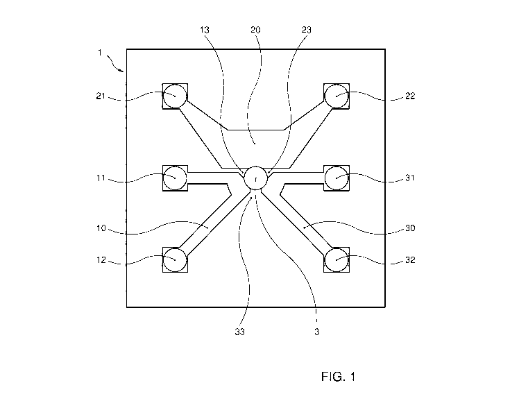
Figure 1 shows a top view of an embodiment of a microfluiclic device
according to the invention which may be used for in vitro 3D cell culture
experimentation.
15 Figure 2 shows a side view of a cross section of the embodiment of a
microfluidic device according to the invention shown in figure 1.
Figure 3 shows an enlarged schematic view of a culture chamber defined by
end walls of the body of the embodiment of the microfluidic device according
to the invention as shown in figure 1 and figure 2.
Figure 4a shows a set of microfluiclic devices in another embodiment
according to the invention.
Figures 4b-4d show a detailed view of a microfluidic device according to the
embodiment of figure 4a in respectively a top view, bottom view and
enlarged view of a central part of the device.
The microfluidic device according to the invention may be configured as a
multiple units organ-on-a-chip. As shown in figures 1-3 the microfluiclic
device comprises a body 1 which is substantially formed from PDMS in
which each of the multiple units consists of three fluid paths 10, 20, 30
being configured as channels, each channel extending between a
CA 02986562 2017-11-20
WO 2016/186503 PCT/NL2016/050361
16
corresponding inlet port 11, 21, 31 at an outer surface of the body 2 and an
outlet port 12, 22, 32. The channels accumulate in a central culture chamber
3 in the body 1. The culture chamber 3 is provided with a scaffolding
substance in the form of a block of gel (hatched block in culture chamber 3
in figure 2). The block of gel is maintained in the culture chamber 3 resting
on a bottom plate 2 of the body. The lateral sides of the block of gel are
bound by the end parts of the walls 13, 23, 33 (figure 1 and figure 3) of the
body separating the channels 10,20, 30 from each other. The block of gel
communicates with the channels for uptake of nutrients, metabolites, or
other components or compounds from the fluid flowing through the channels
by diffusion or perfusion. The flow is however directed from each inlet
opening of each channel to each outlet opening of each channel along the
block of gel, which poses a fluid flow barrier preventing direct flow of fluid
into the culture chamber 3. The culture chamber 3 on an upper side opens
into an access port 5 of the body 1, which access port is open at the outer
top
surface of the body 1. The access port 5 is provided directly above the
culture chamber 3 so that the culture chamber is directly accessible from
the outside through the access port. Between the access port 5 and the
culture chamber 3 there is a restricting wall (figure 2) defining an opening
between the culture chamber 3 and the access port 5 of which a cross
sectional size is smaller than the cross sectional size of either the culture
chamber 3 and the access port 5.
With this device the challenges of intestinal tissue engineering such as a
coculture of different 2-D and 3-D cell types under well-defined conditions
may be addressed. For example, a lumen-to-blood barrier of a human small
intestine exists of 2-D cell types such as blood/lymphatic endothelial or
intestinal epithelial cells and 3-D cell types such as different adherent and
migrating cells of intestinal interstitium such as (myo)fibroblasts, neural
and immune cells. In this regard direct contact of the fluid paths 10, 20, 30
CA 02986562 2017-11-20
WO 2016/186503
PCT/NL2016/050361
17
with the scaffolding substance of the open access culture chamber 3 of the
present embodiment allows: 1) 2-D cell culture of blood endothelial, lymph
endothelial and intestinal epithelial cells under well-defined conditions on
the scaffolding surface communicating with a particular fluid path; 2) 3-D
cell culture within the scaffolding substance in the open access chamber; 3)
direct mutual contact of all 2-D monolayers with the 3-D cell populated
scaffolding including autocrine and paracrine cellular signaling and
communications; 4) direct live imaging of cellular autocrine and paracrine
communications via the bottom transparent plate 2 and/or the open access
port 5 directly above, thus in line with, the culture chamber 3; 5) direct
access and sampling from the 3-D cell populated scaffolding through the
open access port above the culture chamber.
As an example a fully differentiated crypt-villus unit of intestinal
epithelium of the human small intestine can reach 1,5 mm height. The
larger fluid path 20 as compared to the smaller fluid paths 10, 30 of the
body according to this embodiment due to its 3 mm width and 0,35 mm
height allows fully 3-D development and differentiation of the crypt-villus
unit in a parallel to a planar line direction in the microfluiclic device.
Biomechanical and biochemical stimulation of cultured cells via different
channels 10,20,30 with shear stress, pressure and biochemical stimuli
allows in vitro simulation of some complex situations like for instance
kinetic motion of plasma from blood capillary through interstitium into a
lymphatic vessel; biochemical gradient of different compounds within
interstitium like oxygen, different signaling molecules and metabolites; and
cellular migration from a capillary, intestinal lumen of interstitium into
lymphatic system. Separated fluid paths offer a formidable opportunity to
sample medium (supernatant) for evaluation from different fluid paths.
CA 02986562 2017-11-20
WO 2016/186503 PCT/NL2016/050361
18
In figures 4a-4d an embodiment of a microfluiclic device, particularly a chip,
according to the invention is shown which is largely similar to the
embodiment of figures 1-3 but mainly differs in that instead of three fluid
paths the body comprises four fluid paths which communicate with a culture
chamber centrally located in the body. As particularly shown in figure 4a
the microfluidic device comprises multiple culture chambers and
corresponding access ports and fluid paths which form separate
experimentation units. Accordingly the microfluiclic chip may be used for
multiple and/or parallel experiments. The four fluid paths are configured to
communicate with the culture chamber at four different sides,
approximately 90 degrees apart. The inlet and outlet openings of the fluid
paths are all provided in the outer top surface of the body along the lateral
ends thereof. The access ports are positioned more centrally and are aligned
with respect to each other. The microfluidic device may be used in
accordance with the foregoing description.
Interconnection of several units of the present embodiment in one
microfluidic device, for example in one body such as a chip, allows creation
of more complex systems such as a human digestive system (e.g. mouth-
stomach-intestine) or a human body (e.g. intestine-liver-hart) using only one
such microfluidic chip. Capabilities of this embodiment of the microfluidic
device according to the invention can help to address the challenges not only
in tissue and microfluidic engineering, but also in systems biology. The in
vitro model provides experimentation for learning about the communication
and control of biological systems at the scale of individual organs-on-chips.
This complex, powerful, and integrated system allows recapitulating inter-
and intra-organ signaling and dynamics of a human gastrointestinal tract.
Unless otherwise defined, all technical and scientific terms used herein have
the same meaning as commonly understood by one of ordinary skill in the
CA 02986562 2017-11-20
WO 2016/186503 PCT/NL2016/050361
19
art to which this invention belongs. Methods and materials are described
herein for use in the present invention. However other suitable methods and
materials known in the art can also be used. The materials and examples
are illustrative only and not intended to be limiting, unless so indicated.
The
following definitions are used unless otherwise described.
As used herein, the term "microfluidic device" refers to any device that
allows for a precise control and manipulation of fluids that are constrained
geometrically to a small, typically sub-millimeter, scale and is suitable for
experimentation on cell cultures, particularly 3D cell cultures. The device is
a tool that allows for control of the cellular environment. A particular
embodiment of a microfluidic device as used herein is a microfluidic chip. A
microfluidic device such as particularly a microfluidic chip is preferably
made out of one or more of the materials Si02, glass and synthetic
polymers. As synthetic polymer a polysiloxane is preferred, and in
particular polyclimethylsiloxane (PDMS), although other polymers may be
used, such as polycarbonate (PC), polystyrol (PS), polytetrafluoroethylene
(PTFE) or cyclic olefin copolymer (COC). Particularly PDMS as material for
microfluidic devices allows easy implementation of desired geometric
structures and offers excellent live cell imaging conditions as PDMS is
relatively transparent and has stable optical features, and particularly a low
level of auto-fluorescence. Microfluidic cell culture devices made of PDMS
therefore allow use of fluorescent live cell imaging, providing a powerful
characterization of a multitude of cellular responses on a single cell as well
as population level. Soft lithography of poly-dimethylsiloxane (PDMS) is a
convenient method for the manufacturing of a microfluidic device for cell
culture applications. With this technique, structures of micrometer
resolution are molded from a hard master into PDMS. Such a microfluidic
device allows for exact spatial and temporal control of fluid flow and
delivery of media, drugs and signaling factors to live cells.
CA 02986562 2017-11-20
WO 2016/186503
PCT/NL2016/050361
Although mixtures of two or more of the above materials may be employed
for the device or parts of the device, it is preferred that the microfluidic
device comprises at most a few different materials, and preferably is made
at least almost wholly of a single material, for instance from COC which
5 provides a high resistance against deforming of the body.
As used herein, the term "3D cell culture" refers to a cell culture with cells
positioned relative to each other in three dimensions, i.e. width, depth and
height. Such 3D cell culture for most cells better mimics cell to cell
10 environments in tissues, organs and subjects. The microfluidic device
according to the present invention is particularly suitable for culturing such
3D cell cultures, but may also be used for 2D cell cultures.
As used herein, the term "scaffolding substance" refers to any substance
15 capable of maintaining living cells in a spatial relation in multiple
dimensions, preferably 3 dimensions. Scaffolding substances may be liquids,
gels or solids. Non limiting examples of scaffolding substance for use in a
microfluidic device according to the invention are water, hydrogel, agar gel,
micropore scaffold, microfiber scaffold, membrane and hollow fiber.
As used herein, the term "fluid" refers to any substance that continually
flows under an applied shear stress. Fluids as used herein may include
liquids, gases and plasmas.
As used herein, the term "fluid path" refers to any space through which a
quantity of fluid may flow, such as a compartment, channel, chamber, or
cavity.
For the purpose of clarity and a concise description, features are described
herein as part of the same or separate aspects and preferred embodiments
CA 02986562 2017-11-20
WO 2016/186503
PCT/NL2016/050361
21
thereof, however, it will be appreciated that the scope of the invention may
include embodiments having combinations of all or some of the features
described.