Note : Les descriptions sont présentées dans la langue officielle dans laquelle elles ont été soumises.
CA 02987211 2017-11-24
WO 2016/189530 PCT/1L2016/050541
AN INTRAOCULAR LENS AND METHODS FOR ACCOMMODATING EXISTING
ADAPTIVE INTRAOCULAR LENSES
FIELD OF THE INVENTION
The present invention relates to multifocal intraocular lens. More
specifically, the present invention
relates to an eyelid controlled zonal multifocal intraocular lens and methods
to accommodate
existing intraocular lens.
BACKGROUND OF THE INVENTION
An intraocular lens (I0L) is a device that is able to simulate the ability of
the natural lens of a young
individual to focus at different distances effortlessly. This ability usually
diminishes with age
culminating in presbyopia (inability to focus at near) around the age of 45 to
50. There are several
reasons for this phenomenon, the rigidity of the aging lens being the main.
The artificial intraocular lenses available today are monofocal lenses or
multifocal by design. These
lenses, while flexible during insertion, are not intended to move or to focus
inside the eye. The
multifocal lenses have two or more focal distances and the amount of focused
light is necessarily
reduced, because part of the lens is focused for distance and another part is
focused for near. That
means that part of the lens is always not focusing the image properly. That
also means that
multifocal lenses create visual aberrations due to dispersed light coming from
the part of the lens
that is not focusing properly.
Accommodative intraocular lens is an artificial lens in which its optical part
needs to be flexible to
be able to change focus. The obvious solution for that is a lens that is at
least partially liquid.
The accommodative intraocular lens should be mechanically coupled to the
contracting ciliary
muscle that is in charge of accommodation. This muscle is located
circumferentially behind the iris
and its contraction normally pulls the zonular fibers that are normally
attached to the lens capsule.
However, weakening of the zonular fibers may contribute to the lack of
focusing. Thus part of the
accommodative lens haptics should be in direct contact with the ciliary
muscle, pressing against it.
That means that the lens should be located in the ciliary sulcus, between the
natural lens capsule
and the iris.
1
CA 02987211 2017-11-24
WO 2016/189530 PCT/1L2016/050541
Since the size of the eye varies, a flexible lens allows the haptics to open
in a spring-like fashion
where the haptics rest on the diametrically opposing sides of the ciliary
muscle and the optic is
centered in front of the pupil. It also allows insertion of the lens through a
small cut.
However the same flexibility will prevent the transmission of forces from the
ciliary muscle to the
optical part. The flexible haptics will absorb whatever contracting forces of
the ciliary muscle exerts,
preventing any significant force to reach the optical part of the lens.
It is therefore an object of the present invention to provide a method and
apparatus that provide
reduced amount of unfocused light and a method and an apparatus that reduce
the absorption of
the contracting forces that the ciliary muscle exerts.
SUMMARY OF THE INVENTION
According to one embodiment of the present invention the flexible parts of the
lens haptics are
made at least partially from a material that can change its properties and
become rigid.
According to another embodiment of the present invention the flexible parts of
the lens haptics are
made of a polymer that becomes rigid after exposure to ultraviolet light. The
haptics and the joints
between the haptics and the IOL are the parts that become rigid by curing them
after of the surgical
implantation.
According to another embodiment of the present invention different flexible
parts of the lens are
made of a UV-sensitive material which are cured after of the surgical
implantation.
According to another embodiment of the present invention the internal liquid
pressure of a liquid
lens is accommodated by puncturing bubbles located in the liquid lens and thus
accommodating its
optical power.
According to another embodiment of the present invention the multifocal IOL
comprises of several
not-circular symmetric regions with different focal lengths and different
relative areas, where the
proportions of the relative areas of the different regions where light is
propagating through, are
controlled by the eyelids.
2
CA 02987211 2017-11-24
WO 2016/189530 PCT/1L2016/050541
BRIEF DESCRIPTION OF THE DRAWINGS
In order to understand the invention and to see how it may be carried out in
practice,
embodiments will now be described, by a way of non-limiting examples only,
with reference to the
accompanying drawing, in which:
Fig. la describes the side view of a first embodiment of an intraocular lens
(I0L) according the
present invention.
Fig. lb describes the top view of the first embodiment of an IOL according the
present invention.
Fig. 2 describe another embodiment of a flexible IOL according the present
invention.
Fig. 3 shows a top view of an embodiment of an IOL according to the present
invention wherein the
liquid pressure inside the lens can be adjusted.
Fig. 4a shows a front view of a multifocal IOL according to the present
invention.
Fig. 4b shows a cross section view of a multifocal IOL according to the
present invention.
Fig. 5a shows the rays of light when the eyelid is open.
Fig. 5b shows the rays of light when the eyelid is half closed.
Fig. 6a shows a front view of another embodiment of a multifocal intraocular
lens (I0L) according to
the present invention.
Fig. 6b shows a cross section view of another embodiment of a multifocal IOL
according to the
present invention.
Fig. 7a shows a front view of another embodiment of a multifocal IOL according
to the present
invention.
Fig. 7b shows a cross section view of another embodiment of a multifocal IOL
according to the
present invention.
Fig. 8a shows a front view of another embodiment of a multifocal IOL according
to the present
invention.
Fig. 8b shows a cross section view of another embodiment of a multifocal IOL
according to the
present invention.
3
CA 02987211 2017-11-24
WO 2016/189530 PCT/1L2016/050541
Fig. 9a shows a front view of another embodiment of a multifocal IOL according
to the present
invention.
Fig. 9b shows a cross section view of another embodiment of a multifocal IOL
according to the
present invention.
Fig. 10 shows a front view of another embodiment of a multifocal IOL according
to the present
invention.
DETAILED DESCRIPTION OF INVENTION
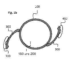
Fig. la and lb show the side view and the top view, respectively, of one
embodiment of a flexible
intraocular lens (IOL) according to the present invention. This embodiment is
only for the purpose
of illustrating the main idea of the present invention. In this embodiment,
the IOL 100 comprises a
flexible lens 150 which may be of zero optical power for add-on sulcus lens or
any other optical
power, a liquid lens 200 in which its curvature may by modified by the liquid
pressure and thus the
overall optical power of the IOL 100, haptics 300 which are made of a UV
sensitive material that is
initially flexible but may be turned into rigid by UV radiation, flexible
cushions filled of liquid 400
which are resting on the ciliary body and communicating with the liquid lens
through a pipe 500
that joins the liquid of the flexible cushions and the liquid of the liquid
lens. The overall optical
power composes the optical power of the lens 150 and the optical power of the
liquid lens 200. The
inner surface of the liquid lens 200 may or may not be in contact with the
inner surface of the lens
150. When the liquid pressure of the liquid lens changes, the curvature of the
outer surface of the
liquid lens is changed too, and thus its optical power is modified. The liquid
pressure of the liquid
lens may depend on multiple factors inside the eye. The most important factor
is the haptics
pressure on the ciliary body that will result in deformation of liquid
cushions and change the liquid
pressure. When the ciliary muscle changes its contraction, the pressure on the
ciliary body will be
changed and this results in changes of the deformation of the liquid cushions
and thus the liquid
pressure. Since the liquid of the cushions 400 is communicating with the
liquid of the liquid lens,
the changes of the liquid pressure modifies the outer surface of the liquid
lens and thus its optical
power. However since the haptics are initially flexible, this flexibility will
prevent the complete
transmission of forces from the ciliary muscle to the optical part. To stop
this from happening the
flexible parts of the lens haptics are made, at least partially, from a
material that can change its
physical properties and become rigid. This material may be for example, a
polymer that becomes
rigid after exposure to ultraviolet light or a material that becomes rigid
after exposure to higher
4
CA 02987211 2017-11-24
WO 2016/189530 PCT/1L2016/050541
temperature. Thus, the haptics and the joints between the haptics and the IOL
100 may become
rigid by curing them. After the surgery, when the IOL is located in its place,
a UV or IR radiation is
directed to the flexible haptics and the joints between the haptics and the
liquid lens and turns
them into rigid.
Fig. 2 shows a side view of another embodiment of a flexible intraocular lens
(IOL) according to the
present invention. In this embodiment, the IOL 100 comprises a first lens 150
made of flexible UV
sensitive material which may be of zero power for add-on sulcus lens or any
other power and a
second lens 200 also made of flexible UV sensitive material. There is a space
between both lenses
150 and 200 filled of liquid or some other material that can deliver the
forces from the ciliary
mussels. The IOL also comprises haptics 300 which also are made of a UV
sensitive material, flexible
cushions filled of liquid 400 which are resting on the ciliary body and
communicating with the liquid
lens through a pipe 500 that joins the liquid of the flexible cushions and the
liquid between the
lenses 150 and 200. After the surgery, UV or IR illumination is directed to
the lenses 100 and 200,
the haptics 300 and the joints between the haptics and the IOL 100 and
transform them into rigid.
Thus, when the ciliary muscle changes its contraction, the pressure on the
ciliary body will be
changed and this results in changes of the deformation of the liquid cushions
and the liquid
pressure. Since the liquid of the cushions 400 is connected to liquid in the
space between the two
lenses 100 and 200, the changes of the liquid pressure modifies the distance
between both lenses
and thus the optical power of the IOL changes.
It may be emphasized that the embodiments described above are only for
illustration and the main
idea of the present invention is to describe a method for turning parts of a
flexible IOL into rigid
after the surgery, thus, on one hand, before the surgery the IOL is flexible
and may allow its
insertion through a small cut, but after the surgery, parts of the IOL are
turned into rigid inside the
eye to create an accommodating IOL with higher efficiency.
It is also noted that the material that deliver the forces from the ciliary
mussels to change the
distances or/and the structure of any of the optical parts of the IOL to
modify its optical power may
also be any material such as gas, gel or solid.
It is also noted that the material that forms the parts of the IOL that are
transformed from flexible
to rigid may be any material that can be transformed from flexible into rigid
by any physical or
chemical process or any combination of them.
CA 02987211 2017-11-24
WO 2016/189530 PCT/1L2016/050541
The said parts of the IOL to be transformed from flexible to rigid may be any
part or parts of the IOL
or any combination of them.
Fig. 3 shows a top view of an embodiment of an intraocular lens (IOL)
according to the present
invention wherein the liquid pressure inside the lens can be adjusted. This
embodiment may be
similar to the previous embodiment which includes a liquid lens 200, except
for additional multiple
bubbles 220 and 240 that are added at the circumference of the liquid lens
200. The liquid pressure
may depend on multiple factors inside the eye where the most important of
these is the haptics
pressure on the ciliary body that will result in deformation of liquid
cushions as described in the
previous embodiment. However, the final factors that determine the liquid
pressure inside the lens
are difficult to predict before surgery, so the final curvature of the liquid
lens inside the eye is also
difficult to predict. Some adjustment mechanism is needed. We suggest the
following adjustment
mechanism. Multiple bubbles made of semi-rigid material will be placed at the
circumference of the
liquid lens. These bubbles will share a flexible wall with the liquid lens.
Some of these bubbles (220)
will be initially inflated to have a high liquid pressure thus resulting in
some bulging of the shared
wall into the liquid lens space. The bubbles will be filled with a liquid that
is bio-compatible with the
aqueous humor. Puncturing such a bubble (such as shown in 225) with YAG laser
or a mechanically
will reduce its internal pressure thus the flexible shared wall will stop
bulging into the lens. This will
effectively reduce the internal liquid pressure inside the lens. Puncturing
several of such bubbles
will allow to reduce the curvature of the liquid lens in a step-wise manner.
Some other similar
bubbles (240) may be fashioned to have low pressure (vacuum) resulting in
outward bulging of the
shared wall of the liquid lens. Puncturing a low-pressure bubble (such as
shown in 245) will result in
elevation of the pressure inside the liquid lens, thus increasing its
curvature and the optical power.
After proper adjustment and stabilization of the lens, the lens may be cured
to become rigid, no
longer flexible except for the central optics, by irradiating it with UV or IR
light.
Alternatively, at least one flexible cushion or a flexible tire filled with
liquid may be added at the
circumference of the liquid lens 200. The adjustment of the liquid pressure at
the liquid lens may be
controlled by filling the flexible cushion or the tire with more liquid or
draining it in a step-wise
manner. After proper adjustment and stabilization of the lens, parts of the
lens may be cured to
become rigid by irradiating it with UV or IR light.
It is emphasized here that the embodiment described here is only for
illustration to describe a
method for adjusting the liquid pressure inside an accommodating IOL that
includes liquid inside,
after surgery. Accordingly, the IOL may be any kind of known 10Ls that
includes liquid inside. The
6
CA 02987211 2017-11-24
WO 2016/189530 PCT/1L2016/050541
said adjusting of the liquid pressure with said bubbles or said flexible tire
may control surface
curvatures of optical elements which are parts of the 10L, distances between
different parts of the
IOL that may be varied according the liquid pressure inside the IOL or any
other physical
parameters that may be varied according the liquid pressure inside the 10L.
Figs 4a and 4b show a front and a cross section views of a multifocal
intraocular lens (IOL) according
to the present invention. The IOL 100 is divided to at least two different
regions 200 and 300 with
different focal lengths and different areas. The IOL is divided to non-
circular symmetric regions but
from up to down. When the light from any point of the scene penetrates through
the eye pupil it
propagates through the IOL 100. However, since the IOL has different regions
with different focal
lengths the light rays from any point of the scene are focused at different
planes which only one of
them may coincide with the retina. The light rays that are not focused on the
retina may cause a
blurred image of that point. In order to reduce the effect of the not focused
light it suggested here
to divide the IOL 100 to at least two different regions with non-equal areas
such that the
proportions of the areas of the various regions are controlled by the position
of the eyelids. In this
scheme, according to the position of the eyelid the proportions of the areas
of the various regions
are varied and the largest amount of light penetrates to the eye at the region
with the largest area,
and thus the dominant focal length is the focal length with the largest area.
An illustration is shown
schematically in figs 5a and 5b. In this illustration, the IOL 100 has two
regions 200 and 300, where
the upper region 200 has longer focal length and larger area and the lower
region 300 has shorter
focal length and smaller area. When the eyelid 600 is open as shown in fig.
5a, most of the light rays
50 penetrate to the eye through region 200 with the longer focal length. If
the patient is looking at
a distant object most of the rays are focused on the retina 500 and the effect
of blurring due the
non-focused light from the lower region 300 is small. On the other hand, when
the eyelid is half
closed as shown in fig. 5b, the largest area now is region 300 which has the
shortest focal length. If
the patient is looking at a close object most of the rays are focused on the
retina 500 and the effect
of blurring due the non-focused light from the upper region 200 is small.
It may be noted that the embodiment described above are only for illustration
and the opposite
situation where the upper region 200 has shorter focal length and larger area
and the lower region
300 has longer focal length and smaller area or the opposite can also be
applied. Intermediate focal
lengths may also be applied.
The different focal lengths of the different regions can be obtained by
several methods and/or their
combinations:
7
CA 02987211 2017-11-24
WO 2016/189530 PCT/1L2016/050541
Figs 6a and 6b show schematically a side and a front view of one embodiment of
a multifocal IOL
100 with two regions with different focal lengths due to different refractive
indices. The two
regions have different areas as described above. In this illustration, region
200 has long focal length
due to one refractive index and region 300 has short length due to a different
refractive index. The
process of how it works is similar to what described above.
Figs 7a and 7b show schematically a side and a front view of another
embodiment of a multifocal
IOL 100 with two regions with different focal lengths due to surface's
curvatures. The two regions
have different areas as described above. In this illustration, region 200 has
long focal length due to
one surface's curvatures and region 300 has short length due to a different
surface's curvatures.
The process of how it works is similar to what described above.
Figs 8a and 8b show schematically a side and a front view of another
embodiment of a multifocal
IOL 100 with two regions with different focal lengths due to combination of
several optical
elements with different optical powers. The two regions have different areas
as described above. In
this illustration, region 200 has long focal length due to one combination of
several optical
elements with different optical powers and region 300 has short length due to
a different
combination of several optical elements with different optical powers. The
process of how it works
is similar to what described above.
Figs 9a and 9b show schematically a side and a front view of another
embodiment of a multifocal
IOL 100 with two regions with different areas as described above. In this
illustration, both regions
have the same optical power but they are located in different distances
relative to the retina, one
region is far from the retina and one region is close to the retina. Thus, the
two parts of the lens
focus the rays coming from distant or close object's points at different
locations. Rays from distant
object's point that penetrate trough the region with the longer distance from
the retina are focused
on the retina but those that penetrate trough the region with the shorter
distance from the retina
are focused previous to the retina. Rays from close object's point that
penetrate trough the region
with the longer distance from the retina are focused behind the retina but
those that penetrate
trough the region with the shorter distance from the retina are focused on the
retina. If the patient
is looking at a distant object most of the rays are focused on the retina and
the effect of blurring
due the non-focused light from the lower region is small. On the other hand,
when the eyelid is half
closed, the largest area now is the lower region which is closer to the retina
and if the patient is
looking at a close object most of the rays are focused on the retina and the
effect of blurring due
the non-focused light from the upper region is small.
8
CA 02987211 2017-11-24
WO 2016/189530 PCT/1L2016/050541
The above embodiments are only for illustration and some parts of the IOL such
as the haptics etc.
are omitted in order to illustrate the idea. In real 10L, these parts may be
added.
The different areas of the IOL may also be separated by any curved line as
shown schematically in
front view in Fig. 10, provided that the eyelid movement controls the relative
effective area of the
lens such that in different eyelid positions the dominant desired focus will
changed accordingly.
According to the present invention, the optical system of the IOL is a
bistable system, that is, it is
stable only in discrete states and it is not stable in all continues states.
In the different stable
discrete states the IOL has different focal lengths. Several examples are
described in the following
where the focal length is controlled by the gravity and the position of the
head:
a) In one position of the head, the gravity causes a curving of a membrane
(like trampoline)
where in both sides of the membrane there are lenses, one is fixed and one is
moveable
with the membrane.
b) In one position of the head (down), the gravity causes an additional
optical element to
move and to be placed in the optical axis. In the other position (head up) the
gravity
causes the additional optical element to move from the optical axis (like
doll's eyes that
are open or closed according to its position).
c) In one position of the head, the gravity causes a fluid with a different
refractive index to
be positioned in the optical axis (head down). In the other position (head up)
the gravity
causes the additional the fluid with a different refractive index to be
positioned in the
optical axis ¨ according to the communicating vessels law.
d) In one position of the head, the gravity causes a fluid to push an air
bubble to be
positioned in the optical axis (head down) between two lenses. In the other
position
(head up) the gravity causes the fluid to push the air bubble out of the
optical axis. This
causes two effects: 1. A refractive index change in the space between the two
lenses. 2.
Changes the relative positions of the two lenses.
e) In one position of the head, the gravity causes a fluid to push an air
bubble to be
positioned in the optical axis (head down) between two lenses. In the other
position
(head up) the gravity causes the fluid to push the air bubble out of the
optical axis. This
causes two effects: 1. A refractive index change in space between the two
lenses. 2.
Changes the relative positions of the two lenses.
9
CA 02987211 2017-11-24
WO 2016/189530 PCT/1L2016/050541
According to the present invention, the focal length of the IOL is controlled
by magnetowetting. A
transparent fluid with magnetowetting characteristics is place on a
transparent material. The fluid
changes its surface's curvature due its wetting characteristics and the
surface tension in
corresponding to the material is placed on, due to a magnetic field that is
applied. The magnetic
field can be changed and controlled by the eyelids or the eyelashes positions
whereby a magnetic
powder is sprinkled on, or by an auxiliary device.
According to the present invention, the focal length of the IOL is controlled
by a smartphone where
inside the IOL there is an electronic device and/or a mechanical system. The
distances and/or the
surface's curvatures of the optical elements in the 10L, are controlled by a
smartphone or some
other remote system.