Note : Les descriptions sont présentées dans la langue officielle dans laquelle elles ont été soumises.
CA 02989839 2017-12-15
WO 2016/209865 PCT/US2016/038622
NON-HLA MATCHED HUMANIZED NSG MOUSE MODEL WITH
PATIENT-DERIVED XENOGRAFT
REFERENCE TO RELATED APPLICATION
This application claims the benefit of the filing date under 35 USC 119(e) to
U.S.
Provisional Patent Application No. 62/183,386, filed on June 23, 2015, the
entire content of
which is hereby incorporated by reference.
BACKGROUND OF THE INVENTION
The immune system of vertebrates is extremely complex and disorders of the
immune
system are likewise complicated. The vertebrate immune system comprises the
innate
immune system and the adaptive immune system. The innate immune system, also
called the
non-specific immune system, includes cells that defend an organism in a non-
specific
manner. The innate immune system is distinct from the adaptive immune system
which
specifically recognizes antigens and provides long-term protection. The innate
immune
system is characterized by antigen-independent response, and exposure of the
innate immune
system does not result in immunologic memory. Cells of the innate immune
system include
dendritic cells, mast cells, macrophages, natural killer cells, neutrophils,
basophils and
eosinophils.
Due to the complexity of the vertebrate immune system, diseases and defects
are often
difficult to characterize and treat. There is a continuing need for animal
models which allow
for isolation of aspects of the immune response, providing methods and
compositions useful,
for example, for identification of effective medical and pharmaceutical
treatments of diseases
and defects of the immune system.
Immunodeficient mice are frequently used as models of growth and
differentiation of
normal and abnormal xenogeneic cells. Immunodeficient mice are characterized
by one or
more of: a lack of functional immune cells, such as T cells and B cells; a DNA
repair defect;
a defect in the rearrangement of genes encoding antigen-specific receptors on
lymphocytes;
and a lack of immune functional molecules such as IgM, IgGl, IgG2a, IgG2b,
IgG3 and IgA.
Immunodeficient mice can be characterized by one or more deficiencies in a
gene involved in
immune function, such as Ragl and Rag2 (Oettinger, M. A et al., Science,
248:1517-1523,
1990; and Schatz, D. G. et al., Cell, 59:1035-1048, 1989). Immunodeficient
mice may have
any of these or other defects which result in abnormal immune function in the
mice.
- 1 -
CA 02989839 2017-12-15
WO 2016/209865 PCT/US2016/038622
Particularly useful immunodeficient mouse strains are NOD.Cg-Prkdcsad
//2ren/SzJ, commonly referred to as NOD scid gamma (NSG) mice, described in
detail in
Shultz et al., J. Immunol., 174:6477-6489, 2005; and NOD.Cg-RagltmlMom
Il2renlwil/SzJ,
Shultz et al., (lin. Exp. Immunol., 154(2):270-284, 2008, commonly referred to
as NRG
mice.
In some experiments, such immunodeficient mouse strains are humanized by
engrafting parts of the human immune system into the immunodeficient mouse.
Such
humanized mouse models are particularly powerful research tools. While most
experimental
studies are done in rodents, such as mouse, the outcomes predicted by murine
studies are not
always representative of actual outcomes in humans. Creating humanized mouse
model
permits study of human-specific infections and therapies in mice, thus
enabling clinically
relevant in vivo studies of human cells, tissues, and immune systems, without
the drawback
of putting patients at risk.
While various immunodeficient mouse strains are available, each has drawbacks
and
limitations in use. In particular, efficient engraftment of xenogeneic stem
cells, such as
xenogeneic hematopoietic stem cells (HSC), in immunodeficient mice requires
irradiation of
the recipient mouse or conditioning by radiomimetic drugs such as busulfan.
Irradiation of
newborn mice results in small, frail mice, and some of the irradiated mice die
prematurely.
Further, there is concern about the effect of irradiation on hematopoietic
development of the
treated animals. See, for example, Nielsen et al., Blood, 110(3):1076-1077,
2007.
Thus, there is a continuing need for methods and compositions for engraftment
of
xenogeneic hematopoietic stem cells in immunodeficient mouse strains and using
the same.
SUMMARY OF THE INVENTION
One aspect of the invention provides a humanized immunodeficient non-obese
diabetic (NOD) mouse, wherein the mouse: (1) is homozygous for the scid
mutation; (2) has
an IL-2 receptor gamma chain deficiency; (3) is engrafted with CD34+ human
hematopoietic
stem cells (HSCs); (4) is inoculated with a human patient-derived xenograft
(PDX); wherein
the HSCs and the PDX are non-HLA matched (e.g., only partially matched or not
matched).
In certain embodiments, the scid mutation is Cg-Prkdcsad.
- 2 -
CA 02989839 2017-12-15
WO 2016/209865 PCT/US2016/038622
In certain embodiments, the IL-2 receptor gamma chain deficiency is a genetic
null
mutation, such as 112relwil. In other embodiments, the IL-2 receptor gamma
chain
deficiency is a truncation mutation in the IL-2R gamma chain (e.g., latching
the extracellular
or intracellular domain).
In certain embodiments, the mouse is NOD.Cg-Prkdcsad 112relwillSzJ (i.e., NOD
scid gamma (NSG)).
In certain embodiments, the mouse is a female NSG mice further surgically
implanted
with human thymus and liver fragments, e.g., the hu-BLT NSGTM mouse (BLT mouse
ot
BLT humanized mouse).
In certain embodiments, the mouse is engrafted with human peripheral blood
mononuclear cells, e.g., the hu-PBMC NSGTM mouse (or PBMC humanized mouse).
In certain embodiments, the mouse further comprises transgenes constitutively
expressing human interleukin-3 (IL-3), human granulocyte/macrophage-
stimulating factor
(GM-CSF), and/or human Steel factor (SF).
In certain embodiments, the CD34+ human HSCs are engrafted through tail vein
injection (preferably the mouse is female), facial vein injection,
intracardiac injection, or
intrahepatic injection.
In certain embodiments, the CD34+ human HSCs are engrafted to the mouse at the
age of about 2-4 weeks, e.g., about 2 weeks, 3 weeks, or 4 weeks.
In certain embodiments, the CD34+ human HSCs are engrafted to the mouse at the
age of about 24-72 hrs, e.g., about 24 hrs, 36 hrs, 48 hrs, 60 hrs, 72 hrs, or
90 hrs.
In certain embodiments, the CD34ig. human HSCs are engrafted after whole body
irradiation of the mouse (e.g., at a dose of about 1,2, 3, 4, 5, 10, 20, 100,
200, 300, 400, 500,
600, 700, 800, 900, 1000, 1100, 1200, or 1300 cGy, or between any of the two
recited doses
herein, such as 100-300 cGy or 700-1300 cGy, etc.).
In certain embodiments, the human PDX is inoculated to the mouse about 2 weeks
after the mouse is engrafted with the CD34+ human HSCs. In certain
embodiments, the
human PDX is inoculated to the mouse about 12 weeks after the mouse is
engrafted with the
CD34+ human HSCs. In certain embodiments, the human PDX is inoculated to the
mouse
about 1, 2, 3, 4, 5, 6, 7, 8,9, 10, 11, 12, 13, 14, or 15 weeks (or a range
defined by any of the
two numeric values) after the mouse is engrafted with the CD34+ human HSCs.
- 3 -
CA 02989839 2017-12-15
WO 2016/209865 PCT/US2016/038622
In certain embodiments, the human PDX is from a primary patient sample. In
certain
embodiments, the human PDX is from an archived tumor sample that has been
passaged as a
xenograft for at least one generation. In certain embodiments, the human PDX
has a low
passage number, e.g., one that has been passaged as a xenograft or in culture
for no more than
5, 4, 3, 2, or 1 generations. In certain embodiments, the human PDX retains
genetic and/or
phenotypic heterogeneity of the human cancer from which it is derived. In
certain
embodiments, the human PDX is from a treatment-naive patient. In certain
embodiments, the
human PDX is from a treatment -resistant patient.
In certain embodiments, the human PDX is any one or more of the PDX from the
PDX LIVETM tumor maintained and available from the Jackson Laboratory. The
Jackson
Laboratory provides access to a wider range of patient-derived xenograft (PDX)
cancer
models at earlier passage numbers, as a collection of PDX LIVETM tumor
engrafted NSG
mice. This collection of readily available, off-the-shelf, PDX tumors can be
maintained in
the NSG mouse background, and any of the PDX can also be in the subject non-
HLA
matched humanized immune-deficient mouse (e.g., NSG, NSGS, BLT, etc.).
For example, in certain embodiments, the PDX tumor is a breast tumor,
including
invasive ductal carcinoma. Representative breast tumor includes TM00089,
TM00095-
TM00099, TM00103 and TM 00129 (The TM numbers represent the PDX Model ID in
the
Mouse Tumor Biology Database at tumor dot informatics dot jax dot org slash
mtbwi slash
index dot do). In certain embodiments, the PDX tumor is a lung cancer, such as
one with
mutations in ALK, KRAS, TP53, EGFR, or combination thereof. See TM00046,
TM00186,
TM00192-TM00194, TM00200, TM00202-TM00204, TM00206, TM00208, TM00213,
TM00214, TM00219, TM00222, TM00226, TM00233, TM00253, TM00302, TM00355,
TM00784, TM00832. In certain embodiments, the PDX tumor is a bladder cancer
(e.g.,
TM00015). In certain embodiments, the PDX tumor is a brain cancer (e.g.,
TM00058 and
TM01087). In certain embodiments, the PDX tumor is a colon cancer (e.g.,
TM00164 and
TM00165). In certain embodiments, the PDX tumor is an ovarian cancer (e.g.,
TM00334,
TM00335, TM00391).
In certain embodiments, the human PDX is a xenograft from an ovarian cancer, a
lung
cancer such as a non-small cell lung cancer (NSCLC), a bladder cancer, a
lymphoma (such as
AML, CML, ALL, CLL, DLBCL (diffuse large B-cell lymphoma)), a breast cancer
such as a
triple-negative breast cancer (TNBC), a brain cancer, a pancreatic cancer, a
prostate cancer, a
colon cancer, a colorectal cancer, an endometrial cancer, a gastric/GIST
cancer, a
- 4 -
CA 02989839 2017-12-15
WO 2016/209865 PCT/US2016/038622
heptocellular cancer, a kidney / renal cancer, a skin cancer (such as
melanoma), a soft tissue
carcinoma, a sarcoma, or a cancer cell line.
In certain embodiments, the human PDX is a xenograft from a tumor / cancer
that
expresses PD-Li and/or PD-L2.
In certain embodiments, about 0.5-10x106 cells (e.g., about 1-9x10⁶ cells,
about 2-
8x10⁶ cells, about 3-7x10⁶ cells, about 4-6x10⁶ cells, or about 5x10⁶ cells)
of the human
PDX are inoculated.
In certain embodiments, percentage of human CD45+ cells in peripheral blood of
the
mouse reaches about 20-30% at about 50 days post PDX inoculation (or at about
9 weeks
post HSCs engrafment).
In certain embodiments, the mouse is administered an anti-cancer compound. For
example, the anti-cancer compound may be 5-FU, Avastin, cisplatin,
carboplatin, keytruda,
docetaxel, or combination thereof. In certain embodiments, the anti-cancer
compound is a
chemotherapeutic reagent. In certain embodiments, the anti-cancer compound is
a preclinical
drug. In certain embodiments, the anti-cancer compound is an immuno-modulator,
such as a
modulator of PD-1 or ligand / receptor thereof, or a modulator of CTLA-4 or
ligand / receptor
thereof. In certain embodiments, the anti-cancer compound is an anti-PD-1
and/or anti-PD-
Li agent, such as an anti-PD-1 antibody and/or an anti-PD-Li antibody.
The anti-PD-1 antibody blocks interactions between PD-1 and its ligands, PD-Li
and
PD-L2, while the anti-PD-Li antibody blocks interactions between PD-Li and
both PD-1 and
B7-1 (CD80), which is implicated in the down-modulation of T-cell responses.
Several PD-1 and PD-Li inhibitors are in clinical development in early- and
late-stage
clinical trials across a wide variety of cancers. Any one or more of the PD-1
and PD-Li
inhibitors can be used as anti-cancer agent of the invention.
Representative anti-PD-Li agents include the following agents in Table 1, and
representative anti-PD-1 agents include the following in Table 2, both adapted
from Dolan
and Gupta, Cancer Control, 21(3):231-7, 2014 (incorporated by reference).
For example, BMS-936559/MDX-1105 is a fully human, high affinity,
immunoglobulin (Ig) G4 monoclonal antibody to PD-Li. MPDL3280A is an
engineered
human monoclonal antibody targeting PD-Li. CT-011/pidilizumab is a humanized
IgG1
monoclonal antibody that binds to PD-1. BMS-936558/MDX-1106/nivolumab is a
fully hu
- 5 -
CA 02989839 2017-12-15
WO 2016/209865 PCT/US2016/038622
man IgG4 monoclonal antibody against PD-1. Pembrolizumab is a highly
selective,
humanized IgG4-kappa monoclonal antibody with activity against PD-1.
Table 1. ¨ Selected Ongoing Clinical Trials at Anti¨PD-1.1 Drugs
dltithinti:i.:::::i.:::::::::::::::::::::i.:::::i.::::::::::::::::::::::::::::i
.:::::i:i.:::::::::::::::::::i.::::::::::.:i.................i.......i.........
.................................i.................i:::::::::::::::::::::::::::
:::::::::.....................CimpoliAd.....
A,.iva:lo..Ã,j :;oi:d turr,or;; etvis-93t.t.9 N
C.100729664 ,
MEDE4735 hIC101693562 1
...............................................................................
................................. ....... . . . . ... . . ......
..............
...............................................................................
..................................... ......... ........ .........
....
kie=tlfIFfla MML3286A=71)Ur36 iii........
NC1.016642fliiiiiii
1
........MEDt4?38 * doreteitib + itomtin v tmroamt) olov ::::::::::::::: :::..
Nt.,V20.??9bt::::::::
NSCLC MPDL3280A + erlotinib NCT02013219 lb
NIPDL,'3280A NC T01846416 2
:
l'APDL3280A NCT02031458 t 2
t
MPDL328CA vs docet.xel NCT01903993 1 2
1VIPD1.3280A vs docetaxei NOT02008227 3
MED14736 + trentelinttituab NCTO2C0t1:447 i
lb
t
iiiiiiiiiiiiiiiiiiiiiiiiiin-iM
:
Sofid or he;uatological inalignancies 11PDL3280A
NCT01375842 1 I
,...................
,.:,kolitl tumors
::::::::::::::::::::=::=::=::=::=::=::=::=::=::=::=::=::=::=::=::=::=::=::=::=:
:=::=::=::=::=::=::=::=::=::=:: =:::::Nit-1)3..;3.`4dt,',0:+ tteveoixttteb
eitdc'ttr cltIvIllerswi ill-AR.Itt$.'.179 t
1:.....=:::=:::=:::=:::=:::=:::=:::=:::=:::=:::=:::=:::
:::::::::::::::::::::::::::::::::::::::::::::::::::::::..111111111111111111Iiii
ii iiii=:1.41'01,328lA + cobirdetNb bli;l899f5 1 1
1EDI4739 NCT31 938912t
1,..:::?:::?:::?:::?:::?:::?::
, ..............................
fAED14736 4 b.erlelintuutalt IslCTO1S7.5831 t
:.::::::::::::.:
...... ,
............................
.............. ,
............................
NtS+)8810718C NCVIN3461::::::::::::
#k,,ISBni=)iii MC1111772004 t
1 :.:.:.:::::=::=::=::=::=::=::=::
Pi".).1.1 = programined fieari ligand 1. tiSCIJ.; .. ric::---rirnaell lung
cancer, RCG :-. renal celi
- 6 -
CA 02989839 2017-12-15
WO 2016/209865 PCT/US2016/038622
Table 2. ¨ Ongoing Clinical Trials of Arili-PD-1 Drugs for Solid Tumors
............ii4iii.............................................................
...............................................................................
..................................................1............................
...............................................................................
...............................................................................
...............................................................................
......i;4......................................................................
...............................................................................
...............................................................................
...............................................................iiVaiiMF4140
. ', .................................!`............
.................................... ...
Advanced cancer l AMP-224 NCT01352884
= 1
I====================
rIchtanCed solid T.1113301'3 ; fbOlurnab
+ ilioltritiLvir tar3ii-XIR). NCP.11714T39 .:::::::.=:::::::::::::::::
,. = ..:::::.-
..tt.....:::::::::::::::::::::::::::::::
Castration-resistant postala cancer, l
Ni=,,oliirrran NCT00730639 ; lb
ritelanoma, NSCLC, RC l
: =
,
...............:=:::;=:::;=:::;=:::;=:::;=:::.:=:::.:=:::.:=:::.:=:::.:=:::.:=:
::.:=:::.:=:::.:=:::.:=:::.:=:::.:=:::.:=:::.:=:::.:=:::.:=:::.:=:,:::.:=::::::
:::::::::::::::::.:=:::::::::::;=:::::::::::::::;=:::.ttttttttttttttttttttttttt
tttttttttttttttttttttttttttttttttttttttttttttttttttttttt
.............tt::::.===========================================================
; ===============================
WOO NcT0I8765
11 = 2
................................................
...............................................................................
............................... .
,
Gastric. head and neck. Tkil3C, urolltalial
Pembralizomab NCT01.S418834 = 1
,
,
.=:::::::::::::::::::- ;
Gastri.c, parscrmlic.,
:::::::::::::::::::::: t tboluiltab ipilitiminnitt ItICP,11,W.8394
= 1/2
::::::::::::::::::::.= , =
sitlail-corit lung cancer, Tisl9C
==================== I =
Glicblastoirkt t Nivolurnab ;pilirtitimab vs bevataz.timah NC I
L12017717 : 2
, ,
==::::*:.::.:.:.:.:.:.:.:.:.:.:.:.:.:.:.:.:.:.:.:.:..:---arrsarrs:::::::::: l
,
....:::::::::::::::::::::::::::::::............................................
.......................:::::::::::: uu*Kuuu*Kuu*Kuuuuu::::::::::
1101 -0#6 ..._5,88re a
==,=============::::=,t:::::::::::::
flermtraoellular.:::::::mttmm::::::::..--Kuuu*:::::::::: t Ilwo/un4.1
::::::::::::ttttttttttttttttttttttttttttttttttttttt:::
riuu*Kuu*Kuuu*Kuuuusttt:::::. , ::::m
-, ====================:,:tttt u*Kuu:tt:::::::
:::::::.itt....................................................................
.......................................
:::::::::::::::::::::::::::::::::::::::::::::::::::::::::::::::::::::::::::::::
:::::: ::::::::::::::::::::
i
1-lodgi,:n ly:Iti.litorns, rnyekma, l
PembroliziiinabNCT01 , =
953692 : 1
l
ntyelotlysplastio syndrome. l =
,
non-Hodgkin lymphoma =
=
,
mairiatiant
otic,,,Iv;;....:::::::M::::::..iiiiiiiiiiiiiiiiiiiiiiiiiiiiiiiiiii.:;:=:::=:::.
fiiitlikOP..Iiiiiiiiiiiiiiiiiiiiiiiiiiiiiiiiiiiiiiiiiiiiiiiiiiiiiiiiiiiiiiiiii
iiiiiiiiiiiiiiiiiiiiiiiiiiiiiiiiiiiiiiiiiiiiiiiiiiiiiiiiiiiiiiiiiiiiiiiiiiiiiii
iiiiiiiiiiiiiiiiig t1w,1761) = 1j2
-4. Z, ......:::-..Yrce,$$$$$$$$$$$$$$$$$$$ :::::::::::: ='''
....;,.:O
eieieieieieieieieieieieieieieieieieieieieieieieieieieieieieieieieieieieieieieie
K O::::: =."- ::
Melanoma NNOitilltab + ipilimurnab vs ipilinlurriab NCI
01844505 3
Nivolumab + ipilirtitiirutb vs ipilirituillab NC101927419
: 2
=
Nivoluittab + ipilimtirriab NCT01024231
t 1
Nivoltimal) sequentially with ipilimurnab NCT91783938
2
,
Nit,oltrmab vs DTIC or c.arboplatinipaclitaxel after ipilunicinan NCT0172 I
746 3
Nivolumab vs DTIC
kICT01721772 : 3
=
Nivokimab + multiple class 1 peptides and rrIontarride. ISA 51 VG
NCT01176461 t 1
,
Nivotuntab + multiple class 1 peptides and montanide ISA 51 VG NCT01178474
= 1
,
Nivoliirriab NCT01621490
1
Pellibrolizurnah vs cilemotherapy NCT01704287
: 2
: ,
Pembrolizoinab vs ip:timuittab NCT01866319
t 3
t,
::::::,.õ..a.:::::,...:::::::::::::::::::ttt:,:.::::,:.::_:.::::::,:.::::::xttt
tttttttttttttttttttttttt..........tt:=,, _ . ,
:::::mirWteitik:lifspikkri:=:::::::::::::::::::::::::::::::::::::::::::::::::::
:::::::::::::=:== t Fumbroll,?,irrnan NOTOI .?9,3kr7 : 1
,
NSCLC .,i N1'3,0E11131413) gerncitabineir.isplati!l.
fMfrtetre.xedicispiattn. NCT01454102 : 1
carboplaiinipaclitayel, bevacizumab. erlolinib. Ipilimumab
NiV0filMab vs docetaxel
Nivolumab vs doc.etaxel
Nivoliirri,b
PembrolizItinab vs docetax.el ,
=
,
NCT01673867 3
kit3T016420t)4 : 3
NCT01721 759 3
Nivolumab
NCT01928ts.76 : 2
,
NCT01905657 2/3
Pembroliztirnah NCT02007070
: 1
exlm.41..Ø......,:::.
i.iii.iiii.iiii.iiii.iiii.iiii.iiii.iiii.iiii.iiii.iiii.iiii.iiii.iiii.iiii.iii
i.iiii.iiiii.iiiii.iiii:.:::.::::.:::.:::.:: :.=_1 0õ.. p 4grca, an
====:::::::ttt gaiaKuuggaigaigaigaigaigaigaigaigaigaigai:tt:::::::
===_=e====r,:;::_,3õ0 41, t 4
::::::t::::::ttttttttttt:: i*imaimaimaimaimai,:ttf::=:: :::::::::::::
...,
, .
Prostate t Pidiii2lirriab + sipuleAcel-T + cyclephosphamide
kICT0142t1085 = ..,µ
, -
:
..... ...........::::::g
uu*Kuu*Kuuu*Kuuuuuuuuu:tt::::::::::l==================== .====:.:=:=:==
=
HCC.::::::::illitt:::,...iggiiggggiiggiiiiiiiiiingagg8::::::::::::l
ItI3t)}0331-$'...3 + stirirtkl:4, pUopanlb, or ipili ornalr ::::::=:tt
iiiiiggiiiiiiiiiiiiiiii8:t:=::=::=:: NCTUI47.:"..081 t 1
...tt:=:::: :::........itt ,,,,,,,,,,,maimaimaimusiusiusisis :tf:::::: :
1601=6
...............................................................................
.......................:::::::::::: uu*Kuuuuu:::::::::: NC:P:11564481 =
2
..ttt:itt:ittt::....imaimaimaimaimaimai,:tf::=::=:: :.. =
......... ........................................................ , .
...............................................................................
.................................................... ,
vs. ever.4015.1,1::::::it. -
::isisisisisisisisisisisisisisisisiiiiiiiiiiiiiiiiiiiiiiiiiiiiiiiiiiiii:t:=::::
:: NCT01668784 = 2
................................................................... ,
...............................................................................
..................................................õ
...............................................................................
...............................................................................
........................................õ. ,,tholuinab
===õ..............õ...................................õ........................
.............................................................................õ.
.-................õ.
NCP3135872 1
,
iliiiiiiiiiiiiiiiiiiiiiiiiiiiiiiiiiiiiiiiiiiiiiiiiiiiiiiiiiiiiiiiiiiiiiiiiiiiii
iiiiiiiiiiiiiiiiiiiiiiiiiiiiiiiiiiiiiiiiiiiiin pembro=izt,tmat) t pazt-pauk
.:::::Mllllllllllllllllllllllllll:::::IiiiiiiiiiiiiiiiiiiiiiiiiiiiiiiiiiE
NCTtY4'0146313 t 1
asarsarsarsarsarsarsarsarsarsarsarsarsarsarsas :::::::::: :..
.......:.:::::=::::::::.:=:::.:=:::.:=:::.:=:::::::.:=:::.:=:::.:=:::.::=:::;=.
.......tttt
Pirlililtimah derldritio cri/liPCC fttar033 3k11G.^4M3310.:.:E
iiiiiiiiiiiiiiiiiiiiiiiiiiiiiiiiii g.:::::: tIC70/.441785 ; 2
:.g::::::::::::::::::::::::::::::::::::::::::::::::::::::::::::::::::::::::::::
:::::::tttt
iiiiiiiiiiiiiiiiiiiiiiiiiiiiiiiiiiiiiiiiiiiiiiiiiiiiiiiiiiiiiiiiii:t::::=::=::
t ..:.:.
.......:::::tttttttttttttttt:_igaiiiiiiiiiiiiiiiiiiiiiiiiiitt::::::::. ,
':':'.4q= - _ .;,'= - k= = = = = = = = = = = = '.::::::O:
KOKOKOKOKOKOKOKOKOKOKO :O:::::: , 4, , 4 4, 4.4 4.,'.:.4:.:.44::%. ,,,,,, .
, = 4 4 4,,44,4.4,. .4 4 ,,44. ' 4
'.9#0.'. tit 11,141.4::::::::::::
iiiiiiiiiiiiiiiiiiiiiiiiiiiiiiiiiiiiiiiiiiiiiiiiiiiiiiiiiiiiiiiiii ::::::::::
A.33B-134t3,3 kpf4teprV03)3.310".1..f.tr.i.P3t333:3*,3......::::ii:::
iiiiiiiiiiiiiiiiiiiiiiiiiiiiiiiiiiiiiiiiiiiiiiiiiiiiiiiiiiiiiiiiii::::::::::
err, r U 3V=00 i LV ; 3
emõ o, ruro, ::::::::::::::?==:
.:::::::::::::::::::::::::::::::::::::::::::::::*i::::::: *i i i i i i i i i i
i i i i i*K:::::::::: I,,,. N = A
tkvohurretaiiifedilli.W41Mill'::.=..::: NCT01629758
1
................................................................... ,
...............................................................................
...............................................................................
......................õ
.::::::::::::::::::::::::::::::::::::::::::::::::::::::::::::::::::::::::::::::
::;;;;:iiiiiiiiiiiiiiiiiiiiiiiiiiiiiiiiiiiiN:=:::õI
iktts4t.s...t,44:::::::::::::::t:=t:=t:=t::=H
iiiiiiiiiiiiiiiiiiiiiiiiiiiiiiiiiiiiiiiiiiiiiiiiiiiiiiiiiiiiiiiiiiiiiiiiiiiiiii
iiiiiiiiiiiiiiiiiiiiiiiiiiiiiiiiiiiH:::: .....õ itroT020131304 t 1
,..::::::::::::::::::::::::::::::::::::::::::::::::::::::::::::::::::::::::::::
:::::::::::::::::::::::::::::::::::tttttt:mrrrrrrrrrrrrrrttttt:
::::::::=:tttttttttttttttt::. sarsarsarsarsarsarsarsarsarsarsarsarsasasattttt
:tt:=:::::. ...,
Soli(' Itittiois, NSCLC Peiribrolizilinib
, NCT01840579 : 1
PC-1 r. prc:aiiiitied ti;fatti 1, NSCLC ., rion--sroa11-oell lung cancer, PCC
. renal cell ctirciripriia TlIFiC. r. triple zle,i;;_ilive bretist car . ci
- 7 -
CA 02989839 2017-12-15
WO 2016/209865 PCT/US2016/038622
In certain embodiments, the anti-cancer agent is a CTLA-4 antagonist, such as
an
anti-CTLA-4 antibody (e.g., ipilimumab - FDA-approved CTLA-4 inhibitor for
treating
melanoma; and Tremelimumab, formerly ticilimumab or CP-675,206, a fully human
IgG2
monoclonal antibody produced by Pfizer, and is undergoing human clinical
trials for the
treatment of cancer).
In certain embodiments, the anti-cancer agent is a combination of a CTLA-4
antagonist and a PD-1 antagonist / PD-Li antagonist. Since CTLA-4 and PD-1
regulate
distinct immune inhibitory pathways, concurrent inhibition of both immune
inhibitory
pathways may be more efficacious than inhibiting either one alone.
CTLA-4 is a key inhibitory cell surface protein on T cells, and cancer growth
may be
associated with an imbalance in the natural feedback mechanisms that modulate
the immune
response. For example, tumors may down-regulate co-stimulatory pathways for T-
cell
activation, including CD28, CD40, 0X40, and CD137. Meanwhile or alternatively,
tumors
may up-regulate inhibitory immune checkpoint pathways, including LAG-3, CTLA-
4, and
B7-H3. Preclinical and/or clinical evidence suggests that advanced cancers
have been
associated with decreased T-cell expression of 0X40; tumor evasion of normal
immune
attack by exploitation of the CTLA-4 immune checkpoint pathway; T-cell
expression of
CTLA-4 inhibits the anti-tumor response by restricting T-cell activation and
proliferation;
increased T-cell expression of the immune checkpoint LAG-3 (thus increasing
the inhibitory
effect on T-cell activation and function); and tumor cell expression of B7-H3,
which may
impair T-cell-mediated immune responses. Thus the subject mouse may be used to
determine
whether up-regulating CD28, CD40, 0X40, and/or CD137 co-stimulatory pathways,
or
down-regulating LAG-3, CTLA-4, and/or B7-H3 inhibitory immune checkpoint
pathways,
can treat any of the PDX tumors.
Tumors also use mechanisms in addition to those mediated by CTLA-4 and PD-1 to
evade immune responses. For example, multiple myeloid growth factors are
released within
the microenvironment of many tumors to signal immature myeloid cells with
unique
immunosuppressive capacities to expand, including the myeloid cell
subpopulations called
tumor-associated macrophages (TAMs). TAMs are an abundant population of
leukocytes in
solid tumors that, in many settings, facilitate, rather than limit tumor
progression by, for
example, suppressing TIL activity and increasing tumor angiogenesis.
- 8 -
CA 02989839 2017-12-15
WO 2016/209865 PCT/US2016/038622
Regulatory T cells (Tõg) and T helper 2 cells (TH2) promoted by TAMs generate
strong immunosuppressive actions in the tumor. These cells are normally
associated with
maintenance of immune tolerance.
Other myeloid cells found in tumors include myeloid-derived suppressor cells
(MDSCs), which represent an heterogeneous group of immature cells that include
precursors
of macrophages, granulocytes, and dendritic cells, defined by their ability to
suppress T cell
proliferation and to promote angiogenesis. MDSCs use a spectrum of
immunosuppressive
mechanisms to help tumors evade immunity, most of their effects are directed
at suppressing
T cells.
Other immune cell populations important in tumor immunity include dendritic
cells
(DCs) and natural killer (NK) cells. DCs are "professional antigen presenting
cells" and are
capable of processing unique tumor-specific antigens to activate T and B
cells. DCs,
therefore, are at the center of research devoted to developing tumor vaccines
and to
expanding tumor-specific CTLS ex vivo for subsequent adoptive immunotherapy.
NK cells have unique cell-surface receptors that are important for immune
surveillance of self-tissues and whose activities are mediated by binding of
HLA class I
antigen-presenting molecules that are found on most normal cells and tumors.
Tumors that
retain HLA class I expression evade NK cell-mediated cytotoxicity, but those
that lose
expression are no longer recognized by NK cells as "self' and are killed.
Compounds that
promote NK cell activation and adoptive immunotherapies that use allogeneic NK
cells are
active areas of preclinical and clinical investigation.
In certain embodiments, the anti-cancer agent is an antibody (e.g., a
monoclonal
antibody or mAb) or antigen-binding fragment thereof. In certain embodiments,
the antibody
blocks or enhances ligand-receptor interactions between cells (e.g., between a
tumor cell and
an immune cell, such as a T cell, a TAM, an MDSC, a DC, an NK cell, etc.). In
certain
embodiments, the antibody acts as agonists or antagonists of ligand-receptor
interactions
between cells (e.g., between a tumor cell and an immune cell, such as a T
cell, a TAM, an
MDSC, a DC, an NK cell, etc.). In certain embodiments, the antibody targets
cellular
destruction by antibody-dependent cellular cytotoxicity (ADCC). In certain
embodiments,
the antibody delivers conjugated drug payloads to specific target cells.
In certain embodiments, the anti-cancer agent is a genetically engineered
lymphocyte
that expresses conventional T cell receptors or chimeric antigen receptors
(CARs), which can
be used in an adoptive cell transfer immunotherapy. In certain embodiments,
the genetically
- 9 -
CA 02989839 2017-12-15
WO 2016/209865 PCT/US2016/038622
engineered lymphocyte is a T cell that expresses an antibody against a cancer
associated
antigen, wherein the antibody is linked or fused to a transmembrane and/or
signaling domain
of a CAR. Such T cells can be used for adoptive T-cell therapy.
In certain embodiments, the anti-cancer agent is a bi-specific T-cell Engager
(BiTE)
that comprises binding specificity regions from two antibodies fused into a
single molecule,
in order to directly bind CTLs to antigens on tumor cells to enhance tumor
killing.
In certain embodiments, the anti-cancer agent is re-infused TILs expanded ex
vivo,
wherein the TILs are genetically engineered to express T-cell receptors (TCR)
that are
specific for unique tumor antigens. In certain embodiments, the tumor is
cervical cancer,
lymphoma, or leukemia. In certain embodiments, the anti-cancer agent further
comprises an
inhibitor of immune checkpoint, such as an anti-CTLA-4 antibody, an anti-PD-1
antibody, or
an anti-PD-Li antibody.
In certain embodiments, the anti-cancer agent is an allogeneic donor
lymphocyte
infusion (DLI), or allogeneic NK cell infusion.
In certain embodiments, the anti-cancer agent is adaptively transferred
dendritic cells
that have been primed by tumor-specific antigens prior to the adaptive
transfer.
In certain embodiments, the anti-cancer agent is a vaccine comprising tumor-
specific
antigen, wherein the vaccine amplifies endogenous tumor-specific T cell
response.
In certain embodiments, the mouse is homozygous or hemizygous for the IL-2
receptor gamma chain deficiency.
Another aspect of the invention provides a method of generating humanized
immunodeficient non-obese diabetic mouse with patient-derived xenograft, the
method
comprising: (1) introducing, into an immunodeficient non-obese diabetic mouse,
CD34+
human hematopoietic stem cells (HSCs), wherein the mouse: (a) is homozygous
for the scid
mutation; and, (b) has an IL-2 receptor gamma chain deficiency; (2)
inoculating said mouse
with a human patient-derived xenograft (PDX), wherein said HSCs and said PDX
are non-
HLA matched.
Another aspect of the invention provides a method for xenogeneic stem cell
engraftment in an immunodeficient non-obese diabetic mouse having a severe
combined
immunodeficiency, comprising: administering xenogeneic stem cells to the
mouse.
Another aspect of the invention provides a method of predicting efficacy rank
order
for a plurality of anti-tumor agents for treating a tumor, the method
comprising: (1)
- 10 -
CA 02989839 2017-12-15
WO 2016/209865 PCT/US2016/038622
administering each one of the plurality of anti-tumor agents as single agent
to a subject (non-
HLA matched PDX) mouse (e.g., an NSG mouse), and determining efficacy, wherein
the
PDX represents the tumor; (2) comparing and/or ranking efficacy for each one
of the plurality
of anti-tumor agents, thereby predicting efficacy rank order for said
plurality of anti-tumor
agents for treating the tumor.
Another aspect of the invention provides a method of testing combination
therapy for
treating a tumor using two or more candidate agents, the method comprising:
(1)
administering said two or more candidate agents, either as single agent or as
a combination,
to a mouse of claim 1, and determining efficacy, wherein said PDX represents
said tumor; (2)
comparing efficacy for the combination and efficacy for the single agents,
wherein a higher
efficacy for the combination compared to the additive efficacy of the single
agents is
indicative that the combination is superior.
Another aspect of the invention provides a method to determine the efficacy
and/or
safety of a dosing regimen for treating a tumor using an agent, the method
comprising:(1)
administering said agent to a mouse of claim 1, wherein said PDX represents
said tumor, and
wherein said agent is administered according to said dosing regimen; (2)
determining efficacy
and/or safety.
It is contemplated that any one of the embodiments described herein, including
those
only described in the Examples and those only described under one aspect of
the invention,
can be combined with any one or more other embodiments unless explicitly
disclaimed or
inapplicable as one of skill in the art would understand.
BRIEF DESCRIPTION OF THE DRAWINGS
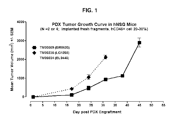
FIG. 1 shows growth curve for non-HLA matched tumors in humanized NSG mouse.
FIG. 2 shows the growth curve of the non-HLA matched SKOV3 ovarian cancer
xenograft in humanized NSG mouse (n=7).
FIG. 3 shows hCD45+ cells (%) in peripheral blood at 50 days post SKOV3 cancer
cell inoculation.
FIGs. 4A-4C show growth curves for non-HLA matched tumors (BR0744, LG0977,
and 5A0209, respectively) in NSG vs. humanized NSG mice.
FIGs. 5A-5C show percentage of hCD45+ cells over total tumor (BR0744, LG0977,
and 5A0209, respectively) population in NSG vs. humanized NSG models.
- 11-
CA 02989839 2017-12-15
WO 2016/209865 PCT/US2016/038622
FIGs. 6A-6C show human lymphocyte percentage of the total infiltrating CD45+
cells
in the three non-HLA matched tumor PDX (BR0744, LG0977, and SA0209,
respectively).
FIG. 7A shows tumor volume curves of the colon cancer CN1572P5 PDX in non-
HLA matched humanized NSG model, treated by 5-FU, Avastin, and vehicle control
at the
indicated dosing regimens. FIG. 7B shows mean tumor volume on Study Day 21 in
the three
groups.
FIG. 8A shows tumor volume curves of the breast cancer MDA-MB-231 PDX in non-
HLA matched humanized NSG model, treated by cisplatin, pembrolizumab
(Keytruda), and
vehicle control at the indicated dosing regimens. FIG. 8B shows mean tumor
volume on
Study Day 20 in the pembrolizumab (Keytruda) and vehicle groups. A similar
experiment as
the one in FIG. 8A was run and the result was shown in FIG. 8C.
FIGs. 9A-9D show that human T cells (both CD3+CD4+ and CD3+CD8 ) and B cells
(CD19 ) are present in the peripheral blood of the subject Hu-CD34 NSGTM non-
HLA
matched MDA-MB-231 PDX mice.
FIG. 10A-10C show that human T cells are present in the tumor tissue of the
subject
Hu-CD34 NSGTM non-HLA matched MDA-MB-231 PDX mice.
FIG. 11A shows tumor volume curves of the breast cancer BR1126 PDX in non-HLA
matched humanized NSG model, treated by cisplatin, pembrolizumab (Keytruda),
and
vehicle control at the indicated dosing regimens. FIG. 11B shows mean tumor
volume on
Study Day 17 in the three groups. A similar experiment as the one in FIG. 11A
was run and
the result was shown in FIG. 11C.
FIGs. 12A-12D show that human T cells (both CD3+CD4+ and CD3+CD8 ) and B
cells (CD19 ) are present in the peripheral blood of the subject Hu-CD34 NSGTM
non-HLA
matched BR1126 PDX mice.
FIG. 13A-13D show that human T and B cells are present in the tumor tissue of
the
subject Hu-CD34 NSGTM non-HLA matched BR1126 PDX mice. FIG. 13E-13H show that
human T and B cells are present in the spleens of the subject Hu-CD34 NSGTM
non-HLA
matched BR1126 PDX mice.
FIG. 14A shows tumor volume curves of the lung cancer LG1306 PDX in non-HLA
matched humanized NSG model, treated by pembrolizumab (Keytruda), with or
without
Decetaxol, and vehicle control at the indicated dosing regimens. FIG. 14B
shows mean
- 12-
CA 02989839 2017-12-15
WO 2016/209865 PCT/US2016/038622
tumor volume on Study Day 24 in the three groups. A similar experiment as the
one in FIG.
14A was run and the result was shown in FIG. 14C.
FIGs. 15A-15D show that human T cells (both CD3+CD4+ and CD3+CD8 ) and B
cells (CD19 ) are present in the peripheral blood of the subject Hu-CD34 NSGTM
non-HLA
matched LG1306 PDX mice. FIG. 15E-15H show that human T and B cells are
present in
the spleens of the subject Hu-CD34 NSGTM non-HLA matched LG1306 PDX mice. FIG.
15I-15K show that human T and B cells are present in the tumor tissue of the
subject Hu-
CD34 NSGTM non-HLA matched LG1306 PDX mice.
FIG. 16 shows immuno-staining for the presence of CD45 CD8+ infiltrating T
cells in
PDX samples treated by vehicle (control), chemotherapy alone, anti-PD1
(Keytruda), and
anti-CTLA4 agent (ipilimumab). The data shows that anti-PD1 and anti-CTLA4
therapies
led to strong presence of infiltrating T cells in the PDX tumors.
DETAILED DESCRIPTION OF THE INVENTION
1. Overview
The traditional approach to cancer treatment utilizes broad-acting chemical
agents that
are toxic to rapidly dividing cells, such as tumor / cancer cells. This
chemotherapeutic
approach can be successful but can be complicated by a wide array of off-
target toxicities and
has the risk of inducing drug resistance. Mammalian immune systems have
developed a
number of efficient, highly specific mechanisms for eliminating target cells,
including cells
that are infected with pathogens and those that have become cancerous. In
response, tumor
cells have developed their own suite of mechanisms for evading
immunedetection. Hence,
gaining a better understanding of the interaction between immune effector
cells and tumors
opens a new and promising avenue of treatment strategies that stimulate
durable, immune-
mediated tumor regression for clinical use. This class of new immuno-oncology
treatment
strategies are highly encouraging, yet further research in this field can
benefit from the
subject humanized, small animal model-based (e.g., mouse-based) in vivo
testing platform
that permits insights into a better biological understanding of human immune
and tumor cell
interactions, and enables preclinical testing of new therapies that have a
higher likelihood for
success when translated to clinical application.
The invention described herein is partly based on the surprising discovery
that growth
rate of the patient-derived xenograft (PDX) in the human CD34 -engrafted NSG
mice does
- 13 -
CA 02989839 2017-12-15
WO 2016/209865 PCT/US2016/038622
not require complete HLA-type matching between the PDX and the engrafted human
immune
cells.
The invention described herein is also partly based on the surprising
discovery that
timing of cancer cell line engraftment, relative to humanization, has no
significant impact on
xenograft growth. On the other hand, timing of cancer cell line engraftment
also has no
significant effect on CD45+ cell population.
Thus one aspect of the invention provides a humanized immunodeficient non-
obese
diabetic (NOD) mouse, wherein the mouse: (1) is homozygous for the scid
mutation; (2) has
an IL-2 receptor gamma chain deficiency; (3) is engrafted with CD34+ human
hematopoietic
stem cells (HSCs); (4) is inoculated with a human patient-derived xenograft
(PDX); wherein
the HSCs and the PDX are non-HLA matched.
As used herein, "non-HLA matched" refers to not complete HLA-matched,
including
only partial HLA-match, or not HLA-matched. In certain embodiments, there is
only partial
HLA-match between the HSCs and the PDX. In certain embodiments, there is no
HLA-
match between the HSCs and the PDX.
In certain embodiments, the mouse is an NSG mouse, or a closely related
derivative
such as an NSGS mouse, an NSG-SGM3 mouse, or a human CD34+ engrafted BLT-
mouse.
The humanized mouse of the invention can be used in a broad spectrum of
biological,
medical, and clinical research, including cancer biology, immuno-oncology,
regenerative
medicine, human hematopoiesis, infectious diseases, transplantation,
preclinical drug efficacy
testing studies, and immunity and autoimmunity, just to name a few.
For example, the subject humanized mouse models can be used to study immune
response in cancer therapy, treatment of infectious disease, gene therapy, and
immunogenecity of large molecule drugs, etc.
The subject humanized mouse models can also be used in preclinical prediction
studies, such that a patient-specific xenograft (such as a PDX from a cancer)
can be studied in
the subject mouse model, in the presence of engrafted human hematopoietic
systems. The
effect, safety (e.g., any associated side effect on immune system), and
efficacy of any test
compounds or drugs can be studied by administering such test compounds or
drugs under one
or more dosing regimens to the subject mouse model. This is particularly
powerful in
studying immuno-oncology or immuno-modulators, or any study involving the
interaction
between a diseased tissue (e.g., cancer, autoimmune disease) and the immune
system.
- 14 -
CA 02989839 2017-12-15
WO 2016/209865 PCT/US2016/038622
The subject humanized mouse models can further be used to study any PDX in the
presence of engrafted human hematopoietic system. This includes conducting
tumor
histology studies, omic studies or profiling (proteomic, genomic, metablomic,
etc.). In
certain embodiments, information concerning the PDX under study may be
obtained from the
Mouse Tumor Biology Database (MTB), which was designed to aid researchers in
such areas
as choosing experimental models, reviewing patterns of mutations in specific
cancers, and
identifying genes that are commonly mutated across a spectrum of cancers.
The subject humanized mouse models can also be used to study human immune
system development and function, including development of humanized mouse
models,
analysis of innate immune cell function, examination of T cell homeostasis,
and/or
characteristics of the BLT (fetal thymus / fetal liver) mouse model.
With the general aspects of the invention described above, certain aspects or
embodiments of the invention are further described in the sections below.
2. Definitions
Unless indicated otherwise, scientific and technical terms used herein are
intended to
have the meanings commonly understood by those of ordinary skill in the art.
Such terms are
found defined and used in context in various standard references
illustratively including J.
Sambrook and D. W. Russell, Molecular Cloning: A Laboratory Manual, Cold
Spring Harbor
Laboratory Press; 3rd Ed., 2001; F. M. Ausubel, Ed., Short Protocols in
Molecular Biology,
Current Protocols; 5th Ed., 2002; B. Alberts et al., Molecular Biology of the
Cell, 4th Ed.,
Garland, 2002; D. L. Nelson and M. M. Cox, Lehninger Principles of
Biochemistry, 4th Ed.,
W.H. Freeman & Company, 2004; Herdewijn, P. (Ed.), Oligonucleotide Synthesis:
Methods
and Applications, Methods in Molecular Biology, Humana Press, 2004; A. Nagy,
M.
Gertsenstein, K. Vintersten, R. Behringer (Eds.) 2002, Manipulating the Mouse
Embryo: A
Laboratory Manual, 3rd edition, Cold Spring Harbor Laboratory Press, ISBN-10:
0879695919; and K. Turksen (Ed.), "Embryonic Stem Cells: Methods And Protocols
in
Methods," MoL Biol., 185:499, 2002, Humana Press; Current Protocols in Stem
Cell Biology,
ISBN: 9780470151808.
The singular terms "a," "an," and "the" are not intended to be limiting and
include
plural referents unless explicitly stated otherwise or the context clearly
indicates otherwise.
- 15 -
CA 02989839 2017-12-15
WO 2016/209865 PCT/US2016/038622
The terms "express," "expression," "expressing" and "expresses" with reference
to a
gene or refer to transcription of the gene to produce a corresponding mRNA
and/or
translation of the mRNA to produce a functional corresponding encoded protein.
The term "immunodeficient non-human animal" refers to a non-human animal
(e.g.,
mouse) characterized by one or more of: a lack of functional immune cells,
such as T cells
and B cells; a DNA repair defect; a defect in the rearrangement of genes
encoding antigen-
specific receptors on lymphocytes; and a lack of immune functional molecules
such as IgM,
IgGl, IgG2a, IgG2b, IgG3 and IgA.
The term "immunodeficient mouse" refers to a mouse characterized by one or
more
of: a lack of functional immune cells, such as T cells and B cells; a DNA
repair defect; a
defect in the rearrangement of genes encoding antigen-specific receptors on
lymphocytes;
and a lack of immune functional molecules such as IgM, IgGl, IgG2a, IgG2b,
IgG3 and IgA.
Immunodeficient mice can be characterized by one or more deficiencies in a
gene involved in
immune function, such as Ragl and Rag2 (Oettinger et al., Science, 248:1517-
1523, 1990;
and Schatz et al., Cell, 59:1035-1048, 1989). Immunodeficient mice may have
any of these
or other defects which result in abnormal immune function in the mice.
Particularly useful immunodeficient mouse strains are NOD.Cg-PrkdcsadI12rel
wil/SzJ, commonly referred to as NOD scid gamma (NSG) mice, described in
detail in Shultz
et al.õ J. Immunol., 174:6477-6489, 2005; and NOD.Cg-Ragrmim'2reiwillSzJ,
Shultz et
al., Clin. Exp. Immunol., 154(2):270-284, 2008, commonly referred to as NRG
mice.
The term "severe combined immune deficiency (SCID)" refers to a condition
characterized by absence of T cells and lack of B cell function.
Common forms of SCID include: X-linked SCID which is characterized by gamma
chain gene mutations in the IL2RG gene and the lymphocyte phenotype TO BO NKO;
and
autosomal recessive SCID characterized by Jak3 gene mutations and the
lymphocyte
phenotype TO BO NKO, ADA gene mutations and the lymphocyte phenotype TO BO
NKO, IL-7R alpha-chain mutations and the lymphocyte phenotype TO BO NK( ), CD3
delta or epsilon mutations and the lymphocyte phenotype TO BO NK( ), RAG1/RAG2
mutations and the lymphocyte phenotype TO BO NK( ), Artemis gene mutations and
the
lymphocyte phenotype TO BO NK( ), CD45 gene mutations and the lymphocyte
phenotype
TO BO NK( ).
A genetically modified mouse according to aspects of the present invention has
the
severe combined immunodeficiency mutation (Prkdcsad), commonly referred to as
the scid
- 16-
CA 02989839 2017-12-15
WO 2016/209865 PCT/US2016/038622
mutation. The scid mutation is well-known and located on mouse chromosome 16
as
described in Bosma et al., Immunogenetics, 29:54-56, 1989. Mice homozygous for
the scid
mutation are characterized by an absence of functional T cells and B cells,
lymphopenia,
hypoglobulinemia and a normal hematopoetic microenvironment. The scid mutation
can be
detected, for example, by detection of markers for the scid mutation using
well-known
methods, such as PCR or flow cytometry.
A genetically modified mouse according to aspects of the present invention has
an
IL2 receptor gamma chain deficiency. The term "IL2 receptor gamma chain
deficiency"
refers to decreased IL2 receptor gamma chain. Decreased IL2 receptor gamma
chain can be
due to gene deletion or mutation. Decreased IL2 receptor gamma chain can be
detected, for
example, by detection of IL2 receptor gamma chain gene deletion or mutation
and/or
detection of decreased IL2 receptor gamma chain expression using well-known
methods. In
certain embodiments, the IL2 receptor gamma chain deficiency is a null
mutation of the IL2
receptor gamma chain gene. In certain embodiments, the animal having IL2
receptor gamma
chain deficiency is a homozygous mutant for the IL2 receptor gamma chain.
Genetically modified immunodeficient mice having the scid mutation, or an IL2
receptor gamma chain deficiency in combination with the scid mutation are
provided
according to aspects of the present invention. Genetically modified NOD scid
gamma mice
are provided according to aspects of the present invention.
The terms "NOD scid gamma" and "NSG" are used interchangeably herein to refer
to
a well-known immunodeficient mouse strain NOD .Cg-Prkdcsad NSG mice combine
multiple
immune deficits from the NOD/ShiLtJ background, the severe combined immune
deficiency
(scid) mutation, and a complete knockout of the interleukin-2 receptor gamma
chain. As a
result, NSG mice lack mature T, B and NK cells, and are deficient in cytokine
signaling.
NSG mice are characterized by lack of IL2R-y (gamma c) expression, no
detectable serum
immunoglobulin, no hemolytic complement, no mature T lymphocytes, and no
mature natural
killer cells.
Genetically modified immunodeficient non-human animals (e.g., mice) having
severe
combined immunodeficiency or an IL2 receptor gamma chain deficiency in
combination with
severe combined immunodeficiency are provided according to aspects of the
present
invention.
- 17 -
CA 02989839 2017-12-15
WO 2016/209865 PCT/US2016/038622
Generation of a genetically modified immunodeficient non-human animal can be
achieved by introduction of a gene targeting vector into a preimplantation
embryo or stem
cells, such as embryonic stem (ES) cells or induced pluripotent stem (iPS)
cells.
The term "gene targeting vector" refers to a double-stranded recombinant DNA
molecule effective to recombine with and mutate a specific chromosomal locus,
such as by
insertion into or replacement of the targeted gene.
The term "wild-type" refers to a naturally occurring or unmutated organism,
protein
or nucleic acid.
Optionally, genetically modified immunodeficient non-human animals (e.g.,
mice) of
the present invention are produced by selective breeding. A first parental
strain of non-
human animal which has a first desired genotype may be bred with a second
parental strain of
non-human animal which has a second desired genotype to produce offspring
which are
genetically modified non-human animals having the first and second desired
genotypes.
Genetically modified immunodeficient non-human animals of the present
invention
are preferably non-human mammals, particularly rodents, such as mice, rats or
guinea pigs.
A genetically modified immunodeficient mouse having an IL2 receptor gamma
chain
deficiency in combination with the scid mutation provided according to aspects
of the present
invention may be an NSG mouse, an NSGS mouse, a human CD34 + HSC engrafted NSG
/
NSGS mouse, or a human CD34 + engrafted BLT-mouse.
The term "xenogeneic" is used herein with reference to a host cell or organism
to
indicate that the material referred to as "xenogeneic" is derived from another
species than that
of the host cell or organism.
The term "hematopoietic stem cells" as used herein refers to multipotent stem
cells
functional to give rise to an immune system. Hematopoietic stem cells from
mice express c-
Kit receptor. C-Kit receptor is well-known in the art, for example as
described in
Vandenbark et al., "Cloning and structural analysis of the human c-kit gene,"
Onco gene,
7(7): 1259-1266, 1992 ; and Edling & Hallberg, "c-Kit--a hematopoietic cell
essential
receptor tyrosine kinase," Int. J. Biochem. Cell Biol., 39(11):1995-1998,
2007. Human
hematopoietic stern cells express CD34. CD34 is a well-known protein, for
example as
described in Simmons et al., "Molecular cloning of a cDNA encoding CD34, a
sialomucin of
human hematopoietic stem cells," J. Immunol., 148(1):267-271, 1992.
- 18 -
CA 02989839 2017-12-15
WO 2016/209865 PCT/US2016/038622
According to aspects of the present invention, xenogeneic (e.g., human)
hematopoietic stem cells are administered to a genetically modified
immunodeficient non-
human animal (e.g., mouse) of the present invention, wherein the xenogeneic
hematopoietic
stem cells differentiate into xenogeneic immune cells in the genetically
modified
immunodeficient non-human animal.
According to aspects of the present invention, human hematopoietic stem cells
are
administered to a genetically modified immunodeficient mouse of the present
invention,
wherein the human hematopoietic stem cells differentiate into human immune
cells in the
genetically modified immunodeficient mouse.
Hematopoietic stem cells for administration to a genetically modified
immunodeficient animal can be obtained from any tissue containing HSC such as,
but not
limited to, umbilical cord blood, bone marrow, GM-CSF-mobilized peripheral
blood and fetal
liver.
Optionally, hematopoietic stem cells for administration to a genetically
modified
immunodeficient animal can be obtained as cells cultured in vitro prior to
administration to
expand the population of cells obtained from one or more tissues containing
HSC such as, but
not limited to, umbilical cord blood, bone marrow, GM-CSF-mobilized peripheral
blood and
fetal liver.
HSC can be administered into newborn animals by administration via various
routes,
such as, but not limited to, into the heart (intracardiac injection), liver
(intrahepatic injection)
and/or facial vein. HSC can be administered into adult animals by various
routes, such as,
but not limited to, administration into the tail vein, into the femur bone
marrow cavity or into
the spleen. In a further example, the HSC as fetal liver and/or fetal thymus
can be engrafted
under the renal capsule (e.g., as 1 mm3 cube organoids in BLT mouse).
Optionally, HSC are administered to a conditioned animal. Conditioning of a
recipient animal in preparation for receipt of HSC is performed to deplete or
suppress the
HSCs and progenitor cells endogenous to the recipient animal prior to receipt
of the
xenogeneic HSCs. Conditioning of a recipient animal includes administration of
radiation
and/or one or more chemical agents effective to deplete or suppress the HSCs
and progenitor
cells endogenous to the recipient animal prior to receipt of the xenogeneic
HSCs. Busulfan is
a well-known example of a chemical agent effective to deplete or suppress the
HSCs and
progenitor cells endogenous to the recipient animal prior to receipt of the
xenogeneic HSCs.
Conditioning by radiation and/or one or more chemical agents effective to
deplete or suppress
- 19-
CA 02989839 2017-12-15
WO 2016/209865 PCT/US2016/038622
the HSCs and progenitor cells endogenous to the recipient animal prior to
receipt of the
xenogeneic HSCs is performed according to well-known protocols to produce a
conditioned
animal.
Engraftment of xenogeneic HSC can be assessed by any of various methods, such
as,
but not limited to, flow cytometric analysis of cells in the animals to which
the xenogeneic
HSC are administered at one or more time points following the administration
of HSC.
Exemplary methods for isolation of xenogeneic HSC, administration of the
xenogeneic HSC to a host organism and methods for assessing engraftment
thereof are
described herein and in T. Pearson et al., Curr. Protoc. Immunol., 81:15.21.1-
15.21.21, 2008;
Ito et al., Blood, 100:3175-3182, 2002; Traggiai et al., Science, 304:104-107,
2004; Ishikawa
et al., Blood, 106:1565-1573, 2005; Shultz et al., J. Immunol. 174: 6477-6489,
2005;
Holyoake et al., Exp Hematol., 27(9):1418-1427, 1999, all incorporated by
reference.
The HSCs administered are isolated from an original source material to obtain
a
population of cells enriched in HSCs. The isolated HSCs may or may not be
pure.
In certain embodiments, HSCs are purified by selection for a cell marker, such
as
CD34.
In certain embodiments, administered human HSCs are a population of human
cells in
which CD34+ cells constitute about 1-100% of total cells, although a
population of human
cells in which CD34+ cells constitute fewer than 1% of total cells can be
used. In certain
embodiments, administered human HSCs are T cell depleted umbilical cord blood
cells in
which CD34+ cells make up about 1-3% of total cells, lineage depleted
umbilical cord blood
cells in which CD34+ cells make up about 50% of total cells, or CD34+
positively selected
cells in which CD34+ cells make up about 90% of total cells.
The number of HSCs administered is not considered limiting with regard to
generation of a xenogeneic immune system in an immunodeficient mouse. A single
HSC can
generate cells of an immune system. Thus, the number of administered HSCs is
generally in
the range of 1-10 x 106 HSCs where the recipient is a mouse, although more can
be used. For
other species, the number of cells can be adjusted if necessary using only
routine
experimentation.
In general, HSCs are present as a subpopulation of CD34+ cells in a larger
population
of CD34 . Thus, administration of a population of CD34+ cells obtained from
any tissue
containing HSC such as, but not limited to, umbilical cord blood, bone marrow,
GM-CSF-
mobilized peripheral blood and fetal liver is administered to deliver the HSC
subpopulation
- 20 -
CA 02989839 2017-12-15
WO 2016/209865 PCT/US2016/038622
to the recipient animal to be engrafted. The number of CD34+ cells obtained
from any tissue
containing HSC such as, but not limited to, umbilical cord blood, bone marrow,
GM-CSF-
mobilized peripheral blood and fetal liver administered to deliver the HSC
subpopulation to
the recipient animal to be engrafted is not limited and can be in the range of
1 cell-1 billion
cells, such as 1 cell-500 million cells, 1 cell-100 million cells, 1 cell-10
million cells, 1 cell-5
million cells, 1 cell-1 million cells, 1 cell-500,000 cells, 1 cell-100,000
cells, 1 cell 50,000
cells, 1 cell-10,000 cells, 1 cell-1,000 cells, of such CD34+ cells. Further,
the number of
CD34+ cells administered is in the range of 100 cells-10 million cells, 100
cells-5 million
cells, 100 cells-1 million cells, 100 cells-500,000 cells, 100 cells-100,000
cells, 100 cells-
50,000 cells, 100 cells-10,000 cells or 100 cells-1,000 cells. Still further,
the number of
CD34+ cells administered is in the range of 1000 cells-10 million cells, 1000
cells-5 million
cells, 1000 cells-1 million cells, 1000 cells-500,000 cells, 1000 cells-
100,000 cells, 1000
cells-50,000 cells or 1000 cells-10,000 cells.
Engraftment is successful where xenogeneic HSCs and cells differentiated from
the
HSCs in the recipient animal are detected at a time when the majority of any
administered
non-HSC have degenerated. The hallmark of successful human HSC engraftment is
multi-
lineage human immune cell differentiation and homing to bone marrow, thymus,
spleen, and
PBL, etc. NSG mice support multi-lineage engraftment and immune cell homing
into nearly
all of the appropriate organs and tissues. The full range of the human immune
cell
populations detected in hu-CD34 NSG mice are summarized in Ishikawa et al.
(Blood,
106(5): 1565-1573, 2005); and Tanaka et al. (J. Immunol., 188(12): 6145-6155,
2012).
Detection of differentiated HSC cells can be achieved by detection of
xenogeneic
DNA in the recipient animal or detection of intact xenogeneic HSCs and cells
differentiated
from the HSCs, for example. Serial transfer of CD34+ cells into a secondary
recipient and
engraftment of a xenogeneic hematopoietic system is a further test of HSC
engraftment in the
primary recipient. Engraftment can be detected by flow cytometry as 0.05% or
greater
xenogeneic CD45+ cells in the blood at 10-12 weeks after administration of the
HSC.
Methods are provided according to aspects of the present invention which
include
delivery of xenogeneic stem cell factor (SCF) to the xenogeneic hematopoietic
stem cells in
the immunodeficient animals. The SCF may be delivered acutely or chronically
to the
animals. According to aspects of the present invention, the immunodeficient
non-human
animals may further include a transgene encoding a xenogeneic SCF operably
linked to a
promoter. In a further option, where the animals express the xenogeneic SCF,
the animals are
- 21 -
CA 02989839 2017-12-15
WO 2016/209865 PCT/US2016/038622
not conditioned by administration of a radiomimetic agent prior to
administering the
xenogeneic stem cells.
Methods for identifying modulators of an immune system response according to
aspects of the present invention include providing a non-human genetically
modified
immunodeficient animal; administering xenogeneic hematopoietic stem cells to
the non-
human genetically modified immunodeficient animal, wherein the xenogeneic
hematopoietic
stem cells differentiate to produce xenogeneic immune cells in the non-human
genetically
modified immunodeficient animal; administering an immune system stimulator to
the animal;
administering a test compound to the animal; assaying a response of the
xenogeneic immune
cells to the immune system stimulator; and comparing the response to a
standard to determine
the effect of the test compound on the response of the xenogeneic immune cells
to the
stimulator, wherein an effect of the test substance identifies a modulator of
the xenogeneic
immune system in the animal.
A test compound used in a method of the present invention can be any chemical
entity, illustratively including a synthetic or naturally occurring compound
or a combination
of a synthetic or naturally occurring compound, a small organic or inorganic
molecule, a
protein, a peptide, a nucleic acid, a carbohydrate, an oligosaccharide, a
lipid or a combination
of any of these.
A sample as used herein can be a sample obtained from a non-human animal,
illustratively includes spleen, bone marrow, blood, blood plasma and blood
serum.
Optionally, particular cell populations of the immune system are assayed, such
as
dendritic cells, plasmacytoid dendritic cells, myeloid dendritic cells, mast
cells,
monocytes/macrophages, natural killer cells, neutrophils, basophils and
eosinophils, T
lymphocytes (CD3+CD4+ or CD3+CD8+ T cells), B lymphocytes (e.g., CD19+ B
cells).
Isolated bone marrow cells of genetically modified immunodeficient non-human
animals having an engrafted human immune system are provided by the present
invention.
Isolated bone marrow cells of genetically modified immunodeficient non-human
animals
having an engrafted human immune system are provided by the present invention.
Isolated cells of genetically modified immunodeficient non-human animals are
provided by the present invention. Such isolated cells can be cultured in
vitro for use in
various assays. For example, such isolated cells are useful as controls in
assays for
assessment of a test substance to determine the activity of the test
substance. In a further
- 22 -
CA 02989839 2017-12-15
WO 2016/209865 PCT/US2016/038622
example, such isolated bone marrow cells are useful to determine the activity
of the test
substance on activity of the immune system.
Immunoassay methods can be used to assay a target analyte or an indicator of
immune
cell response in a sample, including, but not limited to, enzyme-linked
immunosorbent assay
(ELISA), enzyme-linked immunofiltration assay (ELIFA), flow cytometry,
immunoblot,
immunoprecipitation, immunohistochemistry, immunocytochemistry, luminescent
immunoassay (LIA), fluorescent immunoassay (FIA), and radioimmunoassay. Assay
methods may be used to obtain qualitative and/or quantitative results.
Specific details of
suitable assay methods for both qualitative and quantitative assay of a sample
are described in
standard references, illustratively including E. Harlow and D. Lane,
Antibodies: A
Laboratory Manual, Cold Spring Harbor Laboratory Press, 1988; F. Breitling and
S. Dubel,
Recombinant Antibodies, John Wiley & Sons, New York, 1999; H. Zola, Monoclonal
Antibodies: Preparation and Use of Monoclonal Antibodies and Engineered
Antibody
Derivatives, Basics: From Background to Bench, BIOS Scientific Publishers,
2000; B. K. C.
Lo, Antibody Engineering: Methods and Protocols, Methods in Molecular Biology,
Humana
Press, 2003; F. M. Ausubel et al., Eds., Short Protocols in Molecular Biology,
Current
Protocols, Wiley, 2002; S. Klussman, Ed., The Aptamer Handbook: Functional
Oligonucleotides and Their Applications, Wiley, 2006; Ormerod, M. G., Flow
Cytometry: A
Practical Approach, Oxford University Press, 2000; Givan, A. L., Flow
Cytometry: First
Principles, Wiley, New York, 2001; Gorczyca, W., Flow Cytometry in Neoplastic
Hematology: Morphologic-Immunophenotypic Correlation, Taylor & Francis, 2006;
Crowther, J. R., The ELISA Guidebook (Methods in Molecular Biology), Humana
Press,
2000; Wild, D., The Immunoassay Handbook, 3rd Edition, Elsevier Science, 2005;
and J.
Sambrook and D. W. Russell, Molecular Cloning: A Laboratory Manual, Cold
Spring Harbor
Laboratory Press, 3rd Ed., 2001.
Antibodies and methods for preparation of antibodies are well-known in the
art. As
used herein, the terms "antibody" and "antibodies" encompass monoclonal
antibodies,
polyclonal antibodies, bispecific antibodies, multispecific antibodies, human
antibodies,
humanized antibodies, chimeric antibodies, camelized antibodies, single domain
antibodies,
single-chain Fvs (scFv), single chain antibodies, disulfide-linked Fvs (sdFv),
and anti-
idiotypic (anti-Id) antibodies and antigen-binding fragments of any of the
above. In
particular, antibodies include immunoglobulin molecules and immunologically
active
fragments of immunoglobulin molecules, i.e., molecules that contain an antigen
binding site.
- 23 -
CA 02989839 2017-12-15
WO 2016/209865 PCT/US2016/038622
Immunoglobulin molecules are of any type (e.g., IgG, IgE, IgM, IgD, IgA and
IgY), class
(e.g., IgGl, IgG2, IgG3, IgG4, IgAl and IgA2), or subclass.
As used herein, the terms "antibody fragment" and "antigen-binding fragment"
defines a fragment of an antibody that immuno specifically binds to a target
analyte. Antibody
fragments may be generated by any technique known to one of skill in the art.
For example,
Fab and F(ab')2 fragments may be produced by proteolytic cleavage of
immunoglobulin
molecules, using enzymes such as papain (to produce Fab fragments) or pepsin
(to produce
F(ab')2 fragments). Antibody fragments are also produced by recombinant DNA
technologies.
Antibodies, antigen-binding fragments, methods for their generation and
methods for
screening of generated antibodies for substantially specific binding to an
antigen are known
in the art and such antibodies, antigen binding fragments and methods are
described in further
detail, for instance, in Antibody Engineering, Kontermann, R. and Dubel, S.
(Eds.), Springer,
2001; Harlow, E. and Lane, D., Antibodies: A Laboratory Manual, Cold Spring
Harbor
Laboratory Press, 1988; F. Breitling and S. Dubel, Recombinant Antibodies,
John Wiley &
Sons, New York, 1999; H. Zola, Monoclonal Antibodies: Preparation and Use of
Monoclonal Antibodies and Engineered Antibody Derivatives, Basics: From
Background to
Bench, BIOS Scientific Publishers, 2000; Ausubel, F. et al., (Eds.), Short
Protocols in
Molecular Biology, Wiley, 2002; J. D. Pound (Ed.) Immunochemical Protocols,
Methods in
Molecular Biology, Humana Press, 2nd ed., 1998; B. K. C. Lo (Ed.), Antibody
Engineering:
Methods and Protocols, Methods in Molecular Biology, Humana Press, 2003; and
Kohler, G.
and Milstein, C., Nature, 256:495-497 (1975). Antibodies for target analytes,
such as toll-
like receptor 4 or indicators of innate immune cell response, can be produced
in animals,
synthesized, produced by recombinant methods and/or obtained commercially.
Detecting binding between a target analyte present in a sample and a binding
partner
is achieved by any of various methods known in the art, illustratively
including detection of a
detectable label directly or indirectly attached to the target analyte or the
binding partner. The
term "detectable label" refers to a material capable of producing a signal
indicative of the
presence of the detectable label by any appropriate method illustratively
including
spectroscopic, optical, photochemical, biochemical, enzymatic, electrical
and/or
immunochemical. Examples of detectable labels illustratively include a
fluorescent moiety, a
chemiluminescent moiety, a bioluminescent moiety, an electron dense particle,
a magnetic
particle, an enzyme, a substrate, a radioisotope and a chromophore.
- 24 -
CA 02989839 2017-12-15
WO 2016/209865 PCT/US2016/038622
The identity of a particular detectable label or labels used depends on the
detection
process used. Such detection processes are incorporated in particular assay
formats
illustratively including ELISA, Western blot, immunoprecipitation,
immunocytochemistry,
immuno-fluorescence assay, liquid chromatography, flow cytometry, other
detection
processes known in the art, or combinations thereof.
A binding assay can incorporate a binding partner attached to a support. A
support
with attached binding partner used in a binding assay can be solid or semi-
solid and can be
any of various materials such as glass, silicon, paper, a synthetic or
naturally occurring
polymer, such as polystyrene, polycarbonate, polypropylene, PVDF, nylon,
cellulose,
agarose, dextran, and polyacrylamide or any other material to which a binding
partner can be
stably attached for use in a binding assay.
A support used can include functional groups for binding to binding partners,
such as,
but not limited to carboxyl, amine, amino, carboxylate, halide, ester,
alcohol, carbamide,
aldehyde, chloromethyl, sulfur oxide, nitrogen oxide, epoxy and/or tosyl
functional groups.
Attachment of binding partners to a support is achieved by any of various
methods,
illustratively including adsorption and chemical bonding. In one example, 1-
Ethy1-343-
dimethylaminopropyl] carbodiimide hydrochloride, EDC or EDAC chemistry, can be
used to
attach binding partners to particles. The binding partners can be bonded
directly or indirectly
to the material of the support, for example, via bonding to a coating or
linker disposed on the
support. Functional groups, modification thereof and attachment of a binding
partner to a
support are known in the art, for example as described in Fitch, R. M.,
Polymer Colloids: A
Comprehensive Introduction, Academic Press, 1997.
Such supports can be in any of a variety of forms and shapes including, but
not
limited to, microtiter plates, microtiter wells, pins, fibers, beads, slides,
silicon chips and
membranes such as a nitrocellulose or PVDF membrane.
Any of various spectroscopy methods can be used to assay a target analyte,
such as
toll-like receptor 4 or an indicator of innate immune cell response, according
to aspects of the
present invention, including, but not limited to, gas chromatography, liquid
chromatography,
ion mobility spectrometry, mass spectrometry, liquid chromatography-mass
spectrometry
(LC-MS or HPLC-MS), ion mobility spectrometry-mass spectrometry, tandem mass
spectrometry, gas chromatography-mass spectrometry, matrix-assisted desorption
ionization
time-of-flight (MALDI-TOF) mass spectrometry, surface-enhanced laser
desorption
- 25 -
CA 02989839 2017-12-15
WO 2016/209865 PCT/US2016/038622
ionization (SELDI) and nuclear magnetic resonance spectroscopy, all of which
are well-
known to the skill artisan.
Optionally, spectrometric analysis is used to assay a sample for a target
analyte such
as toll-like receptor 4 or an indicator of innate immune cell response. Mass
analysis can be
used in an assay according to aspects of the present invention. Mass analysis
is conducted
using, for example, time-of-flight (TOF) mass spectrometry or Fourier
transform ion
cyclotron resonance mass spectrometry. Mass spectrometry techniques are known
in the art
and exemplary detailed descriptions of methods for protein and/or peptide
assay are found in
Li J., et al., Clin Chem., 48(8):1296-1304, 2002; Hortin, G. L., Clinical
Chemistry, 52: 1223-
1237, 2006; A. L. Burlingame, et al. (Eds.), Mass Spectrometry in Biology and
Medicine,
Humana Press, 2000; and D. M. Desiderio, Mass Spectrometry of Peptides, CRC
Press, 1990.
3. The Humanized Tumor-Bearing NSG Mice
The Jackson Laboratory NSG mice (NOD.Cg-Prkdcsad Il2reniwfilSzJ, Stock No.
005557) are also commonly known as NOD scid gamma; NSG; NOD-scid
IL2Rgamma1ull;
and NOD-scid IL2Rell. They combine the features of the NOD/ShiLtJ background
(Jackson Laboratory Stock No. 001976), the severe combined immune deficiency
mutation
(scid), and IL2 receptor gamma chain deficiency. As a result, The NSG mice
lack mature T
cells, B cells (and thus does not generate mouse antibodies), and functional
NK cells, has no
complement system, and are deficient in cytokine signaling, leading to better
engraftment of
human hematopoietic stem cells (HSCs) and peripheral-blood mononuclear cells
(PBMCs)
than any other published mouse strain. The NSG mice also has defective
macrophages and
dendritic cells. Recent publications have demonstrated this strain's
outstanding utility in the
studies of islet transplantation, hematopoietic stem cells and cancer stem
cells.
Specifically, the NSG mice do not express the Prkdc gene nor the X-linked
Il2rg
gene. NSG mice are viable, fertile, normal in size and do not display any
gross physical or
behavioral abnormalities. Histological examination of lymphoid tissues reveals
absence of
lymphoid cells and some cystic structures in the thymus, an absence of
follicles in the spleen
and markedly diminished celluarity of lymph nodes. NSG mice are deficient in
mature T-
and B-lymphocytes, serum Ig is not detectable and natural killer (NK) cell
cytotoxic activity
is extremely low. These mice are resistant to lymphoma development even after
sublethal
irradiation treatment. These mutant mice have been shown to readily support
engraftment of
human CD34+ hematopoietic stem cells and represent a superior, long-lived
(median survival
is over 89 weeks) model suitable for studies employing xenotransplantation
strategies.
- 26 -
CA 02989839 2017-12-15
WO 2016/209865 PCT/US2016/038622
The NSG mice carry the true null interleukin-2 receptor gamma chain mutation,
as
opposed to other strains that express a truncated interleukin-2 receptor gamma
chain (see
Ohbo et al., Blood, 87:956-967, 1996). The NSG mice are available to non-
profit research
institutions under a material transfer agreement (MTA), and the Jackson
Laboratory
distributes NSG mice under an agreement with the NIH.
To create the subject humanized NSG mouse (HU-NSGTm), hematopoietic stem
cells,
such as the human CD34+ HSCs, are introduced into the Jackson Laboratory NSG
mice by,
for example, tail vein injection, intracardiac injection, or intrahepatic
injection. The HSCs can
be introduced into about 2-, 3-, or 4-week old NSG mice. Typically, 25-gauge
needles can be
used for tail vein injection. Smaller gauge needles can also be used, with
potentially increased
shearing of the cells in the inocula.
Alternative sites for delivery of HSCs include the retroorbital venous sinus,
the bone
marrow cavity itself, and the spleen. Injection into the retroorbital sinus is
easier to perform,
but more invasive, than using the tail vein, and it requires the recipient
mouse to be
anesthetized. The homing of stem cells to the marrow is dependent on molecules
such as
stromal-derived factor 1 and stem cell factor that guide the stem cells from
the peripheral
blood to the marrow cavity. Therefore, delivery of the stem cells into the
circulation (or
orthotopically into the marrow) increases the likelihood that the cells will
establish residence
in the bone marrow of the new host.
Optionally, just prior to HSCs introduction, the NSG mice are exposed whole-
or
total-body irradiation (TBI) for myeloablation, which can be achieved by
placing the NSG
mice in specifically designed irradiators, with a dose of whole-body gamma
irradiation
designed to causes the animals to become either transiently or chronically
immuno suppressed.
Successful survival of the human immune system in the NSG mice may require
suppression of the host's immune system in some manner to prevent HVG (host-vs-
graft)
rejections. In addition to suppressing the host's immune system, irradiation
also helps
deplete the bone marrow niche of host progenitor cells, thereby allowing space
for
engraftment of donor stem cells. For NSG mice, this preparation is commonly
accomplished
through whole-body gamma irradiation. Irradiators may vary in size depending
on their
intended use. Small irradiators (for example, the Mark-I irradiator from JL
Shepherd and
Associates, San Fernando, CA) are the size of a refrigerator and commonly are
used to
irradiate both cells and a small number of mice. In contrast, one commonly
used larger (6600
- 27 -
CA 02989839 2017-12-15
WO 2016/209865 PCT/US2016/038622
lb) gamma irradiator (the Gammacell-40, MDS Nordion, Ottawa, ON) can be used
to
irradiate several dozen mice at once. Animals are generally irradiated for
short periods of
time (less than 15 min). The amount of time spent inside the irradiator varies
depending on
the radioisotope decay charts, amount of irradiation needed, and source of
ionizing energy
(that is, X-rays versus gamma rays, for which a cesium or cobalt source is
needed). Larger
irradiators, such as Clinac 4/80 linear accelerator (Varian Medical Systems,
Palo Alto, CA)
may also be used if necessary. In general, the mice need not be anesthetized
for irradiation.
The myeloablative irradiation dose is usually 700 to 1300 cGy, though in some
embodiments, lower doses such as 1-100 cGy (e.g., about 2, 5, or 10 cGy), or
300-700 cGy
may be used. It can be either cesium- or X-ray irradiation.
In certain embodiments, female NSG mice are surgically implanted with human
thymus and liver fragments and injected with donor-matched human CD34+
hematopoietic
stem cells (humanized BLT NSG-mice). Such humanized BLT-mice (hu-BLT) have the
most functional immune system of any current humanized mouse model, and offer
distinct
advantages and improved performance in certain studies, such as mucosal-based
immune
responses.
In certain embodiments, instead of using NSG mice for humanization, the NSGS
or
NSG-SGM3 mice (NOD.Cg-Prkdcsad 112relwil Tg(CMV-1L3,CSF2,KITLG)1Eav/MloySzJ,
Jackson Laboratory Stock No. 013062, also commonly known as NOD-scid IL2Rgnull-
3/GM/SF) can be used. This is a multi-allelic mouse line combines an
immunodeficient
environment with the presence of several transgenic human cyotokines
supportive of human
myeloid cell expansion, and represents an especially useful model for the
hosting of
xenografts. In particular, these mice harbor three transgenes, human
interleukin-3 (IL-3),
human granulocyte/macrophage-stimulating factor (GM-CSF), and human Steel
factor (SF)
gene, each driven by a human cytomegalovirus promoter/enhancer sequence. These
mice are
maintained on the NSG (NOD.Cg-Prkdcsad 112relwillSzJ) mice background, and
constitutively produce 2-4 ng/mL serum levels of human IL-3, GM-CSF, and SF.
The I12rg-/-
specific NOD.SCID background supports human and murine hematopoietic cell
engraftment,
and suppresses human erythropoiesis, enhances human myelopoiesis, and reduces
human B-
lymphopoiesis in mice after transplant of human bone marrow or fetal liver
cells.
In certain embodiments, the mouse is engrafted with human peripheral blood
mononuclear cells, e.g., the hu-PBMC NSGTM mouse (or PBMC humanized mouse).
- 28 -
CA 02989839 2017-12-15
WO 2016/209865 PCT/US2016/038622
For simplicity, in certain embodiments, the various NSG or NSG derived mouse
strains may be collectively referred to as NSG mouse.
In certain embodiments, the human CD34+ HSCs are introduced into NSG mice (or
NSGS mice, or BLT-NSG mice) when the mice are around 3 weeks of age, and
mature
human B cells appear around week-12, and mature human T cells appear around
week-15.
In certain embodiments, human CD34+ engrafted NSG mice (or NSGS mice, or BLT-
NSG mice) have at least about 20%, 25%, 30% or more human CD45+ cells in the
peripheral
blood of the mice about 12 weeks post HSCs engraftment.
To create the subject humanized NSG mice (or NSGS mice, or BLT-NSG mice) with
patient-derived xenograft (PDX), the NSG mice (or NSGS mice, or BLT-NSG mice)
repopulated by human CD34+ HSCs are injected with an appropriate amount of
patient-
derived cells, such as 1-10 x 106 human cancer cells. The human origin of the
cancer cells
can be verified by Ki67 staining. In certain embodiments, the PDX is
introduced into the
mice at about 2 weeks post HSCs engraftment. In certain embodiments, the PDX
is
introduced into the mice at about 1, 2, 3, 4, 5, 6, 7, 8, 9, 10, 11, 12, 13,
14, 15 weeks post
HSCs engraftment. In certain embodiments, the PDX is introduced into the mice
before the
engrafted human immune cells (e.g., human B- or T-cells or NK cells) appear.
In certain embodiments, the HLA type of the PDX does not match the HLA type of
the donor human HSCs.
EXAMPLES
Any patents or publications mentioned in this specification are incorporated
herein by
reference to the same extent as if each individual publication is specifically
and individually
indicated to be incorporated by reference.
The non-human animals (e.g., mouse), compositions, and methods of the present
invention described herein are presently representative of certain
illustrative embodiments,
exemplary embodiments, and are otherwise not intended as limitations on the
scope of the
invention. Changes therein and other uses will occur to those skilled in the
art. Such changes
and other uses can be made without departing from the scope of the invention
as set forth in
the claims.
- 29 -
CA 02989839 2017-12-15
WO 2016/209865 PCT/US2016/038622
Example 1 Mice
NOD .Cg-Prkdcsad wil/Sa (NOD- scid NSG)
mice were obtained from
colonies developed and maintained at The Jackson Laboratory (Bar Harbor, ME).
All
animals were housed in a specific pathogen free facility, in microisolator
cages, and given
autoclaved food and maintained on sulfamethoxazole-trimethoprim medicated
water
(Goldline Laboratories, Ft. Lauderdale, Fla.) and acidified autoclaved water
on alternating
weeks.
Example 2 Engraftment of Mice with Human Hematopoietic Stem Cells (HSCs)
Groups of 24 to 72 hour-old (newborn) NSG mice are irradiated with 100 cGy as
described in Pearson et al. (Curr. Protoc. Immunol. 81:15.21.1-15.21.21,
2008). Irradiated
mice are injected with CD3 T cell-depleted human umbilical cord blood (UCB)
containing
3x10⁴ CD34+ hematopoietic stem cells (HSC) in a 25-50 [IL volume via
intracardiac injection
as described in Brehm et al. (Clinic. Immunol. 135(1):84-98, 2010). After 12
weeks, flow
cytometry analyses of the blood of HSC recipients quantifies the engraftment
of the human
immune system. For experimental studies only mice with >20% peripheral human
CD45+
cells and >5% human CD3 + T cells are used.
Similarly, sub-lethally irradiated newborn NSG mice can also be injected with
CD3 T
cell-depleted human umbilical cord blood (UCB) containing 3x10⁴ CD34+
hematopoietic
stem cells (HSC) via intrahepatic injection or through facial vein injection
as described in
Brehm et al. (Clinic. Immunol., 135(1):84-98, 2010).
Furthermore, groups of about 3-weeks old NSG mice can be subject to whole-body
sublethal irradiation at a dose of about 200 to 1300 cGy, before 0.2-1x10⁶CD34
HSCs are
injected via lateral tail vein using standard techniques. For example, each
animal is weighed
before injection, and up to about 1% of the animal's body weight in volume can
be
administered per injection. Prior to injection, warm animals for 5-10 minutes
(e.g., in a
commercially available warming box, or under an overheard heat lamp, or using
a warm
water circulating pad placed under the cage) to dilate the veins. Then lightly
anesthetize
animals are positioned on their side, on a rechargeable heat pack or
circulating warm water
pad to keep them warm during anesthesia. Then a small gauge (28-30) needle is
inserted,
bevel up, into the vein towards the direction of the animal head, trying to
keep the needle and
syringe parallel to the tail. The needle should advance smoothly into the
vein. After
-30-
CA 02989839 2017-12-15
WO 2016/209865 PCT/US2016/038622
injection, the animals are to cage and observed for 5-10 minutes to make sure
that bleeding
has not resumed.
For the BLT (bone marrow / liver / thymus) mouse model, about 1 mm3 cubes of
fetal
liver and fetal thymus are implanted as organoids into the host NSG mouse,
which has
previously been subject to about 200 cGy of whole-body irradiation. Then about
0.2-1x10⁶
CD34 HSCs are injected via lateral tail vein using standard techniques. The
BLT mouse
model allows robust and consistent xenograft (e.g., human) immune system
development,
comprising multiple hematopoietic lineages; exhibits sustained, high level T
cell
development; and the T cells are educated on autologous thymic tissues. Such
mouse model
typically has detectable T and B cell responses to viral infection (e.g., EBV
and HIV).
In any of the suitable humanized mouse models, such as in the humanized NSG
mice,
patient-derived xenograft (PDX) or cancer cell line is inoculated at specific
time points, such
as 2 or 12 weeks post human CD34+ cell injection. The xenografts are allowed
to grow, e.g.,
for about 7 weeks, with body weight and tumor volume monitored frequently
(e.g., twice per
week).
Peripheral blood from the mice may be collected at the end of the study, for
analyzing
the extent of human CD45+ donor cell engraftment. Preferably, human CD45+
donor cells
must reach at least about 20-30% in the peripheral blood.
Example 3 Recapitulation of Expected PDX Growth Rates Does Not Require HLA-
type Matching
Humanization of the NSG mouse were performed substantially as described above
in
Example 2. Briefly, groups of about 3 weeks old NSG mice were subject to whole-
body
irradiation (about 200 cGy) before about 0.2-1x10⁶ CD34 HSCs are injected via
lateral tail
vein using standard techniques. PDX xenografts using three different cancer
samples (breast
cancer cells BR0620, lung cancer cells LG1208, and bladder cancer cells
BL0440) were
implanted subcutaneously into a subject humanized NSG mouse model - i.e., hu-
CD34 NSG
mice with established and functionally mature human immune cells derived from
an
HLAmismatched human HSC donor, and the growth of the PDX engraftments were
monitored for up to about 55 days post PDX engraftment. All three tumors
showed robust
growth and no obvious indication of rejection (FIG. 1).
The results show that HLA-type matching is not required to recapitulate
expected
PDX growth rate in the subject humanized NSG mouse model.
-31 -
CA 02989839 2017-12-15
WO 2016/209865 PCT/US2016/038622
Example 4 Temporal Evaluation of PDX Engraftment on Tumor Growth in Hu-
CD34 NSGTM Mice
To evaluate any temporal effect on PDX xenograft tumor growth in non-HLA
matched humanized hCD34+ NSG mouse, about 5x10⁶ human SKOV3 ovarian cancer
cells
were inoculated either 2 or 12 weeks post non-HLA matched human CD34+ cell
injection
into the NSG mouse, according to a procedure substantially as described in
Examples 2 and 3
above. The xenografts were allowed to grow for about 7 more weeks, with body
weight and
tumor volume monitored twice per week. Peripheral blood from the mice was
collected at
the end of the study, for analyzing the extent of human CD45+ donor cell
engraftment.
Average results from two groups of 7 mice each (2 weeks vs. 12 weeks) were
shown in FIG.
2.
At 2 weeks post-engraftment, human immunity has not yet developed. Indeed,
mature human T and B cells require at least 12 weeks to become detectable in
the PBL of hu-
CD34 NSG mice. In the tumor engraftment studies, however, tumor take was 100%
in both
groups (N = 7 for both) and the increase in tumor volume over time in the 2
week group
slightly outpaced the 12 week group (FIG. 2).
The results showed that there is no significant difference between the two
groups of
mice, suggesting that timing of cancer cell line engraftment, relative to
humanization, has no
significant (if any) impact on growth of SKOV3 ovarian cancer cells.
The mice were tested for human hematopoietic chimerism 50 days after cancer
cell
inoculation and all showed 25-50% huCD45+ cells in the PBL, indicating
successful
engraftment (FIG. 3). The results showed that timing of cancer cell line
engraftment has no
significant (if any) effect on CD45+ cell population, in the non-HLA matched
humanized
NSG PDX model.
Example 5 Humanization Has No Significant Impact on PDX Growth Kinetics
An important question not addressed by these above experiments was whether the
presence of human immune cells influenced tumor growth rates when compared to
their
growth rates in normal, non-humanized NSG mice.
To determine whether humanization in the NSG mouse model has any significant
impact on growth kinetics of non-HLA matched PDX, three fresh PDX tumor
samples, breast
cancer BR0744 (FIG. 4A), lung cancer LG0977 (FIG. 4B), and soft tissue sarcoma
SA0209
-32-
CA 02989839 2017-12-15
WO 2016/209865 PCT/US2016/038622
(FIG. 4C), were independently engrafted in parallel into either the NSG mouse
model, or the
huCD34-humanized NSG mouse model, using substantially the same methods as
described
above, and the PDX growth curves were measured and plotted accordingly.
In both the NSG and humanized NSG models, take rate for all three tumors were
100%, and tumors developed in each of the engrafted hosts. Only the breast
tumor grew at a
slightly faster rate in NSG versus hu-CD34 NSG recipients (FIG. 4A); the other
two tumors
grew at the same rate in both hosts (FIGs. 4B and 4C). But overall, there is
no significant
difference in tumor growth curves in NSG vs. hNSG models in the PDX tumors
over the
entire 40-70-day post engraftment experimental period.
In all hNSG experimental groups, hCD45+ cells in peripheral blood were above
20%,
suggesting that the humanization was successful. Also see FIG. 5A-5C, showing
that no
hCD45+ cells were observed in the non-humanized NSG mice.
At the end of the tumor growth study, tumors were also collected and analyzed
by
flow cytometry for the presence of TILs. All three tumors contained human CD4+
and CD8+
T-cells, but few CD19+ B cells were detected (See FIGs. 6A-6C). While not
wishing to be
bound by any particular theory, the failure of the TILs to slow tumor growth
in the hu-CD34
NSG recipients suggests that T-cells that recognized the tumor may have become
anergic.
Together, these results demonstrate that hu-CD34 NSG mice support non-HLA
matched tumor growth and that the presence of human immune cells does not
significantly
impact tumor take or growth rates.
Example 6 Hu-CD34 NSGTM PDX Mice are Functional Platform for Evaluating
Drug Efficacy
The ability of the humanized NSG mice to support the growth of non-HLA matched
human tumors, as demonstrated above, was an important finding in the
development of this
preclinical testing platform.
To test whether the subject Hu-CD34 NSGTM mice with non-HLA matched PDX can
be used to evaluate drug efficacy against the PDX, using clinically relevant
standard-of-care
(SOC) treatments, tumor growth curves over 21 days were obtained for a
negative control /
vehicle group, and two treatment groups using 5-FU and Avastin, respectively,
against the
colon cancer CN1572P5 PDX. Specifically, 5-FU was administered i.v. at a dose
of about 20
mg/kg body weight, Q7dx2 (every 7 days, 2 total doses). Avastin was
administered i.p. at a
- 33 -
CA 02989839 2017-12-15
WO 2016/209865 PCT/US2016/038622
dose of about 10 mg/kg body weight, twice a week, 5 total doses. Vehicle (D5W)
was
administered i.v., Q7dx3.
The results in FIGs. 7A and 7B show that both 5-FU and Avastin are both
effective
against the non-HLA matched colon cancer PDX in the subject humanized NSG
model. This
demonstrates that the subject non-HLA matched Hu-CD34 NSG PDX model is a
functional
platform for evaluating the efficacy of clinically relevant standard-of-care
(SOC) drugs.
Example 7 Keytruda and Cisplatin Inhibit the Growth of PD-L1+ MDA-MB-231
Breast Cancer Tumor Model in Hu-CD34 NSGTM
Programmed cell death protein 1 (also known as PD-1 and CD279), is a protein
that in
humans is encoded by the PDCD1 gene. PD-1 is an immunoinhibitory cell surface
receptor
that belongs to the CD28 family of immunoglobulin (Ig) superfamily, and is
expressed on T
cells, pro-B cells, monocytes, natural killer cells, and many tumor-
infiltrating lymphocytes
(TILs). It is an important immune "checkpoint" receptor that inhibits the T-
cell response and
plays a key role in modulating T-cell function.
PD-1 binds two ligands, PD-Li and PD-L2, both of which have been found on
tumor
cells and both of which have been used by tumor cells to engage the PD-1
receptor on
activated T cells to suppress the function of the activated T cells, thus
evading immune
response against tumor cells. Hence, PD-1 and its ligands play an important
role in down
regulating the immune system by preventing the activation of T-cells, which in
turn down-
regulates immune response against cancer, but also reduces autoimmunity and
promotes self-
tolerance. The inhibitory effect of PD-1 is thought to be accomplished through
a dual
mechanism of promoting apoptosis (programmed cell death) in antigen specific T-
cells in
lymph nodes, while simultaneously reducing apoptosis in regulatory T cells
(suppressor T
cells).
A new class of drugs that block PD-1, the PD-1 inhibitors, activate the immune
system to attack tumors and are therefore useful to treat cancer. For example,
monoclonal
antibodies against PD-1 may boost the immune system, thus useful for treatment
of cancer.
In addition, many tumor cells express the immunosuppressive PD-1 ligand PD-Li.
Thus
inhibition of the interaction between PD-1 and PD-Li can enhance T-cell
responses in vitro
and mediate preclinical antitumor activity.
One such anti-PD-1 antibody drug, nivolumab, (Opdivo - Bristol Myers Squibb),
produced complete or partial responses in non-small-cell lung cancer,
melanoma, and renal-
- 34-
CA 02989839 2017-12-15
WO 2016/209865 PCT/US2016/038622
cell cancer, in a clinical trial with a total of 296 patients. Nivolumab also
targets PD-1
receptors, and was approved in Japan in July 2014 and by the US FDA in
December 2014 to
treat metastatic melanoma.
Another such anti-PD-1 antibody drug, pembrolizumab (Keytruda, MK-3475,
Merck),
targets PD-1 receptors, and was approved by the FDA in Sept 2014 to treat
metastatic
melanoma. Pembrolizumab has been made accessible to advanced melanoma patients
in the
UK via UK Early Access to Medicines Scheme (EAMS) in March 2015. It is also
being used
in clinical trials in the US for lung cancer and mesothelioma.
Other drugs in early stage development targeting PD-1 receptors include
Pidilizumab
(CT-011, Cure Tech), BMS 936559 (Bristol Myers Squibb), and MPDL3280A (Roche).
On the other hand, cisplatin is the first member of now a class of platinum-
containing
anti-cancer chemotherapy drugs, which also includes carboplatin and
oxaliplatin. These
platinum complexes react in vivo, binding to and causing crosslinking of DNA,
which
ultimately triggers apoptosis (programmed cell death).
To determine if engrafted PDX tumors would also respond to clinically relevant
immuno-oncology checkpoint inhibitors, or if such checkpoint inhibitors could
reactivate
anti-tumor responses in the resident human immune cells, a series of
experiments were
designed and conducted to address these questions.
First, to determine if the checkpoint inhibitor pembrolizumab (Keytruda) is
efficacious in the subject non-HLA matched PDX humanized NSG model, a PDX
huNSG
model was established according to the methods of the invention. Specifically,
humanized
NSG mice (human CD45+ cells were found to be more than 25% in the peripheral
blood of
the hNSG mice, demonstrating successful huCD34 engraftment) were engrafted
with 5x10⁶
of non-HLA matched PD-Li-positive breast cancer cell line MDA-MB-231 cells per
mouse,
via s.c. inoculation with matrigel. This cell line expresses very high levels
of PD-Li on the
tumor cell surface that can bind to PD-1 on T-cells and induce anergy - about
94.3% of the
MDA-MB-231 cells expressed PD-Li. The tumor-engrafted humanized NSG mice were
then
treated either with vehicle, Cisplatin, or Prembrolizumab (Keytruda).
Tumor growth curves over 21 days were obtained for a negative control /
vehicle
group, and two treatment groups using cisplatin and pembrolizumab (Keytruda),
respectively,
against the MDA-MB-231 PDX. Specifically, cisplatin was administered i.v. at a
dose of
about 2 mg/kg body weight, Q7dx2 (every 7 days, 2 total doses). Pembrolizumab
(Keytruda)
- 35 -
CA 02989839 2017-12-15
WO 2016/209865 PCT/US2016/038622
was administered i.p. at a dose of about 5-10 mg/kg body weight, Q5dx4 (every
5 days, 4
total doses). Vehicle (Saline) was administered i.p. , Q5dx4.
Cisplatin is a platinum-containing chemotherapeutic that causes DNA cross-
linking
and apoptosis in rapidly dividing cells. Cisplatin treatment only marginally
reduced the
growth rate of the MDA-MB-231 tumors in the humanized mice. In contrast,
Keytruda
delayed tumor growth significantly within ¨2 weeks after treatment was started
(FIGs. 8A
and 8B).
At the termination of the growth study, peripheral blood of the experimental
mice
were collected and assayed for human CD19+ B cells and human CD4+ and CD8+ T-
cells.
FIGs. 9A-9D show that human T cells, including both CD3+CD4+ and CD3+CD8+ T
cells,
and CD19+ B cells, are present in the peripheral blood of the subject Hu-CD34
NSGTM non-
HLA matched MDA-MB-231 PDX mice.
Tumors were also collected and assayed for human CD4+ and CD8+ infiltrating T-
cells. FIG. 10A-10C show that human T cells (both CD3+CD4+ and CD3+CD8+ T
cells) are
present in the tumor tissue of the subject Hu-CD34 NSGTM non-HLA matched MDA-
MB-
231 PDX mice.
Thus all three treatment groups showed similar percentages of these cells
irrespective
of treatment. The absence of additional TILs in the Keytruda-treated tumors
suggests that the
slower tumor growth resulted from re-activation of resident TILs and not from
additional
stimulation of TIL infiltration from PBL or spleen.
The data again demonstrates that the subject non-HLA matched Hu-CD34 NSG PDX
model is a functional platform for evaluating drug efficacy, including
immunomodulatory
drugs that may rely on the function of the engrafted human immune cells.
Example 8 Keytruda and Cisplatin Inhibit the Growth of PD-L1+ TNBC BR1126
Breast Cancer Tumor Model in Hu-CD34 NSGTM
Substantially the same result as in Example 7 was obtained in this example,
where the
non-HLA matched PD-Li-positive breast cancer cell line MDA-MB-231 cells were
replaced
with the non-HLA matched PD-Li-positive breast cancer cell line BR1126 cells -
a triple
negative breast cancer (TNBC) cell line. Triple negative breast cancer is an
aggressive subset
of breast cancer with limited treatment options. PD-Li expression has been
reported in
patients with TNBC. When PD-Li expression was evaluated in TILs, it correlated
with
- 36 -
CA 02989839 2017-12-15
WO 2016/209865 PCT/US2016/038622
higher grade and larger-sized tumors. Tumor PD-Li expression also correlates
with the
infiltration of T-regulatory cells in TNBC, findings that suggest the role of
PD-Li¨expressing
tumors and the PD-1/PD-L1¨expressing TILs in regulating immune response in
TNBC.
Specifically, humanized NSG mice were engrafted with 5x10⁶ of non-HLA matched
PD-Li-positive breast cancer cell line BR1126 cells per mouse via s.c.
inoculation. This can
be done with the presence of matrigel. About 56.9% of the BR1126 cells
expressed PD-Li.
Human CD45+ cells were found to be more than 25% in the peripheral blood of
the hNSG
mice.
Tumor growth curves over 21 days were obtained for a negative control /
vehicle
group, and two treatment groups using cisplatin and pembrolizumab (Keytruda),
respectively,
against the BR1126 PDX. Specifically, cisplatin was administered i.v. at a
dose of about 2
mg/kg body weight, Q7dx3 (every 7 days, 3 total doses). Pembrolizumab
(Keytruda) was
administered i.p. at a dose of about 5-10 mg/kg body weight, Q5dx4 (every 5
days, 4 total
doses). Vehicle (Saline) was administered i.p., Q5dx4.
The results in FIGs. 11A and 11B show that both Cisplatin and Keytruda
significantly
reduced tumor growth compared to the vehicle control, against the non-HLA
matched PD-
L1+ breast cancer PDX in the subject humanized NSG model.
FIGs. 12A-12D further show that human T cells, including both CD3+CD4+ and
CD3+CD8+ T cells, and CD19+ B cells, are present in the peripheral blood of
the subject Hu-
CD34 NSGTM non-HLA matched BR1126 PDX mice.
Tumors from the Keytruda-treated mice were collected at the end of the study
and
examined for lymphocyte infiltration. FIG. 13A-13D show that human T cells
(both
CD3+CD4+ and CD3+CD8+ T cells) and CD19+ B cells are present in the tumor
tissue of the
subject Hu-CD34 NSGTM non-HLA matched BR1126 PDX mice. Thus, as in the cancer
cell
line experiment, the tumors were also infiltrated with human CD4+ and CD8+ T-
cells, as well
as with human B cells, and treatment with Keytruda did not increase tumor
infiltration
compared to the vehicle treated mice, suggesting again that the slower tumor
growth resulted
from re-activation of resident immune effector cells.
FIG. 13E-13H further show that human T cells (both CD3+CD4+ and CD3+CD8+ T
cells) and CD19+ B cells are present in the spleens of the subject Hu-CD34
NSGTM non-HLA
matched BR1126 PDX mice.
In a similar experiment, immuno-staining was conducted on PDX tumor samples
treated by chemotherapy alone, anti-PD1 agent Keytruda, and anti-CTLA4 agent
ipilimumab,
- 37 -
CA 02989839 2017-12-15
WO 2016/209865 PCT/US2016/038622
for the presence of CD45 CD8+ infiltrating T lymphocytes. FIG. 16 shows that
treatments
using the anti-PD1 agent Keytruda, and the anti-CTLA4 agent ipilimumab, led to
strong
presence of the infiltrating CD45 CD8+ T lymphocytes, compared to chemotherapy
alone.
This again demonstrates that the subject non-HLA matched Hu-CD34 NSG PDX
model is a functional platform for evaluating drug efficacy, including
immunomodulatory
drugs that may rely on the function of the engrafted human immune cells.
Example 9 Keytruda +/- Docetaxol Inhibit the Growth of PD-L1+ LG1306 Lung
Cancer Tumor Model in Hu-CD34 NSGTM
The experiment was conducted to determine if combinatorial treatment of a
tumor in
hu-CD34 NSG mice would show greater efficacy than either single agent therapy.
Substantially the same result as in Examples 7 and 8 was obtained in this
example,
where the non-HLA matched PD-Li-positive breast cancer cell line MDA-MB-231
cells or
BR1126 cells were replaced with the non-HLA matched PD-Li-positive lung cancer
cell line
LG1306.
Specifically, humanized NSG mice were engrafted with 5x10⁶ of non-HLA matched
PD-Li-positive PDX lung cancer cell line LG1306 per mouse. About 89.1% of the
LG1306
cells expressed PD-Li. Human CD45+ cells were found to be more than 20% in the
peripheral blood of the hNSG mice.
Tumor growth curves over 24 days were obtained for a negative control /
vehicle
group, and two treatment groups using pembrolizumab (Keytruda), with or
without the anti-
mitotic chemotherapeutic agent Decotaxol, against the LG1306 PDX.
Specifically,
Decetaxol, when present, was administered i.v. at a dose of about 10 mg/kg
body weight,
Q7dx4 (every 7 days, 4 total doses). Pembrolizumab (Keytruda) was administered
i.p. at a
dose of about 5 mg/kg body weight, Q5dx6 (every 5 days, 6 total doses).
Vehicle (Saline)
was administered i.p., Q5dx6.
Tumors from mice treated with Keytruda alone showed reduced growth compared to
those from vehicle-treated controls, but their responses were highly variable
due to one of 10
mice not responding to Keytruda. When Keytruda was combined with Docetaxel,
tumor
growth was significantly suppressed within 10 days following treatment, with
very little
mouse-to-mouse (tumor-to-tumor) variability. When the one mouse that did not
respond to
Keytruda was taken out of the calculations, however, there was no difference
between the
- 38 -
CA 02989839 2017-12-15
WO 2016/209865 PCT/US2016/038622
Keytruda and the combination arms. Both arms showed significant decrease in
tumor growth
and no additive effects were observed when combining Keytruda and Docetaxel.
Thus, the results as shown in FIGs. 14A and 14B, showed that pembrolizumab
(Keytruda), with or without Decetaxol, are effective against the non-HLA
matched PD-L1+
breast cancer PDX in the subject humanized NSG model.
Together, the experiments described herein, particularly the experiments in
Examples
6-9 demonstrated that human tumors engrafted in hu-CD34 NSG mice are able to
respond to
standard-of-care chemotherapeutics. An even more significant finding, however,
is that the
engrafted tumors appear to evade human immunity much as they do in the
patients from
which they were derived. Moreover, treatment with a TIL check-point inhibitor
presumably
releases T-cells from anergy and stimulates their cytotoxicity towards the
tumor.
The data demonstrate the hu-CD34 NSG mice as a powerful platform for gathering
new insights into the interactions of human immune cells and tumors, and for
testing
immuno-oncology and combination therapies.
Overall, the results demonstrated in the examples herein demonstrates the
engraftment
and growth of PDX tumors in the subject humanized mice (e.g., NSG mice), the
responses of
the engrafted mice to standard-of-care (SOC) treatments, and immune-mediated
tumor
regression following treatment with a check-point inhibitor. These results
support the use of
the subject humanized mice (e.g., NSG mice) as a new preclinical bridge for
immuno-
oncology therapies.
All references, including patent literature, as cited herein are incorporated
by
reference.
- 39 -